dr. maninder kaur - govt.college for girls sector 11...
TRANSCRIPT

Dr. Maninder KaurAssociate Professor Botany
Post Graduate Government College for Girls
Sector-11, Chandigarh

Systematic Position Gymnospermae
• Division: Coniferophyta
Class: Coniferopsida
• Order: Coniferales
• Family: Pinaceae
Genus: Pinus

Occurrence & Distribution Widely distributed in the
Northern Hemisphere
Of the 75 species, 6 species areIndian
P. excelsa, P. longifolia, P.gerardiana, P. insularis, P.armandi
Forms dense evergreen forestsin hilly regions (Himalayas)

Sporophytic Plant Body Adult plants are tall trees up to 200 feet in height
Perennial, xerophytic plants appearing pyramidal orconical due to radial branching
Branches are dimorphic – long shoots and dwarfshoots (spurs)
Leaves are dimorphic – Scale leaves and green acicularleaves
Male and female cones present on the same plant,hence monoecious

External Morphology – Stem Erect, tall, cylindrical, woody and
branched
Branching monopodial andexcurrent
Lower branches longer andhorizontal giving the conicalshape to the plant
Pinus -trunk

External Morphology – Stem Branches of unlimited growth are the long
shoots
Arranged spirally around the main trunk
Bear scale leaves and dwarf shoots in axilsof scale leaves
Branches of limited growth or dwarf shootlacks apical bud
Possess 8-10 spirally arranged scale leavesterminating into 1-5 needle like foliageleaves at apex

External Morphology – Leaves Scale leaves thin, brown and small
Main function is to protect young buds &conserve water around the branches
Foliage leaves are long & acicular (needlelike)
Remains green for a number of years (3-10yrs) hence plants are evergreen
No. of needles per spur varies from 1-5 withspecies (monofoliar to pentafoliar)

External Morphology – Root Plant possesses tap root
Elongated structure with strong lateral branches
Root-hairs scanty; function taken up by ectotrophicmycorrhiza (fungus roots)
It is symbiotic association of fungal mycelium on the root’ssurface
Helps in absorption of nutrients & protection frompathogens
Fungal species identified are Rhizopogon, Amanita,Boletus, Entoloma, etc. – mostly members ofBasidiomycetes

Anatomy – Root Resembles typical dicotyledonous root
Piliferous epiblema bear unicellular root hair (seen only inyoung roots)
Broad parenchymatous cortex follows
Endodermis and pericycle layers seen next
Vascular tissue is radially arranged in 2-6 groups of xylemand phloem
This tissue lacks true vessels and companion cells
Resin canals present in xylem patch making it Y-shaped
Old roots show secondary growth

Anatomy – Stem Typically dicotyledonous stem
Cuticularized epidermis encloses the lignifiedsclerechymatous hypodermal layer below
Inner cortex is thin walled parenchyma containingchloroplasts and resin canals
Vascular bundles are conjoint, collateral, endarch ,open and form a ring
Medullary rays are narrow
Vessels in xylem and companion cells in phloem areabsent

Anatomy – Stem
Pinus – T.S. of Old Stem

Secondary growth in stem Ring of vascular cambium develops
Remains active each year forming spring wood & autumnwood – annual rings
Important in dendrology for estimation of the age of theplant
Secondary medullary rays usually uniseriate
Pinus wood is dense and massive with few parenchymacells – pycnoxylic
Cork cambium (phellogen) formed in outer cortical layer
Forms secondary cortical cells (phelloderm) towards innerside and cork (phellem) on outer side

Anatomy – Leaf Xeromorphic
P. longifolia is trifoliar; so the needle shows triangularoutline
Outermost epidermal layer has thick-walled cellswhich are cuticularized
Stomata are sunken
Hypodermis is sclerenchymatous

Anatomy – Leaf
Pinus – T.S. Needle

Anatomy – Leaf Mesophyll not differentiated further
These cells have peg-like infoldings of celluloseprojecting in their cavities
Have a large number of chloroplasts & starch grains
Resin canals with secretory tissue present
Two vascular bundles with conjoint
tissue present in the middle
Resin Duct

REPRODUCTION Takes place by means of spores –microspores (male)
and megaspores (female).The plants are thereforeheterosporous
The male and female cones occur on the same plant,but different branches i.e. monoecious
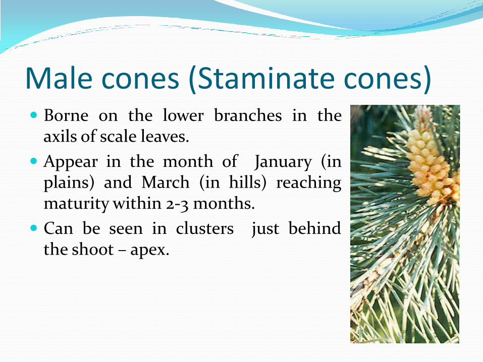
Male cones (Staminate cones) Borne on the lower branches in the
axils of scale leaves.
Appear in the month of January (inplains) and March (in hills) reachingmaturity within 2-3 months.
Can be seen in clusters just behindthe shoot – apex.
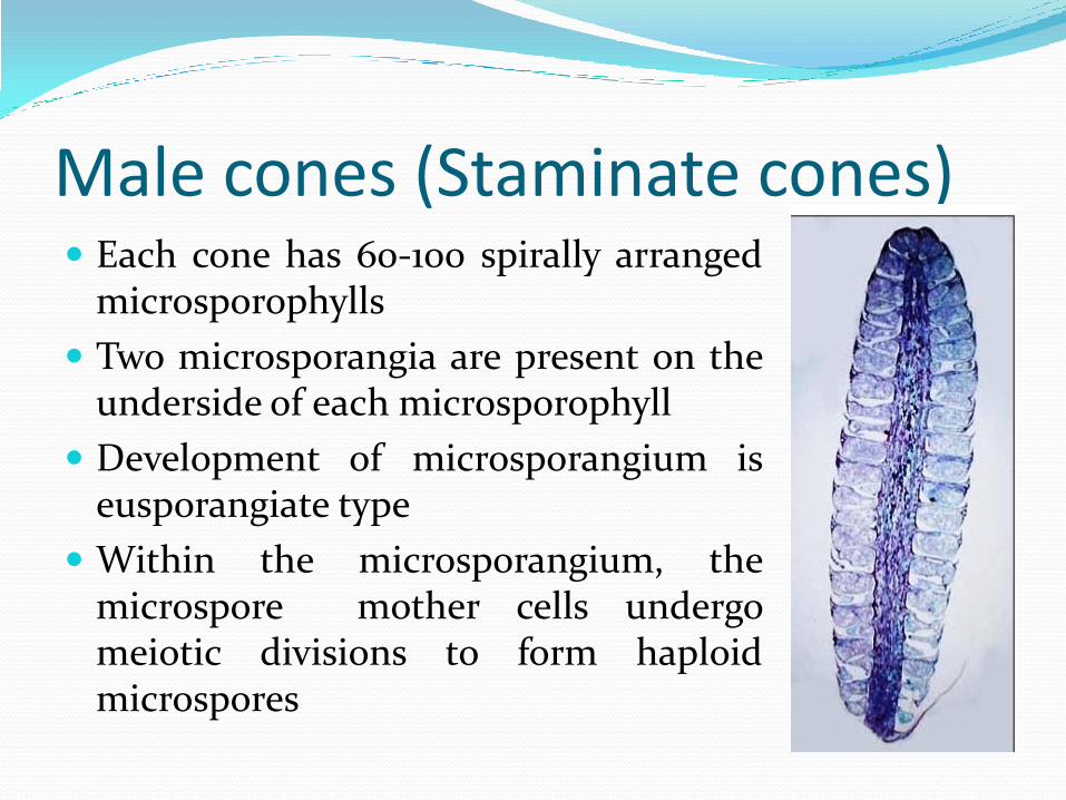
Male cones (Staminate cones) Each cone has 60-100 spirally arranged
microsporophylls
Two microsporangia are present on theunderside of each microsporophyll
Development of microsporangium iseusporangiate type
Within the microsporangium, themicrospore mother cells undergomeiotic divisions to form haploidmicrospores

Microspore (Pollen grain) It is surrounded by a 3-layered wall
Exine heavily cuticularized on one side of themicrospore
Middle layer (exo-intine) projected outwardsinto two large balloon-like air sacs or wings
Inner layer(intine) is very thin
On maturation the spores germinate in situ.Hence, early gametophytic development isprecocious
At the time of dehiscence, huge quantities ofmicrospores form yellow clouds around thepine forests. It’s called the “Shower of sulphurdust”
Pollen grains
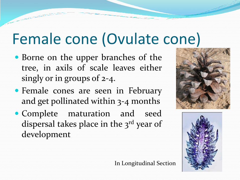
Female cone (Ovulate cone) Borne on the upper branches of the
tree, in axils of scale leaves eithersingly or in groups of 2-4.
Female cones are seen in Februaryand get pollinated within 3-4 months
Complete maturation and seeddispersal takes place in the 3rd year ofdevelopment
In Longitudinal Section

Female cone (Ovulate cone) Each cone consists of central axis
bearing spirally arrangedovuliferous scales (60-70)
On young cones a small thin &leathery bract scale can be belowthe ovuliferous scale
Each ovuliferous scale has twoovules on its upper surface
Cone on maturity is usuallycylindrical and 15-20cms inlength

Megasporophyll The ovuliferous scale is thick, large, woody & brownish
structure
More or less triangular in outline – broad, terminalportion is apophysis with its centrally projected area –the umbo
Basal portion is narrow and bears two naked, sessileanatropous ovules on its upper surface

Ovule Structure Micropyle of the ovule faces the central axis of the
cone
The single integument is fused to the nucleus exceptfor a short distance near the micropyle
Embedded in the nucellus ,the archesporial celldivides meiotically to form four megaspores

Female Gametophyte The inner most functional megaspore further gives
rise to the haploid female gametophyte tissue whereinthe archegonia develop.
The venter of the archegonia contains the upperventral canal cell and the larger egg cell.
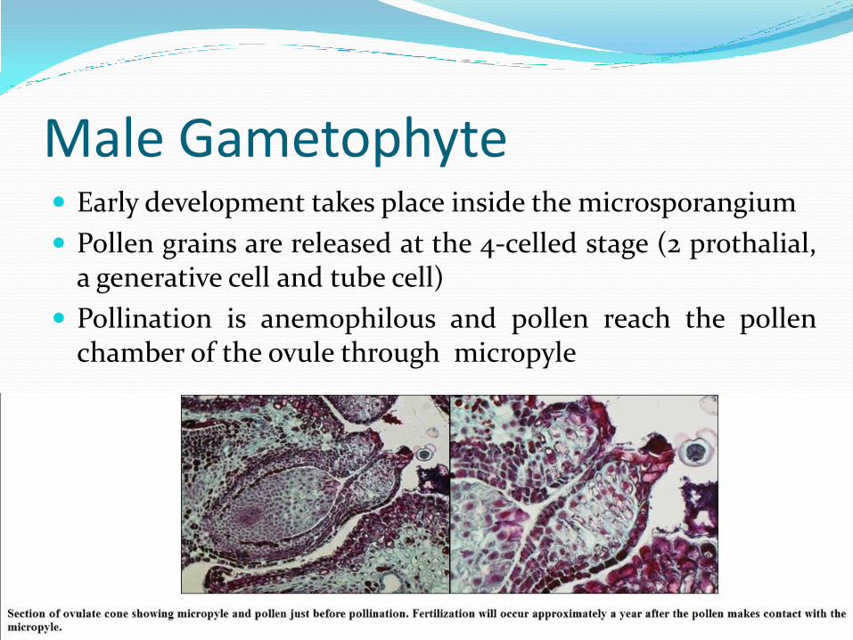
Male Gametophyte Early development takes place inside the microsporangium
Pollen grains are released at the 4-celled stage (2 prothalial,a generative cell and tube cell)
Pollination is anemophilous and pollen reach the pollenchamber of the ovule through micropyle

Male Gametophyte Further development here, results in the formation of
pollen tube which carries the two unequal malegametes to the neck of the archegonium
The released male gametes will fertilize the egg cellresulting in zygote formation
Time gap of 12-14 months is seen between pollinationand fertilization
Young Sporophyte Embryo development is meroblastic
In early stages the embryonal tier of the proembryosplits apart forming 4 apical segments each with itssuspensor
Each of these terminal embryonal cell give rise to amature embryo, thus Cleavage polyembryony isobserved

Seed Structure Seeds are naked (not enclosed in fruit)
Seeds are winged – the latter being derived fromportion of upper surface of the ovuliferous scale
Outer fleshy layer of ovule disintegrates
Testa formed from the middle stony layer
Tegmen is the inner fleshy layer of the ovule
Nucellus is almost consumed during embryodevelopment.
Remnants of nucellus , at micropylar end can be seenas reddish papery structure – the perisperm

Seed Structure The haploid female gametophyte surrounding the
embryo forms the oily white kernel (edible part).
Mature embryo has the radicle towards the micropyleand plumule away from it.
Plumule is surrounded by 8-14 cotyledons, which aregreen in colour.
Germination is epigeal.