dr. chandresh s. jardoshimagsb.com/uploads/bulletin/december-2019.pdf · i.m.a.g.s.b. news bulletin...
TRANSCRIPT
I.M.A.G.S.B. NEWS BULLETIN / DECEMBER - 2019 Vol. 14 No.12
Dr. Chandresh S. Jardosh
Dr. Manjit Nayak
Dr. Prakash Gandhi
Dr. Heming Agrawal
Dr. Yatish Lapsiwala
Dr. C. H. Saini
Dr. Rajen Desai
Dr. Piyush Unadkat
Ahmedabad
Modasa
Daman
Surat
Vadodara
Bhavnagar
Rajkot

GMJ 16
Dear Members,
Seasons Greetings,
Anti-Microbial Resistance (AMR) is one of the major public health problems especially in developing countries
where relatively easy availability and higher consumption of antibiotics have led to higher incidence of inappropriate
use of antibiotics and greater levels of resistance compared to developed countries. In 2010, India was the world's
largest consumer of antibiotics for human health.
As you are aware inappropriate consumption of Antibiotics directly leads to drug resistance. Reducing
unnecessary consumption would go a long way in preserving antibiotic effectiveness
Indian Medical Association to create awareness on Appropriate Prescription of Antibiotics.
Extended Action Committee meeting was held at IMA HQs, New Delhi this month & declared War against AMR.
Following decision was taken as IMA North Zone Declaration, 2019.
• Stop Quackery.
• Stop over the counter sale of Antibiotics without prescription.
• Stop online sale of antibiotics.
• Judicious use of antibiotics.
• Maintain sterility and monitoring of O.T./ICU/Labour Rooms and other critical areas in your hospital.
• Antibiotic policy should be created for every region/state/healthcare establishments after proper scientific
evidence.
• Policy on prophylactic antibiotics in clean surgical patients be implemented.
• All health care sector employees should be vaccinated for Tetanus, Hepatitis B at least and if possible, for
influenza and pneumococcus.
• Hand hygiene and other infection control practice should be promoted among healthcare workers and
society at large.
• Restricted antibiotic list should be identified and published according to antibiograms and to be used only
after enough scientific evidence.
• Public awareness poster against self-medications and depicting ill effect of unnecessary use of antibiotics should
be pasted in patient waiting area.
• Marketing of antibiotics to AYUSH practitioners must be stopped.
• Strict regulations to restrict the use of antibiotic in poultry, animal husbandry and agriculture.
• Unethical practice by pharma companies to promote target-oriented sale of antibiotics should be stopped.
• Publication of antibiogram of different micro-organism should be published on regular basis.
• Government should fund for research on development of new antibiotics as the pharma companies are not
coming forward for the same.
Long Live IMA, Jai IMA
I.M.A.G.S.B. NEWS BULLETIN / DECEMBER - 2019 Vol. 14 No.12
DR. KAMLESH B. SAINIDR. C. S. JARDOSH

Dear friends,
We are thankful to all the central council members of GSB IMA for putting their faith, trust and confidence in
us and giving the charge of prestigious Gujarat Medical Journal (GMJ) for this year. On our side, we promise to see
that the faith and trust that is put in us is full filled and for that, we shall try our best. We are well aware that in
these years GMJ has carved out its name as a journal of research oriented and academic minded people, in the
medical field. All the editors in past, have tried their best to give a name and fame to this journal and we are
enjoying their fruits. But we are aware, that increases our responsibility also.We shall have to work hard and will
have to be vigilant to maintain that standard of our journal.
Here, I want to tell our members about the procedure that we are adopting in selection of an article for GMJ.
We ask the author to send the article on CD, and three physical copies, of which one copy bears names, addresses,
etcs., of authors but two other copies, don't have any name or address of authors, they contain only the material
of the article. On receiving this our office clerk puts code number on it. Articles are known from its code number
only. GMJ editor is given the copy which doesn't have the name, etcs. of the author. And editor then sends the said
article for review to a retired professor or HOD or having that level of expertise in the subject ( whom we call
“referee” or “ reviewer”). So the reviewer also doesn't know about the author. This procedure is adopted since
years.
Without making any compromise with our laid down policy, we have made all the efforts to make GMJ more
informative, more interesting and more popular so that large number of our colleagues read it and utilize the
knowledge and information provided in it. For this, we welcome your suggestions and comments also.
You all know now, that GMJ is indexed in Index Copernicus International” (ICI), and all the issues of GMJ
since 2015 can be viewed on;
https://journals.indexcopernicus.com/search/details?id=43553
Our sincere thanks to GSB president Dr. Chandresh Jardosh and hon. secretary Dr. Kamlesh Saini for
encouragement and suggestions, and giving us free hand in publication of this journal. We are also grateful to GSB
past presidents Dr Kirtibhai Patel, Dr. Jitubhai Patel and Dr. Mahendrabhai Desai for their guidance and help. How
can we forget IMA GSB past president Dr. Yogendra Modi for his help, guidance and support ? A particular thanks
to Dr. Yogendrabhai. We are also thankful to our ex editor Dr. Amit Shah and also to Dr. Urvesh Shah (GCS Medical
college) for their guidance and help.
With regards,
GMJ 17 I.M.A.G.S.B. NEWS BULLETIN / DECEMBER - 2019 Vol. 14 No.12

GMJ 19 I.M.A.G.S.B. NEWS BULLETIN / DECEMBER - 2019 Vol. 14 No.12
DR. CHANDRESH S. JARDOSH
MOB. 98791 32526
DR. JASWANTSINH DARBAR
DR. JITESH DESAI
DR. BHASKAR MAHAJAN
DR. VINOD NOTICEWALA
DR. VINOD MEHTA
DR. NARESH JOSHI
DR. HIREN KOTHARI
DR. JITENDRA H. SHAH
DR. RAJNIKANT PATEL
DR. MAYUR N. BHAGAT
DR. VINESH B. SHAH
DR. PARESH MAJMUDAR
DR. KASHYAP C. DAVE
DR. AMIT AGRAVAT
SURAT
2019-2020
ORIGINAL ARTICLE
GMJ 20
I.M.A. G.S.B. NEWS BULLETIN (Gujarat Medical Journal)
Vol. : 14 DECEMBER-2019 Issue : 12
CONTENTS
* State President and Hon. Secretary's Message ............................................................................................ 16
* From the Desk of Editors....................................................................................................................................17
* A Study on Type of Respiratory Infections and Its Correlation with CD4 Count in HIV Positive Patients
Dr. Mehul R Marwadi*, Dr. Rakeshkumar M Raval**, Dr. Rajesh S Roy*, Dr. R. K.Chavda***.............................22
* A Study of Maternal and fetal out Come in Cases of Gestational Diabetes Mellitus
Dr. Tushar M. Shah*, Dr. Bruhal Patel, Dr. Vishwa Patel**.................................................................................26
* Bone Marrow Aspiration - One year study in tertiary care hospital at Rajkot
Dr. Amit H. Agravat*, Dr. Avani S. Nimavat**, Dr. Gauravi A. Dhruva***.................................................................29
* “Is upper ureteric kink a real nightmare for the Urologists while performing Flexible Ureteroscopy
(fURS)? A single center experience of 15 cases of Retrograde Intra Renal Surgery (RIRS) in unstented
patients with upper ureteric kink”.
Dr. Kandar P. Parikh, Dr. Ravi Jain, Dr. Aditya K. Parikh ....................................................................................33
* A Comparative Study of Macular Thickness in Primary open Angle Glaucoma Patients and Normal
Patients.
Dr. Nilesh V. Parekh*, Dr. Sagar S.Patel**..............................................................................................................38
* A study to evaluate pain relief and pancreatic function after lateral pancreati cojejunotomy for chronic pancreatitis
Dr. Akash Shah*, Dr. Hiren Vaidya**, Dr. Aditya Vaidya***, Dr. Harish Chauhan**..................................................48
* Feto-maternal outcome in Gestational Diabetes Mellitus
V. M. Kansara*, Akash Chikani**, A. N.Desai***............................................................................................59
* Study of Minor and Major adverse drug reaction of intravenous Iron Sucrose in the treatment of severe
iron deficiency anemia.
Dr.Tushar M. Shah, Dr.Vishakha Tyagi, Dr.Vishwa Patel..............................................................................................64
* Evaluation of different techniques for management of postpartum hemorrhage.
*Dr. Megha S. Patel, **Dr. Satvik Patel, ***Dr. Pankti Jayswal, ****Dr. Jigar Thakkar,..........................................67
I.M.A.G.S.B. NEWS BULLETIN / DECEMBER - 2019 Vol. 14 No.12
REVIEW ARTICLES
CASE REPORT
GMJ 21
* Iatrogenic Bile Duct Injury What And When?
Dr. Nupoor Raval*, Dr. Maulik Mehta**, Dr. PankajModi***, Dr. Prujal Parekh***, Dr. Pruthvi Patel***.....................71
* Anesthetic Management of Post Acid Ingestion Esophagopleural Fistula Posted for Fistula
Repair – Case Report
Dr. Shakuntala Goswami*, Dr. Dhwani Trambadia**, Dr. Artivaghasia**, Dr. Payal Yadav**, Dr. Anjali Sahoo**,
Dr. Monica Yadav**...........................................................................................................................................74
* Study Of Effectiveness And Safety Of Inj DMPA In Postpartum Period
Dr. Kruti J. Deliwala* Dr Margi Shah** Dr Parul T. Shah** Dr Reena V. Patel** Dr. Devangi S. Munshi**
Dr Zalak Patel**...............................................................................................................................................77
* Association Of Retinitis Pigmentosa(Rp) With Mental Retardation(Mr) and Subluxated Lens
Dr. Sneha T. Shah*(Corresponding Author) ,Dr. Nilesh V. Parekh**, Dr. Neepa R. Gohil***, Dr. Aditya Parekh****..............80
* Straw coloured Right sided Pleuropancreatic effusion : a diagnosis not to be missed
Dr. Arvind Vala*, Dr. Meghna Patel**, Dr. Kiran Rami***, Dr. Kaushal Bhavsar............................................................82
* Mandatory Submission Form ........................................................................................................................84
I.M.A. G.S.B. NEWS BULLETIN (Gujarat Medical Journal)
Vol. : 14 DECEMBER-2019 Issue : 12
I.M.A.G.S.B. NEWS BULLETIN / DECEMBER - 2019 Vol. 14 No.12
DISCLAIMER
Opinions in the various articles are those of the authors and do not reflect the views of I
ndian Medical Association, Gujarat State Branch. The appearance of advertisement is not a guarantee
or endorsement of the product or the claims made for the product by the manufacturer.
I.M.A.G.S.B. NEWS BULLETIN / DECEMBER - 2019 Vol. 14 No. 1222
Correspondence Address : Dr. Rakeshkumar Raval 66, Ruxmaninagar Society, Katargam, Surat - 385004.
ABSTRACT
Background : Several important changes in the pattern of pulmonary diseases that have become recently
apparent have implications for the diagnosis, management and outcome of AIDS patients. The present study was
planned with an objective to study pattern of various respiratory infections and to correlate CD4 count with various
respiratory infections in HIV patients.
Methodology : The present cross-sectional study was carried out in 50 HIV positive patients admitted in Medicine
ward/ ICU/TB ward in a tertiary care hospital. The information regarding clinical examination, sputum examination
and laboratory parameters including CD4 counts were recorded.
Results : The most common respiratory infection was pulmonary tuberculosis (76%) followed by pneumonia
(12%). M. tuberculosis was found in 40 cases (80%), P. aeruginosa in 3 cases (6%), K. pneumonia in 3 cases (6%),
S. Pneumonia in 2 cases (4%) and S. aureus in 1 case (2%). Mean CD4 count in sputum positive TB patients was
224.4±159.75 while for sputum negative TB patients it was 167.90±60.88. Mean CD4 for patients of
Pseudomonas pneumonia in this study was 286.66±160.74, while for K. pneumoniae it was 421±222.03 and for P.
aeruginosa it was 270±124.45.
Conclusion : It is concluded from present study that there is a strong correlation between CD4 count and pattern
of respiratory complications in HIV-seropositive patients. Patients with CD4 count <200 cells/μL are more prone for
respiratory complications. Hence, high level of clinical suspicion required for diagnosis of respiratory
complications in HIV-infected individuals particularly with patients having CD4 count <200 cells/μL.
Keywords : HIV-AIDS, Respiratory infections, sputum, CD4 count
Dr. Mehul R Marwadi*, Dr. Rakeshkumar M Raval**, Dr. Rajesh S Roy*, Dr. R. K.Chavda***
*Assistant Professor in Medicine dept.,Parul Inst.Of Medical Sciences & Research,Limda,Vadodara-391760
**Senior Resident in Medicine dept.,Govt.Medical College, Surat
***Professor & Head of dept.,Medicine dept., Parul Inst.Of Medical Sciences & Research,Limda,Vadodara-391760
INTRODUCTION
The lungs are portal of entry for many infectious agents in
our body that either may cause acute illness or may cause 1latent infection. Among these, the majority are pulmonary
tuberculosis and pneumonia in India. As these pulmonary
infections indicate underlying progression of HIV
infection, we need to update our knowledge of these
diseases for better diagnosis and management.
Over the past decade, several changes in the pattern of
disease have occurred. While Pneumocystis Carinnii
Pneumonia (PCP) is the most common opportunistic
pathogen in AIDS patients in developed countries,
infection with mycobacterium tuberculosis & other
organisms causing pneumonia are major health problems 2in developing countries. It appears unbelievable that
bacterium identified in the 1880's would become partner
with a virus isolated a century later, and in combination
poses a challenge in the field of medicine. The pattern of
TB in AIDS is distinct from non-immuno compromised
persons. The association of TB and compromised HIV
has caused so much concern that strategies for
diagnosis, effective treatment and control of TB have to 3 be reframed. For diagnostic and therapeutic reasons,
especially those concerning prevention, it is far more
useful to consider the entire continum of HIV infection
than only the last and invariably fatal stage that we call
AIDS.1
Again, the pattern of respiratory disease is different in HIV
seropositive patients in developed countries and in
developing country like India. Above all, because several
important changes in the pattern of pulmonary diseases
that have become recently apparent have implications for
the diagnosis management and outcome of AIDS
patients, it is worthwhile to update our knowledge of
A Study on Type of Respiratory Infections and Its Correlation with CD4 Count in HIV Positive Patients
Original Articles
23
pulmonary disease in HIV infected individuals as we
approach the end of the forth AIDS decade.
MATERIALS AND METHODS
The present cross-sectional study was carried out in 50
HIV positive patients admitted in Medicine ward/ ICU/TB
ward in a tertiary care hospital. Considering proportion of
HIV patients admitted with respiratory infections, which
was calculated by two week pilot surveyed at Medicine
ward/ ICU/TB ward at a tertiary hospital as p=3.22 %, Z
(level of significance)= 1.96, L (Allowable error) = 5% the
calculated sample size (n) = Z2pq / L2 was 50.
Adults HIV positive patients above 18 years of age and
with abnormal X ray findings were included in the study
while patients with known case of respiratory disorder
such as asthma, chronic obstructive airway disease and
lung cancer were excluded from the study. The study was
conducted after getting ethical clearance certificate and
participants are included after taking voluntary informed
consent from them.
Detailed history and clinical examination of the
participants were done. Two sputum samples (One spot
and one early morning expectorated sputum [induced
sputum if required]) were collected separately in sterile
containers from all patients. Induction of sputum was
done using a Nebulizer (model - Medel Aero Family) and
3% hypertonic saline for 15 minutes. Microscopic
examination of sputum was done for the presence of
trophozoites and cysts of P.carini i ,whi le the
expectorated sputum was examined for bacterial and
fungal pathogens. Bartlett's scoring method was used for 4microscopic evaluation of the expectorated sputum.
A sputum was considered unsuitable if it had a final score
of 0 or less. All unsuitable specimens were discarded and
a repeat specimen was collected. The sputum specimens
were inoculated into blood agar with 10% sheep blood,
Chocolate agar with 10% sheep blood, McConkey's agar
and Brain Heart Infusion (BHI) agar. Any significant
bacterial growth was further processed as per the
5standard procedure to identify the pathogens. The
sputum was also inoculated onto Sabouraud dextrose
agar (SDA) with antibiotics, SDA without antibiotics in
duplicate (incubated at 37°C and 25°C) and BHI agar
(incubated at 37°C). Any significant growth of a fungal 6species was further identified as per standard protocol.
In addition to sputum, 10 ml of blood was collected from all
patients included in the study for investigations like
C o m p l e t e B l o o d C o u n t ( C B C ) , E r y t h r o c y t e
Sedimentation rate (ESR), Renal Function Test (RFT),
Liver Function Test (LFT), Lactate Dehydrogenase (LDH)
(if needed), Arterial Blood Gas (if needed) etc. Pleural
fluid examination was done in cases of pleural effusion if
required, and it was sent for routine microscopy, protein,
sugar, LDH and ADA.
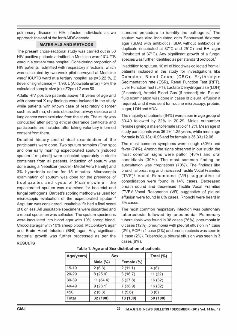
The majority of patients (64%) were seen in age group of
30-49 followed by 22% in 20-29. Males outnumber
females giving a male to female ratio of 1.7:1. Mean age of
study participants was 36.2±11.25 years, while mean age
for male is 36.13±10.96 and for female is 36.33±12.06.
The most common symptoms were cough (80%) and
fever (74%). Among the signs observed in our study, the
most common signs were pallor (48%) and oral
candidiasis (30%). The most common finding on
auscultation was crepitations (70%). The findings like
bronchial breathing and increased Tactile Vocal Framitus
(TVF)/ Vocal Resonance (VR) suggest ive of
consolidation were found in 14% cases. Decreased
breath sound and decreased Tactile Vocal Framitus
(TVF)/ Vocal Resonance (VR) suggestive of pleural
effusion were found in 8% cases. Rhonchi were heard in
8% cases.
The most common respiratory infection was pulmonary
tuberculosis followed by pneumonia. Pulmonary
tuberculosis was found in 38 cases (76%), pneumonia in
6 cases (12%), pneumonia with pleural effusion in 1 case
(2%), PCP in 1 case (2%) and bronchiectesis was seen in
1 case (2%). Tuberculous pleural effusion was seen in 3
cases (6%).RESULTS
Table 1: Age and Sex distribution of patients
Age(years) Sex Total (%)
Male (%) Female (%)
15-19 2 (6.3) 2 (11.1) 4 (8)
20-29 8 (25.0) 3 (16.7) 11 (22)
30-39 11 (34.4) 5 (27.8) 16 (32)
40-49 9 (28.1) 7 (38.9) 16 (32)
>50 2 (6.3) 1 (5.6) 3 (6)
Total 32 (100) 18 (100) 50 (100)
I.M.A.G.S.B. NEWS BULLETIN / DECEMBER - 2019 Vol. 14 No. 12

M. tuberculosis was found in 40 cases (80%), P.
aeruginosa in 3 cases (6%), K. pneumonia in 3 cases
(6%), S. Pneumonia in 2 cases (4%) and S. aureus in 1
case (2%). Hemoglobin level was normal (≥12 gm/dl) in
15 cases (30%) and anemia was observed in 35 cases
(70%). Among anemia, most cases had moderate anemia
(16 cases, 32%). Mild anemia was seen in 11 cases (22%)
and severe anemia was seen in 8 cases (16%).
Mean CD4 for sputum positive tuberculosis is
224.4±159.75 whi le for sputum negative i t is
167.90±60.88. Thus it states that sputum negativity is
more frequent with lower CD4 count. Mean CD4 for
pneumonia in this study is 286.66±160.74, while for K.
pneumoniae it is 421±222.03 and for P. aeruginosa it is
270±124.45.
DISCUSSION
In the study conducted by K.C.Mohanty et al7, maximum
43.5% of patients were found in age group 15-30 years,
followed by 28.3% in 30-39 years and 7.8% in 40-49 age 8,group. Another study carried out by Sunderam et al
similar results were observed being maximum 56.3% in
same age group as above and then in 30-39 age group.
Fairly comparable results were obtained in the present
study.7In the study conducted by K.C.Mohanty et al , sputum
smear positivity was seen in 47.8% while negative in 9 1052.2%. Another study of Theur et al , Jayswal et al and
sputum smear positivity was seen in 47% and 27.6%. In
the present study, Sputum smear positivity was seen in
47.62% while negative in 52.38%
In the present study, tuberculosis including tuberculous
pleural effusion is the most common disease seen in 84% 7cases. K.C.Mohanty et al observed that most common
pulmonary disease was tuberculosis [including pleural
effusion] (88.8%) followed by pneumonitis (9.4%). The
other disease like PCP was seen in 0.9% and
bronchiectesis in 0.9%. Another study by Sara Chako et 11al , similar results were noted. Tuberculosis was the most
common and found in 52% cases followed by
pneumonitis in 18% cases. 3.2% had nonspecific
interstitial pneumonitis.12A study carried out by Toshniwal et al showed that 69.1%
cases had respiratory tract infections when their CD4
count was <200. Patients with CD4 count >500 had less
frequency of respiratory infections as compare to other
group in the study. As the CD4 count decreases,
frequency of respiratory infections increases. In present
study, comparable results were found. Early diagnosis of
opportunistic infections and prompt treatment with
24
Table 2: Distribution of clinical signs and symptoms
Symptoms No. (%)
Cough 40 (80)
Fever 37 (74)
Breathlessness 20 (40)
Chest pain 10 (20)
Weight loss 33 (66)
Diarrhea 18 (36)
Hemoptysis 4 (4)
Oral candidiasis 15 (30)
Genital ulcer 4 (8)
Signs
Pallor 24 (48)
Icterus 03 (6)
Cyanosis 1 (2)
Clubbing 3 (6)
Pedal edema 2 (4)
Lymphadenopathy 5 (10)
Oral candidiasis 15 (30)
Dermatitis 1 (2)
Genital ulcer 4 (8)
Table 3: Spectrum of respiratory manifestations
in HIV positive patients
Diseases No. (%)
Tuberculosis 38 (76)
Pneumonia 6 (12)
Pneumonia with pleural effusion 1 (2)
Pleural effusion 3 (6)
Infective Bronchiectesis 1 (2)
PCP 1 (2)
Total 50(100)
Table 4 : Spectrum of respiratory manifestations
in HIV positive patients
Micro-organism No. (%)
M. Tuberculosis 20 (40)
Ps. Aeruginosa 2 (4)
K. Pneumonia 2 (4)
Str. Pneumonia 1 (2)
No any pathogen isolated 25 (50)
I.M.A.G.S.B. NEWS BULLETIN / DECEMBER - 2019 Vol. 14 No. 12
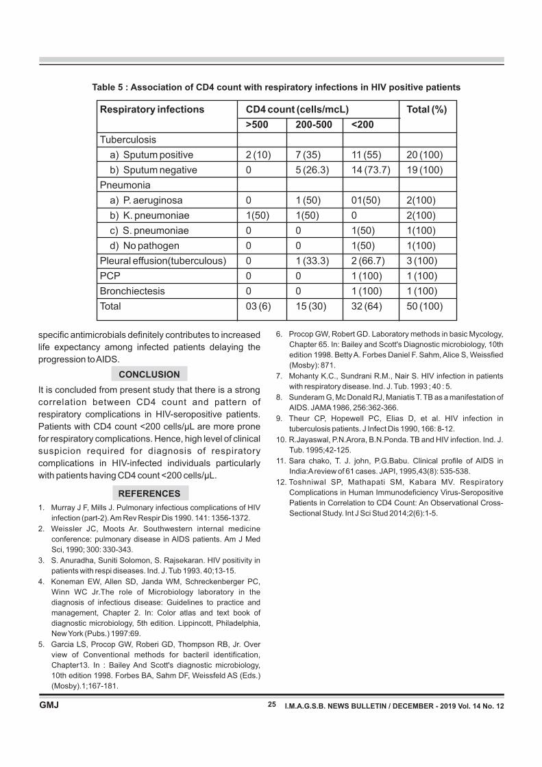
Table 5 : Association of CD4 count with respiratory infections in HIV positive patients
Respiratory infections CD4 count (cells/mcL) Total (%)
>500 200-500 <200
Tuberculosis
a) Sputum positive 2 (10) 7 (35) 11 (55) 20 (100)
b) Sputum negative 0 5 (26.3) 14 (73.7) 19 (100)
Pneumonia
a) P. aeruginosa 0 1 (50) 01(50) 2(100)
b) K. pneumoniae 1(50) 1(50) 0 2(100)
c) S. pneumoniae 0 0 1(50) 1(100)
d) No pathogen 0 0 1(50) 1(100)
Pleural effusion(tuberculous) 0 1 (33.3) 2 (66.7) 3 (100)
PCP 0 0 1 (100) 1 (100)
Bronchiectesis 0 0 1 (100) 1 (100)
Total 03 (6) 15 (30) 32 (64) 50 (100)
25
6. Procop GW, Robert GD. Laboratory methods in basic Mycology,
Chapter 65. In: Bailey and Scott's Diagnostic microbiology, 10th
edition 1998. Betty A. Forbes Daniel F. Sahm, Alice S, Weissfied
(Mosby): 871.
7. Mohanty K.C., Sundrani R.M., Nair S. HIV infection in patients
with respiratory disease. Ind. J. Tub. 1993 ; 40 : 5.
8. Sunderam G, Mc Donald RJ, Maniatis T. TB as a manifestation of
AIDS. JAMA 1986, 256:362-366.
9. Theur CP, Hopewell PC, Elias D, et al. HIV infection in
tuberculosis patients. J Infect Dis 1990, 166: 8-12.
10. R.Jayaswal, P.N.Arora, B.N.Ponda. TB and HIV infection. Ind. J.
Tub. 1995;42-125.
11. Sara chako, T. J. john, P.G.Babu. Clinical profile of AIDS in
India:A review of 61 cases. JAPI, 1995,43(8): 535-538.
12. Toshniwal SP, Mathapati SM, Kabara MV. Respiratory
Complications in Human Immunodeficiency Virus-Seropositive
Patients in Correlation to CD4 Count: An Observational Cross-
Sectional Study. Int J Sci Stud 2014;2(6):1-5.
specific antimicrobials definitely contributes to increased
life expectancy among infected patients delaying the
progression to AIDS.
CONCLUSION
It is concluded from present study that there is a strong
correlation between CD4 count and pattern of
respiratory complications in HIV-seropositive patients.
Patients with CD4 count <200 cells/μL are more prone
for respiratory complications. Hence, high level of clinical
suspicion required for diagnosis of respiratory
complications in HIV-infected individuals particularly
with patients having CD4 count <200 cells/μL.
REFERENCES
1. Murray J F, Mills J. Pulmonary infectious complications of HIV
infection (part-2). Am Rev Respir Dis 1990. 141: 1356-1372.
2. Weissler JC, Moots Ar. Southwestern internal medicine
conference: pulmonary disease in AIDS patients. Am J Med
Sci, 1990; 300: 330-343.
3. S. Anuradha, Suniti Solomon, S. Rajsekaran. HIV positivity in
patients with respi diseases. Ind. J. Tub 1993. 40;13-15.
4. Koneman EW, Allen SD, Janda WM, Schreckenberger PC,
Winn WC Jr.The role of Microbiology laboratory in the
diagnosis of infectious disease: Guidelines to practice and
management, Chapter 2. In: Color atlas and text book of
diagnostic microbiology, 5th edition. Lippincott, Philadelphia,
New York (Pubs.) 1997:69.
5. Garcia LS, Procop GW, Roberi GD, Thompson RB, Jr. Over
view of Conventional methods for bacteril identification,
Chapter13. In : Bailey And Scott's diagnostic microbiology,
10th edition 1998. Forbes BA, Sahm DF, Weissfeld AS (Eds.)
(Mosby).1;167-181.
I.M.A.G.S.B. NEWS BULLETIN / DECEMBER - 2019 Vol. 14 No. 12
26
ABSTRACT
Content : Gestational diabetes mellitus (GDM) is defined as Carbohydrate/Glucose intolerance of varying
degrees of severity with onset or first recognition during pregnancy”. Pregnancy itself is a Diabetogenic State.
GDM complicates 7% of the pregnancy. There is much higher rate of maternal and fetal compromise in a diabetic
pregnancy as compared to normal pregnancy.
Aims : This Study is carried out to study proportion of gestational diabetes in pregnancy and its fetal and maternal
outcome.
Setting and Designs : This Prospective study was conducted among 32 cases of gestational diabetes mellitus in
Department of Obstetrics and Gynaecology ,Civil Hospital , Ahmedabad.
Results and Conclusions : In this Study , Prevalence of GDM was 0.205%, Most common maternal complication
was PIH among37.5% of GDM pregnancy and Most common fetal complication was Prematurity among 18.8% of
GDM pregnancies.
A Study of Maternal and fetal out Come in Cases of Gestational Diabetes Mellitus Dr. Tushar M. Shah*, Dr. Bruhal Patel, Dr. Vishwa Patel**
*HOU and Associate Professor, **Resident Doctor ***Medical Officer, Shalin Hospital, Ahmedabad.
Keywords : Macrosomia, Oral Glucose, Tolerance Test
INTRODUCTION
Gestational Diabetes Mellitus (GDM) is defined as:
carbohydrate/glucose intolerance of varying degrees of
severity with onset or first recognition during pregnancy.
Pregnancy It self is a diabetogenic state. GDM 1complicates 7% of the pregnancy . There is much higher
rate of maternal and fetal compromise in a diabetic 2pregnancy as compared to normal pregnancy.
Placental Lactogen, estrogen, progesterone and cortisol
also there is increased destruction by kidney and placenta
to increase in insulin resistance. All in all leading to 3physiological insulin resistance of late pregnancy.
The adverse intrauterine environment causes epigenetic
changes in the fetus that may contribute to metabolic 4disorders, the so-called vicious cycle of diabetes .
The mainstay of GDM treatment is dietary and lifestyle
advice, which includes medical nutrition therapy, weight 5management, and physical activity . Women monitor their
fasting and post meal glucose levels and adjust their
individual diet and lifestyle to meet their glycemic targets.
This pragmatic approach achieves the glycemic targets in
approximately two-thirds of women with GDM5. However,
despite the importance of medical nutrition therapy and its
widespread recommendation in clinical practice, there
are limited data regarding the optimal diet for achieving 5–8maternal euglycemia . It is also unknown whether the
dietary interventions for achieving maternal glycemia are
also effective for reducing excessive fetal growth and 9adiposity .
Different dietary strategies have been reported including
low glycemic index (GI), energy restriction, increase or
decrease in carbohydrates, and modifications of fat or 10protein quality or quantity .
Risk factors for gestational diabetes are family history of
diabetes, obesity, BMI, history of GDM (recurrence rate of
30 to 50% in the subsequent pregnancy), elderly gravida,
> 25 years, smoking, sedentary lifestyle. GDM
complicates 7% of the pregnancies , therefore it is
important to study its feto-maternal outcome.
Therefore, detection of GDM becomes an important
health issue. Detection and treatment of GDM not only
reduces the risk for fetus but also provides an opportunity
to warn the mother to adopt preventive measures like
controlled diet, exercise, and achieve ideal body weight to
haltor delay the process of onset of overt diabetes.
Insulin production and insulin sensitivity is normal in early
pregnancy, but as pregnancy advances there is increased
production of insulin. Therefore it is important to study its
feto- maternal outcome.
Aims :
1 To Study the proportion of Gestational diabetes in
Pregnancy.
Original Articles
Correspondence Address : Dr. Tushar M. Shah A-902, Status Tower, Opp. Doordarshan Kendra, Drive in Road, Nr. SAL Hospital, Thaltej, Ahmedabad-380059. Email :- [email protected]
I.M.A.G.S.B. NEWS BULLETIN / DECEMBER - 2019 Vol. 14 No. 12
27
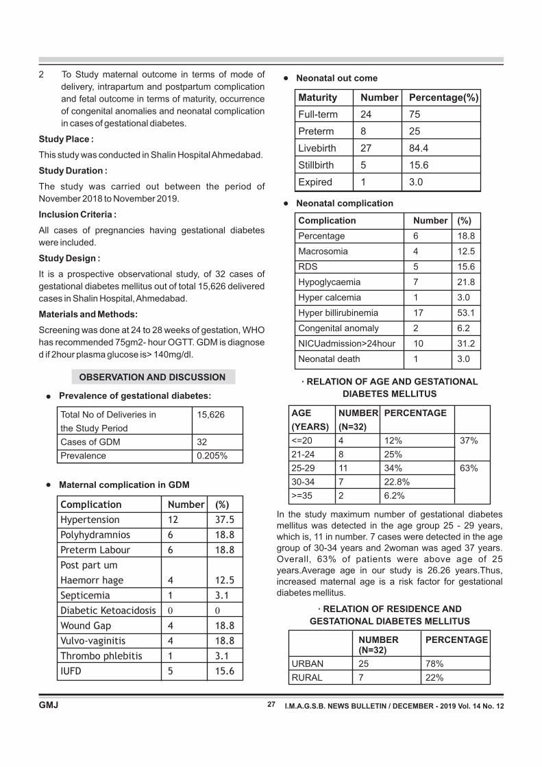
2 To Study maternal outcome in terms of mode of
delivery, intrapartum and postpartum complication
and fetal outcome in terms of maturity, occurrence
of congenital anomalies and neonatal complication
in cases of gestational diabetes.
Study Place :
This study was conducted in Shalin Hospital Ahmedabad.
Study Duration :
The study was carried out between the period of
November 2018 to November 2019.
Inclusion Criteria :
All cases of pregnancies having gestational diabetes
were included.
Study Design :
It is a prospective observational study, of 32 cases of
gestational diabetes mellitus out of total 15,626 delivered
cases in Shalin Hospital, Ahmedabad.
Materials and Methods:
Screening was done at 24 to 28 weeks of gestation, WHO
has recommended 75gm2- hour OGTT. GDM is diagnose
d if 2hour plasma glucose is> 140mg/dl.
OBSERVATION AND DISCUSSION
Prevalence of gestational diabetes:
Total No of Deliveries in 15,626
the Study Period
Cases of GDM 32
Prevalence 0.205%
Maternal complication in GDM
Complication Number (%)
Hypertension 12 37.5
Polyhydramnios 6 18.8
Preterm Labour 6 18.8
Post part um
Haemorr hage 4 12.5
Septicemia 1 3.1
Diabetic Ketoacidosis 0 0
Wound Gap 4 18.8
Vulvo-vaginitis 4 18.8
Thrombo phlebitis 1 3.1
IUFD 5 15.6
Neonatal out come
Maturity Number Percentage(%)
Full-term 24 75
Preterm 8 25
Livebirth 27 84.4
Stillbirth 5 15.6
Expired 1 3.0
Neonatal complication
Complication Number (%)
Percentage 6 18.8
Macrosomia 4 12.5
RDS 5 15.6
Hypoglycaemia 7 21.8
Hyper calcemia 1 3.0
Hyper billirubinemia 17 53.1
Congenital anomaly 2 6.2
NICUadmission>24hour 10 31.2
Neonatal death 1 3.0
· RELATION OF AGE AND GESTATIONAL
DIABETES MELLITUS
AGE NUMBER PERCENTAGE
(YEARS) (N=32)
<=20 4 12% 37%
21-24 8 25%
25-29 11 34% 63%
30-34 7 22.8%
>=35 2 6.2%
· RELATION OF RESIDENCE AND
GESTATIONAL DIABETES MELLITUS
NUMBER PERCENTAGE (N=32)
URBAN 25 78%
RURAL 7 22%
In the study maximum number of gestational diabetes mellitus was detected in the age group 25 - 29 years, which is, 11 in number. 7 cases were detected in the age group of 30-34 years and 2woman was aged 37 years. Overall, 63% of patients were above age of 25 years.Average age in our study is 26.26 years.Thus, increased maternal age is a risk factor for gestational diabetes mellitus.
I.M.A.G.S.B. NEWS BULLETIN / DECEMBER - 2019 Vol. 14 No. 12
●
●
●
●
28
In the study out of 32 patients which were detected to be positive for gestational diabetes mellitus, 25 patients belong to urban area, which is approx 78% and 7 patients belong to rural area, which is approx 22%.
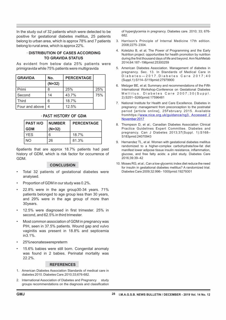
· DISTRIBUTION OF CASES ACCORDING
TO GRAVIDA STATUS
As evident from below data 25% patients were primigravida while 75% patients were multigravida
GRAVIDA No. PERCENTAGE
(N=32)
Primi 8 25% 25%
Second 14 43.7% 75%
Third 6 18.7%
Four and above 4 12.5%
· PAST HISTORY OF GDM
PAST H/O NUMBER PERCENTAGE
GDM (N=32)
YES 6 18.7%
NO 26 81.3%
6patients that are approx 18.7% patients had past history of GDM, which is risk factor for occurrence of GDM.
CONCLUSION
• Total 32 patients of gestational diabetes were analysed.
• Proportion of GDM in our study was 0.2%.
• 22.8% were in the age group30-34 years. 71% patients belonged to age group less than 30 years, and 29% were in the age group of more than 30years.
• 12.5% were diagnosed in first trimester. 25% in second, and 62.5% in third trimester.
• Most common association of GDM in pregnancy was PIH, seen in 37.5% patients. Wound gap and vulvo vaginitis was present in 18.8% and septicemia in3.1%.
• 25%neonateswerepreterm
• 15.6% babies were still born. Congenital anomaly was found in 2 babies. Perinatal mortality was 22.2%.
REFERENCES
1. American Diabetes Association Standards of medical care in diabetes 2010. Diabetes Care.2010;33;676-682.
2. International Association of Diabetes and Pregnancy study groups recommendations on the diagnosis and classification
of hyperglycemia in pregnancy. Diabetes care. 2010; 33; 676-682.
3. Harrison's Principle of Internal Medicine 17th edition. 2008;2275- 2304.
4. Koletzko B, et al; The Power of Programming and the Early Nutrition project: opportunities for health promotion by nutrition during the first thousand days of life and beyond. Ann NutrMetab 2014;64:187–196pmid:25300259
5. American Diabetes Association. Management of diabetes in pregnancy. Sec. 13. In Standards of Medical Care in D i a b e t e s — 2 0 1 7 . D i a b e t e s C a r e 2 0 1 7 ; 4 0 (Suppl.1):S114–S119pmid:27979900
6. Metzger BE, et al; Summary and recommendations of the Fifth International Workshop-Conference on Gestational Diabetes M e l l i t u s . D i a b e t e s C a r e 2 0 0 7 ; 3 0 ( S u p p l . 2):S251–S260pmid:17596481
7. National Institute for Health and Care Excellence. Diabetes in pregnancy: management from preconception to the postnatal period [art icle onl ine], 25February 2015. Avai lable fromhttps://www.nice.org.uk/guidance/ng3. Accessed 2 November 2017
8. Thompson D, et al.; Canadian Diabetes Association Clinical Practice Guidelines Expert Committee. Diabetes and pregnancy. Can J Diabetes 2013;37(Suppl. 1):S168– S183pmid:24070943
9. Hernandez TL, et al. Women with gestational diabetes mellitus randomized to a higher-complex carbohydrate/low-fat diet manifest lower adipose tissue insulin resistance, inflammation, glucose, and free fatty acids: a pilot study. Diabetes Care 2016;39:39–42
10. Moses RG, et al.; Can a low-glycemic index diet reduce the need for insulin in gestational diabetes mellitus? A randomized trial. Diabetes Care 2009;32:996– 1000pmid:19279301
I.M.A.G.S.B. NEWS BULLETIN / DECEMBER - 2019 Vol. 14 No. 12
INTRODUCTION
Bone marrow examination is useful in the diagnosis of
both hematological and non-hematological disorders.
The two most important techniques used for the diagnosis
of hematological disorders are bone marrow aspiration
and trephine biopsy. Bone marrow aspiration is an
invasive procedure where bone marrow is obtained
through a needle aspiration for diagnostic evaluations [1][2][3].especially cytology and stem cell harvest
Bone marrow examination was first done by Mosler in
1876 using a regular wood drill to aspirate bone marrow [1].particles from a patient with leukemia
Bone marrow aspiration specimens are useful in further
diagnostic assays including cytochemical/special
stainings, immunophenotyping, microbiologic tests, [1][2][3].cytogenetic analysis and molecular studies
It may be useful in establishing the diagnosis of storage
diseases and metastatic non-haemopoietic malignancies
or when a leucoerythroblastic peripheral blood picture is
Keywords : Bone marrow, Pancytopenia, Megaloblastic anemia
Bone Marrow Aspiration - One year study in tertiary care hospital at Rajkot
Original Articles
Dr. Amit H. Agravat*, Dr. Avani S. Nimavat**, Dr. Gauravi A. Dhruva***
*Associate Professor, **Resident Doctor, *** Professor and Head, Pathology Department P.D.U. Medical College,
Rajkot
29
Correspondence Address : D r. Avani Nimavat, 101, Ashutosh avenue, nakum street, Shroff road, Rajkot Email id: [email protected]
ABSTRACT
INTRODUCTION : Bone marrow aspiration (BMA) is crucial in evaluation, diagnosis, and management of anemia
and other hematological disorders, especially in situations where diagnosis remains cryptic after detailed clinical
history, physical examination and peripheral blood analysis. This is relatively safe and simple procedure.
AIMS AND OBJECTIVES : The aim of this study is to know prevalence of various hematological disorders and to
compare findings of peripheral smear with diagnosis given on bone marrow aspiration.
MATERIALS AND METHODS : Bone marrow examination of 68 cases of suspected hematological disorders was
carried out. Bone marrow aspiration was done under aseptic conditions. Slides were stained with Field's stain and
where needed Leishman's stain was also done.
RESULT : A total of 68 cases were included in this study. The age range of cases was from 11months to 80 years.
Males were 29 (43%) and Females were 39 (57%). In our study pancytopenia was most common indication
followed by anemia, thrombocytopenia &suspected malignancy. Megaloblastic anemia was most common finding
on bone marrow aspiration in this study.
CONCLUSION : Bone marrow aspiration is relatively simple, safe, and cheap, mildly invasive technique which can
diagnose many hematological and non-hematologic diseases that can be confirmed by more advanced
investigations.
[4].present Deviations from the normal may be qualitative
with abnormal cellular morphology or quantitative with [5]aplasia, hypoplasia or hyperplasia .
This study reports on age and sex distribution, the
spectrum of common indications and diagnosis of bone-
marrow aspiration.
MATERIALS AND METHODS
This was a study of one year (October 2018-September
2019) conducted in the Central Clinical Laboratory,
Department of Pathology ,P.D.U. Government Medical
College & Hospital, Rajkot. Posterior superior iliac crest
was the site of choice for Bone marrow aspiration in most
of the patients and sternum in case of obese patients.
Records regarding the patient detailed information,
consent, clinical history, physical examination, clinical
indication for the procedure and all laboratory tests
findings including peripheral smear reports were
recorded.
The Bone marrow aspiration material was collected and
smears were prepared by wedge-spread method and
I.M.A.G.S.B. NEWS BULLETIN / DECEMBER - 2019 Vol. 14 No. 12
30
stained with Field's stain & Leishman's stain. Wherever
needed, special stains such as Myeloperoxidase stain,
Periodic Acid–Schiff stain and Pearl's stain were used. All
slides were examined by the expert pathologist and the
data were manually collected and subsequently
analyzed.
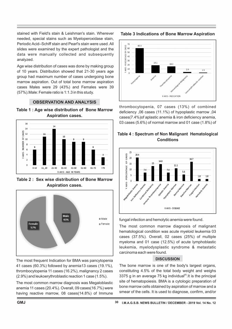
Age wise distribution of cases was done by making group
of 10 years. Distribution showed that 21-30 years age
group had maximum number of cases undergoing bone
marrow aspiration. Out of total bone marrow aspiration
cases Males were 29 (43%) and Females were 39
(57%).Male: Female ratio is 1:1.3 in this study.
OBSERVATION AND ANALYSIS
Table 1 : Age wise distribution of Bone Marrow
Aspiration cases.
Table 2 : Sex wise distribution of Bone Marrow
Aspiration cases.
The most frequent Indication for BMA was pancytopenia
41 cases (60.3%) followed by anemia13 cases (19.1%),
thrombocytopenia 11 cases (16.2%), malignancy 2 cases
(2.9%) and leukoerythroblastic reaction 1 case (1.5%).
The most common marrow diagnosis was Megaloblastic
anemia 11 cases (20.4%). Overall, 09 cases(16.7%) were
having reactive marrow, 08 cases(14.8%) of Immune
Table 3 Indications of Bone Marrow Aspiration
thrombocytopenia, 07 cases (13%) of combined
deficiency ,06 cases (11.1%) of hypoplastic marrow ,04
cases(7.4%)of aplastic anemia & iron deficiency anemia,
03 cases (5.6%) of normal marrow and 01 case (1.8%) of
Table 4 : Spectrum of Non Malignant Hematological
Conditions
fungal infection and hemolytic anemia were found.
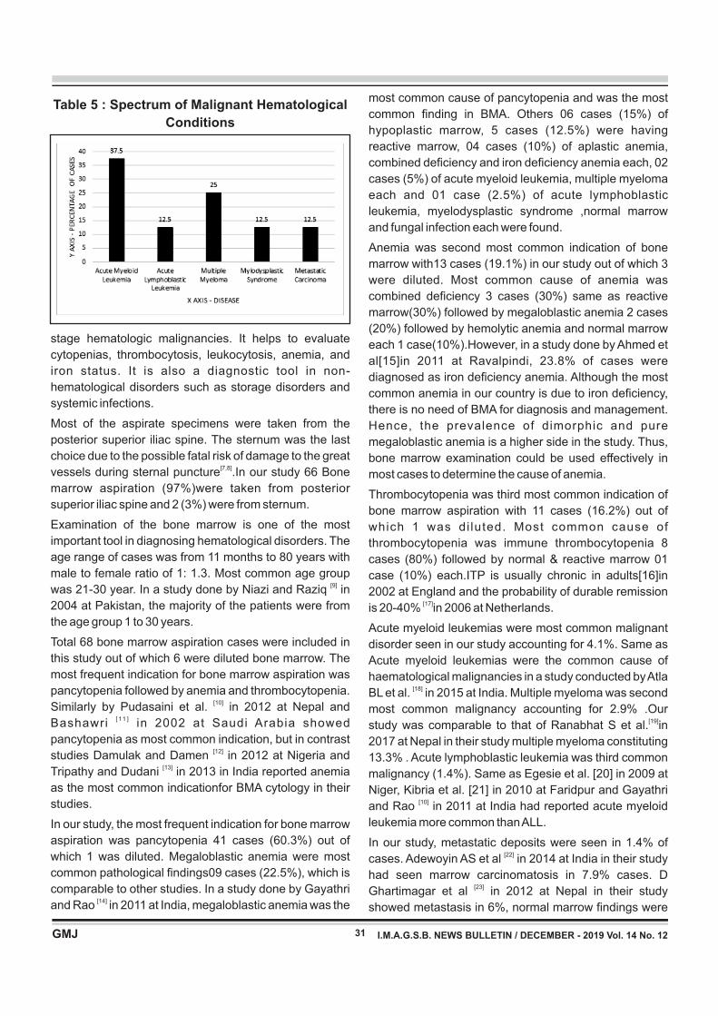
The most common marrow diagnosis of malignant
hematological condition was acute myeloid leukemia 03
cases (37.5%). Overall, 02 cases (25%) of multiple
myeloma and 01 case (12.5%) of acute lymphoblastic
leukemia, myelodysplastic syndrome & metastatic
carcinoma each were found.
DISCUSSION
The bone marrow is one of the body's largest organs,
constituting 4.5% of the total body weight and weighs [6]3375 g in an average 75 kg individual .It is the principal
site of hematopoiesis. BMA is a cytologic preparation of
bone marrow cells obtained by aspiration of marrow and a
smear of the cells. It is used to diagnose, confirm, and/or
I.M.A.G.S.B. NEWS BULLETIN / DECEMBER - 2019 Vol. 14 No. 12
31
Table 5 : Spectrum of Malignant Hematological
Conditions
stage hematologic malignancies. It helps to evaluate
cytopenias, thrombocytosis, leukocytosis, anemia, and
iron status. It is also a diagnostic tool in non-
hematological disorders such as storage disorders and
systemic infections.
Most of the aspirate specimens were taken from the
posterior superior iliac spine. The sternum was the last
choice due to the possible fatal risk of damage to the great [7,8]vessels during sternal puncture .In our study 66 Bone
marrow aspiration (97%)were taken from posterior
superior iliac spine and 2 (3%) were from sternum.
Examination of the bone marrow is one of the most
important tool in diagnosing hematological disorders. The
age range of cases was from 11 months to 80 years with
male to female ratio of 1: 1.3. Most common age group [9]was 21-30 year. In a study done by Niazi and Raziq in
2004 at Pakistan, the majority of the patients were from
the age group 1 to 30 years.
Total 68 bone marrow aspiration cases were included in
this study out of which 6 were diluted bone marrow. The
most frequent indication for bone marrow aspiration was
pancytopenia followed by anemia and thrombocytopenia. [10]Similarly by Pudasaini et al. in 2012 at Nepal and
[ 11 ]Bashawri in 2002 at Saudi Arabia showed
pancytopenia as most common indication, but in contrast [12]studies Damulak and Damen in 2012 at Nigeria and
[13]Tripathy and Dudani in 2013 in India reported anemia
as the most common indicationfor BMA cytology in their
studies.
In our study, the most frequent indication for bone marrow
aspiration was pancytopenia 41 cases (60.3%) out of
which 1 was diluted. Megaloblastic anemia were most
common pathological findings09 cases (22.5%), which is
comparable to other studies. In a study done by Gayathri [14]and Rao in 2011 at India, megaloblastic anemia was the
most common cause of pancytopenia and was the most
common finding in BMA. Others 06 cases (15%) of
hypoplastic marrow, 5 cases (12.5%) were having
reactive marrow, 04 cases (10%) of aplastic anemia,
combined deficiency and iron deficiency anemia each, 02
cases (5%) of acute myeloid leukemia, multiple myeloma
each and 01 case (2.5%) of acute lymphoblastic
leukemia, myelodysplastic syndrome ,normal marrow
and fungal infection each were found.
Anemia was second most common indication of bone
marrow with13 cases (19.1%) in our study out of which 3
were diluted. Most common cause of anemia was
combined deficiency 3 cases (30%) same as reactive
marrow(30%) followed by megaloblastic anemia 2 cases
(20%) followed by hemolytic anemia and normal marrow
each 1 case(10%).However, in a study done by Ahmed et
al[15]in 2011 at Ravalpindi, 23.8% of cases were
diagnosed as iron deficiency anemia. Although the most
common anemia in our country is due to iron deficiency,
there is no need of BMA for diagnosis and management.
Hence, the prevalence of dimorphic and pure
megaloblastic anemia is a higher side in the study. Thus,
bone marrow examination could be used effectively in
most cases to determine the cause of anemia.
Thrombocytopenia was third most common indication of
bone marrow aspiration with 11 cases (16.2%) out of
which 1 was di luted. Most common cause of
thrombocytopenia was immune thrombocytopenia 8
cases (80%) followed by normal & reactive marrow 01
case (10%) each.ITP is usually chronic in adults[16]in
2002 at England and the probability of durable remission [17]is 20-40% in 2006 at Netherlands.
Acute myeloid leukemias were most common malignant
disorder seen in our study accounting for 4.1%. Same as
Acute myeloid leukemias were the common cause of
haematological malignancies in a study conducted by Atla [18]BL et al. in 2015 at India. Multiple myeloma was second
most common malignancy accounting for 2.9% .Our [19]study was comparable to that of Ranabhat S et al. in
2017 at Nepal in their study multiple myeloma constituting
13.3% . Acute lymphoblastic leukemia was third common
malignancy (1.4%). Same as Egesie et al. [20] in 2009 at
Niger, Kibria et al. [21] in 2010 at Faridpur and Gayathri [10]and Rao in 2011 at India had reported acute myeloid
leukemia more common than ALL.
In our study, metastatic deposits were seen in 1.4% of [22]cases. Adewoyin AS et al in 2014 at India in their study
had seen marrow carcinomatosis in 7.9% cases. D [23]Ghartimagar et al in 2012 at Nepal in their study
showed metastasis in 6%, normal marrow findings were
I.M.A.G.S.B. NEWS BULLETIN / DECEMBER - 2019 Vol. 14 No. 12
32
seen in 6.8% cases. Normal marrow study was seen in [18]3.8% cases in the study of Atla et al in 2015 at India
while 10.5% cases had a normal marrow in study by [9]Pudasaini et al. in 2012 at Nepal.
CONCLUSION
Bone marrow examination is an important and easy
investigation to arrive at the confirmatory diagnosis of
hematological disorders. The procedure remains a
veritable tool in the diagnosis and management of a wide
range of hematological and some non-hematological
diseases included as the cause of pancytopenia.
REFERENCES 1. Gluckman E. Choice of the donor according to HLA typing and
stem cell source. In: Apperley J, Carreras E, Gluckman E,
Masszi T, editors. EBMT Handbook Haemotopoietic Stem cell
Transplantation. 6th ed., Vol. 6. Nigeria: EBMT Handbook;
2012. p. 90-107.
2. Ryan DH, Felgar RE. Examination of the marrow. In: Lichtman
MA, Kipps TJ, Seligsohn U, Kaushansky K, Prchal JT. Lichtman
MA, editor. William's Haematology. 7th ed., Vol. 3. New York:
McGraw Hill; 2006. p. 21-31.
3. Abla O, Friedman J, Doyle J. Performing bone marrow
aspiration and biopsy in children: Recommended guidelines.
Paediatr Child Health 2008;13:499-501.
4. Dimitrios V. Bone marrow aspiration and biopsy, emedicine last
updated in June 28, 2004.
5. Mwangi, J. Bone marrow aspirate cytologies. Medicom. 1999;
14: 15 - 19.
6. Reich C. A Clinical Atlas of Sternal Bone Marrow. Chicago:
Abbott Laboratories; 1946.
7. Trewhitt KG. Bone marrow aspiration and biopsy: Collection and
interpretation. Oncol Nurs Forum 2001;28:1409-15.
8. Thieml H, Diem H, Haferlach T, editors. Procedures, assays and
normal values. In: Color Atlas of Hematology. Practical
Microscopic and Clinical Diagnosis. 2nd ed., Vol. 2. New York:
Thieme Stuttgart; 2002. p. 9-28.
9. Niazi M, Raziq FI. The incidence of underlying pathology in
pancytopenia-an experience of 89 cases. J Postgrad Med Inst
2004;18:76-9.
10. Pudasaini S, Prasad KB, Rauniyar SK, Shrestha R, Gautaam K,
Pathak R, et al. Interpretation of bone marrow aspiration in
haematological disorders. J Pathol Nepal 2012;2:309-12.
11. Bashawri LA. Bone marrow examination. Indication and
diagnostic value. Saudi Med J 2002;23:191-6.
12. Damulak OD, Damen JG. Diagnostic outcome of bone marrow
aspiration in a new centre in Nigeria. Glob Adv Res J Med Sci
2012;1:166-71.
13. Tripathy S, Dudani S. Comparative evaluation of simultaneous
bone marrow aspiration and trephine biopsy. Experience from
routine haematology practice. Indian J Clin Pract 2013;
14. Gayathri BN, Rao KS. Pancytopaenia: A clinico hematological
study. J Lab Physician 2011;3:315-20.
15. Ahmed SQ, Khan OU, Zafar N. Utilization of bone marrow
examination in a secondary care hospital. J Rawalpindi Med
Coll 2011;15:40-1.
16. Cines, D.B. and Blanchette, V.S. Immune thrombocytopenic
purpura. N. Engl. J. Med. 2002; 346: 995-1008.
17. Stevens, W., Koene, H., Zwaginga, J.J. and Vreugdenhil , G .
Chronicidiopathic thrombocytopenic purpura: present strategy,
guidelines and new insights. Netherlands J. Med. 2006; 64: 356-
363.
18. Atla BL, Anem V, Dasari A. Prospective study of bone marrow in
haematological disorders. Int J Res Med Sci. 2015
Aug;3(8):1917-1921. 9
19. Ranabhat S, Maharjan S, Tiwari M, Bhandari A, Osti BP. Bone
marrow aspiration cytology in the diagnosis of hematologic and
non-hematologic diseases in a multi-specialty hospital in Nepal.
Inter J Res Med Sci. 2017 Feb 20;5(3):922-6.
20. Egesie OJ, Joseph DE, Egesie UG, Ewuga OJ. Epidemiology of
anemia necessitating bone marrow aspiration cytology in Jos.
Niger Med J 2009;50:61-3.
21. Kibria SG, Islam MD, Chowdhury AS, Ali MY, Haque MR,
Mustanzid SM, et al. Prevalence of hematological disorder: A
bone marrow study of 177 cases in a private hospital at Faridpur.
Faridpur Med Coll J 2010;5:11-3.
22. Adewoyin AS, Nwogoh B. Peripheral blood film-a review. Ann
IbPostgrad Med2014;12:71-9.
23. Ghartimagar D, Ghosh A, Narasimhan R, Talwar OP. Patterns of
hematological and nonhematological malignancies in bone
marrow in a tertiary care hospital in Nepal-11 years study.
NMCJ. 2012 Sep;14(3):187-92.
I.M.A.G.S.B. NEWS BULLETIN / DECEMBER - 2019 Vol. 14 No. 12
“Is upper ureteric kink a real nightmare for the Urologists while performing Flexible
Ureteroscopy (fURS)? A single center experience of 15 cases of Retrograde Intra
Renal Surgery (RIRS) in unstented patients with upper ureteric kink”.
Original Articles
Dr. Kandar P. Parikh, Dr. Ravi Jain, Dr. Aditya K. Parikh
33
INTRODUCTION
“Well begun is half done.” This holds true in Urology
espec ia l l y i n su rge r i es l i ke Pe rcu taneous
Nephrolithotomy (PCNL) and Retrograde Intra Renal
Surgery (RIRS). As a successful Initial Puncture is vital
for a successful PCNL, successful deployment of
Flexible Ureteroscope (fURS) across the ureter defines
the success of RIRS. Anatomical difficulties such as
upper ureteric kink however sometimes challenge the
Urologist. Manoeuvres such as using a Double Lumen
Ureteric Catheter, parking a safety guide wire, placing
guide wire with the help of flexible ureteroscope may be
required. We report our experience of 15 cases of RIRS
in upper ureteric kink in unstented patients and evaluate
the feasibility, technical difficulties and outcome and
follow up of RIRS in such cases.
AIM
To evaluate the feasibility and success of Retrograde
Intra Renal Surgery (RIRS) in unstented patients with
Upper ureteric kink.
Correspondence Address : D r Ravi Jain 102, Aryaman Flats, 24, Shantinagar Soceity, Opp. Shah Hospital, Usmanpura, Ahmedabad-380013. E-mail : [email protected]
KEY WORDS : UU Kink, RIRS, FURS, PCNL, DLC, SFR, RGP
ABSTRACT:
INTRODUCTION: As a successful Initial Puncture defines the success of Percutaneous Nephro Lithotripsy
(PCNL), similarly successful deployment of the Flexible Ureteroscope (fURS) defines the success of Retrograde
Intra Renal Surgery (RIRS). We report our experience of 15 cases of RIRS in upper ureteric kink in unstented
patients and evaluate the feasibility, technical difficulties, outcome and follow up. METHODS: Out of 16 patients
with upper ureteric kink, RIRS could be performed in 15 patients (94%). Intra operative maneuversrequired,
Stone free rates (SFR) at 1 month post-operative follow up and number of auxiliary procedures required was
noted. RESULTS: In 13 patients (87%), sensor guide wire was placed through semi-rigid Ureteroscope. In 2
patients (13%), guide wire was negotiated across the ureteric kink with fURS placed distal to ureteric kink. We
placed Double Lumen Ureteric Catheter and safety guide wire in 11 patients (69%) which helped keeping the
ureter straight. SFR after 1st procedure was in 10/15 patients (67%) and in 14/15 patients (94%) after 2nd
procedure.SFR after 1st RIRS in stone burden < 2 cm is 9/11 (82%) as compared to ¼ (25%) in stone burden > 2
cm (p < 0.01). 5/15 patients (33%) developed contrast extravasation, successfully managed with DJ stenting. No
serious complication was noted. CONCLUSION: RIRS is feasible and safe for upper tract calculi in patients with
ureteric kink for stones < 2cm. Maneuvers such as Retro Grade Pyelography (RGP), Double Lumen Ureteric
catheter, safety guide wire improve the success rate.
Ethics:The study protocol was reviewed and approved by
ethics committee of our hospital. Each patient was
informed about the merits and demerits of RIRS and was
given the option between PCNL and RIRS. All patients
opted for RIRS and informed consent was taken.
MATERIALS AND METHODS
Out of 96 patients operated by RIRS for single/multiple
renal and/or upper ureteric stones at our institute between
February 2017 to December 2017, 16 patients were
identified with upper ureteric kink by pre-operative
imaging with NCCT KUB Scan and were confirmed on
intra operative Retro Grade Pyelography (RGP). RIRS
could be successfully performed in 15/16 patients while 1
patient required Open Ureterolithotomy. The data
included clinical history, pre-operative work up, NCCT
KUB Scan and Urine culture. Intra operative events such
as Retro Grade Pyelography (RGP), technical difficulties
faced and the maneuvers required to overcome them
were noted. Immediate post-operative course and follow
up at 1 month with repeat NCCT KUB were noted.
I.M.A.G.S.B. NEWS BULLETIN / DECEMBER - 2019 Vol. 14 No. 12
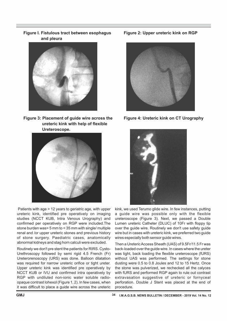
Figure I. Fistulous tract between esophagus
and pleura
Figure 2: Upper ureteric kink on RGP
Figure 3: Placement of guide wire across the
ureteric kink with help of flexible
Ureteroscope.
Figure 4: Ureteric kink on CT Urography
34
Patients with age > 12 years to geriatric age, with upper
ureteric kink, identified pre operatively on imaging
studies (NCCT KUB, Intra Venous Urography) and
confirmed per operatively on RGP were included.The
stone burden was> 5 mm to < 35 mm with single/ multiple
renal and /or upper ureteric stones and previous history
of stone surgery. Paediatric cases, anatomically
abnormal kidneys and stag horn calculi were excluded.
Routinely we don't pre stent the patients for RIRS. Cysto-
Urethroscopy followed by semi rigid 4.5 French (Fr)
Ureterorenoscopy (URS) was done. Balloon dilatation
was required for narrow ureteric orifice or tight ureter.
Upper ureteric kink was identified pre operatively by
NCCT KUB or IVU and confirmed intra operatively by
RGP with undiluted non-ionic water soluble radio-
opaque contrast Iohexol (Figure 1, 2). In few cases, when
it was difficult to place a guide wire across the ureteric
kink, we used Terumo glide wire. In few instances, putting
a guide wire was possible only with the flexible
ureteroscope (Figure 3). Next, we passed a Double
Lumen ureteric Catheter (DLUC) of 10Fr with floppy tip
over the guide wire. Routinely we don't use safety guide
wire but in cases with ureteric kink, we preferred two guide
wires especially both sensor guide wires.
Then a Ureteric Access Sheath (UAS) of 9.5Fr/11.5 Fr was
back-loaded over the guide wire. In cases where the ureter
was tight, back loading the flexible ureteroscope (fURS)
without UAS was performed. The settings for stone
dusting were 0.5 to 0.8 Joules and 12 to 15 Hertz. Once
the stone was pulverized, we rechecked all the calyces
with fURS and performed RGP again to rule out contrast
extravasation suggestive of ureteric or fornyceal
perforation. Double J Stent was placed at the end of
procedure.
I.M.A.G.S.B. NEWS BULLETIN / DECEMBER - 2019 Vol. 14 No. 12
35
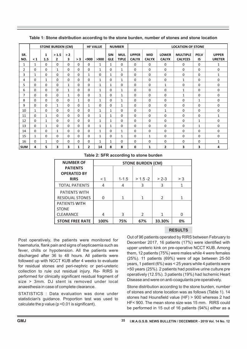
Table 1: Stone distribution according to the stone burden, number of stones and stone location
Table 2: SFR according to stone burden
Post operatively, the patients were monitored for
haematuria, flank pain and signs of septicaemia such as
fever, chills or hypotension. All the patients were
discharged after 36 to 48 hours. All patients were
followed up with NCCT KUB after 4 weeks to evaluate
for residual stones and peri-nephric or peri-ureteric
collection to rule out residual injury. Re- RIRS is
performed for clinically significant residual fragment of
size > 3mm. DJ stent is removed under local
anaesthesia in case of complete clearance.
STATISTICS : Data evaluation was done under
statistician's guidance. Proportion test was used to
calculate the p value (p <0.01 is significant).
1-1.5 > 1.5 -2 > 2-3 > 3
RESULTS
Out of 96 patients operated by RIRS between February to
December 2017, 16 patients (17%) were identified with
upper ureteric kink on pre-operative NCCT KUB. Among
them, 12 patients (75%) were males while 4 were females
(25%). 11 patients (69%) were of age between 25-50
years, 1 patient (6%) was < 25 years while 4 patients were
>50 years (25%). 2 patients had positive urine culture pre
operatively (12.5%). 3 patients (19%) had Ischemic Heart
Disease and were on anti-coagulants pre operatively.
Stone distribution according to the stone burden, number
of stones and stone location was as follows (Table 1). 14
stones had Hounsfield value (HF) > 900 whereas 2 had
HF< 900. The mean stone size was 15 mm. RIRS could
be performed in 15 out of 16 patients (94%) either as a
I.M.A.G.S.B. NEWS BULLETIN / DECEMBER - 2019 Vol. 14 No. 12
36
grading or classification system for diagnosing the
tortuosity of the ureter exists till date. No case series of
RIRS in upper ureteric kink is reported yet. We performed
Retrograde Pyelography in all patients identified with
ureteric kink on pre-operative NCCT KUB or IVU.
Semi rigid Ureteroscopy before the fURS helped in
ureteric dilatation, identifying and dusting any ureteric
stone and discovering any surprise pathology like
Transitional Cell Carcinoma of ureter. In difficult fURS,
DLUC 10 French is yet available very helpful and serves
several purposes: ureteric dilatation, performing RGP,
parking safety guide wire and straightening the ureteric
kink. We preferred two sensor guide wires as Terumo tip
of the sensor guide wire prevents mucosal perforation
whereas zebra shaft keeps the ureter straight, facilitates
repeated introduction of fURS and guide wire doesn't slip
out easily. We used UAS of 9.5 Fr/11.5 Fr as successful
UAS placement ensures better stone free rates and
decreases the ureteric mucosal trauma, intra pelvic
pressures, risk of septicemia and damage to the fURS.
[4, 5]
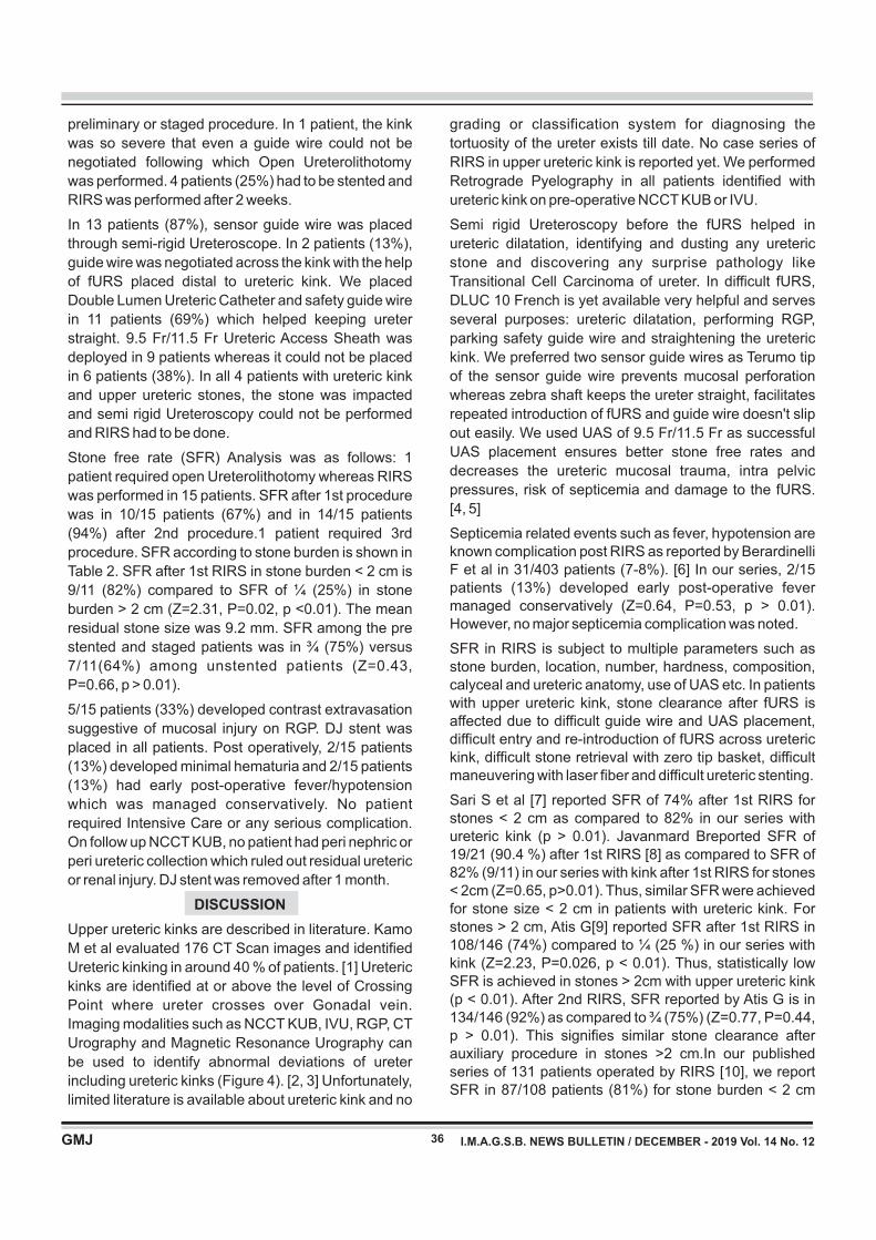
Septicemia related events such as fever, hypotension are known complication post RIRS as reported by Berardinelli F et al in 31/403 patients (7-8%). [6] In our series, 2/15 patients (13%) developed early post-operative fever managed conservatively (Z=0.64, P=0.53, p > 0.01). However, no major septicemia complication was noted.
SFR in RIRS is subject to multiple parameters such as stone burden, location, number, hardness, composition, calyceal and ureteric anatomy, use of UAS etc. In patients with upper ureteric kink, stone clearance after fURS is affected due to difficult guide wire and UAS placement, difficult entry and re-introduction of fURS across ureteric kink, difficult stone retrieval with zero tip basket, difficult maneuvering with laser fiber and difficult ureteric stenting.
Sari S et al [7] reported SFR of 74% after 1st RIRS for stones < 2 cm as compared to 82% in our series with ureteric kink (p > 0.01). Javanmard Breported SFR of 19/21 (90.4 %) after 1st RIRS [8] as compared to SFR of 82% (9/11) in our series with kink after 1st RIRS for stones < 2cm (Z=0.65, p>0.01). Thus, similar SFR were achieved for stone size < 2 cm in patients with ureteric kink. For stones > 2 cm, Atis G[9] reported SFR after 1st RIRS in 108/146 (74%) compared to ¼ (25 %) in our series with kink (Z=2.23, P=0.026, p < 0.01). Thus, statistically low SFR is achieved in stones > 2cm with upper ureteric kink (p < 0.01). After 2nd RIRS, SFR reported by Atis G is in 134/146 (92%) as compared to ¾ (75%) (Z=0.77, P=0.44, p > 0.01). This signifies similar stone clearance after auxiliary procedure in stones >2 cm.In our published series of 131 patients operated by RIRS [10], we report SFR in 87/108 patients (81%) for stone burden < 2 cm
preliminary or staged procedure. In 1 patient, the kink
was so severe that even a guide wire could not be
negotiated following which Open Ureterolithotomy
was performed. 4 patients (25%) had to be stented and
RIRS was performed after 2 weeks.
In 13 patients (87%), sensor guide wire was placed
through semi-rigid Ureteroscope. In 2 patients (13%),
guide wire was negotiated across the kink with the help
of fURS placed distal to ureteric kink. We placed
Double Lumen Ureteric Catheter and safety guide wire
in 11 patients (69%) which helped keeping ureter
straight. 9.5 Fr/11.5 Fr Ureteric Access Sheath was
deployed in 9 patients whereas it could not be placed
in 6 patients (38%). In all 4 patients with ureteric kink
and upper ureteric stones, the stone was impacted
and semi rigid Ureteroscopy could not be performed
and RIRS had to be done.
Stone free rate (SFR) Analysis was as follows: 1
patient required open Ureterolithotomy whereas RIRS
was performed in 15 patients. SFR after 1st procedure
was in 10/15 patients (67%) and in 14/15 patients
(94%) after 2nd procedure.1 patient required 3rd
procedure. SFR according to stone burden is shown in
Table 2. SFR after 1st RIRS in stone burden < 2 cm is
9/11 (82%) compared to SFR of ¼ (25%) in stone
burden > 2 cm (Z=2.31, P=0.02, p <0.01). The mean
residual stone size was 9.2 mm. SFR among the pre
stented and staged patients was in ¾ (75%) versus
7/11(64%) among unstented patients (Z=0.43,
P=0.66, p > 0.01).
5/15 patients (33%) developed contrast extravasation
suggestive of mucosal injury on RGP. DJ stent was
placed in all patients. Post operatively, 2/15 patients
(13%) developed minimal hematuria and 2/15 patients
(13%) had early post-operative fever/hypotension
which was managed conservatively. No patient
required Intensive Care or any serious complication.
On follow up NCCT KUB, no patient had peri nephric or
peri ureteric collection which ruled out residual ureteric
or renal injury. DJ stent was removed after 1 month.
DISCUSSION
Upper ureteric kinks are described in literature. Kamo
M et al evaluated 176 CT Scan images and identified
Ureteric kinking in around 40 % of patients. [1] Ureteric
kinks are identified at or above the level of Crossing
Point where ureter crosses over Gonadal vein.
Imaging modalities such as NCCT KUB, IVU, RGP, CT
Urography and Magnetic Resonance Urography can
be used to identify abnormal deviations of ureter
including ureteric kinks (Figure 4). [2, 3] Unfortunately,
limited literature is available about ureteric kink and no
I.M.A.G.S.B. NEWS BULLETIN / DECEMBER - 2019 Vol. 14 No. 12
37
against elevated renal pressures during routine flexible ureteroscopic stone manipulat ion.J Endourol . 2004 Feb;18(1):33-Available from
6. Berardinelli F, De Francesco P, Marchioni M, Cera N, Proietti S, Hennessey D et al. Infective complications after retrograde intrarenal surgery: a new standardized classification system.Int Urol Nephrol. 2016 Nov;48(11):1757-1762.
7. Sari S, Ozok HU, Cakici MC, Ozdemir H, Bas O, Karakoyunlu N,et al. A Comparison of Retrograde Intrarenal Surgery and Percutaneous Nephrolithotomy for Management of Renal Stones ?2 CM.Urol J. 2017 Jan 18;14(1):2949-2954.
8. Javanmard B, Kashi AH, Mazloomfard MM, Ansari Jafari A, Arefanian S. Retrograde Intrarenal Surgery Versus Shock Wave Lithotripsy for Renal Stones Smaller Than 2 cm: A Randomized Clinical Trial.Urol J. 2016 Oct 10;13(5):2823-2828.
9. Atis G, Culpan M, Pelit ES, Canakci C, Ulus I, Gunaydin B, Yildirim A, Caskurlu T. Comparison of Percutaneous Nephrolithotomy and Retrograde Intrarenal Surgery in Treating 20-40 mm Renal Stones.Urol J. 2017 Mar 16;14(2):2995-2999.
10. Parikh KP, Jain RJ, Kandarp AP. Is retrograde intrarenal surgery the game changer in the management of upper tract calculi? A single-center single-surgeon experience of 131 cases. Urol Ann 2018;10:29-34.
without ureteric kink, similar to 9/11 patients (81%) with stone burden < 2 cm and ureteric kink (p > 0.01). Whereas, SFR in stone burden > 2 cm in patients with ureteric kink is in ¼ patients (25%) which is significantly low as compared to SFR in 12/23 patients (52%) without kink for stone burden > 2 cm (p < 0.01). The complication rate of fever/chills/ hypotension in our series of 131 patients was in 8/131 patients (6%) as compared to 2/15 (13%) patients with ureteric kink (p > 0.01).
Out of 4 patients who required to be stented and staged, SFR was noted in ¾ (75%) versus 7/11 patients (7/11= 64%) in unstented patients (Z=0.43, P=0.66, p>0.01). Thus, no significant difference in SFR was noted among the pre-stented and staged patients versus non-stented patients in ureteric kink. Also, ureteric stenting is technically difficult in ureteric kink. Thus, pre DJ stenting does not improve stone clearance in ureteric kink as the kink reappears after stent removal.
The limitations of the study are single center small size study, limited number of patients, lack of adequate literature on the anatomy and pathology of upper ureteric kinks and lack of comparison between RIRS v/s PCNL in Upper Ureteric kink. In the future, an anatomical or radiological classification for ureteric anatomy would guide pre-operative planning and predicting the success in terms of SFR after RIRS.
CONCLUSION
RIRS is a feasible and safe modality for upper tract calculi in patients with upper ureteric kink. Maneuvers such as RGP, use of DLUC, safety guide wire improve the success in technically difficult fURS. Successful and comparable SFR have been achieved by RIRS in patients with ureteric kink for stone burden < 2 cm. With auxiliary procedure, successful stone clearance can be achieved in stone burden < 2.0 cm. Owing to the low complication rate, RIRS is a better option as compared to PCNL even in ureteric kink. Ureteric stenting is technically difficult and prior stenting does not improve the stone clearance in patients with ureteric kink.
REFERENCES1. Kamo M, Nozaki T, Yoshida K, Tateishi U, Akita K. Kinking of
the upper ureter in CT urography: anatomic and clinical significance.Surg Radiol Anat. 2016 Dec;38(10):1115-1121.
2. Potenta SE, D’Agostino R, Sternberg KM, Tatsumi K, Perusse K. CT Urography for Evaluation of the Ureter.Radiographics. 2015 May-Jun;35(3):709-26.
3. Reisner DC, Elgethun MT, Heller MT, Klepchick PR, Hartman MS. Congenital and Acquired Disorders of Ureteral Course.Curr Probl Diagn Radiol. 2017 Mar - Apr;46(2):151-160.
4. Kaplan AG, Lipkin ME, Scales CD Jr, Preminger GM. Use of ureteral access sheaths in ureteroscopy.Nat Rev Urol. 2016 Mar;13(3):135-40.
5. Auge BK, Pietrow PK, Lallas CD, Raj GV, Santa-Cruz RW, Preminger GM. Ureteral access sheath provides protection
I.M.A.G.S.B. NEWS BULLETIN / DECEMBER - 2019 Vol. 14 No. 12
INTRODUCTION
Primary open angle glaucoma is a chronic , progressive
optic neuropathy in adults in which there is a
characteristic acquired atrophy of the optic nerve and loss [1]of retinal ganglion cells and their axons . Because 50%
[2]of the RGCs are located within the macula and the
macular shape is generally less variable than the ONH,
macular thickness assessment has been considered for
evaluating structural changes of glaucoma. High-
resolution imaging of retinal structure is done through
optical coherence tomography.[3]Zeimer et al first suggested imaging of the macula as a
potential location for glaucoma evaluation. Macular
thickness measurements by optical coherence
tomography (OCT) have been shown in previous studies
A Comparative Study of Macular Thickness in Primary open Angle Glaucoma Patients and Normal Patients.
Original Articles
Dr. Nilesh V. Parekh*, Dr. Sagar S.Patel**
* Professor and Head,Department of Ophthalmology, Government medical college,Bhavnagar.
Institution: Government Medical College and New Opd room no-135,Sir-T Hospital, Bhavnagar,Gujarat,364001.
** Senior Resident, Department of Ophthalmology, GMERS Medical College,Valsad.
38
Correspondence Address : D r. Sagar Patel D-19, Palliniu - 3, Road - D, Plot No. 165, Tithal Road, Valsad-396001 Email : [email protected]
ABSTRACT
Aim : To compare difference of macular thickness using optical coherence tomography (OCT) in primary open
angle glaucoma (POAG)patients and normal subjects..
Materials and methods :
This Observational case control study included primary open angle glaucoma(POAG) patients(n=60eyes)and
healthy subjects in the control group(n=60eyes).All subjects underwent detailed history,general examination,and
systemic examination. Complete ocular examination included best corrected visual acuity(BCVA),slit lamp
examination, intraocular pressure(IOP),gonioscopy,dilated fundus biomicroscopy.visual fied analysis was done
using haag streit octopus 900 machine.
Optical coherence tomography imaging machine was performed using Topcon 3D OCT machine, version
8.42003.01.In both these groups,parameters analysed were macular thickness and macular volume.
Results :
The POAG group had significantly decreased values of macular thickness(11.3%) macular thickness in POAG as
compared to control (265.09 ± 12.60 vs. 235.16 ± 7.64, p<0.001). and macular volume (7.68 ± 0.46 vs. 7.00 ±
0.48, p<0.001).Thus ,macular thickness and macular volume parameters may be used for making the diagnosis of
glaucoma, especially in patients with abnormalities of disc.
to be significantly thinner in glaucomatous eyes [4]compared to healthy eyes
RETINA
Retina is a thin membrane extending from the optic disc to
the ora serrata in front. It varies in thickness from 0.4mm
near the optic nerve to 0.15mm anteriorly at the ora [5]serrata.
Macular thickness
The macula contains over 50% of all retinal ganglion cells
and is an ideal area for detection of early cell loss and [5]changes over the time because of high cell density. In
the macular area, ganglion cells are arranged in 4 to 6
layers making up 30 to 35% of retinal macular thickness,
so that the loss of macular ganglion cells results in
significant retinal or retinal nerve fiber layer thinning.
I.M.A.G.S.B. NEWS BULLETIN / DECEMBER - 2019 Vol. 14 No. 12
KEY WORDS : Macular thickness,Glaucoma,Optical coherence tomography
39
OPTICAL COHERENCE TOMOGRAPHY
OCT uses low-coherence interferometry to produce a
two-dimensional image of optical scattering from internal
tissue microstructures in a way that is analogous to
ultrasonic pulse-echo imaging..
The diagnosis and management of glaucoma are
currently difficult clinical problems. Intraocular pressure
measurements often do not adequately predict the
progression of glaucoma. [6]
METHODOLOGY
Method of collection of data : Data was collected after
approval from Institutional Review Board(IRB)
,government medical college.
Study Area : Department of Ophthalmology and
Government Medical College.
Sample Size : 60 patients-30 patients of primary open
angle glaucoma and 30 normal patients(control group).
Study period : Study was carried out over a period of 6
months.
Ethical consideration : Informed and written consent
from each participant was taken.
Statistical analysis : Comparision between the values of
macular thickness between primary open angle
glaucoma patients and normal patients will be done
.Independent Student's t test and chi-square (χ2) test
were used for statistical analysis.
Inclusion criteria
• Patients age more than 30 years of either sex.
• Patients who will be ready for written and inform
consent.
• A case of primary open angle glaucoma and normal
subjects(control group).
Exclusion criteria
• Patients who are not ready to give written and inform
consent.
• Patients with any other retinal disease.
• Patients with any other associated ocular disease or
deformity that hampers posterior segment evaluation
by oct like dense cataract,corneal opacity.
• Patients on steroid therapy and on any other
medications known to affect retina.
MATERIAL
• The study was performed on patients diagnosed as
pr imary open angle g laucoma and normal
patients(control group).
• All patients underwent :
1. Visual assesment using Snellen's visual acuity chart.
2. Examination of anterior segment in detail using slit
lamp biomicroscopy.
3. Refraction.
4. Intraocular pressure measurement using goldmann
applanation tonomertry.
5. Gonioscopy using 3 mirror lens.
6. Perimetry by haag streit octopus 900 machine.
7. Dilated fundus examination was done using 90 D lens
on slit lamp biomicroscope. The participant's both
eyes were dilated using tropicamide (0.8%)+
phenylephrine (5%). Dilated fundus findings were
noted which included media,disc ,macula,blood
vessels and background.
8. All patients then underwent macular thickness
evaluation using optical coherence tomography. OCT
was done using TOPCON 3D OCT machine, version
8.42003.01.
OBSERVATION
DEMOGRAPHIC CHARACTERISTICS :
The demographic characteristics (age and sex) of two
groups is summarised as summarised in Table I and also
shown in Figure I and II, respectively. The age of control
and POAG ranged from 34-77 yrs and 34-65 yrs
respectively with mean (± SD) 50.93 ± 9.79 yrs and 52.53
± 7.78 yrs respectively and median 49 yrs and 54 yrs
respectively. The mean age of POAG group was slightly
higher than control group. Comparing the mean age of
two groups, Student's t test showed similar age between
the two groups (50.93 ± 9.79 vs. 52.53 ± 7.78, t=0.70,
p=0.486) i.e. did not differ significantly
Further, in control group, there were 14 (46.7%) females
and 16 (53.3%) males whereas it were 15 (50.0%) and 15
(50.0%) in POAG group. Comparing the sex proportion
(M/F) of two groups, χ2 test showed similar sex proportion
between the two groups (χ2=0.07, p=0.796) i.e. also not
differ significantly.
SECONDARY OUTCOME MEASURES
I. RIGHT EYE
The secondary outcome measures (Best corrected visual
acuity , Slit lamp examination, Intraocular Pressure,
MEDIA, CDR, MACULA, Blood vessels, Background,
gonioscopy ) of two groups at right eye is summarised in
Table II and also shown in Fig.III-VII. Comparing the
I.M.A.G.S.B. NEWS BULLETIN / DECEMBER - 2019 Vol. 14 No. 12
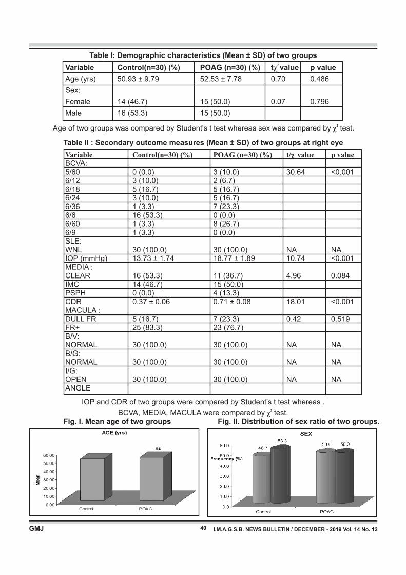
40
Table I: Demographic characteristics (Mean ± SD) of two groups 2
Variable Control(n=30) (%) POAG (n=30) (%) tχ value p value
Age (yrs) 50.93 ± 9.79 52.53 ± 7.78 0.70 0.486
Sex:
Female 14 (46.7) 15 (50.0) 0.07 0.796
Male 16 (53.3) 15 (50.0)
Variable Control(n=30) (%) POAG (n=30) (%) t/ value p value2 χ BCVA: 5/60 0 (0.0) 3 (10.0) 30.64 <0.0016/12 3 (10.0) 2 (6.7)6/18 5 (16.7) 5 (16.7)6/24 3 (10.0) 5 (16.7)6/36 1 (3.3) 7 (23.3)6/6 16 (53.3) 0 (0.0)6/60 1 (3.3) 8 (26.7)6/9 1 (3.3) 0 (0.0) SLE: WNL 30 (100.0) 30 (100.0) NA NAIOP (mmHg) 13.73 ± 1.74 18.77 ± 1.89 10.74 <0.001MEDIA : CLEAR 16 (53.3) 11 (36.7) 4.96 0.084IMC 14 (46.7) 15 (50.0) PSPH 0 (0.0) 4 (13.3)CDR 0.37 ± 0.06 0.71 ± 0.08 18.01 <0.001MACULA : DULL FR 5 (16.7) 7 (23.3) 0.42 0.519FR+ 25 (83.3) 23 (76.7) B/V: NORMAL 30 (100.0) 30 (100.0) NA NAB/G: NORMAL 30 (100.0) 30 (100.0) NA NAI/G: OPEN 30 (100.0) 30 (100.0) NA NAANGLE
2Age of two groups was compared by Student's t test whereas sex was compared by χ test.
Fig. I. Mean age of two groups Fig. II. Distribution of sex ratio of two groups.
Table II : Secondary outcome measures (Mean ± SD) of two groups at right eye
IOP and CDR of two groups were compared by Student's t test whereas .2
BCVA, MEDIA, MACULA were compared by χ test.
I.M.A.G.S.B. NEWS BULLETIN / DECEMBER - 2019 Vol. 14 No. 12
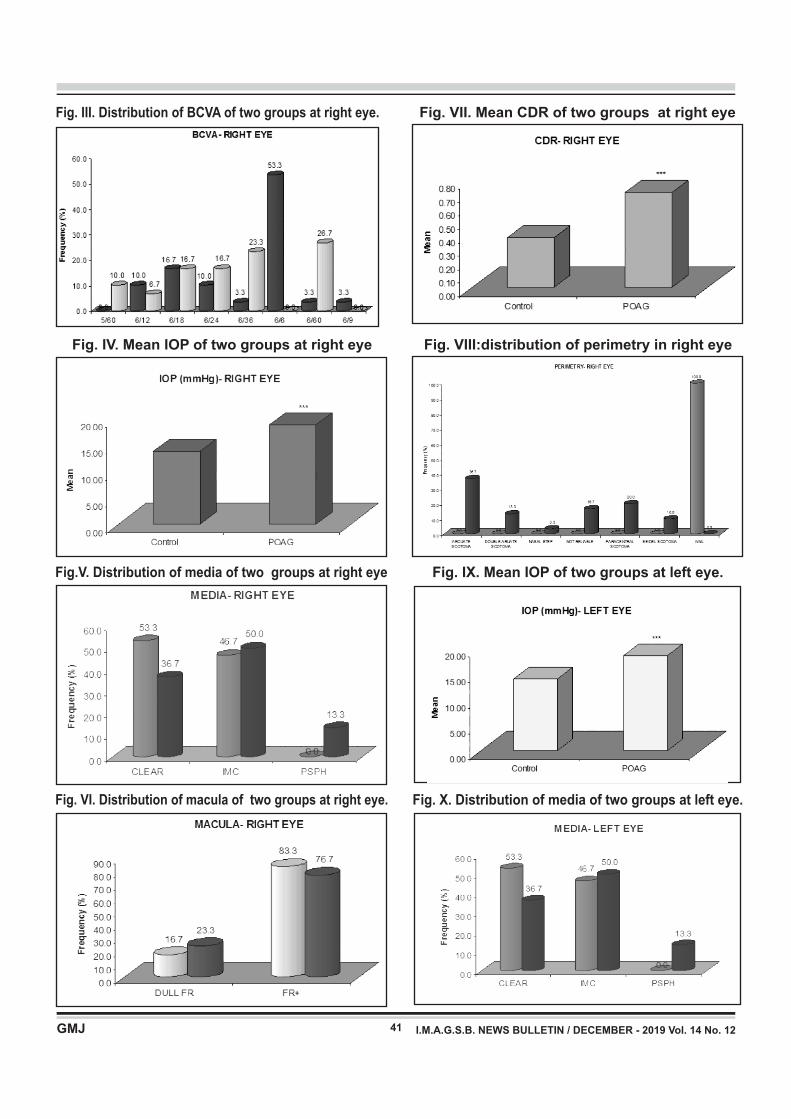
Fig. IX. Mean IOP of two groups at left eye.
41
Fig. III. Distribution of BCVA of two groups at right eye.
Fig. IV. Mean IOP of two groups at right eye
Fig.V. Distribution of media of two groups at right eye
Fig. VI. Distribution of macula of two groups at right eye.
Fig. VII. Mean CDR of two groups at right eye
Fig. VIII:distribution of perimetry in right eye
I.M.A.G.S.B. NEWS BULLETIN / DECEMBER - 2019 Vol. 14 No. 12
Fig. X. Distribution of media of two groups at left eye.
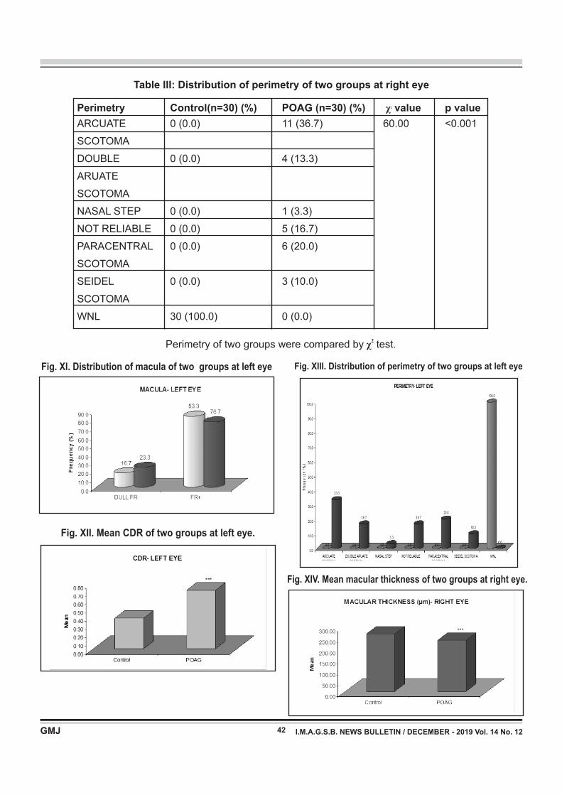
42
Perimetry Control(n=30) (%) POAG (n=30) (%) value p value2 χ
ARCUATE 0 (0.0) 11 (36.7) 60.00 <0.001
SCOTOMA
DOUBLE 0 (0.0) 4 (13.3)
ARUATE
SCOTOMA
NASAL STEP 0 (0.0) 1 (3.3)
NOT RELIABLE 0 (0.0) 5 (16.7)
PARACENTRAL 0 (0.0) 6 (20.0)
SCOTOMA
SEIDEL 0 (0.0) 3 (10.0)
SCOTOMA
WNL 30 (100.0) 0 (0.0)
Table III: Distribution of perimetry of two groups at right eye
2Perimetry of two groups were compared by χ test.
Fig. XI. Distribution of macula of two groups at left eye
Fig. XIV. Mean macular thickness of two groups at right eye.
Fig. XII. Mean CDR of two groups at left eye.
I.M.A.G.S.B. NEWS BULLETIN / DECEMBER - 2019 Vol. 14 No. 12
Fig. XIII. Distribution of perimetry of two groups at left eye
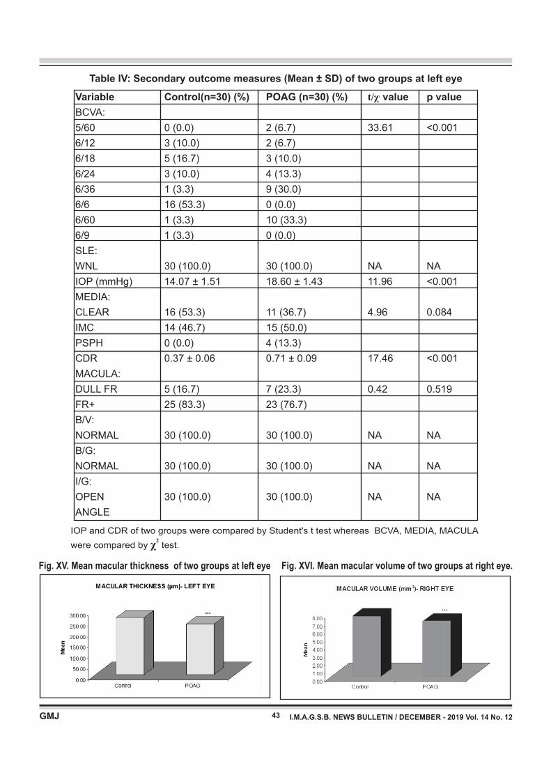
43 I.M.A.G.S.B. NEWS BULLETIN / DECEMBER - 2019 Vol. 14 No. 12
Variable Control(n=30) (%) POAG (n=30) (%) t/ value p value2 χ
BCVA:
5/60 0 (0.0) 2 (6.7) 33.61 <0.001
6/12 3 (10.0) 2 (6.7)
6/18 5 (16.7) 3 (10.0)
6/24 3 (10.0) 4 (13.3)
6/36 1 (3.3) 9 (30.0)
6/6 16 (53.3) 0 (0.0)
6/60 1 (3.3) 10 (33.3)
6/9 1 (3.3) 0 (0.0)
SLE:
WNL 30 (100.0) 30 (100.0) NA NA
IOP (mmHg) 14.07 ± 1.51 18.60 ± 1.43 11.96 <0.001
MEDIA:
CLEAR 16 (53.3) 11 (36.7) 4.96 0.084
IMC 14 (46.7) 15 (50.0)
PSPH 0 (0.0) 4 (13.3)
CDR 0.37 ± 0.06 0.71 ± 0.09 17.46 <0.001
MACULA:
DULL FR 5 (16.7) 7 (23.3) 0.42 0.519
FR+ 25 (83.3) 23 (76.7)
B/V:
NORMAL 30 (100.0) 30 (100.0) NA NA
B/G:
NORMAL 30 (100.0) 30 (100.0) NA NA
I/G:
OPEN 30 (100.0) 30 (100.0) NA NA
ANGLE
Table IV: Secondary outcome measures (Mean ± SD) of two groups at left eye
IOP and CDR of two groups were compared by Student's t test whereas BCVA, MEDIA, MACULA 2
were compared by χ test.
Fig. XV. Mean macular thickness of two groups at left eye Fig. XVI. Mean macular volume of two groups at right eye.
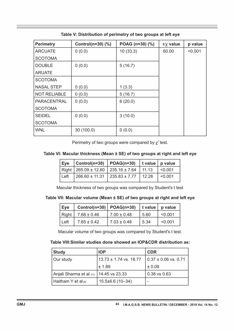
44
Perimetry Control(n=30) (%) POAG (n=30) (%) t/ value p value2 χ
ARCUATE 0 (0.0) 10 (33.3) 60.00 <0.001
SCOTOMA
DOUBLE 0 (0.0) 5 (16.7)
ARUATE
SCOTOMA
NASAL STEP 0 (0.0) 1 (3.3)
NOT RELIABLE 0 (0.0) 5 (16.7)
PARACENTRAL 0 (0.0) 6 (20.0)
SCOTOMA
SEIDEL 0 (0.0) 3 (10.0)
SCOTOMA
WNL 30 (100.0) 0 (0.0)
Table V: Distribution of perimetry of two groups at left eye
2Perimetry of two groups were compared by χ test.
Table VI: Macular thickness (Mean ± SE) of two groups at right and left eye
Eye Control(n=30) POAG(n=30) t value p value
Right 265.09 ± 12.60 235.16 ± 7.64 11.13 <0.001
Left 266.60 ± 11.31 235.83 ± 7.77 12.28 <0.001
Macular thickness of two groups was compared by Student's t test
Table VII: Macular volume (Mean ± SE) of two groups at right and left eye
Eye Control(n=30) POAG(n=30) t value p value
Right 7.68 ± 0.46 7.00 ± 0.48 5.60 <0.001
Left 7.65 ± 0.42 7.03 ± 0.48 5.34 <0.001
Macular volume of two groups was compared by Student's t test.
Table VIII:Similar studies done showed an IOP&CDR distribution as:
Study IOP CDR
Our study 13.73 ± 1.74 vs. 18.77 0.37 ± 0.06 vs. 0.71
± 1.89 ± 0.08
Anjali Sharma et al [11] 14.45 vs 23.33 0.38 vs 0.63
Haitham Y et al [8] 15.5±6.6 (10–34) -
I.M.A.G.S.B. NEWS BULLETIN / DECEMBER - 2019 Vol. 14 No. 12

Table IX:Similar studies done showed an age distribution as:
Table X:Similar studies done showed a Macular thickness distribution as
Study Total subjects with Mean age distribution
(POAG)
Our study 30 52.53 ± 7.78 yrs[7]
David s et al 30 56.7 ± 20.3 yrs[8]
Haitham Y et al 42 53.7 ± 3.7 years
Behzad FALLAHI 104 59.96 ± 8.75 years[9]MOTLAGH et al
Study Macular thickness
Our study 265.09 ± 12.60 vs. 235.16 ± 7.64[11]Anjali Sharma et al 263.56 vs 243.96 mm
[7]David s et al 278+_24m vs 304+-15m[9]Behzad FALLAHI MOTLAGH et al 276.96 ± 7.80 (262.00 to 298.00)
Table XI : Similar studies done showed a Macular volume distribution as
Study Macular thickness
Our study 7.68 ± 0.46 vs. 7.00 ± 0.48[11]
Anjali Sharma et al 6.64 ± 0.17 vs 6.18 ± 0.39
45 I.M.A.G.S.B. NEWS BULLETIN / DECEMBER - 2019 Vol. 14 No. 12
quantitative IOP and CDR of two groups at right eye,
Student's t test showed significantly different and higher
IOP (26.8%) (13.73 ± 1.74 vs. 18.77 ± 1.89, t=10.74,
p<0.001) and CDR (47.9%) (0.37 ± 0.06 vs. 0.71 ± 0.08,
t=18.01, p<0.001) both in POAG patients as compared to
control subjects
Further, comparing the distribution of PERIMETRY of two
groups at right eye, χ2 test showed significantly different
distribution of PERIMETRY (χ2=60.00, p<0.001)
between the two groups (Table III and Fig. VII).
Fig. XVII. Mean macular volume of two groups at left eye Similarly, comparing the distributions of categorical
BCVA, MEDIA and MACULA of two groups at right eye, χ2
test showed significantly different distribution of BCVA
(χ2=30.64, p<0.001) between the two groups while
distributions of both MEDIA and MACULA were found
similar (p>0.05) between the two groups i.e. did not differ
significantly.
II. LEFT EYE
The secondary outcome measures (BCVA, SLE, IOP,
MEDIA, CDR, MACULA, B/V, B/G and I/G) of two groups
at left eye is summarised in Table IV and also shown in Fig
IX-XII. Comparing the quantitative IOP and CDR of two
groups at left eye, Student's t test showed significantly
different and higher IOP (24.4%) (14.07 ± 1.51 vs. 18.60 ±
1.43, t=11.96, p<0.001) and CDR (47.9%) (0.37 ± 0.06 vs.
0.71 ± 0.09, t=17.46, p<0.001) both in POAG patients as
compared to control subjects.
Similarly, comparing the distributions of categorical
BCVA, MEDIA and MACULA of two groups at left eye, χ2
test showed significantly different distribution of BCVA
(χ2=33.61, p<0.001) between the two groups while
distributions of both MEDIA and MACULA were found
46 I.M.A.G.S.B. NEWS BULLETIN / DECEMBER - 2019 Vol. 14 No. 12
In our study it was found the mean age of POAG group
was slightly higher than control group. Comparing the
mean age of control vs POAG (50.93 ± 9.79 vs. 52.53 ±
7.78, t=0.70, p=0.486)
In varies studies it has been noticed that there is a
negative relationship between retinal thickness and age,
total macular volume and RNFL thickness Eriksson u [10]et al
We compared the quantitative IOP and CDR of two
groups at right eye, test showed significantly different and
higher IOP (26.8%) (13.73 ± 1.74 vs. 18.77 ± 1.89,
t=10.74, p<0.001) and CDR (47.9%) (0.37 ± 0.06 vs. 0.71
± 0.08, t=18.01, p<0.001) both in POAG as compared to
control. Similarly, comparing the two groups at right eye,
χ2 test showed significantly different distribution of BCVA
(χ2=30.64, p<0.001) between the two groups while
distributions of both MEDIA and MACULA were found
similar (p>0.05) between the two groups.
Further, χ2 test showed significantly different distribution
of PERIMETRY (χ2=60.00, p<0.001) between the two
groups.
We are comparing the quantitative IOP and CDR of two
groups in left eye, test showed significantly different and
higher IOP (24.4%) (14.07 ± 1.51 vs. 18.60 ± 1.43,
t=11.96, p<0.001) and CDR (47.9%) (0.37 ± 0.06 vs. 0.71
± 0.09, t=17.46, p<0.001) both in POAG as compared to
control. Similarly, χ2 test showed significantly different
distribution of BCVA (χ2=33.61, p<0.001) between the
two groups while distributions of both MEDIA and
MACULA were found similar (p>0.05) between the two
groups.
Further, χ2 test showed significantly different distribution
of PERIMETRY (χ2=60.00, p<0.001) between the two
groups .
We compare the macular thickness of two groups in right
eye, test showed significantly different and lower (11.3%)
macular thickness in POAG as compared to control
( 2 6 5 . 0 9 ± 1 2 . 6 0 v s . 2 3 5 . 1 6 ± 7 . 6 4 , m e a n
difference=29.94, t=11.13, p<0.001). Similarly left eye,
test showed significantly different and lower (11.5%)
macular thickness in POAG as compared to control
( 2 6 6 . 6 0 ± 11 . 3 1 v s . 2 3 5 . 8 3 ± 7 . 7 7 , m e a n
difference=30.77, t=12.28, p<0.001)
Thus it was seen that macular thickness was decreased in
glaucomatous patients as compared to normal subjects.
These findings are in correlation with studies discussed
above and published literature. [12], [13]
similar (p>0.05) between the two groups i.e. did not differ
significantly.
Further, comparing the distribution of PERIMETRY of two
groups at left eye, χ2 test showed significantly different
distribution of PERIMETRY (χ2=60.00, p<0.001)
between the two groups (table V, figure XIII).
PRIMARY OUTCOME MEASURES
I. MACULAR THICKNESS
The macular thickness of two groups at both right and left
eye is summarised in Table VI and also depicted in Fig.
XIV and XV, respectively. Comparing the macular
thickness of two groups at right eye, Student's t test
showed significantly different and lower (11.3%) macular
thickness in POAG patients as compared to control
subjects (265.09 ± 12.60 vs. 235.16 ± 7.64, mean
difference=29.94, t=11.13, p<0.001) (Table VI and
Fig.XIV ).
Similarly, comparing the macular thickness of two groups
at left eye, Student's t test showed significantly different
and lower (11.5%) macular thickness in POAG patients as
compared to control subjects (266.60 ± 11.31 vs. 235.83 ±
7.77, mean difference=30.77, t=12.28, p<0.001) (Table VI
and Fig. XV)
II. MACULAR VOLUME
The macular volume of two groups at both right and left
eye is summarised in Table VII and also shown in Fig.XVI
and XVII , respectively. Comparing the macular volume
of two groups at right eye, Student's t test showed
significantly different and lower (8.8%) macular volume in
POAG patients as compared to control subjects (7.68 ±
0.46 vs. 7.00 ± 0.48, mean difference=0.68, t=5.60,
p<0.001) (Table VII and Fig. XVI).
Similarly, comparing the macular volume of two groups at
left eye, Student's t test showed significantly different and
lower (8.1%) macular volume in POAG patients as
compared to control subjects (7.65 ± 0.42 vs. 7.03 ± 0.48,
mean difference=0.42, t=5.34, p<0.001) (Table VII and
Fig. XVI)
DISCUSSION
In our study we compare and correlate macular thickness
in primary open angle glaucoma patients and normal
subjects using optical coherence tomography.
A total of 60 patients enrolled in the study. Out of which 30
were diagnosed case of primary open angle glaucoma
(POAG) and 30 age and sex matched normal subjects
without glaucoma were recruited served as control.
47 I.M.A.G.S.B. NEWS BULLETIN / DECEMBER - 2019 Vol. 14 No. 12
We compare the macular volume of two groups in right
eye, test showed significantly different and lower (8.8%)
macular volume in POAG as compared to control (7.68 ±
0.46 vs. 7.00 ± 0.48, mean difference=0.68, t=5.60,
p<0.001). Similarly left eye, test showed significantly
different and lower (8.1%) macular volume in POAG as
compared to control (7.65 ± 0.42 vs. 7.03 ± 0.48, mean
difference=0.42, t=5.34, p<0.001)
CONCLUSION
With the increasing literature regarding the role of
macular imaging by optical coherence tomography (OCT)
in glaucoma care,Spectral domain OCT (SD-OCT) has
allowed for high resolution imaging of the total macula and
macular segments. It proves that macular thickness and
volume shows a significant correlation with the
glaucomatous damage. It may be useful method of
document ing ear ly g laucoma and moni tor ing
progression.
Thus, in our study we conclude that macular parameters,
such as total macular volume & macular thickness was
significantly lower in open angle glaucoma patients when
compared with the normal subjects. Thus in addition to
RNFL thickness to aid in the diagnosis of early glaucoma
using OCT, in certain conditions, such as disc
abnormalities or peripapillary atrophy, where RNFL
parameters may be distorted macular parameters may be
relied upon.
REFERENCES
1. "Facts About Glaucoma". National Eye Institute. Archived from the
original on 28 March 2016. Retrieved 29 March 2016
2. Van Buren JM. The Retinal Ganglion Cell Layer Springfield. IL:
Charles C Thomas; 1963. pp. 36–64
3. Quantitative detection of glaucomatous damage at the posterior
pole by retinal thickness mapping. A pilot study. Zeimer R, Asrani S,
Zou S, Quigley H, Jampel H Ophthalmology. 1998 Feb; 105(2):224-
31
4. Detection of glaucoma progression with stratus OCT retinal nerve
fiber layer, optic nerve head, and macular thickness measurements.
Medeiros FA, Zangwill LM, Alencar LM, Bowd C, Sample PA,
Susanna R Jr, Weinreb RN Invest Ophthalmol Vis Sci. 2009 Dec;
50(12):5741-8
5. Yoon SW, Park WH, Baek SH, Kong SM. Thicknesses of Macular
Retinal Layer and Peripapillary Retinal Nerve Fiber Layer in
Patients with Hyperopic Anisometropic Amblyopia. Korean J
O p h t h a l m o l . 2 0 0 5 M a r ; 1 9 ( 1 ) : 6 2 - 6 7 .
https://doi.org/10.3341/kjo.2005.19.1.62
6. Intraocular pressure and glaucoma. Sommer AAm J Ophthalmol.
1989 Feb 15; 107(2):186-8.
7. David s. Greenfield, MD; Harmohina bagga, MD; Robert
W.knighton. Macular thickness changes in glaucomatous optic
neuropathy detected using optical coherence tomography. Arch
ophthalmol. 2003;121.41-46
8. Haitham Y. Al-Nashar, Ahmad M.B. Awad. Correlation between
peripapillary choroidal thickness and nerve fiber layer thickness in
primary open-angle glaucoma. Delta Journal of Ophthalmology,
Vol. 19 No. 2, April-June 2018
9. behzad fallahi motlagh 1; ali sadeghi 1 et al Med Hypothesis Discov
Innov Ophthalmol. 2017; 6(2)
10. Eriksson u et al. Macular edema and visual outcome following
cataract surgery in patients with diabetic retinopathy and controls.
Graefes arch clin exp ophthalmol. 2011 mar; 249(3):349-59.doi:
10,1007/s00417-010-1484-9. Epub 2010 sep 9
11. Sharma A, Agarwal P, Sathyan P, Saini VK. Macular Thickness
Variability in Primary Open Angle Glaucoma Patients using Optical
Coherence Tomography. J Current Glau Prac 2014;8(1):10-14.
12. Quigley HA, West SK, Rodriguez J, et al. The prevalence of
glaucoma in a population-based study of Hispanic subjects:
Proyecto VER. Arch Ophthalmol. 2001;119:1819–26.
13. Dielemans I, Vingerling JR, Algra D, et al. Primary open angle
glaucoma, intraocular pressure, and systemic blood pressure in the
general elderly population. The Rotterdam Study. Ophthalmology.
1995;102:54–60.
INTRODUCTION
Chronic pancreatitis is a progressive inflammatory
disease in which there is irreversible destruction of
pancreatic tissue. Its clinical course is charecterised by
severe abdominal pain and in its later stages exocrine and
endocrine pancreatic insufficiency. In India the
prevalence is 100-200 per 100000. The male : female
ratio is 4:11. There are many causes of pain in chronic
pancreatitis, but increased pancreatic ductal pressure is 2 ,3 .one of them Chronic fibrocalcific pancreatitis
associated with pancreatic ductal dilation and intractable
pain have been historically managed successfully with 2,3lateral pancreaticojejunostomy (LPJ) . The primary
objective of surgical treatment in chronic pancreatitis is to 4relieve pain . A second objective is to preserve or even
improve pancreatic function by restoring the flow of
pancreatic secretions to the intestine and by reducing
pancreatic inflammation. Eleven patients with intractable
pain due to chronic pancreatitis were treated by lateral 5pancreaticojejunostomy (modified Puestow procedure)
after preoperative assessment in SMIMER hospital,
Surat. All patients were evaluated on follow up visits for
pain relief and endocrine or exocrine insufficiency.
Keywords : Chronic pancreatitis, Lateral pancreaticojejunostomy
A study to evaluate pain relief and pancreatic function after lateral pancreati
cojejunotomy for chronic pancreatitis
Original Articles
Dr. Akash Shah*, Dr. Hiren Vaidya**, Dr. Aditya Vaidya***, Dr. Harish Chauhan**
*Resident in Department of Surgery, Surat Municipal Institute of Medical Education and Research, Surat
**Associate Professor, Department of Surgery, SMIMER, Surat)
48
Correspondence Address : Dr. Hiren Vaidya Aditya Hospital, Pramukh Doctor House, Besides Hundai Showroom, Parvat patiya, Surat-395010 Email : [email protected]
ABSTRACT
Introduction : Chronic pancreatitis is the persistent inflammation and irreversible fibrosis of pancreatic tiisue. It is
charecterised by chronic abdominal pain and pancreatic insufficiency that decreses the quality of life of the
patients.
Material and Methods : Eleven patients with severe pain due to chronic pancreatitis were treated by lateral
pancreaticojejunostomy after preoperative assessment in SMIMER hospital, Surat. All patients were evaluated on
follow up visits for pain relief and endocrine and exocrine insufficiency.
Results : Eight patients (73%) are free of pain after operation. Two patients (18%) had decresed pain after
surgery. One patient (9%) had no relief of pain and died after surgery. There is no improvement in steatorrhea and
diabetes mellitus after surgery.
Conclusion : Longitudinal pancreaticojejunostomy has a role for the control of pain in chronic pancreatitis.
MATERIAL AND METHODS
Eleven patients with recurrent or persistent pain due to
chronic pancreatitis admited to SMIMER hospital were
included in the study. These patients had severe pain
ranging from 8 to 10 on VAS (visual analog scale). The
pain was uncontrolled by analgesics. The average
duration of pain was 11 months, with a range of 5 months
to 2 years. All patients had a dilated pancreatic duct on
magnetic resonanace cholangiopancreatography with a
minimum diameter of 9 mm and changes of chronic
pancreatitis. Preoperative assessment included history of
diabetes mellitus, habit of alcohol consumption and
tobacco chewing. Blood investigations included serum
amylase, s. lipase, s. calcium, total leucocyte count, s.
bilirubin, and s. alkaline phosphatase levels. Radiological
investigation included ultrasonography of the abdomen,
contrast enhanced computed tomography abdomen and
magnetic resonanace cholangiopancreatography.
Evaluation of pancreatic function was done by doing
fasting blood sugar level, post prandial blood sugar level
and stool fat assesment. The operation performed in 10
patients was a open lateral pancreaticojejunostomy
utilizing a Roux-en-Y loop of jejunum. One patient was
I.M.A.G.S.B. NEWS BULLETIN / DECEMBER - 2019 Vol. 14 No. 12
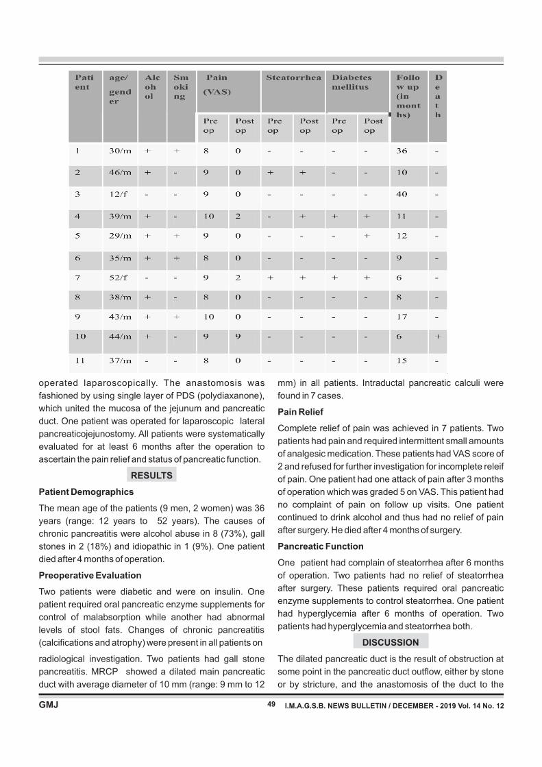
49
operated laparoscopically. The anastomosis was
fashioned by using single layer of PDS (polydiaxanone),
which united the mucosa of the jejunum and pancreatic
duct. One patient was operated for laparoscopic lateral
pancreaticojejunostomy. All patients were systematically
evaluated for at least 6 months after the operation to
ascertain the pain relief and status of pancreatic function.
RESULTS
Patient Demographics
The mean age of the patients (9 men, 2 women) was 36
years (range: 12 years to 52 years). The causes of
chronic pancreatitis were alcohol abuse in 8 (73%), gall
stones in 2 (18%) and idiopathic in 1 (9%). One patient
died after 4 months of operation.
Preoperative Evaluation
Two patients were diabetic and were on insulin. One
patient required oral pancreatic enzyme supplements for
control of malabsorption while another had abnormal
levels of stool fats. Changes of chronic pancreatitis
(calcifications and atrophy) were present in all patients on
radiological investigation. Two patients had gall stone
pancreatitis. MRCP showed a dilated main pancreatic
duct with average diameter of 10 mm (range: 9 mm to 12
mm) in all patients. Intraductal pancreatic calculi were
found in 7 cases.
Pain Relief
Complete relief of pain was achieved in 7 patients. Two
patients had pain and required intermittent small amounts
of analgesic medication. These patients had VAS score of
2 and refused for further investigation for incomplete releif
of pain. One patient had one attack of pain after 3 months
of operation which was graded 5 on VAS. This patient had
no complaint of pain on follow up visits. One patient
continued to drink alcohol and thus had no relief of pain
after surgery. He died after 4 months of surgery.
Pancreatic Function
One patient had complain of steatorrhea after 6 months
of operation. Two patients had no relief of steatorrhea
after surgery. These patients required oral pancreatic
enzyme supplements to control steatorrhea. One patient
had hyperglycemia after 6 months of operation. Two
patients had hyperglycemia and steatorrhea both.
DISCUSSION
The dilated pancreatic duct is the result of obstruction at
some point in the pancreatic duct outflow, either by stone
or by stricture, and the anastomosis of the duct to the
I.M.A.G.S.B. NEWS BULLETIN / DECEMBER - 2019 Vol. 14 No. 12
50
4. Fischer JE, Ellison EC, Upchurch GR, Gould JC, Klimberg VS,
Henke P, Hochwald SN, Tiao GM. Selection of surgical therapy
for chronic pancreatitis. In: Muscarella P, Nealon WH, editors.
Fischer's mastery of surgery, 7th ed. Wolters kluwer; 2018;
122: 1492-1504
5. Fischer JE, Ellison EC, Upchurch GR, Gould JC, Klimberg VS,
Henke P, Hochwald SN, Tiao GM. Operative interventions for
chronic pancreatitis. In: Lillemoe KD, Ball CG, editors. Fischer's
mastery of surgery, 7th ed. Wolters kluwer; 2018; 123: 1504-
1513
6. Jordan GL, Strug BS, Crowder WE. Current status of
pancreatojejunostomy in the management of chronic
pancreatitis. Am J Surg 1977; 133: 46-51
7. Way LW, Gadacz T, Goldman L. Surgical treatment of chronic
pancreatitis. Am J Surg 1974; 127: 202-209
8. David B. Adams, Margaret C. Ford, Marion C. Anderson.
Outcome after lateral pancreaticojejunostomy for chronic
pancreatitis. Ann Surg 1994; 219: 481-489
9. Little JM. Alcohol abuse and chronic pancreatitis. Surgery 1987;
101: 357-360
10. Mannell A, Adson MA, Mcllrath DC, Ilstrup DM. Surgical
management of chronic pancreatitis: long-term results in 141
patients. Br J Surg 1988; 75: 467-472
11. Partington PF, Rochelle RF: Modified Puestow procedure for
retrograde drainage of the pancreatic duct. Ann Surg 1966;
152: 1037-1643
12. Cahow CE, Hayes MA: Operative treatment of chronic
recurrent pancreatitis. Am J Surg 1973; 125: 390-398
13. Leger L, Lenriot JP, LeMaigre G. Five to twenty year follow up
after surgery for chronic pancreatitis in 148 patients. Ann Surg
1974; 180:185-191
14. Andrew L. Warshaw, John W. Popp, Robert H. Schapiro. Long
term patency, pancreatic function and pain relief after lateral
pancreat ico je junostomy for chron ic pancreat i t i s .
Gastroenterology 1980; 79: 289-293
jejunum provides an alternate vent for decompression.
Thus lateral pancreaticojejunostomy provides relief from
pain3,6,7. Outcome after procedures that provide ductal
drainage for chronic pancreatitis is influenced by several
variables8. Chronic pancreatitis is almost exclusively a
result of chronic alcohol abuse9. This population might
have other substance abuse problems including tobacco,
that also may affect their health status and have effect on
deveopement of chronic pancreatitis3. Patients were
selected for LPJ on the basis of unrelenting pain and
evidence of a dilated pancreatic duct. However,
associated other conditions were present in a few of the
patients. One patient had gall bladder and common bile
duct calculi. He was managed by theraputic ERCP and
s t e n t i n g o f C B D f o l l o w e d b y L a p a r o s c o p i c
cholecystectomy fol lwed later by open lateral
pancreaticojejunostomy. We did modified Puestow
procedure for LPJ in all cases5. In all cases, the duct
incision was carried on the full length of the dilated duct.
MPD stones were found only in 4 cases (radiologically
they were present in 7 patients). The average duration of
surgery was 4 hours. Morbidity was within an acceptable
range after surgery7. There were no deaths in the first 30
days after surgery. Mortality rates in the range of 0% to 5%
have been reported in several recent studies10,11,12.
One patient had jejunojejunal anastomosis leak on 6th
postoperative day. It was refashioned. One patient was
operated laparosopically. In common with other studies,
persistant pain was found in two patients with decresed
severity10-14. These patients had first episode of pain
within one month after surgery. Eight patients (73%) were
free of pain after operation. One patient continued to
drink alcohol and thus had no relief of pain after surgery.
He died after 4 months of surgery
CONCLUSION
Longitudinal pancreaticojejunostomy has a role for the
control of pain in chronic pancreatitis.
REFERENCES 1. Bailey HH, Love RJ. The pancreas. In: Williams NS, Mccaskie
AW and O'Connell PR, editors. Bailey and Love's short practice
of surgery. 27th ed. FL: CRC Press, 2018; 1230-35
2. Townsend CM, Beauchamp RD, Evers BM, Mattox KL. Exocrine
pancreas. In: Dudeja V, Christein JD, Jensen EH, Vickers SM,
editors. Sabiston textbook of surgery: The biological basis of
modern surgical practice vol II, 1st south asia ed. Elsevier Inc.
2016; 1531-36
3. Brunicardi F, Andersen D, Billiar T, Dunn D, Hunter J. The
pancreas. In: Fischer WE, Endersem DK, Winor JA, Saluja AK,
editors. Schwartz's principles of surgery, 10th ed. New York:
McGraw-Hill Education; 2015; 1361-90
I.M.A.G.S.B. NEWS BULLETIN / DECEMBER - 2019 Vol. 14 No. 12
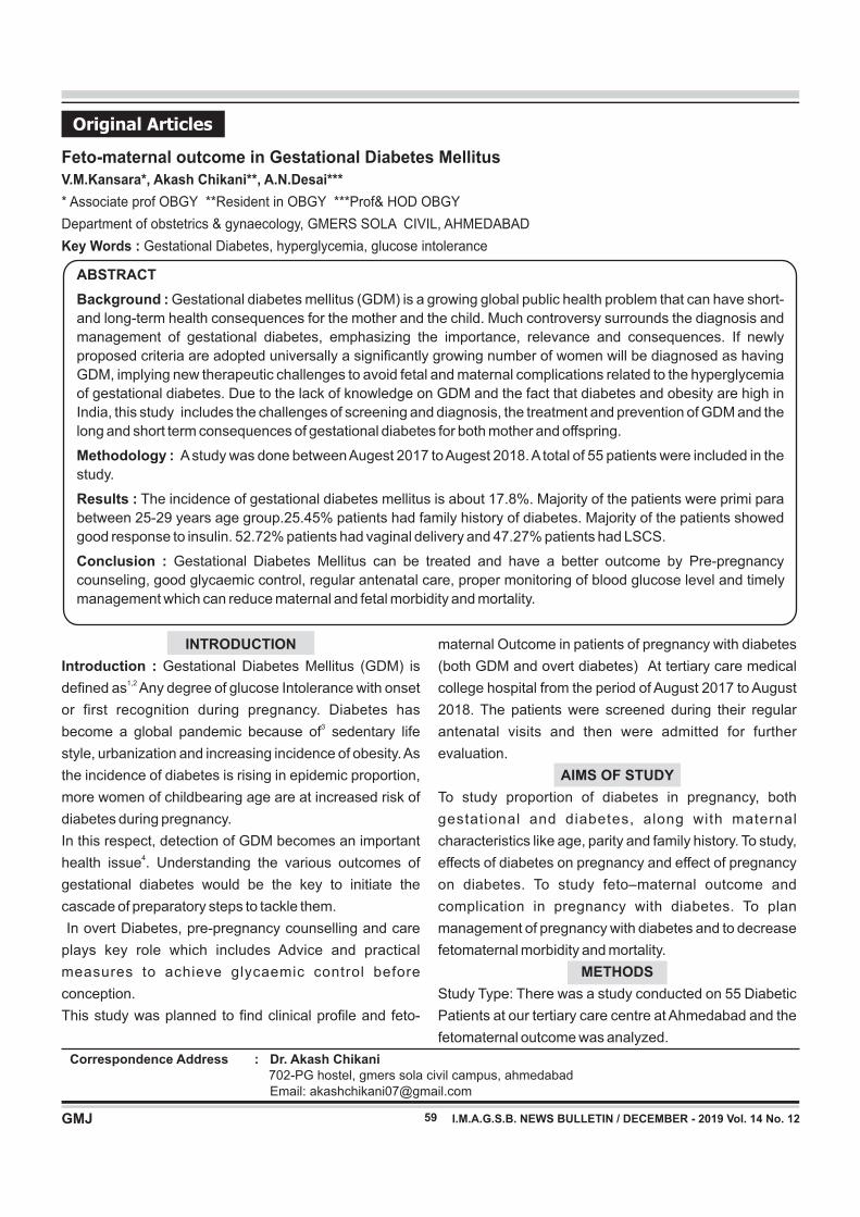
INTRODUCTION
Introduction : Gestational Diabetes Mellitus (GDM) is 1,2defined as Any degree of glucose Intolerance with onset
or first recognition during pregnancy. Diabetes has 3become a global pandemic because of sedentary life
style, urbanization and increasing incidence of obesity. As
the incidence of diabetes is rising in epidemic proportion,
more women of childbearing age are at increased risk of
diabetes during pregnancy.
In this respect, detection of GDM becomes an important 4health issue . Understanding the various outcomes of
gestational diabetes would be the key to initiate the
cascade of preparatory steps to tackle them.
In overt Diabetes, pre-pregnancy counselling and care
plays key role which includes Advice and practical
measures to achieve glycaemic control before
conception.
This study was planned to find clinical profile and feto-
Feto-maternal outcome in Gestational Diabetes Mellitus
Original Articles
V.M.Kansara*, Akash Chikani**, A.N.Desai***
* Associate prof OBGY **Resident in OBGY ***Prof& HOD OBGY
Department of obstetrics & gynaecology, GMERS SOLA CIVIL, AHMEDABAD
Key Words : Gestational Diabetes, hyperglycemia, glucose intolerance
59
Correspondence Address : Dr. Akash Chikani 702-PG hostel, gmers sola civil campus, ahmedabad Email: [email protected]
ABSTRACT
Background : Gestational diabetes mellitus (GDM) is a growing global public health problem that can have short-
and long-term health consequences for the mother and the child. Much controversy surrounds the diagnosis and
management of gestational diabetes, emphasizing the importance, relevance and consequences. If newly
proposed criteria are adopted universally a significantly growing number of women will be diagnosed as having
GDM, implying new therapeutic challenges to avoid fetal and maternal complications related to the hyperglycemia
of gestational diabetes. Due to the lack of knowledge on GDM and the fact that diabetes and obesity are high in
India, this study includes the challenges of screening and diagnosis, the treatment and prevention of GDM and the
long and short term consequences of gestational diabetes for both mother and offspring.
Methodology : A study was done between Augest 2017 to Augest 2018. A total of 55 patients were included in the
study.
Results : The incidence of gestational diabetes mellitus is about 17.8%. Majority of the patients were primi para
between 25-29 years age group.25.45% patients had family history of diabetes. Majority of the patients showed
good response to insulin. 52.72% patients had vaginal delivery and 47.27% patients had LSCS.
Conclusion : Gestational Diabetes Mellitus can be treated and have a better outcome by Pre-pregnancy
counseling, good glycaemic control, regular antenatal care, proper monitoring of blood glucose level and timely
management which can reduce maternal and fetal morbidity and mortality.
maternal Outcome in patients of pregnancy with diabetes
(both GDM and overt diabetes) At tertiary care medical
college hospital from the period of August 2017 to August
2018. The patients were screened during their regular
antenatal visits and then were admitted for further
evaluation.
AIMS OF STUDY
To study proportion of diabetes in pregnancy, both
gestational and diabetes, along with maternal
characteristics like age, parity and family history. To study,
effects of diabetes on pregnancy and effect of pregnancy
on diabetes. To study feto–maternal outcome and
complication in pregnancy with diabetes. To plan
management of pregnancy with diabetes and to decrease
fetomaternal morbidity and mortality.
METHODS
Study Type: There was a study conducted on 55 Diabetic
Patients at our tertiary care centre at Ahmedabad and the
fetomaternal outcome was analyzed.
I.M.A.G.S.B. NEWS BULLETIN / DECEMBER - 2019 Vol. 14 No. 12
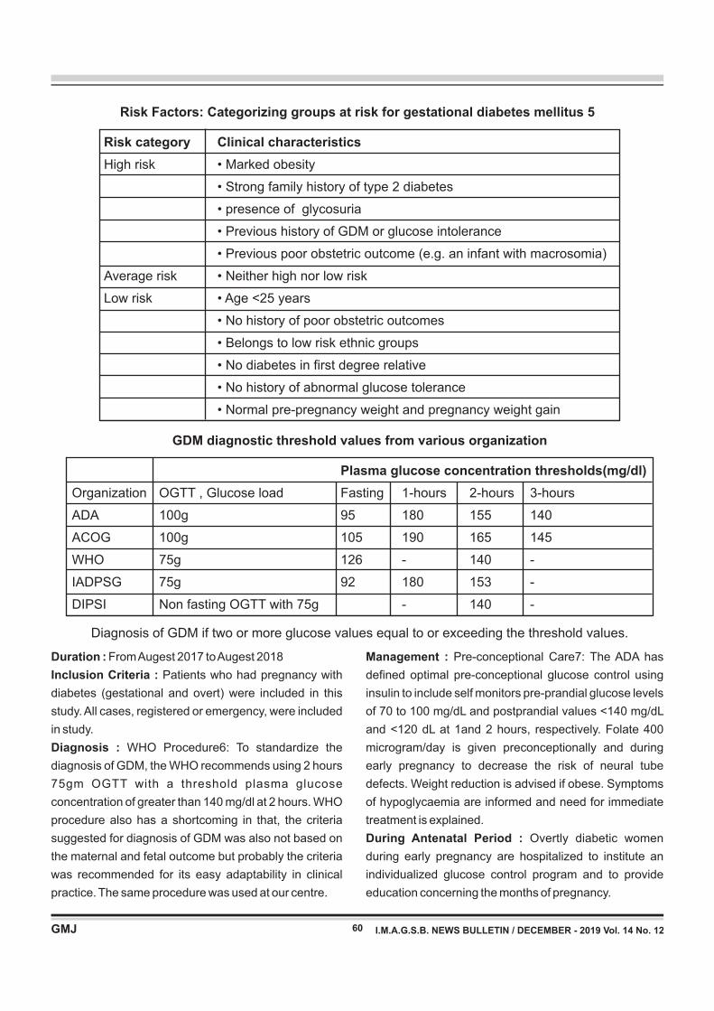
60
Risk Factors: Categorizing groups at risk for gestational diabetes mellitus 5
Risk category Clinical characteristics
High risk • Marked obesity
• Strong family history of type 2 diabetes
• presence of glycosuria
• Previous history of GDM or glucose intolerance
• Previous poor obstetric outcome (e.g. an infant with macrosomia)
Average risk • Neither high nor low risk
Low risk • Age <25 years
• No history of poor obstetric outcomes
• Belongs to low risk ethnic groups
• No diabetes in first degree relative
• No history of abnormal glucose tolerance
• Normal pre-pregnancy weight and pregnancy weight gain
Duration : From Augest 2017 to Augest 2018
Inclusion Criteria : Patients who had pregnancy with
diabetes (gestational and overt) were included in this
study. All cases, registered or emergency, were included
in study.
Diagnosis : WHO Procedure6: To standardize the
diagnosis of GDM, the WHO recommends using 2 hours
75gm OGTT with a threshold plasma glucose
concentration of greater than 140 mg/dl at 2 hours. WHO
procedure also has a shortcoming in that, the criteria
suggested for diagnosis of GDM was also not based on
the maternal and fetal outcome but probably the criteria
was recommended for its easy adaptability in clinical
practice. The same procedure was used at our centre.
Management : Pre-conceptional Care7: The ADA has
defined optimal pre-conceptional glucose control using
insulin to include self monitors pre-prandial glucose levels
of 70 to 100 mg/dL and postprandial values <140 mg/dL
and <120 dL at 1and 2 hours, respectively. Folate 400
microgram/day is given preconceptionally and during
early pregnancy to decrease the risk of neural tube
defects. Weight reduction is advised if obese. Symptoms
of hypoglycaemia are informed and need for immediate
treatment is explained.
During Antenatal Period : Overtly diabetic women
during early pregnancy are hospitalized to institute an
individualized glucose control program and to provide
education concerning the months of pregnancy.
GDM diagnostic threshold values from various organization
Plasma glucose concentration thresholds(mg/dl)
Organization OGTT , Glucose load Fasting 1-hours 2-hours 3-hours
ADA 100g 95 180 155 140
ACOG 100g 105 190 165 145
WHO 75g 126 - 140 -
IADPSG 75g 92 180 153 -
DIPSI Non fasting OGTT with 75g - 140 -
Diagnosis of GDM if two or more glucose values equal to or exceeding the threshold values.
I.M.A.G.S.B. NEWS BULLETIN / DECEMBER - 2019 Vol. 14 No. 12
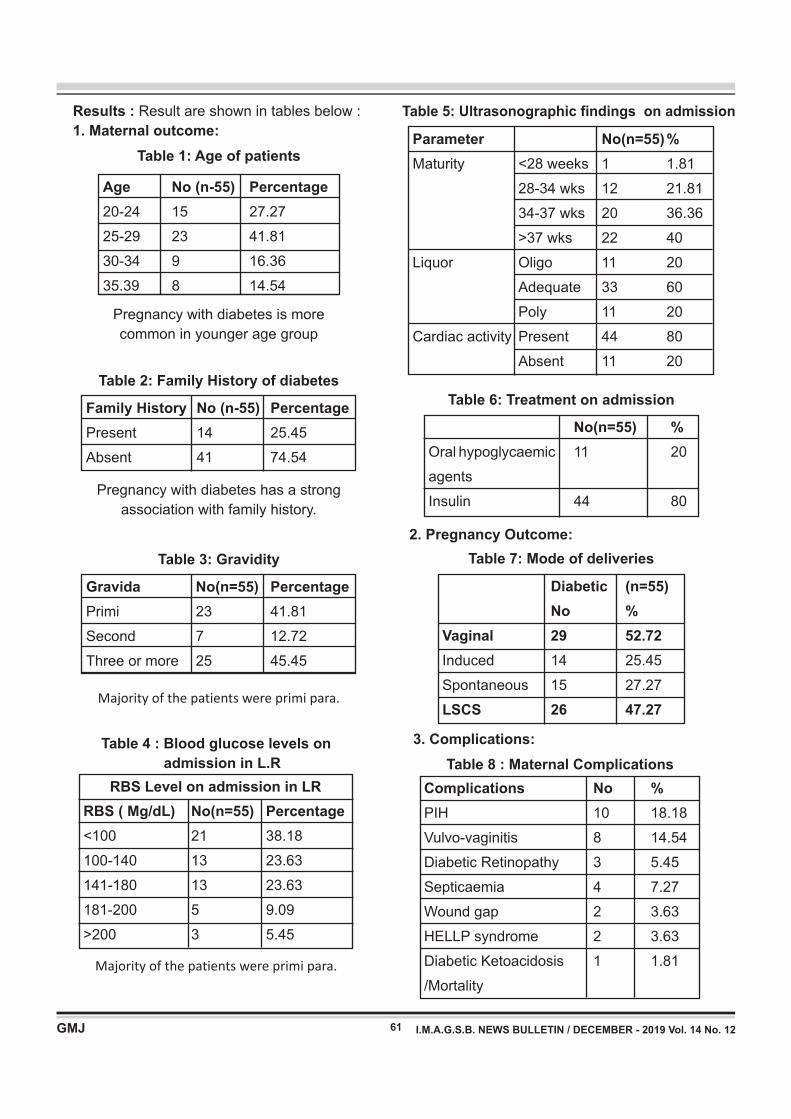
61
Results : Result are shown in tables below :
1. Maternal outcome:
Table 1: Age of patients
Age No (n-55) Percentage
20-24 15 27.27
25-29 23 41.81
30-34 9 16.36
35.39 8 14.54
Pregnancy with diabetes is more
common in younger age group
Family History No (n-55) Percentage
Present 14 25.45
Absent 41 74.54
Gravida No(n=55) Percentage
Primi 23 41.81
Second 7 12.72
Three or more 25 45.45
Pregnancy with diabetes has a strong
association with family history.
Majority of the patients were primi para.
Table 2: Family History of diabetes
Table 3: Gravidity
RBS ( Mg/dL) No(n=55) Percentage
<100 21 38.18
100-140 13 23.63
141-180 13 23.63
181-200 5 9.09
>200 3 5.45
Majority of the patients were primi para.
Table 4 : Blood glucose levels on
admission in L.R
RBS Level on admission in LR
Parameter No(n=55) %
Maturity <28 weeks 1 1.81
28-34 wks 12 21.81
34-37 wks 20 36.36
>37 wks 22 40
Liquor Oligo 11 20
Adequate 33 60
Poly 11 20
Cardiac activity Present 44 80
Absent 11 20
Table 5: Ultrasonographic findings on admission
No(n=55) %
Oral hypoglycaemic 11 20
agents
Insulin 44 80
Table 6: Treatment on admission
2. Pregnancy Outcome:
Table 7: Mode of deliveries
Diabetic (n=55)
No %
Vaginal 29 52.72
Induced 14 25.45
Spontaneous 15 27.27
LSCS 26 47.27
3. Complications:
Table 8 : Maternal Complications
Complications No %
PIH 10 18.18
Vulvo-vaginitis 8 14.54
Diabetic Retinopathy 3 5.45
Septicaemia 4 7.27
Wound gap 2 3.63
HELLP syndrome 2 3.63
Diabetic Ketoacidosis 1 1.81
/Mortality
I.M.A.G.S.B. NEWS BULLETIN / DECEMBER - 2019 Vol. 14 No. 12
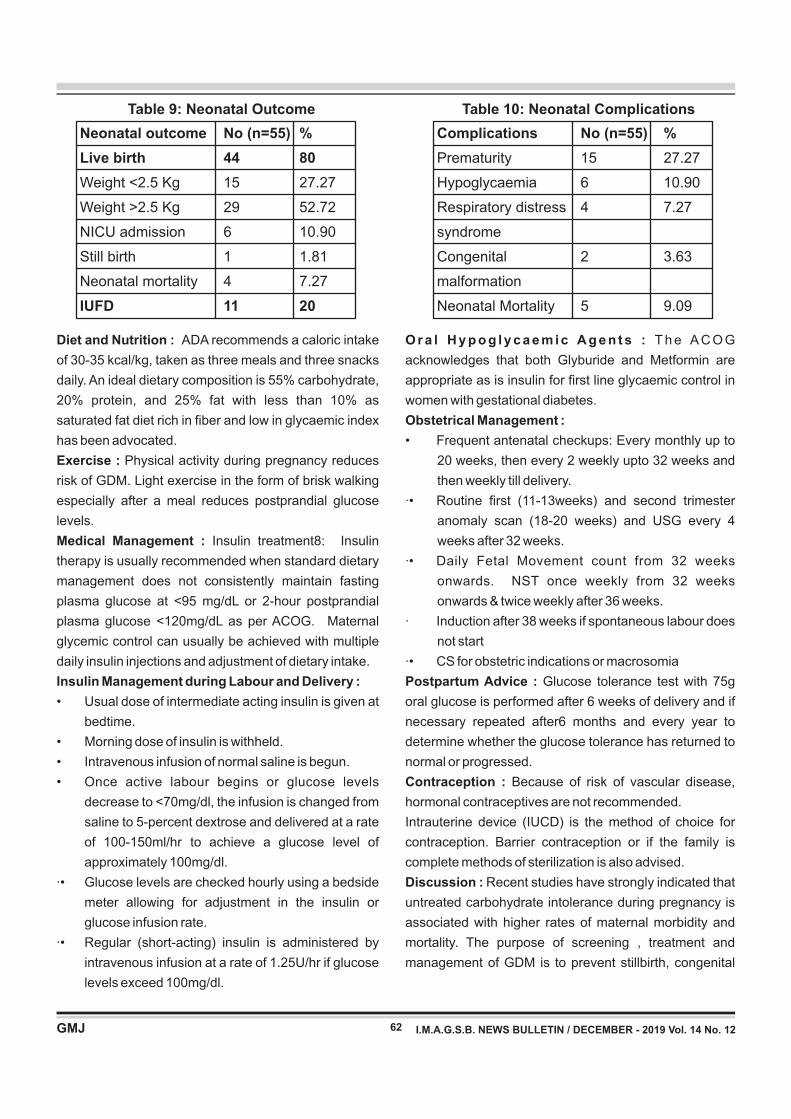
Table 10: Neonatal Complications
Complications No (n=55) %
Prematurity 15 27.27
Hypoglycaemia 6 10.90
Respiratory distress 4 7.27
syndrome
Congenital 2 3.63
malformation
Neonatal Mortality 5 9.09
Table 9: Neonatal Outcome
Neonatal outcome No (n=55) %
Live birth 44 80
Weight <2.5 Kg 15 27.27
Weight >2.5 Kg 29 52.72
NICU admission 6 10.90
Still birth 1 1.81
Neonatal mortality 4 7.27
IUFD 11 20
62
Diet and Nutrition : ADA recommends a caloric intake
of 30-35 kcal/kg, taken as three meals and three snacks
daily. An ideal dietary composition is 55% carbohydrate,
20% protein, and 25% fat with less than 10% as
saturated fat diet rich in fiber and low in glycaemic index
has been advocated.
Exercise : Physical activity during pregnancy reduces
risk of GDM. Light exercise in the form of brisk walking
especially after a meal reduces postprandial glucose
levels.
Medical Management : Insulin treatment8: Insulin
therapy is usually recommended when standard dietary
management does not consistently maintain fasting
plasma glucose at <95 mg/dL or 2-hour postprandial
plasma glucose <120mg/dL as per ACOG. Maternal
glycemic control can usually be achieved with multiple
daily insulin injections and adjustment of dietary intake.
Insulin Management during Labour and Delivery :
• Usual dose of intermediate acting insulin is given at
bedtime.
• Morning dose of insulin is withheld.
• Intravenous infusion of normal saline is begun.
• Once active labour begins or glucose levels
decrease to <70mg/dl, the infusion is changed from
saline to 5-percent dextrose and delivered at a rate
of 100-150ml/hr to achieve a glucose level of
approximately 100mg/dl.
·• Glucose levels are checked hourly using a bedside
meter allowing for adjustment in the insulin or
glucose infusion rate.
·• Regular (short-acting) insulin is administered by
intravenous infusion at a rate of 1.25U/hr if glucose
levels exceed 100mg/dl.
O r a l H y p o g l y c a e m i c A g e n t s : T h e A C O G
acknowledges that both Glyburide and Metformin are
appropriate as is insulin for first line glycaemic control in
women with gestational diabetes.
Obstetrical Management :
• Frequent antenatal checkups: Every monthly up to
20 weeks, then every 2 weekly upto 32 weeks and
then weekly till delivery.
·• Routine first (11-13weeks) and second trimester
anomaly scan (18-20 weeks) and USG every 4
weeks after 32 weeks.
·• Daily Fetal Movement count from 32 weeks
onwards. NST once weekly from 32 weeks
onwards & twice weekly after 36 weeks.
· Induction after 38 weeks if spontaneous labour does
not start
·• CS for obstetric indications or macrosomia
Postpartum Advice : Glucose tolerance test with 75g
oral glucose is performed after 6 weeks of delivery and if
necessary repeated after6 months and every year to
determine whether the glucose tolerance has returned to
normal or progressed.
Contraception : Because of risk of vascular disease,
hormonal contraceptives are not recommended.
Intrauterine device (IUCD) is the method of choice for
contraception. Barrier contraception or if the family is
complete methods of sterilization is also advised.
Discussion : Recent studies have strongly indicated that
untreated carbohydrate intolerance during pregnancy is
associated with higher rates of maternal morbidity and
mortality. The purpose of screening , treatment and
management of GDM is to prevent stillbirth, congenital
I.M.A.G.S.B. NEWS BULLETIN / DECEMBER - 2019 Vol. 14 No. 12
63
anomalies, NICU admissions, recurrent abortion, pre-
eclampsia, intra-uterine deaths and decrease incidence
of macrosomic babies, thereby reducing maternal and
perinatal morbidity and mortality. The findings of the
present study confirmed that GDM patients are liable to
have adverse pregnancy outcomes.
Pregnancy with diabetes was more common in younger
age group, has a strong association with family history
(25.45%). Majority of these patients were primi para
(41.81%)
On admission, majority of the patients had a random
blood sugar level between 141-180. Ultrasound on
admission showed polyhydramnios(20%) and 20%
fetuses on ultrasound showed intra uterine death. 80% of
patients were given insulin and had controlled blood
sugar level with the same.
Mode of delivery was often dictated by the increased size
of the baby, poor past obstetric history, fetal distress and
glycaemic control of patient. Gestational diabetes was
associated with an increased incidence of cesarean
delivery (47.27%) The main indications for CS being
cepha lope l v i c d i sp ropo r t i on , f e ta l d i s t ress ,
malpresentation and macrosomic babies.
Increased incidence of complications were found to be
associated with GDM. 10 patients developed associated
pre-eclampsia, 8 patients developed vulvovaginitis,3
patients developed diabetic retinopathy, 4 patients
developed septicemia, and 1 patient developed diabetic
ketoacidosis. Thus, gestational diabetes is associated
with high maternal morbidity and mortality.
27.27% of neonates were low birth weight, 10.90% of
neonates were admitted in neonatal intensive care unit.
Therefore, neonates of diabetic mothers are at an
increased risk of prematurity and respiratory distress
syndrome.
The above results suggest that good glycemic control is
essential in gestational diabetes to improve maternal and
neonatal outcome.
Conclusion: GDM is one of the most common medical and
metabolic complication seen in pregnancy. Women who
are at high risk of developing GDM should be
appropriately screened to reduce maternal and fetal
morbidity.
The management of GDM should be based on a team
approach involving diabetologist, obstetrician, dietician
I.M.A.G.S.B. NEWS BULLETIN / DECEMBER - 2019 Vol. 14 No. 12
and paediatrician to reduce the incidence of associated
complications.universal screening, Pre-conceptional
care, antepartum and intrapartum management and
contraceptive counselling are of equal importance to
improve feto-maternal outcome.
Patients with GDM are at risk of developing type 2
diabetes in the future and should be monitored regularly.
Similarly the offspring of diabetic pregnancies are at risk
of developing obesity, IGT and diabetes and should also
be followed up.
REFERENCES
1. Fernando A, Bhide A & Arulkumaran S Arias practical guide to
High risk pregnancy and delivery. new delhi: elsevier; 2015.
2. Williams, Cunningham FG & Leveno KJ Williams obstetrics.
united states: McGraw Hill; 2014.
3. Mpondo BC, Ernest A, Dee HE. Gestational diabetes mellitus:
challenges in diagnosis and management. Journal of Diabetes
& Metabolic Disorders. 2015 May 12;14(1):42.
4. Agarwal MM. Gestational diabetes mellitus: An update on the
current international diagnostic criteria. World journal of
diabetes. 2015 Jun 25;6(6):782.
5. Agarwal MM, Dhatt GS, Punnose J, Zayed R. Gestational
diabetes: fasting and postprandial glucose as first prenatal
screening tests in a high-risk population. The Journal of
reproductive medicine. 2007 Apr;52(4):299-305.
6. World Health Organization. Report of a WHO consultation:
diagnostic criteria and classification of hyperglycaemia first
detected in pregnancy: a World Health Organization guideline.
Diabetes Res Clin Pract. 2014;103:341-63.
7. Hod M, Kapur A, Sacks DA, Hadar E, Agarwal M, Di Renzo GC,
Roura LC, McIntyre HD, Morris JL, Divakar H. The International
Federation of Gynecology and Obstetrics (FIGO) Initiative on
gestational diabetes mellitus: A pragmatic guide for diagnosis,
management, and care. International Journal of Gynecology &
Obstetrics. 2015 Oct 1;131(S3).
8. Kampmann U, Madsen LR, Skajaa GO, Iversen DS, Moeller N,
Ovesen P. Gestational diabetes: A clinical update. World journal
of diabetes. 2015 Jul 25;6(8):1065.
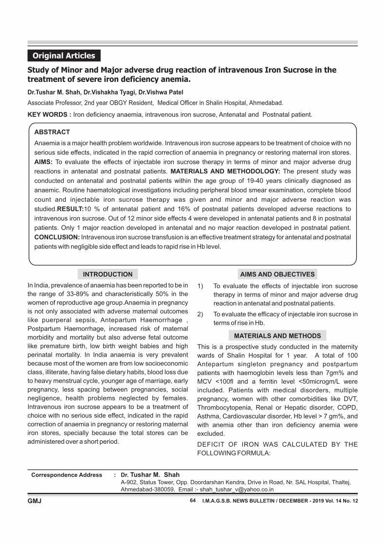
64
Correspondence Address : Dr. Tushar M. Shah A-902, Status Tower, Opp. Doordarshan Kendra, Drive in Road, Nr. SAL Hospital, Thaltej, Ahmedabad-380059. Email :- [email protected]
ABSTRACT
Anaemia is a major health problem worldwide. Intravenous iron sucrose appears to be treatment of choice with no
serious side effects, indicated in the rapid correction of anaemia in pregnancy or restoring maternal iron stores.
AIMS: To evaluate the effects of injectable iron sucrose therapy in terms of minor and major adverse drug
reactions in antenatal and postnatal patients. MATERIALS AND METHODOLOGY: The present study was
conducted on antenatal and postnatal patients within the age group of 19-40 years clinically diagnosed as
anaemic. Routine haematological investigations including peripheral blood smear examination, complete blood
count and injectable iron sucrose therapy was given and minor and major adverse reaction was
studied.RESULT:10 % of antenatal patient and 16% of postnatal patients developed adverse reactions to
intravenous iron sucrose. Out of 12 minor side effects 4 were developed in antenatal patients and 8 in postnatal
patients. Only 1 major reaction developed in antenatal and no major reaction developed in postnatal patient.
CONCLUSION: Intravenous iron sucrose transfusion is an effective treatment strategy for antenatal and postnatal
patients with negligible side effect and leads to rapid rise in Hb level.
Study of Minor and Major adverse drug reaction of intravenous Iron Sucrose in the treatment of severe iron deficiency anemia.
Dr.Tushar M. Shah, Dr.Vishakha Tyagi, Dr.Vishwa Patel
Associate Professor, 2nd year OBGY Resident, Medical Officer in Shalin Hospital, Ahmedabad.
KEY WORDS : Iron deficiency anaemia, intravenous iron sucrose, Antenatal and Postnatal patient.
INTRODUCTION
In India, prevalence of anaemia has been reported to be in
the range of 33-89% and characteristically 50% in the
women of reproductive age group.Anaemia in pregnancy
is not only associated with adverse maternal outcomes
like puerperal sepsis, Antepartum Haemorrhage ,
Postpartum Haemorrhage, increased risk of maternal
morbidity and mortality but also adverse fetal outcome
like premature birth, low birth weight babies and high
perinatal mortality. In India anaemia is very prevalent
because most of the women are from low socioeconomic
class, illiterate, having false dietary habits, blood loss due
to heavy menstrual cycle, younger age of marriage, early
pregnancy, less spacing between pregnancies, social
negligence, health problems neglected by females.
Intravenous iron sucrose appears to be a treatment of
choice with no serious side effect, indicated in the rapid
correction of anaemia in pregnancy or restoring maternal
iron stores, specially because the total stores can be
administered over a short period.
AIMS AND OBJECTIVES
1) To evaluate the effects of injectable iron sucrose
therapy in terms of minor and major adverse drug
reaction in antenatal and postnatal patients.
2) To evaluate the efficacy of injectable iron sucrose in
terms of rise in Hb.
MATERIALS AND METHODS
This is a prospective study conducted in the maternity
wards of Shalin Hospital for 1 year. A total of 100
Antepartum singleton pregnancy and postpartum
patients with haemoglobin levels less than 7gm% and
MCV <100fl and a ferritin level <50microgm/L were
included. Patients with medical disorders, multiple
pregnancy, women with other comorbidities like DVT,
Thrombocytopenia, Renal or Hepatic disorder, COPD,
Asthma, Cardiovascular disorder, Hb level > 7 gm%, and
with anemia other than iron deficiency anemia were
excluded.
DEFICIT OF IRON WAS CALCULATED BY THE
FOLLOWING FORMULA:
Original Articles
I.M.A.G.S.B. NEWS BULLETIN / DECEMBER - 2019 Vol. 14 No. 12
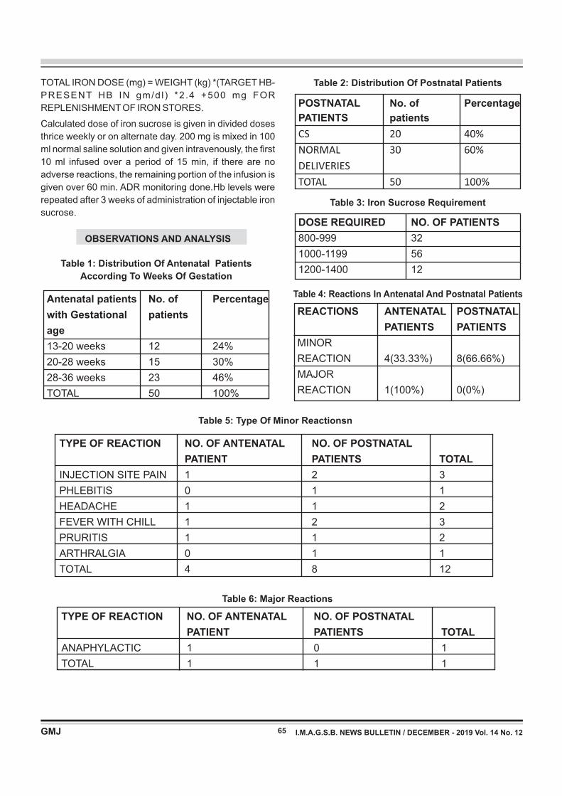
Table 2: Distribution Of Postnatal Patients
POSTNATAL No. of Percentage
PATIENTS patients
CS 20 40%
NORMAL 30 60%
DELIVERIES
TOTAL 50 100%
65
Table 4: Reactions In Antenatal And Postnatal Patients
Table 5: Type Of Minor Reactionsn
TYPE OF REACTION NO. OF ANTENATAL NO. OF POSTNATAL
PATIENT PATIENTS TOTAL
INJECTION SITE PAIN 1 2 3
PHLEBITIS 0 1 1
HEADACHE 1 1 2
FEVER WITH CHILL 1 2 3
PRURITIS 1 1 2
ARTHRALGIA 0 1 1
TOTAL 4 8 12
TOTAL IRON DOSE (mg) = WEIGHT (kg) *(TARGET HB-
PRESENT HB IN gm/d l ) *2 .4 +500 mg FOR
REPLENISHMENT OF IRON STORES.
Calculated dose of iron sucrose is given in divided doses
thrice weekly or on alternate day. 200 mg is mixed in 100
ml normal saline solution and given intravenously, the first
10 ml infused over a period of 15 min, if there are no
adverse reactions, the remaining portion of the infusion is
given over 60 min. ADR monitoring done.Hb levels were
repeated after 3 weeks of administration of injectable iron
sucrose.
I.M.A.G.S.B. NEWS BULLETIN / DECEMBER - 2019 Vol. 14 No. 12
OBSERVATIONS AND ANALYSIS
Table 1: Distribution Of Antenatal Patients
According To Weeks Of Gestation
Antenatal patients No. of Percentage
with Gestational patients
age
13-20 weeks 12 24%
20-28 weeks 15 30%
28-36 weeks 23 46%
TOTAL 50 100%
REACTIONS ANTENATAL POSTNATAL
PATIENTS PATIENTS
MINOR
REACTION 4(33.33%) 8(66.66%)
MAJOR
REACTION 1(100%) 0(0%)
Table 3: Iron Sucrose Requirement
DOSE REQUIRED NO. OF PATIENTS
800-999 32
1000-1199 56
1200-1400 12
Table 6: Major Reactions
TYPE OF REACTION NO. OF ANTENATAL NO. OF POSTNATAL
PATIENT PATIENTS TOTAL
ANAPHYLACTIC 1 0 1
TOTAL 1 1 1
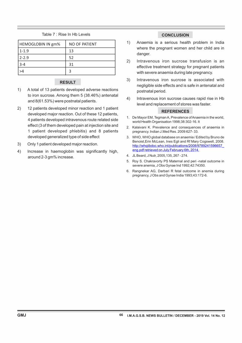
66
Table 7 : Rise In Hb Levels
HEMOGLOBIN IN gm% NO OF PATIENT
1-1.9 13
2-2.9 52
3-4 31
>4 3
RESULT
1) A total of 13 patients developed adverse reactions
to iron sucrose. Among them 5 (38.46%) antenatal
and 8(61.53%) were postnatal patients.
2) 12 patients developed minor reaction and 1 patient
developed major reaction. Out of these 12 patients,
4 patients developed intravenous route related side
effect (3 of them developed pain at injection site and
1 patient developed phlebitis) and 8 patients
developed generalized type of side effect
3) Only 1 patient developed major reaction.
4) Increase in haemoglobin was significantly high,
around 2-3 gm% increase.
I.M.A.G.S.B. NEWS BULLETIN / DECEMBER - 2019 Vol. 14 No. 12
CONCLUSION
1) Anaemia is a serious health problem in India
where the pregnant women and her child are in
danger.
2) Intravenous iron sucrose transfusion is an
effective treatment strategy for pregnant patients
with severe anaemia during late pregnancy.
3) Intravenous iron sucrose is associated with
negligible side effects and is safe in antenatal and
postnatal period.
4) Intravenous iron sucrose causes rapid rise in Hb
level and replacement of stores was faster.
REFERENCES
1. De Mayor EM, Tegman A, Prevalence of Anaemia in the world, world Health Organisation 1998;38:302-16. It
2. Kalaivani K. Prevalence and consequences of anaemia in pregnancy. Indian J.Med Res. 2009:627- 33.
3. WHO, WHO global database on anaemia / Edited by Bruno de Benoist,Erin McLean, Ines Egli and Rf Mary Cogswell, 2008. http://whqiibdoc.who.int/publications/2008/9789241596657_eng.pdf retrieved on July February 6th, 2014.
4. JL Beard, J Nutr, 2005,135, 267 - 274.
5. Roy S. Chakravorty PS Maternal and peri -natal outcome in severe anemia, J Obs Gynae Ind 1992;42:74350.
6. Rangnekar AG, Darbari R fetal outcome in anemia during pregnancy, J Obs and Gynae India 1993;43:172-6.
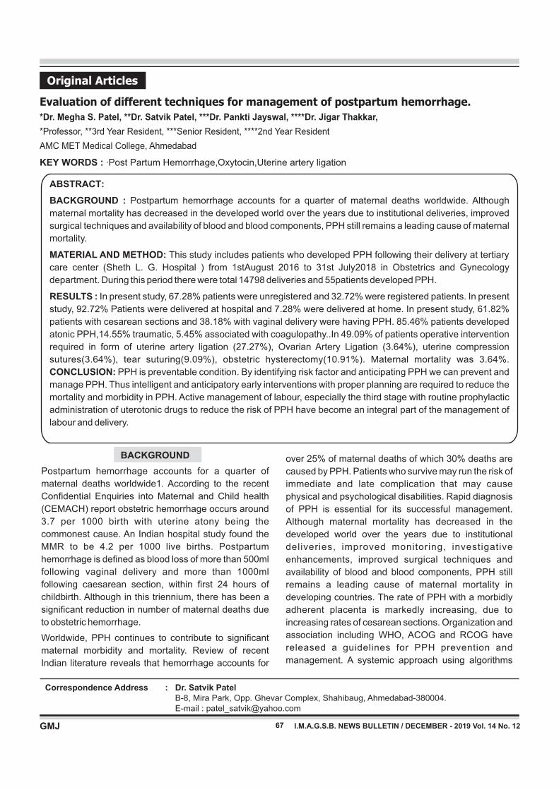
KEY WORDS : · Post Partum Hemorrhage,Oxytocin,Uterine artery ligation
Original Articles
Evaluation of different techniques for management of postpartum hemorrhage.
*Dr. Megha S. Patel, **Dr. Satvik Patel, ***Dr. Pankti Jayswal, ****Dr. Jigar Thakkar,
*Professor, **3rd Year Resident, ***Senior Resident, ****2nd Year Resident
AMC MET Medical College, Ahmedabad
67
ABSTRACT:
BACKGROUND : Postpartum hemorrhage accounts for a quarter of maternal deaths worldwide. Although
maternal mortality has decreased in the developed world over the years due to institutional deliveries, improved
surgical techniques and availability of blood and blood components, PPH still remains a leading cause of maternal
mortality.
MATERIAL AND METHOD: This study includes patients who developed PPH following their delivery at tertiary
care center (Sheth L. G. Hospital ) from 1stAugust 2016 to 31st July2018 in Obstetrics and Gynecology
department. During this period there were total 14798 deliveries and 55patients developed PPH.
RESULTS : In present study, 67.28% patients were unregistered and 32.72% were registered patients. In present
study, 92.72% Patients were delivered at hospital and 7.28% were delivered at home. In present study, 61.82%
patients with cesarean sections and 38.18% with vaginal delivery were having PPH. 85.46% patients developed
atonic PPH,14.55% traumatic, 5.45% associated with coagulopathy..In 49.09% of patients operative intervention
required in form of uterine artery ligation (27.27%), Ovarian Artery Ligation (3.64%), uterine compression
sutures(3.64%), tear suturing(9.09%), obstetric hysterectomy(10.91%). Maternal mortality was 3.64%.
CONCLUSION: PPH is preventable condition. By identifying risk factor and anticipating PPH we can prevent and
manage PPH. Thus intelligent and anticipatory early interventions with proper planning are required to reduce the
mortality and morbidity in PPH. Active management of labour, especially the third stage with routine prophylactic
administration of uterotonic drugs to reduce the risk of PPH have become an integral part of the management of
labour and delivery.
BACKGROUND
Postpartum hemorrhage accounts for a quarter of
maternal deaths worldwide1. According to the recent
Confidential Enquiries into Maternal and Child health
(CEMACH) report obstetric hemorrhage occurs around
3.7 per 1000 birth with uterine atony being the
commonest cause. An Indian hospital study found the
MMR to be 4.2 per 1000 live births. Postpartum
hemorrhage is defined as blood loss of more than 500ml
following vaginal delivery and more than 1000ml
following caesarean section, within first 24 hours of
childbirth. Although in this triennium, there has been a
significant reduction in number of maternal deaths due
to obstetric hemorrhage.
Worldwide, PPH continues to contribute to significant
maternal morbidity and mortality. Review of recent
Indian literature reveals that hemorrhage accounts for
Correspondence Address : D r. Satvik Patel B-8, Mira Park, Opp. Ghevar Complex, Shahibaug, Ahmedabad-380004. E-mail : [email protected]
over 25% of maternal deaths of which 30% deaths are
caused by PPH. Patients who survive may run the risk of
immediate and late complication that may cause
physical and psychological disabilities. Rapid diagnosis
of PPH is essential for its successful management.
Although maternal mortality has decreased in the
developed world over the years due to institutional
deliveries, improved monitoring, investigative
enhancements, improved surgical techniques and
availability of blood and blood components, PPH still
remains a leading cause of maternal mortality in
developing countries. The rate of PPH with a morbidly
adherent placenta is markedly increasing, due to
increasing rates of cesarean sections. Organization and
association including WHO, ACOG and RCOG have
released a guidelines for PPH prevention and
management. A systemic approach using algorithms
I.M.A.G.S.B. NEWS BULLETIN / DECEMBER - 2019 Vol. 14 No. 12
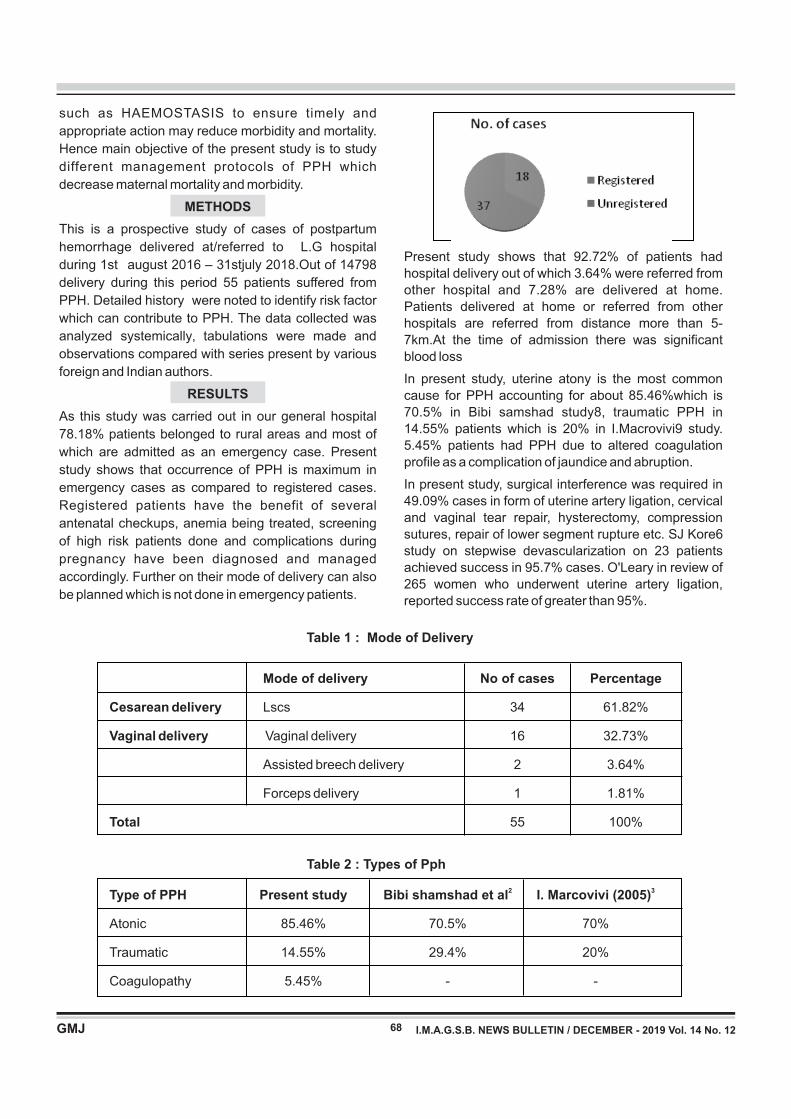
68
Present study shows that 92.72% of patients had
hospital delivery out of which 3.64% were referred from
other hospital and 7.28% are delivered at home.
Patients delivered at home or referred from other
hospitals are referred from distance more than 5-
7km.At the time of admission there was significant
blood loss
In present study, uterine atony is the most common
cause for PPH accounting for about 85.46%which is
70.5% in Bibi samshad study8, traumatic PPH in
14.55% patients which is 20% in I.Macrovivi9 study.
5.45% patients had PPH due to altered coagulation
profile as a complication of jaundice and abruption.
In present study, surgical interference was required in
49.09% cases in form of uterine artery ligation, cervical
and vaginal tear repair, hysterectomy, compression
sutures, repair of lower segment rupture etc. SJ Kore6
study on stepwise devascularization on 23 patients
achieved success in 95.7% cases. O'Leary in review of
265 women who underwent uterine artery ligation,
reported success rate of greater than 95%.
Table 1 : Mode of Delivery
Table 2 : Types of Pph
Mode of delivery No of cases Percentage
Cesarean delivery Lscs 34 61.82%
Vaginal delivery Vaginal delivery 16 32.73%
Assisted breech delivery 2 3.64%
Forceps delivery 1 1.81%
Total 55 100%
2 3Type of PPH Present study Bibi shamshad et al I. Marcovivi (2005)
Atonic 85.46% 70.5% 70%
Traumatic 14.55% 29.4% 20%
Coagulopathy 5.45% - -
such as HAEMOSTASIS to ensure timely and
appropriate action may reduce morbidity and mortality.
Hence main objective of the present study is to study
different management protocols of PPH which
decrease maternal mortality and morbidity.
METHODS
This is a prospective study of cases of postpartum
hemorrhage delivered at/referred to L.G hospital
during 1st august 2016 – 31stjuly 2018.Out of 14798
delivery during this period 55 patients suffered from
PPH. Detailed history were noted to identify risk factor
which can contribute to PPH. The data collected was
analyzed systemically, tabulations were made and
observations compared with series present by various
foreign and Indian authors.
RESULTS
As this study was carried out in our general hospital
78.18% patients belonged to rural areas and most of
which are admitted as an emergency case. Present
study shows that occurrence of PPH is maximum in
emergency cases as compared to registered cases.
Registered patients have the benefit of several
antenatal checkups, anemia being treated, screening
of high risk patients done and complications during
pregnancy have been diagnosed and managed
accordingly. Further on their mode of delivery can also
be planned which is not done in emergency patients.
I.M.A.G.S.B. NEWS BULLETIN / DECEMBER - 2019 Vol. 14 No. 12
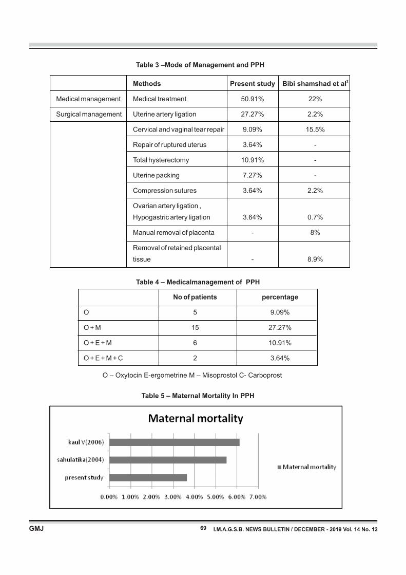
69
2 Methods Present study Bibi shamshad et al
Medical management Medical treatment 50.91% 22%
Surgical management Uterine artery ligation 27.27% 2.2%
Cervical and vaginal tear repair 9.09% 15.5%
Repair of ruptured uterus 3.64% -
Total hysterectomy 10.91% -
Uterine packing 7.27% -
Compression sutures 3.64% 2.2%
Ovarian artery ligation ,
Hypogastric artery ligation 3.64% 0.7%
Manual removal of placenta - 8%
Removal of retained placental
tissue - 8.9%
Table 3 –Mode of Management and PPH
Table 5 – Maternal Mortality In PPH
Table 4 – Medicalmanagement of PPH
No of patients percentage
O 5 9.09%
O + M 15 27.27%
O + E + M 6 10.91%
O + E + M + C 2 3.64%
O – Oxytocin E-ergometrine M – Misoprostol C- Carboprost
I.M.A.G.S.B. NEWS BULLETIN / DECEMBER - 2019 Vol. 14 No. 12
70
Accurate diagnosis of cause of bleeding , Adequate and
prompt restoration of circulatory volume, Any form of
operative procedure should be performed timely before
patient is in extremis and guided by an experienced
person ,Measures should be taken for subsequent
recovery.
REFERENCES
1. World health organization. Attending to 136 million births, every
year: make every mother and child count: the world record
2005.2005:62-63
2. Shamshad Bibi, Nargis Danish et al : an audit of primary PPH,
j. ayub med. Coll. Abbottabad: 19(4) 102
3. I. macrovivi, postpartum hemorrhage . Journal W de chirurgic ,
Iasi 2005 vol 1 no 4
4. Walter prendiville et al : effects of routine oxytocin
administration in management of third stage of labour . Br.Jr.
of obstetrics and gynecology Jan 1988 vol 95 pp 3- 16
5. Merrikay A. olum, Mariano, controlling refractory atonic PPH
with hemabate sterile solution. Am. J. obst. Gynecol. Vol 162:
205 , 1990
6. SJ Kore et al : stepwise uterine devascularization, Bombay
hospital journal , Aug. 2010
7. Sahulatika et al : hysterectomy for obstetric emergencies, j.
obstet and gynecolind vol 54 no 1 Jan / feb 2004
8. Kaul V et al: the impact of primary postpartum hemorrhage in
“Near-Miss” morbidity and mortality at a tertiary care hospital in
north India. Indian j. med. Sci. 2006:60:233-40
Medical means of management were used in all
patients to no avail. In present study, about 50.91% of
patients responded to medical treatment alone.9.09%
responds to only oxytocin and 27.27% responds after
adding tablet misoprostol to oxytocin.3.64% patients
required all for agents to stop the bleeding.
Walter Prendiville et al (1988)4 stated that routine
administration of oxytocin does have an effect on the
rate of PPH, reducing it about 40% Merrikay A. et al
(1990)5 study states that hemorrhage was successfully
controlled immediately after the administration of
hemabate sterile solution (15methyl PGF2alpha) in
87.8% cases (208/237)
Out of 55, two patients were expired In present study
mortality rate is 3.64% which is comparable to
sahulatika7 5.5% and kaul V8 6.1%. one patient died
because of complications of PPH like DIC and
septicemia & second patient died because of
hypovolemic shock and cardiopulmonary arrest. Low
incidence is due to better antenatal care, good tertiary
care facilities and skilled staff.
CONCLUSION
Despite the keen interest of international health
agencies, worldwide maternal mortality has not
declined substantially due to high rates of PPH . This
can be attributed to non-availability of health care
resources in rural settings, lack of means of rapid
transport system and local cultural practices. Once
PPH is diagnosed coordinated functioning of
multidisciplinary practitioners such as – midwife,
obstetrician, anesthesiologist, hematologist, and
laboratory team including blood bank services is
required. PPH is preventable condition. By identifying
risk factor and anticipating PPH we can prevent and
manage PPH. Active management of labour, especially
the third stage with routine prophylactic administration
of uterotonic drugs to reduce the risk of PPH have
become an integral part of the management of labour
and delivery. Since the last decade conservative
surgical procedures have been successfully used in
various forms. Conservative surgical approaches not
only control PPH, but also preserve a woman's
reproductive functions and avoids hysterectomy and its
related complications and consequences.
Thus following are the measures to be taken to reduce
the mortality and morbidity in PPH :
Regular and adequate antenatal check up, Aseptic
vaginal examination, Avoidance of prolonged labour
,Follow the protocol of AMTLS to prevent PPH,
I.M.A.G.S.B. NEWS BULLETIN / DECEMBER - 2019 Vol. 14 No. 12
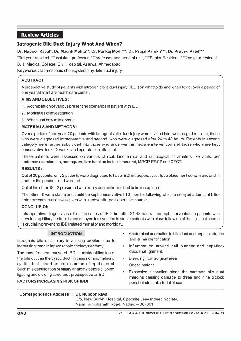
INTRODUCTION
Iatrogenic bile duct injury is a rising problem due to
increasing trend in laparoscopic cholecystectomy.
The most frequent cause of IBDI is misidentification of
the bile duct as the cystic duct; in cases of anomalies of
cystic duct insertion into common hepatic duct.
Such misidentification of biliary anatomy before clipping,
ligating and dividing structures predisposes to IBDI.
FACTORS INCREASING RISK OF IBDI
• Anatomical anomalies in bile duct and hepatic arteries
and its misidentification.
• Inflammation around gall bladder and hepatico-
duodenal ligament.
• Bleeding from surgical area
• Obese patient
• Excessive dissection along the common bile duct
margins causing damage to three and nine o'clock
pericholedochal arterial plexus.
Keywords : laparoscopic cholecystectomy, bile duct injury
Iatrogenic Bile Duct Injury What And When?
Review Articles
Dr. Nupoor Raval*, Dr. Maulik Mehta**, Dr. Pankaj Modi***, Dr. Prujal Parekh***, Dr. Pruthvi Patel***
*3rd year resident, **assistant professor, ***professor and head of unit, ***Senior Resident, ***2nd year resident
B. J. Medical College, Civil Hospital, Asarwa, Ahmedabad.
71
Correspondence Address : Dr. Nupoor Raval C/o, New Surbhi Hospital, Opposite Jeevandeep Society, Nana Kumbhanath Road, Nadiad – 387001
ABSTRACT
A prospective study of patients with iatrogenic bile duct injury (IBDI) on what to do and when to do; over a period of
one year at a tertiary health care center.
AIMS AND OBJECTIVES :
1. A compilation of various presenting scenarios of patient with IBDI.
2. Modalities of investigation.
3. When and how to intervene.
MATERIALS AND METHODS :
Over a period of one year, 20 patients with iatrogenic bile duct injury were divided into two categories – one, those
who were diagnosed intraoperative and second, who were diagnosed after 24 to 48 hours. Patients in second
category were further subdivided into those who underwent immediate intervention and those who were kept
conservative for 8-12 weeks and operated on after that.
These patients were assessed on various clinical, biochemical and radiological parameters like vitals, per
abdomen examination, hemogram, liver function tests, ultrasound, MRCP, ERCP and CECT.
RESULTS :
Out of 20 patients, only 2 patients were diagnosed to have IBDI intraoperative, t-tube placement done in one and in
another the proximal end was tied.
Out of the other 18 – 2 presented with biliary peritonitis and had to be re-explored.
The other 16 were stable and could be kept conservative till 3 months following which a delayed attempt at bilio-
enteric reconstruction was given with a uneventful post operative course.
CONCLUSION
Intraoperative diagnosis is difficult in cases of IBDI but after 24-48 hours – prompt intervention in patients with
developing biliary peritonitis and delayed intervention in stable patients with close follow up of their clinical course
is crucial in preventing IBDI related mortality and morbidity.
I.M.A.G.S.B. NEWS BULLETIN / DECEMBER - 2019 Vol. 14 No. 12
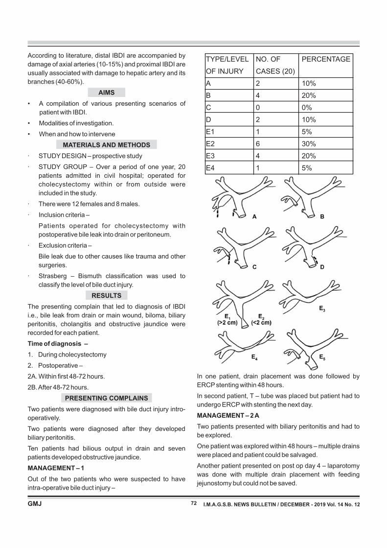
72
According to literature, distal IBDI are accompanied by
damage of axial arteries (10-15%) and proximal IBDI are
usually associated with damage to hepatic artery and its
branches (40-60%).
AIMS
• A compilation of various presenting scenarios of
patient with IBDI.
• Modalities of investigation.
• When and how to intervene
MATERIALS AND METHODS
· STUDY DESIGN – prospective study
· STUDY GROUP – Over a period of one year, 20
patients admitted in civil hospital; operated for
cholecystectomy within or from outside were
included in the study.
· There were 12 females and 8 males.
· Inclusion criteria –
Patients operated for cholecystectomy with
postoperative bile leak into drain or peritoneum.
· Exclusion criteria –
Bile leak due to other causes like trauma and other
surgeries.
· Strasberg – Bismuth classification was used to
classify the level of bile duct injury.
RESULTS
The presenting complain that led to diagnosis of IBDI
i.e., bile leak from drain or main wound, biloma, biliary
peritonitis, cholangitis and obstructive jaundice were
recorded for each patient.
Time of diagnosis –
1. During cholecystectomy
2. Postoperative –
2A. Within first 48-72 hours.
2B. After 48-72 hours.
PRESENTING COMPLAINS
Two patients were diagnosed with bile duct injury intro-
operatively.
Two patients were diagnosed after they developed
biliary peritonitis.
Ten patients had bilious output in drain and seven
patients developed obstructive jaundice.
MANAGEMENT – 1
Out of the two patients who were suspected to have
intra-operative bile duct injury –
In one patient, drain placement was done followed by
ERCP stenting within 48 hours.
In second patient, T – tube was placed but patient had to
undergo ERCP with stenting the next day.
MANAGEMENT – 2 A
Two patients presented with biliary peritonitis and had to
be explored.
One patient was explored within 48 hours – multiple drains
were placed and patient could be salvaged.
Another patient presented on post op day 4 – laparotomy
was done with multiple drain placement with feeding
jejunostomy but could not be saved.
TYPE/LEVEL NO. OF PERCENTAGE
OF INJURY CASES (20)
A 2 10%
B 4 20%
C 0 0%
D 2 10%
E1 1 5%
E2 6 30%
E3 4 20%
E4 1 5%
I.M.A.G.S.B. NEWS BULLETIN / DECEMBER - 2019 Vol. 14 No. 12
73
MANAGEMENT – 2 B
16 patients along with 1 who had survived laparotomy
were monitored daily on basis of –
1. Vital parameters like temperature, pulse rate, blood
pressure, respiratory rate and air entry.
2. Per abdomen findings like distention, tenderness or
guarding, peristalsis.
3. Development of signs of obstructive jaundice like
passage of clay coloured stool, icterus and pruritus.
4. Biochemical markers like hemogram and liver
function tests.
5. Ultrasound was serially done at monthly interval or
according to symptom of patient to monitor
presence or absence of collection, biliary tree – size
and level of cut off, echotexture of liver.
6. ERCP was attempted in 2 patients but had failed.
7. MRCP was done and used as a main guideline for
plan of management.
8. Decision was taken to operate this patients after the
bile leak had stopped, proximal duct had dilated to
at least 1.5 cm, there was no collection and patient
had been built up nutritionally but before patient
started developing liver cirrhosis.
9. This was usually between 2-3 months following
surgery.
OPERATIVE TECHNIQUE OF BILE DUCT DISSECTION
AND ROUX-EN-Y HEPATICO-JEJUNOSTOMY
RECONSTRUCTION
• Extra hepatic biliary tree was explored upto confluence
and even beyond according to level of injury.
• Satisfactory bleeding from the cut end was ensured.
• Roux loop of jejunum was transferred to upper
abdomen via transverse colon.
• Single layer duct to mucosa hepatico-jejunostomy was
performed with absorbable suture material vicryl or
PDS – 3-0 or 4-0.
• Drain was placed in all patients.
CONCLUSION
Intraoperative diagnosis is difficult in cases of IBDI but after
24-48 hours – prompt intervention in patients with
developing biliary peritonitis and delayed intervention in
stable patients with close follow up of their clinical course is
crucial in preventing IBDI related mortality and morbidity.
REFERENCES 1. Fischer's mastery of surgery, 7th edition
2. Sabiston textbook of surgery, 1st south Asian edition
3. Shackelfords's surgery of alimentary tract, 8th edition
4. https://www.ncbi.nlm.nih.gov/pmc/articles/PMC6408920/
5. https://www.researchgate.net/figure/Strasberg-classification-of-
b i le -duc t - in ju r ies -and-s t r i c tu res -C lass -A- in ju ry - to -
small_fig1_46035997
I.M.A.G.S.B. NEWS BULLETIN / DECEMBER - 2019 Vol. 14 No. 12
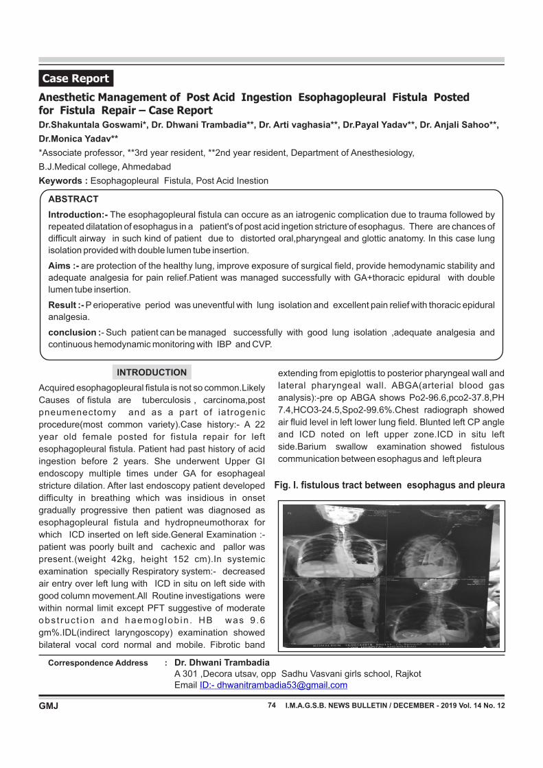
74
Correspondence Address : Dr. Dhwani Trambadia A 301 ,Decora utsav, opp Sadhu Vasvani girls school, Rajkot Email ID:- [email protected]
ABSTRACT
Introduction:- The esophagopleural fistula can occure as an iatrogenic complication due to trauma followed by
repeated dilatation of esophagus in a patient's of post acid ingetion stricture of esophagus. There are chances of
difficult airway in such kind of patient due to distorted oral,pharyngeal and glottic anatomy. In this case lung
isolation provided with double lumen tube insertion.
Aims :- are protection of the healthy lung, improve exposure of surgical field, provide hemodynamic stability and
adequate analgesia for pain relief.Patient was managed successfully with GA+thoracic epidural with double
lumen tube insertion.
Result :- P erioperative period was uneventful with lung isolation and excellent pain relief with thoracic epidural
analgesia.
conclusion :- Such patient can be managed successfully with good lung isolation ,adequate analgesia and
continuous hemodynamic monitoring with IBP and CVP.
Case Report
Anesthetic Management of Post Acid Ingestion Esophagopleural Fistula Posted for Fistula Repair – Case Report
Dr.Shakuntala Goswami*, Dr. Dhwani Trambadia**, Dr. Arti vaghasia**, Dr.Payal Yadav**, Dr. Anjali Sahoo**,
Dr.Monica Yadav**
*Associate professor, **3rd year resident, **2nd year resident, Department of Anesthesiology,
B.J.Medical college, Ahmedabad
Keywords : Esophagopleural Fistula, Post Acid Inestion
INTRODUCTION
Acquired esophagopleural fistula is not so common.Likely
Causes of fistula are tuberculosis , carcinoma,post
pneumenectomy and as a part of iatrogenic
procedure(most common variety).Case history:- A 22
year old female posted for fistula repair for left
esophagopleural fistula. Patient had past history of acid
ingestion before 2 years. She underwent Upper GI
endoscopy multiple times under GA for esophageal
stricture dilation. After last endoscopy patient developed
difficulty in breathing which was insidious in onset
gradually progressive then patient was diagnosed as
esophagopleural fistula and hydropneumothorax for
which ICD inserted on left side.General Examination :-
patient was poorly built and cachexic and pallor was
present.(weight 42kg, height 152 cm).In systemic
examination specially Respiratory system:- decreased
air entry over left lung with ICD in situ on left side with
good column movement.All Routine investigations were
within normal limit except PFT suggestive of moderate
obs t ruc t ion and haemog lob in . HB was 9 .6
gm%.IDL(indirect laryngoscopy) examination showed
bilateral vocal cord normal and mobile. Fibrotic band
extending from epiglottis to posterior pharyngeal wall and
lateral pharyngeal wall. ABGA(arterial blood gas
analysis):-pre op ABGA shows Po2-96.6,pco2-37.8,PH
7.4,HCO3-24.5,Spo2-99.6%.Chest radiograph showed
air fluid level in left lower lung field. Blunted left CP angle
and ICD noted on left upper zone.ICD in situ left
side.Barium swallow examination showed fistulous
communication between esophagus and left pleura
Fig. I. fistulous tract between esophagus and pleura
I.M.A.G.S.B. NEWS BULLETIN / DECEMBER - 2019 Vol. 14 No. 12
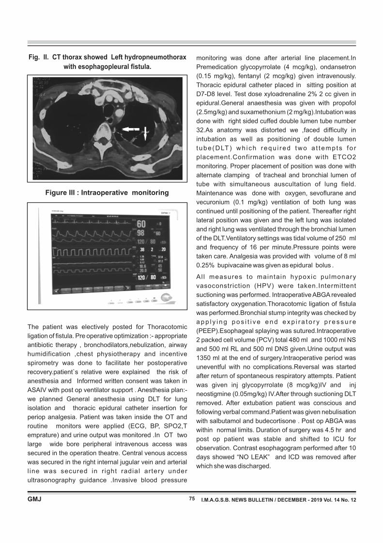
75
The patient was electively posted for Thoracotomic
ligation of fistula. Pre operative optimization :- appropriate
antibiotic therapy , bronchodilators,nebulization, airway
humidification ,chest physiotherapy and incentive
spirometry was done to facilitate her postoperative
recovery.patient`s relative were explained the risk of
anesthesia and Informed written consent was taken in
ASAIV with post op ventilator support . Anesthesia plan:-
we planned General anesthesia using DLT for lung
isolation and thoracic epidural catheter insertion for
periop analgesia. Patient was taken inside the OT and
routine monitors were applied (ECG, BP, SPO2,T
emprature) and urine output was monitored .In OT two
large wide bore peripheral intravenous access was
secured in the operation theatre. Central venous access
was secured in the right internal jugular vein and arterial
l ine was secured in r ight radial artery under
ultrasonography guidance .Invasive blood pressure
Fig. II. CT thorax showed Left hydropneumothorax
with esophagopleural fistula. monitoring was done after arterial line placement.In
Premedication glycopyrrolate (4 mcg/kg), ondansetron
(0.15 mg/kg), fentanyl (2 mcg/kg) given intravenously.
Thoracic epidural catheter placed in sitting position at
D7-D8 level. Test dose xyloadrenaline 2% 2 cc given in
epidural.General anaesthesia was given with propofol
(2.5mg/kg) and suxamethonium (2 mg/kg).Intubation was
done with right sided cuffed double lumen tube number
32.As anatomy was distorted we ,faced difficulty in
intubation as well as positioning of double lumen
tube (DLT) wh i ch requ i red two a t t emp ts f o r
placement.Confirmation was done with ETCO2
monitoring. Proper placement of position was done with
alternate clamping of tracheal and bronchial lumen of
tube with simultaneous auscultation of lung field.
Maintenance was done with oxygen, sevoflurane and
vecuronium (0.1 mg/kg) ventilation of both lung was
continued until positioning of the patient. Thereafter right
lateral position was given and the left lung was isolated
and right lung was ventilated through the bronchial lumen
of the DLT.Ventilatory settings was tidal volume of 250 ml
and frequency of 16 per minute.Pressure points were
taken care. Analgesia was provided with volume of 8 ml
0.25% bupivacaine was given as epidural bolus .
Al l measures to maintain hypoxic pulmonary
vasoconstriction (HPV) were taken.Intermittent
suctioning was performed. Intraoperative ABGA revealed
satisfactory oxygenation.Thoracotomic ligation of fistula
was performed.Bronchial stump integrity was checked by
a p p l y i n g p o s i t i v e e n d e x p i r a t o r y p r e s s u r e
(PEEP).Esophageal splaying was sutured.Intraoperative
2 packed cell volume (PCV) total 480 ml and 1000 ml NS
and 500 ml RL and 500 ml DNS given.Urine output was
1350 ml at the end of surgery.Intraoperative period was
uneventful with no complications.Reversal was started
after return of spontaneous respiratory attempts. Patient
was given inj glycopyrrolate (8 mcg/kg)IV and inj
neostigmine (0.05mg/kg) IV.After through suctioning DLT
removed. After extubation patient was conscious and
following verbal command.Patient was given nebulisation
with salbutamol and budecortisone . Post op ABGA was
within normal limits. Duration of surgery was 4.5 hr and
post op patient was stable and shifted to ICU for
observation. Contrast esophagogram performed after 10
days showed “NO LEAK” and ICD was removed after
which she was discharged.
Figure III : Intraoperative monitoring
I.M.A.G.S.B. NEWS BULLETIN / DECEMBER - 2019 Vol. 14 No. 12
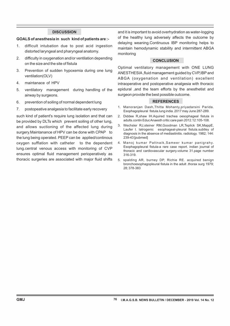
76
and it is important to avoid overhydration as water-logging
of the healthy lung adversely affects the outcome by
delaying weaning.Continuous IBP monitoring helps to
maintain hemodynamic stability and intermittent ABGA
monitoring
CONCLUSION
Optimal ventilatory management with ONE LUNG
ANESTHESIA ,fluid management guided by CVP,IBP and
ABGA (oxygenation and venti lat ion) excel lent
intraoperative and postoperative analgesia with thoracic
epidural ,and the team efforts by the anesthetist and
surgeon provide the best possible outcome.
REFERENCES
1. Manoranjan Dash,Thitta Mohanty,priyadarsini Parida. Esophagopleural fistula.lung india. 2017 may June 287-289.
2. Diddee R,shaw IH.Aquired trachea oesophageal fistula in adults.contin Educ Anaesth critic care pain 2012.12:105-108.
3. Wechsler RJ,steiner RM,Goodman LR,Teplick SK,MappE, Laufer I. Iatrogenic esophageal-pleural fistula.subtley of diagnosis in the absence of mediastinitis. radiology. 1982; 144: 239-43 [pubmed]
4. Mano j kumar Pa t tna ik ,Sameer kumar pan ig rahy. Esophagopleural fistula-a rare case report. indian journal of thoracic and cardiovascular surgery.volume 31.page number 316-319.
5. spalding AR, burney DP, Richie RE. acquired benign bronchoesophagopleural fistula in the adult .thorax surg 1979; 28; 378-383
DISCUSSION
GOALS of anesthesia in such kind of patients are :-
1. difficult intubation due to post acid ingestion
distorted laryngeal and pharyngeal anatomy.
2. difficulty in oxygenation and/or ventilation depending
on the size and the site of fistula
3. Prevention of sudden hypoxemia during one lung
ventilation(OLV)
4. maintanace of HPV
5. ventilatory management during handling of the
airway by surgeons.
6. prevention of soiling of normal dependent lung
7. postopeative analgesia to facilitate early recovery
such kind of patient's require lung isolation and that can
be provided by DLTs which prevent soiling of other lung,
and allows suctioning of the affected lung during
surgery.Maintanance of HPV can be done with CPAP to
the lung being operated. PEEP can be applied/continous
oxygen sufflation with catheter to the dependent
lung.central venous access with monitoring of CVP
ensures optimal fluid management perioperatively as
thoracic surgeries are associated with major fluid shifts
I.M.A.G.S.B. NEWS BULLETIN / DECEMBER - 2019 Vol. 14 No. 12
Study Of Effectiveness And Safety Of Inj DMPA In Postpartum Period
Case Report
Dr. Kruti J. Deliwala* Dr Margi Shah** Dr Parul T. Shah** Dr Reena V. Patel** Dr. Devangi S. Munshi** Dr Zalak patel**
77
Correspondence Address : Dr. Margi Shah 3/B, J. K. Plaza, Nr. Bank of Baroda, Maninagar Cross Roads, Maninagar, Ahmedabad. Email : [email protected]
INTRODUCTION
Population explosion is one of the major problem of the
developing countries like India. Contraceptive prevalence [1]in our country is 54.8% . Moreover contraception in
postpartum period is a major challenge. Popular
methods offered are barrier, progesterone only pills,
IUCD, sterilization, Depot medroxy progesterone
(DMPA). Inj Depot medroxy progesterone has been found
to provide effective, long acting and reversible
contraception. Its 12 week's dosing results in patient
convenience, avoiding need for daily compliance or use at [2]time of intercourse . It is to be given deep intramuscular
so not much training of staff is required. One injection at
every three months and less side effects makes
compliance better in temporary motivated postpartum
women specially in low resource settings.
Ideal time to initiate DMPA is within 5 days of onset of
menses this ensures patient is not pregnant and prevents
ovulation during first month of use. WHO recommends
DMPA in breastfeeding women be initiated at 6 weeks due [2]to theoretical concerns about infant safety . In indian set
up delaying contraception until 6 weeks postpartum may
increase risk of unwanted pregnancy and large amount
lost to follow up. ACOG recognizes the need for
contraception against theoretical neonatal risks and [3]endorses earlier initiation of DMPA.
Injection DMPA has typical use failure rate of 0.3 per 100 [4]woman years . Major side effects with DMPA includes
menstrual disturbance, weight gain, mood changes etc.
Inj DMPA does not have any permanent effect on fertility. It
has been shown that after discontinuation of DMPA;
normal period cycle returns by 8 months and normal
fertility returns by 18 months.
Here we aim to study acceptance and effect of inj DMPA in
immediate postpartum period
MATERIALS AND METHODS
This is a prospective study conducted at obstetrics and
gynaecology department, tert iary care centre,
Ahmedabad. After approval from institute, and patients
consent before joining this study.
Study conducted for total duration of 9 months from
September 2017 to January 2019. A total of 200 women
who were apparently healthy, aged 21 to 35 years,
breatfeeding, ready to follow up and wanted a long term
reversible contraception were recruited for this study.
Eligibility criteria
• Age 21 to 35 years
• Patient desiring long term, non coitus depended
contraceptive method
• Willing for regular follow up and gives consent for
study
• Apparently healthy with no associated medical
condition like anaemia, diabetes, hypertension,
cardiac disease.
Keywords : injection DMPA, contraception, postpartum, fertility.
ABSTRACT
Depot medroxyprogesterone acetate(DMPA) provides safe and effective contraception in postpartum period
Aims and objectives : to study acceptance and compliance of DMPA, observe side effects related to usage of
DMPA and establish its effectiveness as contraception method.
Methods : this is prospective study carried out at Obstetrics and Gynaecology department of tertiary care hospital.
Over period of 9 months followed up on OPD basis.
Result : out of 200 cases selected 42% were in age group of 26 to 30, 51% were from lower socio economic class.
Amenorrhoea and irregular spotting found in 68% and 42% respectively. There was no cases of pregnancy in
present study.
Conclusion : DMPA is an effective, safe and convenient method of contraception in lactating period. Proper
counseling will increase acceptance of DMPA.
I.M.A.G.S.B. NEWS BULLETIN / DECEMBER - 2019 Vol. 14 No. 12
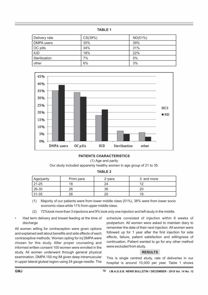
78
TABLE 1
Delivery rate CS(39%) ND(51%)
DMPA users 35% 39%
OC pills 34% 31%
IUD 18% 22%
Sterilization 7% 5%
other 6% 3%
TABLE 2
nd rdAge/parity Primi para 2 para 3 and more
21-25 18 24 12
26-30 26 36 20
31-35 26 20 18
• Had term delivery and breast feeding at the time of
discharge
All women willing for contraception were given options
and explained well about benefits and side effects of each
contraceptive methods. Women opting for inj DMPA were
chosen for this study. After proper counseling and
informed written consent 100 women were enrolled in the
study. All women underwent through general physical
examination. DMPA 150 mg IM given deep intramuscular
in upper lateral gluteal region using 24 gauge needle. The
schedule consisted of injection within 6 weeks of
postpartum. All women were asked to maintain diary to
remember the date of their next injection. All women were
followed up for 1 year after the first injection for side
effects, failure, patient satisfaction and willingness of
continuation. Patient wanted to go for any other method
were excluded from study.
RESULTS
This is single centred study, rate of deliveries in our
hospital is around 10,000 per year. Table 1 shows
PATIENTS CHARACTERISTICS
(1) Age and parity
Our study included apparenty healthy women in age group of 21 to 35.
(1) Majority of our patients were from lower middle class (51%), 38% were from lower socio
economic class while 11% from upper middle class.
(2) 72%took more than 3 injections and 9% took only one injection and left study in the middle.
I.M.A.G.S.B. NEWS BULLETIN / DECEMBER - 2019 Vol. 14 No. 12
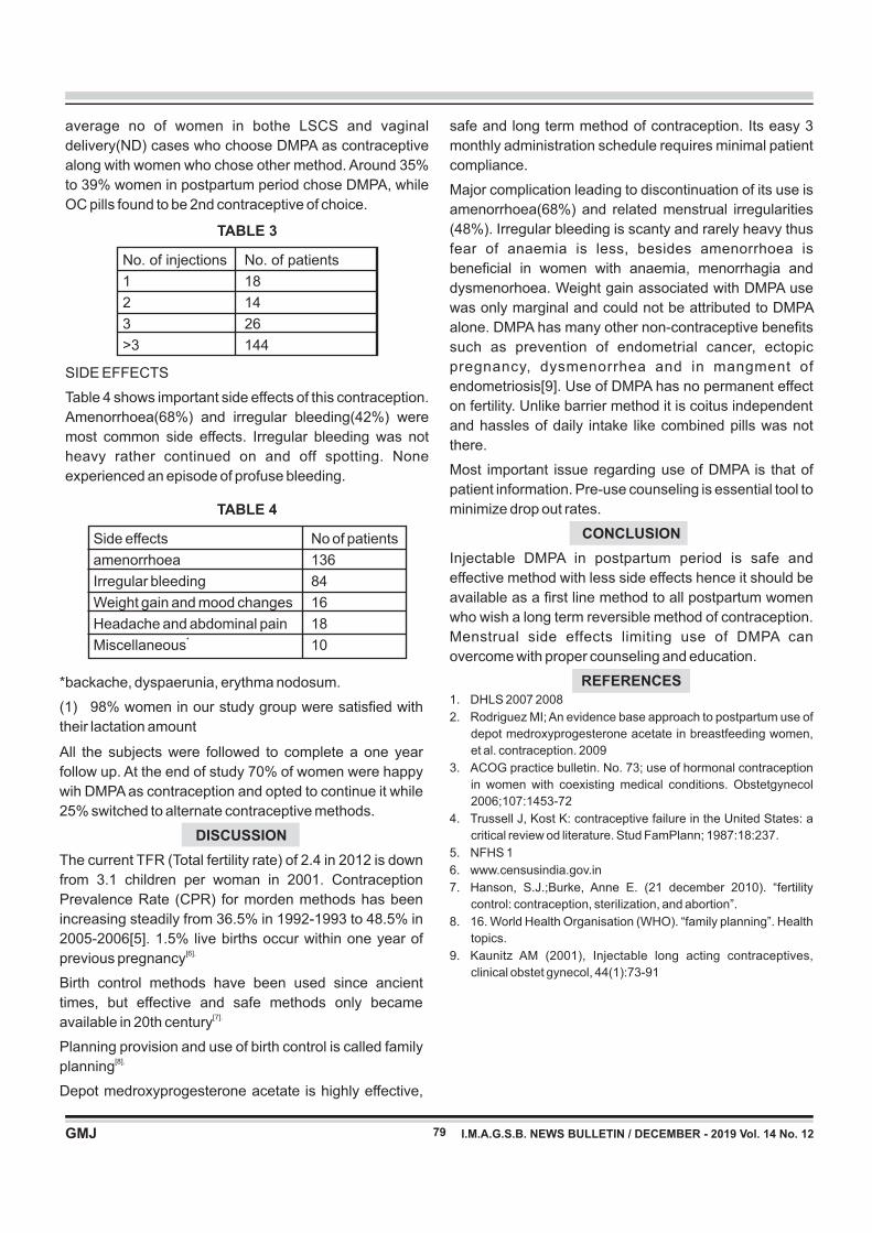
TABLE 4
Side effects No of patients
amenorrhoea 136
Irregular bleeding 84
Weight gain and mood changes 16
Headache and abdominal pain 18* Miscellaneous 10
79
average no of women in bothe LSCS and vaginal
delivery(ND) cases who choose DMPA as contraceptive
along with women who chose other method. Around 35%
to 39% women in postpartum period chose DMPA, while
OC pills found to be 2nd contraceptive of choice.
All the subjects were followed to complete a one year
follow up. At the end of study 70% of women were happy
wih DMPA as contraception and opted to continue it while
25% switched to alternate contraceptive methods.
DISCUSSION
The current TFR (Total fertility rate) of 2.4 in 2012 is down
from 3.1 children per woman in 2001. Contraception
Prevalence Rate (CPR) for morden methods has been
increasing steadily from 36.5% in 1992-1993 to 48.5% in
2005-2006[5]. 1.5% live births occur within one year of [6].previous pregnancy
Birth control methods have been used since ancient
times, but effective and safe methods only became [7].available in 20th century
Planning provision and use of birth control is called family [8].planning
Depot medroxyprogesterone acetate is highly effective,
TABLE 3
No. of injections No. of patients
1 18
2 14
3 26
>3 144
SIDE EFFECTS
Table 4 shows important side effects of this contraception.
Amenorrhoea(68%) and irregular bleeding(42%) were
most common side effects. Irregular bleeding was not
heavy rather continued on and off spotting. None
experienced an episode of profuse bleeding.
*backache, dyspaerunia, erythma nodosum.
(1) 98% women in our study group were satisfied with
their lactation amount
safe and long term method of contraception. Its easy 3
monthly administration schedule requires minimal patient
compliance.
Major complication leading to discontinuation of its use is
amenorrhoea(68%) and related menstrual irregularities
(48%). Irregular bleeding is scanty and rarely heavy thus
fear of anaemia is less, besides amenorrhoea is
beneficial in women with anaemia, menorrhagia and
dysmenorhoea. Weight gain associated with DMPA use
was only marginal and could not be attributed to DMPA
alone. DMPA has many other non-contraceptive benefits
such as prevention of endometrial cancer, ectopic
pregnancy, dysmenorrhea and in mangment of
endometriosis[9]. Use of DMPA has no permanent effect
on fertility. Unlike barrier method it is coitus independent
and hassles of daily intake like combined pills was not
there.
Most important issue regarding use of DMPA is that of
patient information. Pre-use counseling is essential tool to
minimize drop out rates.
CONCLUSION
Injectable DMPA in postpartum period is safe and
effective method with less side effects hence it should be
available as a first line method to all postpartum women
who wish a long term reversible method of contraception.
Menstrual side effects limiting use of DMPA can
overcome with proper counseling and education.
REFERENCES 1. DHLS 2007 2008
2. Rodriguez MI; An evidence base approach to postpartum use of
depot medroxyprogesterone acetate in breastfeeding women,
et al. contraception. 2009
3. ACOG practice bulletin. No. 73; use of hormonal contraception
in women with coexisting medical conditions. Obstetgynecol
2006;107:1453-72
4. Trussell J, Kost K: contraceptive failure in the United States: a
critical review od literature. Stud FamPlann; 1987:18:237.
5. NFHS 1
6. www.censusindia.gov.in
7. Hanson, S.J.;Burke, Anne E. (21 december 2010). “fertility
control: contraception, sterilization, and abortion”.
8. 16. World Health Organisation (WHO). “family planning”. Health
topics.
9. Kaunitz AM (2001), Injectable long acting contraceptives,
clinical obstet gynecol, 44(1):73-91
I.M.A.G.S.B. NEWS BULLETIN / DECEMBER - 2019 Vol. 14 No. 12
Association Of Retinitis Pigmentosa(Rp) With Mental Retardation(Mr) and Subluxated Lens
Case Report
Dr. Sneha T. Shah*(Corresponding Author) ,Dr. Nilesh V. Parekh**, Dr. Neepa R. Gohil***, Dr. Aditya Parekh****
*2nd Year Resident Doctor, **Professor and Head of Department of Ophthalmology, ***Assistant Professor of
Ophthalmology, Department of Ophthalmology, Sir T. Hospital, Bhavnagar 364001,Gujarat, India.
****MBBS,22 Behind CSMCRI,Bhavnagar 364001,Gujarat,India.
80
Correspondence Address : D r. Sneha T. Shah E-mail : [email protected] Mob no. : 07977893689/09969673287
Keywords : Mental Retardation, Retinitis Pigmentosa, Subluxated Lens.
ABSTRACT
Retinitis Pigmentosa is pigmentary retinal dystrophy characterized by triad of perivascular pigmentary changes
resembling bone corpuscles,presence of pale and waxy optic disc and attenuated thread like retinal blood vessels
on fundus examination which is associated with common syndromes.We report a case of 35 year old Mentally
Retarded Male having Subluxated lens with Retinitis Pigmentosa.
I.M.A.G.S.B. NEWS BULLETIN / DECEMBER - 2019 Vol. 14 No. 12
INTRODUCTION
Retinitis Pigmentosa is slowly progressive bilateral
primary pigmentary retinal dystrophy due to apoptosis
affecting rods more than cones resulting in night
blindness(earliest feature), which is inherited as
Autosomal Recessive condition, affecting Males more
commonly, showing characteristic triad of perivascular
p i g m e n t o r y c h a n g e s r e s e m b l i n g b o n e
corpuscles,presence of pale and waxy optic disc and
attenuated thread like retinal blood vessels on fundus
examination which is associated with common
syndromes.
CASE REPORT
35 year old Mentally Retarded Male presented with chief
complaint of Left eye Redness,Pain and Watering since 3
days.Torch Light findings were confirmed by Slit Lamp
Examination on which Left eye was Edematous
Congested having Corneal Edema,Shallow Anterior
Chamber,Semidilated Fixed Pupil and Anteriorly
Subluxated Lens with Intraocular Pressure(IOP) not
recordable on Non Contact Tonometer(NCT) and patient
being uncooperative for applanation tonometry.Right eye
having Inferotemporally Subluxated Lens with 15 mmHg
IOP on NCT.Due to Mental Retardation,Patient was
unable to cooperate for Visual Acuity testing.Glow not
seen on Fundus examination with Subluxated Lens in
both eyes.Both eye having Vitreous Degeneration on B
Scan Ultrasonography.
Surgical Management planned after written and informed
consent from legal guardian.Psychiatric evaluation of
patient was done.Preoperatively Tobramycin eye
drops(eds) four times/day(QDS),Prednisolone eds 6
times/day and Hypertonic saline eds 5 times/day was
started in Left eye with Intravenous 500ml 20% Mannitol
Stat and Tablet Acetazolamide (250 mg) QDS with
banana or coconut water.On IOP control next day Left eye
cataract extraction with Iris Claw Lens Implantation was
d o n e u n d e r G e n e r a l A n e s t h e s i a ( F i g u r e
1).Postoperatively Fundus Examination of Left eye had
tilted disc with pallor,generalized vessel attenuation and
bony spicules indicating Retinitis Pigmentosa(Figure 2).5
months later Elective Right Eye Cataract Extraction with
Iris Claw Lens Implantation was done under General
Anesthes ia(F igure 3,4) .Postoperat ive fundus
examination of right eye revealed similar picture of
Retinitis Pigmentosa(Figure 5).
DISCUSSION
Common Syndromes associated with Retinit is
Pigmentosa:
1. Laurence Moon Biedl Syndrome
Most Common
Mental Retardation + Polydactyly + Obesity +
Hypogonadism
2. Cockayne Syndrome
Mental Retardation + Ataxia + Nystagmus
3. Kearns-Sayre Syndrome
Ocular Myopathy + Heart defects
81 I.M.A.G.S.B. NEWS BULLETIN / DECEMBER - 2019 Vol. 14 No. 12
4. Refsum disease
Polyneuropathy + Cerebellar Ataxia + Deafness
5. Usher's Syndrome
Labyrinthine deafness
6. Barren-Kornweig Syndrome
Abetalipoproteinemia + Acanthocytosis
7. NARP Syndrome
Neuropathy + Ataxia
But association of Retinitis Pigmentosa in Mentally
Retarded with Subluxated Lens is a new presentation.
Have we found a New Syndrome?
CONCLUSION
Common Syndromes are associated with Retinitis
Pigmentosa but we present case of 35 year old Mentally
Retarded having Subluxated Lens with Retinitis
Pigmentosa which is a new presentation.
REFERENCES
1. Ammann F, Klein D, Franceschetti A: Genetic and
epidemiological investigation of pigmentary degeneration of
the retina and allied disorders in Switzerland. J Neurol Sci.
1965, 2: 183-196. 10.1016/0022-510X(65)90079-1.
2. Boughman JA, Vernon M, Shaver KA: Usher syndrome:
definition and estimate of prevalence from two high-risk
populations. J Chronic Dis. 1983, 36: 595-603. 10.1016/0021-
9681(83)90147-9.
3. Beales PL, Elcioglu N, Woolf AS, Parker D, Flinter FA: New
criteria for improved diagnosis of Bardet-Biedl syndrome:
results of a population survey. J Med Genet. 1999, 36: 437-
446.
4. Dryja TP, McGee TL, Reichel E, Hahn LB, Cowley GS, Yandell
DW, Sandberg MA, Berson EL: A point mutation of the
rhodopsin gene in one form of retinitis pigmentosa. Nature.
1990, 343: 364-366. 10.1038/343364a0.
5. Maubaret C, Hamel CP: Génétique des rétinites pigmentaires
: c l a s s i f i c a t i o n m é t a b o l i q u e e t c o r r é l a t i o n s
phénotype/génotype. J Fr Ophtalmol. 2005, 28: 71-92.
6. Fain GL: Why photoreceptors die (and why they don't).
Bioessays. 2006, 28: 344-354. 10.1002/bies.20382.
82
Correspondence Address : Dr. Arvind Vala B-246, Sitaram society, Bombay market to Puna gam road, Surat - 395010 Email ID:[email protected]
ABSTRACT
Pleural effusion secondary to chronic pancreatitis is an uncommon condition accounting for less than 1% of
patients and usually left sided, haemorrhagic. Rarely it may be right sided and straw coloured causing difficulty in
establishing the diagnosis, especially if the chest symptoms are disproportionally more than abdominal
symptoms. We report a rare case of 41 years old alcoholic male who had history of right sided chest pain, cough
with expectoration, breathlessness, abdominal pain for 2 months. Pleural fluid examination suggested straw
coloured, lymphocytic predominant exudate with law adenosine deaminase and high lipase and amylase level. To
rule out alcoholic pancreatitis CT scan thorax and abdomen was done, which demonstrated pancreatic
pseudocyst and right sided gross pleural effusion. Conservative treatment was given in form of intercostal chest
tube drainage, higher antibiotics, somatostatin analogue low fat diet for three to four weeks.
Case Report
Straw coloured Right sided Pleuropancreatic effusion: a diagnosis not to be missed
Dr Arvind Vala*, Dr Meghna Patel**, Dr Kiran Rami***, Dr. Kaushal Bhavsar****
* Resident(R2), ** Associate professor, *** Professor and HOD, **** Assistant professor,
Department of Respiratory medicine, GMERS Sola, Ahmedabad
Keywords : Pleural effusion, Pseudocyst of Pancreas, Pancreatitis
INTRODUCTION
Pancreatic pleural effusion due to chronic pancreatitis
with pseudocyst accounting for less than 1% of cases.
Pleural fluid is usually haemorrhagic, left sided with
markedly increased amylase activity. Because of low
incidence, we are presenting a case of right sided pleural
effusion secondary to chronic pancreatitis managed
conservatively. Pancreatic pleural effusion usually
presents on left side. Gross pleural effusion is a known but
rare complication of chronic pancreatitis.
CASE REPORT
A 41-year-old male patient presented with right side chest
pain, cough, sputum and upper abdominal pain for a
period of 2 months. He had history of tobacco chewing
since 5 years and consumed 90 ml country liquor/day for
20 years and last intake was before 2 months with no
history of smoking. His vital signs (temperature, pulse,
Blood pressure and spo2) were within normal limit.
Expansion of the right hemi-thorax was diminished.
Percussion tone was dull and breath sounds were
diminished and vocal resonance decreased over right
side of chest. Laboratory reports showed complete blood
counts, renal function test, liver function test and RBS
within normal limits. On the chest X-ray- right sided
homogenous opacity, obliteration of right costophrenic
angle with shifting of trachea and mediastinum opposite
side suggestive of right sided pleural effusion (fig. A) and
also confirmed by USG Thorax.
His pleural fluid examination revealed uniformly straw
coloured fluid with a total cell count of 160/cu mm with
90%neutrophils, 10% lymphocytes, Malignant cells were
not found in fluid cytology report. Microorganisms, AFB
and fungal elements were not found. Biochemical
analysis showed exudative fluid with total proteins 3.4
g/dl, glucose 101 mg/dl, and Adenosine Deaminase
(ADA) 45.66 U/L.
After all these investigations we could not found
etiological diagnosis. As patient is alcoholic we undergo
for USG abdomen to rule out alcoholic pancreatitis and
reports showed pancreatic pseudocyst with atrophic
pancreas so we sent pleural fluid lipase and amylase,
which was 1997 U/L and 57 U/L respectively.
Patient underwent CT thorax and abdomen and it showed
pancreatic pseudocyst with atrophic pancreas (fig. b) with
massive right sided pleural effusion. Intercostal drainage
tube was inserted and surgical consultant opinion was
taken and was put on higher antibiotics for 4 weeks,
somatostatin analogue and low fat diet as per their advice.
By above management patient was improved, ICD
removed and patient was discharged.
The chest X-ray- right sided homogenous opacity,
obliteration of right costophrenic angle with shifting of
trachea and mediastinum opposite side suggestive of
right sided pleural effusion (fig. A)
I.M.A.G.S.B. NEWS BULLETIN / DECEMBER - 2019 Vol. 14 No. 12
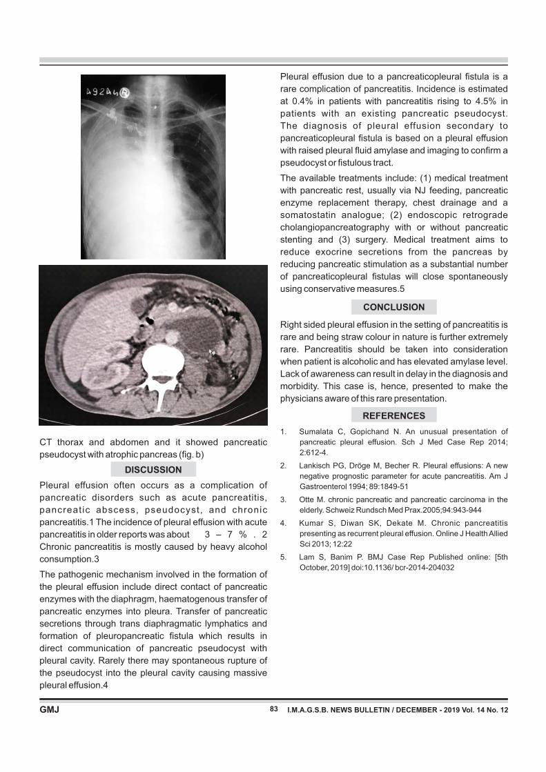
83 I.M.A.G.S.B. NEWS BULLETIN / DECEMBER - 2019 Vol. 14 No. 12
CT thorax and abdomen and it showed pancreatic
pseudocyst with atrophic pancreas (fig. b)
DISCUSSION
Pleural effusion often occurs as a complication of
pancreatic disorders such as acute pancreatitis,
pancreat ic abscess, pseudocyst, and chronic
pancreatitis.1 The incidence of pleural effusion with acute
pancreatitis in older reports was about 3 – 7 % . 2
Chronic pancreatitis is mostly caused by heavy alcohol
consumption.3
The pathogenic mechanism involved in the formation of
the pleural effusion include direct contact of pancreatic
enzymes with the diaphragm, haematogenous transfer of
pancreatic enzymes into pleura. Transfer of pancreatic
secretions through trans diaphragmatic lymphatics and
formation of pleuropancreatic fistula which results in
direct communication of pancreatic pseudocyst with
pleural cavity. Rarely there may spontaneous rupture of
the pseudocyst into the pleural cavity causing massive
pleural effusion.4
Pleural effusion due to a pancreaticopleural stula is a
rare complication of pancreatitis. Incidence is estimated
at 0.4% in patients with pancreatitis rising to 4.5% in
patients with an existing pancreatic pseudocyst.
The diagnosis of pleural effusion secondary to
pancreaticopleural stula is based on a pleural effusion
with raised pleural uid amylase and imaging to conrm a
pseudocyst or stulous tract.
The available treatments include: (1) medical treatment
with pancreatic rest, usually via NJ feeding, pancreatic
enzyme replacement therapy, chest drainage and a
somatostatin analogue; (2) endoscopic retrograde
cholangiopancreatography with or without pancreatic
stenting and (3) surgery. Medical treatment aims to
reduce exocrine secretions from the pancreas by
reducing pancreatic stimulation as a substantial number
of pancreaticopleural stulas will close spontaneously
using conservative measures.5
CONCLUSION
Right sided pleural effusion in the setting of pancreatitis is
rare and being straw colour in nature is further extremely
rare. Pancreatitis should be taken into consideration
when patient is alcoholic and has elevated amylase level.
Lack of awareness can result in delay in the diagnosis and
morbidity. This case is, hence, presented to make the
physicians aware of this rare presentation.
REFERENCES
1. Sumalata C, Gopichand N. An unusual presentation of
pancreatic pleural effusion. Sch J Med Case Rep 2014;
2:612-4.
2. Lankisch PG, Dröge M, Becher R. Pleural effusions: A new
negative prognostic parameter for acute pancreatitis. Am J
Gastroenterol 1994; 89:1849-51
3. Otte M. chronic pancreatic and pancreatic carcinoma in the
elderly. Schweiz Rundsch Med Prax.2005;94:943-944
4. Kumar S, Diwan SK, Dekate M. Chronic pancreatitis
presenting as recurrent pleural effusion. Online J Health Allied
Sci 2013; 12:22
5. Lam S, Banim P. BMJ Case Rep Published online: [5th
October, 2019] doi:10.1136/ bcr-2014-204032
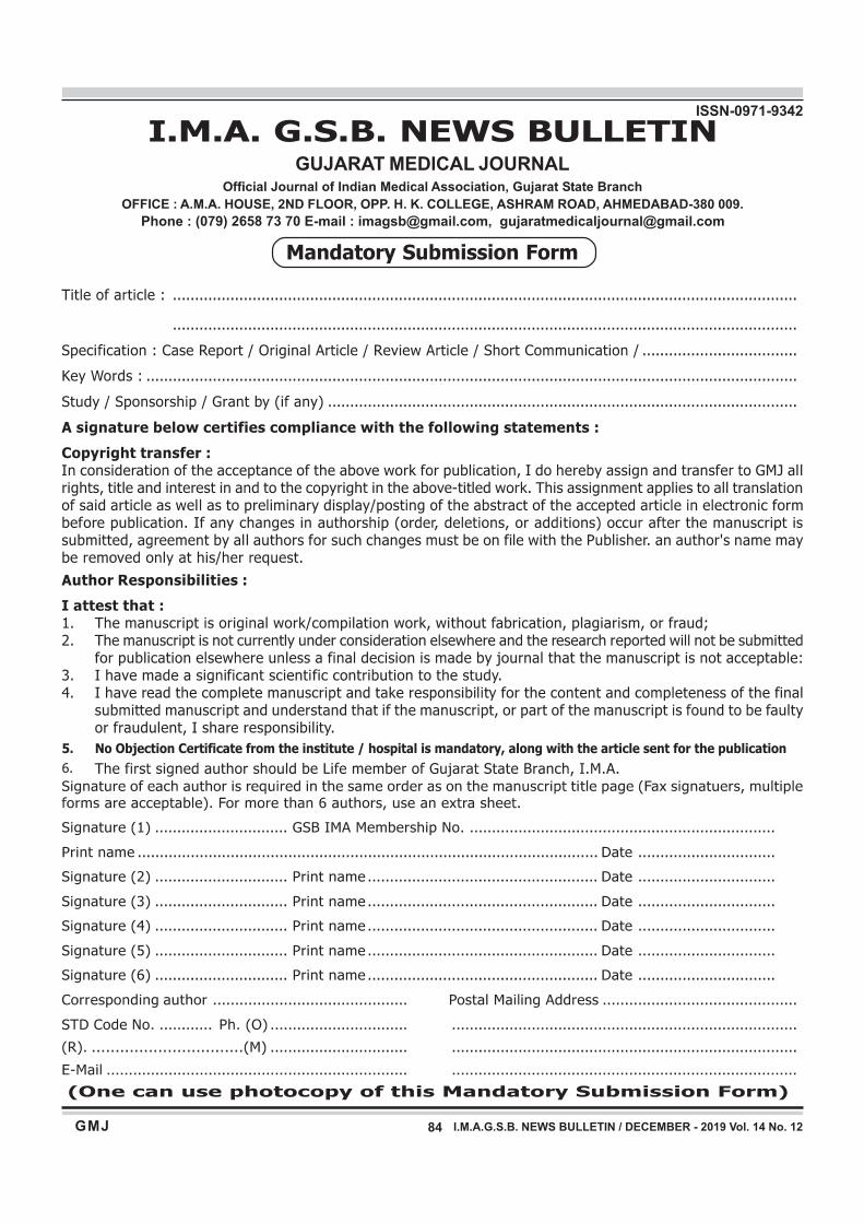
5. No Objection Certificate from the institute / hospital is mandatory, along with the article sent for the publication
6.
Phone : (079) 2658 73 70 E-mail : [email protected], [email protected]
84 I.M.A.G.S.B. NEWS BULLETIN / DECEMBER - 2019 Vol. 14 No. 12
Phone : (079) 2658 73 70 E-mail : [email protected], [email protected]
85 I.M.A.G.S.B. NEWS BULLETIN / DECEMBER - 2019 Vol. 14 No. 12