dr. areefa al bahri ch. 5 the birth experience physiological, psychological, and emotional changes...
TRANSCRIPT

Dr. Areefa Al Bahri
Ch. 5 The Birth Experience
Physiological, psychological, and emotionalchanges that take place during pregnancy help to preparethe woman for labor and birth. Near the end of the pregnancy,the fetus continues to develop physiological abilitiesthat facilitate successful adaptation for the transitionfrom in utero life to the outside environment.
The Process of Labor and BirthThe Process of Labor and Birth
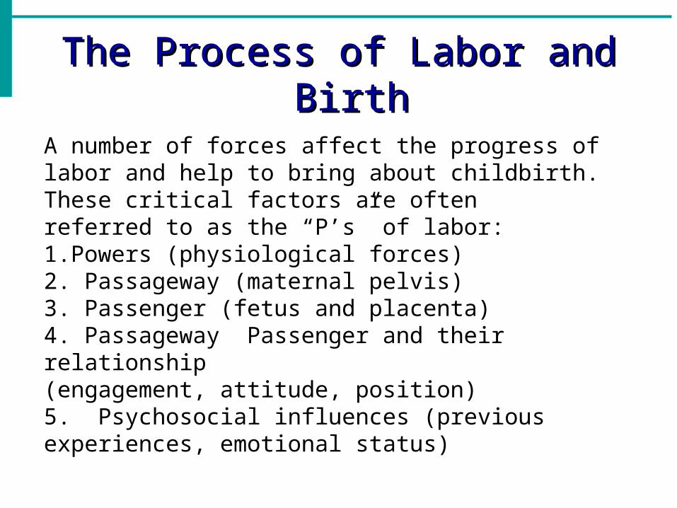
A number of forces affect the progress of labor and help to bring about childbirth. These critical factors are oftenreferred to as the “P’s” of labor:1.Powers (physiological forces)2. Passageway (maternal pelvis)3. Passenger (fetus and placenta)4. Passageway Passenger and their relationship(engagement, attitude, position)5. Psychosocial influences (previous experiences, emotional status)


POWERSThe powers are the physiological forces of labor and birth that include the uterine contractions and the maternal pushing efforts. The uterine muscular contractions, primarily responsible for causing cervical effacement and dilation, also move the fetus down toward the birth canal during the fi rst stage of labor. Uterine contractions are considered the primary force of labor. Once the cervix is fully dilated, the maternal pushing efforts serve as an additional force. During the second stage of labor, use of the maternal abdominal muscles for pushing (the secondary force of labor) adds to the primary force to facilitatechildbirth.

Characteristics of Uterine ContractionsContractions are a rhythmic tightening of the uterus that occurs intermittently. Over time, this action shortens the individual uterine muscle fi bers and aids in the process of cervical effacement and dilation, birth, and postpartal involution (the reduction in uterine size after birth). Each contraction consists of three distinct components: the increment (building of the contraction), the acme (peak of the contraction) and the decrement (decrease in the contraction). Between contractions, the uterus normally returns to a state of complete relaxation. This rest period allows the uterine muscles to relax and provides the woman with a short recovery period that helps her to avoid exhaustion. In addition, uterine relaxation between contractions is important for fetal oxygenation as it allows for blood fl owfrom the uterus to the placenta to be restored.

The lower uterine segment becomes thin-walled and passive. The boundary between the upper lower uterine segments becomes marked by a ridge on the inner uterine surface, known as the “physiological retraction ring.” With each contraction, the uterus elongates. Elongation causes a straightening of the fetal body so that the upper body is pressed against the fundus and the lower, presenting part is pushed toward the lower uterine segment and the cervix. The pressure exerted by the fetus is called the fetal axis pressure. As the uterus elongates, the longitudinal muscle fibers are stretched upward over the presenting part. This force, along with the hydrostatic pressure of the fetal membranes, causes the cervix to dilate (open).


The coordinated efforts of the contractions help to bring about effacement and dilatation of the cervix. Effacement is the process of shortening and thinning of the cervix. As contractions occur, the cervix becomes progressively shorter until the cervical canal eventually disappears. The amount of cervical effacement is usually expressed as a percentage related to the length of the cervical canal, as compared to a non effaced cervix.
For example, if a cervix has thinned to half the normal length of a cervix it is considered to be 50% effaced. Dilation is the opening and enlargement of the cervix that progressively occurs throughout the first stage of labor. Cervical dilation is expressed in centimeters and full dilation is approximately 10 cm. With continued uterine contractions, the cervix eventually opens large enough to allow the fetal head to come through. At this point, the cervix is considered fully dilated or completely dilated and measures 10 cm.


Maternal Pushing EffortsAfter the cervix has become fully dilated, the laboringwoman usually experiences an involuntary “bearing down”sensation that assists with the expulsion of the fetus. Atthis time, the woman can use her abdominal muscles to aidin the expulsion. It is important to remember that the cervixmust be fully dilated before the patient is encouragedto push. Bearing down on a partially dilated cervix cancause cervical edema and damage and adversely affect theprogress of the labor. For most women, the urge to beardown generally occurs when the fetal head reaches thepelvic fl oor. Women who have a strong urge to push oftendo so more effectively than women who force themselvesto push without experiencing any sensations of pressure.

PASSAGEWAY
The passageway consists of the maternal pelvis and the softtissues. The bony pelvis through which the fetus must pass is divided into three sections: the inlet, midpelvis (pelvic cavity), and outlet. Each of these pelvic components has aunique shape and dimension through which the fetus must maneuver to be born vaginally. In human females, the fourclassic types of pelvis are the gynecoid, android, platypelloid, and anthropoid.

PASSENGERThe passenger is referred to as the fetus and the fetal membranes. In the majority (96%) of pregnancies, the fetus presents in a head-fi rst position. The fetal skull, usually the largest body structure, is also the least flexible part of the fetus. However, because of the sutures and fontanels, there is some flexibility in the fetal skull. These structures allow the cranial bones the capability of movement and they overlap in response to the powers of labor. The overlapping or overriding of the cranial bones is called molding.

The fetal skull, or cranium, consists of three major components:the face, the base of the skull, and the vault of the cranium (roof). The facial bones and the cranial base are fused and fixed. The cranial base is made up of two temporal bones. The cranial vault is composed of five bones: two frontal bones, two parietal bones, and the occipital bone. These bones, which are not fused, meet at the sutures. The sutures of the fetal skull are composed of strong but flexible connective tissue that fills the spaces that lie betweenthe cranial bones.

The sagittal suture lies between the parietal bones and runs in an anteroposterior direction between the fontanels, dividing the head into a right and a left side. The lambdoidal suture extends from the posterior fontanel and separates the occipital bones from the parietal bones. The coronal sutures are located between the frontal and parietal bones. They extend from the anterior fontanel laterally and separate the parietal from the frontal bones. The frontal (mitotic) suture lies between the frontal bones and extends from the anterior fontanel to the prominence between the eyebrows. Two membrane-fi lled spaces are present where the suture lines meet. These spaces are referred to as the anterior and posterior fontanels. The anterior fontanel is the larger of the two and measures approximately 0.8 1.2 inch (2 3 cm). It is diamond shaped and is positioned where the sagittal, frontal, and coronal suturesintersect. The anterior fontanel remains open until approximately 18 months of age to allow normal brain growth to occur. The posterior fontanel is triangular in shape and is much smaller than the anterior fontanel. It measures approximately 0.8 inch (2 cm) at its widest point. The posterior fontanel is positioned where the lambdoidal and sagittal sutures meet. Shaped like a smalltriangle, it closes at approximately 6 to 8 weeks after birth.

Fetal PresentationThe fetal presentation refers to the fetal part that enters the pelvic inlet first and leads through the birth canal during labor. The fetal presentation may be cephalic, breech, or shoulder. The part of the fetal body first felt by the examining finger during a vaginal examination is the “presenting part.” The presenting part is determined by the fetal lie and attitude.CEPHALIC PRESENTATIONfetal head will be first to come into contact with the maternal cervix. Cephalic presentations occur in approximately 95% of pregnancies. There are four types of cephalic presentations Vertex. The fetal head presents fully flexed. This is themost frequent and optimal presentation as it allows the smallest suboccipitalbregmatic diameter to present. It is called a “vertex presentation.”Military. In the military position, the fetal head presentsin a neutral position, which is neither flexed nor extended. The occipitofrontal diameter presents to the maternal pelvis and the top of the head is the presentingpart. Brow. In the brow position, the fetal head is partlyextended. This is an unstable presentation that converts toFace. Face presentation. the fetal head is fully extended. The submentobregmatic diameter presents to the maternal pelvis and the face is the presenting part


The following advantages are associated with a cephalic presentation:• The fetal head is usually the largest part of the infant.Once the fetal head is born, the rest of the body usually delivers without complications.• The fetal head is capable of molding. There is sufficient time during labor and descent for molding of the fetal head to occur. Molding helps the fetus to maneuver through the maternal birth passage.• The fetal head is smooth and round, which is the optimal shape to apply pressure to the cervix and aid in dilation.Other presentations (e.g., breech, shoulder) are associated with difficult, prolonged labor and often require cesarean births. They are called malpresentations.

BREECH PRESENTATIONA breech presentation occurs when the fetal buttocks enter the maternal pelvis first. Breech presentations occur in approximately 3% of births and are classified according to the attitude of the fetalhips and knees. Breech presentations are more likely to occur in preterm births or in the presence of a fetal abnormality such as hydrocephaly (head enlargement due to fluid) that prevents the head from entering the pelvis. They are also associated with abnormalities of the maternal uterus or pelvis. Since many factors can compromisethe normal labor and birth process associated with breech presentations, delivery is usually accomplished via cesarean section. There are three types of breech presentations
Frank. The frank breech is the most common of allbreech presentations
Complete (Full). The complete, or full, breech position isthe same as the flexed position with the fetal buttockspresenting first. The legs are typically flexed.
Footling. In the footling breech position, one or both ofthe fetal leg(s) are extended with one foot (“single footling”) or both feet (“double footling”) presenting first into the maternal pelvis.

Several disadvantages are associated with a breech presentation:1.An increased risk for umbilical cord prolapsed because the presenting part may not be covering the cervix (i.e., footling breech).2. The presenting part (buttocks, feet) is not as smooth and hard as the fetal head and is less effective in dilating the cervix.3. Once the fetal body (abdomen) is delivered, the umbilical cord can become compressed.
Rapid delivery may be difficult since the fetal head is usually the largest body part and in this situation, there is no time to allow for molding. In response to adverse outcomes that have been associated with vaginal breech births, the American College of Obstetricians and Gynecologists (ACOG, 2006) has published a Committee Opinion concerning planned breech deliveries.

SHOULDER PRESENTATIONThe shoulder presentation is a transverse lie (Fig. below). This presentation is rare and occurs in fewer than 1% of births. When a transverse lie is present, the maternal abdomen appears large from side to side, rather than up and down. In addition, the woman may demonstrate a lower than expected (for the gestational age) fundal height measurement. Although the shoulder is usually the presenting part, the fetal arm back, abdomen, or side may present in a transverse lie. This presentation occurs most often with preterm birth, high parity, prematurely ruptured membranes, hydramnios, and placenta previa. It is important for the nurse to promptly identify a transverse lie or shoulder presentation since the infant will almost always require a cesarean birth.

StationStation refers to the level of the presenting part in relation to the maternal ischial spines. In the normal female pelvis, the ischial spines represent the narrowest diameter through which the fetus must pass. The ischial spines is a landmark to identify station zero. To visualize the location of station zero, an imaginary line may be drawn between the ischial spines.
Engagement has occurred when the presenting part is at station zero. When the presenting part lies above the maternal ischial spines, it is at a minus station. Therefore, a station of minus 5 (–5) cm indicates that the presenting part is at the pelvic inlet. Positive numbers indicate that the presenting part has descended past the ischial spines. During labor, the presenting part should continue to descend into the pelvis, indicating labor progress. As labor advances and the presenting part descends, the station should also progress to a numerically higher positive station. If the station does not change in the presence of strong, regular contractions, this finding may indicate a problem with the relationship between the maternal pelvis and the fetus (“cephalopelvic disproportion”).

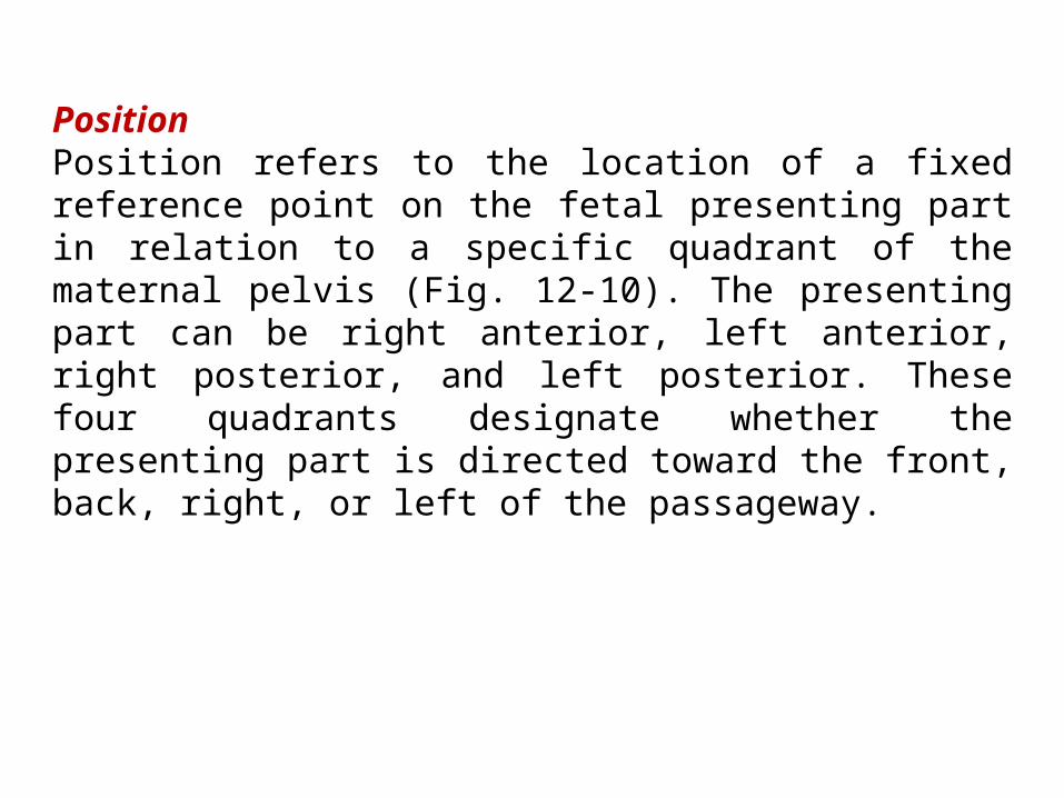
PositionPosition refers to the location of a fixed reference point on the fetal presenting part in relation to a specific quadrant of the maternal pelvis (Fig. 12-10). The presenting part can be right anterior, left anterior, right posterior, and left posterior. These four quadrants designate whether the presenting part is directed toward the front, back, right, or left of the passageway.

Passageway (passenger)The passageway and the passenger have been identified as two of the factors that affect labor. The next “P” is the relationship between the passageway (maternal pelvis) and the passenger (fetus and membranes). The nurse assesses the relationship between the two when determining the engagement, station, and fetal position.EngagementEngagement is said to have occurred when the widest diameter of the fetal presenting part has passed through the pelvic inlet. In a cephalic presentation, the largest diameter is the biparietal; in breech presentations, it is the intertrochanteric diameter. Engagement can bedetermined by external palpation or by vaginal examination. In primigravidas, engagement usually occurs

PSYCHOSOCIAL INFLUENCESThe first four P’s discussed address the physical forces of labor. The last “P” (psychosocial influences) acknowledges the many other critical factors that have an effect on parents such as their readiness for labor and birth, level of educational preparedness, previous experience with labor and birth, emotional readiness, cultural influences, and ethnicity. Transition into the maternal role, and most likely, into the paternal role as well, is facilitated by a positive childbirth experience. A number of internal and external influences can affect the woman’s psychologicalwell-being during labor and birth.

Culturally oriented views of childbirth help to shape the woman’s expectations and ongoing perceptions of the birth experience. The nurse’s understanding of the cultural values and expectations attached to childbirth provide a meaningful framework upon which to plan and deliver sensitive, appropriate care. Cultural considerations for the laboring woman encompass many elements of the birth experience including choice of a birth support person strategies for coping with contractions, pain expression and relief and food preferences.
Signs and Symptoms of Impending LaborBefore the onset of labor, a number of physiological changes occur that signal the readiness for labor and birth. These changes are usually noted by the primigravid woman at about 38 weeks of gestation. In multigravidas, they may not take place until labor begins. It is important for nurses to empower pregnant women and their families by teaching them about the signs and symptoms of impending labor. Providing guidelines about when to contact the health care provider or come to the birth facility helps to demystify the sometimes confusing events that surround birth and lessen the anxieties that can accompany the onset of labor.

LIGHTENINGAt about 38 weeks in the primigravid pregnancy, the presenting part (usually the fetal head) settles downward into the pelvic cavity, causing the uterus to move downward as well. This process, called lightening, marks the beginning of engagement. This downward settling of the uterus may decrease the upward pressure on the diaphragm and result in easier breathing. The downward settling may also lead to the following maternal symptoms:• Leg cramps or pains• Increased pelvic pressure• Increased urinary frequency• Increased venous stasis, causing edema in the lowerextremities• Increased vaginal secretions, due to congestion in thevaginal mucosa

BRAXTON-HICKS CONTRACTIONSAs the pregnancy approaches term, most women become more aware of irregular contractions called Braxton-Hicks contractions. As the contractions increase in frequency (they may occur as often as every 10 to 20 minutes), they may be associated with increased discomfort. Braxton-Hicks contractions are usually felt in the abdomen or groin region and patients may mistake them for true labor. It is believed that these contractions contribute to the preparation of the cervix and uterus for the advent of true labor. Braxton-Hicks contractions do not lead to dilation or effacement of the cervix, and thus are often termed “false labor.”CERVICAL CHANGESIn the non pregnant woman, the cervix is normally rigid. In preparation for passage of the fetus, the cervix undergoes many physiological changes. The cervix softens (“cervical ripening”), stretches, and thins, and eventually is taken up into the lower segment of the uterus. This softening and thinning is called cervical effacement.

BLOODY SHOWDuring pregnancy the cervix is plugged with mucus. The mucus plug acts as a protective barrier for the uterus and its contents throughout the pregnancy. As the cervix begins to soften, stretch, and thin through effacement, there may be rupture of the small cervical capillaries. The added pressure created by engagement of the presenting part may lead to the expulsion of a blood mucus plug, called bloody show. Its presence often indicates that labor will begin within 24 to 48 hours. Late in pregnancy, vaginal examination that involves cervical manipulation may also produce a bloody discharge that can be confused with bloody show.

Rupture Of The MembranesAbout 12% of pregnant women experience spontaneous rupture of the amniotic sac (“ruptured membranes” or “ruptured bag of waters”) prior to the onset of labor. In the majority of pregnancies, the amniotic membranes rupture once labor is well established, either spontaneously or by amniotomy, the artificial rupture of the membranes by the primary care provider.
Rupture of the membranes is a critical event in pregnancy. If the membranes do rupture at home, the woman should be taught to immediately contact the birthing center who will advise her to report for an examination.
It is important for the woman to note the color, amount, and odor of the amniotic fluid. The fluid should be clear and odorless. A yellow green tinged amniotic fluid may indicate infection or fetal passage of meconium and this finding always signals the need for further assessment and fetal heart rate monitoring. Urinary incontinence (frequently associated with urgency, coughing, and sneezing) is sometimes confused with ruptured membranes. The presence of amniotic fluid can be confirmed by a Nitrazine tape test or by a fern test.


First Stage of LaborThis stage begins with the onset of regular uterine contractions and ends with complete dilation of the cervix. woman may not always recognize when true labor actually begins. The first stage of labor is most often the longest stage and its duration can vary considerably among women. The first stage of labor is divided into three distinct phases: latent, active, and transition. Factors such as analgesia, maternal and fetal position, the woman’s body size and her level of physical fitness can also affect the length of labor. LATENT PHASELabor pains are often initially felt as sensations similar to painful menstrual cramping and are usually accompanied by low back pain. Contractions during this phase are typically about 5 minutes apart, last 30 to 45 seconds, and are considered to be mild. During the latent phase cervical effacement and early dilation (0 to 3 cm) occurs. The latent phase of labor can last as long as 10 to 14 hours as the contractions are mild and cervical changes occur slowly.

ACTIVE PHASEThe active phase of labor is characterized by more active contractions. The contractions become more frequent (every 3 to 5 minutes), last longer (60 seconds), and are of a moderate to strong intensity. During the active laborphase, the woman becomes more focused on each contraction and tends to draw inward in an attempt to cope with the increasing demands of the labor. Cervical dilation during this phase advances more quickly as the contractions are often more efficient. While the length of the active phase is variable, nulliparous women generally progress at an average speed of 1 cm of dilation per hourand multiparas at 1.5 cm of cervical dilation per hour.
TRANSITION PHASEThe transition phase is the most intense phase of labor. Transition is characterized by frequent, strong contractions that occur every 2 to 3 minutes and last 60 to 90 seconds on average. Fortunately, this phase often does not take long because dilation usually progresses at a pace equal to or faster than active labor (1 cm/hr for a nullipara and 1.5 cm/hr for a multipara).


Assessment of the Fetus During Labor and BirthFetal assessments include the identification of fetal position and presentation, and the evaluation of the fetal status. Nurses use a variety of assessment techniques including observation, palpation, and auscultation. When assessing a woman in labor, the nurse is able to use observation and interview skills from the moment the woman comes through the door. Astute observation assists the nurse in assessing the patient’s level of pain, her coping abilities

Baseline Fetal Heart RateThe normal baseline fetal heart rate at term is 110 to 160 beats per minute (bpm). There are two abnormal variations of the baseline: tachycardia (baseline above 160 bpm); and bradycardia (baseline below 110 bpm).
TACHYCARDIA. Tachycardia is generally defi ned as a sustained baseline fetal heart rate greater than 160 beats per minute for a duration of 10 minutes or longer. A number of conditions are associated with fetal tachycardia:• Fetal hypoxia: The fetus attempts to compensate for reduced blood flow by increasing sympathetic stimulation of the central nervous system (CNS). Maternal fever: • Maternal medications: Both parasympathetic drugs(i.e., atropine, scopolamine) and beta-sympathetic drugs (tocolytic drugs used to halt contractions) can have a stimulant effect and increase the fetal heart rate.• Infection: uterine infection (amnionitis) • Fetal anemia: In response to a decrease in hemoglobin, the FHR increases to compensate and improve tissue metabolism.• Maternal hyperthyroidism: Thyroid-stimulating hormone (TSH) may cross the placenta and stimulate the fetal heart rate (Tucker, 2004).

BRADYCARDIABradycardia is defined as baseline FHR of less than 110 to 120 bpm. Fetal bradycardia may be associated with:• Late hypoxia: Myocardial activity becomes depressed and lowers the fetal heart rate.• Medications: Beta-adrenergic blocking drugs (e.g., propanolol [Inderal]).• Maternal hypotension:• Prolonged umbilical cord compression• Bradyarrhythmias: With complete heart block, the FHRbaseline is often as low as 70 to 90 bpm.

VariabilityVariability of the FHR is manifested by fluctuations in the baseline fetal heart rate observed on the fetal monitor. The pattern denotes an irregular, changing FHR rather than a straight line that indicates few changes in the rate. The variability of the FHR is a result of the interplay between the fetal sympathetic nervous system, which assists to increase the heart rate and the parasympathetic nervous system, which acts to decrease the heart rate.
The absence of or undetected variability is considered non-reassuring.
FHR variability is indicative of an adequately oxygenated neurological pathway in which impulses are transmitted from the fetal brain to the cardiac conduction system (Fox, Kilpatrick, King, & Parer, 2000).
Conversely, the absence of variability may indicate normal variations such as fetal sleep (the sleep state should not last longer than 30 minutes), a response to certain drugs that depress the CNS, such as analgesics (meperidine [Demerol], tranquilizers (diazepam [Valium]),

ACCELERATIONSAn acceleration is defined as an increase in the FHR of 15 bpm above the fetal heart baseline that lasts for at least 15 to 30 seconds. Accelerations are considered a sign of fetal well-being when they accompany fetal movement. Thus, when a fetus is active in utero, accelerations are normally present. When contractions are present, accelerations are often noted as a response to the contraction.

DECELERATIONSDecelerations are defined as any decrease in FHR below the baseline FHR. Decelerations are further defined according to their onset and are characterized as early, variable, and late.Early DecelerationsEarly decelerations are characterized by a deceleration in the FHR that resembles a mirror image to the contraction. Therefore, the onset of the deceleration begins near the onset of the contraction, and the FHRreturns to baseline by the end of the contraction. Early decelerations are usually repetitive and are commonly observed during active labor and descent of the fetus


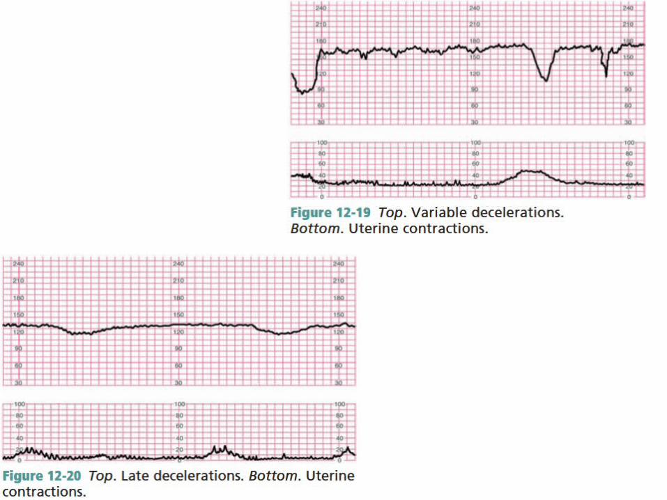
Variable DecelerationsVariable decelerations, as the name implies, are decelerations that are variable in terms of their onset, frequency, duration, and intensity. The decrease in FHR below the baseline is 15 bpm or more, lasts at least 15 seconds, and returns to the baseline in less than 2 minutes from the time of onset (NICHD, 1997) (Fig. 12-19). The deceleration is unrelated to the presence of uterine contractions. Variable decelerations are thought to be a result of umbilical cord compression. Thus, the degree by which the cordis compressed (partially versus completely) can affect the severity of the deceleration. The American College of Obstetricians and Gynecologists(ACOG, 2005) classifies variable decelerations as significant when the FHR falls below 70 bpm and lasts longer than 60 seconds. In addition, the Society of Obstetricians and Gynaecologists of Canada (SOGC, 2005)concurs and further identifi es “non-reassuring” or “atypical” variable decelerations as:

Late DecelerationsThis type of deceleration does not resolve until after the contraction has ended. Late decelerations indicate the presence of uteroplacental insufficiency, a decline in placental function. a decrease in blood flow from the uterus to the placenta results in fetal hypoxia and late decelerations. Late decelerations require prompt attention and reporting. The longer the late decelerations persist, the more serious they become. For example, late decelerations in the presence of an oxytocin infusion may signal a need to immediatelydiscontinue the oxytocin infusion, especially if uterine hyperstimulation is suspected. Nursing interventions that should be implemented immediately include reporting the late decelerations, changing the maternal position, discontinuing the oxytocin infusion,increasing the intravenous fl uids, and administering oxygen by mask.


THE CARDINAL MOVEMENTSThe cardinal movements, or mechanisms of labor, havebeen used to describe how the fetus (in a vertex presentation)passes through the birth canal and the positionalchanges required to facilitate birth (Fig. 12-23). The cardinalmovements are presented in the order in which theyoccur.

DescentFour forces facilitate descent, which is the progression of the fetal head into the maternal pelvis: (1) pressure of the amniotic fluid; (2) direct pressure of the uterine fundus on the fetal breech; (3) contraction of the maternal abdominal muscles; and (4) extension and straightening of the fetal body. The fetal head enters the maternal inlet in the occiput transverse or the oblique position because the pelvic inlet is widest from side to side. The sagittal suture is equidistant from the maternal symphysis pubisand sacral promontory. The degree of fetal descent is measuredby stations.

FlexionFlexion occurs as the fetal head descends and comes into
contact with the soft tissues of the pelvis, the muscles of the maternal pelvic floor, and the cervix. The resistance
encountered with these structures causes the fetal chin to flex downward onto the chest. This position allows the smallest
fetal diameters to enter the maternal pelvis.

Internal RotationTo fit into the maternal pelvic cavity, which is widest in theanteroposterior diameter, the fetal head must rotate. ExtensionAs the fetal head passes under the maternal symphysis pubis, it meets with resistance from the pelvic floor. The head pivots and extends with each maternal pushing effort. The head is born in extension as the occiput slides under the symphysis and the face is directed toward therectum. The fetal brow, nose, and chin then emerge. Restitution Internal rotation causes the fetal shoulders to enter the maternal pelvis in an oblique position. After the head is delivered in the extended position, it rotates briefly to the position it occupied when it was engaged in the inlet. This movement is termed restitution. The 45-degree turnof the fetal head facilitates realignment with the long axis of the body.
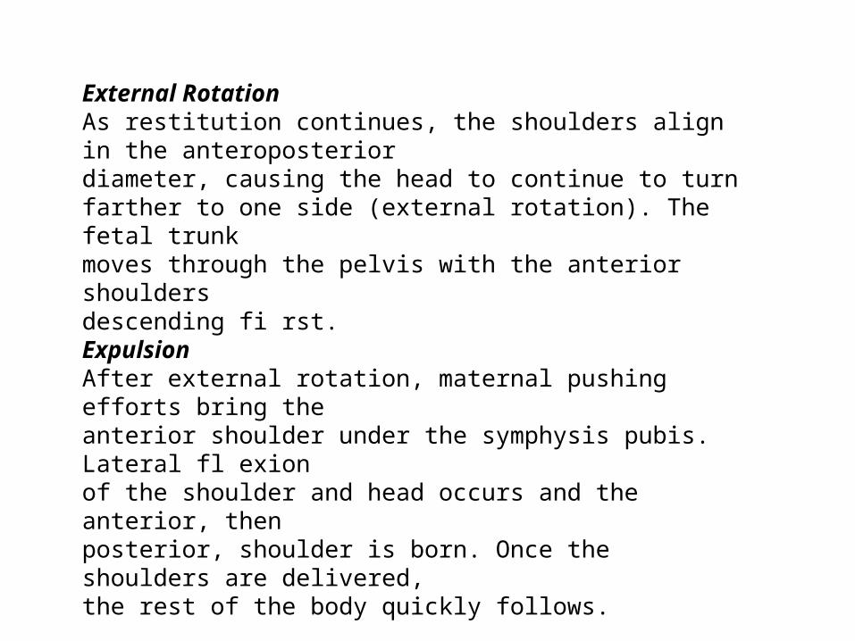
External RotationAs restitution continues, the shoulders align in the anteroposteriordiameter, causing the head to continue to turnfarther to one side (external rotation). The fetal trunkmoves through the pelvis with the anterior shouldersdescending fi rst.ExpulsionAfter external rotation, maternal pushing efforts bring theanterior shoulder under the symphysis pubis. Lateral fl exionof the shoulder and head occurs and the anterior, thenposterior, shoulder is born. Once the shoulders are delivered,the rest of the body quickly follows.

