
VETERINARY HEMATOLOGYATLAS OF COMMON DOMESTIC AND NON-DOMESTIC SPECIES
SECOND EDITION
COPYRIG
HTED M
ATERIAL


3
C H A P T E R O N E
HEMATOPOIESIS
GENERAL FEATURES
All blood cells have a fi nite life span, but in normal animals, the number of cells in circulation is main-tained at a fairly constant level. To accomplish this, cells in circulation need to be constantly replenished, which occurs via the production and release of cells from the bone marrow. Production sites in the bone marrow are commonly referred to as medullary sites.
In times of increased demand, production can also occur outside the bone marrow in sites such as spleen, liver, and lymph nodes. These sites are called extra-medullary sites. In rodents, in the normal steady state, extramedullary production of blood cells occurs in the spleen.
Hematopoiesis, the production of blood cells, is a complex and highly regulated process. Some differ-ences in hematopoiesis exist between species and are
Figure 1.1 Overview of hematopoiesis.
4 H E M AT O P O I E S I S
beyond the scope of this text; readers are referred to the detailed coverage in some of the Selected Refer-ences. The dog will be used to demonstrate some of the basic principles of hematopoiesis. All blood cells in the bone marrow arise from a common stem cell. This pluripotent stem cell gives rise to several stages of committed progenitor cells, which then differen-tiate into cells of the erythrocytic, granulocytic, megakaryocytic, and agranulocytic (monocytic and lymphocytic) lineages. The end result of this develop-ment process is the release of red blood cells, white blood cells, and platelets into the circulation. At the light microscopic level, without the use of immunocy-tochemistry or enzyme cytochemistry, it is impossible to accurately identify the early stem cells in the bone marrow, but the more differentiated stages of devel-opment can be identifi ed and are graphically depicted in Figure 1.1.
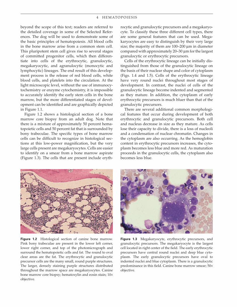
Figure 1.2 shows a histological section of a bone marrow core biopsy from an adult dog. Note that there is a mixture of approximately 50 percent hema-topoietic cells and 50 percent fat that is surrounded by bony trabeculae. The specifi c types of bone marrow cells can be diffi cult to recognize in histological sec-tions at this low-power magnifi cation, but the very large cells present are megakaryocytes. Cells are easier to identify on a smear from a bone marrow aspirate (Figure 1.3). The cells that are present include eryth-
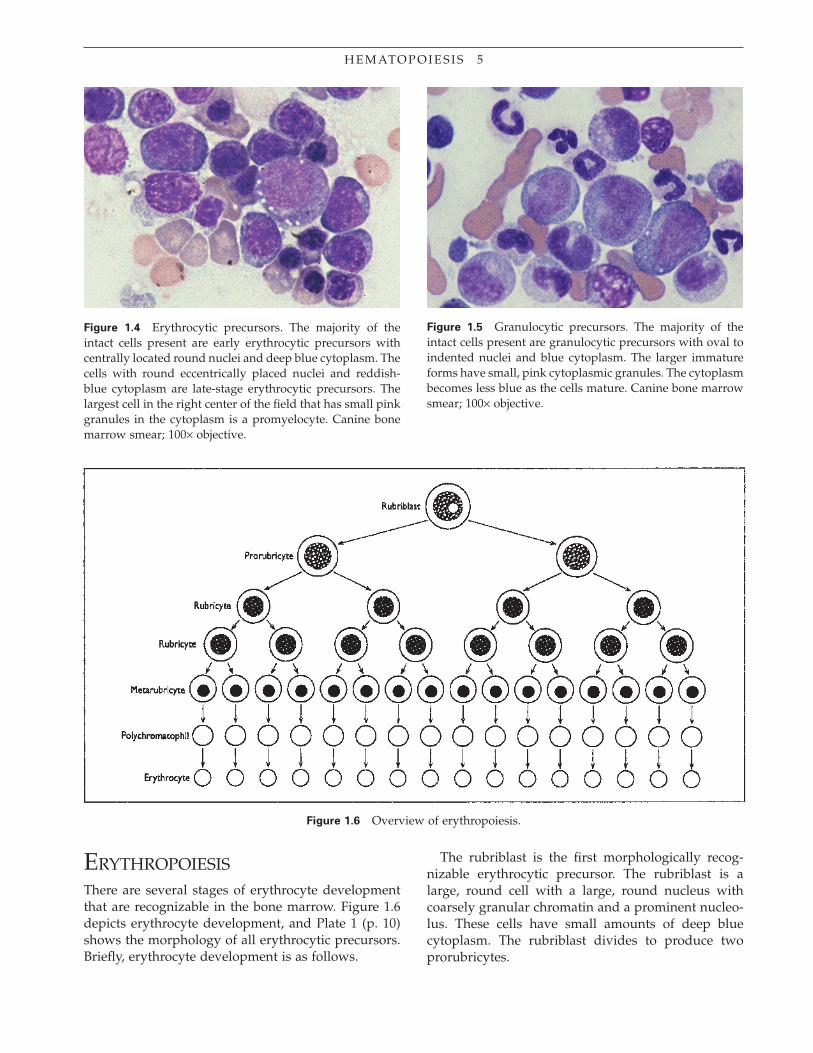
rocytic and granulocytic precursors and a megakaryo-cyte. To classify these three different cell types, there are some general features that can be used. Mega-karyocytes are easy to distinguish by their very large size; the majority of them are 100–200 μm in diameter compared with approximately 20–30 μm for the largest granulocytic or erythrocytic precursors.
Cells of the erythrocytic lineage can be initially dis-tinguished from those of the granulocytic lineage on the basis of their nuclear shape and color of cytoplasm (Figs. 1.4 and 1.5). Cells of the erythrocytic lineage have very round nuclei throughout most stages of development. In contrast, the nuclei of cells of the granulocytic lineage become indented and segmented as they mature. In addition, the cytoplasm of early erythrocytic precursors is much bluer than that of the granulocytic precursors.
There are several additional common morphologi-cal features that occur during development of both erythrocytic and granulocytic precursors. Both cell and nucleus decrease in size as they mature. As cells lose their capacity to divide, there is a loss of nucleoli and a condensation of nuclear chromatin. Changes in the cytoplasm are also occurring. As the hemoglobin content in erythrocytic precursors increases, the cyto-plasm becomes less blue and more red. As maturation proceeds in the granulocytic cells, the cytoplasm also becomes less blue.
Figure 1.2 Histological section of canine bone marrow. Pink bony trabeculae are present in the lower left corner, lower right corner, and top of the photomicrograph and surround the hematopoietic cells and fat. The round to oval clear areas are the fat. The erythrocytic and granulocytic precursor cells are the many small, round purple structures. The larger, densely staining purple structures distributed throughout the marrow space are megakaryocytes. Canine bone marrow core biopsy; hematoxylin and eosin stain; 10× objective.
Figure 1.3 Megakaryocyte, erythrocytic precursors, and granulocytic precursors. The megakaryocyte is the largest cell located in right center of the fi eld. The early erythrocytic precursors have central round nuclei and deep blue cyto-plasm. The early granulocytic precursors have oval to indented nuclei and blue cytoplasm. There is a granulocytic predominance in this fi eld. Canine bone marrow smear; 50× objective.
H E M AT O P O I E S I S 5
The rubriblast is the fi rst morphologically recog-nizable erythrocytic precursor. The rubriblast is a large, round cell with a large, round nucleus with coarsely granular chromatin and a prominent nucleo-lus. These cells have small amounts of deep blue cytoplasm. The rubriblast divides to produce two prorubricytes.
Figure 1.4 Erythrocytic precursors. The majority of the intact cells present are early erythrocytic precursors with centrally located round nuclei and deep blue cytoplasm. The cells with round eccentrically placed nuclei and reddish-blue cytoplasm are late-stage erythrocytic precursors. The largest cell in the right center of the fi eld that has small pink granules in the cytoplasm is a promyelocyte. Canine bone marrow smear; 100× objective.
Figure 1.5 Granulocytic precursors. The majority of the intact cells present are granulocytic precursors with oval to indented nuclei and blue cytoplasm. The larger immature forms have small, pink cytoplasmic granules. The cytoplasm becomes less blue as the cells mature. Canine bone marrow smear; 100× objective.
ERYTHROPOIESISThere are several stages of erythrocyte development that are recognizable in the bone marrow. Figure 1.6 depicts erythrocyte development, and Plate 1 (p. 10) shows the morphology of all erythrocytic precursors. Briefl y, erythrocyte development is as follows.
Figure 1.6 Overview of erythropoiesis.

6 H E M AT O P O I E S I S
The prorubricyte is round and is of equal size or is sometimes larger than the rubriblast. The nucleus is round, with a coarsely granular chromatin pattern. A nucleolus is typically not present. There is a small amount of deep blue cytoplasm, often with a promi-nent perinuclear clear zone. Each prorubricyte divides to form two rubricytes.
The rubricyte is smaller than the prorubricyte. The nucleus is still round, and the coarsely granular chro-matin is more condensed compared with the earlier stages. There is a small amount of deep blue cyto-plasm, although some of the more mature rubricytes have reddish-blue cytoplasm. At the rubricyte stage, there are two divisions; the rubricytes then mature into metarubricytes.
The metarubricyte is smaller than the rubricyte. The nucleus is round to slightly oval, is centrally to eccen-trically located, and has very condensed chromatin. There is a moderate amount of blue to reddish-blue cytoplasm. From the metarubricyte stage on, there is no further division of the cells, just maturation.
The highly condensed pyknotic nucleus of the meta-rubricyte is extruded from the cell, and this cell becomes a polychromatophil. Polychromatophils are round cells without a nucleus and have bluish cyto-plasm. As a polychromatophil matures, it becomes less blue and more red, becoming a mature red blood cell. The mature red blood cells have species-depen-
dent morphological features, which are described in Chapter 2.
GRANULOPOIESIS
Granulopoiesis is depicted in Figure 1.7 and Plate 2 (p. 11). In the bone marrow, there are three types of granulocytes, which include cells of the neutrophilic, eosinophilic, and basophilic lineages. Cells of the neu-trophilic lineage are the predominant type of granu-locyte present, and their development is described fi rst.
The myeloblast is the fi rst recognizable granulocytic precursor in the bone marrow. It is a large cell with a round to oval nucleus with a fi nely granular chroma-tin pattern and one or more prominent nucleoli. The amount of cytoplasm is small to moderate and blue. Each myeloblast divides to form two promyelocytes. Promyelocytes look similar to myeloblasts except they may not have nucleoli, and they may have a perinu-clear clear zone within the cytoplasm. The distinguish-ing feature of promyelocytes is that they contain multiple, very small, pink to purple granules in the cytoplasm; these are known as primary granules. Pro-myelocytes divide to produce myelocytes.
The myelocyte is smaller than the earlier precursors and has a round to oval to slightly indented nucleus with fi nely to moderately granular chromatin. These
Figure 1.7 Overview of neutrophilic granulopoiesis.

H E M AT O P O I E S I S 7
cells have moderate amounts of blue cytoplasm. At this stage, primary granules are no longer being pro-duced, and now secondary granules are formed. These secondary granules are larger than the primary granules.
In neutrophilic myelocytes, the secondary granules are light pink and are very diffi cult to recognize with the light microscope. The myelocyte goes through two divisions, and the resulting progeny mature into metamyelocytes. From the metamyelocyte stage forward, the cells no longer divide.
The metamyelocyte is smaller than the myelocyte and has a kidney-shaped nucleus. The chromatin is moderately granular and is more condensed and clumped than that in the myelocyte. The cytoplasm is blue and contains primary and secondary granules. Both types of granules in the metamyelocyte and subsequent stages of development are not easily seen light microscopically in most animal species. Metamyelocytes develop into band neutrophils. Band neutrophils are round and smaller than meta-myelocytes, have horseshoe-shaped nuclei, and have moderate amounts of blue to light blue cyto-plasm. The band neutrophil will mature into a segmented neutrophil, which is a small cell with faintly blue to pink cytoplasm and a segmented nucleus. The nuclear chromatin is coarsely granular and clumped.
Mature eosinophils and basophils and their precur-sors are found in very low numbers in the normal bone marrow. The production of these cells is very similar to that of neutrophils, and the only major dif-ferences are described in Figure 1.1. The development is identical until the myelocytic stage, which is when eosinophilic and basophilic myelocytes can be distin-guished from neutrophilic myelocytes by the color of the secondary granules. The eosinophilic and baso-philic myelocytes contain reddish to reddish-orange and purple secondary granules, respectively. Eosino-philic and basophilic metamyelocytes and bands can also be recognized by the presence of the unique secondary granules.
The last stage of development is the mature eosino-phil and basophil. The eosinophil is often slightly larger than the mature neutrophil, and the nucleus is not as tightly segmented. The cytoplasm contains reddish to reddish-orange granules. The mature baso-phil is a round cell that is slightly larger than the neutrophil, with a segmented nucleus with condensed chromatin. The cytoplasm is light purple and may contain granules. There are some unique species-dependent features of mature eosinophils and baso-phils, which are described in Chapter 5.
MONOCYTOPOIESIS
The precursors of monocytes arise from committed stem cells, which are common precursors for both cells of the granulocytic and monocytic lineage. Monocyte development is depicted in Figure 1.1. In normal bone marrow, very few cells of the monocytic lineage are present. Monoblasts are the fi rst microscopically rec-ognizable precursors in bone marrow, although they can be impossible to differentiate from myeloblasts. Monoblasts give rise to promonocytes. A promono-cyte is a large cell with an oval to sometimes indented nucleus with a reticular (netlike) or lacy chromatin pattern. These cells have small to moderate amounts of blue cytoplasm and can be diffi cult to distinguish from neutrophilic myelocytes or metamyelocytes. Promonocytes give rise to monocytes, which are larger than segmented neutrophils. The nucleus of the mono-cyte has multiple indentations. The nuclear chromatin has areas of condensation but has a lacy or reticular pattern compared with the condensed chromatin pattern of the mature neutrophil. The cytoplasm is moderate in amount and is typically blue-gray, often with discrete multiple vacuoles.
MEGAKARYOCYTOPOIESIS AND PLATELET PRODUCTION
Megakaryocytopoiesis is quite unique compared with the development of the other blood cells and is depicted in Figure 1.1. The megakaryoblasts are the fi rst morphologically recognizable precursors of the megakaryocytic lineage in bone marrow but can be impossible to differentiate from other blast cells. The megakaryoblast is a large cell with a single round nucleus and prominent nucleolus. This cell differenti-ates into a promegakaryocyte, which is larger than the megakaryoblast and has a multilobed nucleus with dark blue agranular cytoplasm. The promegakaryo-cyte gives rise to the megakaryocyte (Figure 1.8), which is easily recognized in the bone marrow because of its large size (typically 100–200 μm). This large cell has a large, multilobulated nucleus and abundant granular cytoplasm.
Platelets are formed from the cytoplasm of mega-karyocytes by the formation of a structure known as a proplatelet. The proplatelet is fragmented into mul-tiple platelets. The resulting platelets are discoid-shaped small cells that do not have nuclei and that have light pink cytoplasm with sometimes distinct purple granules.

8 H E M AT O P O I E S I S
LYMPHOPOIESIS
Lymphocytes arise from the same common stem cell precursor as the other bone marrow cells (Fig. 1.1). Multiple stages of differentiation of lymphocytes in bone marrow cannot be recognized light microscopi-cally, but there are two main types of lymphocytes that can be identifi ed by immunophenotyping in the peripheral blood: B and T lymphocytes. These two cell types look similar and cannot be differentiated on the basis of morphology alone, but their functions are quite different. In bone marrow, low numbers of small lymphocytes and rarely seen medium and large lym-phocytes are present (Fig. 1.9). The exact number of lymphocytes present in bone marrow is species depen-dent; however, rodents have a relatively greater abun-dance of bone marrow lymphocytes compared with the common domestic species.
The small lymphocyte is a small, round cell with a round to slightly indented nucleus. In some areas, the nuclear chromatin has a very smooth glassy appear-ance, and in other areas it is more clumped or smudged. Overall, the chromatin is not as condensed as that of a rubricyte, which is the cell type with which it is most often confused. The lymphocyte has a small amount of light blue cytoplasm. The medium and large lymphocytes, as the names imply, are larger than the small lymphocytes. The nuclei are round, and the chromatin is fi nely granular, with some areas of con-densation. The nucleus of the large lymphocyte typi-cally has a nucleolus and is known as a lymphoblast. Both cell types have small amounts of light to moder-ate blue cytoplasm.
In addition to lymphocytes, low numbers of plasma cells can be seen in bone marrow (Fig. 1.10). These cells are the end stage of differentiation of B lympho-cytes and are round with eccentrically placed round nuclei. The nuclear chromatin is very condensed and clumped, with clear areas between the clumps. Plasma cells have moderate amounts of deep blue cytoplasm with a prominent perinuclear clear zone.
Figure 1.8 Megakaryocyte. The megakaryocyte is the large cell in the center with a multilobulated irregular nucleus and abundant granular cytoplasm. Canine bone marrow smear; 50× objective.
Figure 1.9 Lymphocytes. The two smallest round cells (left center) that are slightly larger than red blood cells, with round to oval nuclei and small amounts of light blue cytoplasm, are small lymphocytes. The largest cell in the center is a neutrophilic granulocytic precursor. The round cell (above the granulocytic precursor) with a round nucleus, very condensed chromatin, and a rim of deep blue cyto-plasm is a rubricyte. Feline bone marrow smear; 100× objective.
Figure 1.10 Plasma cells. The three cells (center) with eccentrically placed round nuclei; coarse, clumped chroma-tin; and a moderate amount of deep blue cytoplasm with perinuclear clear zones are plasma cells. The other cells are mainly granulocytic precursors. Canine bone marrow smear; 100× objective.
H E M AT O P O I E S I S 9
OTHER CELLS OF THE BONE MARROW
Macrophage
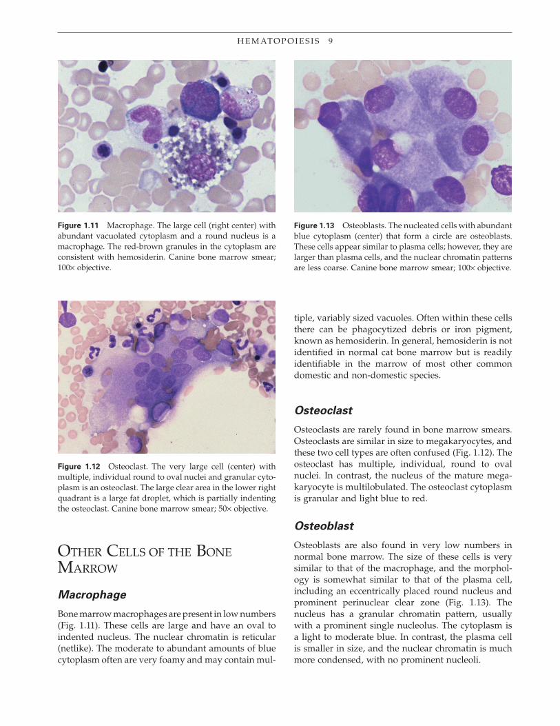
Bone marrow macrophages are present in low numbers (Fig. 1.11). These cells are large and have an oval to indented nucleus. The nuclear chromatin is reticular (netlike). The moderate to abundant amounts of blue cytoplasm often are very foamy and may contain mul-
Figure 1.11 Macrophage. The large cell (right center) with abundant vacuolated cytoplasm and a round nucleus is a macrophage. The red-brown granules in the cytoplasm are consistent with hemosiderin. Canine bone marrow smear; 100× objective.
Figure 1.12 Osteoclast. The very large cell (center) with multiple, individual round to oval nuclei and granular cyto-plasm is an osteoclast. The large clear area in the lower right quadrant is a large fat droplet, which is partially indenting the osteoclast. Canine bone marrow smear; 50× objective.
Figure 1.13 Osteoblasts. The nucleated cells with abundant blue cytoplasm (center) that form a circle are osteoblasts. These cells appear similar to plasma cells; however, they are larger than plasma cells, and the nuclear chromatin patterns are less coarse. Canine bone marrow smear; 100× objective.
tiple, variably sized vacuoles. Often within these cells there can be phagocytized debris or iron pigment, known as hemosiderin. In general, hemosiderin is not identifi ed in normal cat bone marrow but is readily identifi able in the marrow of most other common domestic and non-domestic species.
Osteoclast
Osteoclasts are rarely found in bone marrow smears. Osteoclasts are similar in size to megakaryocytes, and these two cell types are often confused (Fig. 1.12). The osteoclast has multiple, individual, round to oval nuclei. In contrast, the nucleus of the mature mega-karyocyte is multilobulated. The osteoclast cytoplasm is granular and light blue to red.
Osteoblast
Osteoblasts are also found in very low numbers in normal bone marrow. The size of these cells is very similar to that of the macrophage, and the morphol-ogy is somewhat similar to that of the plasma cell, including an eccentrically placed round nucleus and prominent perinuclear clear zone (Fig. 1.13). The nucleus has a granular chromatin pattern, usually with a prominent single nucleolus. The cytoplasm is a light to moderate blue. In contrast, the plasma cell is smaller in size, and the nuclear chromatin is much more condensed, with no prominent nucleoli.

PLATE 1. Red Blood Cell Development
Rubriblast
The rubriblast is a large, round cell with a large, round nucleus; coarsely granular chromatin; and a nucleolus. This cell has small amounts of deep blue cytoplasm.
Prorubricyte
The prorubricyte is a large, round cell with a round nucleus with a coarsely granular chromatin pattern. This cell typically lacks a nucleolus. There is a small amount of deep blue cytoplasm often with a prominent perinuclear clear zone.
Rubricyte
The rubricyte is a round cell with a round, centrally located nucleus; it is smaller than the prorubricyte. The coarsely granular chromatin is more con-densed compared with the earlier stages of development, and irregular clear areas are present between the chromatin clumps. The cytoplasm varies from deep blue to reddish-blue. Early rubricytes typically have more bluish cyto-plasm, and later rubricytes stain more red as the amount of hemoglobin increases.
Metarubricyte
The metarubricyte is smaller than the rubricyte. The nucleus is round to oval, usually slightly eccentrically located, and has very condensed chromatin. There are small to moderate amounts of blue to reddish-blue cytoplasm. The metarubricytes, with more-reddish cytoplasm, contain more hemoglobin.
Polychromatophil
The polychromatophil does not have a nucleus, and cytoplasm is blue to reddish-blue. As polychromatophils mature, they become less blue and more red as a result of their increased amounts of hemoglobin.
Red Blood Cell
The red blood cell does not have a nucleus, and the cytoplasm is reddish to reddish-orange. The central pallor present here is a result of the biconcave discoid shape of the cells.
1 0
11
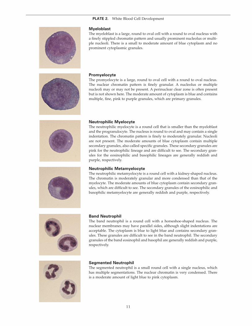
Myeloblast
The myeloblast is a large, round to oval cell with a round to oval nucleus with a fi nely stippled chromatin pattern and usually prominent nucleolus or multi-ple nucleoli. There is a small to moderate amount of blue cytoplasm and no prominent cytoplasmic granules.
Promyelocyte
The promyelocyte is a large, round to oval cell with a round to oval nucleus. The nuclear chromatin pattern is fi nely granular. A nucleolus or multiple nucleoli may or may not be present. A perinuclear clear zone is often present but is not shown here. The moderate amount of cytoplasm is blue and contains multiple, fi ne, pink to purple granules, which are primary granules.
Neutrophilic Myelocyte
The neutrophilic myelocyte is a round cell that is smaller than the myeloblast and the progranulocyte. The nucleus is round to oval and may contain a single indentation. The chromatin pattern is fi nely to moderately granular. Nucleoli are not present. The moderate amounts of blue cytoplasm contain multiple secondary granules, also called specifi c granules. These secondary granules are pink for the neutrophilic lineage and are diffi cult to see. The secondary gran-ules for the eosinophilic and basophilic lineages are generally reddish and purple, respectively.
Neutrophilic Metamyelocyte
The neutrophilic metamyelocyte is a round cell with a kidney-shaped nucleus. The chromatin is moderately granular and more condensed than that of the myelocyte. The moderate amounts of blue cytoplasm contain secondary gran-ules, which are diffi cult to see. The secondary granules of the eosinophilic and basophilic metamyelocyte are generally reddish and purple, respectively.
Band Neutrophil
The band neutrophil is a round cell with a horseshoe-shaped nucleus. The nuclear membranes may have parallel sides, although slight indentations are acceptable. The cytoplasm is blue to light blue and contains secondary gran-ules. These granules are diffi cult to see in the band neutrophil. The secondary granules of the band eosinophil and basophil are generally reddish and purple, respectively.
Segmented Neutrophil
The segmented neutrophil is a small round cell with a single nucleus, which has multiple segmentations. The nuclear chromatin is very condensed. There is a moderate amount of light blue to pink cytoplasm.
PLATE 2. White Blood Cell Development

1 2 H E M AT O P O I E S I S
Mast Cell
Mast cells are found in very low numbers in bone marrow in most species but can be found in higher numbers in the bone marrow of rats. These cells are round, with a round, centrally located nucleus (Fig. 1.14). These cells are easily recognized by the small purple granules that fi ll the cytoplasm. Often, in these cells the granularity is so great that it hides the nuclei. Mast cells are often present within intact bone marrow particles.
Figure 1.14 Mast cell. The cell (center) with abundant purple cytoplasmic granules is a mast cell. The granules almost obscure the round nucleus. A small fat droplet (round clear structure) is partially indenting the right side of the cell. Canine bone marrow smear; 100× objective.


