The Complete Blood Cell Count (CBC)
CBC - Part 1: The Hemogram
Louisiana State University Medical Center
Department of Pathology
New Orleans, Louisiana
by
Carolyn Sue Walters, MHS, MT(ASCP)
A Clinical Pathology 201 Study Module
click here to continue
School of Medicine
©01-07-03
DO NOT REPRODUCE THIS EXERCISE.

C. Sue Walters, MHS, MT(ASCP)
click here to continue
Associate Professor
Department of Pathology
LSU Health Sciences Center
New Orleans, LA
csw
lsuhsc
2001
csw
lsuhsc
2001
This study exercise is the property of Carolyn Sue
Walters, MHS, MT(ASCP) and the Department of
Pathology, LSU Health Sciences Center in New
Orleans, LA. All rights are reserved. It is intended
for use solely within the LSUHSC campus network.
No part of this exercise may be reproduced, stored
in a retrieval system, or transmitted in any form or
by any means (to include but not be restricted to
electronic, mechanical, recording, and photo-
copying) without prior written permission from the
author.
The CBC
click here to continue

csw
lsuhsc
2001
Special thanks is given to Angela Foley, MS,
MT(ASCP), Department of Clinical Laboratory
Sciences, LSUHSC School of Allied Health in New
Orleans, LA for the use of some of her images of
blood cells and for her assistance in the art of
creating image files…
Special Acknowledgment
click here to continue
…and to W. Douglas Scheer, PhD, Department of
Pathology, LSUHSC School of Medicine in New
Orleans, LA for converting the document for internet
access.

csw
lsuhsc
2001 The CBC
click here to continue
CBC – Part 1 The hemogram
CBC – Part 2 WBC differential & blood morphology
CBC – Part 3 RBC morphology & platelet estimate
CBC – Part 4 Post-test
This is the first module of a 4-part study exercise
regarding the CBC. The four parts are entitled:

Feedback
Feedback as to the quality and usefulness of
this exercise is solicited and suggestions for
improvement are welcomed. Please forward
your remarks by E-mail [email protected]
or via US MAIL:
C. Sue Walters, MHS, MT(ASCP)
LSUHSC Department of Pathology
1901 Perdido Street
New Orleans, LA 70112
click here to continue
csw
lsuhsc
2001

Directions
csw
lsuhsc
2001
The directions for navigating through the exer-
cise are given on the next 3 pages. They are the
same as those used in the other modules of this
4-part exercise. Click on:
to visit the directions before continuing
with the exercise.
to bypass the directions.
or
Directions, d
csw
lsuhsc
2001
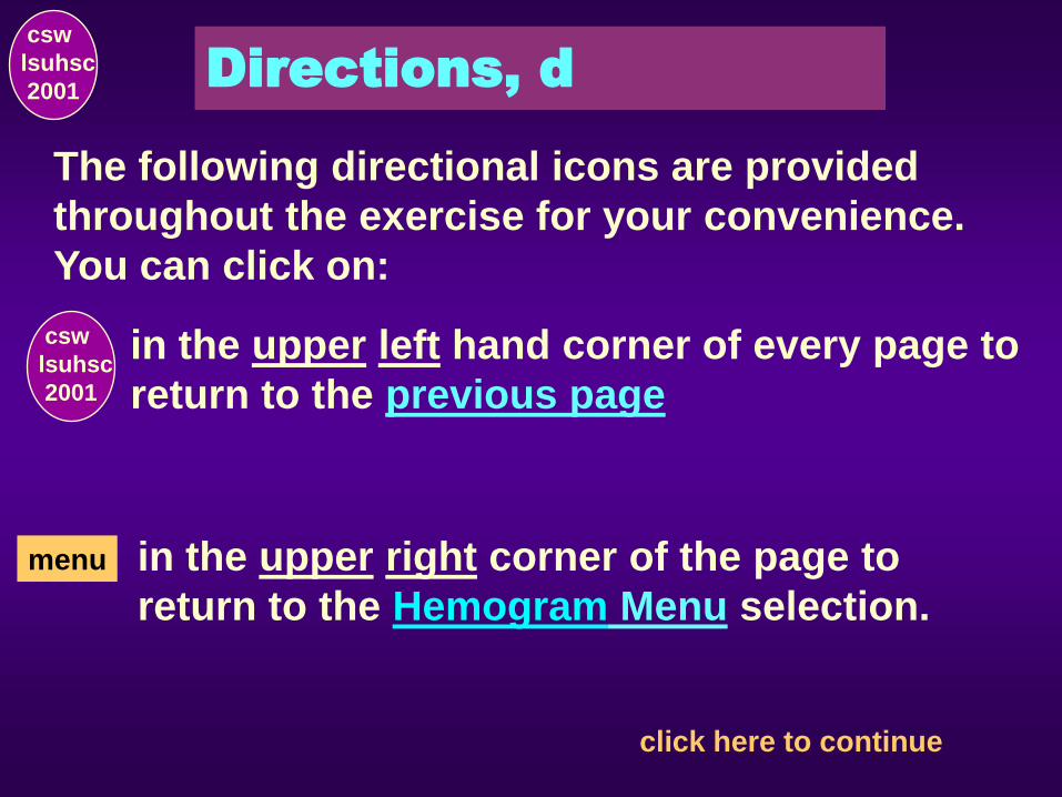
in the upper left hand corner of every page to
return to the previous page
menu in the upper right corner of the page to
return to the Hemogram Menu selection.
The following directional icons are provided
throughout the exercise for your convenience.
You can click on:
csw
lsuhsc
2001
click here to continue
Directions, d
csw
lsuhsc
2001
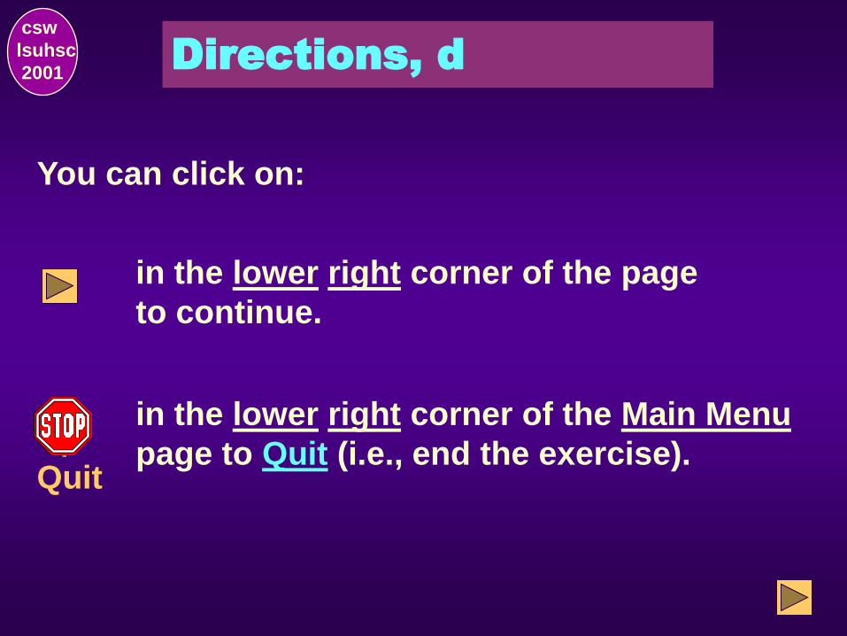
You can click on:
in the lower right corner of the page
to continue.
Quit
in the lower right corner of the Main Menu
page to Quit (i.e., end the exercise).
Directions, d
csw
lsuhsc
2001
“Hot points” (symbols, words, phrases) have been
inserted on the pages as navigational tools and can
be identified by their “gold” color. If it’s “gold”, click
on it to move to the next text/data entry. Also, sounds
have been added in a few places for emphasis.
Remember, if it’s gold, click on it. Try it!
Caution, failure to follow the structured order of the
“hot points” may result in confusion. If you use the
mouse without placing the cursor directly on the “gold
hot point” or click without waiting for the “gold” to
appear, you may skip over vital information.
Special Comments csw
lsuhsc
2001
This exercise has numerous images. You may
note that, when a page contains images, there
may be a rather long delay before you regain
control of the cursor. Please be patient. I think
you will find the images are worth the wait.
NOTE:
Some animation and/or interactive affects may
be lost if you attempt to replay a page by re-
turning to the previous page and then advanc-
ing to that page again.
Now, click on the gold to begin.
csw
lsuhsc
2001 CBC – Part 1
The Complete Blood Cell Count
(CBC)
Part 1 - The Hemogram
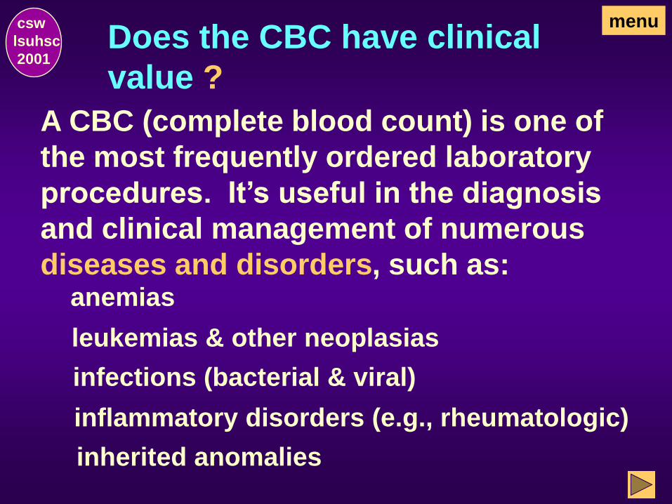
Does the CBC have clinical
value ?
A CBC (complete blood count) is one of
the most frequently ordered laboratory
procedures. It’s useful in the diagnosis
and clinical management of numerous
diseases and disorders, such as:
csw
lsuhsc
2001
menu
anemias
leukemias & other neoplasias
infections (bacterial & viral)
inflammatory disorders (e.g., rheumatologic)
inherited anomalies

What specimen is required?
A CBC (complete blood count) can be performed
by automated electronic instruments or by
manual methods on a whole blood specimen
collected: by
venipuncture - in a tube containing EDTA
anticoagulant
or capillary stick
(e.g., finger,
heel, ear)
- in a vial containing a measured
volume of diluent appropriate
for the method used
csw
lsuhsc
2001
menu
What is a CBC? csw
lsuhsc
2001
A CBC is a battery of hematologic tests. The
values obtained provide valuable information
regarding the three types of blood cells found
in peripheral blood, which are red blood cells
(RBC), white blood cells (WBC), and platelets
(PLT).
menu
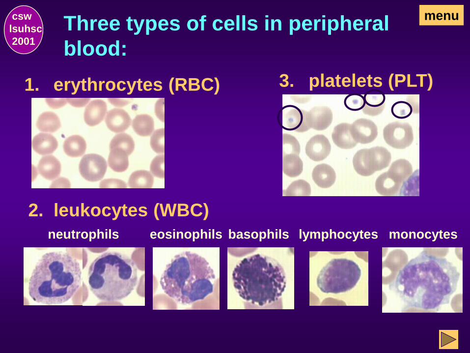
Three types of cells in peripheral
blood:
csw
lsuhsc
2001
2. leukocytes (WBC)
platelets (PLT) 3. erythrocytes (RBC) 1.
eosinophils basophils lymphocytes monocytes neutrophils
menu
What information is provided by a CBC?
csw
lsuhsc
2001
Basically, the CBC provides information regard-
ing the:
frequency distribution of white blood cells
hemoglobin content of red blood cells
morphologic features of the blood cells
relationship of red blood cells to total blood
volume and hemoglobin concentration
menu
number of red cells, white cells, and platelets
in circulating peripheral blood
What are the components of a CBC?
csw
lsuhsc
2001
In most laboratories, due to advanced technolo-
gies now available, an automated CBC is per-
formed on a multi-channel instrument employing
a variety of techniques. It usually includes a:
1 Hemogram
2 Differential WBC count
3 General description of blood cell
morphology (WBC, RBC, and PLT)
4 Platelet estimate
menu
HEMOGRAM
csw
lsuhsc
2001
hemogram menu

HEMOGRAM MENU
Total WBC Count
Total RBC Count
Hemoglobin
Hematocrit
Erythrocyte (RBC) Indices
Platelet Count
Red Cell Distribution Width (RDW)
csw
lsuhsc
2001
Quit
Corrected WBC Count
Mean platelet volume
Introduction

Introduction
csw
lsuhsc
2001
menu

What is a hemogram? csw
lsuhsc
2001
menu
The hemogram components of a CBC are hema-
tologic assays/procedures that provide useful
information regarding the red blood cells (RBC),
white blood cells (WBC), and platelets (PLT).
Automated electronic instruments are able to:
• enumerate the number of each of the three
blood cell types
• differentiate normal from abnormal cells
• provide a variety of information related to each
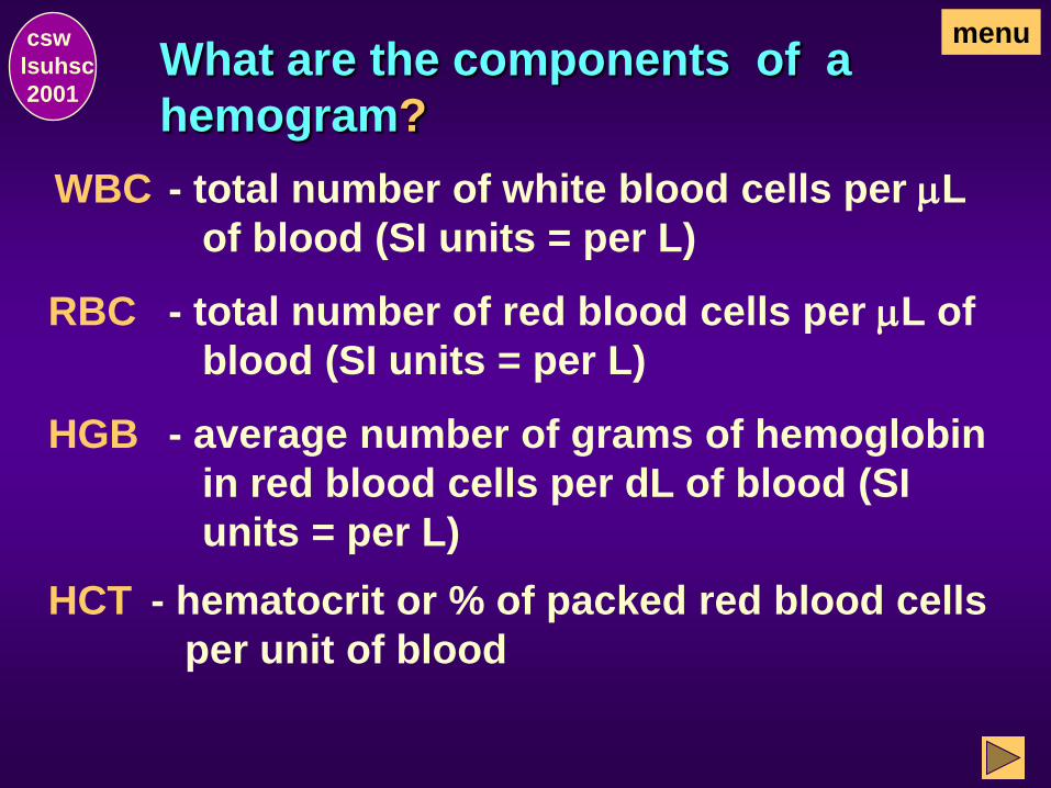
What are the components of a
hemogram?
WBC
RBC
HGB
HCT
menu csw
lsuhsc
2001
- total number of white blood cells per mL
of blood (SI units = per L)
- total number of red blood cells per mL of
blood (SI units = per L)
- average number of grams of hemoglobin
in red blood cells per dL of blood (SI
units = per L)
- hematocrit or % of packed red blood cells
per unit of blood
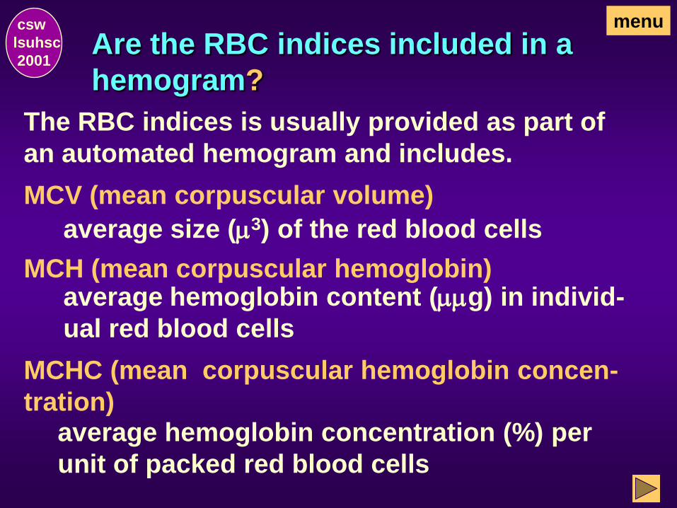
Are the RBC indices included in a
hemogram?
menu
MCV (mean corpuscular volume)
MCH (mean corpuscular hemoglobin)
MCHC (mean corpuscular hemoglobin concen-
tration)
csw
lsuhsc
2001
The RBC indices is usually provided as part of
an automated hemogram and includes.
average size (m3) of the red blood cells
average hemoglobin content (mmg) in individ-
ual red blood cells
average hemoglobin concentration (%) per
unit of packed red blood cells
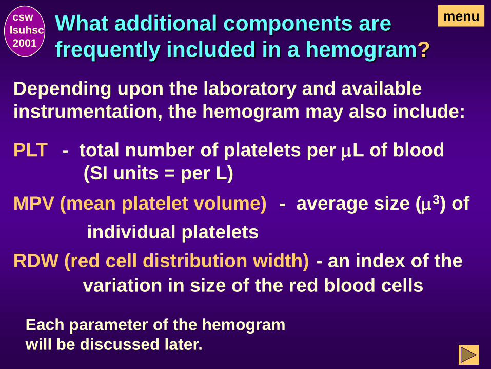
What additional components are
frequently included in a hemogram?
Depending upon the laboratory and available
instrumentation, the hemogram may also include:
PLT
MPV (mean platelet volume)
RDW (red cell distribution width)
csw
lsuhsc
2001
Each parameter of the hemogram
will be discussed later.
- total number of platelets per mL of blood
(SI units = per L)
- average size (m3) of
individual platelets
variation in size of the red blood cells
- an index of the
menu
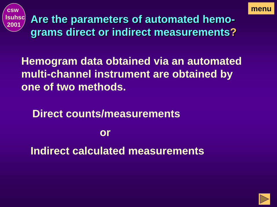
Are the parameters of automated hemo-
grams direct or indirect measurements?
csw
lsuhsc
2001
Hemogram data obtained via an automated
multi-channel instrument are obtained by
one of two methods.
Direct counts/measurements
or
Indirect calculated measurements
menu
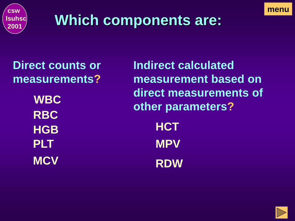
Which components are: csw
lsuhsc
2001
Direct counts or
measurements?
Indirect calculated
measurement based on
direct measurements of
other parameters? WBC
RBC
HGB
PLT
MCV RDW
HCT
MPV
menu
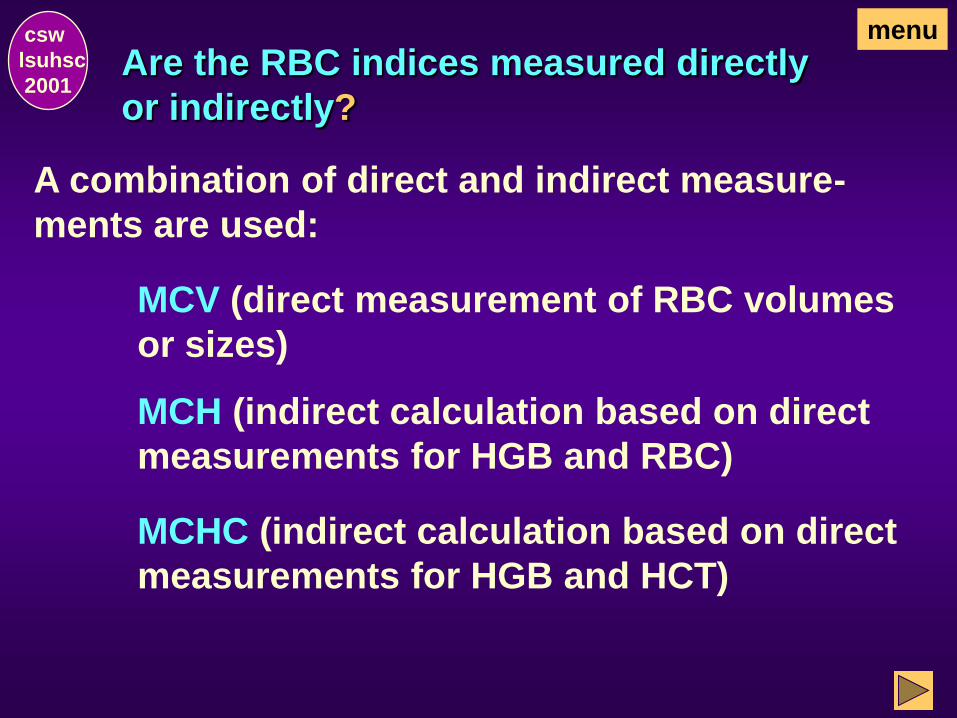
Are the RBC indices measured directly
or indirectly?
csw
lsuhsc
2001
A combination of direct and indirect measure-
ments are used:
MCV (direct measurement of RBC volumes
or sizes)
MCH (indirect calculation based on direct
measurements for HGB and RBC)
MCHC (indirect calculation based on direct
measurements for HGB and HCT)
menu
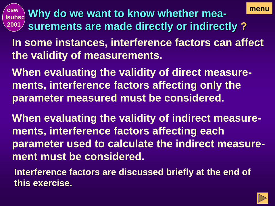
Why do we want to know whether mea-
surements are made directly or indirectly ?
csw
lsuhsc
2001
In some instances, interference factors can affect
the validity of measurements.
When evaluating the validity of direct measure-
ments, interference factors affecting only the
parameter measured must be considered.
When evaluating the validity of indirect measure-
ments, interference factors affecting each
parameter used to calculate the indirect measure-
ment must be considered.
Interference factors are discussed briefly at the end of
this exercise.
menu
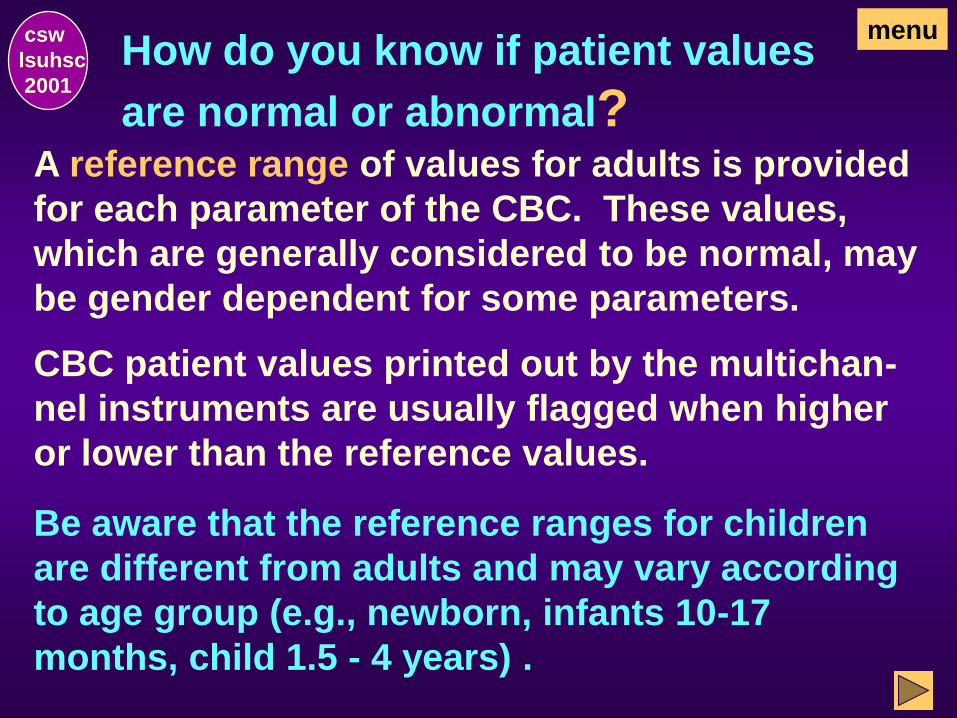
How do you know if patient values
are normal or abnormal?
csw
lsuhsc
2001
CBC patient values printed out by the multichan-
nel instruments are usually flagged when higher
or lower than the reference values.
Be aware that the reference ranges for children
are different from adults and may vary according
to age group (e.g., newborn, infants 10-17
months, child 1.5 - 4 years) .
menu
A reference range of values for adults is provided
for each parameter of the CBC. These values,
which are generally considered to be normal, may
be gender dependent for some parameters.
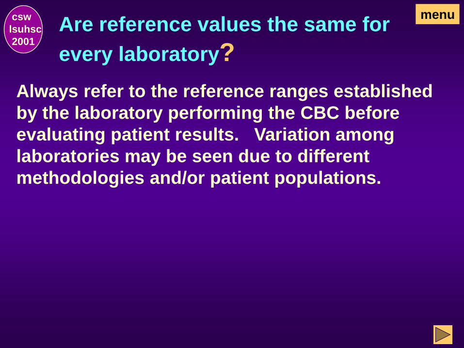
Are reference values the same for
every laboratory?
csw
lsuhsc
2001
Always refer to the reference ranges established
by the laboratory performing the CBC before
evaluating patient results. Variation among
laboratories may be seen due to different
methodologies and/or patient populations.
menu
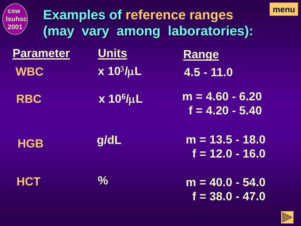
Examples of reference ranges
(may vary among laboratories):
Parameter Range Units
WBC x 103/mL 4.5 - 11.0
RBC m = 4.60 - 6.20
f = 4.20 - 5.40 x 10⁶/mL
HGB m = 13.5 - 18.0
f = 12.0 - 16.0 g/dL
HCT
csw
lsuhsc
2001
m = 40.0 - 54.0
f = 38.0 - 47.0
%
menu
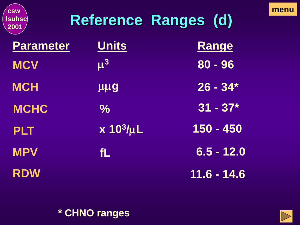
Reference Ranges (d)
MCV
Parameter Range Units
80 - 96 m3
MCH 26 - 34* mmg
MCHC 31 - 37* %
PLT 150 - 450 x 103/mL
MPV 6.5 - 12.0 fL
RDW 11.6 - 14.6
csw
lsuhsc
2001
menu
* CHNO ranges
End of Introduction
csw
lsuhsc
2001
This concludes the Introduction to the
Hemogram Section. Select one of the
following:
Go to Total WBC Count, the next section, to
continue with the exercise as designed.
Return to the Hemogram Menu and make an
alternate selection.
OR

Total WBC Count
csw
lsuhsc
2001
menu
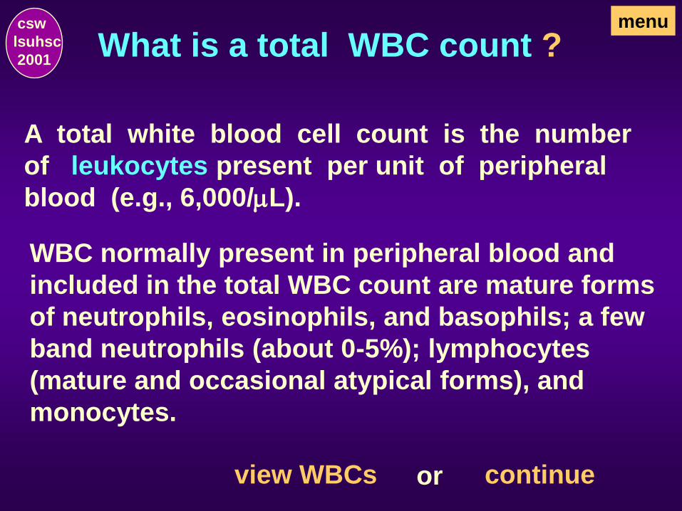
What is a total WBC count ? csw
lsuhsc
2001
A total white blood cell count is the number
of leukocytes present per unit of peripheral
blood (e.g., 6,000/mL).
continue
WBC normally present in peripheral blood and
included in the total WBC count are mature forms
of neutrophils, eosinophils, and basophils; a few
band neutrophils (about 0-5%); lymphocytes
(mature and occasional atypical forms), and
monocytes.
view WBCs or
menu
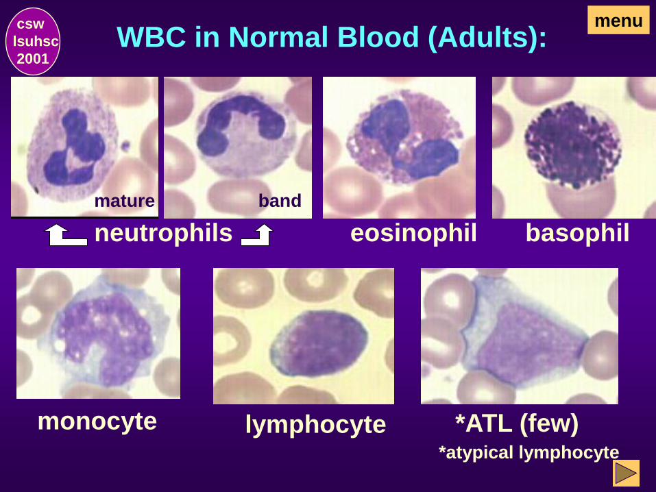
WBC in Normal Blood (Adults):
eosinophil basophil
monocyte lymphocyte *ATL (few) *atypical lymphocyte
csw
lsuhsc
2001
menu
neutrophils
band mature
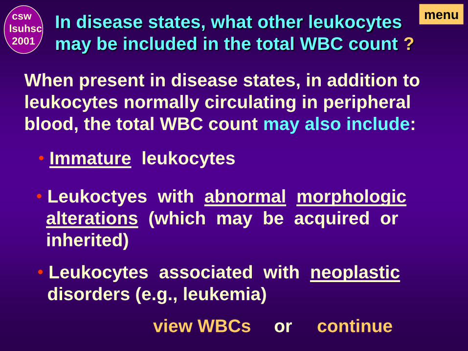
In disease states, what other leukocytes
may be included in the total WBC count ?
csw
lsuhsc
2001
When present in disease states, in addition to
leukocytes normally circulating in peripheral
blood, the total WBC count may also include:
• Immature leukocytes
• Leukoctyes with abnormal morphologic
alterations (which may be acquired or
inherited)
• Leukocytes associated with neoplastic
disorders (e.g., leukemia)
continue view WBCs or
menu

Examples of immature WBC:
granulocytes
(various stages)
myeloblasts
lymphoblasts
monoblasts
csw
lsuhsc
2001
menu
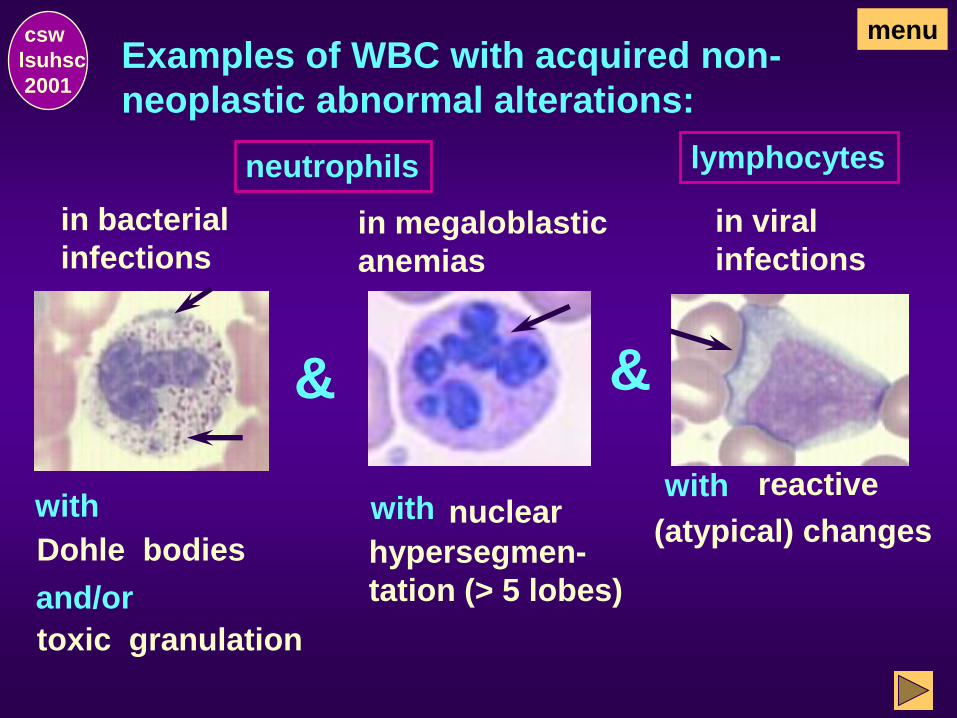
Examples of WBC with acquired non-
neoplastic abnormal alterations:
neutrophils
in bacterial
infections
with
Dohle bodies
and/or
toxic granulation
&
in megaloblastic
anemias
with nuclear
hypersegmen-
tation (> 5 lobes)
&
lymphocytes
in viral
infections
with reactive
(atypical) changes
csw
lsuhsc
2001
menu
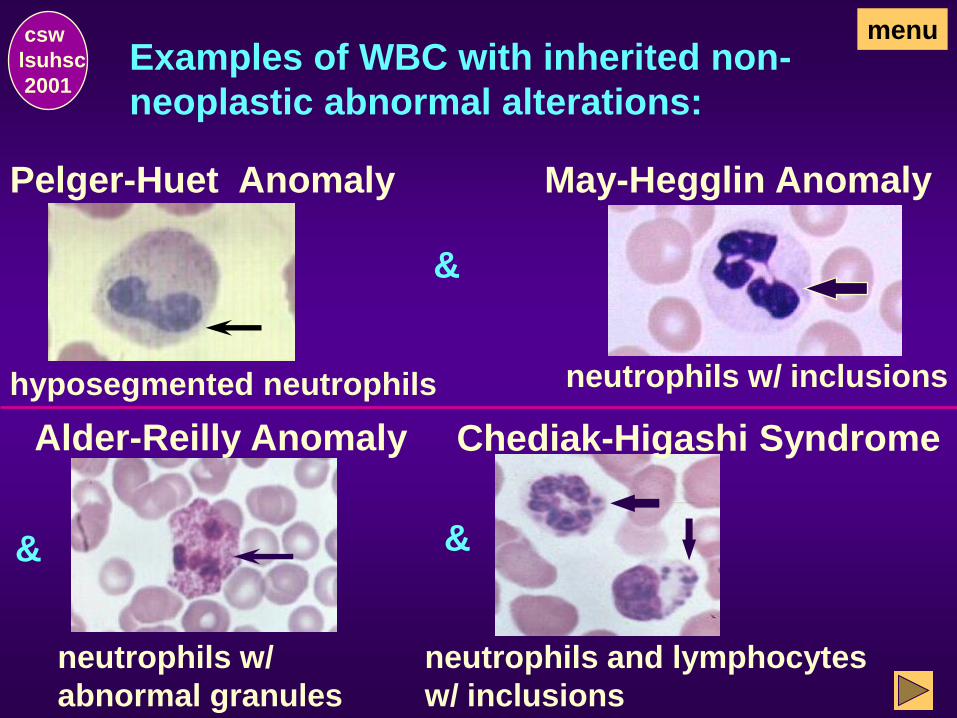
Examples of WBC with inherited non-
neoplastic abnormal alterations:
Pelger-Huet Anomaly
hyposegmented neutrophils
&
May-Hegglin Anomaly
neutrophils w/ inclusions
&
Alder-Reilly Anomaly
neutrophils w/
abnormal granules
&
csw
lsuhsc
2001
Chediak-Higashi Syndrome
neutrophils and lymphocytes
w/ inclusions
menu
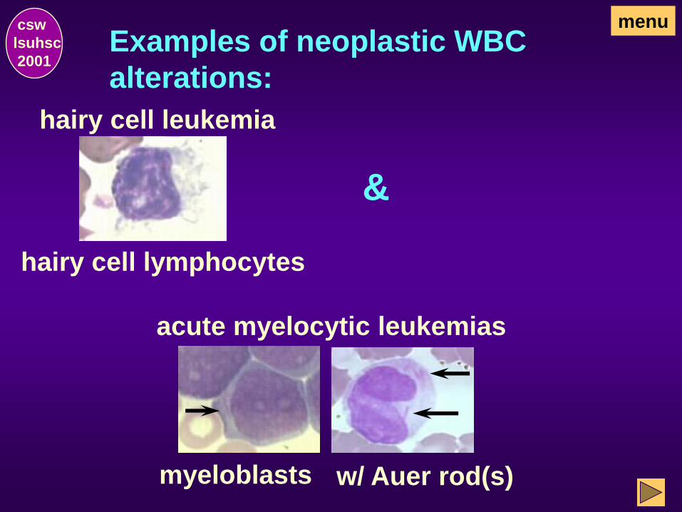
Examples of neoplastic WBC
alterations:
hairy cell lymphocytes
hairy cell leukemia
&
myeloblasts w/ Auer rod(s)
acute myelocytic leukemias
csw
lsuhsc
2001
menu
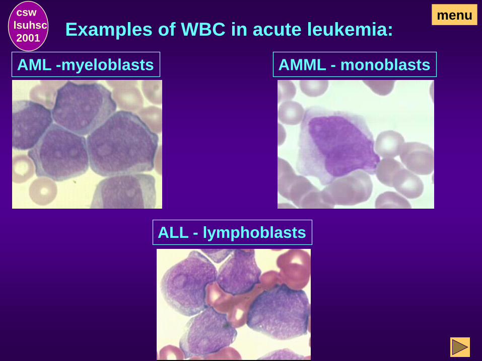
Examples of WBC in acute leukemia:
AML -myeloblasts
ALL - lymphoblasts
AMML - monoblasts
csw
lsuhsc
2001
menu
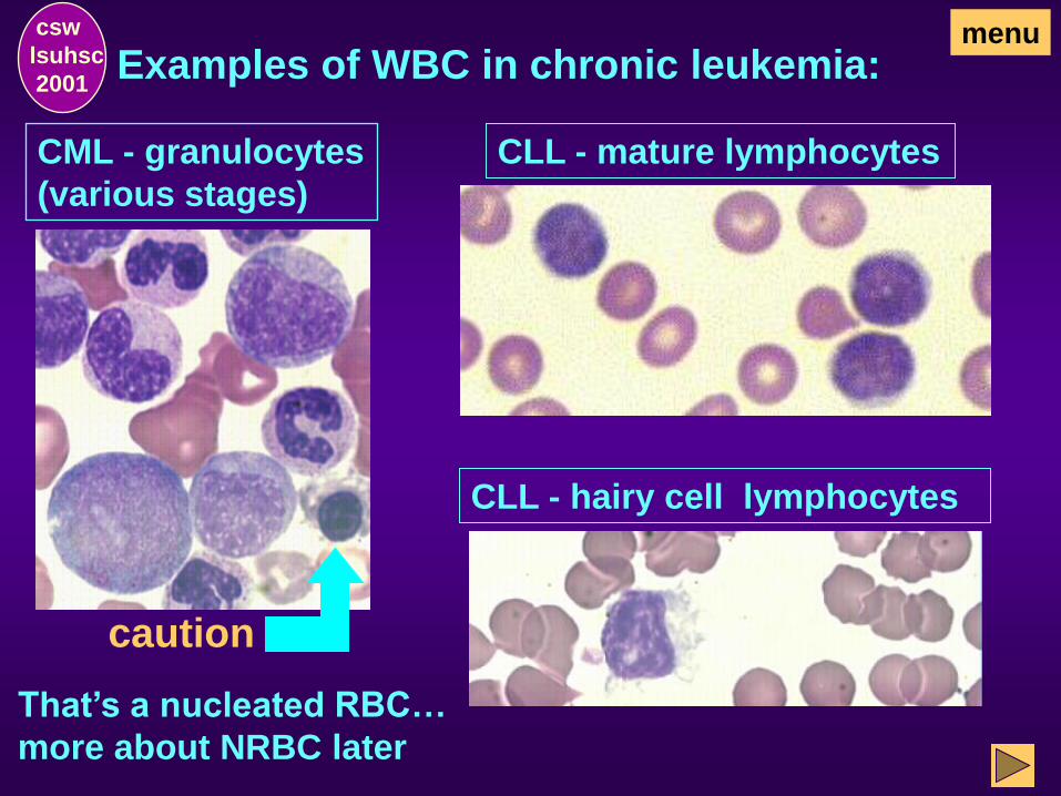
Examples of WBC in chronic leukemia:
CML - granulocytes
(various stages)
csw
lsuhsc
2001
CLL - mature lymphocytes
caution
CLL - hairy cell lymphocytes
menu
That’s a nucleated RBC…
more about NRBC later
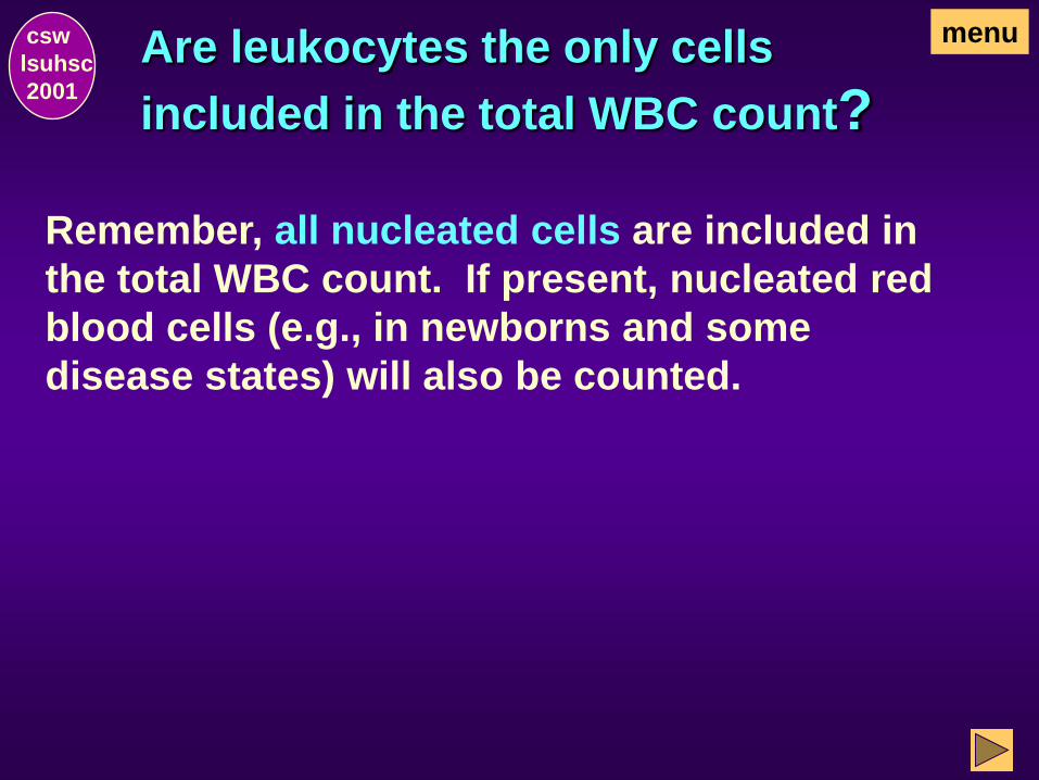
Are leukocytes the only cells
included in the total WBC count?
csw
lsuhsc
2001
Remember, all nucleated cells are included in
the total WBC count. If present, nucleated red
blood cells (e.g., in newborns and some
disease states) will also be counted.
menu

Examples of nucleated RBC in
various stages of maturation:
proerythroblast basophilic erythroblast
polychromatophilic
erythroblast
orthochromatophilic
erythroblast
csw
lsuhsc
2001
menu
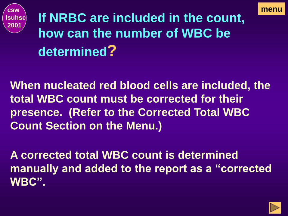
If NRBC are included in the count,
how can the number of WBC be
determined?
csw
lsuhsc
2001
When nucleated red blood cells are included, the
total WBC count must be corrected for their
presence. (Refer to the Corrected Total WBC
Count Section on the Menu.)
A corrected total WBC count is determined
manually and added to the report as a “corrected
WBC”.
menu
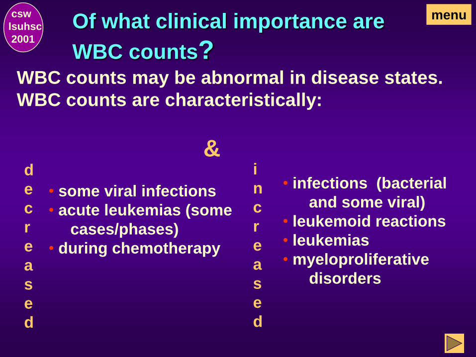
Of what clinical importance are
WBC counts? WBC counts may be abnormal in disease states.
WBC counts are characteristically:
d
e
c
r
e
a
s
e
d
& i
n
c
r
e
a
s
e
d
csw
lsuhsc
2001
menu
• infections (bacterial
and some viral)
• leukemoid reactions
• leukemias
• myeloproliferative
disorders
• some viral infections
• acute leukemias (some
cases/phases)
• during chemotherapy
Does the total WBC count differentiate
WBC as to cell line?
csw
lsuhsc
2001
No, the total WBC count is the total number of
all nucleated cells. In the case of abnormal total
WBC counts, a differential WBC count must be
performed before it can be determined which
cell line is decreased or increased.
It is also important to determine whether the
increase/decrease is a relative percent or
absolute number, which is discussed in the
Differential WBC section presented later.
menu
End of Total WBC Count
csw
lsuhsc
2001
This concludes the Total WBC Count
Section. Select one of the following:
Go to Corrected WBC Count, the next section,
to continue with the exercise as designed.
Return to the Hemogram Menu and make an
alternate selection.
OR

Corrected WBC Count
(for presence of NRBC)
csw
lsuhsc
2001
menu
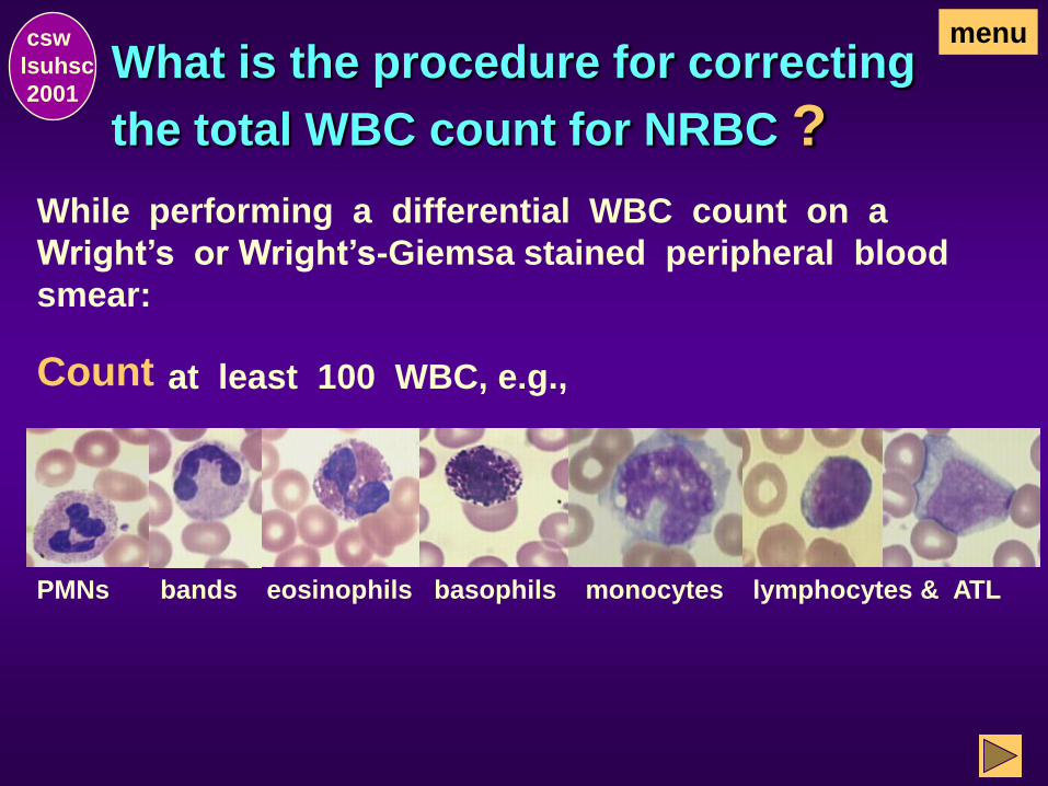
What is the procedure for correcting
the total WBC count for NRBC ?
While performing a differential WBC count on a
Wright’s or Wright’s-Giemsa stained peripheral blood
smear:
Count at least 100 WBC, e.g.,
csw
lsuhsc
2001
menu
PMNs bands eosinophils basophils monocytes lymphocytes & ATL
…procedure for correcting the total WBC count
for NRBC continued…
csw
lsuhsc
2001
menu
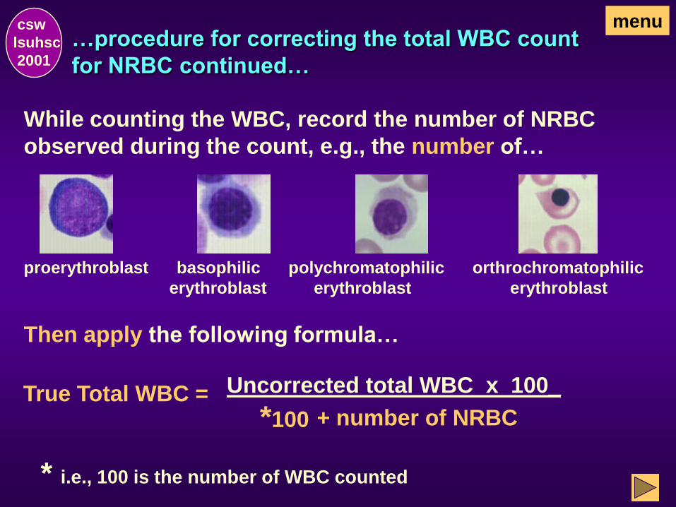
While counting the WBC, record the number of NRBC
observed during the count, e.g., the number of…
proerythroblast basophilic polychromatophilic orthrochromatophilic
erythroblast erythroblast erythroblast
Then apply the following formula…
True Total WBC = Uncorrected total WBC x 100_
*100 + number of NRBC
* i.e., 100 is the number of WBC counted
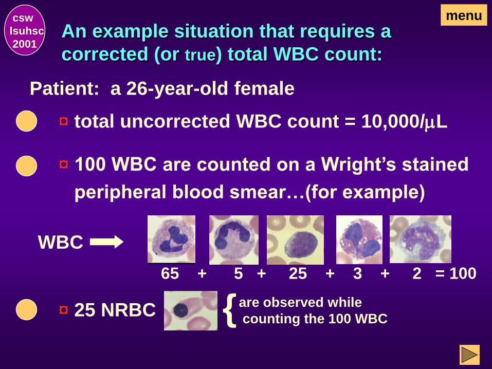
An example situation that requires a
corrected (or true) total WBC count:
csw
lsuhsc
2001
¤ 100 WBC are counted on a Wright’s stained
peripheral blood smear…(for example)
¤ 25 NRBC
Patient: a 26-year-old female
¤ total uncorrected WBC count = 10,000/mL
menu
WBC
65 + 5 + 25 + 3 + 2 = 100
{ are observed while
counting the 100 WBC

The corrected (or true) WBC count
is calculated:
Uncorrected total WBC count = 10,000/mL
NRBC = 25/100 WBC
(i.e., 25 NRBC were noted per 100 WBC counted on
a stained peripheral blood smear)
True/Corrected WBC Count
10,000 x 100
100 + 25 = 1,000,000
125 = 8,000/mL
csw
lsuhsc
2001
menu
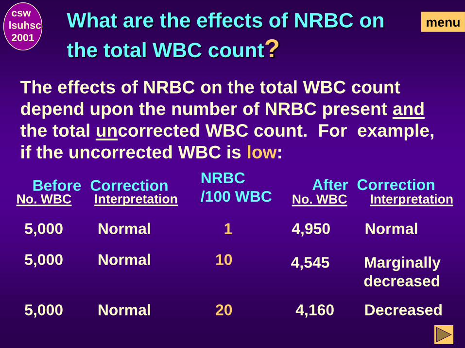
What are the effects of NRBC on
the total WBC count?
The effects of NRBC on the total WBC count
depend upon the number of NRBC present and
the total uncorrected WBC count. For example,
if the uncorrected WBC is low:
No. WBC
NRBC
/100 WBC Before Correction After Correction
Interpretation No. WBC Interpretation
5,000 Normal 1 4,950 Normal
5,000 Normal 10 4,545 Marginally
decreased
5,000 Normal 20 4,160 Decreased
csw
lsuhsc
2001
menu
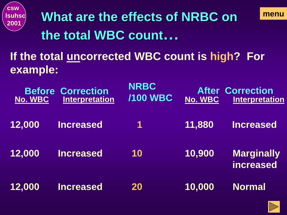
What are the effects of NRBC on
the total WBC count…
If the total uncorrected WBC count is high? For
example:
No. WBC
NRBC
/100 WBC Before Correction After Correction
Interpretation No. WBC Interpretation
12,000 Increased 1 11,880 Increased
12,000 Increased 10 10,900 Marginally
increased
12,000 Increased 20 10,000 Normal
csw
lsuhsc
2001
menu
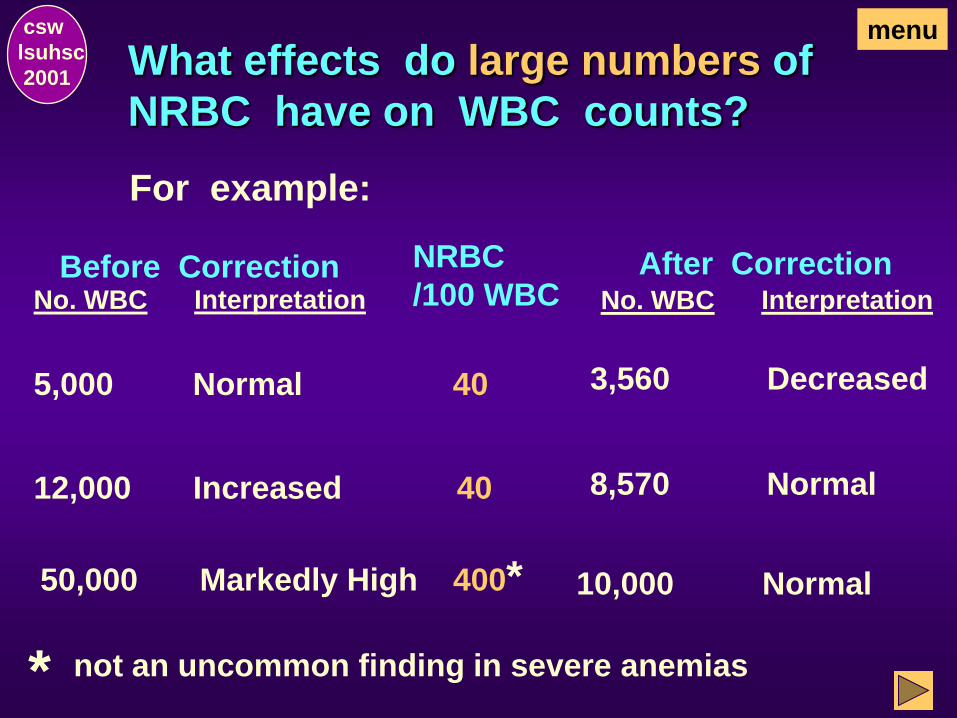
What effects do large numbers of
NRBC have on WBC counts?
For example:
No. WBC
NRBC
/100 WBC Before Correction After Correction
Interpretation No. WBC Interpretation
5,000 Normal 40 3,560 Decreased
12,000 Increased 40 8,570 Normal
50,000 Markedly High 400*
not an uncommon finding in severe anemias *
10,000 Normal
csw
lsuhsc
2001
menu
End of Corrected WBC Count
csw
lsuhsc
2001
This concludes the Corrected WBC
Section. Select one of the following:
Go to Total RBC Count, the next section, to
continue with the exercise as designed.
Return to the Hemogram Menu and make an
alternate selection.
OR
Total RBC Count
csw
lsuhsc
2001
menu
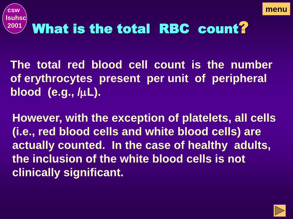
What is the total RBC count?
csw
lsuhsc
2001
The total red blood cell count is the number
of erythrocytes present per unit of peripheral
blood (e.g., /mL).
However, with the exception of platelets, all cells
(i.e., red blood cells and white blood cells) are
actually counted. In the case of healthy adults,
the inclusion of the white blood cells is not
clinically significant.
menu
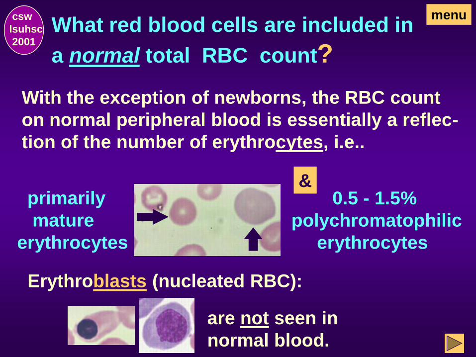
What red blood cells are included in
a normal total RBC count?
With the exception of newborns, the RBC count
on normal peripheral blood is essentially a reflec-
tion of the number of erythrocytes, i.e..
csw
lsuhsc
2001
primarily
mature
erythrocytes
& 0.5 - 1.5%
polychromatophilic
erythrocytes
Erythroblasts (nucleated RBC):
are not seen in
normal blood.
menu
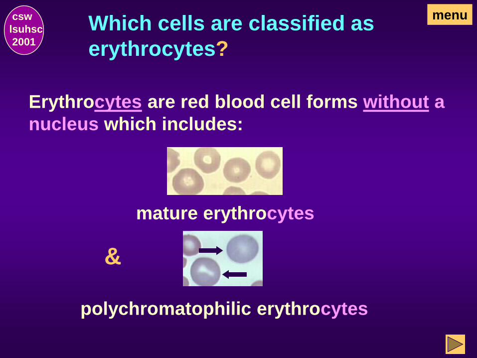
Erythrocytes are red blood cell forms without a
nucleus which includes:
csw
lsuhsc
2001
mature erythrocytes
&
polychromatophilic erythrocytes
Which cells are classified as
erythrocytes?
menu
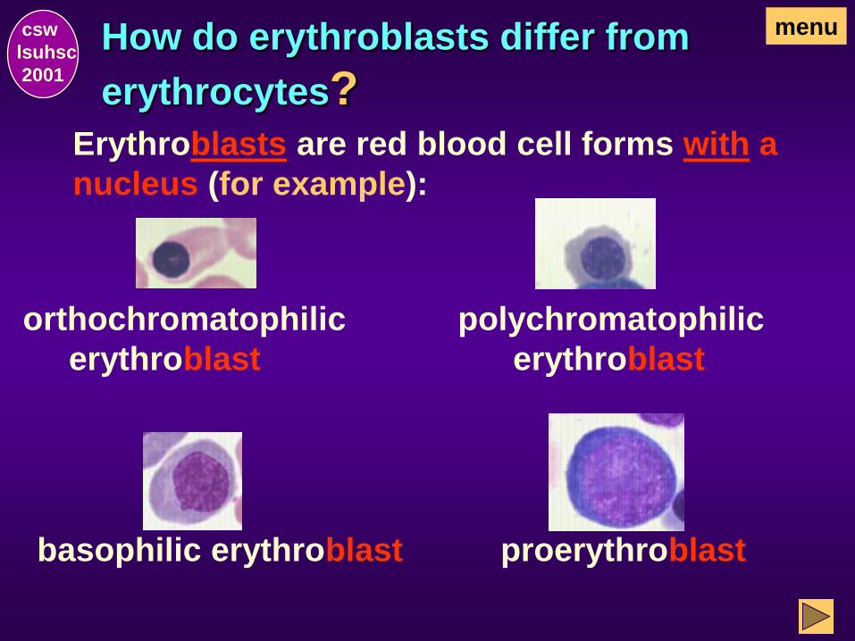
How do erythroblasts differ from
erythrocytes? Erythroblasts are red blood cell forms with a
nucleus (for example):
csw
lsuhsc
2001
orthochromatophilic
erythroblast
polychromatophilic
erythroblast
basophilic erythroblast proerythroblast
menu
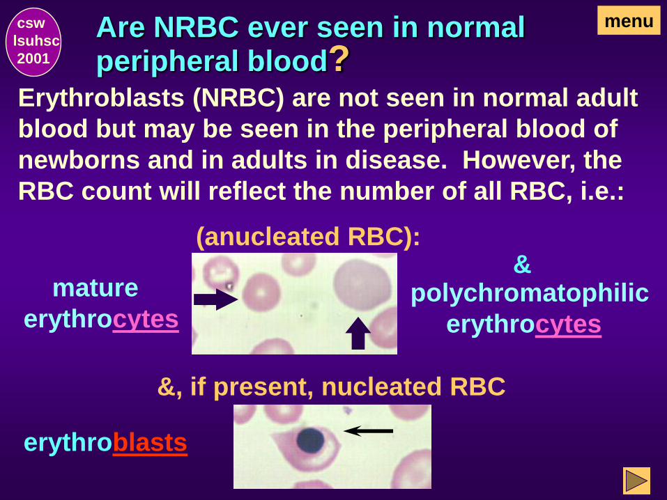
Are NRBC ever seen in normal peripheral blood?
Erythroblasts (NRBC) are not seen in normal adult
blood but may be seen in the peripheral blood of
newborns and in adults in disease. However, the
RBC count will reflect the number of all RBC, i.e.:
(anucleated RBC):
mature
erythrocytes polychromatophilic
erythrocytes
&, if present, nucleated RBC
erythroblasts
csw
lsuhsc
2001
menu
&
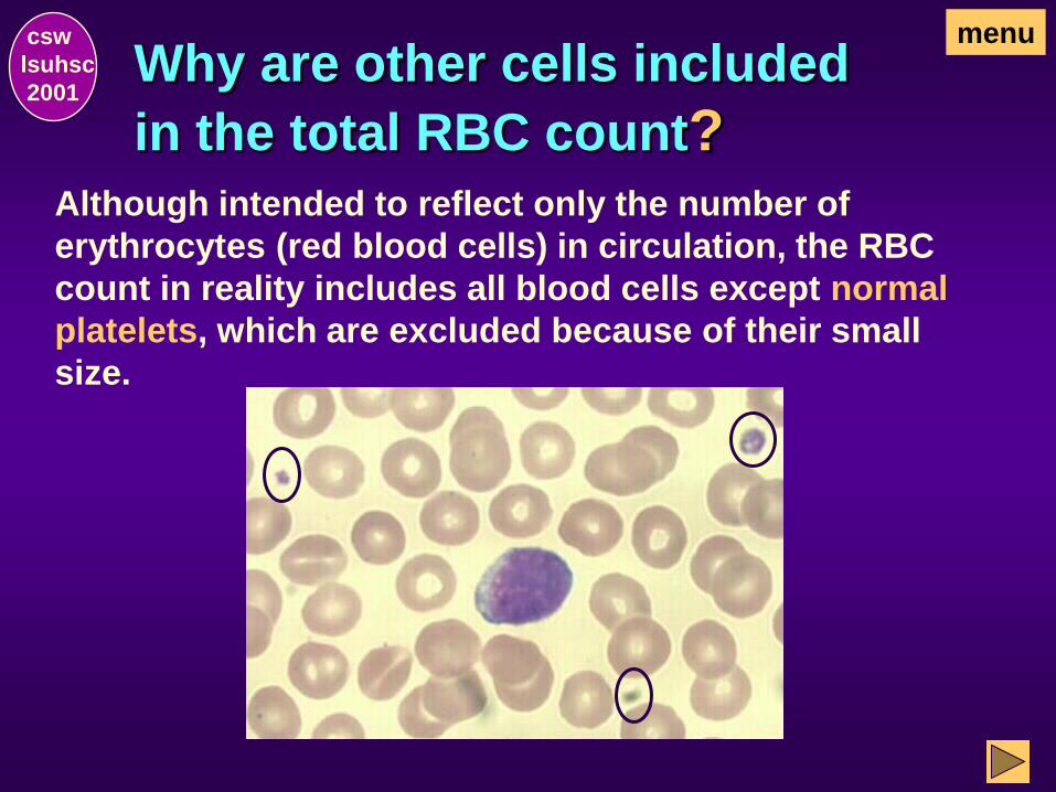
Why are other cells included
in the total RBC count? Although intended to reflect only the number of
erythrocytes (red blood cells) in circulation, the RBC
count in reality includes all blood cells except normal
platelets, which are excluded because of their small
size.
csw
lsuhsc
2001
menu
Why are other cells included
in the total RBC count?
csw
lsuhsc
2001
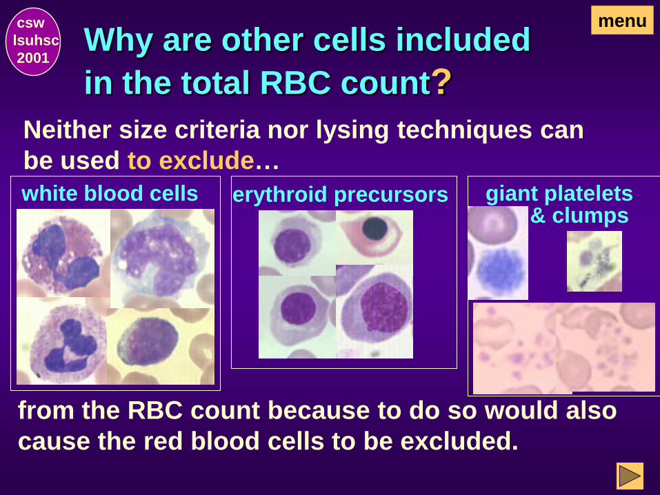
Neither size criteria nor lysing techniques can
be used to exclude…
menu
from the RBC count because to do so would also
cause the red blood cells to be excluded.
white blood cells erythroid precursors & clumps
giant platelets
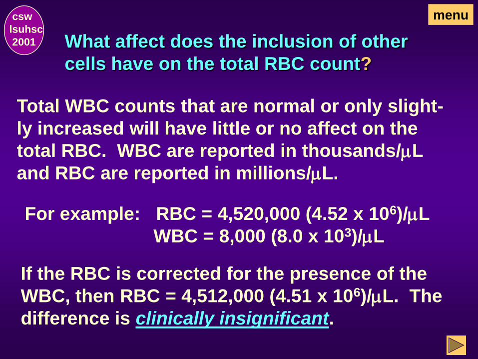
What affect does the inclusion of other
cells have on the total RBC count?
Total WBC counts that are normal or only slight-
ly increased will have little or no affect on the
total RBC. WBC are reported in thousands/mL
and RBC are reported in millions/mL.
csw
lsuhsc
2001
For example: RBC = 4,520,000 (4.52 x 10⁶)/mL
WBC = 8,000 (8.0 x 10³)/mL
If the RBC is corrected for the presence of the
WBC, then RBC = 4,512,000 (4.51 x 10⁶)/mL. The
difference is clinically insignificant.
menu
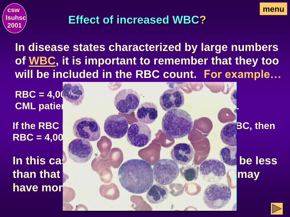
Effect of increased WBC?
In disease states characterized by large numbers
of WBC, it is important to remember that they too
will be included in the RBC count.
In this case, the true RBC population may be less
than that indicated by the RBC count and may
have more clinical significance.
csw
lsuhsc
2001
RBC = 4,000,000 (4.00 x 10⁶)/mL
CML patient with WBC = 300,000 (300.0 x 10³)/mL
If the RBC is corrected for the presence of the WBC, then
RBC = 4,000,000 – 300,000 = 3,700,000/mL.
menu
For example…

Effect of NRBC in disease states?
In disease states characterized by large numbers of
NRBC (e.g., thalassemia), it is important to remember
that they too will be included in the RBC count.
csw
lsuhsc
2001
NRBC = 400/100 WBC (a patient with thalassemia major)
RBC = 3,000,000/mL
Total WBC uncorrected for NRBC = 100,000/mL
menu
If the RBC is corrected for the presence of the
uncorrected WBC (which includes the NRBC), then…
RBC = 3,000,000 - 100,000 = 2,900,000/mL
In this case, the true RBC population may be less than
that indicated by the RBC count and may have more
clinical significance.
For example…
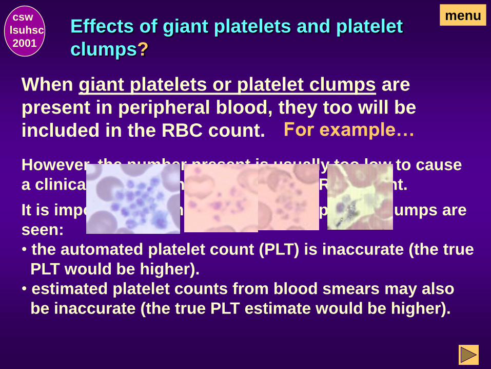
Effects of giant platelets and platelet
clumps?
When giant platelets or platelet clumps are
present in peripheral blood, they too will be
included in the RBC count.
csw
lsuhsc
2001
menu
However, the number present is usually too low to cause
a clinically significant decrease in the RBC count.
It is important to remember that when platelet clumps are
seen:
• the automated platelet count (PLT) is inaccurate (the true
PLT would be higher).
• estimated platelet counts from blood smears may also
be inaccurate (the true PLT estimate would be higher).
For example…
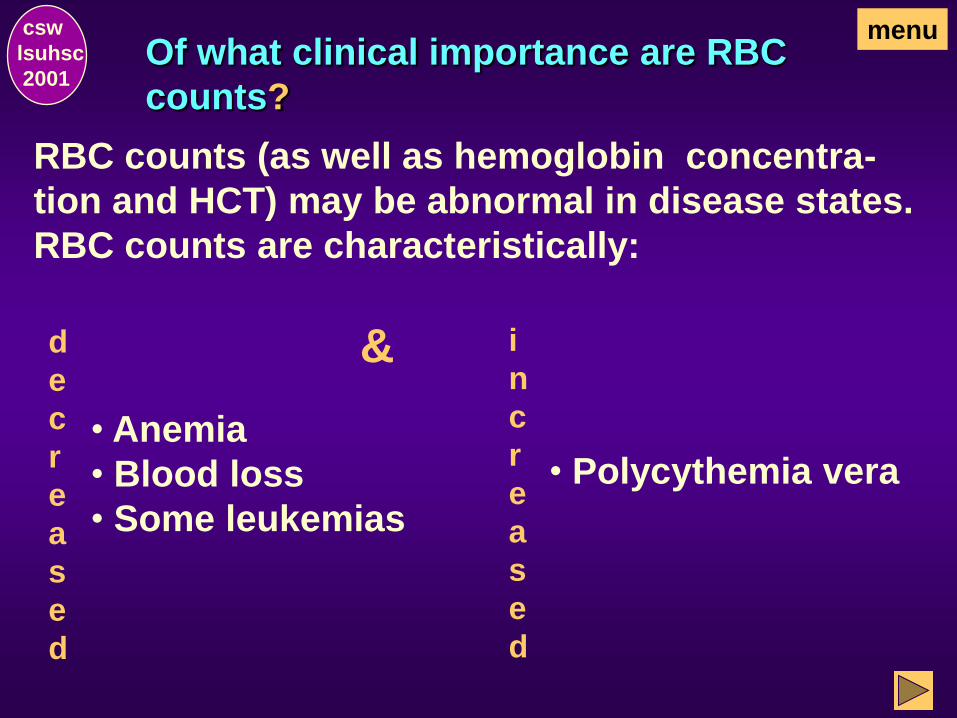
Of what clinical importance are RBC
counts?
RBC counts (as well as hemoglobin concentra-
tion and HCT) may be abnormal in disease states.
RBC counts are characteristically:
d
e
c
r
e
a
s
e
d
• Anemia
• Blood loss
• Some leukemias
& i
n
c
r
e
a
s
e
d
• Polycythemia vera
csw
lsuhsc
2001
menu
End of Total RBC Count
csw
lsuhsc
2001
This concludes the Total RBC Count
Section. Select one of the following:
Go to Hemoglobin, the section, to with the
exercise as designed.
Return to the Hemogram Menu and make an
alternate selection.
OR
Hemoglobin
csw
lsuhsc
2001
menu
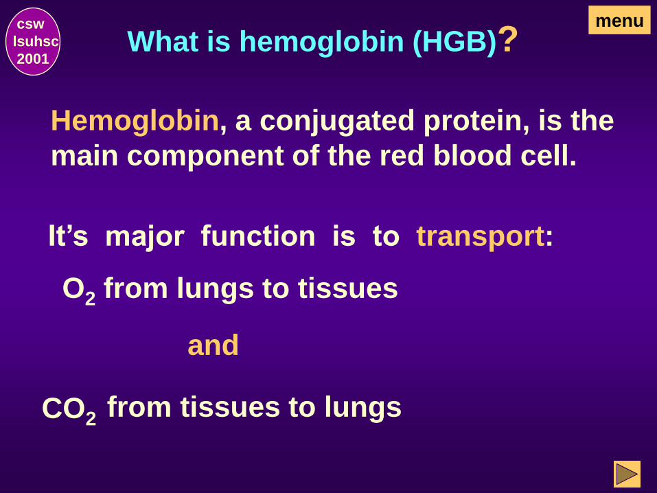
What is hemoglobin (HGB)?
Hemoglobin, a conjugated protein, is the
main component of the red blood cell.
It’s major function is to transport:
from lungs to tissues O2
and
CO2 from tissues to lungs
csw
lsuhsc
2001
menu

How does HGB transport O2 and CO2?
from
lungs
(high
tension)
Hb
O2
in RBC
and
in exchange from
tissues CO2
transported
Hb
CO2
in RBC
csw
lsuhsc
2001
to
tissues
(eg, hand)
(low
tension)
O2 transported
back
to
lungs
menu
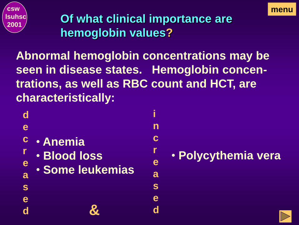
Of what clinical importance are
hemoglobin values?
Abnormal hemoglobin concentrations may be
seen in disease states. Hemoglobin concen-
trations, as well as RBC count and HCT, are
characteristically:
d
e
c
r
e
a
s
e
d
• Anemia
• Blood loss
• Some leukemias
&
i
n
c
r
e
a
s
e
d
• Polycythemia vera
csw
lsuhsc
2001
menu
End of Hemoglobin
csw
lsuhsc
2001
This concludes the Hemoglobin Sec-
tion. Select one of the following:
Go to Hematocrit, the next section, to continue
with the exercise as designed.
Return to the Hemogram Menu and make an
alternate selection.
OR
menu

Hematocrit
csw
lsuhsc
2001
menu
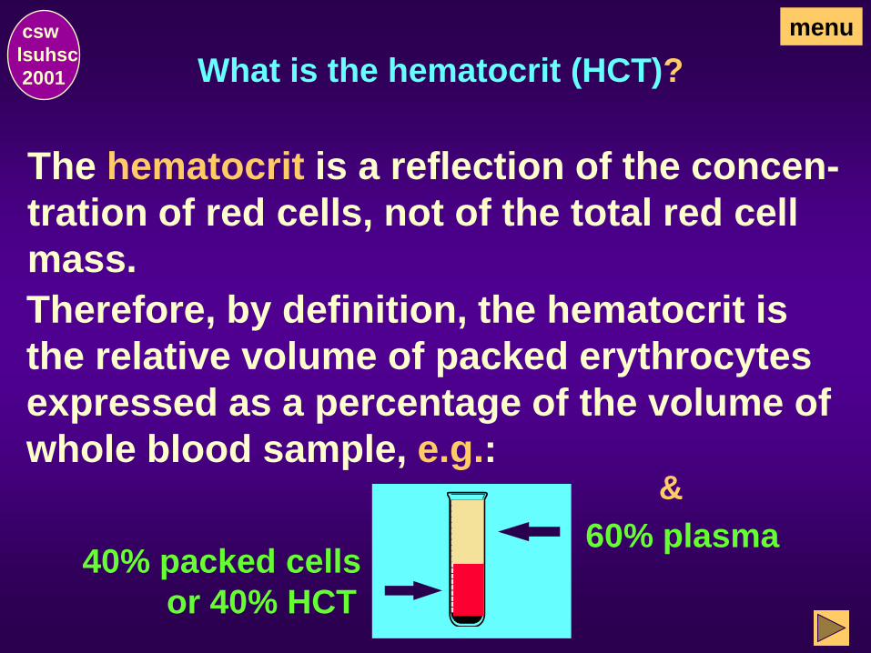
What is the hematocrit (HCT)?
The hematocrit is a reflection of the concen-
tration of red cells, not of the total red cell
mass.
Therefore, by definition, the hematocrit is
the relative volume of packed erythrocytes
expressed as a percentage of the volume of
whole blood sample, e.g.:
40% packed cells
or 40% HCT
&
60% plasma
csw
lsuhsc
2001
menu
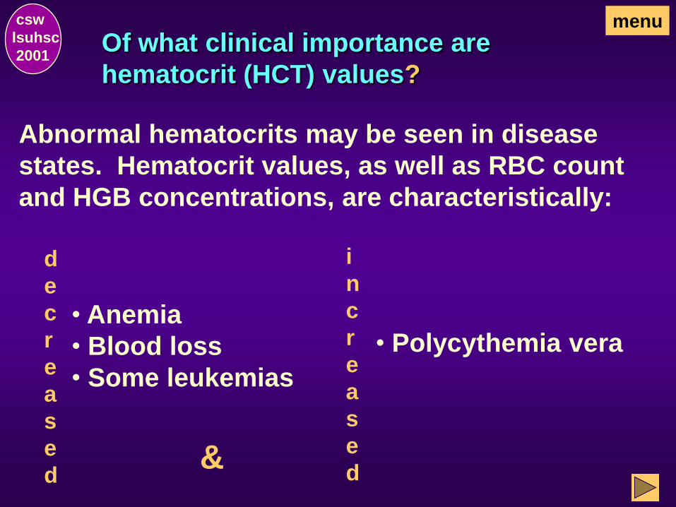
Of what clinical importance are
hematocrit (HCT) values?
Abnormal hematocrits may be seen in disease
states. Hematocrit values, as well as RBC count
and HGB concentrations, are characteristically:
d
e
c
r
e
a
s
e
d
• Anemia
• Blood loss
• Some leukemias
&
i
n
c
r
e
a
s
e
d
• Polycythemia vera
csw
lsuhsc
2001
menu
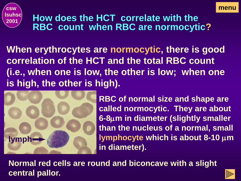
How does the HCT correlate with the RBC count when RBC are normocytic?
When erythrocytes are normocytic, there is good
correlation of the HCT and the total RBC count
(i.e., when one is low, the other is low; when one
is high, the other is high).
RBC of normal size and shape are
called normocytic. They are about
6-8mm in diameter (slightly smaller
than the nucleus of a normal, small
lymphocyte which is about 8-10 mm
in diameter). lymph
csw
lsuhsc
2001
menu
Normal red cells are round and biconcave with a slight
central pallor.

How does the HCT correlate with the RBC
count when the RBCs are macrocytic?
csw
lsuhsc
2001
lymphocyte
the total RBC count may be lower than expected
based upon HCT values.
menu
because the RBC are larger In macrocytosis, than normal,
(normocytic cells)

How does the HCT correlate with the RBC
count when the RBC are microcytic?
In microcytosis,
lymph
because the cells are smaller
csw
lsuhsc
2001
the total RBC count may be higher than expected
based upon HCT values.
menu
than normal,
lymphocyte
(normocytic cells)
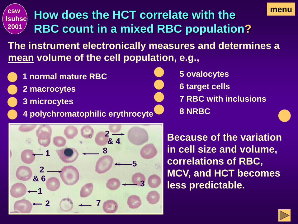
How does the HCT correlate with the
RBC count in a mixed RBC population?
The instrument electronically measures and determines a
mean volume of the cell population, e.g.,
1
csw
lsuhsc
2001
1 normal mature RBC
2 macrocytes
3 microcytes
4 polychromatophilic erythrocyte
5 ovalocytes
6 target cells
7 RBC with inclusions
8 NRBC
Because of the variation
in cell size and volume,
correlations of RBC,
MCV, and HCT becomes
less predictable.
menu
3
& 4
& 6
7
8
2
2
2
1
1
5
What are some examples of true
HCT/RBC inconsistencies?
In hydremia of pregnancy:
the HCT is low although there is no reduction in
the total number of circulating red cells.
In shock accompanied by hemoconcentration:
the HCT may be normal or even high though
blood loss may have caused a considerable
decrease in the total red cell mass.
csw
lsuhsc
2001
menu
and
What is the correlation between
HCT and hemoglobin (HGB)?
HCT (expressed in per cent) is usually roughly
3 times the HGB (expressed in g/dL).
For example:
An adult with hematocrit of 45% would normally
have a hemoglobin of about 15 g/dL.
Further discussion:
The relationships between the HCT and
total RBC count and the HCT and HGB
concentration are discussed further in the
section on RBC indices.
csw
lsuhsc
2001
menu
End of Hematocrit
csw
lsuhs
c
2001
This concludes the Hematocrit Sec-
tion. Select one of the following:
Go to Erythrocyte (RBC) Indices, the next
section to continue with the exercise as
designed.
Return to the Hemogram Menu and make an
alternate selection.
OR
menu
Erythrocyte (RBC) Indices
csw
lsuhsc
2001
menu
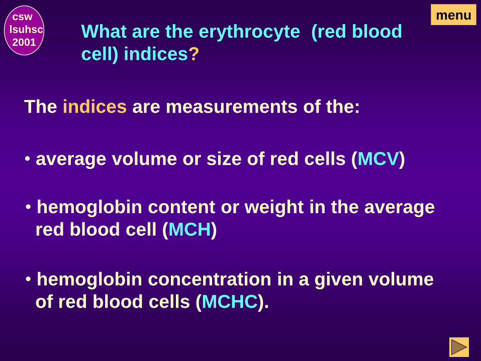
What are the erythrocyte (red blood
cell) indices?
csw
lsuhsc
2001
The indices are measurements of the:
• average volume or size of red cells (MCV)
• hemoglobin content or weight in the average
red blood cell (MCH)
• hemoglobin concentration in a given volume
of red blood cells (MCHC).
menu

Of what clinical value are the erythrocyte
(red blood cell) indices?
csw
lsuhsc
2001
size of the red cells (MCV)
relationship between individual blood cells and the
hemoglobin concentration (MCH)
red blood cell population as a whole and the
hemoglobin concentration (MCHC).
menu
The indices are valuable tools in the study of anemias
because they provide an objective quantitative standard
for assessing the
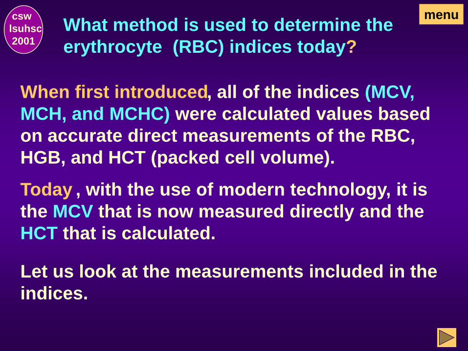
What method is used to determine the
erythrocyte (RBC) indices today?
csw
lsuhsc
2001
, all of the indices (MCV,
MCH, and MCHC) were calculated values based
on accurate direct measurements of the RBC,
HGB, and HCT (packed cell volume).
, with the use of modern technology, it is
the MCV that is now measured directly and the
HCT that is calculated.
Let us look at the measurements included in the
indices.
menu
When first introduced
Today
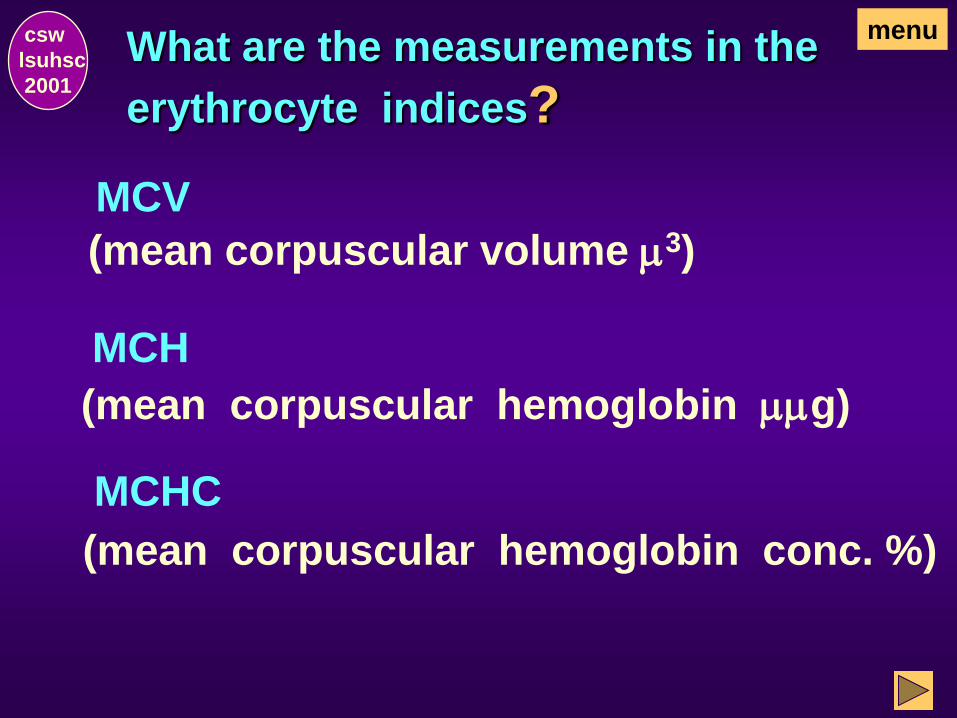
What are the measurements in the
erythrocyte indices?
MCV
(mean corpuscular volume m3)
MCH
(mean corpuscular hemoglobin mmg)
MCHC
(mean corpuscular hemoglobin conc. %)
csw
lsuhsc
2001
menu

What is the MCV? csw
lsuhsc
2001
MCV is the average volume (or size) of red cells
expressed in m3 or femtoliters (fL).
Individuals with normal peripheral blood will
have a normal MCV (i.e., 80-100 m3).
menu
The normal small mature lymphocyte, because it is
relatively consistent in size with a nucleus that is about
10-12m in diameter, is a useful tool in a visual assess-
ment of red cell size on stained blood smears.)
normal RBC
(6-8m)
lymphocyte
(nucleus 10-12m)
10-12m

What affect do abnormally large or
small red cells have on the MCV?
csw
lsuhsc
2001
Individuals with red cells that are predominantly larger
than normal (>8m diameter) will have an MCV >100 m3.
Individuals with red cells that are predominantly smaller
than normal (<6m diameter) will have an MCV < 80 m3.
normal low
high normal
menu

E.g., macrocytic and normocytic RBC: it is important to
remember that the MCV is the average volume (or size) of
the cells. For example, if an MCV is high, it does not mean
that all of the red cells are larger. Some of the cells may
be normal or even smaller than normal. It is an indication,
however, that large cells are present in sufficient numbers
to cause an increased MCV, e.g..
csw
lsuhsc
2001
x
x x
x x
x
x
x
x
x
What if there are various sizes of red cells?
menu
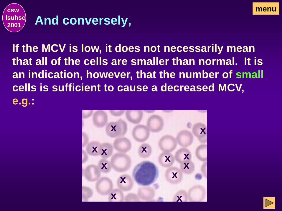
And conversely, csw
lsuhsc
2001
If the MCV is low, it does not necessarily mean
that all of the cells are smaller than normal. It is
an indication, however, that the number of small
cells is sufficient to cause a decreased MCV,
e.g.:
x
x x
x
x
x
x
x x
x
x
x
x
menu
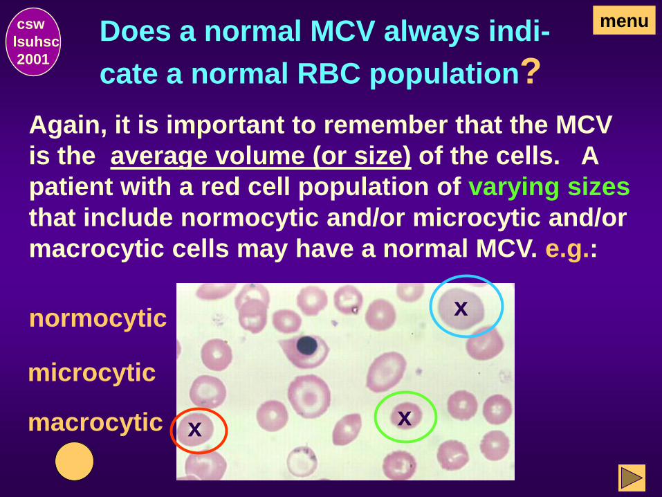
Does a normal MCV always indi-
cate a normal RBC population?
csw
lsuhsc
2001
Again, it is important to remember that the MCV
is the average volume (or size) of the cells. A
patient with a red cell population of varying sizes
that include normocytic and/or microcytic and/or
macrocytic cells may have a normal MCV. e.g.:
x
normocytic
x
microcytic
x
macrocytic
menu
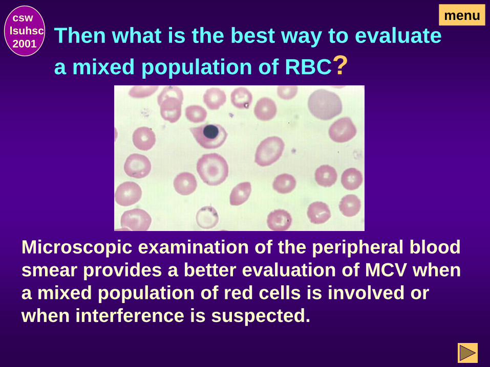
Then what is the best way to evaluate
a mixed population of RBC?
csw
lsuhsc
2001
Microscopic examination of the peripheral blood
smear provides a better evaluation of MCV when
a mixed population of red cells is involved or
when interference is suspected.
menu
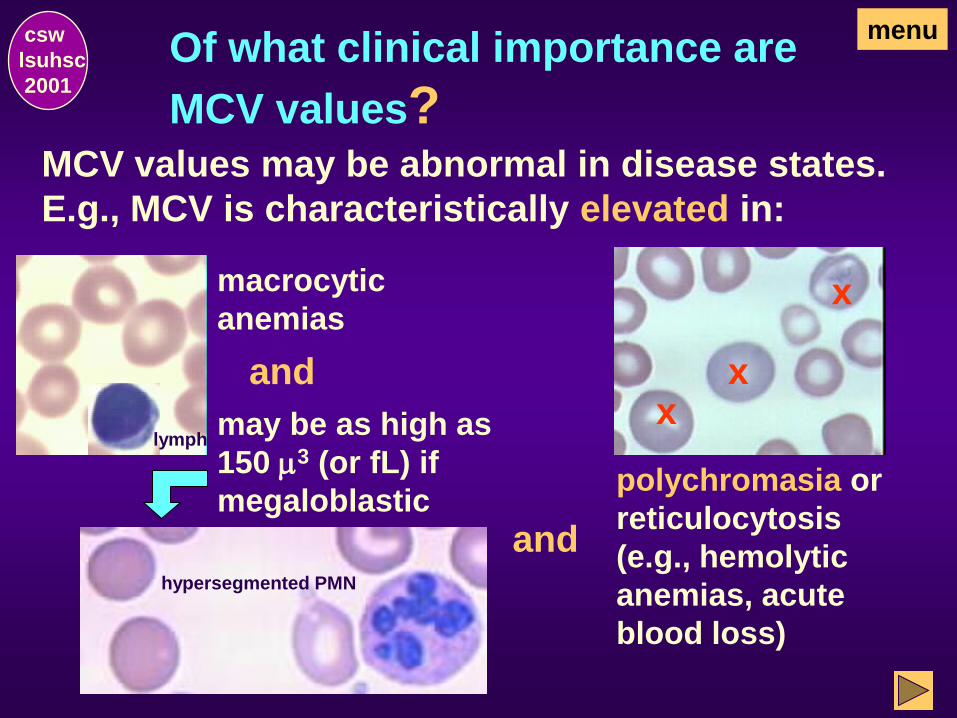
Of what clinical importance are
MCV values?
MCV values may be abnormal in disease states.
E.g., MCV is characteristically elevated in:
and
csw
lsuhsc
2001
polychromasia or
reticulocytosis
(e.g., hemolytic
anemias, acute
blood loss)
x x
x
menu
and
macrocytic
anemias
lymph may be as high as
150 m3 (or fL) if
megaloblastic
hypersegmented PMN
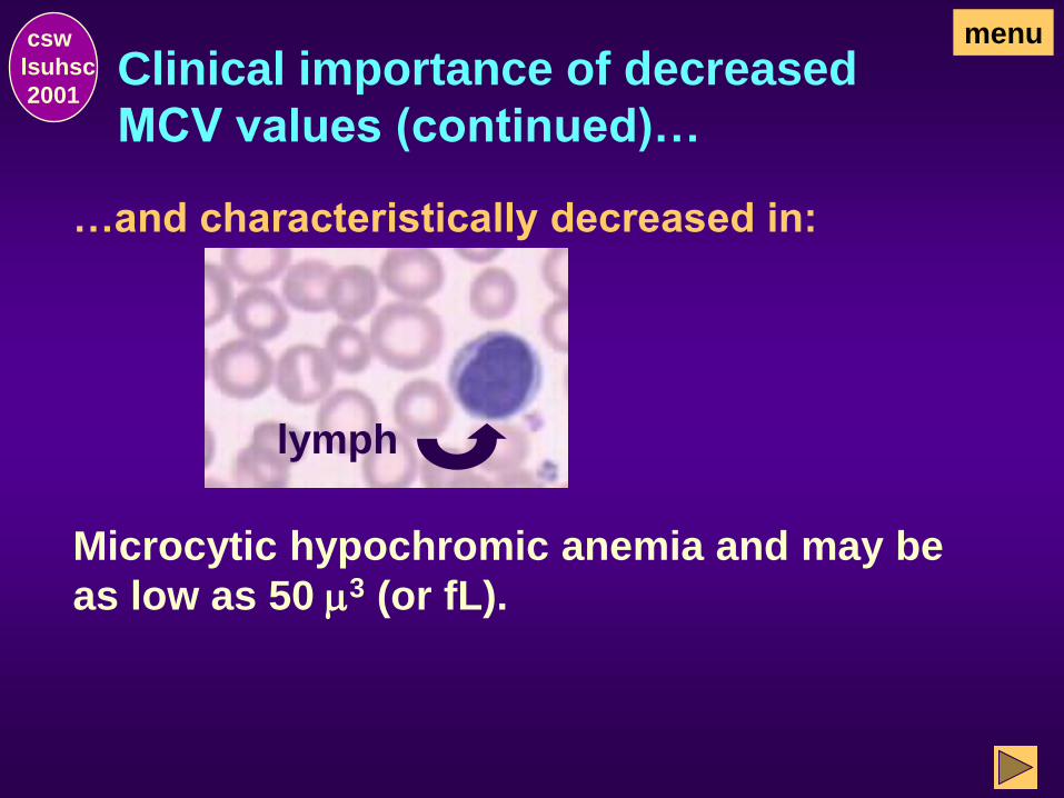
Clinical importance of decreased
MCV values (continued)…
…and characteristically decreased in:
csw
lsuhsc
2001
Microcytic hypochromic anemia and may be
as low as 50 m3 (or fL).
lymph
menu
What is the second measurement
included in the RBC indices?
csw
lsuhsc
2001
In addition to the:
MCV (mean corpuscular volume m3)
There is the MCH.
menu
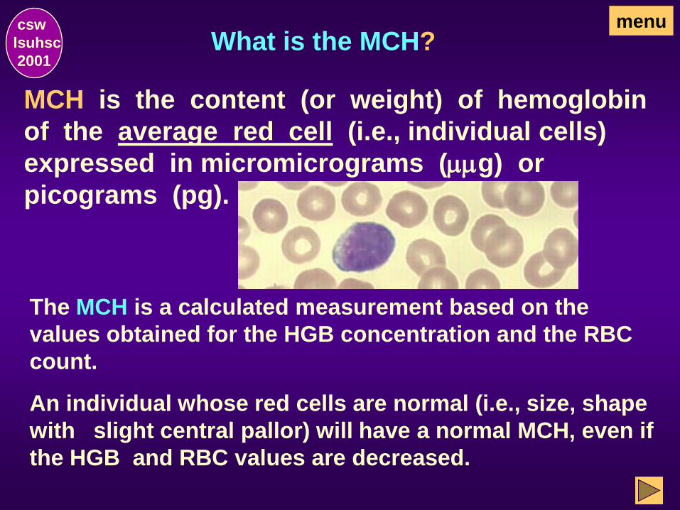
What is the MCH?
MCH is the content (or weight) of hemoglobin
of the average red cell (i.e., individual cells)
expressed in micromicrograms (mmg) or
picograms (pg).
The MCH is a calculated measurement based on the
values obtained for the HGB concentration and the RBC
count.
csw
lsuhsc
2001
An individual whose red cells are normal (i.e., size, shape
with slight central pallor) will have a normal MCH, even if
the HGB and RBC values are decreased.
menu
What about individuals with a
decreased MCH?
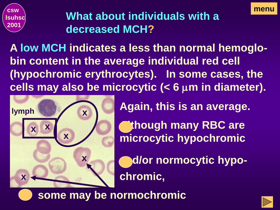
A low MCH indicates a less than normal hemoglo-
bin content in the average individual red cell
(hypochromic erythrocytes). In some cases, the
cells may also be microcytic (< 6 mm in diameter).
lymph Again, this is an average.
Although many RBC are
microcytic hypochromic
some may be normochromic
csw
lsuhsc
2001
x x
and/or normocytic hypo-
chromic, x
x
x
x
menu
Of what clinical importance are MCH
values?
MCH values may be abnormal in disease states.
E.g., MCH is characteristically elevated in:
csw
lsuhsc
2001
menu
macrocytic
anemias
lymph may be as high as
50 mmg (or pg) if
megaloblastic
hypersegmented PMN
and
polychromasia or
reticulocytosis
(e.g., hemolytic
anemias, acute
blood loss)
x x
x
and

Clinical importance of MCH values,
(continued)
Characteristically decreased in:
csw
lsuhsc
2001
Microcytic hypochromic anemia and may be as
low as 15 mmg (or pg).
lymph
menu
What is the third measurement
included in the RBC indices?
csw
lsuhsc
2001
In addition to the:
MCV (mean corpuscular volume m3) and
MCH(mean corpuscular hemoglobin mmg)
There is the MCHC.
menu
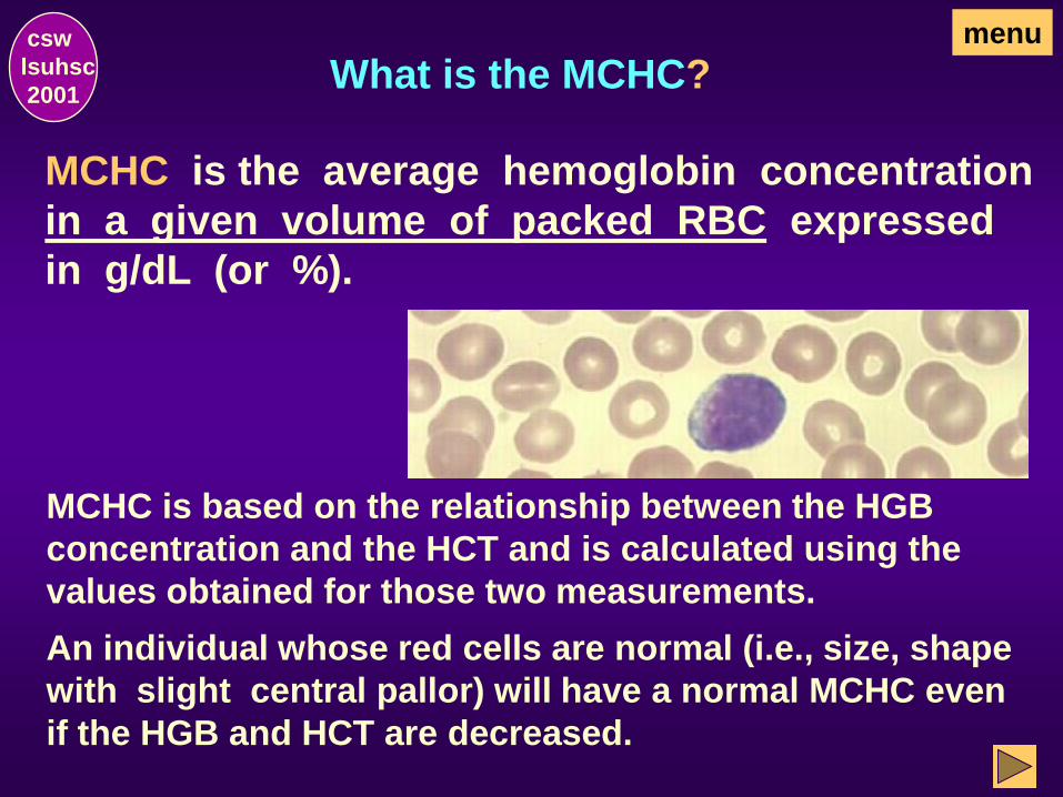
What is the MCHC? csw
lsuhsc
2001
MCHC is the average hemoglobin concentration
in a given volume of packed RBC expressed
in g/dL (or %).
MCHC is based on the relationship between the HGB
concentration and the HCT and is calculated using the
values obtained for those two measurements.
menu
An individual whose red cells are normal (i.e., size, shape
with slight central pallor) will have a normal MCHC even
if the HGB and HCT are decreased.

Of what clinical importance is the
MCHC value?
MCHC values may be abnormal in disease
states.
A true elevated MCHC is seen only in
spherocytosis.
csw
lsuhsc
2001
menu
x x
x x
x
x x
x
x x
x x x
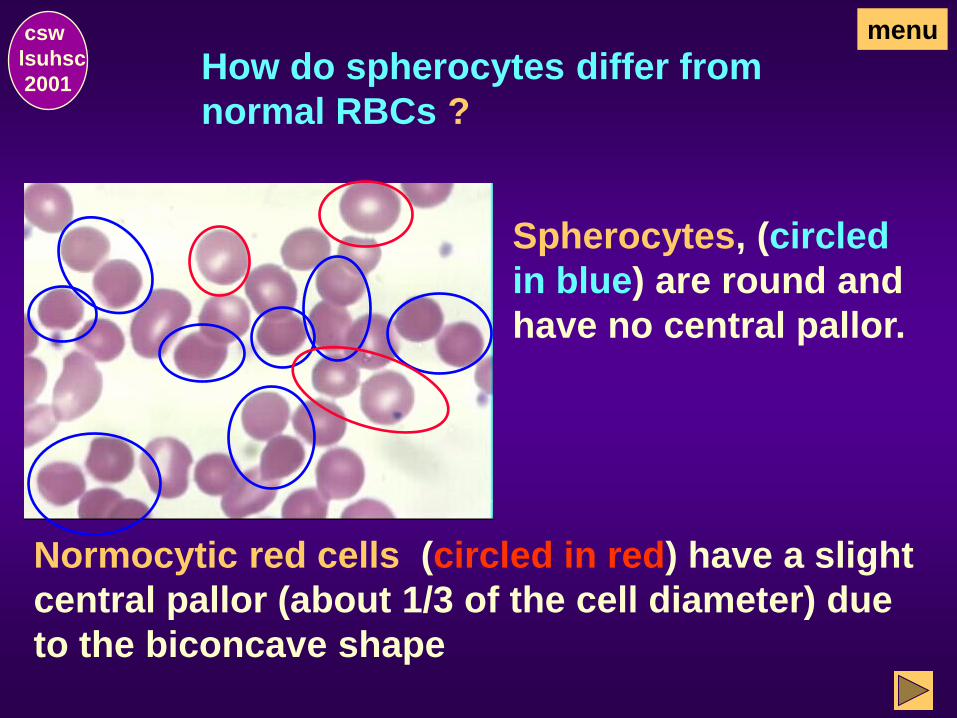
How do spherocytes differ from
normal RBCs ?
Spherocytes, (circled
in blue) are round and
have no central pallor.
Normocytic red cells (circled in red) have a slight
central pallor (about 1/3 of the cell diameter) due
to the biconcave shape
csw
lsuhsc
2001
menu
Why is a true elevated MCHC
seen only in spherocytosis?
csw
lsuhsc
2001
It’s the only situation
in which the cells are
spherical and,
therefore, have a
greater capacity for
hemoglobin than
biconcave cells.
menu
What other disorders are characterized
by an MCHC?
In the absence of spherocytosis, an
elevated MCHC may be an indication of
A falsely elevated MCV and decrease in
HCT (e.g., caused by cold agglutinins)
or
Falsely elevated HGB (e.g., lipemia or
some other interfering factor).
csw
lsuhsc
2001
menu
What disorders are characterized by a
MCHC?
Normal or decreased:
macrocytic anemias
Decreased:
hypochromic anemia (usually no
lower than 22 %)
Normal MCHC but reduced HGB & HCT:
normochromic anemias
csw
lsuhsc
2001
menu
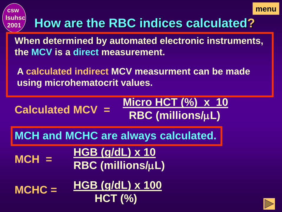
How are the RBC indices calculated?
MCH =
MCHC =
csw
lsuhsc
2001
Micro HCT (%) x 10
RBC (millions/mL)
HGB (g/dL) x 10
RBC (millions/mL)
HGB (g/dL) x 100
HCT (%)
When determined by automated electronic instruments,
the MCV is a direct measurement.
A calculated indirect MCV measurment can be made
using microhematocrit values.
MCH and MCHC are always calculated.
Calculated MCV =
menu
End of RBC Indices
csw
lsuhsc
2001
This concludes the Erythrocyte (RBC) Indices
Section. Select one of the following:
Go to Red Cell Distribution Width (RDW), the next
section, to continue with the exercise as
designed.
Return to the Hemogram Menu and make an
alternate selection.
OR

Red Cell Distribution Width
csw
lsuhsc
2001
menu
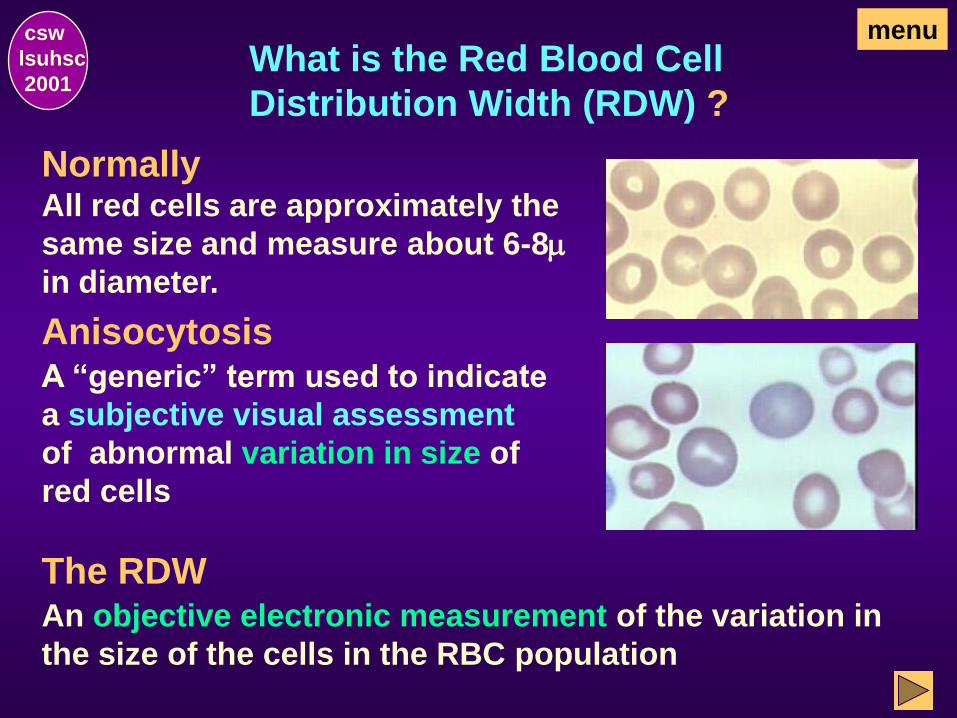
What is the Red Blood Cell
Distribution Width (RDW) ?
Normally All red cells are approximately the
same size and measure about 6-8m
in diameter.
A “generic” term used to indicate
a subjective visual assessment
of abnormal variation in size of
red cells
An objective electronic measurement of the variation in
the size of the cells in the RBC population
csw
lsuhsc
2001
Anisocytosis
The RDW
menu
How is the RDW determined?
Based on data obtained by electronic measurement of the
sizes of the red cells, the RDW is calculated by
enumerating the number of erythrocytes that are:
Thus, based upon objective measurements, the
RDW provides an estimate of anisocytosis (i.e.,
variation in size of the red cells).
csw
lsuhsc
2001
normal MCV MCV
menu
or larger (normal) cell volume (size)
than the reference
MCV
smaller
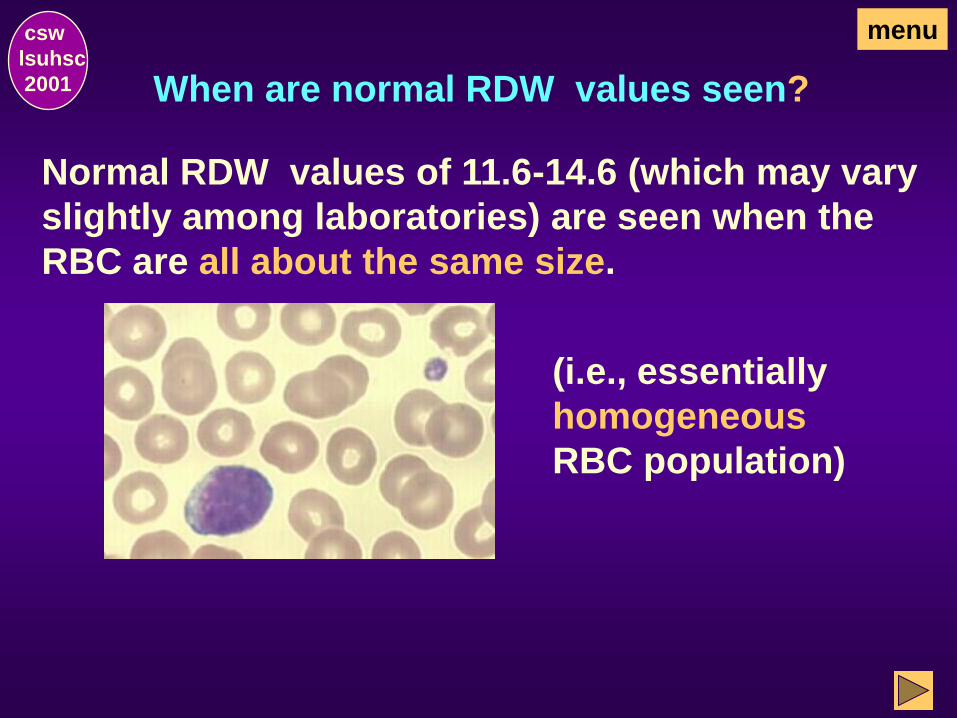
When are normal RDW values seen?
Normal RDW values of 11.6-14.6 (which may vary
slightly among laboratories) are seen when the
RBC are all about the same size.
csw
lsuhsc
2001
(i.e., essentially
homogeneous
RBC population)
menu
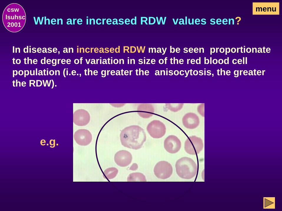
When are increased RDW values seen?
In disease, an increased RDW may be seen proportionate
to the degree of variation in size of the red blood cell
population (i.e., the greater the anisocytosis, the greater
the RDW).
csw
lsuhsc
2001
menu
e.g.
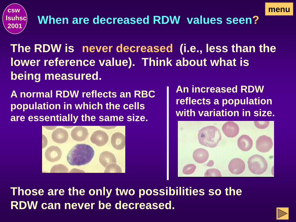
When are decreased RDW values seen? csw
lsuhsc
2001
An increased RDW
reflects a population
with variation in size.
The RDW is (i.e., less than the
lower reference value). Think about what is
being measured.
Those are the only two possibilities so the
RDW can never be decreased.
A normal RDW reflects an RBC
population in which the cells
are essentially the same size.
never decreased
menu
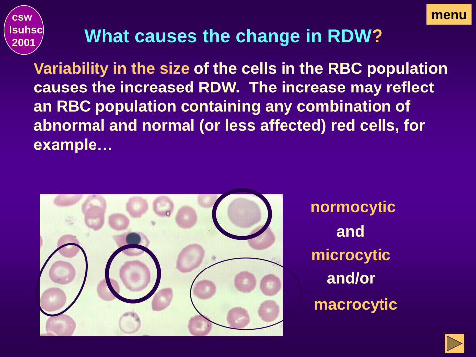
What causes the change in RDW?
Variability in the size of the cells in the RBC population
causes the increased RDW. The increase may reflect
an RBC population containing any combination of
abnormal and normal (or less affected) red cells, for
example…
normocytic
microcytic
and
macrocytic
and/or
csw
lsuhsc
2001
menu
What is the clinical importance of the RDW?
RDW is the most sensitive measurement involving
red cells.
RDW is the first to become abnormal (sometimes before
anemia appears).
RDW is the first to become abnormal in iron deficiency
due to chronic blood loss.
csw
lsuhsc
2001
menu
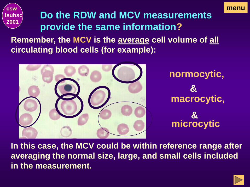
Do the RDW and MCV measurements
provide the same information?
csw
lsuhsc
2001
Remember, the MCV is the average cell volume of all
circulating blood cells (for example):
normocytic,
& macrocytic,
& microcytic
In this case, the MCV could be within reference range after
averaging the normal size, large, and small cells included
in the measurement.
menu
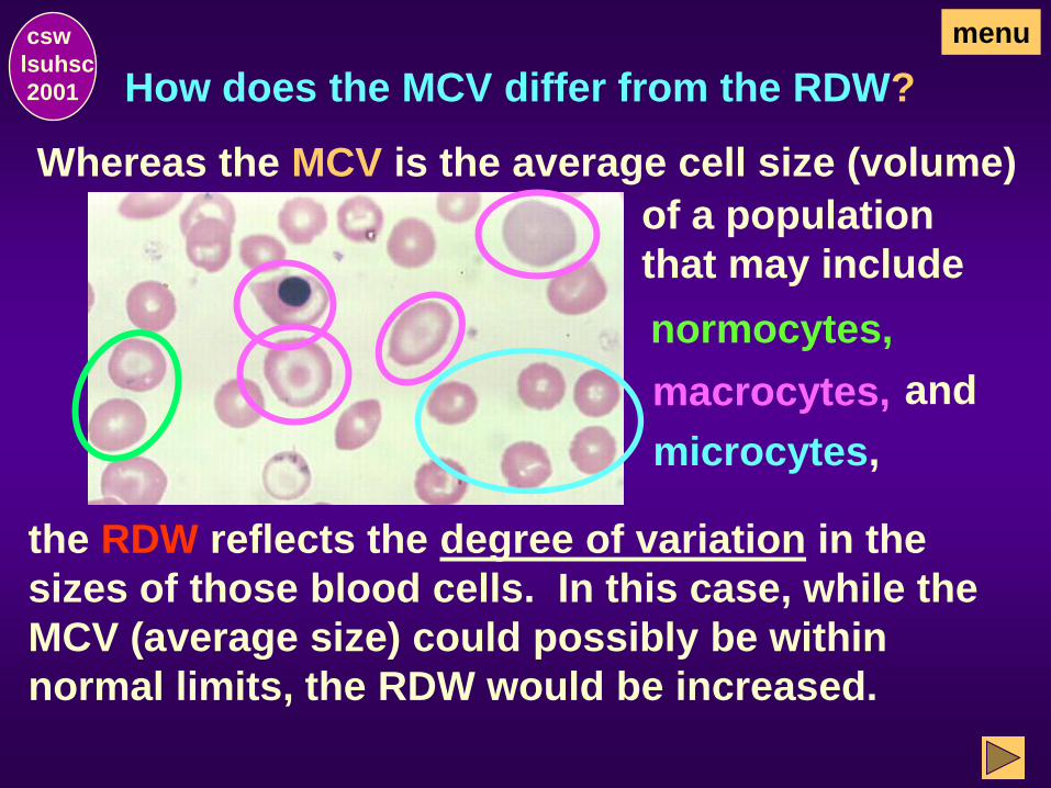
How does the MCV differ from the RDW?
csw
lsuhsc
2001
the RDW reflects the degree of variation in the
sizes of those blood cells. In this case, while the
MCV (average size) could possibly be within
normal limits, the RDW would be increased.
Whereas the MCV is the average cell size (volume)
of a population
that may include
normocytes,
macrocytes,
microcytes,
and
menu
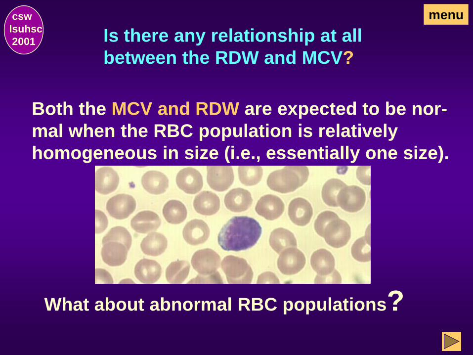
Is there any relationship at all
between the RDW and MCV?
csw
lsuhsc
2001
Both the MCV and RDW are expected to be nor-
mal when the RBC population is relatively
homogeneous in size (i.e., essentially one size).
What about abnormal RBC populations?
menu
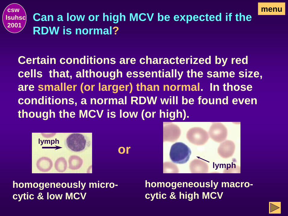
Can a low or high MCV be expected if the
RDW is normal?
csw
lsuhsc
2001
Certain conditions are characterized by red
cells that, although essentially the same size,
are smaller (or larger) than normal. In those
conditions, a normal RDW will be found even
though the MCV is low (or high).
homogeneously micro-
cytic & low MCV
lymph
or
homogeneously macro-
cytic & high MCV
lymph
menu
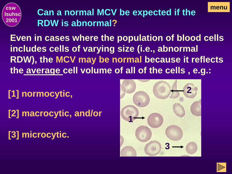
Can a normal MCV be expected if the
RDW is abnormal?
csw
lsuhsc
2001
Even in cases where the population of blood cells
includes cells of varying size (i.e., abnormal
RDW), the MCV may be normal because it reflects
the average cell volume of all of the cells , e.g.:
[1] normocytic,
[2] macrocytic, and/or
[3] microcytic.
menu
1
2
3
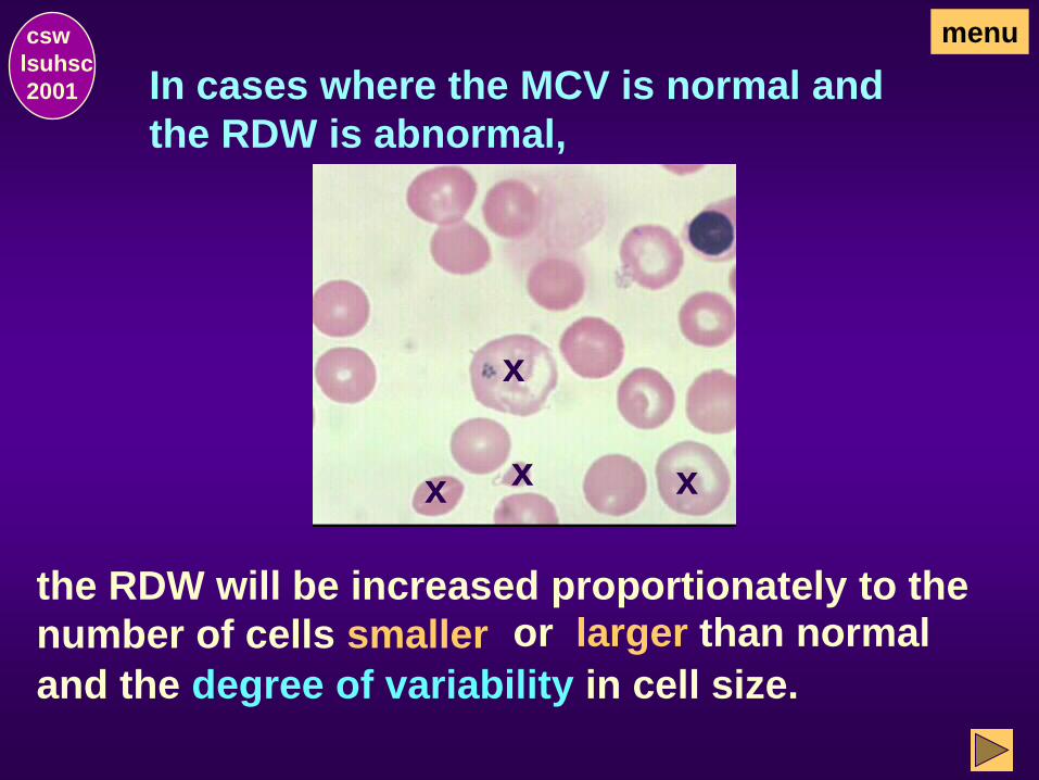
In cases where the MCV is normal and
the RDW is abnormal,
csw
lsuhsc
2001
the RDW will be increased proportionately to the
number of cells smaller
menu
and the degree of variability in cell size.
or larger than normal
x x
x
x
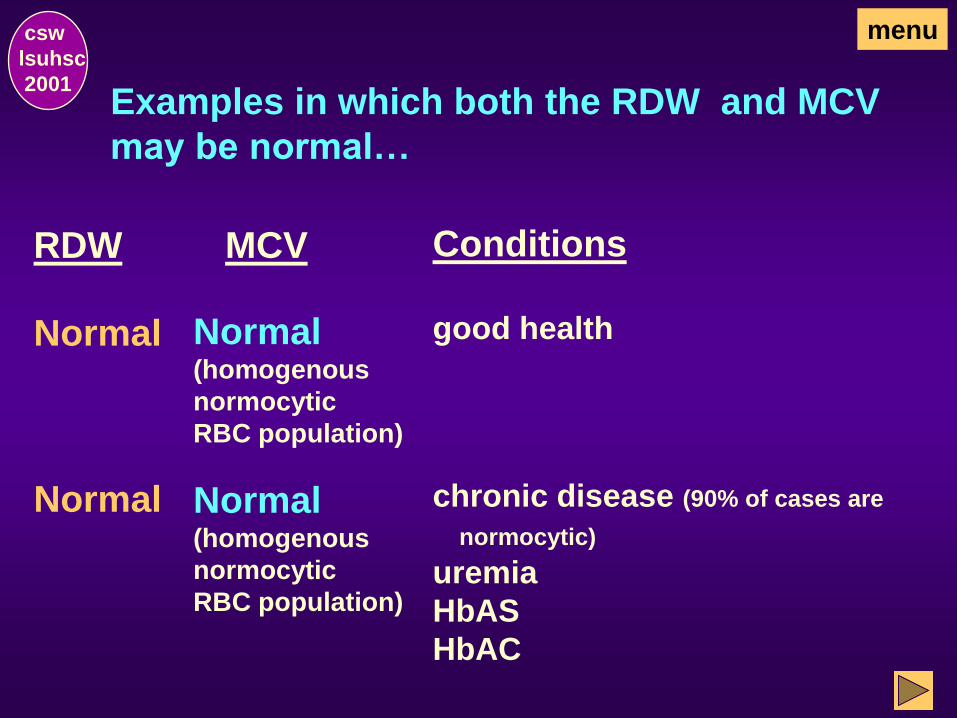
Examples in which both the RDW and MCV
may be normal…
RDW MCV Conditions
Normal chronic disease (90% of cases are
normocytic)
uremia
HbAS
HbAC
Normal (homogenous
normocytic
RBC population)
Normal Normal (homogenous
normocytic
RBC population)
good health
csw
lsuhsc
2001
menu
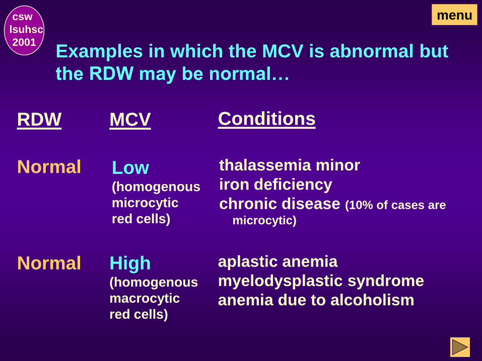
Examples in which the MCV is abnormal but
the RDW may be normal…
RDW MCV Conditions
Normal Low (homogenous
microcytic
red cells)
thalassemia minor
iron deficiency
chronic disease (10% of cases are
microcytic)
Normal High (homogenous
macrocytic
red cells)
aplastic anemia
myelodysplastic syndrome
anemia due to alcoholism
csw
lsuhsc
2001
menu
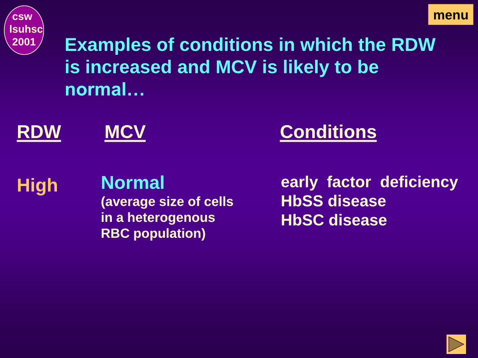
Examples of conditions in which the RDW
is increased and MCV is likely to be
normal…
csw
lsuhsc
2001
RDW MCV Conditions
High Normal (average size of cells
in a heterogenous
RBC population)
early factor deficiency
HbSS disease
HbSC disease
menu
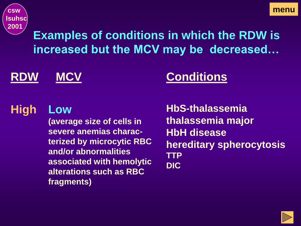
Examples of conditions in which the RDW is
increased but the MCV may be decreased…
csw
lsuhsc
2001
RDW MCV Conditions
High Low (average size of cells in
severe anemias charac-
terized by microcytic RBC
and/or abnormalities
associated with hemolytic
alterations such as RBC
fragments)
HbS-thalassemia
thalassemia major
HbH disease
hereditary spherocytosis TTP
DIC
menu
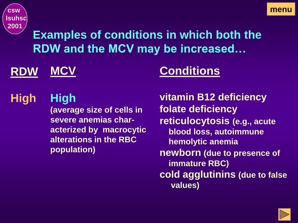
Examples of conditions in which both the
RDW and the MCV may be increased…
csw
lsuhsc
2001
RDW MCV Conditions
High High (average size of cells in
severe anemias char-
acterized by macrocytic
alterations in the RBC
population)
vitamin B12 deficiency
folate deficiency
reticulocytosis (e.g., acute
blood loss, autoimmune
hemolytic anemia
newborn (due to presence of
immature RBC)
cold agglutinins (due to false
values)
menu
Examples of conditions with a decreased RDW
and their expected MCV.
csw
lsuhsc
2001
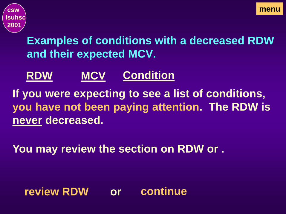
RDW MCV Condition
If you were expecting to see a list of conditions,
you have not been paying attention. The RDW is
never decreased.
You may review the section on RDW or .
review RDW or
menu
continue
End of Red Cell Distribution Width csw
lsuhsc
2001
This concludes the Red Cell Distribution Width
(RDW) Section. Select one of the following:
Go to Platelet Count, the next section, to con-
tinue with the exercise as designed.
OR
Return to the Hemogram Menu to review a section
on one of the parameters of the Hemogram.

Platelet Count
csw
lsuhsc
2001
menu
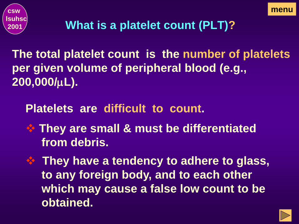
What is a platelet count (PLT)?
The total platelet count is the number of platelets
per given volume of peripheral blood (e.g.,
200,000/mL).
Platelets are difficult to count.
They are small & must be differentiated
from debris.
They have a tendency to adhere to glass,
to any foreign body, and to each other
which may cause a false low count to be
obtained.
csw
lsuhsc
2001
menu
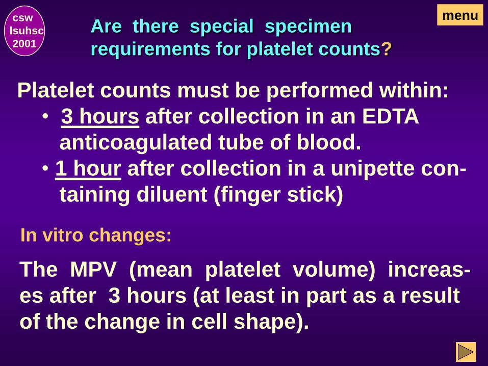
Are there special specimen
requirements for platelet counts?
Platelet counts must be performed within:
• 3 hours after collection in an EDTA
anticoagulated tube of blood.
• 1 hour after collection in a unipette con-
taining diluent (finger stick)
In vitro changes:
The MPV (mean platelet volume) increas-
es after 3 hours (at least in part as a result
of the change in cell shape).
csw
lsuhsc
2001
menu
How can platelet counts be verified?
Questionable counts are verified by microscopic
examination of a stained blood smear. Examine
for:
• platelet clumps
• platelet distribution (evenly throughout smear)
• platelet estimate (1 platelet per oil immersion field
on a smear is equivalent to approximately 15,000 to
20,000 platelets)
Extremely low electronic counts are verified by
phase microscopy (i.e., manual hemacytometer
count).
csw
lsuhsc
2001
menu
Of what clinical importance are
platelet counts?
To be hemostatically effective, platelets must be
present in sufficient numbers and must be func-
tionally normal. Platelet disorders may be classi-
fied as:
Qualitative (i.e., defect in the functional ability
of platelets)
or
Quantitative (i.e., increase or decrease in the
number of platelets), as determined by the
platelet count.
csw
lsuhsc
2001
menu
What disorders are associated with
a quantitative increase in platelets?
Thrombocytosis, an abnormal increase in PLT, may be due to:
Reactive thrombocytosis:
A physiologic response that may be seen as a secondary
phenomenon (eg, in trauma, hemorrhage, iron deficiency)
Temporary rise in platelets:
May be seen following splenectomy (splenic pool is
eliminated)
Autonomous (primary thrombocythemia):
A primary bone marrow disorder commonly seen in
myeloproliferative disorders. Platelets may also have
functional abnormalities.
csw
lsuhsc
2001
menu
What disorders are associated with a
quantitative decrease in platelets?
Thrombocytopenia,
csw
lsuhsc
2001
an abnormal decrease in platelets may be attributed to:
Decreased production:
Due to a quantitative or hypoproliferative defect in mega-
karyocytes (e.g., marrow damage, replacement of normal
marrow by metastatic tumor, intrinsic marrow disease
[leukemia]),
Ineffective thrombopoiesis:
A normal number of marrow megakaryocytes, but platelet
production is decreased (e.g., megaloblastic anemias)
menu
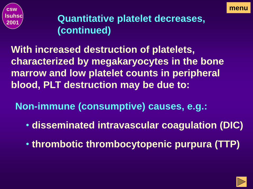
Quantitative platelet decreases,
(continued)
With increased destruction of platelets,
characterized by megakaryocytes in the bone
marrow and low platelet counts in peripheral
blood, PLT destruction may be due to:
Non-immune (consumptive) causes, e.g.:
• disseminated intravascular coagulation (DIC)
• thrombotic thrombocytopenic purpura (TTP)
csw
lsuhsc
2001
menu

Quantitative platelet decreases (continued)
May also be due to immune causes, e.g.:
• drug-induced immune thrombocytopenia
• acute and chronic idiopathic thrombocytopenic
purpura (ITP)
Decreases may also be due to increased spleen
pooling.
csw
lsuhsc
2001
menu
End of Platelet Count csw
lsuhsc
2001
This concludes the Platelet Count
Section. Select one of the following:
Go to Mean Platelet Volume (MPV), the next
section, to continue with the exercise as de-
signed.
Return to the Hemogram Menu and make an
alternate selection.
OR
Mean Platelet Volume
csw
lsuhsc
2001
menu
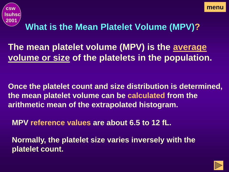
What is the Mean Platelet Volume (MPV)?
The mean platelet volume (MPV) is the average
volume or size of the platelets in the population.
Once the platelet count and size distribution is determined,
the mean platelet volume can be calculated from the
arithmetic mean of the extrapolated histogram.
MPV reference values are about 6.5 to 12 fL.
Normally, the platelet size varies inversely with the
platelet count.
menu csw
lsuhsc
2001
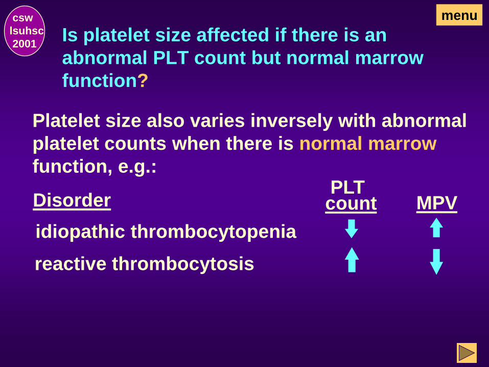
Is platelet size affected if there is an
abnormal PLT count but normal marrow
function?
csw
lsuhsc
2001
Platelet size also varies inversely with abnormal
platelet counts when there is normal marrow
function, e.g.:
MPV Disorder PLT count
idiopathic thrombocytopenia
reactive thrombocytosis
menu
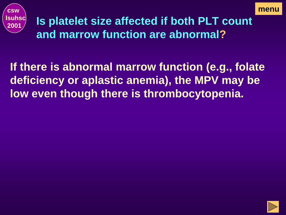
Is platelet size affected if both PLT count
and marrow function are abnormal?
csw
lsuhsc
2001
If there is abnormal marrow function (e.g., folate
deficiency or aplastic anemia), the MPV may be
low even though there is thrombocytopenia.
menu
End of CBC - 1
csw
lsuhsc
2001
This concludes the Mean Platelet Volume
section and Part 1 of the study module,
“The Complete Blood Cell Count (CBC)”.
Click on Hemogram Menu to review a section.
OR
Click on to quit CBC – Part 1
THE END
csw
lsuhsc
2001
quit
The following are additional exercises related to
the CBC:
CBC – Part 2 WBC differential & blood morphology
CBC – Part 3 RBC morphology & platelet estimate
CBC – Part 4 Post-test


