Nº 762. Conferencia
Schistosomal appendicitis: myth busted. ; Apendicitis y Esquistosomiasis: Desafio de un mito.
Charl Hobson [1], Mirta Garcia Jardon[2], Suraj Gaire[3], Lizo Mazwai [4]
(1) Departments of Radiology and Accident & Emergency Nelson Mandela Academic Hospital Mthatha REPUBLICA SUDAFRICANA (2) Department of Anatomical Pathology Walter Sisulu University and Nelson Mandela Academic Hospital Mthatha REPUBLICA SUDAFRICANA (3) Medical Student University of Cape Town Cape Town REPUBLICA SUDAFRICANA (4) Head of Department of Surgery, NMAH and Walter Sisulu University, Mthatha REPUBLICA SUDAFRICANA
Abstract
Objective: To determine whether there is an association between the presence of schistosoma haematobium ova in the appendix and histologically confirmed acute appendicitis in patients with a clinical diagnosis of appendicitis.
Design: A retrospective study into printed histology reports from January 2003 to December 2006 was performed using a standardised data chart, designed through specialist consensus.
Setting: The research was carried out at Nelson Mandela Academic Hospital in Mthatha, the tertiary referral centre for the rural Eastern Cape, South Africa.
Subjects: 979 cases of suspected acute appendicitis were identified.
Outcome measures: A Chi Square Independence Test was conducted as primary outcome measure.
Results: Of the 979 cases, 787 (80.4%) were histologically confirmed as acute appendicitis. Appendiceal schistosomiasis was diagnosed in 55 (5,62%) of the cases, of which 12 cases (1.23%) showed isolated chronic granulomatous reaction secondary to schistosoma ova. 180 (18.39%) of the appendices demonstrated normal histology . The Chi Square Independence Test returned a
value of x2 = 0.180, to the left of the predetermined alpha level of significance (0.05) at 1 degree of freedom, proving that there is no association between the presence of schistosoma ova in the appendix and the histological finding of an acute inflammatory infiltrate in appendices which were removed after a clinical diagnosis of acute appendicitis. Significant differences in age distribution were noted between the group with schistosomiasis and the group without.
Conclusion: This study demostrates that there is no association between the presence of schistosoma haematobium ova in the appendix and histologically confirmed acute appendicitis in the rural Eastern Cape, South Africa, a region with a high prevalence of schistosoma haematobium infection.
Objetivo: Determinar si existe asociación entre la presencia de huevos de esquistosoma hematobium en el apéndice cecal y apendicitis confirmada histológicamente en pacientes con diagnostico clínico de apendicitis.
Diseño: Se realizo un estudio retrospectivo de los reportes de biopsia desde Enero 2003 a Diciembre 2006, utilizando una base de datos diseñada a través de consenso con los especialistas.
Lugar: La investigación se llevo a cabo en el Hospital Académico Nelson Mandela, centro terciario de referencia para la zona rural de Eastern Cape, Sudáfrica.
Muestra: 979 casos sospechosos de apendicitis aguda se identificaron.
Medida pronostica: Se condujo una prueba de Chi cuadrada como medida pronostica primaria.
Resultados: De los 979 casos, 787 (80.4%) tuvieron confirmación histológica de apendicitis aguda. 12 casos (1.23%) mostraron reacción granulomatosa crónica aislada, y 180 (18.39%) de loas apéndices mostraron histología normal. La prueba de Chi cuadrada
mostró un valor de of x2 = 0.180, a la izquierda del nivel de significación predeterminado alfa (0.05) con 1 grado de libertad, demostrando que no existe asociación entre la presencia de huevos aislados de esquistosoma en el apéndice y el hallazgo histológico de infiltrado inflamatorio en apéndices resecadas después del diagnostico clínico de apendicitis aguda. Se encontraron diferencias significativas en la distribución por edades entre el grupo en esquistosomiasis y el grupo sin ella.
Conclusión: El estudio demuestra que no existe asociación entre la presencia de huevos de esquistosoma hematobium en el apéndice cecal y la apendicitis aguda confirmada histológicamente en el área rural de Eastern Cape, Sudáfrica, una zona en la que existe una alta prevalencia de infestación por esquistosoma hematobium.
Page 1 of 99º Congreso Virtual Hispanoamericano de Anatomía Patológica
26/05/2007http://www.conganat.org/9congreso/vistaImpresion.asp?id_trabajo=762&tipo=1
Introduction
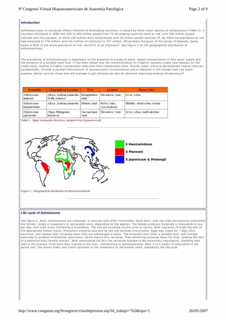
Schistosomiasis, a trematode (fluke) infection of developing countries, is caused by three major species of schistosomes (Table 1). It has been estimated in 1998 that 500 to 600 million people from 74 developing countries were at risk, with 200 million people
infected with this parasite, of which 120 million were symptomatic with 20 million people severely ill1. By 2003 the population at risk had increased to 779 million, and the number of infections to 207 million. Africa bears the grunt of this group of diseases, being
home to 85% of the world population at risk, and 97% of all infections3. See Figure 1 for the geographical distribution of schistosomiasis.
The prevalence of schistosomiasis is dependant on the presence of a body of water, faecal contamination of that water supply and the presence of a suitable snail host. It has been shown that the implementation of irrigation systems create new habitats for the snails hosts, leading to higher transmission rates and more transmission sites. Further water resource development reduce infection paradoxically, through a parallel improvement of socioeconomic circumstances and a reduction in the contact with raw water
supplies. Better care for those who still manage to get infected can also be obtained improving medical infrastructure3.
Table 1 - Major trematode infections, adapted from Gryseels et al2
Figure 1 - Geographical distribution of schistosomiasis18
Life-cycle of Schistosoma
See figure 2. Adult schistosomes are unisexual, in contrast with other trematodes. Adult pairs, with the male permanently embracing the female, reside in mesenteric or perivesicle veins, depending on the species. The female produces hundreds to thousands of ova per day, with each ovum containing a miracidium. The ova are excreted via the urine or faeces, after migration through the wall of the appropriate hollow viscus. Proteolytic enzymes secreted by the ova facilitate this process. Eggs stay viable for 7 days after excretion, and release their miracidia when they are submerged in water. The miracidia then infect a suitable host, and multiply asexually to produce multicellular sporocysts, which mature into cercariae. Free swimming cercariae leave the host, seeking the skin of a definitive host (mostly human). After penetrating the skin the cercariae migrate to the pulmonary vasculature, shedding their tails in the process. From here they migrate to the liver, transforming to schistosomulae. After 4 to 6 weeks of maturation in the portal vein, the worms mate, and travel upstream to the mesenteric or perivesicle veins, completing the life-cycle.
Page 2 of 99º Congreso Virtual Hispanoamericano de Anatomía Patológica
26/05/2007http://www.conganat.org/9congreso/vistaImpresion.asp?id_trabajo=762&tipo=1
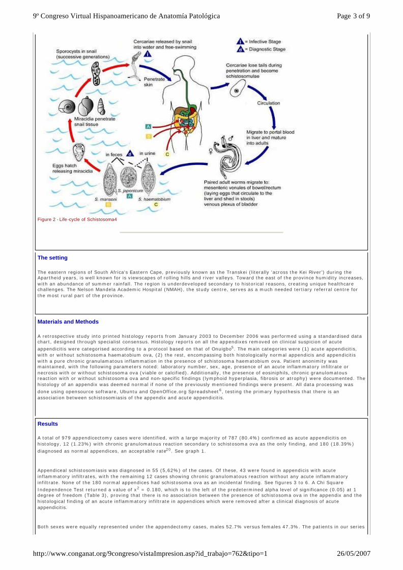
Figure 2 - Life -cycle of Schistosoma4
The setting
The eastern regions of South Africa's Eastern Cape, previously known as the Transkei (literally 'across the Kei River') during the Apartheid years, is well known for is viewscapes of rolling hills and river valleys. Toward the east of the province humidity increases, with an abundance of summer rainfall. The region is underdeveloped secondary to historical reasons, creating unique healthcare challenges. The Nelson Mandela Academic Hospital (NMAH), the study centre, serves as a much needed tertiary referral centre for the most rural part of the province.
Materials and Methods
A retrospective study into printed histology reports from January 2003 to December 2006 was performed using a standardised data chart, designed through specialist consensus. Histology reports on all the appendixes removed on clinical suspicion of acute
appendicitis were categorised according to a protocol based on that of Onuigbo5. The main categories were (1) acute appendicitis, with or without schistosoma haematobium ova, (2) the rest, encompassing both histologically normal appendicis and appendicitis with a pure chronic granulamatous inflammation in the presence of schistosoma haematobium ova. Patient anonimity was maintained, with the following parameters noted: laboratory number, sex, age, presence of an acute inflammatory infiltrate or necrosis with or without schistosoma ova (viable or calcified). Additionally, the presence of eosiniphils, chronic granulomatous reaction with or without schistosoma ova and non-specific findings (lymphoid hyperplasia, fibrosis or atrophy) were documented. The histology of an appendix was deemed normal if none of the previously mentioned findings were present. All data processing was
done using opensource software, Ubuntu and OpenOffice.org Spreadsheet6, testing the primary hypothesis that there is an association between schistosomiasis of the appendix and acute appendicitis.
Results
A total of 979 appendicectomy cases were identified, with a large majority of 787 (80.4%) confirmed as acute appendicitis on histology, 12 (1.23%) with chronic granulomatous reaction secondary to schistosoma ova as the only finding, and 180 (18.39%)
diagnosed as normal appendices, an acceptable rate20. See graph 1.
Appendiceal schistosomiasis was diagnosed in 55 (5,62%) of the cases. Of these, 43 were found in appendicis with acute inflammatory infiltrates, with the remaining 12 cases showing chronic granulomatous reaction without any acute inflammatory infiltrate. None of the 180 normal appendices had schistosoma ova as an incidental finding. See figures 3 to 6. A Chi Square
Independence Test returned a value of x2 = 0.180, which is to the left of the predetermined alpha level of significance (0.05) at 1 degree of freedom (Table 3), proving that there is no association between the presence of schistosoma ova in the appendix and the histological finding of an acute inflammatory infiltrate in appendices which were removed after a clinical diagnosis of acute appendicitis.
Both sexes were equally represented under the appendectomy cases, males 52.7% versus females 47.3%. The patients in our series
Page 3 of 99º Congreso Virtual Hispanoamericano de Anatomía Patológica
26/05/2007http://www.conganat.org/9congreso/vistaImpresion.asp?id_trabajo=762&tipo=1

had a wide age distribution of 2 months to 75 years, with an average of 19 years 4 months, and a standard deviation (SD) of 12 years. These parameters were similar for males and females (Table 2). In both sexes the age group most affected was the 10 to 14 years 11 months group, with the 5 year to 9 year 11 months old female patients relatively more affected than the males of the same age (Graphs 2 to 4).
The appendices demonstrating schistosomiasis on histology tended to be removed from younger patients, with an average age of 15 years 6 months, with a narrower SD of 5 years 3 months. The female patients with schistosoma in their appendices were on average more than two years younger than the male patients (Table 2).
Graph 1 - Main diagnostic categories
Figure 3 - Calcified ova of schistosoma surrounded by inflammatory cell infiltration with predominance of eosinophils. H/E 20X Huevo calcificado de esquistosoma rodeado de infiltrado inflamatorio con predominio de eosinófilos. H/E 20X
Page 4 of 99º Congreso Virtual Hispanoamericano de Anatomía Patológica
26/05/2007http://www.conganat.org/9congreso/vistaImpresion.asp?id_trabajo=762&tipo=1
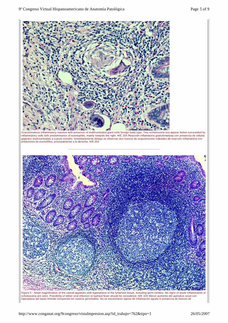
Granulomatous inflammatory reaction with presence of multinucleated giant cells foreign body type. Two schistosoma ova appear below surrounded by inflammatory cells with predominance of eosinophils, mainly towards the right. H/E 20X Reacción inflamatoria granulomatosa con presencia de células gigantes multinucleadas a cuerpo extraño. Inmediatamente debajo se observan dos huevos de esquistosoma rodeados de reacción inflamatoria con predominio de eosinófilos, principalmente a la derecha. H/E 20X
Figure 5 - Small magnification of the caecal appendix with hyperplasia of the lymphoid tissue, including germ centers. No signs of acute inflammation or schistosoma are seen. Possibility of either viral infection or typhoid fever should be considered. H/E 10X Menor aumento del apéndice cecal con hiperplasia del tejido linfoide incluyendo los centros germinales. No se encontraron signos de inflamación aguda ni presencia de huevos de
Page 5 of 99º Congreso Virtual Hispanoamericano de Anatomía Patológica
26/05/2007http://www.conganat.org/9congreso/vistaImpresion.asp?id_trabajo=762&tipo=1
esquistosoma. La posibilidad de infección viral o fiebre tifoidea no puede ser excluida completamente. H/E 10X
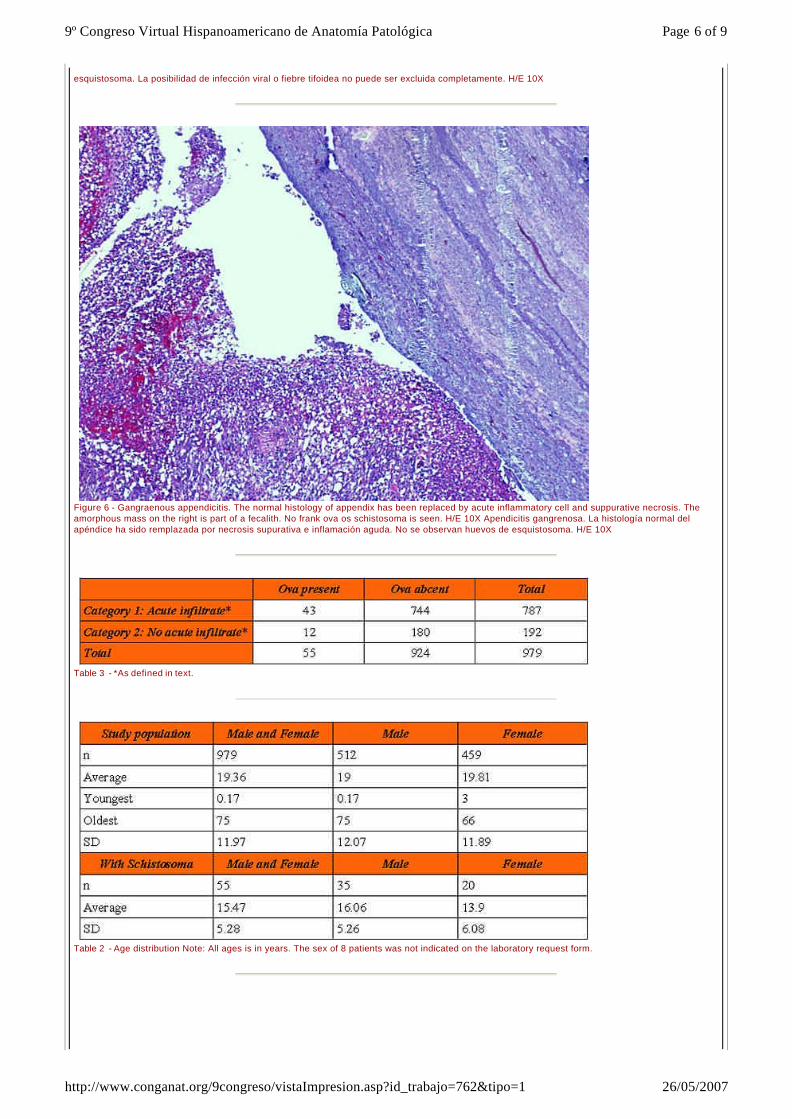
Figure 6 - Gangraenous appendicitis. The normal histology of appendix has been replaced by acute inflammatory cell and suppurative necrosis. The amorphous mass on the right is part of a fecalith. No frank ova os schistosoma is seen. H/E 10X Apendicitis gangrenosa. La histología normal del apéndice ha sido remplazada por necrosis supurativa e inflamación aguda. No se observan huevos de esquistosoma. H/E 10X
Table 3 - *As defined in text.
Table 2 - Age distribution Note: All ages is in years. The sex of 8 patients was not indicated on the laboratory request form.
Page 6 of 99º Congreso Virtual Hispanoamericano de Anatomía Patológica
26/05/2007http://www.conganat.org/9congreso/vistaImpresion.asp?id_trabajo=762&tipo=1
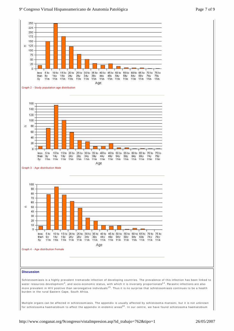
Graph 2 - Study population age distribution
Graph 3 - Age distribution Male
Graph 4 - Age distribution Female
Discussion
Schistosomiasis is a highly prevalent trematode infection of developing countries. The prevalence of this infection has been linked to
water resources development3, and socio-economic status, with which it is inversely proportionate14. Parasitic infections are also
more prevalent in HIV positive than seronegative individuals15. Thus it is no surprise that schistosomiasis continues to be a health burden in the rural Eastern Cape, South Africa.
Multiple organs can be affected in schistosomiasis. The appendix is usually affected by schistosoma mansoni, but it is not unknown
for schistosoma haematobium to affect the appendix in endemic areas20. In our centre, we have found schistosoma haematobium
Page 7 of 99º Congreso Virtual Hispanoamericano de Anatomía Patológica
26/05/2007http://www.conganat.org/9congreso/vistaImpresion.asp?id_trabajo=762&tipo=1
ova in the uterus, vulva, fallopian tubes, rectum, lung as well as in the appendix18. Complications of schistosomiasis include cerebral granulomatous disease, transverse myelitis with flaccid paraplegia, colonic polyposis, portal hypertension, pulmonary hypertension, glomerulonephritis and cystitis which can progress to bladder cancer.
The role of schistosoma in the pathogenesis of appendicitis has long been debated. The clinician could easily be lured into thinking that histologically proven schistosomiasis of the appendix is a rare finding, based on the limited literature available on this topic. Indeed, individually reported cases are few and far in between. These reports typically detail the finding of schistosomiasis of the
appendix in a traveler returning to Europe, or pathology seen in an immigrant from sub-Saharan Africa7, 8, 9, 10, 11. The reports vary from dismissing the schistosomiasis as an incidental finding, to prematurely assigning a causative role.
A large retrospective study12 of 4708 appendectomy specimens conducted in the Asir region in the southwestern part of Saudi Arabia, an endemic area for schistosomiasis, demonstrated schistosomiasis in 63 (1.3%) of the appendices. Migrants from the
African country Egypt had the highest incidence. Another Saudi study produced similar results13. A Nigerian study of 518 appendectomy specimens confirmed a high frequency of 32 (6.2%) cases. This is similar to our study demonstrating a frequency of 5.62%.
Both schistosoma haematobium and mansoni are not kind to the colon, with their local effects previously though to play a role in the pathogenesis of acute appendicitis. It is known that parasitic infections like schistosomiasis elicit a eosinophilic response which causes local tissue damage mainly through the release of cationic proteins from the eosinophil granules, as well as by releasing leukotrienes, platelet -activating factor, reactive oxygen species and lysosomal hydrolases. A number of other mechanism are also
involved: tissue infiltration, direct cytotoxicity and eosinophil-induced hypercoagulability with thromboembolic phenomena16. It has recently been shown that intestinal schistosomiasis in an animal model results in structural, functional and immunological changes in
the affected colon, most notably decreased gastrointestinal transit and increased colonic contractility17.
Our study goes further and conclusively demostrates that there is no association in our clinical setting between the presence of schistosoma ova in the appendix and the histologically confirmed acute inflammatory infiltrate in appendices which were removed after a clinical diagnosis of acute appendicitis, in contrast with previously unproven theories.
Our patient group with schistsomiasis were on average 3 years 11 months younger than the patients without. Recent work have shown that the colons of juvenile rhesus monkeys are more severely affected than those of adult monkeys, while there was no significant difference in worm burden. The juveniles had a more intense and advanced chronic granulomatous response to trapped
schistosoma ova 19. Could it be that this intense reponse presented similar to acute appendicitis clinically, explaining our observation of a younger age of presentation?
Conclusion:
We have demostrated that there is no association between the presence of schistosoma haematobium ova in the appendix and histologically confirmed acute appendicitis in the rural Eastern Cape, South Africa, a region with a high prevalence of schistosoma haematobium infection. Schistosomiasis of the appendix causes a chronic granulomatous reaction in the appendix, but without any association with acute appendicitis. Indeed, schistosoma ova found in the setting of acute appendicitis seem to be an incidental finding in our setting. Nevertheless, all health workers should be reminded of the importance of following up histology results, especially in our region with many barriers to communication and transport. Schistosomiasis, a relatively easily treated disease, has many complications, though acute appendicitis seems not to be one of them.
Aknowledgements:
The authors would like to thank Mr. S Malaoa, Department of Surgery NMAH for his advice during the initial planning of the study, and Dr. B Hobson, Department of Accident & Emergency NMAH, for assistance with data collection.
Bibliografía
1. World Health Organisation. Press Release WHO/91 4 December 1998. Schistosomiasis - the Silent Scourge of Development. Internet. http://www.who.int/inf-pr -1998/en/pr98 -91.html 2. Gryseels B, Polman K, Clerinx J, Kestens L. Human schistosomiasis. Lancet 2006; 368: 1106–18 3. Steinmann P, Keiser J, Bos R, Tanner M, Utzinger J. Schistosomiasis and water resources development: systematic review, meta-analysis, and estimates of people at risk. Lancet Infect Dis 2006; 6: 411–25 4. This image is a work of the United States Department of Health and Human Services, taken or made during the course of an employee's official duties. As a work of the U:S: federal government, the image is in the public domain. http://upload.wikimedia.org/wikipedia/en/8/80/Schistosomiasis_Life_Cycle.jpeg 5. Onuigbo WI. Appendiceal schistosomiasis. Method of classifying oviposition and inflammation. Dis Colon Rectum 1985; 28(6): 397-8 6. www.ubuntu.com 7. Rivasi F. Appendicitis associated with presence of Schistosoma haematobium eggs: an unusual pathology for Europe. Report of three cases. APMIS 2006; 114(1): 72-6 8. Gabbi C. Acute abdomen associated with schistosomiasis of the appendix. Dig Dis Sci 2006; 51(1): 215-7 9. Gottlieb GS, Wald A, Agoff N. Unusual Appendicitis. CID 2003; 37: 1378-9 10.Halkic N, Abdelmoumene A, Gintzburger D, Mosimann F. Schistosomal appendicitis in pregnancy. Swiss Surg 2002; 8(3):
Page 8 of 99º Congreso Virtual Hispanoamericano de Anatomía Patológica
26/05/2007http://www.conganat.org/9congreso/vistaImpresion.asp?id_trabajo=762&tipo=1
121-2 11.Weber G, Borer A, Zirkin HJ, Riesenberg K, Alkan M. Schistosomiasis presenting as acute appendicitis in a traveler. J Travel Med 1998; 5(3): 147-8 12.Abu-Eshy SA, Malik M, Khan A-R, Al-Shehri MY. Schistosomal Appendicitis. Annals of Saudi Medicine 1995 July 13.Khan GM, Grillo IA, Abu-Eshy SA, Khan AR, Mubarak J, Jastaniah S. Pathology of the appendix. J Natl Med Assoc 2000; 92(11): 533-5 14.Raso G. Disparities in parasitic infections, perceived ill health and access to health care among poorer and less poor schoolchildren of rural Côte d'Ivoire. Trop Med Int Health 2005; 10(1): 42-57 15.Hailemariam G. Intestinal parasitic infections in HIV/AIDS and HIV seronegative individuals in a teaching hospital, Ethiopia. Jpn J Infect Dis 2004; 57(2): 41-3 16.Klion AD, Nutman TB. The role of eosinophils in host defense against helminth parasites. J Allergy Clin Immunol 2004; 113: 30-7 17.El Zawawy LA. Effect of Schistosoma mansoni infection on physiological gastrointestinal transit and contractility. J Egypt Soc Parasitol 2006; 36(3): 1057-70 18.Garcia Jardon M, Stepien A, Banach L, Lancaster E, Govender J. Esquistosomiasis: Estudio retrospectivo de biopsias en los últimos dos años. VIII Congreso Virtual Hispanoamericano de Anatomia Patológica 2006 October 19.Bogers JJ. Juvenile rhesus monkeys have more colonic granulomas than adults after primary infection with Schistosoma mansoni. Virchows Arch 2004; 445(3): 285-91 20.Colson M, Skinner K.A, Dunnington G. High negative appendectomy rates are no longer acceptable. Am J Surg 1997; 174:723.
Web mantenido y actualizado por el Servicio de informática uclm. Modificado: 26/05/2007 20:23:23
Page 9 of 99º Congreso Virtual Hispanoamericano de Anatomía Patológica
26/05/2007http://www.conganat.org/9congreso/vistaImpresion.asp?id_trabajo=762&tipo=1


