Rotation Detection in Chest Radiographs based onGeneralized Line Histogram of Rib-Orientations
K.C. Santosh†, S. Candemir†, S. Jaeger†, L. Folio‡, A. Karargyris†, S. Antani†, G. Thoma††Communications Engineering Branch
US National Library of Medicine (NLM), National Institutes of Health (NIH)8600 Rockville Pike, Bethesda, MD 20894, USA‡Department of Radiology and Imaging Sciences
Clinical Center, National Institutes of Health (NIH)Bethesda, MD 20892, USA,
Email: {santosh.kc, sema.candemir, stefan.jaeger, les.folio, alexandros.karargyris, sameer.antani, george.thoma}@nih.gov
Abstract—We present a generalized line histogram techniqueto compute global rib-orientation for detecting rotated lungs inchest radiographs. We use linear structuring elements, such asline seed filters, as kernels to convolve with edge images, andextract a set of lines from the posterior rib-cage. After convolvingkernels in all possible orientations in the range [0, π), we measurethe angle for which the line histogram has maximum magnitude.This measure provides a good approximation of the global chestrib-orientation for each lung. A chest radiograph is said to beupright if the difference between the orientation angles of bothlungs with respect to the horizontal axis, is negligible. We validateour method on sets of normal and abnormal images and arguethat rib orientation can be used for rotation detection in chestradiographs as aid in quality control during image acquisition,and to discard images from training and testing data sets. In ourtest, we achieve a maximum accuracy of 90%.
Index Terms—Chest radiographs; generalized line histogram;rib-orientations; rotation detection.
I. INTRODUCTION
Quality control is a critical issue when large number ofdigital chest radiographs (or chest X-ray (CXR)) need tobe acquired in an automated fashion, such as during masspopulation screening [1]. For this, images need to be inspectedfor proper x-ray penetration, adequate inspiration (inhaling)by the patient, proper angulation, and importantly, the imageshould be devoid of any rotation. Rotation can adversely affectthe performance of subsequent automated processing stepsin screening algorithms or computer-aided diagnosis, such aslung segmentation [2]. Further, rotation can also affect one-to-one zone comparison, viz. upper, middle, and lower betweenlung sections. While rotated CXRs may not necessarily bechallenging for radiologists, they can confuse the machine thatis operating in either a computer-assisted or a fully-automatedfashion. An automatic method for detecting rotated imagesis desirable to enable machines to reject the rotated CXRand/or forward it to a human operator for closer inspection.Rotation in CXRs can often be expected for images acquiredwith portable machines in non-hospital settings or under morechallenging outdoor conditions, such as mobile screening sta-tions in rural areas. In addition to pathology-related rotations,
misaligned body positions are more frequent in these cases dueto hardware limitations of the screening setup used, poorly-observed screening protocols, or other factors [3], [4].
To acquire an upright CXR, a radiology technician needs toalign the patient’s body so that it is perpendicular to the x-raybeam. Any deviation from this position will result in a rotatedimage. The degree of rotation in a CXR can be computed byanalyzing the relationship of the medial heads of the claviclesto the adjacent spinous processes in the upper thorax. Nor-mally, the spinous processes lie equidistant from the medialheads of the clavicles. From a practical point of view, theautomatic detection of clavicle heads and spinous processesneeds to be precise. Even a small deviation of 10 to 20 pixels(in and out) with respect to the actual boundary, can adverselyaffect the decision process. We observe that radiologists canreliably decide if a CXR is rotated by using other contextualinformation independent of the clavicles. Motivated by this,we detect rotation based on the rib-orientation. A rotated lungis likely to affect the positions of clavicles and ribs in thesame way. In addition, this method is likely to be more stablebecause the number of ribs is relatively high and the entirerib-cage can be used.
Humans have 12 pairs of ribs including false and floatingribs [5]. Not all of these ribs are necessarily visible in a typicalradiograph for various reasons. However, the visible subset isusually sufficient to compute the global rib-orientation. Veryfew papers on rib detection and segmentation methods havebeen reported in the literature [6]–[9]. However, the publishedmethods usually do not detect the false and floating ribs.Further some methods detect the ribs only partially due tothe large variation of intensity distributions in CXRs. It isinteresting to note that computing the rib-vertebra anglei.e., theangle between a rib and its corresponding vertebra, which is aclinically accepted method [10], [11]. These works used radio-graphs of scoliosis patients for which they computed the rib-vertebra angle difference (RVAD), for all ribs. Similarly, imageregistration method for interval change detection between twoCXRs based on the difference in anterior-posterior inclinationangles has been reported [12]. Due to low accuracy in rib and
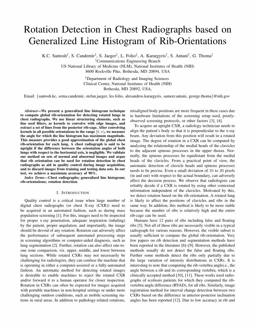
(a) Input image (b) Lung segmentation (c) Right lung (d) Left lung
Fig. 1. An example showing the complete process of segmenting lung sections (right and left) using graph-cut algorithm.
vertebra segmentation, we propose a method for computingthe global rib-orientation for both lungs. Missing a pair of ribsdoes not negatively affect the output of this method because weare only interested in the global rib-orientation and not in theorientation of individual ribs. Contributions of our work to thescience are: 1) we have developed a generalized line histogrammethod to compute rib-orientations; and 2) we apply thismethod as a decision making tool for rotation detection andquality control in acquisition of CXRs, particularly in remoterural areas during mass population screening. Taking rotationinto account using ribs minimizes false positive detectioncaused by overlapping densities in a rotated patient.
The remainder of this paper is organized as follows. We startwith detailing our proposed method in Section II. This includeslung segmentation, line seed filter (kernel) development, linehistogram computation, and rotation decision. In Section III,we evaluate the approach. We conclude the paper in Section IVwith a summary of our results.
II. THE PROPOSED METHOD
A. Lung section segmentation
To effectively compute the rib-cage edge distribution, it isnecessary to segment right and left lung sections from thewhole image. There are several state-of-the-art algorithms, todetect the lung regions. In this work, we use our algorithmwhich is based on graph-cut algorithm guided by patient-specific atlas model [13]. The system first builds a subset ofatlases (which are expert delineation of lung boundaries ofseveral patients) by choosing the most similar x-rays in termsof shape similarity of lungs. Then, it warps these selectedatlases to the target CXRs using a registration algorithm.We use the scale invariant feature transform (SIFT) flow(i.e., SIFT-flow) registration approach [14] which computesthe corresponding pixels of image pairs according to theirSIFT feature similarity. The spatial difference between thecorresponding pixels is used to warp the masks derived fromtraining CXRs to build a lung model for the target CXR. Thelung model and intensity information of the target CXR arecombined by using the objective function: data term, smooth-ness term and lung model term. The data term forces the seg-mentation suitable to intensity information of the CXRs; thesmoothness term produces a smooth solution; and the modelterm guides the algorithm to produce a segmentation result
similar to the patient lung model. The final lung boundaryis computed by solving the objective function with graph cutenergy minimization approach [15]. Fig. 1 shows a couple ofoutput examples of it, where the detected right and left lungsections are separately illustrated.
B. Generalized line histogram
B.1 Kernels.To detect key lines from the image, we define line seed filterkernels that is defined in a normal Gaussian distribution. Wecompute probability density function (pdf) at each of thevalues in X using the normal distribution,
f (x, µ, σ) =1
σ√
2πexp
(−(x− µ)2
2σ2
), (1)
where σ = 1, µ = 0 and X is a vector and its values areconfined in the range [−σ, σ]. To make it simple, in the discretecase, a structuring element can be represented as a set of pixelson a grid, assuming the values 1 if the pixel belongs to thestructuring element or 0, otherwise [16], [17]. Based on this,we define a binary kernel representing a line of any particularlength len and angle θ i.e., f (‘line ′, len, θ). To generate akernel that represents the shape of a Gaussian (‘bell-shaped’)hump g, we perform an element-wise multiplication of binarykernel with the values obtained from Eq. (1),
g = f (‘line ′, len, θ) ◦ f(
[−σ :2σ
len − 1: σ], µ, σ
). (2)
Note that the kernel size depends on the length of the linearstructuring element. Unsurprisingly, larger the size of thekernel matrix, lesser the number of lines.
Considering a set Θ of possible different orientations {θk}which are specified in the range [0◦, 180◦), we have a set Kof kernels {gk},
K = {gk}k=1,...,K , and θk =180◦
bink(k − 1), (3)
where bin is the number of bins and the index k associatedwith kernel g determines the orientation value i.e., θk.
B.2 Line histogram.Given an edge image edg(m,n) of size M × N , our ideais to perform convolution with the kernel gK. Note thatthe edge image is resulted from Canny edge detector after
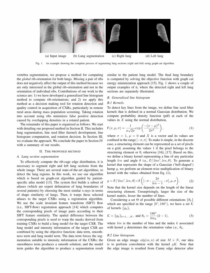
(a) Masked lungs
0◦
30◦
60◦
. . .(b) Kernels (c) Line output at 0 (d) Line output at 30◦ (e) Line output at 60
Fig. 2. An example showing (a) lung sections (cf. Fig. 1), (b) kernels at three different orientations, which are defined in 1D Gaussian distribution and (c-e)the corresponding lines that are via convolution.
global histogram equalization. Convolution in general, can beexpressed as o(m,n) = g ⊗ edg
o(m,n) =∑Ii=1
∑Jj=1 g(i, j)edg(m− i, n− j). (4)
This means that, for each pixel (m,n) in the image, the con-volution output value o(m,n) is calculated by translating theconvolution mask to pixel (m,n) in the image, and then takingthe weighted sum of the pixels in the neighborhood about(m,n), where the individual weights are the correspondingvalues in the convolution mask. Such a convolution producesprominent lines that are appeared in the lung section. Considera complete set K of kernels (cf. Eq. (3)), convolution will pro-duce a complete set L of lines, L = K⊗ edg = {`k}k=1,...,K ,where `k refers to the set of lines in that particular index k.Fig. 2 shows a few examples of it. Formally, any ` can berepresented by a tuple: total number of lines (noL) and totallength of the lines (loL) i.e., ` = 〈noL, loL〉.
To compute histogram, without loss of generality, we useloL in every particular k since lengths of the lines vary fromone line to another (see Fig. 2). Based on this, a complete setH of line histograms hk = loLk of lines in every convolution,designated by kernel gk is
H = {~k}k=1,...,K, and ~k =1
max(hk)hk. (5)
C. Chest radiograph rotation
Global chest rib-orientation can be computed as,arg maxk(~k) i.e., the angle from which the maximummagnitude of line histogram is produced.
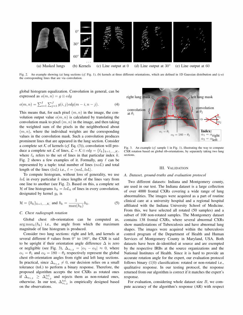
Consider two lung sections: right and left, and kernels atseveral different θ values from 0◦ to 180◦, the CXR is saidto be upright if their orientation angle difference ∆ is zeroor negligible (see Fig. 3), ∆α1,2
= |α1 − α2| ≈ 0, whereα1 = θ1 and α2 = 180− θ2 respectively represent the globalchest rib-orientation angles from right and left lung sections.In practical, since ∆α1,2 6= 0, our decision relies on a smalltolerance (tol.) to perform a binary response. Therefore, theproposed algorithm accepts the test CXRs as rotated onesif ∆α1,2
≥ ∆tol.α1,2
and rejects them as non-rotated ones,otherwise. In our test, ∆tol.
α1,2is empirically designed based
on the observations.
med
ial
axis
...
...
convolutionat θ1
{
right lung mask left lung mask
...
...
} convolutionat θ2
θ2α2θ1
α1 = θ1α2 = 180− θ2
Index:α1 = αrightα2 = αleft
α1
α2⇒
Fig. 3. An example (cf. sample 1 in Fig. 1), illustrating the way to computeCXR rotation based on global rib-orientations, by separately taking two lungsections.
III. VALIDATION
A. Dataset, ground-truths and evaluation protocol
Two different datasets: Indiana and Montgomery county,are used in our test. The Indiana dataset is a large collectionof over 4000 frontal CXRs covering a wide range of lungabnormalities. The images were acquired as a part of routineclinical care at a university hospital and a regional hospitalaffiliated with the Indiana University School of Medicine.From this, we have selected all rotated (50 samples) and asubset of 100 non-rotated samples. The Montgomery datasetcontains 138 frontal CXRs, where several abnormal CXRsshow manifestations of Tuberculosis and have abnormal lungshapes. The images were acquired within the tuberculosiscontrol program of the Department of Health and HumanServices of Montgomery County in Maryland, USA. Bothdatasets have been de-identified at source and are exemptedby the respective IRBs at the source organizations and theNational Institutes of Health. Since it is hard to provide anaccurate rotation angle for the expert, our evaluation protocolfollows binary (1|0) classification: rotated or non-rotated i.e.,qualitative response. In our testing protocol, the responsereturned from our algorithm is correct if it matches the expert’sresponse.
For evaluation, considering whole dataset size B, we com-pute accuracy of the algorithm’s response (AR) with respect
(a) (b)
0.5
1
30
210
60
240
90
270
120
300
150
330
180 0
left
α2=
0◦ri
ght
α1=
0◦
(c)
0.5
1
30
210
60
240
90
270
120
300
150
330
180 0
left
α2=
15◦
righ
tα1=
15◦
(d)
0.5
1
30
210
60
240
90
270
120
300
150
330
180 0
left
α2=
11◦
righ
tα1=
11◦
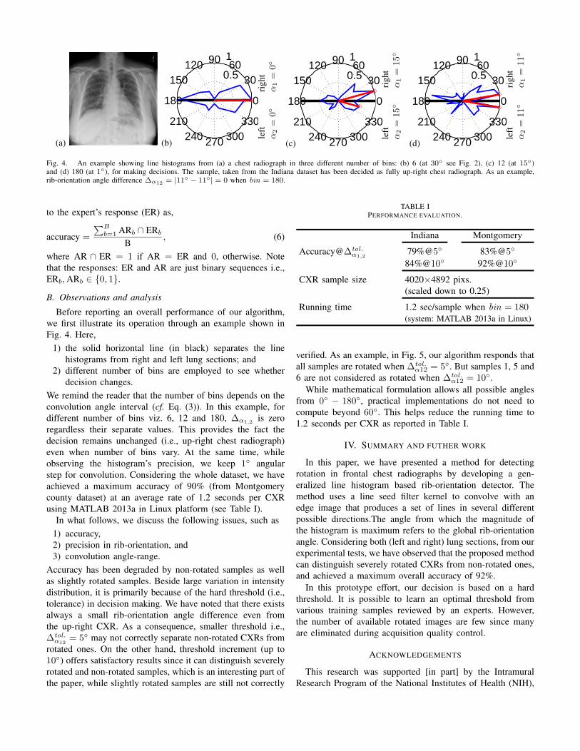
Fig. 4. An example showing line histograms from (a) a chest radiograph in three different number of bins: (b) 6 (at 30◦ see Fig. 2), (c) 12 (at 15◦)and (d) 180 (at 1◦), for making decisions. The sample, taken from the Indiana dataset has been decided as fully up-right chest radiograph. As an example,rib-orientation angle difference ∆α12 = |11◦ − 11◦| = 0 when bin = 180.
to the expert’s response (ER) as,
accuracy =
∑Bb=1 ARb ∩ ERb
B, (6)
where AR ∩ ER = 1 if AR = ER and 0, otherwise. Notethat the responses: ER and AR are just binary sequences i.e.,ERb,ARb ∈ {0, 1}.
B. Observations and analysis
Before reporting an overall performance of our algorithm,we first illustrate its operation through an example shown inFig. 4. Here,
1) the solid horizontal line (in black) separates the linehistograms from right and left lung sections; and
2) different number of bins are employed to see whetherdecision changes.
We remind the reader that the number of bins depends on theconvolution angle interval (cf. Eq. (3)). In this example, fordifferent number of bins viz. 6, 12 and 180, ∆α1,2
is zeroregardless their separate values. This provides the fact thedecision remains unchanged (i.e., up-right chest radiograph)even when number of bins vary. At the same time, whileobserving the histogram’s precision, we keep 1◦ angularstep for convolution. Considering the whole dataset, we haveachieved a maximum accuracy of 90% (from Montgomerycounty dataset) at an average rate of 1.2 seconds per CXRusing MATLAB 2013a in Linux platform (see Table I).
In what follows, we discuss the following issues, such as1) accuracy,2) precision in rib-orientation, and3) convolution angle-range.
Accuracy has been degraded by non-rotated samples as wellas slightly rotated samples. Beside large variation in intensitydistribution, it is primarily because of the hard threshold (i.e.,tolerance) in decision making. We have noted that there existsalways a small rib-orientation angle difference even fromthe up-right CXR. As a consequence, smaller threshold i.e.,∆tol.α12
= 5◦ may not correctly separate non-rotated CXRs fromrotated ones. On the other hand, threshold increment (up to10◦) offers satisfactory results since it can distinguish severelyrotated and non-rotated samples, which is an interesting part ofthe paper, while slightly rotated samples are still not correctly
TABLE IPERFORMANCE EVALUATION.
Indiana Montgomery
Accuracy@∆tol.α1,2
79%@5◦ 83%@5◦
84%@10◦ 92%@10◦
CXR sample size 4020×4892 pixs.(scaled down to 0.25)
Running time 1.2 sec/sample when bin = 180(system: MATLAB 2013a in Linux)
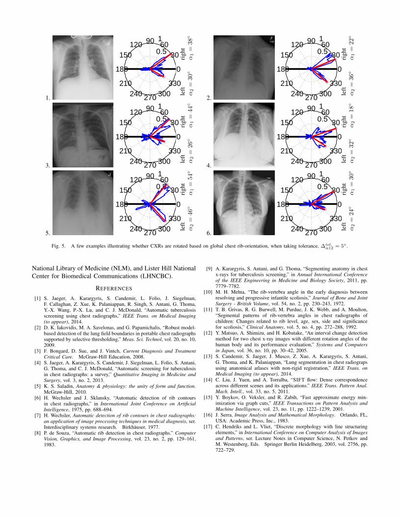
verified. As an example, in Fig. 5, our algorithm responds thatall samples are rotated when ∆tol.
α12 = 5◦. But samples 1, 5 and6 are not considered as rotated when ∆tol.
α12 = 10◦.While mathematical formulation allows all possible angles
from 0◦ − 180◦, practical implementations do not need tocompute beyond 60◦. This helps reduce the running time to1.2 seconds per CXR as reported in Table I.
IV. SUMMARY AND FUTHER WORK
In this paper, we have presented a method for detectingrotation in frontal chest radiographs by developing a gen-eralized line histogram based rib-orientation detector. Themethod uses a line seed filter kernel to convolve with anedge image that produces a set of lines in several differentpossible directions.The angle from which the magnitude ofthe histogram is maximum refers to the global rib-orientationangle. Considering both (left and right) lung sections, from ourexperimental tests, we have observed that the proposed methodcan distinguish severely rotated CXRs from non-rotated ones,and achieved a maximum overall accuracy of 92%.
In this prototype effort, our decision is based on a hardthreshold. It is possible to learn an optimal threshold fromvarious training samples reviewed by an experts. However,the number of available rotated images are few since manyare eliminated during acquisition quality control.
ACKNOWLEDGEMENTS
This research was supported [in part] by the IntramuralResearch Program of the National Institutes of Health (NIH),
1.
0.5
1
30
210
60
240
90
270
120
300
150
330
180 0
left
α2=
30◦
righ
tα1=
38◦
2.
0.5
1
30
210
60
240
90
270
120
300
150
330
180 0
left
α2=
36◦
righ
tα1=
22◦
3.
0.5
1
30
210
60
240
90
270
120
300
150
330
180 0
left
α2=
26◦
righ
tα1=
44◦
4.
0.5
1
30
210
60
240
90
270
120
300
150
330
180 0
left
α2=
32◦
righ
tα2=
18◦
5.
0.5
1
30
210
60
240
90
270
120
300
150
330
180 0
left
α2=
46◦
righ
tα1=
54◦
6.
0.5
1
30
210
60
240
90
270
120
300
150
330
180 0
left
α2=
24◦
righ
tα1=
30◦
Fig. 5. A few examples illustrating whether CXRs are rotated based on global chest rib-orientation, when taking tolerance, ∆tol.α12 = 5◦.
National Library of Medicine (NLM), and Lister Hill NationalCenter for Biomedical Communications (LHNCBC).
REFERENCES
[1] S. Jaeger, A. Karargyris, S. Candemir, L. Folio, J. Siegelman,F. Callaghan, Z. Xue, K. Palaniappan, R. Singh, S. Antani, G. Thoma,Y.-X. Wang, P.-X. Lu, and C. J. McDonald, “Automatic tuberculosisscreening using chest radiographs,” IEEE Trans. on Medical Imaging(to appear), 2014.
[2] D. K. Iakovidis, M. A. Savelonas, and G. Papamichalis, “Robust model-based detection of the lung field boundaries in portable chest radiographssupported by selective thresholding,” Meas. Sci. Technol, vol. 20, no. 10,2009.
[3] F. Bongard, D. Sue, and J. Vintch, Current Diagnosis and TreatmentCritical Care. McGraw-Hill Education, 2008.
[4] S. Jaeger, A. Karargyris, S. Candemir, J. Siegelman, L. Folio, S. Antani,G. Thoma, and C. J. McDonald, “Automatic screening for tuberculosisin chest radiographs: a survey,” Quantitative Imaging in Medicine andSurgery, vol. 3, no. 2, 2013.
[5] K. S. Saladin, Anatomy & physiology: the unity of form and function.McGraw-Hill, 2010.
[6] H. Wechsler and J. Sklansky, “Automatic detection of rib contoursin chest radiographs,” in International Joint Conference on ArtificialIntelligence, 1975, pp. 688–694.
[7] H. Wechsler, Automatic detection of rib contours in chest radiographs:an application of image processing techniques in medical diagnosis, ser.Interdisciplinary systems research. Birkhauser, 1977.
[8] P. de Souza, “Automatic rib detection in chest radiographs.” ComputerVision, Graphics, and Image Processing, vol. 23, no. 2, pp. 129–161,1983.
[9] A. Karargyris, S. Antani, and G. Thoma, “Segmenting anatomy in chestx-rays for tuberculosis screening,” in Annual International Conferenceof the IEEE Engineering in Medicine and Biology Society, 2011, pp.7779–7782.
[10] M. H. Mehta, “The rib-vertebra angle in the early diagnosis betweenresolving and progressive infantile scoliosis,” Journal of Bone and JointSurgery - British Volume, vol. 54, no. 2, pp. 230–243, 1972.
[11] T. B. Grivas, R. G. Burwell, M. Purdue, J. K. Webb, and A. Moulton,“Segmental patterns of rib-vertebra angles in chest radiographs ofchildren: Changes related to rib level, age, sex, side and significancefor scoliosis,” Clinical Anatomy, vol. 5, no. 4, pp. 272–288, 1992.
[12] Y. Matsuo, A. Shimizu, and H. Kobatake, “An interval change detectionmethod for two chest x-ray images with different rotation angles of thehuman body and its performance evaluation,” Systems and Computersin Japan, vol. 36, no. 10, pp. 30–42, 2005.
[13] S. Candemir, S. Jaeger, J. Musco, Z. Xue, A. Karargyris, S. Antani,G. Thoma, and K. Palaniappan, “Lung segmentation in chest radiograpsusing anatomical atlases with non-rigid registration,” IEEE Trans. onMedical Imaging (to appear), 2014.
[14] C. Liu, J. Yuen, and A. Torralba, “SIFT flow: Dense correspondenceacross different scenes and its applications.” IEEE Trans. Pattern Anal.Mach. Intell., vol. 33, no. 5, 2011.
[15] Y. Boykov, O. Veksler, and R. Zabih, “Fast approximate energy min-imization via graph cuts,” IEEE Transactions on Pattern Analysis andMachine Intelligence, vol. 23, no. 11, pp. 1222–1239, 2001.
[16] J. Serra, Image Analysis and Mathematical Morphology. Orlando, FL,USA: Academic Press, Inc., 1983.
[17] C. Hendriks and L. Vliet, “Discrete morphology with line structuringelements,” in International Conference on Computer Analysis of Imagesand Patterns, ser. Lecture Notes in Computer Science, N. Petkov andM. Westenberg, Eds. Springer Berlin Heidelberg, 2003, vol. 2756, pp.722–729.


