Download - Rocas Ígneas En Sección Delgada
1
Igneous rocks in thin section
Contents Image scales (frame widths) Quartz
Plagioclase Potassium feldspars
Myrmeckite Micas
Amphiboles Pyroxenes
Other primary minerals and minor minerals
Secondary (subsolidus) minerals
20x = 6 mm 40x = 3 mm
100x = 1 mm 200x = 0.5 mm
400x = 0.25 mm 1000x = 0.1 mm
Quartz
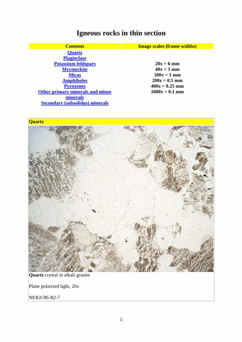
Quartz crystal in alkali granite
Plane polarized light, 20x
NEIGC86-B2-7
2
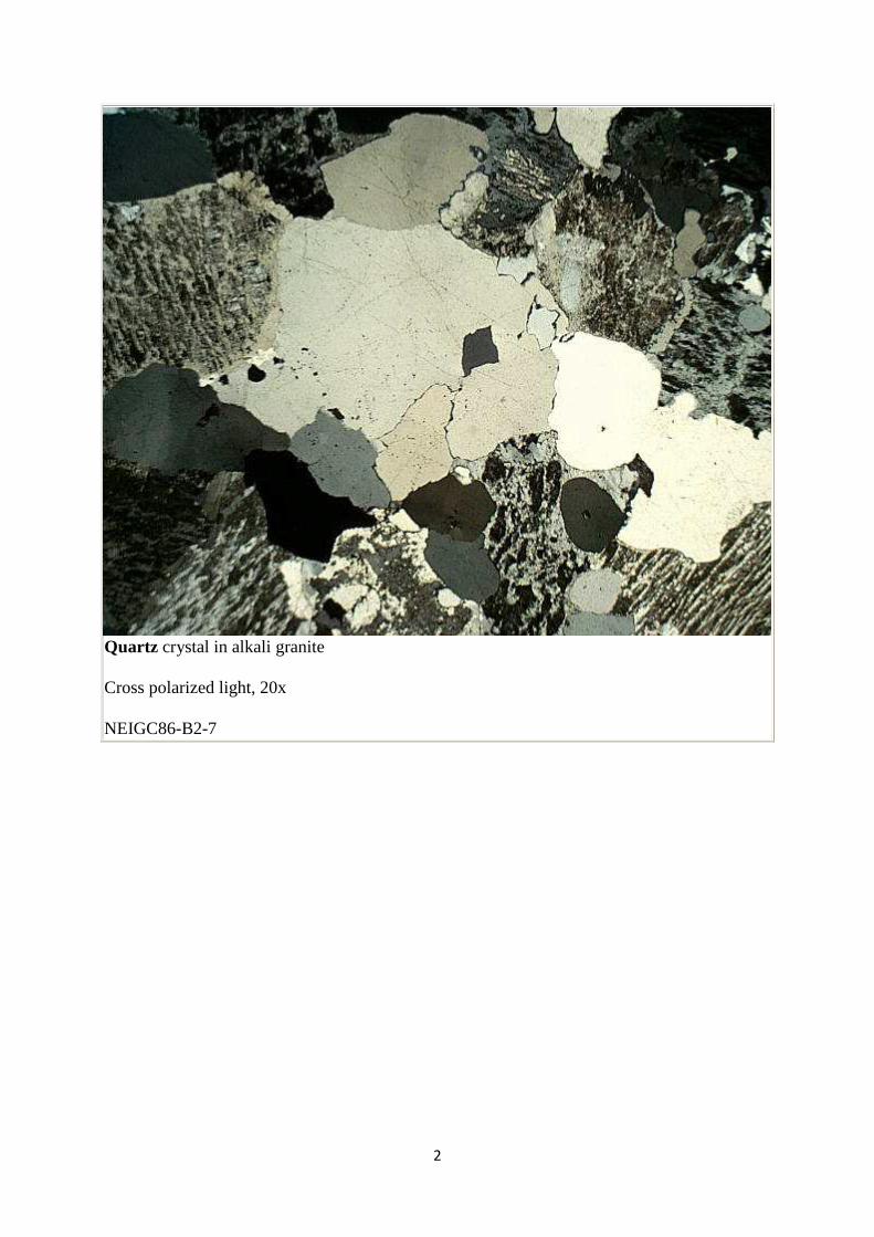
Quartz crystal in alkali granite
Cross polarized light, 20x
NEIGC86-B2-7
3
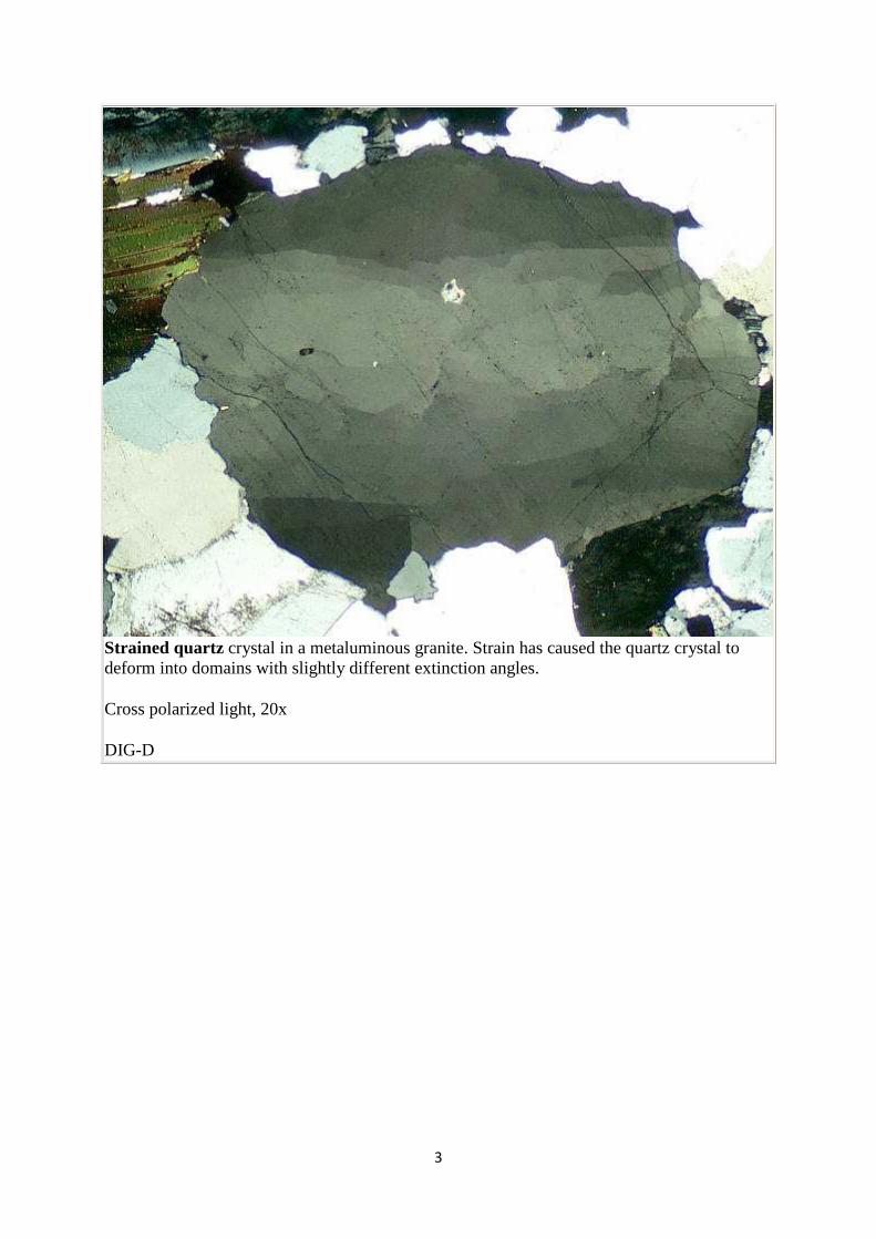
Strained quartz crystal in a metaluminous granite. Strain has caused the quartz crystal to deform into domains with slightly different extinction angles.
Cross polarized light, 20x
DIG-D
4
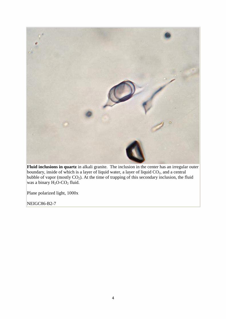
Fluid inclusions in quartz in alkali granite. The inclusion in the center has an irregular outer boundary, inside of which is a layer of liquid water, a layer of liquid CO2, and a central bubble of vapor (mostly CO2). At the time of trapping of this secondary inclusion, the fluid was a binary H2O-CO2 fluid.
Plane polarized light, 1000x
NEIGC86-B2-7
5
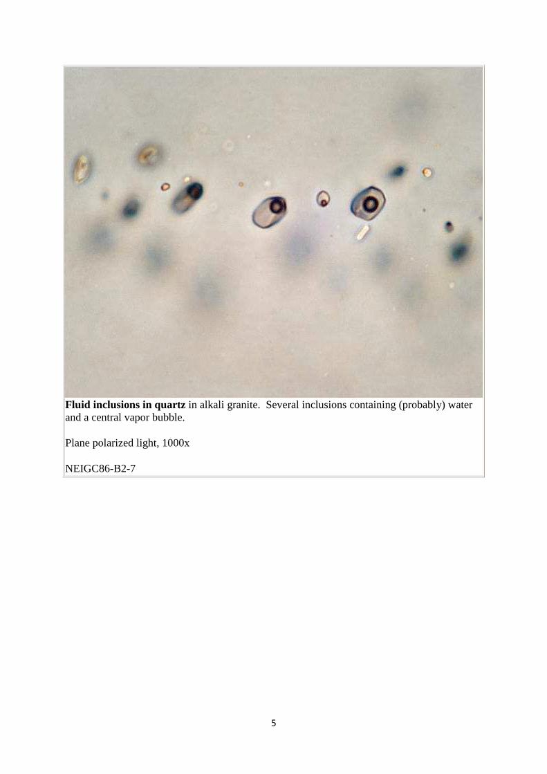
Fluid inclusions in quartz in alkali granite. Several inclusions containing (probably) water and a central vapor bubble.
Plane polarized light, 1000x
NEIGC86-B2-7
6
Plagioclase
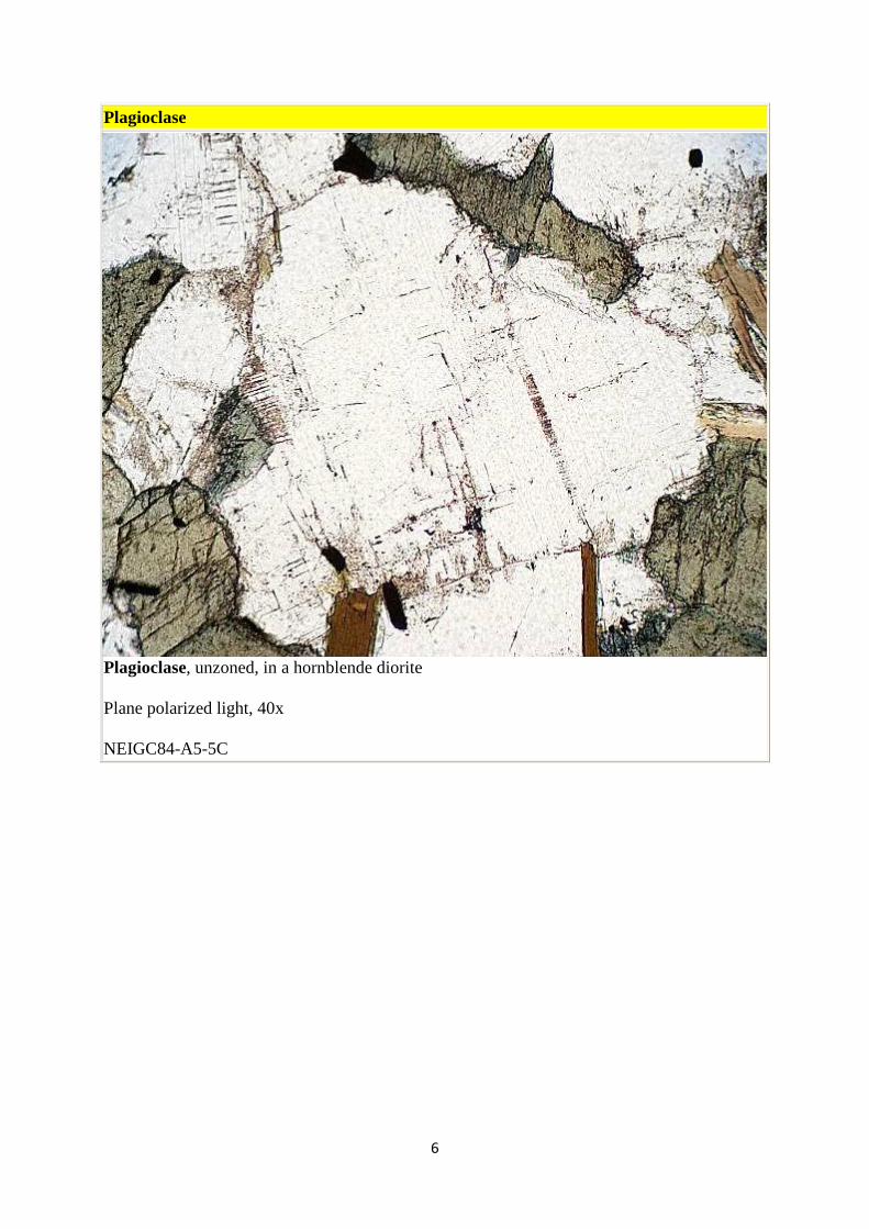
Plagioclase, unzoned, in a hornblende diorite
Plane polarized light, 40x
NEIGC84-A5-5C
7
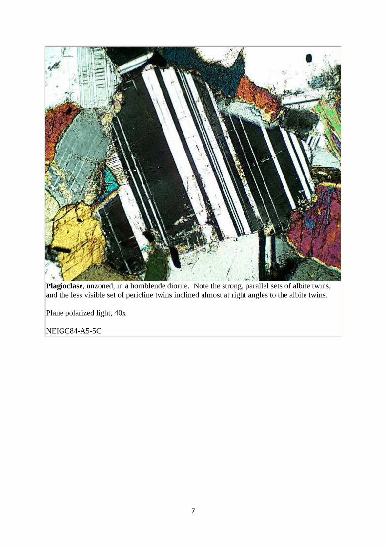
Plagioclase, unzoned, in a hornblende diorite. Note the strong, parallel sets of albite twins, and the less visible set of pericline twins inclined almost at right angles to the albite twins.
Plane polarized light, 40x
NEIGC84-A5-5C
8
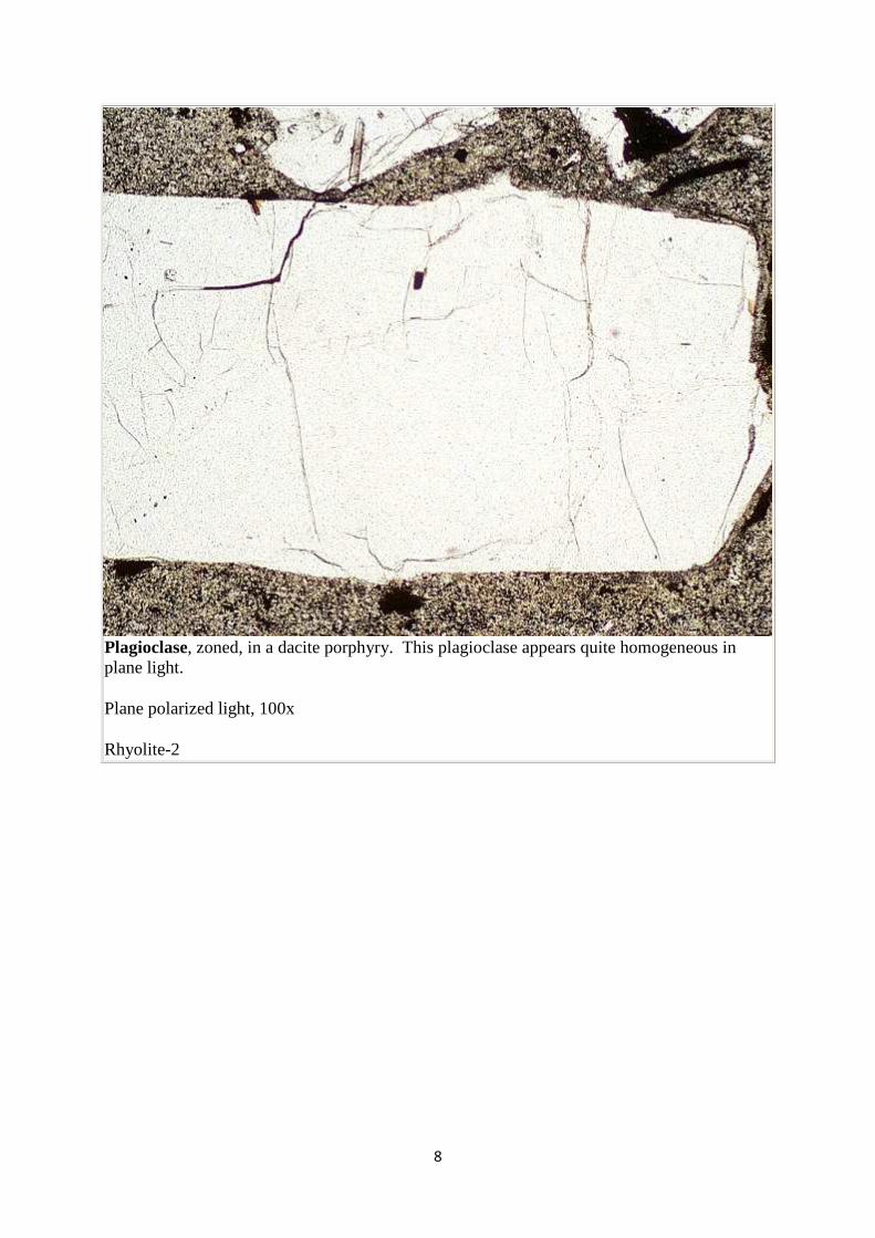
Plagioclase, zoned, in a dacite porphyry. This plagioclase appears quite homogeneous in plane light.
Plane polarized light, 100x
Rhyolite-2
9
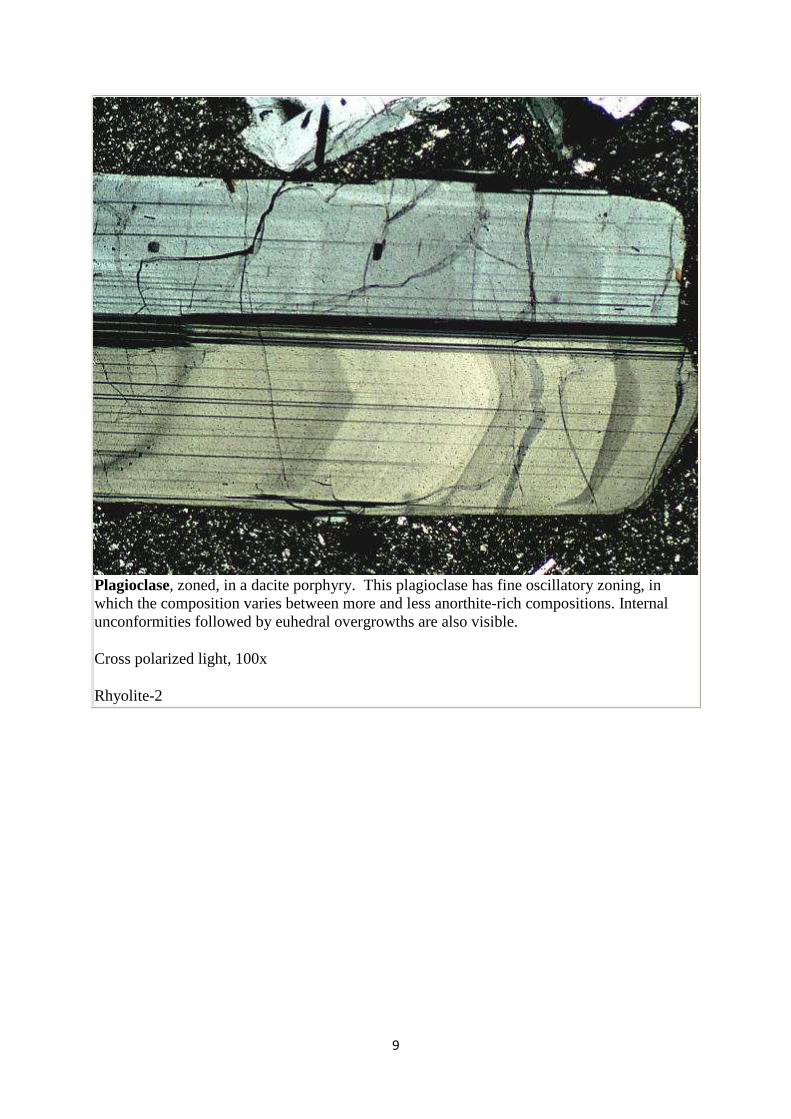
Plagioclase, zoned, in a dacite porphyry. This plagioclase has fine oscillatory zoning, in which the composition varies between more and less anorthite-rich compositions. Internal unconformities followed by euhedral overgrowths are also visible.
Cross polarized light, 100x
Rhyolite-2
10
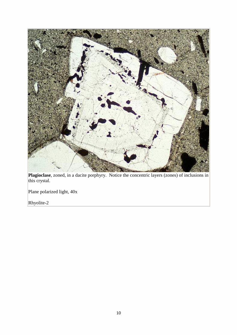
Plagioclase, zoned, in a dacite porphyry. Notice the concentric layers (zones) of inclusions in this crystal.
Plane polarized light, 40x
Rhyolite-2
11
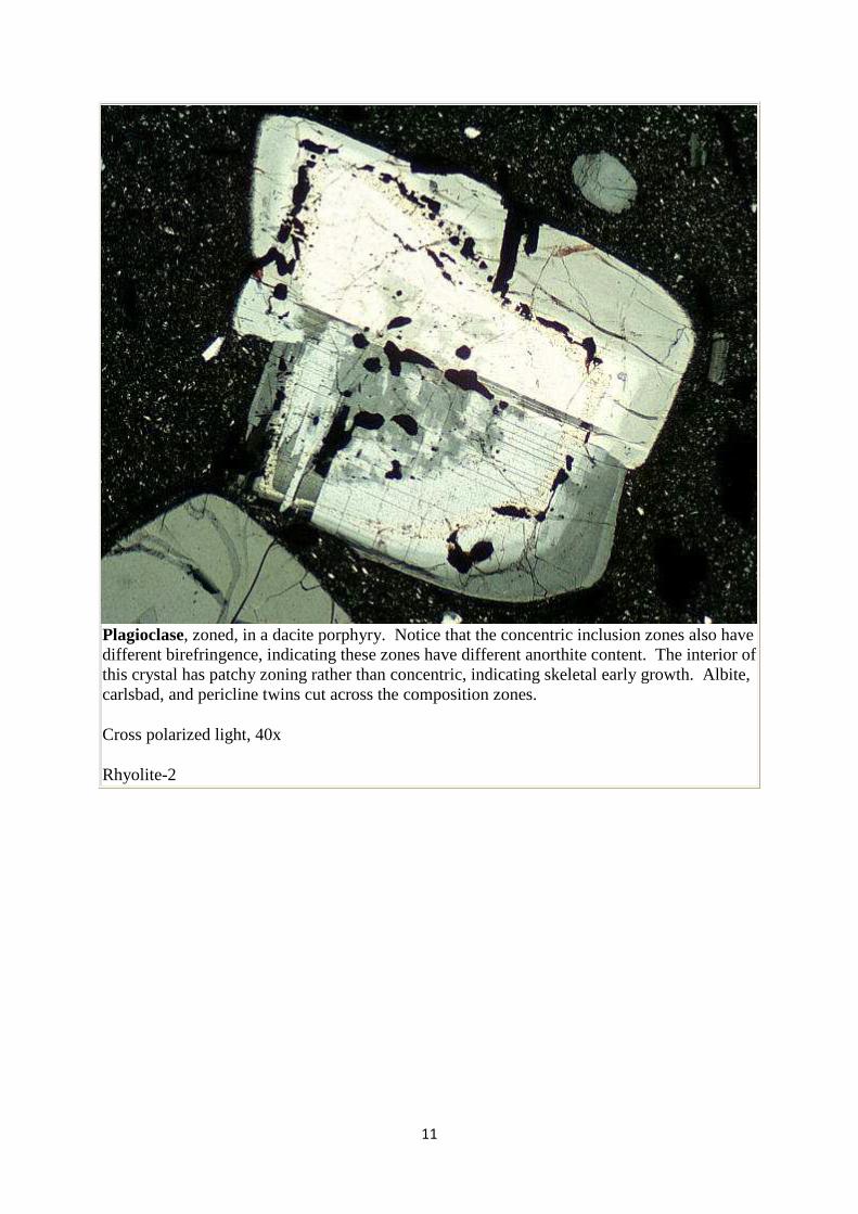
Plagioclase, zoned, in a dacite porphyry. Notice that the concentric inclusion zones also have different birefringence, indicating these zones have different anorthite content. The interior of this crystal has patchy zoning rather than concentric, indicating skeletal early growth. Albite, carlsbad, and pericline twins cut across the composition zones.
Cross polarized light, 40x
Rhyolite-2
12
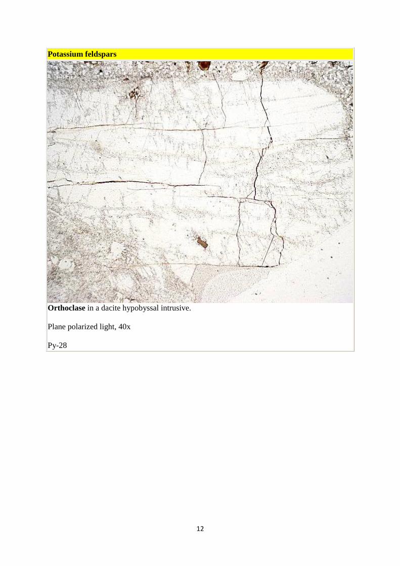
Potassium feldspars
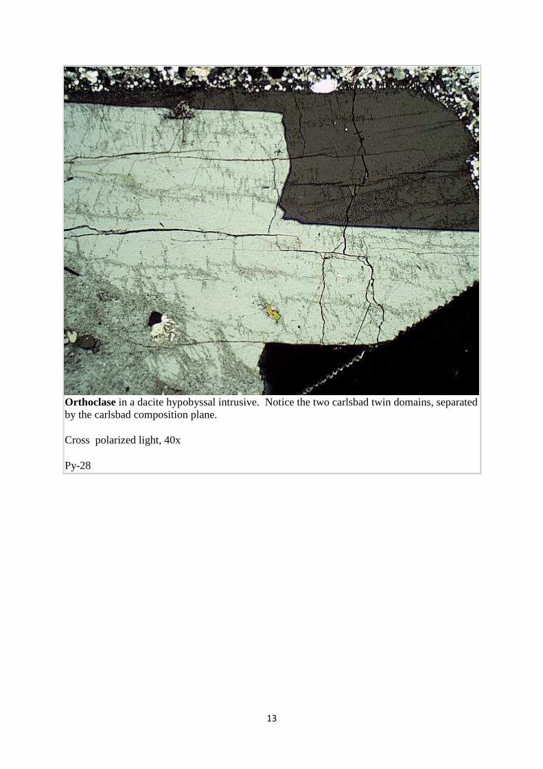
Orthoclase in a dacite hypobyssal intrusive.
Plane polarized light, 40x
Py-28
13
Orthoclase in a dacite hypobyssal intrusive. Notice the two carlsbad twin domains, separated by the carlsbad composition plane.
Cross polarized light, 40x
Py-28
14
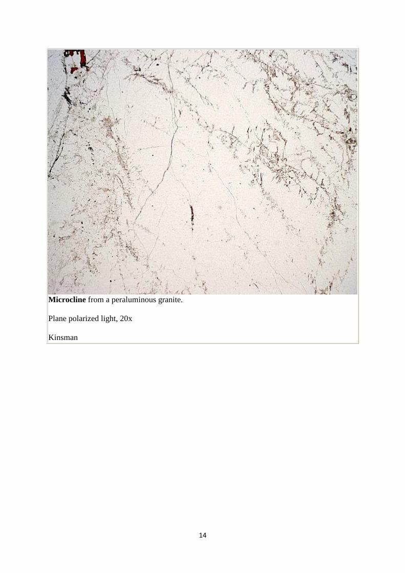
Microcline from a peraluminous granite.
Plane polarized light, 20x
Kinsman
15
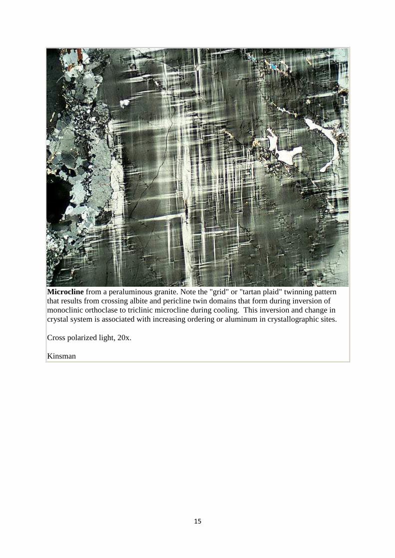
Microcline from a peraluminous granite. Note the "grid" or "tartan plaid" twinning pattern that results from crossing albite and pericline twin domains that form during inversion of monoclinic orthoclase to triclinic microcline during cooling. This inversion and change in crystal system is associated with increasing ordering or aluminum in crystallographic sites.
Cross polarized light, 20x.
Kinsman
16
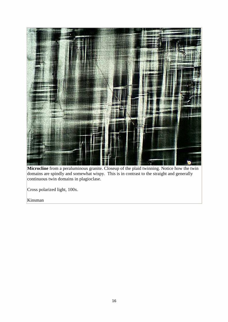
Microcline from a peraluminous granite. Closeup of the plaid twinning. Notice how the twin domains are spindly and somewhat wispy. This is in contrast to the straight and generally continuous twin domains in plagioclase.
Cross polarized light, 100x.
Kinsman
17
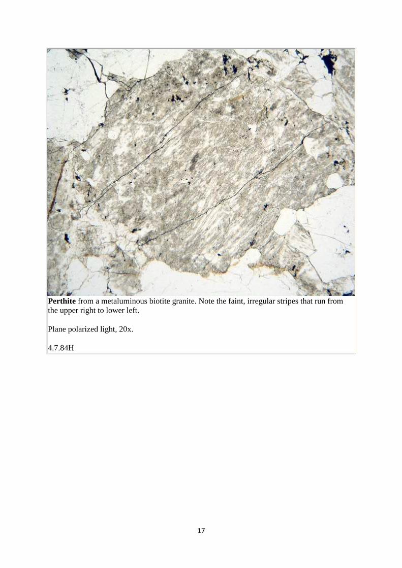
Perthite from a metaluminous biotite granite. Note the faint, irregular stripes that run from the upper right to lower left.
Plane polarized light, 20x.
4.7.84H
18
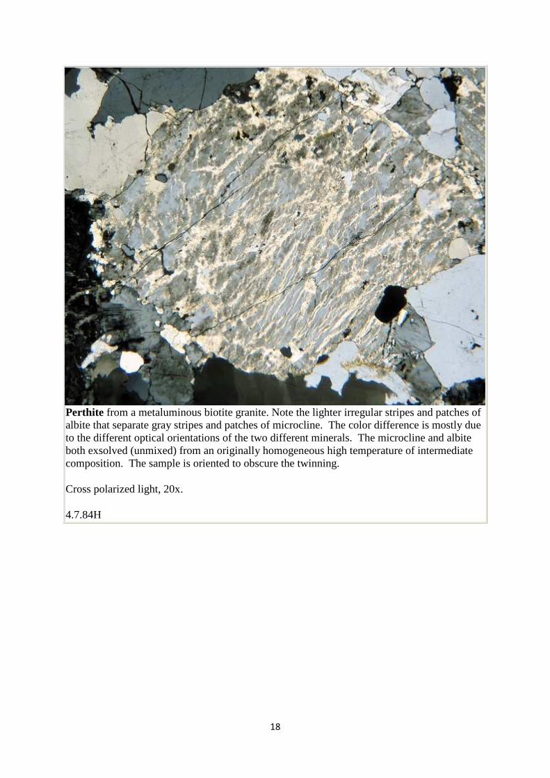
Perthite from a metaluminous biotite granite. Note the lighter irregular stripes and patches of albite that separate gray stripes and patches of microcline. The color difference is mostly due to the different optical orientations of the two different minerals. The microcline and albite both exsolved (unmixed) from an originally homogeneous high temperature of intermediate composition. The sample is oriented to obscure the twinning.
Cross polarized light, 20x.
4.7.84H
19
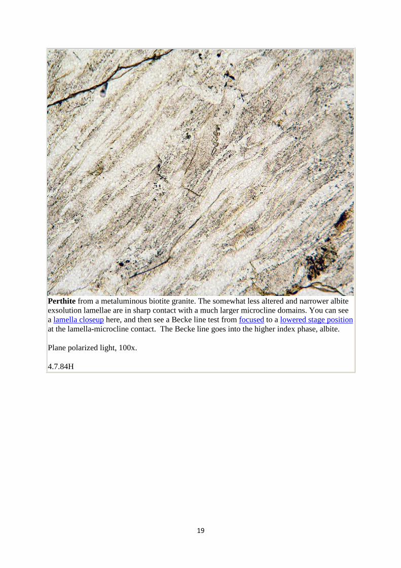
Perthite from a metaluminous biotite granite. The somewhat less altered and narrower albite exsolution lamellae are in sharp contact with a much larger microcline domains. You can see a lamella closeup here, and then see a Becke line test from focused to a lowered stage position at the lamella-microcline contact. The Becke line goes into the higher index phase, albite.
Plane polarized light, 100x.
4.7.84H
20
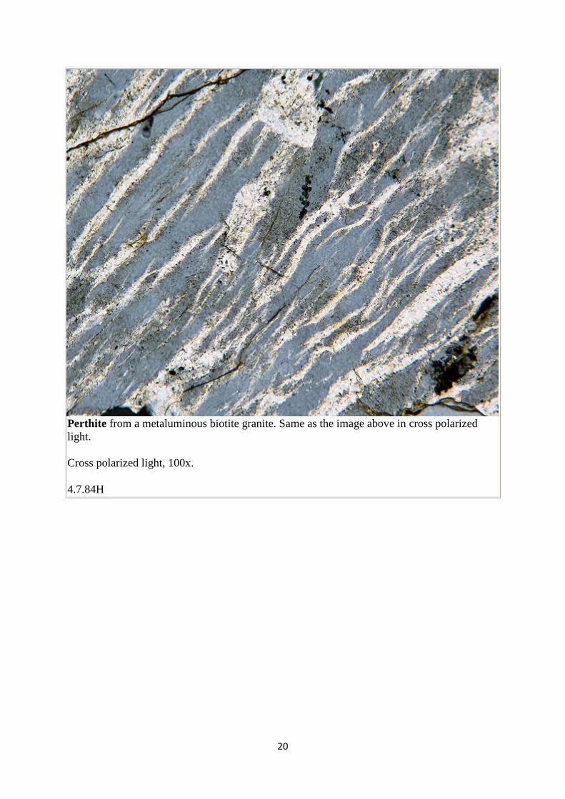
Perthite from a metaluminous biotite granite. Same as the image above in cross polarized light.
Cross polarized light, 100x.
4.7.84H
21
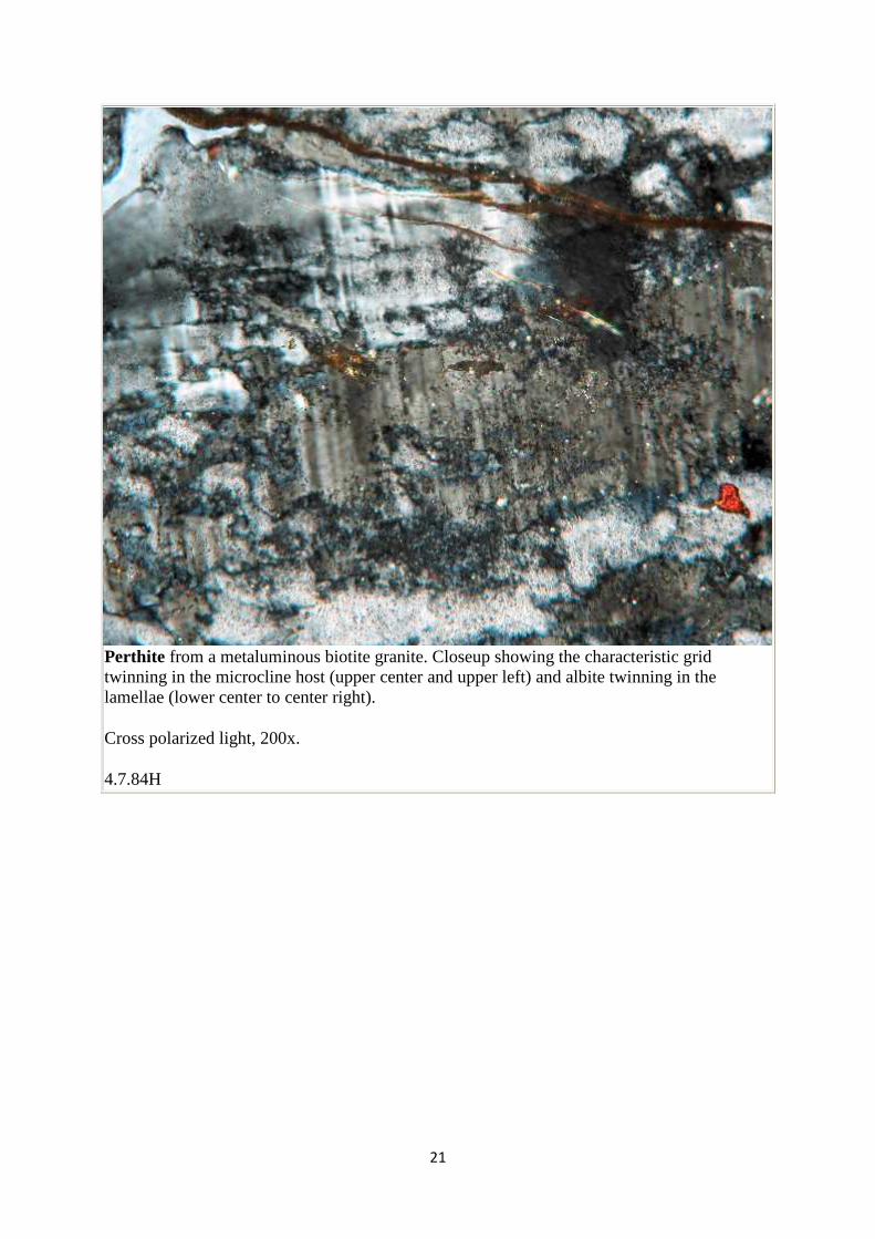
Perthite from a metaluminous biotite granite. Closeup showing the characteristic grid twinning in the microcline host (upper center and upper left) and albite twinning in the lamellae (lower center to center right).
Cross polarized light, 200x.
4.7.84H
22
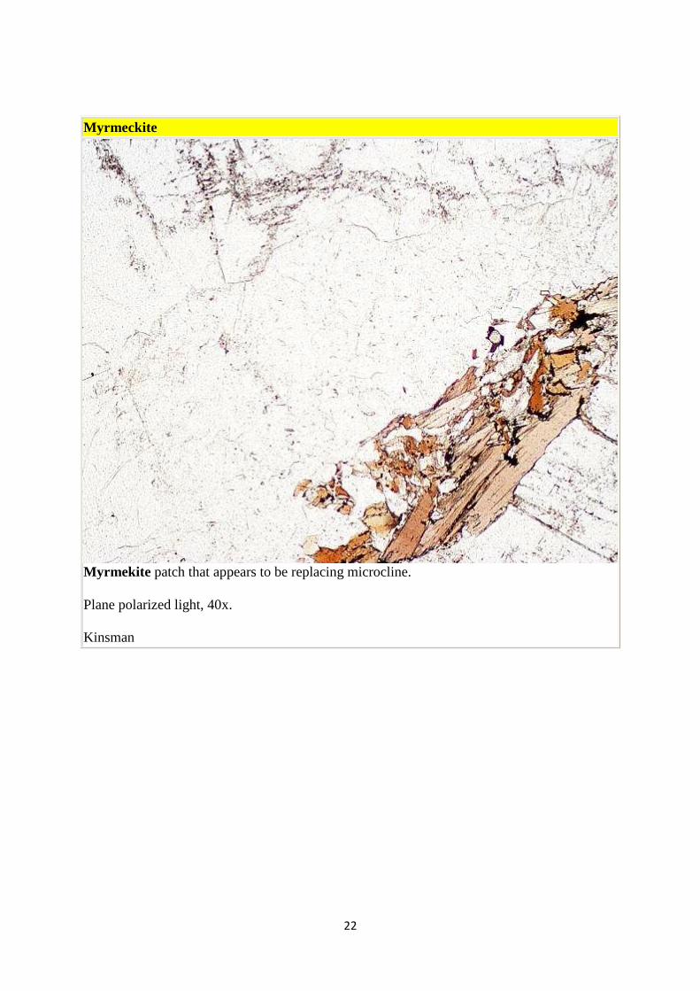
Myrmeckite
Myrmekite patch that appears to be replacing microcline.
Plane polarized light, 40x.
Kinsman
23
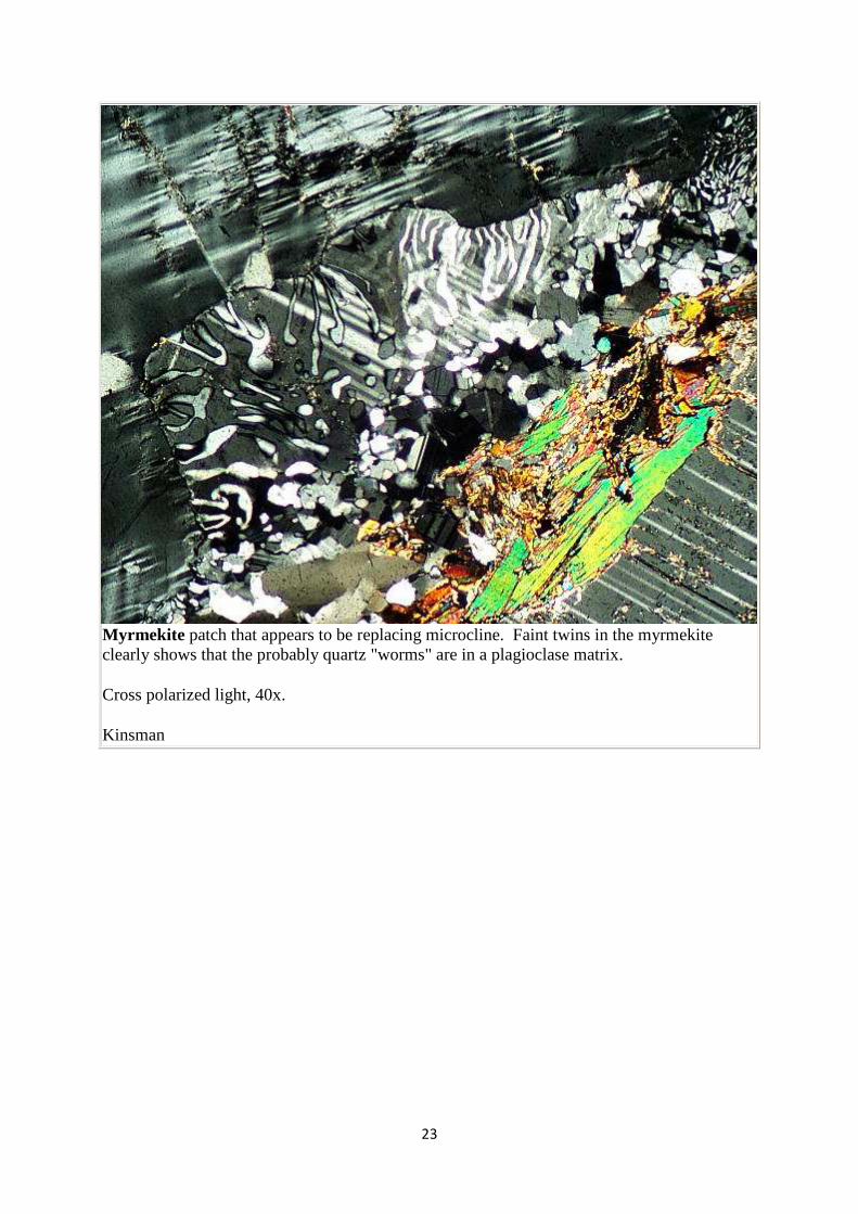
Myrmekite patch that appears to be replacing microcline. Faint twins in the myrmekite clearly shows that the probably quartz "worms" are in a plagioclase matrix.
Cross polarized light, 40x.
Kinsman
24
Micas
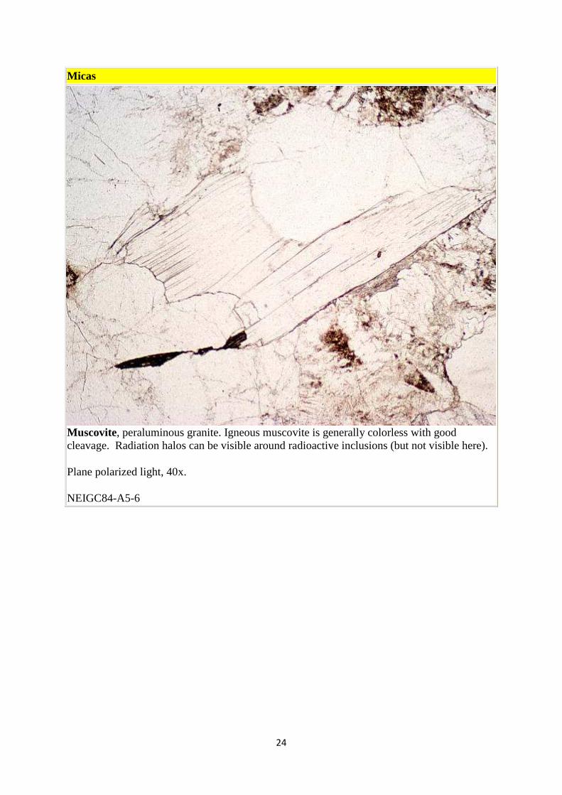
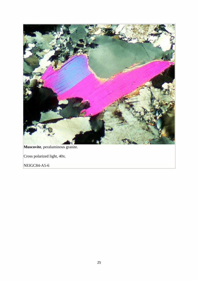
Muscovite, peraluminous granite. Igneous muscovite is generally colorless with good cleavage. Radiation halos can be visible around radioactive inclusions (but not visible here).
Plane polarized light, 40x.
NEIGC84-A5-6
25
Muscovite, peraluminous granite.
Cross polarized light, 40x.
NEIGC84-A5-6
26
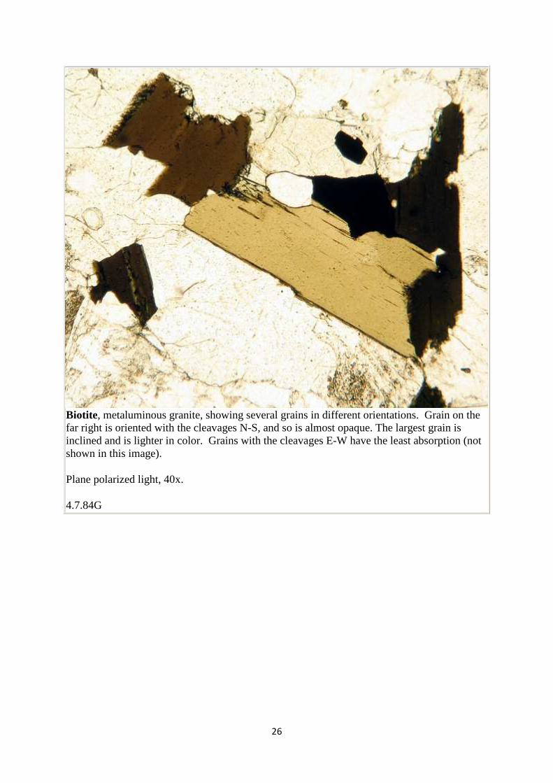
Biotite, metaluminous granite, showing several grains in different orientations. Grain on the far right is oriented with the cleavages N-S, and so is almost opaque. The largest grain is inclined and is lighter in color. Grains with the cleavages E-W have the least absorption (not shown in this image).
Plane polarized light, 40x.
4.7.84G
27
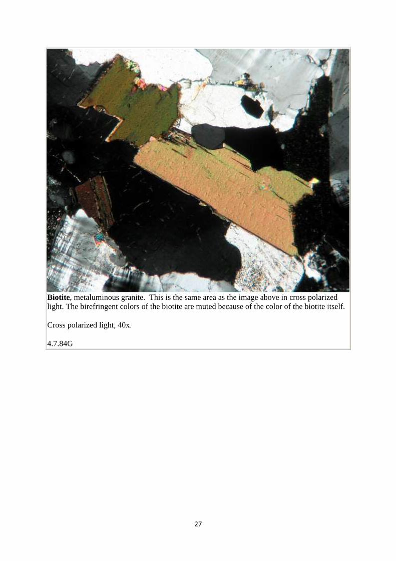
Biotite, metaluminous granite. This is the same area as the image above in cross polarized light. The birefringent colors of the biotite are muted because of the color of the biotite itself.
Cross polarized light, 40x.
4.7.84G
28
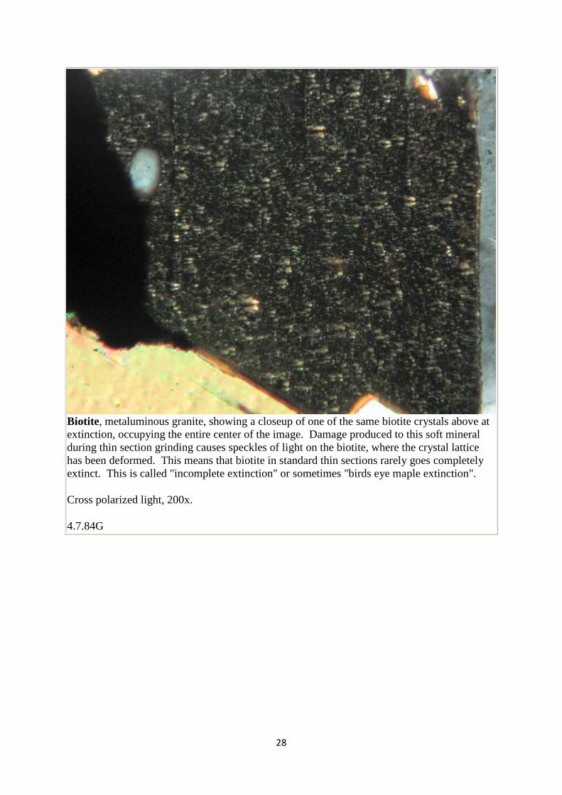
Biotite, metaluminous granite, showing a closeup of one of the same biotite crystals above at extinction, occupying the entire center of the image. Damage produced to this soft mineral during thin section grinding causes speckles of light on the biotite, where the crystal lattice has been deformed. This means that biotite in standard thin sections rarely goes completely extinct. This is called "incomplete extinction" or sometimes "birds eye maple extinction".
Cross polarized light, 200x.
4.7.84G
29
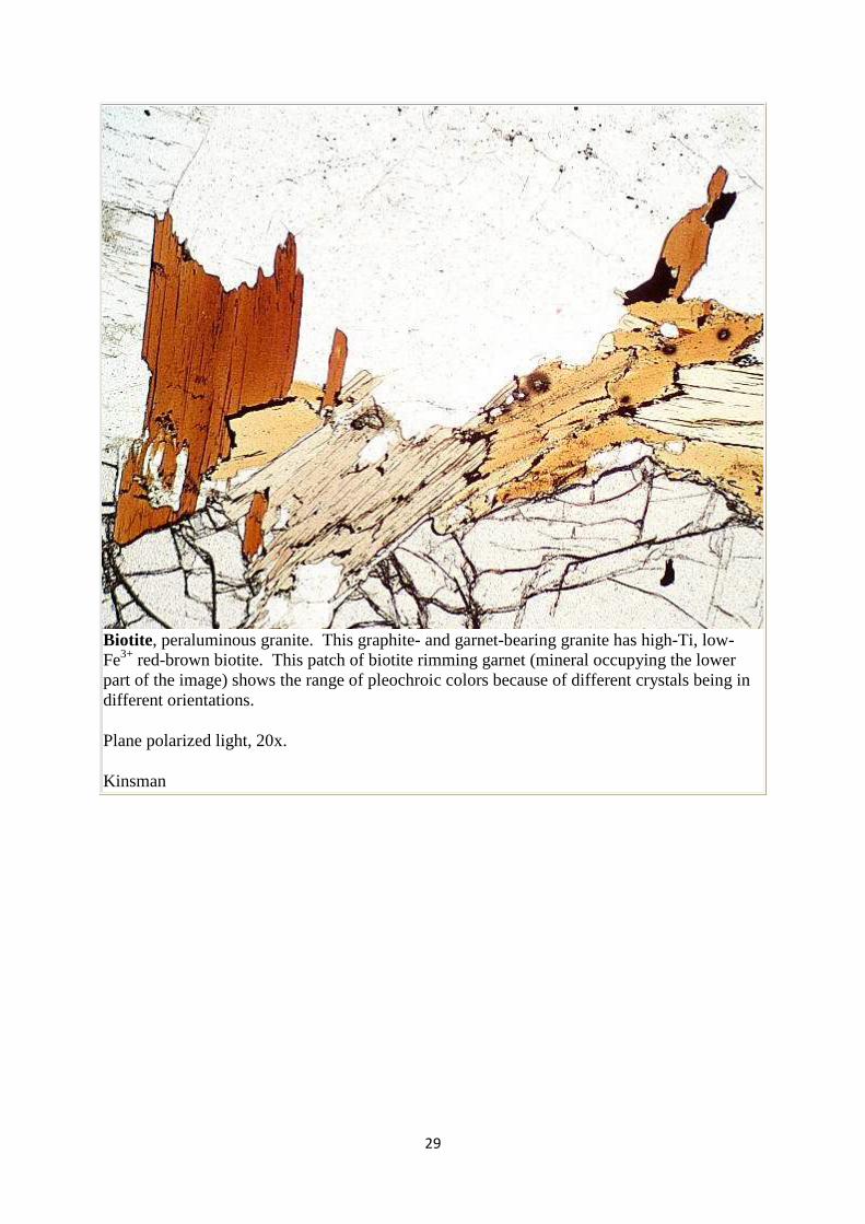
Biotite, peraluminous granite. This graphite- and garnet-bearing granite has high-Ti, low-Fe3+ red-brown biotite. This patch of biotite rimming garnet (mineral occupying the lower part of the image) shows the range of pleochroic colors because of different crystals being in different orientations.
Plane polarized light, 20x.
Kinsman
30
Amphiboles
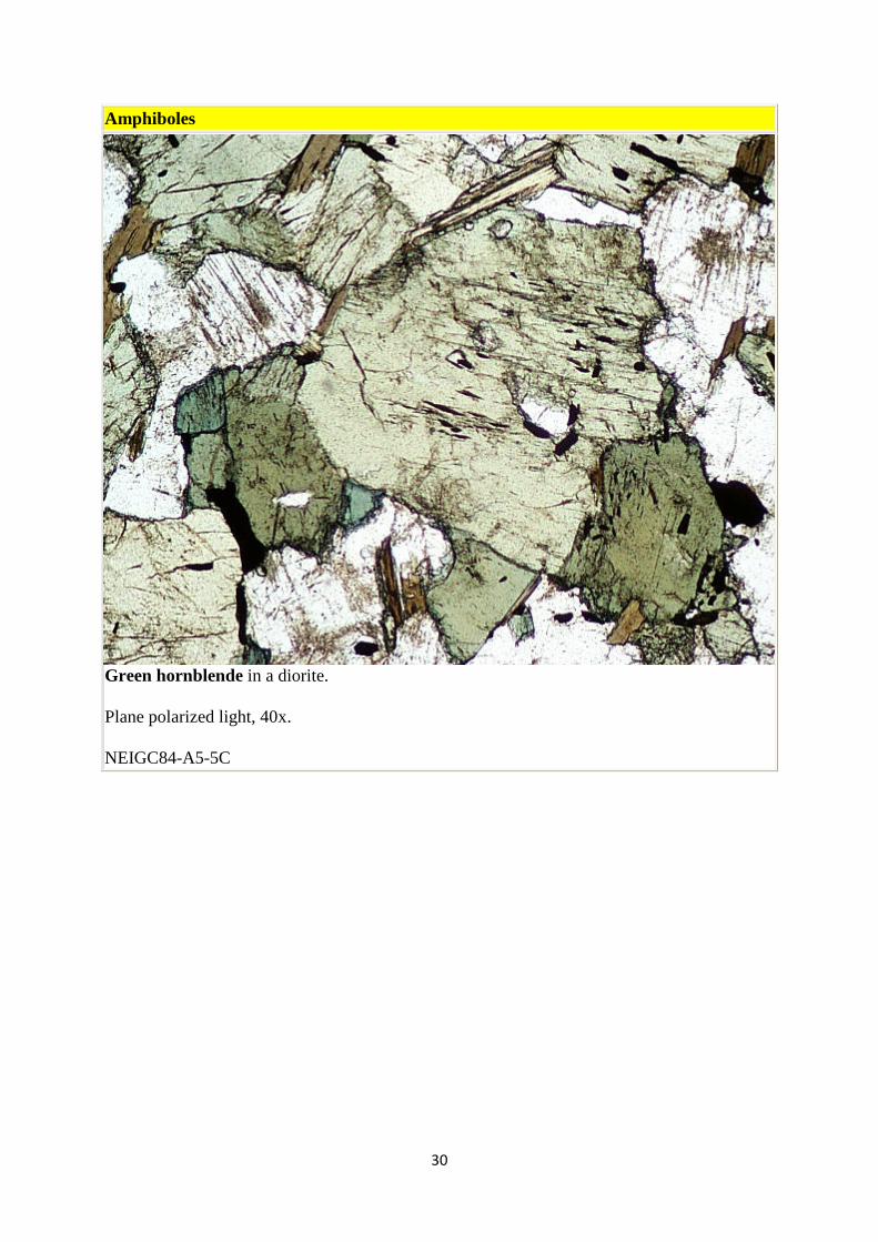
Green hornblende in a diorite.
Plane polarized light, 40x.
NEIGC84-A5-5C
31
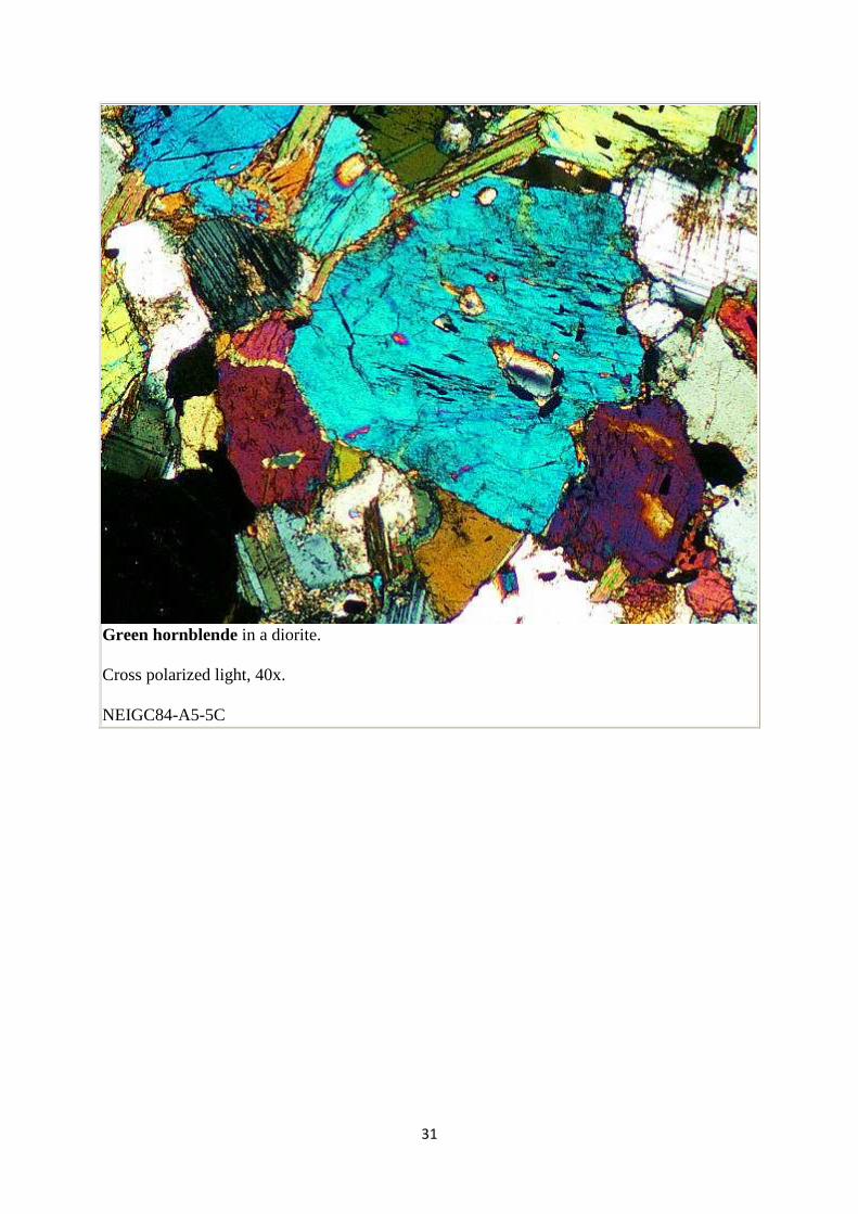
Green hornblende in a diorite.
Cross polarized light, 40x.
NEIGC84-A5-5C
32
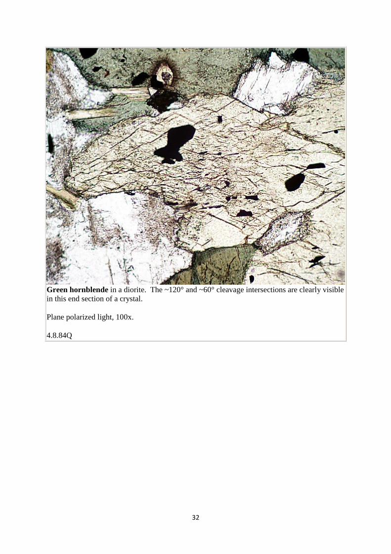
Green hornblende in a diorite. The ~120° and ~60° cleavage intersections are clearly visible in this end section of a crystal.
Plane polarized light, 100x.
4.8.84Q
33
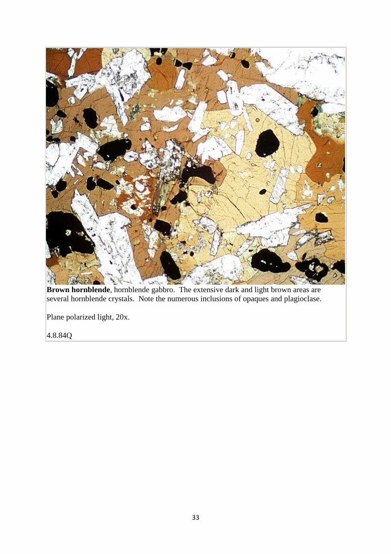
Brown hornblende, hornblende gabbro. The extensive dark and light brown areas are several hornblende crystals. Note the numerous inclusions of opaques and plagioclase.
Plane polarized light, 20x.
4.8.84Q
34
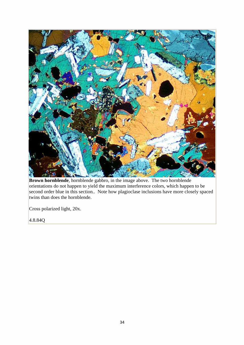
Brown hornblende, hornblende gabbro, in the image above. The two hornblende orientations do not happen to yield the maximum interference colors, which happen to be second order blue in this section.. Note how plagioclase inclusions have more closely spaced twins than does the hornblende.
Cross polarized light, 20x.
4.8.84Q
35
Pyroxenes
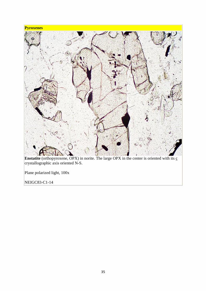
Enstatite (orthopyroxene, OPX) in norite. The large OPX in the center is oriented with its c crystallographic axis oriented N-S.
Plane polarized light, 100x
NEIGC83-C1-14
36
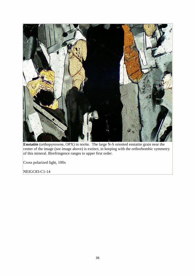
Enstatite (orthopyroxene, OPX) in norite. The large N-S oriented enstatite grain near the center of the image (see image above) is extinct, in keeping with the orthorhombic symmetry of this mineral. Birefringence ranges to upper first order.
Cross polarized light, 100x
NEIGC83-C1-14
37
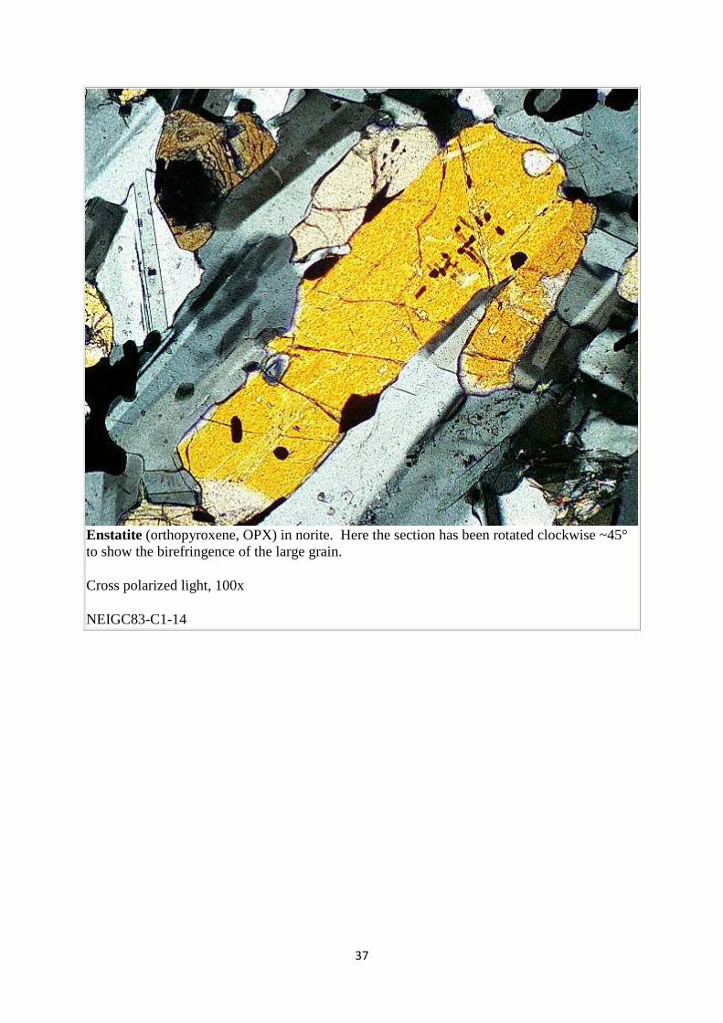
Enstatite (orthopyroxene, OPX) in norite. Here the section has been rotated clockwise ~45° to show the birefringence of the large grain.
Cross polarized light, 100x
NEIGC83-C1-14
38
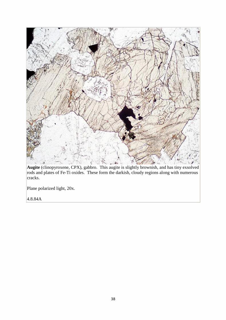
Augite (clinopyroxene, CPX), gabbro. This augite is slightly brownish, and has tiny exsolved rods and plates of Fe-Ti oxides. These form the darkish, cloudy regions along with numerous cracks.
Plane polarized light, 20x.
4.8.84A
39
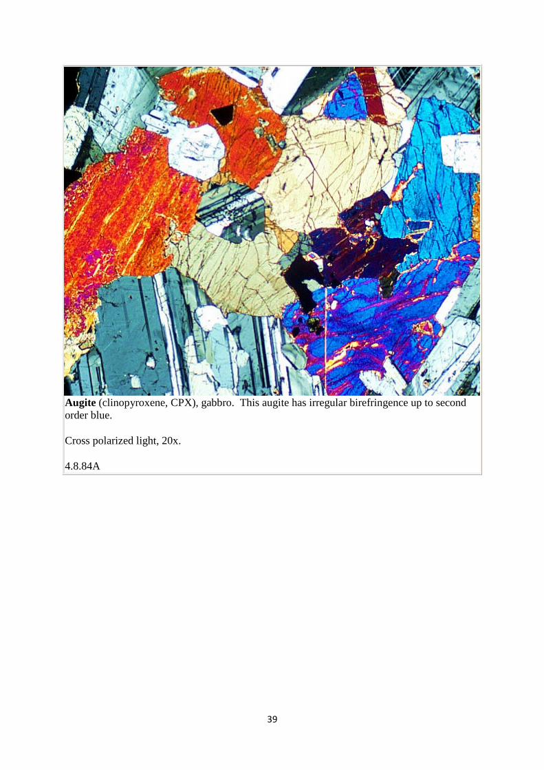
Augite (clinopyroxene, CPX), gabbro. This augite has irregular birefringence up to second order blue.
Cross polarized light, 20x.
4.8.84A
40
Other primary minerals
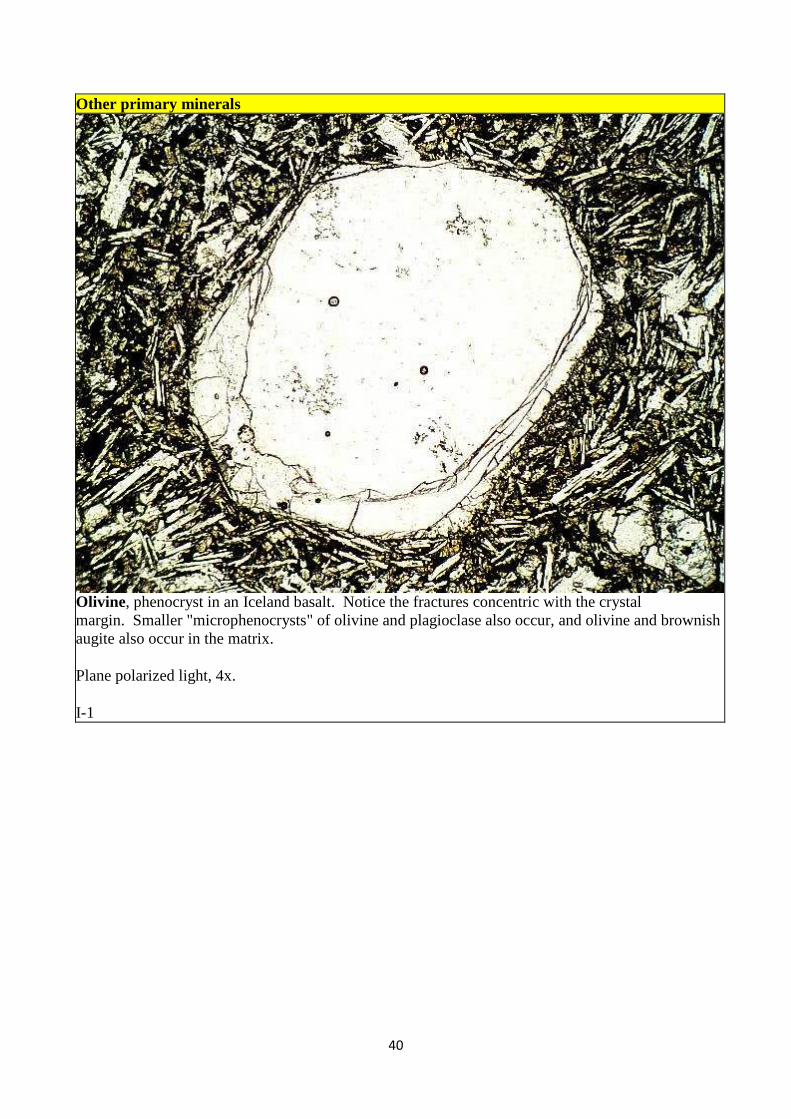
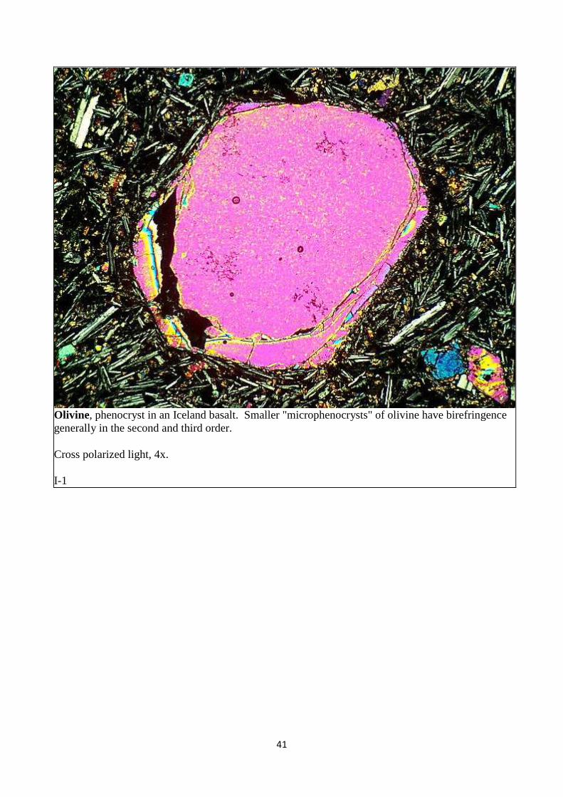
Olivine, phenocryst in an Iceland basalt. Notice the fractures concentric with the crystal margin. Smaller "microphenocrysts" of olivine and plagioclase also occur, and olivine and brownish augite also occur in the matrix.
Plane polarized light, 4x.
I-1
41
Olivine, phenocryst in an Iceland basalt. Smaller "microphenocrysts" of olivine have birefringence generally in the second and third order.
Cross polarized light, 4x.
I-1
42
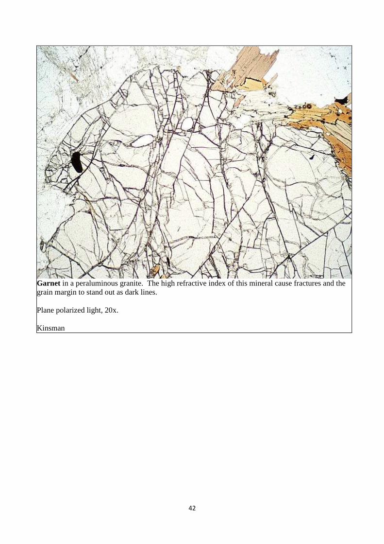
Garnet in a peraluminous granite. The high refractive index of this mineral cause fractures and the grain margin to stand out as dark lines.
Plane polarized light, 20x.
Kinsman
43
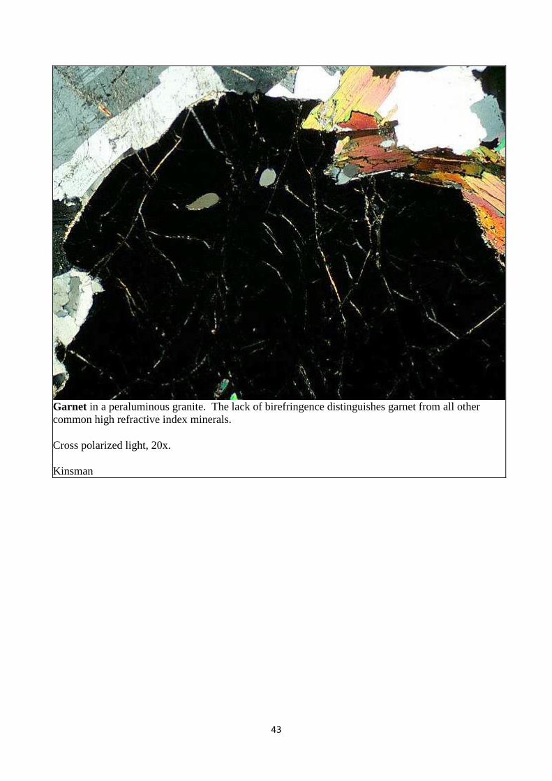
Garnet in a peraluminous granite. The lack of birefringence distinguishes garnet from all other common high refractive index minerals.
Cross polarized light, 20x.
Kinsman

44
Epidote in a calc-alkaline granodiorite. In general, epidote in igneous rocks has rather high Fe3+ content, and is therefore pistacite. It may be pale yellow-green, but more commonly it is essentially colorless. It has high relief and is commonly associated with other Ca-rich minerals such as hornblende, plagioclase, and titanite.
Plane polarized light, 100x.
UN30A
45
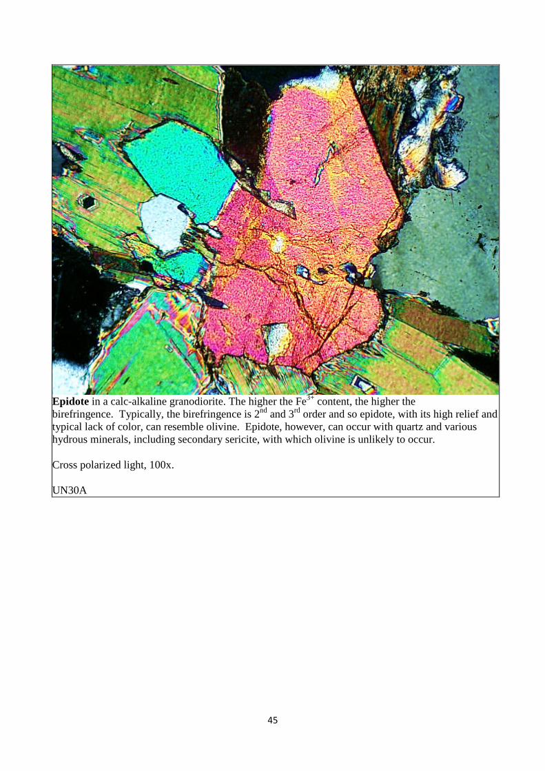
Epidote in a calc-alkaline granodiorite. The higher the Fe3+ content, the higher the birefringence. Typically, the birefringence is 2nd and 3rd order and so epidote, with its high relief and typical lack of color, can resemble olivine. Epidote, however, can occur with quartz and various hydrous minerals, including secondary sericite, with which olivine is unlikely to occur.
Cross polarized light, 100x.
UN30A
46
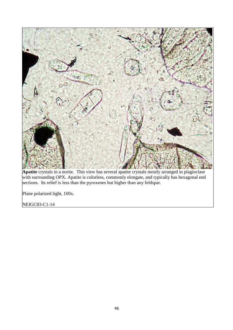
Apatite crystals in a norite. This view has several apatite crystals mostly arranged in plagioclase with surrounding OPX. Apatite is colorless, commonly elongate, and typically has hexagonal end sections. Its relief is less than the pyroxenes but higher than any feldspar.
Plane polarized light, 100x.
NEIGC83-C1-14
47
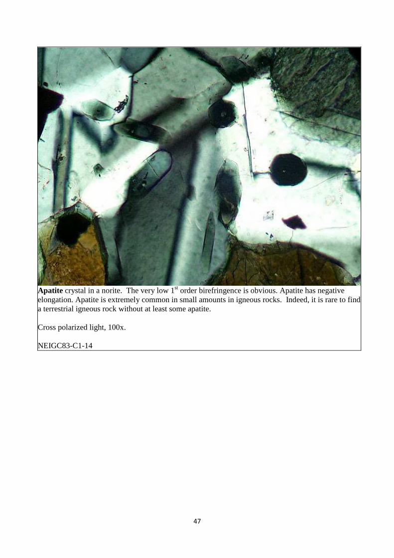
Apatite crystal in a norite. The very low 1st order birefringence is obvious. Apatite has negative elongation. Apatite is extremely common in small amounts in igneous rocks. Indeed, it is rare to find a terrestrial igneous rock without at least some apatite.
Cross polarized light, 100x.
NEIGC83-C1-14
48
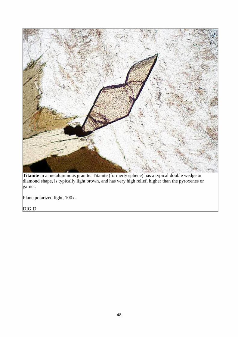
Titanite in a metaluminous granite. Titanite (formerly sphene) has a typical double wedge or diamond shape, is typically light brown, and has very high relief, higher than the pyroxenes or garnet.
Plane polarized light, 100x.
DIG-D
49
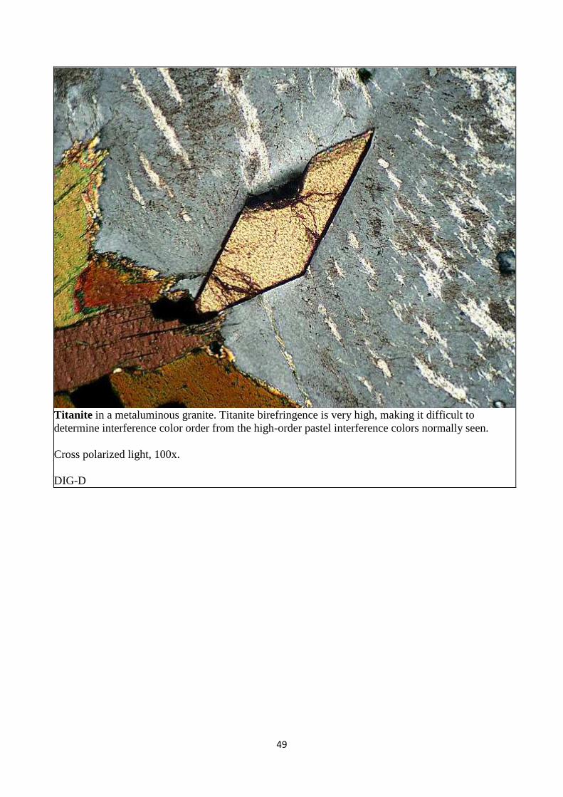
Titanite in a metaluminous granite. Titanite birefringence is very high, making it difficult to determine interference color order from the high-order pastel interference colors normally seen.
Cross polarized light, 100x.
DIG-D
50
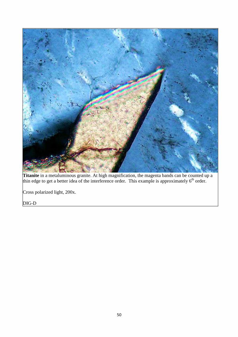
Titanite in a metaluminous granite. At high magnification, the magenta bands can be counted up a thin edge to get a better idea of the interference order. This example is approximately 6th order.
Cross polarized light, 200x.
DIG-D
51
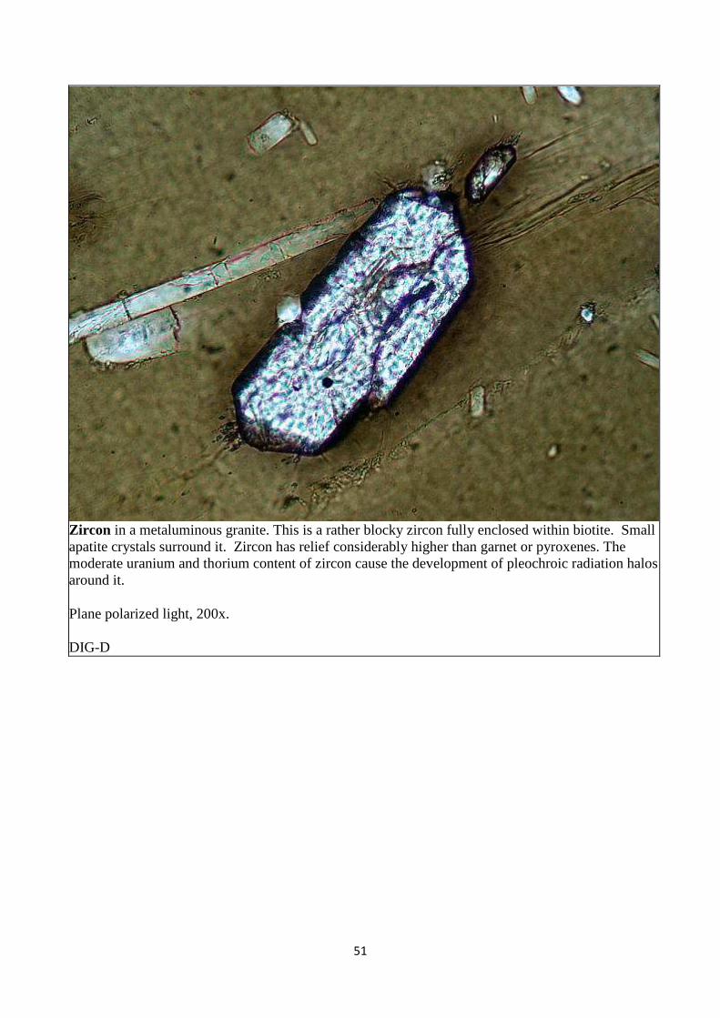
Zircon in a metaluminous granite. This is a rather blocky zircon fully enclosed within biotite. Small apatite crystals surround it. Zircon has relief considerably higher than garnet or pyroxenes. The moderate uranium and thorium content of zircon cause the development of pleochroic radiation halos around it.
Plane polarized light, 200x.
DIG-D
52
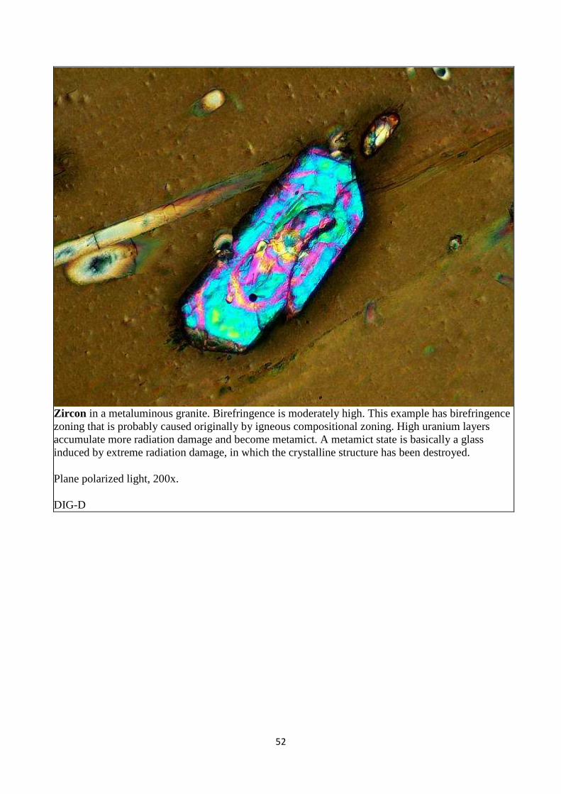
Zircon in a metaluminous granite. Birefringence is moderately high. This example has birefringence zoning that is probably caused originally by igneous compositional zoning. High uranium layers accumulate more radiation damage and become metamict. A metamict state is basically a glass induced by extreme radiation damage, in which the crystalline structure has been destroyed.
Plane polarized light, 200x.
DIG-D
53
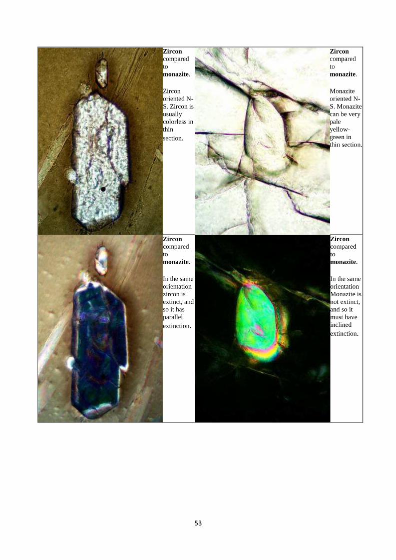
Zircon compared to monazite.
Zircon oriented N-S. Zircon is usually colorless in thin section.
Zircon compared to monazite.
Monazite oriented N-S. Monazite can be very pale yellow-green in thin section.
Zircon compared to monazite.
In the same orientation zircon is extinct, and so it has parallel extinction.
Zircon compared to monazite.
In the same orientation Monazite is not extinct, and so it must have inclined extinction.
54
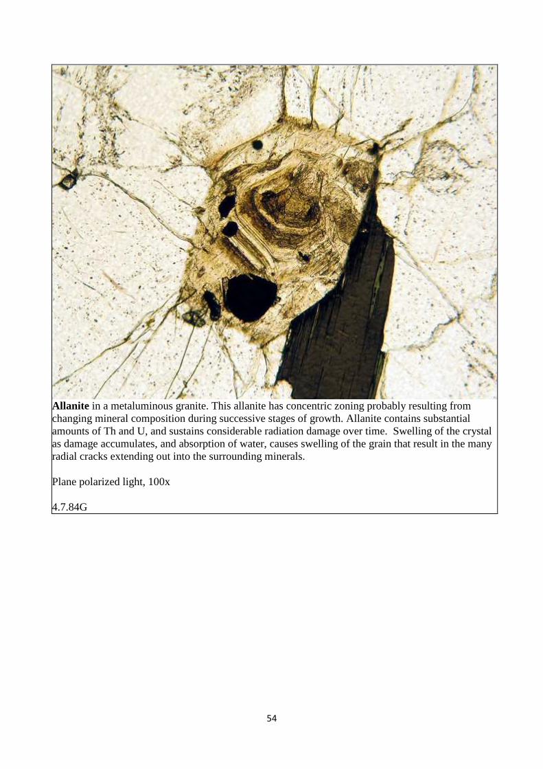
Allanite in a metaluminous granite. This allanite has concentric zoning probably resulting from changing mineral composition during successive stages of growth. Allanite contains substantial amounts of Th and U, and sustains considerable radiation damage over time. Swelling of the crystal as damage accumulates, and absorption of water, causes swelling of the grain that result in the many radial cracks extending out into the surrounding minerals.
Plane polarized light, 100x
4.7.84G
55
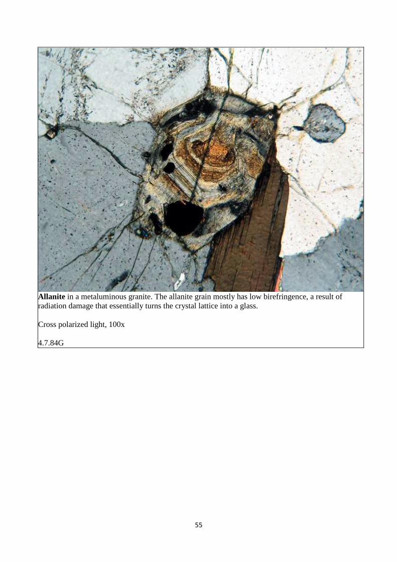
Allanite in a metaluminous granite. The allanite grain mostly has low birefringence, a result of radiation damage that essentially turns the crystal lattice into a glass.
Cross polarized light, 100x
4.7.84G
56
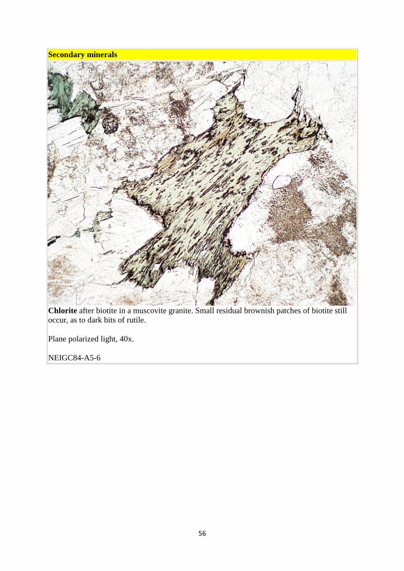
Secondary minerals
Chlorite after biotite in a muscovite granite. Small residual brownish patches of biotite still occur, as to dark bits of rutile.
Plane polarized light, 40x.
NEIGC84-A5-6
57
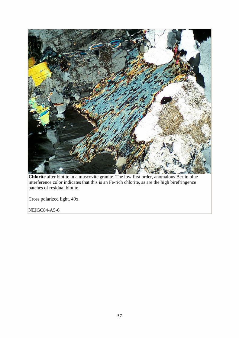
Chlorite after biotite in a muscovite granite. The low first order, anomalous Berlin blue interference color indicates that this is an Fe-rich chlorite, as are the high birefringence patches of residual biotite.
Cross polarized light, 40x.
NEIGC84-A5-6
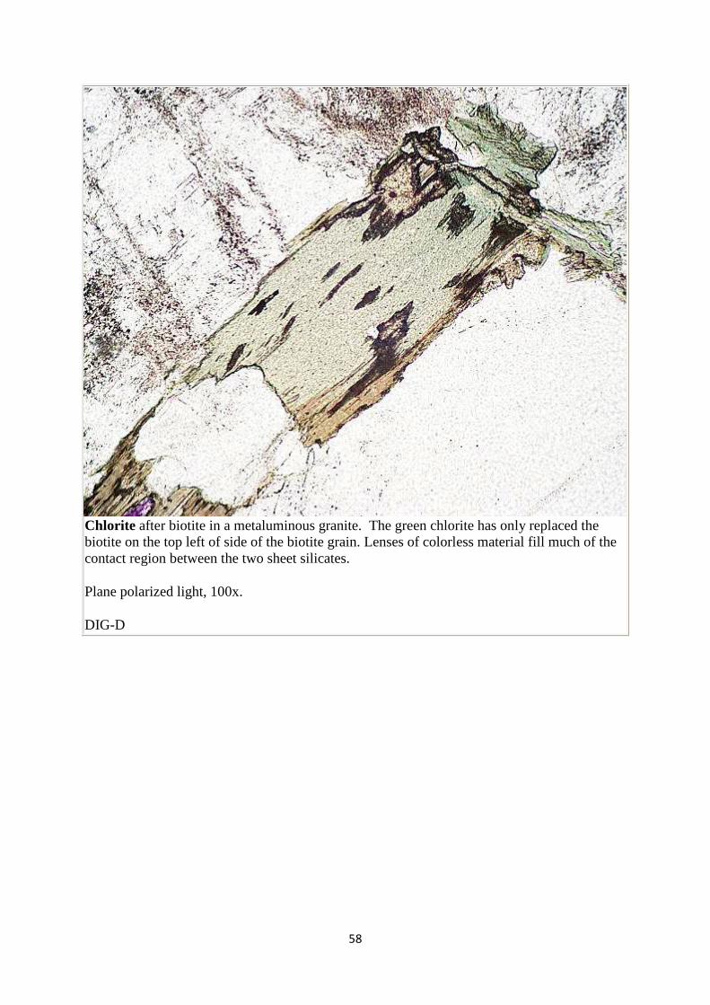
58
Chlorite after biotite in a metaluminous granite. The green chlorite has only replaced the biotite on the top left of side of the biotite grain. Lenses of colorless material fill much of the contact region between the two sheet silicates.
Plane polarized light, 100x.
DIG-D
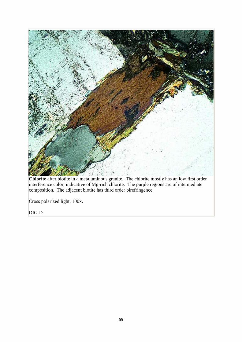
59
Chlorite after biotite in a metaluminous granite. The chlorite mostly has an low first order interference color, indicative of Mg-rich chlorite. The purple regions are of intermediate composition. The adjacent biotite has third order birefringence.
Cross polarized light, 100x.
DIG-D
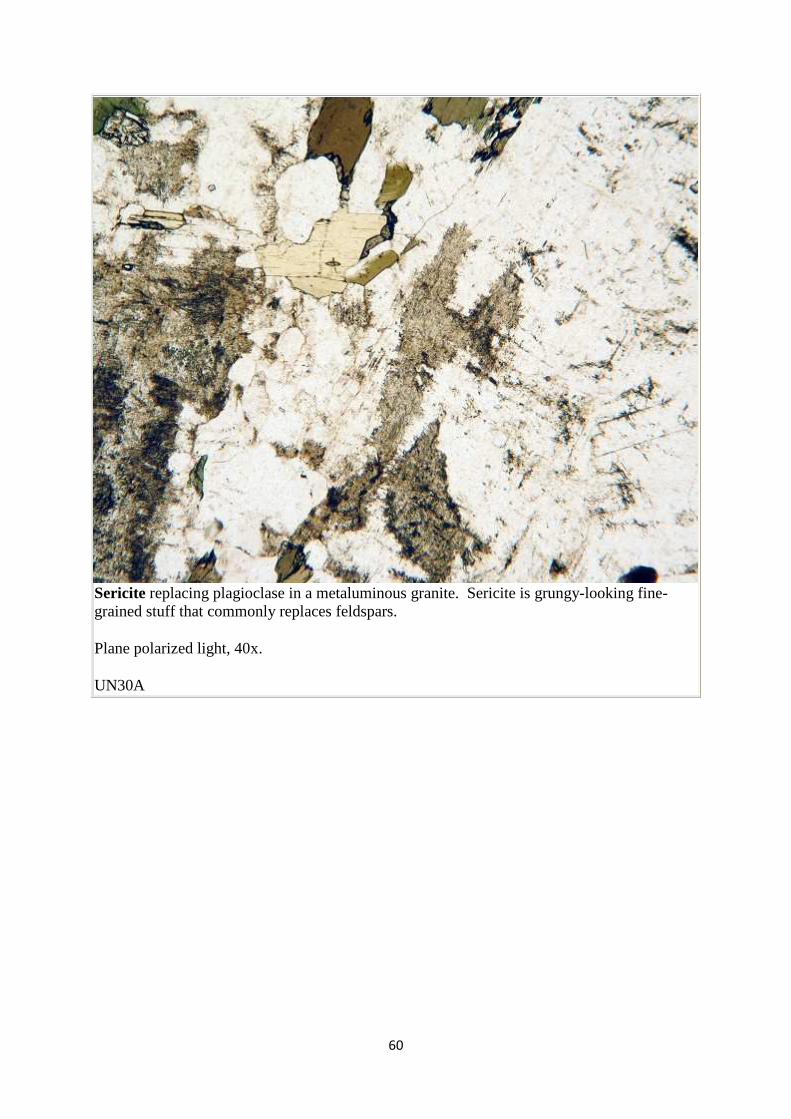
60
Sericite replacing plagioclase in a metaluminous granite. Sericite is grungy-looking fine-grained stuff that commonly replaces feldspars.
Plane polarized light, 40x.
UN30A
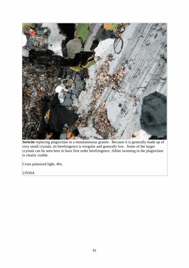
61
Sericite replacing plagioclase in a metaluminous granite. Because it is generally made up of very small crystals, its birefringence is irregular and generally low. Some of the larger crystals can be seen here to have first order birefringence. Albite twinning in the plagioclase is clearly visible.
Cross polarized light, 40x.
UN30A
62
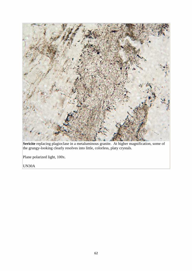
Sericite replacing plagioclase in a metaluminous granite. At higher magnification, some of the grungy-looking clearly resolves into little, colorless, platy crystals.
Plane polarized light, 100x.
UN30A
63
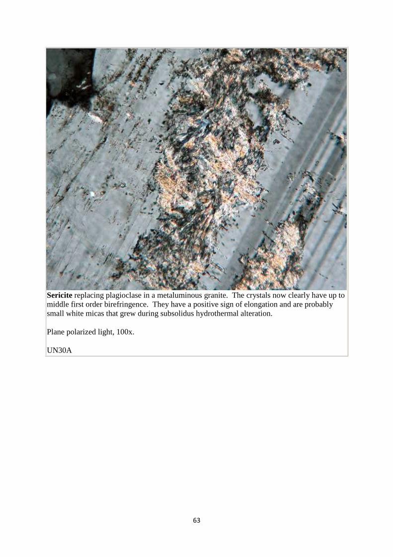
Sericite replacing plagioclase in a metaluminous granite. The crystals now clearly have up to middle first order birefringence. They have a positive sign of elongation and are probably small white micas that grew during subsolidus hydrothermal alteration.
Plane polarized light, 100x.
UN30A
64
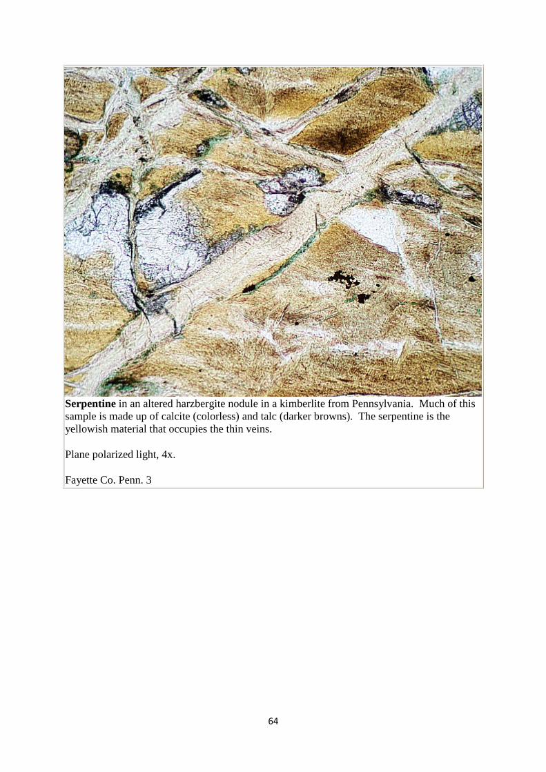
Serpentine in an altered harzbergite nodule in a kimberlite from Pennsylvania. Much of this sample is made up of calcite (colorless) and talc (darker browns). The serpentine is the yellowish material that occupies the thin veins.
Plane polarized light, 4x.
Fayette Co. Penn. 3
65
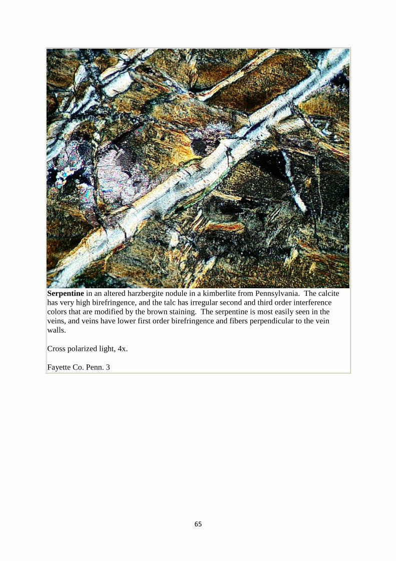
Serpentine in an altered harzbergite nodule in a kimberlite from Pennsylvania. The calcite has very high birefringence, and the talc has irregular second and third order interference colors that are modified by the brown staining. The serpentine is most easily seen in the veins, and veins have lower first order birefringence and fibers perpendicular to the vein walls.
Cross polarized light, 4x.
Fayette Co. Penn. 3

66
Calcite in an alkaline granite. In this image, the calcite grain is oriented so that a N-S line bisects the obtuse angle between the cleavages. This means the c axis of the calcite is approximately N-S, so the N-S polarized light is parallel to the low calcite refractive index. The grain has rather low relief.
Plane polarized light, 100x.
NEIGC86-B2-7
67
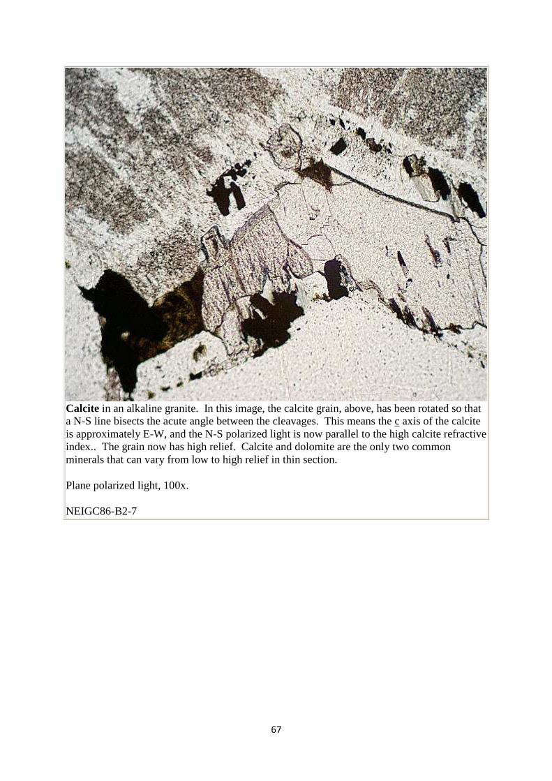
Calcite in an alkaline granite. In this image, the calcite grain, above, has been rotated so that a N-S line bisects the acute angle between the cleavages. This means the c axis of the calcite is approximately E-W, and the N-S polarized light is now parallel to the high calcite refractive index.. The grain now has high relief. Calcite and dolomite are the only two common minerals that can vary from low to high relief in thin section.
Plane polarized light, 100x.
NEIGC86-B2-7
68
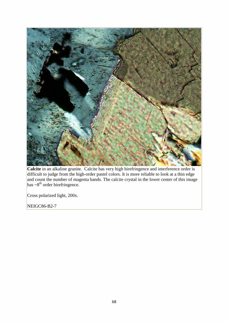
Calcite in an alkaline granite. Calcite has very high birefringence and interference order is difficult to judge from the high-order pastel colors. It is more reliable to look at a thin edge and count the number of magenta bands. The calcite crystal in the lower center of this image has ~8th order birefringence.
Cross polarized light, 200x.
NEIGC86-B2-7
69
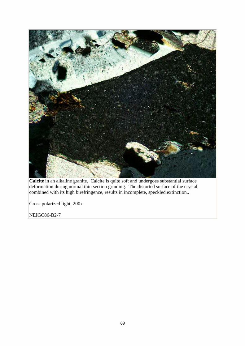
Calcite in an alkaline granite. Calcite is quite soft and undergoes substantial surface deformation during normal thin section grinding. The distorted surface of the crystal, combined with its high birefringence, results in incomplete, speckled extinction..
Cross polarized light, 200x.
NEIGC86-B2-7
70
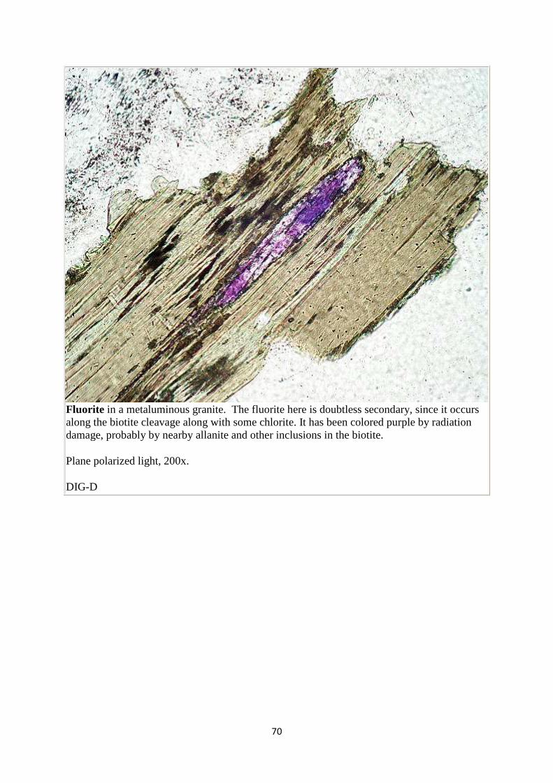
Fluorite in a metaluminous granite. The fluorite here is doubtless secondary, since it occurs along the biotite cleavage along with some chlorite. It has been colored purple by radiation damage, probably by nearby allanite and other inclusions in the biotite.
Plane polarized light, 200x.
DIG-D
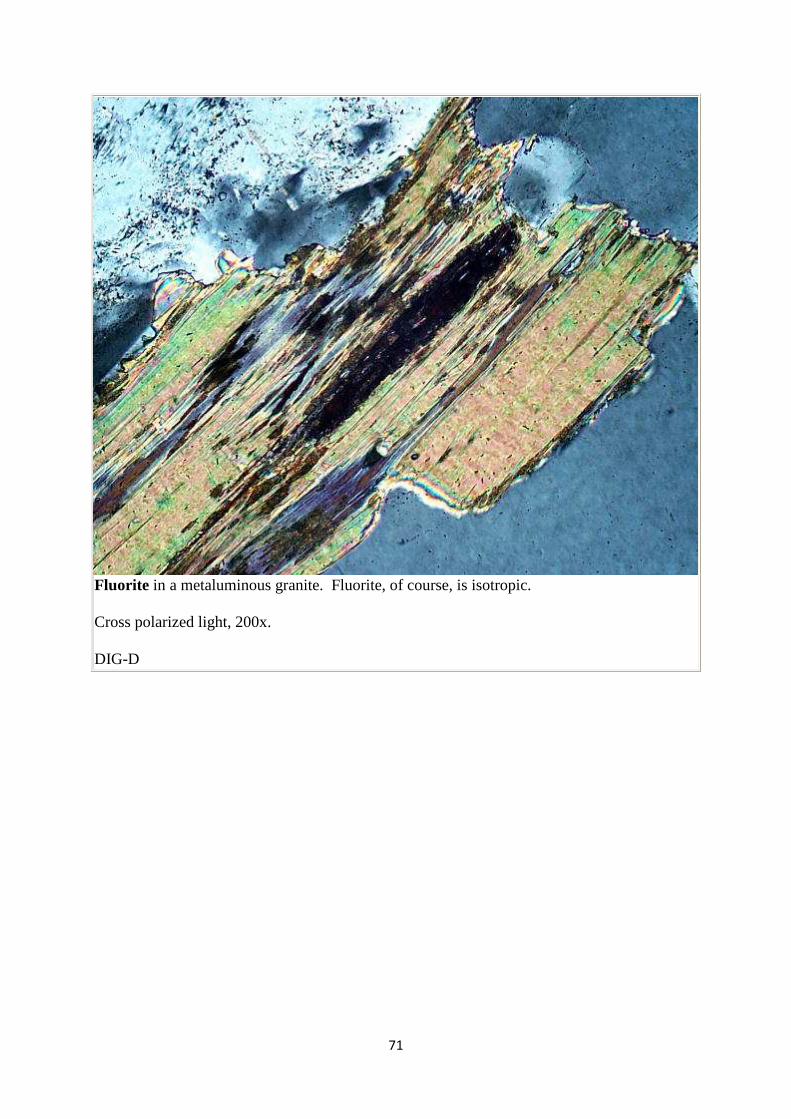
71
Fluorite in a metaluminous granite. Fluorite, of course, is isotropic.
Cross polarized light, 200x.
DIG-D


