Perianal suppuration- Abscess & Fistula
Dr Pankaj Kumar Assistant Professor
Surgical Gastroenterology
Anorectal suppurative disease may manifest as• An acute or Anal sepsis (abscess) • Anal fistula represents the chronic form of the
suppurative process• A fistula and abscess may coexist • May be associated with atypical internal
openings and multiple tracts that result in a complex suppurative process.
Anatomy
• Rectum- hind gut 6 weeks• Anal canal- 8 week – ectoderm.• Dentate line transition from endoderm to
ectoderm.• Anal canal- 4cm, pelvic diaphragm to anal
verge.• 4-8 anal glands drains by crypts at dentate line
• External sphincter: striated muscle. voluntary control
Internal sphincter: smooth muscle Autonomic control contracted at rest
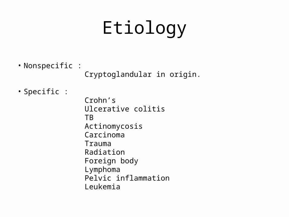
Etiology
• Nonspecific : Cryptoglandular in origin.
• Specific : Crohn’s Ulcerative colitis TB Actinomycosis Carcinoma Trauma Radiation Foreign body Lymphoma Pelvic inflammation Leukemia
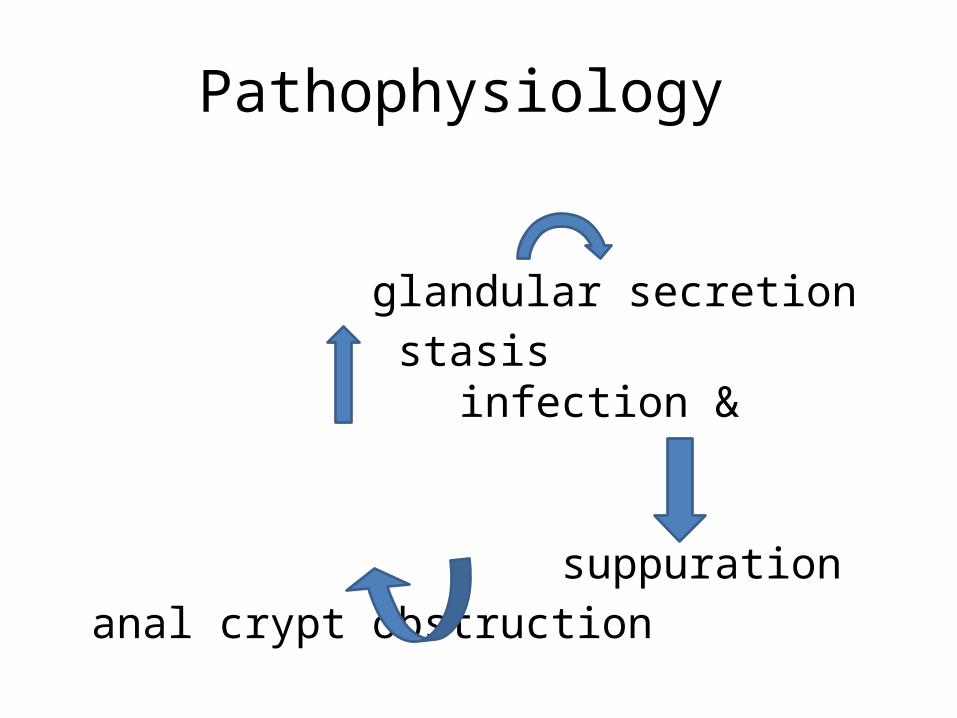
Pathophysiology
glandular secretion stasis infection & suppuration anal crypt obstruction abscess formation
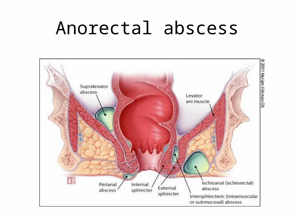
Anorectal abscess
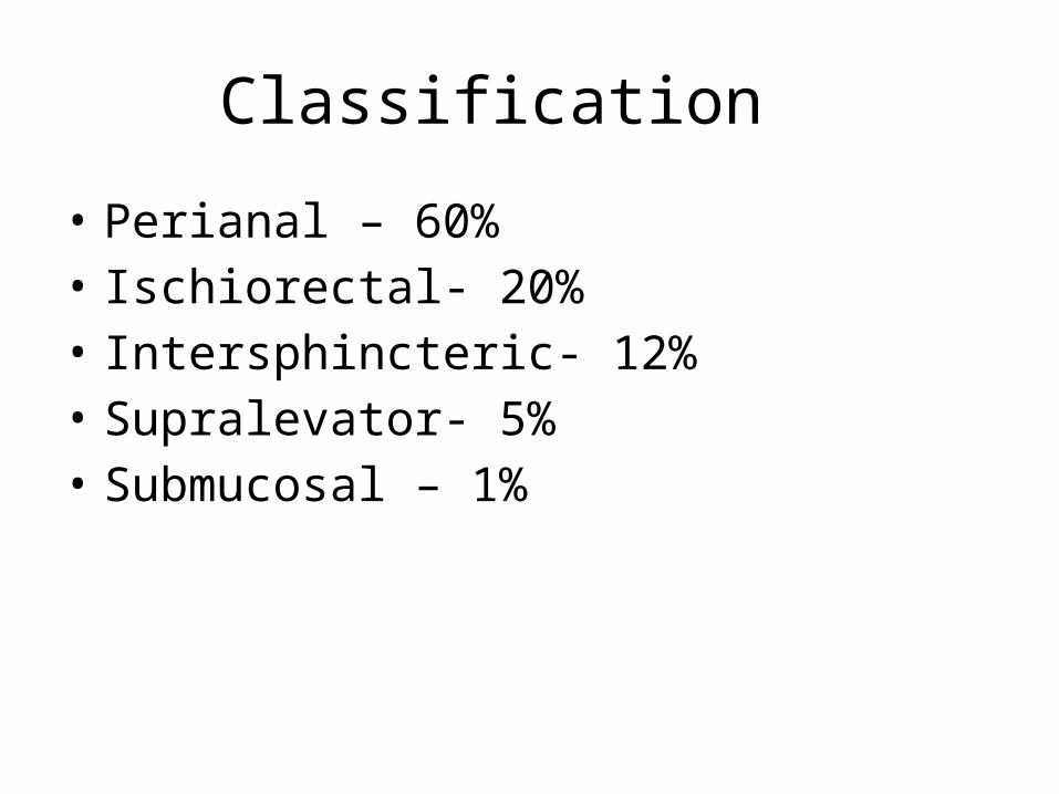
Classification
• Perianal – 60%• Ischiorectal- 20%• Intersphincteric- 12% • Supralevator- 5%• Submucosal – 1%

DIAGNOSIS
History• gradual onset of pain• sensation of pressure and fullness• fever• previous episode of anorectal sepsis
Physical Examination
• Localized swelling, • Hyperemia, • Induration• Tenderness• DRE &/or PV examination• Examination in GA• Can be confirmed by needle aspiration.
TREATMENT
• should be considered a surgical emergency• Incision and drainage.• Antibiotics as adjunctive therapy – valvular heart disease immunosuppression extensive associated cellulitis diabetes
• Perianal Abscess :cruciate incision at tender or fluctuant point as close to the anal verge.
• Ischiorectal Abscess • Intersphincteric Abscess : internal
sphincterotomy overlying the cavity.• Submucosal Abscess : incising the mucosa
over the abscess.
Postanal Abscess and Horseshoe Extension
• Hanley's technique -the posterior midline incision
• muscles attached to the coccyx, the superficial external sphincter, and the lower edge of the internal sphincter are divided
• one or multiple secondary incisions in the skin overlying the ischiorectal space.
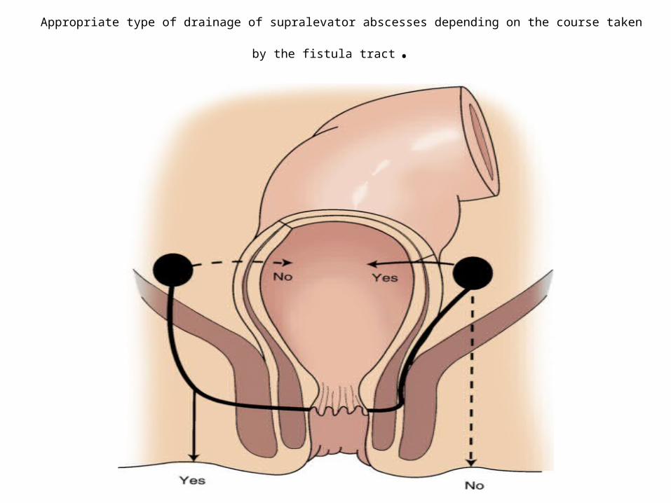
Appropriate type of drainage of supralevator abscesses depending on the course taken by the fistula tract.
Fistula in ano
• a communication between an internal opening in the anal canal and an external opening through which an abscess drained
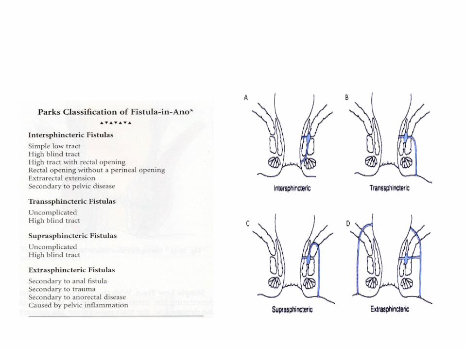
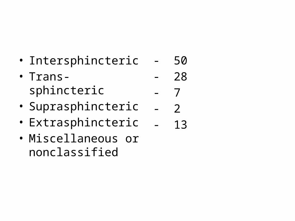
• Intersphincteric• Trans-sphincteric• Suprasphincteric• Extrasphincteric• Miscellaneous or
nonclassified
- 50- 28- 7- 2- 13
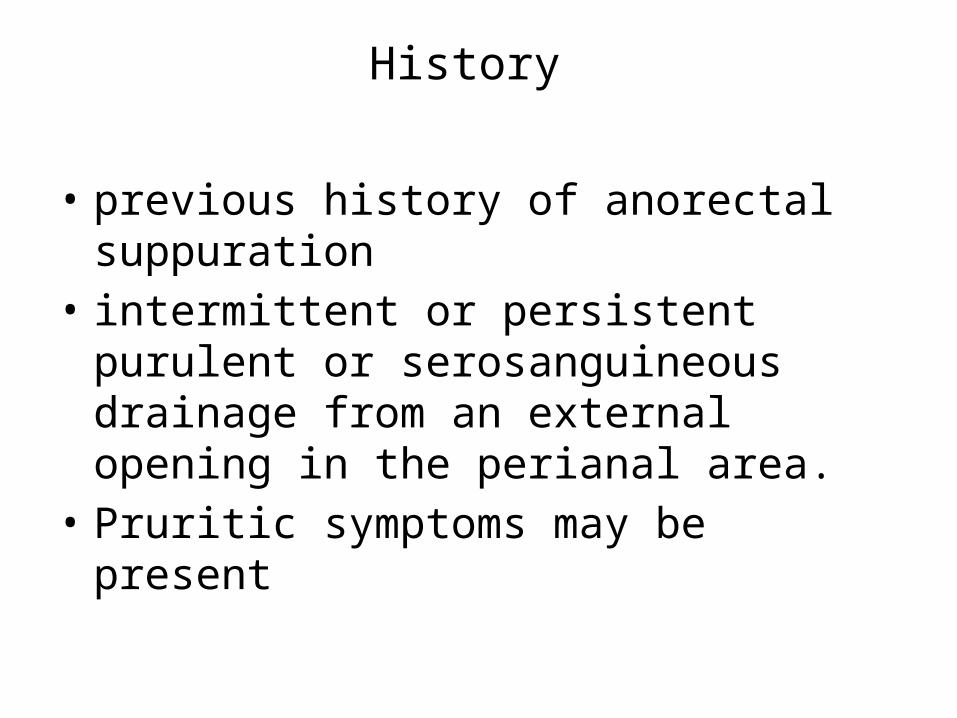
History
• previous history of anorectal suppuration• intermittent or persistent purulent or
serosanguineous drainage from an external opening in the perianal area.
• Pruritic symptoms may be present
Examination
• Perianal examination• DRE• Anoscopy
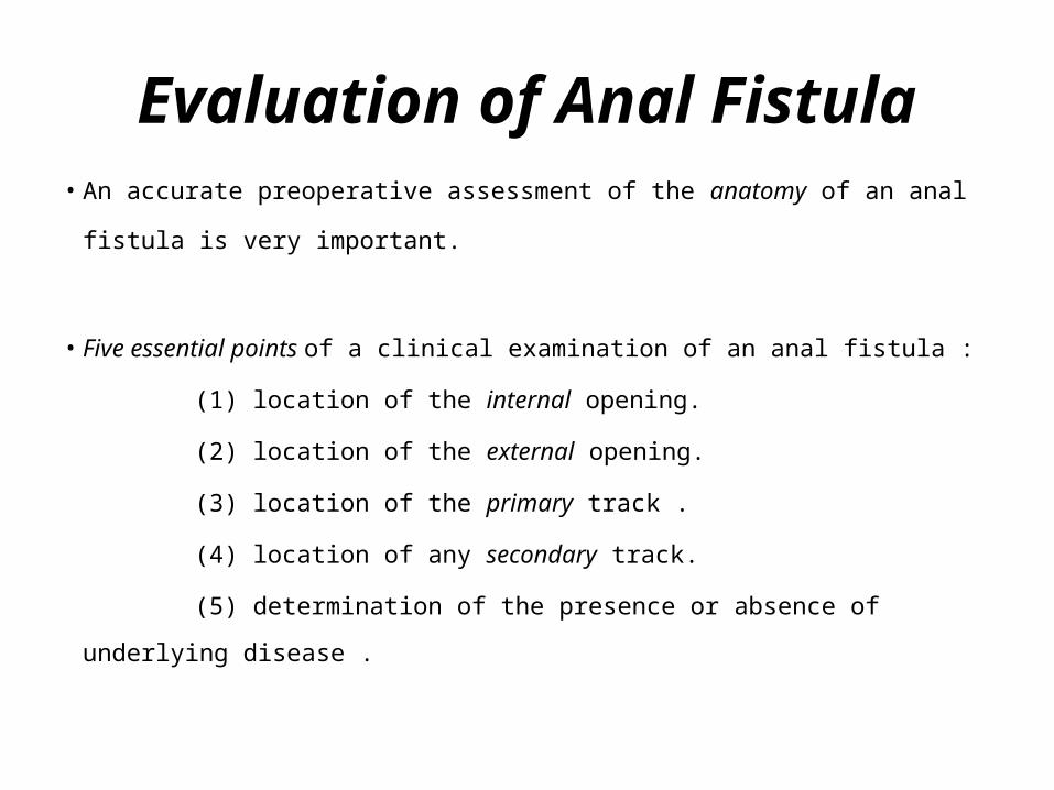
Evaluation of Anal Fistula• An accurate preoperative assessment of the anatomy of an anal fistula is
very important.
• Five essential points of a clinical examination of an anal fistula :
(1) location of the internal opening.
(2) location of the external opening.
(3) location of the primary track .
(4) location of any secondary track.
(5) determination of the presence or absence of underlying disease .
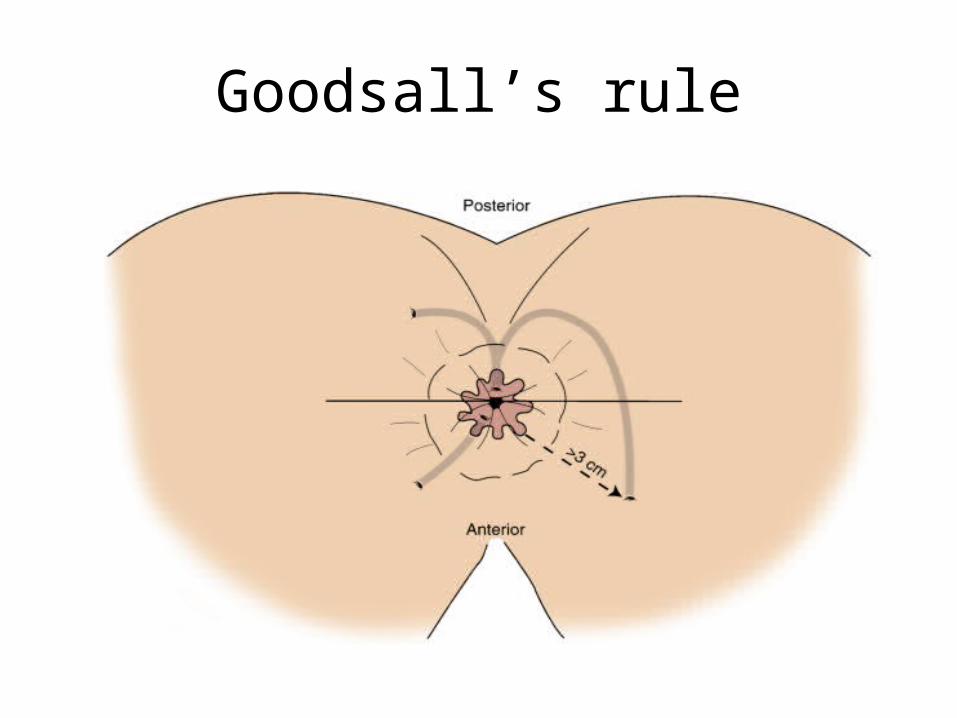
Goodsall’s rule
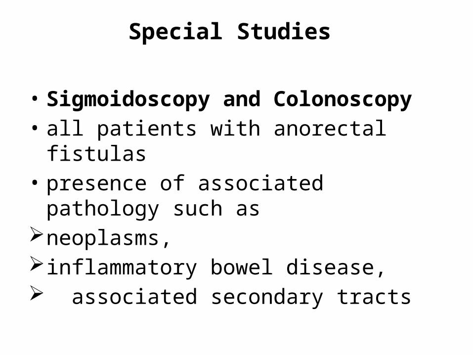
Special Studies
• Sigmoidoscopy and Colonoscopy • all patients with anorectal fistulas• presence of associated pathology such as neoplasms, inflammatory bowel disease, associated secondary tracts
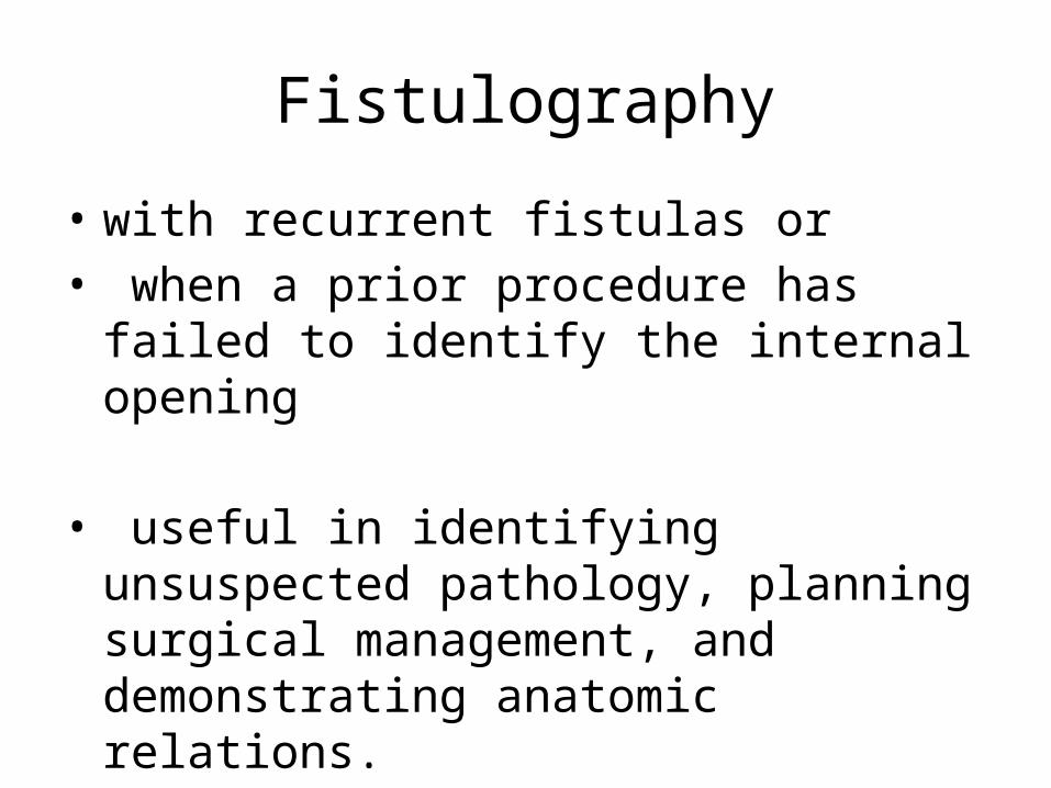
Fistulography
• with recurrent fistulas or• when a prior procedure has failed to identify
the internal opening
• useful in identifying unsuspected pathology, planning surgical management, and demonstrating anatomic relations.
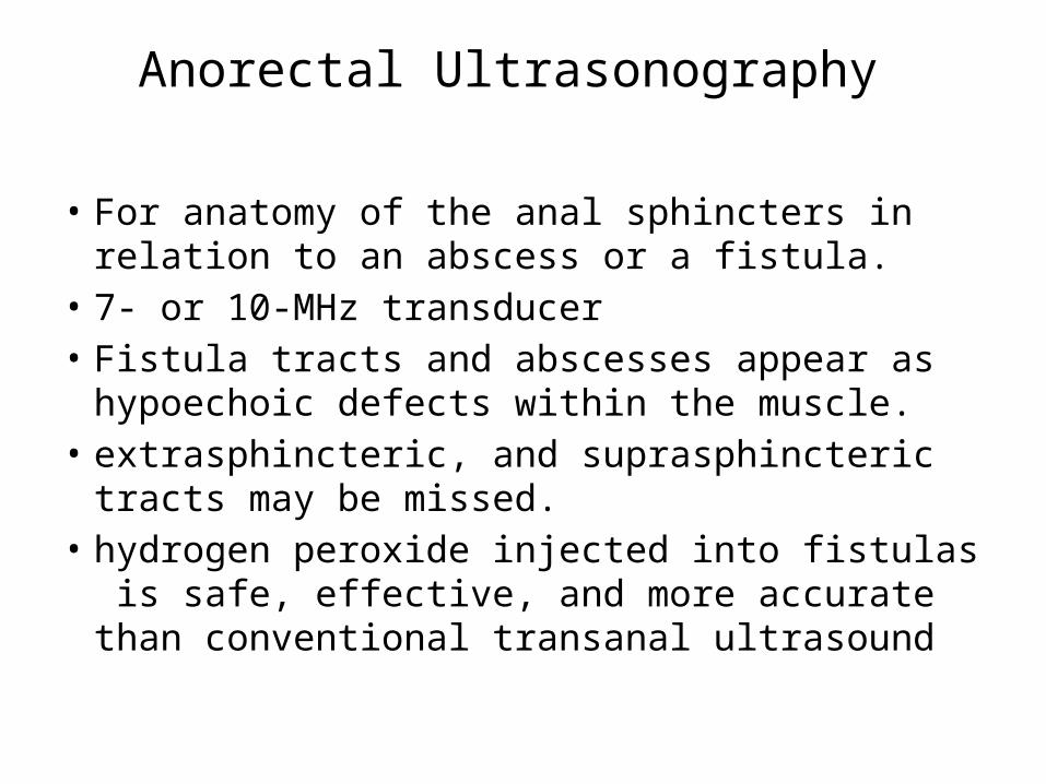
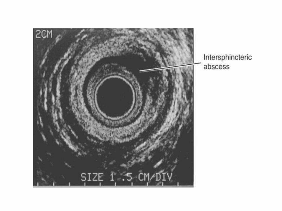
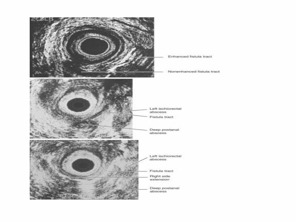
Anorectal Ultrasonography
• For anatomy of the anal sphincters in relation to an abscess or a fistula.
• 7- or 10-MHz transducer • Fistula tracts and abscesses appear as
hypoechoic defects within the muscle.• extrasphincteric, and suprasphincteric tracts may
be missed.• hydrogen peroxide injected into fistulas is safe,
effective, and more accurate than conventional transanal ultrasound
Magnetic Resonance Imaging
• for anatomy• chronic or recurrent fistula• saline solution as a contrast agent • gadolinium enema: enhanced T2 images and
improved lesion identification
• Computed Tomography: • Limited due to poor visualization of the levators
and sphincter complex.• For assessment of associated pelvic pathology
Anorectal Manometry
• assist in identifying patients at the risk for postoperative incontinence.
• Surgical management can be tailored accordingly, improving clinical and functional outcome.
Indications:• suspected sphincter impairment; • needing substantial portions of the external sphincter
divided for fistula cure; • women with a history of multiparity, forceps delivery,
third-degree perineal tear, high birthweight, or prolonged second stage of labor.
Fistuloscopy
• intraoperative technique to identify primary fistula openings, multiple or complex tracts, and iatrogenic tracts
TREATMENT
• should undergo surgical treatment, rarely heal spontaneously.
• prone jackknife position • General, regional, or local anesthesia • The three basic surgical techniques are fistulotomy, use of a seton, endorectal advancement flaps
Fistulotomy
• Most fistulas may be adequately treated. • Recurrence rates are low, • Risks for continence disturbances are minimal• cautery is used to lay it open.• Secondary tracts should be drained through
the fistulotomy incision
Seton Management
• Foreign material that is inserted into the fistula tract to encircle the sphincter muscles.
Indications• Complex anorectal fistulas with risk of
incontinence.• Poor healing: Crohn's disease, immunocompromised incontinent patients, patients with chronic diarrheal states, anterior fistulas in women
• Setons may be used as marking, draining, cutting, staging
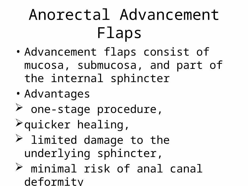
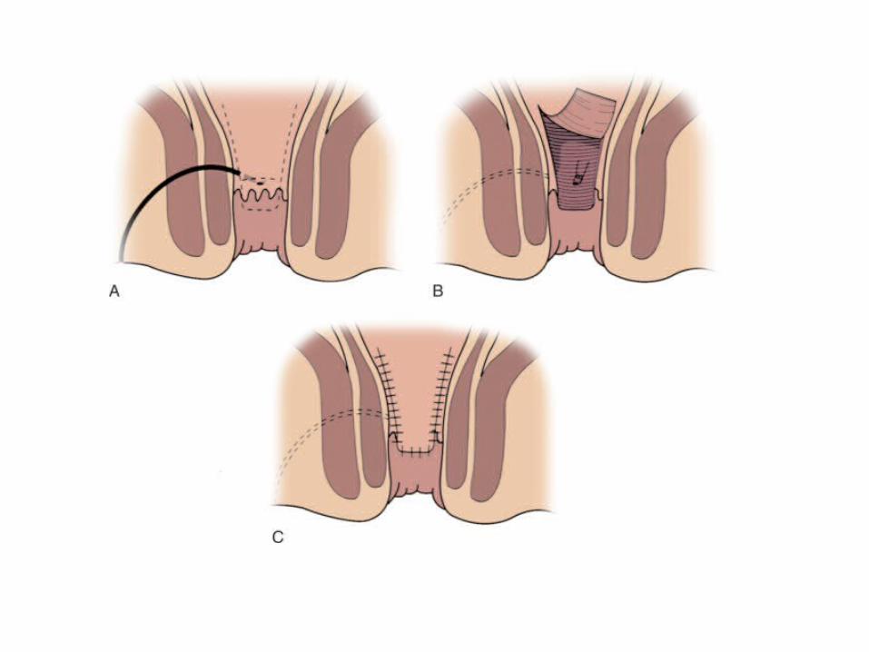
Anorectal Advancement Flaps
• Advancement flaps consist of mucosa, submucosa, and part of the internal sphincter
• Advantages one-stage procedure, quicker healing, limited damage to the underlying sphincter, minimal risk of anal canal deformity
Fibrin Glue
• mixture of fibrinogen and thrombin is injected into the fistula tract .
• POSTOPERATIVE CARE • Sitz baths -for perianal hygiene and comfort. • pain management and • wound care
COMPLICATIONS
• Urinary retention -most common 25% • hemorrhage, • acute external thrombosed hemorrhoids,• cellulitis, • fecal impaction, • stricture, • rectovaginal fistula, • incontinence • recurrence. (3% to 7%).
Sepsis and Fistula in Human Immunodeficiency Virus Disease
• Incidence -6% to 34%.• wound healing increases as the preoperative
CD4+ count increases.• In the absence of risk factors -fistulotomy for
simple fistulas• For complex fistulas and patients with risk
factors for poor healing -draining setons for symptomatic relief.
Crohn's Disease
• incidence of perianal complications-22 -54%• Anorectal abscess - drainage.• A simple fistula in a patient with a normal
rectum -primary fistulotomy .• Complex fistulas in patients with active rectal
Crohn's disease remain a therapeutic challenge. These cases are better served with prolonged drainage
• treatment modalities should be conservative.• Extensive procedures may increase the risk of
incontinence and nonhealing wounds.• long-term administration of metronidazole with symptomatic improvement 71% to 100%• Infliximab -monoclonal antibody to tumor
necrosis factor (TNF)-α

Thanks
MCQ -1
• Most common etiology of perianal suppuration is-
A. Crohn’s diseaseB. Non specific (cryptoglandular)C. Pelvic inflamationD. leukemia
2
• Least common abscess is-A. Perianal B. IschiorectalC. IntersphinctericD. SupralevatorE. Submucosal
3
• Most common fistula is-A. IntersphinctericB. Trans-sphinctericC. SuprasphinctericD. Extrasphincteric
4
• Goodsall’s rule :all are ture exceptA. All posterior external opening have curved
tract.B. All anterior external opening have straight
tract.C. All posterior internal opening have curved
tract.D all anterior internal opening have straight tract.
5
• Treatment for complex fistula in crohns are all except-
A. Draining seton B. Fistulotomy C. metronidazole D. infliximab
6
• In HIV all are true except- A. Incidence -6% to 34%. B. wound healing increases as the preoperative
CD4+ count decreases. C. In the absence of risk factors -fistulotomy for
simple fistulaD. For complex fistulas and patients with risk
factors for poor healing -draining setons for symptomatic relief.


