MUTATION REPORT
Papillon±LefeÁvre Syndrome: Mutations and Polymorphisms inthe Cathepsin C Gene
Aoi Nakano, Kazuo Nomura,* Hajime Nakano, Yoshio Ono,² Sal LaForgia, Leena Pulkkinen,Isao Hashimoto,³ and Jouni UittoDepartments of Dermatology and Cutaneous Biology, and Biochemistry and Molecular Pharmacology, Jefferson Medical College, Jefferson Institute of
Molecular Medicine, and the DebRA Molecular Diagnostics Laboratory, Thomas Jefferson University, Philadelphia, U.S.A.; *Department of
Dermatology and ²Department of Dentistry and Oral Surgery, Aomori Prefectural Central Hospital, Aomori, Japan; ³Department of Dermatology,
Hirosaki University School of Medicine, Hirosaki, Japan
The Papillon±LefeÁvre syndrome, inherited in anautosomal recessive pattern, manifests withpalmoplantar keratoderma and early, destructiveperiodontitis. Recently, mutations in the geneencoding cathepsin C have been disclosed in alimited number of families with Papillon±LefeÁvresyndrome. We have examined two multiplexfamilies with Papillon±LefeÁvre syndrome, andevaluated the gene encoding cathepsin C formutations. The mutation detection strategy con-sisted of polymerase chain reaction ampli®cationof all seven exons and ¯anking intronic sequences,followed by direct nucleotide sequencing. This
strategy identi®ed two missense mutations, W39Sand G301S, affecting highly conserved amino acidresidues within the cathepsin C polypeptide. Theaffected individuals were homozygotes whereasheterozygous carriers of the mutations were clini-cally unaffected, con®rming the recessive nature ofthe mutations. Addition of these cathepsin C genemutations into the expanding Papillon±LefeÁvresyndrome mutation database allows further devel-opment of genotype/phenotype correlationstowards understanding this severe genodermatosis.Key words: keratoderma, periodontitis, genodermatoses.J Invest Dermatol 116:339±343, 2001
The Papillon±LefeÁvre syndrome (PLS; OMIM no.245000) is a relatively rare autosomal recessivecondition manifesting with palmoplantar keratoder-ma, combined with a rapidly progressive periodontitis(Papillon and LefeÁvre, 1924; for reviews see Haneke,
1979; Hart and Shapira, 1994; Siragusa et al, 2000). The estimatedprevalence is one to four per 106 (Verma et al, 1979). The initialclinical signs of skin involvement are usually evident during the ®rst4 y of life, and a histopathologic examination of the affected skinshows hyperkeratosis with psoriasiform parakeratosis. The gingivalinvolvement may be noticeable as early as 3 or 4 y of age. Both thedeciduous teeth and permanent teeth are lost prematurely, and ingeneral the patients affected by PLS are edentulous by the age of15 y. Calci®cation of the dura mater has been suggested to be thethird component of the syndrome (Gorlin et al, 1964).
A number of suggestions for pathoetiology in PLS have beenadvanced over the years, including immune abnormalities andsusceptibility to bacterial infections (see Hart and Shapira, 1994);however, the link between the cutaneous and gingival ®ndings hasnot been clear. Originally, the PLS locus was placed on humanchromosome 11q14 by homozygosity linkage mapping (Fischeret al, 1997; Laass et al, 1997; Hart et al, 1998, 2000c), and quiterecently a number of mutations in patients with PLS have been
reported in the gene encoding human cathepsin C (CTSC)(Toomes et al, 1999; Hart et al, 1999, 2000a, b). This gene consistsof a total of seven exons and the corresponding mRNA, 1.8 kb,encodes a polypeptide of 463 amino acids. The deducedpolypeptide consists of a 24 amino acid signal peptide, a 206amino acid propeptide, and 233 amino acid mature enzyme (Raoet al, 1997). Cathepsin C, also known as dipeptidyl-peptidase I (EC3.4.14.1), is a lysosomal cysteine proteinase, and apparently plays animportant role in intracellular degradation of proteins and inactivation of many serine proteinases within immune/in¯ammatorycells, including polymorphonuclear leukocytes, monocyte-macro-phages, and mast cells.
In this study, we report CTSC mutations in two families withPLS, and the ®rst polymorphisms in the gene.
MATERIALS AND METHODS
Clinical material and diagnostic features
Family 1 The proband (I-2, Family 1 in Fig 1) was a 49-y-old female ofPuerto Rican origin, with severe periodontitis and characteristic cutaneous®ndings diagnostic of PLS. Speci®cally, she had a history of bilateral scalypatches on the feet, palms, and elbows since childhood. She also hadpremature loss of both deciduous and permanent teeth, which requiredreplacement with dentures at the age of 12 y. She denied recurrent skin orsystemic infections.
The proband had 10 siblings, three of which (I-3, I-4, and I-5, Family 1in Fig 1) were similarly affected. For example, the younger brother (I-5)was noted to have yellow, ®ssured hyperkeratotic palms and soles withslight pseudoainhum. Scaly hyperkeratotic plaques were present in elbowsand knees.
Histopathology of the brother's (I-5) skin from right elbow and rightpalm revealed con¯uent parakeratosis, tortuous capillaries in the dermal
Manuscript received August 23, 2000; revised November 10, 2000;accepted for publication November 14, 2000.
Reprint requests to: Dr. Jouni Uitto, Department of Dermatology andCutaneous Biology, Jefferson Medical College, 233 South 10th Street,Suite 450 BLSB, Philadelphia, PA 19107; Email: [email protected]
Abbreviations: PLS, Papillon±LefeÁvre syndrome; CTSC, cathepsin Cgene.
0022-202X/01/$15.00 ´ Copyright # 2001 by The Society for Investigative Dermatology, Inc.
339
papillae, and a sparse in®ltrate of lymphocytes around venules of thesuper®cial plexus, changes reported to be indistinguishable from psoriasis(Angel et al, 2001).
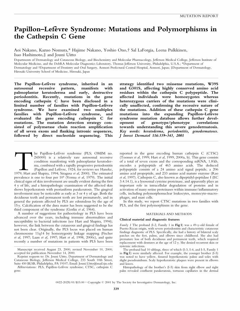
The proband's parents were dead but they were not known to have skinor dental ®ndings similar to their affected children, and there was knownparental consanguinity.
Family 2 The proband at the time of initial examination was a 4-y-oldfemale, the second child of consanguineous Japanese parents (II-2, Family 2in Fig 1). She had an older, clinically unaffected brother (II-1), whereas ayounger brother, 1 y of age (II-3), was clearly affected. The parents were
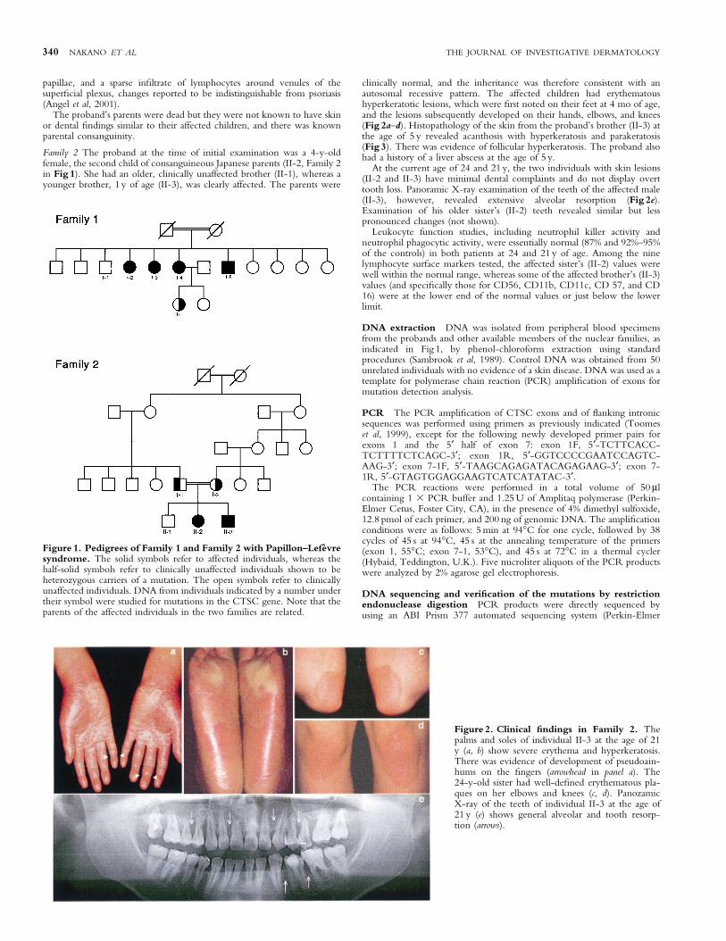
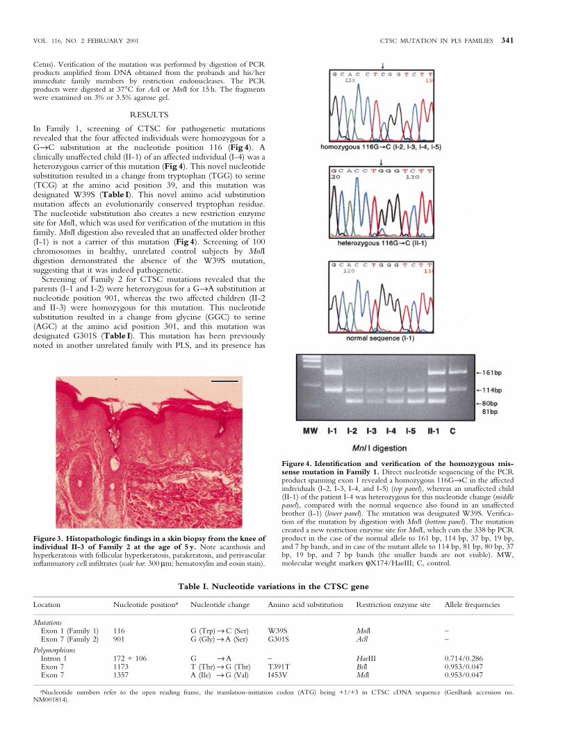
clinically normal, and the inheritance was therefore consistent with anautosomal recessive pattern. The affected children had erythematoushyperkeratotic lesions, which were ®rst noted on their feet at 4 mo of age,and the lesions subsequently developed on their hands, elbows, and knees(Fig 2a±d). Histopathology of the skin from the proband's brother (II-3) atthe age of 5 y revealed acanthosis with hyperkeratosis and parakeratosis(Fig 3). There was evidence of follicular hyperkeratosis. The proband alsohad a history of a liver abscess at the age of 5 y.
At the current age of 24 and 21 y, the two individuals with skin lesions(II-2 and II-3) have minimal dental complaints and do not display overttooth loss. Panoramic X-ray examination of the teeth of the affected male(II-3), however, revealed extensive alveolar resorption (Fig 2e).Examination of his older sister's (II-2) teeth revealed similar but lesspronounced changes (not shown).
Leukocyte function studies, including neutrophil killer activity andneutrophil phagocytic activity, were essentially normal (87% and 92%±95%of the controls) in both patients at 24 and 21 y of age. Among the ninelymphocyte surface markers tested, the affected sister's (II-2) values werewell within the normal range, whereas some of the affected brother's (II-3)values (and speci®cally those for CD56, CD11b, CD11c, CD 57, and CD16) were at the lower end of the normal values or just below the lowerlimit.
DNA extraction DNA was isolated from peripheral blood specimensfrom the probands and other available members of the nuclear families, asindicated in Fig 1, by phenol-chloroform extraction using standardprocedures (Sambrook et al, 1989). Control DNA was obtained from 50unrelated individuals with no evidence of a skin disease. DNA was used as atemplate for polymerase chain reaction (PCR) ampli®cation of exons formutation detection analysis.
PCR The PCR ampli®cation of CTSC exons and of ¯anking intronicsequences was performed using primers as previously indicated (Toomeset al, 1999), except for the following newly developed primer pairs forexons 1 and the 5¢ half of exon 7: exon 1F, 5¢-TCTTCACC-TCTTTTCTCAGC-3¢; exon 1R, 5¢-GGTCCCCGAATCCAGTC-AAG-3¢; exon 7-1F, 5¢-TAAGCAGAGATACAGAGAAG-3¢; exon 7-1R, 5¢-GTAGTGGAGGAAGTCATCATATAC-3¢.
The PCR reactions were performed in a total volume of 50 mlcontaining 1 3 PCR buffer and 1.25 U of Amplitaq polymerase (Perkin-Elmer Cetus, Foster City, CA), in the presence of 4% dimethyl sulfoxide,12.8 pmol of each primer, and 200 ng of genomic DNA. The ampli®cationconditions were as follows: 5 min at 94°C for one cycle, followed by 38cycles of 45 s at 94°C, 45 s at the annealing temperature of the primers(exon 1, 55°C; exon 7-1, 53°C), and 45 s at 72°C in a thermal cycler(Hybaid, Teddington, U.K.). Five microliter aliquots of the PCR productswere analyzed by 2% agarose gel electrophoresis.
DNA sequencing and veri®cation of the mutations by restrictionendonuclease digestion PCR products were directly sequenced byusing an ABI Prism 377 automated sequencing system (Perkin-Elmer
Figure 1. Pedigrees of Family 1 and Family 2 with Papillon±LefeÁvresyndrome. The solid symbols refer to affected individuals, whereas thehalf-solid symbols refer to clinically unaffected individuals shown to beheterozygous carriers of a mutation. The open symbols refer to clinicallyunaffected individuals. DNA from individuals indicated by a number undertheir symbol were studied for mutations in the CTSC gene. Note that theparents of the affected individuals in the two families are related.
Figure 2. Clinical ®ndings in Family 2. Thepalms and soles of individual II-3 at the age of 21y (a, b) show severe erythema and hyperkeratosis.There was evidence of development of pseudoain-hums on the ®ngers (arrowhead in panel a). The24-y-old sister had well-de®ned erythematous pla-ques on her elbows and knees (c, d). PanozamicX-ray of the teeth of individual II-3 at the age of21 y (e) shows general alveolar and tooth resorp-tion (arrows).
340 NAKANO ET AL THE JOURNAL OF INVESTIGATIVE DERMATOLOGY
Cetus). Veri®cation of the mutation was performed by digestion of PCRproducts ampli®ed from DNA obtained from the probands and his/herimmediate family members by restriction endonucleases. The PCRproducts were digested at 37°C for AciI or MnlI for 15 h. The fragmentswere examined on 3% or 3.5% agarose gel.
RESULTS
In Family 1, screening of CTSC for pathogenetic mutationsrevealed that the four affected individuals were homozygous for aG®C substitution at the nucleotide position 116 (Fig 4). Aclinically unaffected child (II-1) of an affected individual (I-4) was aheterozygous carrier of this mutation (Fig 4). This novel nucleotidesubstitution resulted in a change from tryptophan (TGG) to serine(TCG) at the amino acid position 39, and this mutation wasdesignated W39S (Table I). This novel amino acid substitutionmutation affects an evolutionarily conserved tryptophan residue.The nucleotide substitution also creates a new restriction enzymesite for MnlI, which was used for veri®cation of the mutation in thisfamily. MnlI digestion also revealed that an unaffected older brother(I-1) is not a carrier of this mutation (Fig 4). Screening of 100chromosomes in healthy, unrelated control subjects by MnlIdigestion demonstrated the absence of the W39S mutation,suggesting that it was indeed pathogenetic.
Screening of Family 2 for CTSC mutations revealed that theparents (I-1 and I-2) were heterozygous for a G®A substitution atnucleotide position 901, whereas the two affected children (II-2and II-3) were homozygous for this mutation. This nucleotidesubstitution resulted in a change from glycine (GGC) to serine(AGC) at the amino acid position 301, and this mutation wasdesignated G301S (Table I). This mutation has been previouslynoted in another unrelated family with PLS, and its presence has
Figure 4. Identi®cation and veri®cation of the homozygous mis-sense mutation in Family 1. Direct nucleotide sequencing of the PCRproduct spanning exon 1 revealed a homozygous 116G®C in the affectedindividuals (I-2, I-3, I-4, and I-5) (top panel), whereas an unaffected child(II-1) of the patient I-4 was heterozygous for this nucleotide change (middlepanel), compared with the normal sequence also found in an unaffectedbrother (I-1) (lower panel). The mutation was designated W39S. Veri®ca-tion of the mutation by digestion with MnlI (bottom panel). The mutationcreated a new restriction enzyme site for MnlI, which cuts the 338 bp PCRproduct in the case of the normal allele to 161 bp, 114 bp, 37 bp, 19 bp,and 7 bp bands, and in case of the mutant allele to 114 bp, 81 bp, 80 bp, 37bp, 19 bp, and 7 bp bands (the smaller bands are not visible). MW,molecular weight markers jX174/HaeIII; C, control.
Figure 3. Histopathologic ®ndings in a skin biopsy from the knee ofindividual II-3 of Family 2 at the age of 5 y. Note acanthosis andhyperkeratosis with follicular hyperkeratosis, parakeratosis, and perivascularin¯ammatory cell in®ltrates (scale bar: 300 mm; hematoxylin and eosin stain).
Table I. Nucleotide variations in the CTSC gene
Location Nucleotide positiona Nucleotide change Amino acid substitution Restriction enzyme site Allele frequencies
MutationsExon 1 (Family 1) 116 G (Trp) ® C (Ser) W39S Mnll ±Exon 7 (Family 2) 901 G (Gly) ® A (Ser) G301S Acll ±
PolymorphismsIntron 1 172 + 106 G ® A ± HaeIII 0.714/0.286Exon 7 1173 T (Thr) ® G (Thr) T391T Bsll 0.953/0.047Exon 7 1357 A (Ile) ® G (Val) I453V Msll 0.953/0.047
aNucleotide numbers refer to the open reading frame, the translation-initiation codon (ATG) being +1/+3 in CTSC cDNA sequence (GenBank accession no.NM001814).
VOL. 116, NO. 2 FEBRUARY 2001 CTSC MUTATION IN PLS FAMILIES 341
been excluded from a control population of 200 unrelated healthycontrol individuals (Toomes et al, 1999). This nucleotide substitu-tion also abolished a restriction enzyme site for AciI, which wasused for veri®cation of the mutation. AciI digestion con®rmed thatthe parents were heterozygous carriers of the mutation whereas theaffected individuals were homozygotes. The AciI digestion alsorevealed that an unaffected brother (II-1) is not a carrier of thismutation.
During the search for pathogenetic mutations in these twofamilies, three additional sequence variants were discovered(Table I). One of them was an intronic nucleotide substitutionwhereas another was a neutral polymorphism in exon 7 (T391T).The third one, 1357 A®G, substituted an isoleucine in position543 by a valine (I453V). The allele frequencies of these apparentpolymorphisms are indicated in Table I.
DISCUSSION
In this study, we have identi®ed homozygous missense mutationsW39S and G301S in two families with PLS. Discovery of the novelCTSC mutation W39S brings the total number of distinctmutations found in different families with PLS to 15 (Fig 5). Nineof them are missense mutations, whereas ®ve are nonsensemutations or small deletion mutations resulting in prematuretermination codon for translation. In addition, one family has asplicing mutation (486-1G®A) at the intron 2/exon 3 border.
The missense mutations, including W39S, affect critical, highlyconserved amino acids (Hart et al, 2000a), and eight out of ninemissense mutations reside within the mature enzyme domain of thepolypeptides (Fig 5). Also, three of the amino acid substitutionslead to incorporation of a cysteine in the polypeptide. It isconceivable therefore that these missense mutations result in
conformational changes that abolish the catalytic activity of theenzyme. In fact, the activity of this enzyme has been shown to beessentially undetectable in peripheral blood leukocytes of affectedindividuals in two families with PLS as a result of missensemutations V249F and Y347C (Toomes et al, 1999).
The novel missense mutation identi®ed in this study, W39S, wasfound in homozygous state in all four affected individuals in Family1, whereas a heterozygous carrier had no evidence of PLS.Furthermore, this mutation was not present in 100 alleles inunrelated control individuals, thus indicating that it is not apolymorphism. This tryptophan residue is precisely conserved incathepsin C during evolution in various species between humansand Schistosoma japonicum (see Fig 6). Interestingly, however, theW39S mutation resides at the amino-terminal end of the propeptide(Fig 5), which is cleaved off during processing of the polypeptide tomature enzyme (Muno et al, 1993). It is conceivable then that theW39S mutation interferes either with the transport of the proformpolypeptide from endoplasmic reticulum to lysosomes or impedesthe subsequent proteolytic processing of the polypeptide to matureenzyme.
It is of interest that Family 2 with the G301S mutation hadclinical features not entirely typical of PLS. Speci®cally, the twoaffected individuals in this family had retained their teeth up to theircurrent age of 24 and 21 y whereas classic cases with PLS areedentulous by the age of 15 y. Such late onset of periodontitis hasbeen noted before (Willett et al, 1985; Brown et al, 1993; Fardalet al, 1998). It is also of interest that the speci®c cathepsin Cmutation, Y347C, is associated with severe periodontitis but theindividuals homozygous with this mutation have no evidence ofsyndromic skin manifestations (Hart et al, 2000b). Finally, anotherautosomal recessive condition with palmoplantar keratoderma and
Figure 5. Schematic illustration of the cathepsin C polypeptide, deduced from cDNA, with domain organization, and positions of all CTSCmutations disclosed thus far in patients with PLS. The premature termination codon and the putative splice site mutation are shown above themolecule, whereas missense mutations are shown below. The mutations disclosed in this study are shown in bold. The amino acid positions at the domainborders are indicated above the polypeptide.
Figure 6. Conservation of peptide segments encoded by exon 7 of the CTSC gene. The partial human sequences are compared with those in othervertebrates indicated, as well as in Schistosoma japonicum (SchJp) and mansoni (SchMa). The conserved amino acids are boxed, and the positions of missenseand nonsense mutations are indicated by arrows. The dashed lines indicated additional sequences not shown. A similar comparison of sequences encoded byexons 1±6 is presented elsewhere (Hart et al, 2000a).
342 NAKANO ET AL THE JOURNAL OF INVESTIGATIVE DERMATOLOGY
early periodontal destruction, the Haim±Munk syndrome (OMIMno. 245010), has been shown to be allelic with PLS (Hart et al,2000a). A recurrent missense mutation, Q286R, in exon 6 of theCTSC gene has been identi®ed in several nuclear families with theHaim±Munk syndrome, all of the same ancestry (Hart et al, 2000a).The same study reported a nonsense mutation, Q286X, in the samecodon of CTSC in a family with classic features of PLS (Hart et al,2000a). Collectively, this phenotypic variability of the CTSCmutations suggests either phenotype/genotype correlations orphenotypic modulation by associated genetic and/or epigeneticfactors that are not yet evident from the relatively small cohort ofpatients.
The pathomechanistic implications of absent or markedlyreduced cathepsin C activities in PLS are not entirely clear but ithas been suggested that lack of functional CTSC may be associatedwith reduced host response against bacteria in dental plaque andpossibly other sites (Oguzkurt et al, 1996; Czauderna et al, 1999).CTSC plays an essential role in activating serine proteinasesexpressed in the granules of bone marrow derived cells both ofmyeloid and lymphoid series (McGuire et al, 1993). These serineproteinases are implicated in a variety of in¯ammatory and immuneprocesses, including phagocytic destruction of bacteria. In fact,previous leukocyte function studies have suggested depressedneutrophil phagocytic and lytic activity and depressed chemotacticresponse (Bullon et al, 1993; Ghaffar et al, 1999; Liu et al, 2000).Although these leukocyte functions were well within the normallimits in the two patients in Family 2, a number of lymphocytesurface markers were at the low end of normal values or slightlybelow the normal limits, suggesting immunologic de®ciencies.Mechanistically, de®cient activation of leukocyte serine proteinasesdue to lack of CTSC activity could possibly explain the severeperiodontitis in PLS. The mechanisms leading to hyperkeratoticskin lesions are unclear, however. One could speculate that CTSCplays a role in epithelial differentiation leading to characteristiccutaneous ®ndings in PLS.
We thank Drs. Sylvia Hsu-Wong and Steven Kornbleuth for clinical assistance.
This study was supported by USPHS/NIH Grant PO1 AR38923, and by a
grant for clinical research from the Aomori Prefectural Central Hospital.
REFERENCES
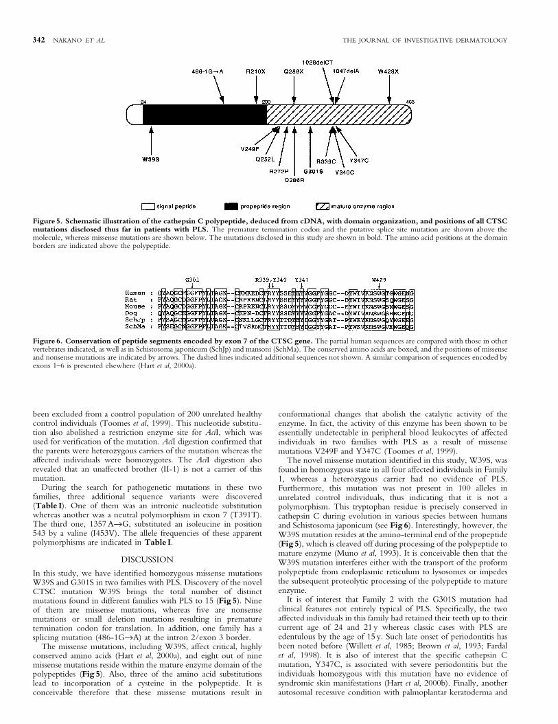
Angel TA, Hsu S, Kornbleuth SI, Kornbleuth J, Kramer EM: Papillon±LefeÁvresyndrome: a case report of four affected siblings. J Am Acad Dermatol in press:2001
Brown RS, Hays GL, Flaitz CM, O'Neill PA, Abramovitch K, White RR: Apossible late onset variation of Papillon±LefeÁvre syndrome: report of 3 cases. JPeriodontol 64:379±386, 1993
Bullon P, Pascual A, Fernandez-Novoa MC, Borobio MV, Muniain MA, CamachoF: Late onset Papillon±LefeÁvre syndrome? A chromosomic, neutrophil functionand microbiological study. J Clin Periodontol 20:662±667, 1993
Czauderna P, Sznurkowska K, Korzon M, Roszkiewicz A, Stoba C: Association ofin¯ammatory pseudotumor of the liver and Papillon±LefeÁvre syndrome: casereport. Eur J Pediatr Surg 9:343±346, 1999
Fardal é, Drangsholt E, Olsen I: Palmar plantar keratosis and unusual periodontal®ndings: observations from a family of 4 members. J Clin Periodontol25:181±184, 1998
Fischer J, Blanchet-Bardon C, Prud'homme J-F, Pavek S, Steijlen PM, Dubertret L,Weissenbach J: Mapping of Papillon±LefeÁvre syndrome to the chromosome11q14 region. Eur J Hum Genet 5:156±160, 1997
Ghaffar KA, Zahran FM, Fahmy HM, Brown RS: Papillon±LefeÁvre syndrome:neutrophil function in 15 cases from 4 families in Egypt. Oral Surg Oral MedOral Pathol Oral Radiol Endod 88:320±325, 1999
Gorlin RJ, Sedano H, Anderson VE: The syndrome of palmar-plantar hyperkeratosisand premature periodontal destruction of the teeth: a clinical and geneticanalysis of the Papillon±LefeÁvre syndrome. J Pediatr 65:895±908, 1964
Haneke E: The Papillon±LefeÁvre syndrome: keratosis palmoplantaris withperiodontopathy. Hum Genet 51:1±35, 1979
Hart TC, Shapira L: Papillon±LefeÁvre syndrome. Periodontology 2000:88±100, 1994Hart TC, Bowden DW, Ghaffar KA, et al: Sublocalization of the Papillon±LefeÁvre
syndrome locus on 11q14-q21. Am J Med Genet 79:134±139, 1998Hart TC, Hart PS, Bowden DW, et al: Mutations of the cathepsin C gene are
responsible for Papillon±LefeÁvre syndrome. J Med Genet 36:881±887, 1999Hart TC, Hart PS, Michalec MD, et al: Haim±Munk syndrome and Papillon±LefeÁvre
syndrome are allelic mutations in cathepsin C. J Med Genet 37:88±94, 2000aHart TC, Hart PS, Michalec MD, et al: Localisation of a gene for prepubertal
periodontitis to chromosome 11q14 and identi®cation of a cathepsin C genemutation. J Med Genet 37:95±101, 2000b
Hart TC, Walker SJ, Bowden DW, Hart PS, Callison SA, Bobby PL, Firatli E: Anintegrated physical and genetic map of the PLS locus interval on chromosome11q14. Mamm Genome 11:243±246, 2000c
Laass MW, Hennies HC, Preis S, et al: Localisation of a gene for Papillon±LefeÁvresyndrome to chromosome 11q14-q21 by homozygosity mapping. Hum Genet101:376±382, 1997
Liu R, Cao C, Meng H, Tang Z: Leukocyte functions in 2 cases of Papillon±LefeÁvresyndrome. J Clin Periodontol 27:69±73, 2000
McGuire MJ, Lipsky PE, Thiele DL: Generation of active myeloid and lymphoidgranule serine proteases requires processing by the granule thiol proteasedipeptidyl peptidase I. J Biol Chem 268:2458±2467, 1993
Muno D, Ishidoh K, Ueno T, Kominami E: Processing and transport of the precursorof cathepsin C during its transfer into lysosomes. Arch Biochem Biophys306:103±110, 1993
Oguzkurt P, Tanyel FC, BuÈyuÈkpamukcËu N, HicËsoÈnmez A: Increased risk ofpyogenic liver abscess in children with Papillon±LefeÁvre syndrome. J PediatSurg 31:955±956, 1996
Papillon MM, LefeÁvre P: Deux cas de keratodermie palmaire et plantairesymmetrique familiale (maladie de Meleda) chez le frere et la soeur:coexistense dans les duex cas d'alterations dentaires gravis. Bull Soc FrDermatol Syphilis 31:81±87, 1924
Rao NV, Rao GV, Hoidal JR: Human dipeptidyl-peptidase I: gene characterization,localization, and expression. J Biol Chem 272:10260±10265, 1997
Sambrook J, Fritsch EF, Maniatis T: Molecular Cloning: a Laboratory Manual.Plainview, NY: Cold Spring Harbor Laboratory Press, 1989, pp 9.16±9.19
Siragusa M, Romano C, Batticane N, Batolo D, Schepis C: A new family withPapillon±LefeÁvre syndrome: effectiveness of etretinate treatment. Cutis65:151±155, 2000
Toomes C, James J, Wood AJ, et al: Loss-of-function mutations in the cathepsin Cgene result in periodontal disease and palmoplantar keratosis. Nat Genet23:421±424, 1999
Verma K, Chadda M, Joshi R: Papillon±LefeÁvre syndrome. Int J Dermatol18:146±149, 1979
Willett L, Gabriel S, Kozma C, Bottomley W: Papillon-LefeÁvre: report of a case. JOral Med 40:43±45, 1985
VOL. 116, NO. 2 FEBRUARY 2001 CTSC MUTATION IN PLS FAMILIES 343


![Institute] Lefevre Duo](https://cdn.vdocuments.mx/doc/165x107/577d2acd1a28ab4e1eaa2116/institute-lefevre-duo.jpg)