Int J Clin Exp Pathol 2013;6(4):561-570www.ijcep.com /ISSN:1936-2625/IJCEP1301008
Original ArticleIdentification of CD68+ neutrophil granulocytes in in vitro model of acute inflammation and inflammatory bowel disease
Ahmad Amanzada1*, Ihtzaz Ahmed Malik1*, Martina Blaschke1, Sajjad Khan1, Hazir Rahman2, Giuliano Ramadori1, Federico Moriconi1
1Division of Gastroenterology and Endocrinology, Department of Internal Medicine, University Medical Center, Georg-August-University Göttingen, Germany; 2Department of Clinical Chemistry, University Medical Center, Georg-August-University Göttingen, Germany. *The first two authors contributed equally to the present work.
Received January 5, 2013; Accepted February 26, 2013; Epub March 15, 2013; Published April 1, 2013
Abstract: CD-68 is widely regarded as a selective marker for human monocytes and macrophages and is commonly used in human pathology studies. The purpose of this study was to investigate the expression of CD-68 in human pe-ripheral blood mononuclear cells (PBMCs), neutrophil granulocytes (NGs) and in inflamed intestinal tissue samples for comparison. PBMCs and NGs were isolated from heparinized human blood samples. Intestinal biopsies were obtained during routine endoscopic procedures from patients with inflammatory bowel disease (IBD), e.g. ulcerative colitis and Crohn’s disease. Gene and protein expression was analyzed by real-time RT-PCR, Western blot and im-munohistochemistry. Both PBMCs and NGs preparations contained cells that were positive for CD-68 and either neutrophil elastase (NE), or myeloperoxidase (MPO). CD-68+/NE-/MPO- cells were regarded as monocytes. CD-68 mRNA expression was detected in PBMCs and NGs preparations. With Western blot and by performing immunopre-cipitation of cell lysate, we could clearly detect CD-68 in NGs, U-937, THP-1, Hep-G2, Jurkat cells and PBMCs. Identi-fication of inflammatory cells in acutely inflamed colonic mucosa obtained from patients with IBD revealed a strong accumulation of CD-68+/MPO+ cells compared to normal colonic mucosa. The uptake of the marker by phagocytosis was excluded by performing a double staining with CD-163/NE and CD-163/MPO in PBMCs, NGs cultures and in inflamed colonic mucosa. These results identify CD-68+ NGs in peripheral blood and inflamed colonic mucosa. CD-68 is not only a marker for the macrophages-monocytes but also for NGs.
Keywords: CD-68, inflammatory bowel disease, monocytes, neutrophil granulocytes, peripheral blood mononucle-ar cells
Introduction
Cells that differ in function have distinct molec-ular structures in their membranes. These structures can serve as markers for particular cell types and can be recognized by specific antisera against the markers. Specific antibod-ies against membrane markers of human cells have a number of current and potential uses [1].
Five antibodies, Y1/82A, Y2/131, EBM11, Ki-M6 and Ki-M7, within the myeloid panel of reagents were recognized as markers of the majority of human tissue macrophages present in tissue sections [2-6]. This group of antigens was designated as CD-68, based on the results
of Micklem et al. [7]. Specific CD-68 is reported to be present in a variety of tissue macro-phages, including Kupffer cells, germinal center macrophages, alveolar macrophages and bone osteoclasts [8].
CD-68 (the human homologue of mouse macro-sialin) is a heavily glycosylated, 110 kDa mem-brane protein. Although it is predominantly located in lysosomal membranes, a small frac-tion is also found on the cell surface [9, 10].
However, in contrast to the generally accepted concept of monocyte/macrophage specificity, many authors have reported some reactivity of anti-human CD-68 antibodies with antigens present on the cell surface of various hemato-
Identification of CD-68 in neutrophil granulocytes
562 Int J Clin Exp Pathol 2013;6(4):561-570
poietic and non-hematopoietic cells [11-13]. Recently published reports demonstrated both at the RNA and protein level that CD-68 was not only expressed in macrophages and mono-cytes but also expressed by non-myeloid cell types, such as T cells, fibroblasts, endothelial and tumor cells [14, 15].
Recently it has been demonstrated that the increased expression of MPO in injured rat and human liver is due to the presence of newly recruited neutrophil granulocytes (NGs) as no MPO expression was evident in Kupffer cells [16]. These reports negate the traditional view that the CD-68 antigen is specific for macro-phages. If the CD-68 antigen is not expressed solely in macrophages but also by NGs, the finding of MPO in CD-68+ cells is explained. This is important especially when physiology of intestinal immune system is studied [17].
In acute tissue injury, the initial inflammatory cell influx consists predominantly of NGs, which in turn orchestrate the recruitment of mono-cytes and the activation of lymphocytes required for a mature inflammatory response. Inflammatory bowel diseases (IBD), Crohn’s dis-ease (CD) and ulcerative colitis (UC), are char-acterized by an excessive recruitment of leuko-cytes from the blood circulation into the inflamed gut wall. During acute flares of UC or CD there is a massive infiltration of NGs into the affected mucosa, which is manifested clini-cally by an increase in stool NGs. In IBD there is also a marked infiltration of macrophages into the inflamed mucosa, manifested by an increase in macrophage-derived inflammatory cytokines IL-1β, IL-6 and TNF-α during acute flares [18]. IBD are characterized by a dense infiltration of tissue by CD-68+ macrophages compared to non-inflamed colonic mucosa [19, 20].
This study investigated whether CD-68 positive NGs is found in peripheral blood and inflamed colonic mucosa of IBD patients.
Methods
Antibodies
A rabbit polyclonal antibody directed against human MPO was purchased from Dako (Hamburg, Germany), a rabbit polyclonal antibody
against human neutrophil elastase from Calbiochem (Germany, Cat. 481001-1ML) and a mouse monoclonal antibody directed against human CD-68 (Clone KP-1, Cat. sc-20060) from Santa Cruz (CA, USA) for Western blot analysis. Results were confirmed with a second mouse monoclonal antibody anti-human CD-68 (Clone KP-1, Cat. ab955, Abcam, Cambridge, UK). For immunohistological analysis, a mono-clonal anti-human CD-68 mouse antibody (Clone PG-M1, Cat. M087601) was bought from Dako. Results were confirmed with a second anti-human CD-68 mouse monoclonal antibody (Clone KP-1, Cat. ab955, Abcam, Cambridge). A monoclonal mouse anti-human CD-11 b-c mouse antibody was purchased from DAKO (Cat. M0741), while the monoclonal mouse anti-human CD-163 antibody (5C6-FAT, Cat. BM4041) came from Novus Biologicals (Cambridge, UK).
Isolation and cell culture condition of human PBMCs and NGs
PBMCs and NGs were isolated from hepara-nized blood of healthy donors (n=3) by Ficoll density gradient centrifugation and dextran sedimentation. Residual red blood cells were hypotonically lysed and cells were washed three times with phosphate-buffered saline (PBS) pH 7.3. Cell preparations were routinely assessed for viability (>95%) by trypan blue exclusion. Cells were stimulated in vitro with phytohaemagglutinin (PHA, 5 µg/ml [Roche Molecular Biochemicals, Mannheim, Germany]) or lipopolysaccharide (LPS, 1 µg/ml [Sigma-Aldrich Chemie, Taufkirchen, Germany]) for 2, 4 and 8 hours as previously described [21].
Isolation of RNA and real-time quantitative RT-PCR
Isolation of RNA, cDNA synthesis and real-time RT-PCR were performed as previously described [22]. Real-time PCR analysis of cDNA was per-formed at 60°C to 95°C for 45 cycles in the Sequence Detection System of ABI Prism 7000 (Applied Biosystems, Darmstadt, Germany) fol-
Table 1. Human primer sequences used for RT-PCRPrimer Forward ReverseMPO 5´-TCGTCAGAACCAAATTGCAG-3´ 5´-ATGTTCAGAGCAGGCAGGTC-3´NE 5´-GCATCTTCGAAAACGGCTAC -3´ 5´-GACCCGTTGAGCTGGAGAAT-3´CD-68 5´-TCAGCTTTGGATTCATGCAG-3´ 5´-TTGTACTCCACCGCCATGTA-3´β-actin 5´- CTGGAACGGTGAAGGTGACA -3´ 5´- GTCCTCGGCCACATTGTGA -3´
Identification of CD-68 in neutrophil granulocytes
563 Int J Clin Exp Pathol 2013;6(4):561-570
lowing the manufacturer’s instructions and by using SYBR Green Reaction Master Mix (ABI Prism; Applied Biosystems) and the primers reported in Table 1. All primers were synthe-sized by Invitrogen (Groningen, Netherlands). In every sample, β-actin was taken as the house-keeping gene. Fold change expression was cal-culated from the threshold cycle (Ct) values. For calculation of the relative changes, gene expression measured in unstimulated cells at 0h was set at unity.
Cytospin preparation and immunofluorescence
Cytospin preparation and immunofluorescent staining of PBMCs and NGs were performed as described previously [21]. Double-staining was performed according to an established proto-col [23]. The primary antibodies were incubat-ed with the cytospins preparations overnight at 4°C. Following a short washing step in phos-phate buffered saline (PBS), an incubation was carried out with Alexa-Fluor® conjugated goat anti-rabbit and anti-mouse secondary antibod-ies (1:200; Molecular Probes, Germany) at room temperature for 1 h and the cytospin preparations were washed three times for 5 min in PBS. Finally, the nuclei were stained with 4,6-diamidino-2-phenylindole (DAPI), and the sections were washed and mounted.
Intestinal tissue sections (paraffin embedded)
Biopsy samples of the colon were obtained from six patients (3 men, 3 women) with CD, six
patients with UC (4 men, 2 women) and three control subjects without detectable colonic dis-ease (2 men, 1 women). The control biopsies were obtained from mucosa without any mac-roscopic evidence of inflammation (confirmed at the microscopic level) in patients who under-went elective colonoscopic screening for can-cer. The IBD patients had clinical and endo-scopical signs of acute inflammation. IBD patients had clinical and endoscopic signs of acute inflammation. All patients gave written informed consent to participate in the study in accordance with the ethical guidelines of the 1975 Declaration of Helsinki and the ethics committee of the University Medical Center. The diagnosis of IBD was based on clinical, radiological, endoscopic and histopathologic criteria. The specific indication for the colono-scopic examination in the IBD patients was a lack of efficacy of their therapy. As a result, all biopsies were obtained from patients with active mucosa inflammation during an acute flare of the disease. The assessment of the severity of the mucosal disease was based on macroscopic and histologic findings. In the IBD group, two patients had distal colitis, two proc-titis, and two had pancolitis. The duration of the IBD ranged from 6 to 22 years (mean, 9.5 years). Serum C-reactive protein (CRP) levels were determined utilizing standard clinical lab-oratory procedures and ranged from 2 to 28.3 mg/L (mean, 8.2 mg/L). All of the CRP levels in the control subjects were within the normal
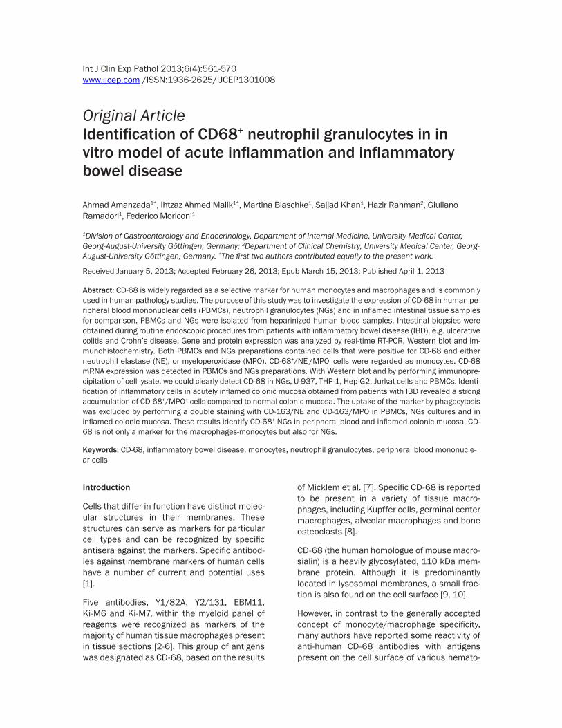
Figure 1. Double staining of PBMCs cytospins with monoclonal antibody directed against CD-68 and CD-163 (green), polyclonal antibody against NE and MPO (red), followed by fluorescence immunodetection. CD-68+/NE+ cells in con-trol cultures and 4h after stimulation with LPS (Figure 2A). CD-68+/MPO+ and CD-163+/NE+ PBMCs in control cells and 4h after stimulation with LPS (Figure 2B and 2C) (Original magnification: x200. Scale bar= 100 µm).

Identification of CD-68 in neutrophil granulocytes
564 Int J Clin Exp Pathol 2013;6(4):561-570
laboratory range (<2 mg/L). The number of biopsy specimens taken from a given patient
ranged from 1 to 3 (mean, 1.6 specimens). Specimens for RNA analysis were immediately
Figure 2. Double staining of NGs cytospins with monoclonal antibody directed against CD-68 (green) and polyclonal antibody against NE (red) followed by fluorescence immunodetection. CD-68+/NE+ cells in control cultures and 4h after stimulation with LPS (Figure 2A). CD-68+/MPO+ NGs in control cultures and 4h after stimulation with LPS (Figure 2B). Double staining of NGs cytospins with polyclonal antibody directed against NE (red) and monoclonal antibody against CD-11b-c and CD-163 (green) followed by fluorescence immunodetection. NE+/CD-11 b-c+ (Figure 2C) and NE+/CD-163+ (Figure 2D) NGs in control cultures and 4h after stimulation with LPS (Original magnification: x200. Scale bar= 100 µm).
Figure 3. Western blot analysis of MPO and β-actin of total protein from human NGs (Figure 3A). Protein was ex-tracted from NGs of control (Co) cells or 12 h after stimulation with PHA or with LPS. In a second set of experiments, protein from NGs cells lysate was immunoprecipitated with a monoclonal antibody anti-CD-68 (clone KP1) (Figure 3B). U-937 and THP-1 cell lines were used as positive controls, whereas NGs without additional incubation with a monoclonal antibody anti-CD-68 were used as negative control. Western blot analysis of MPO in control (Co), PHA- or LPS-stimulated PBMCs was also performed (Figure 3C). β-actin was always used as house-keeping gene. 20 µg of total protein were separated by SDS-PAGE, and blotted onto PVDF membranes. The membranes were subsequently incubated with the antibodies against MPO, NE, CD-68 and β-actin. An immunoprecipitation of cells lysate was also performed (Figure 3D). In order, NGs (unstimulated and after 12h treatment with TNF-α), U-937, THP-1, Hep-G2, Jurkatt cells and PBMCs (unstimulated and after 12h stimulation with PHA or LPS) were subjected to SDS-PAGE and transferred to polyvinylidene fluoride (PVDF) membranes followed by Western blot analysis for CD-68. The molecu-lar weight of MPO is 59 kDa. The molecular weight of NE is 29 kDa, of β-actin is 42 kDa. The molecular weight of CD-68 (KP1) is between 75 and 110 kDa, as described previously. For immuno precipitation of cell lysates a rabbit polyclonal anti-CD68 antibody with a predicted band of 42kDa was used.
Identification of CD-68 in neutrophil granulocytes
565 Int J Clin Exp Pathol 2013;6(4):561-570
Statistical analysis
The data were analyzed with Prism Graph Pad 5 software (San Diego, USA). All experimental errors are shown as SEM. Statistical signifi-cance was calculated using Student’s t-test, one-way analysis of variance, and Dunnett’s post hoc tests. Significance was defined at a level of P<0.05.
Results
In vitro identification of CD-68+/NE+, CD-68+/MPO+, CD-11b-c+/NE+ and CD-163+/NE+ in activated human PBMCs and NGs by immuno-fluorescence double staining
After 4 h of stimulation with PHA or LPS as well as in unstimulated PBMCs, 20% of the total cell populations was found to be CD-68+/NE+ (Figure 1A), and CD-68+/MPO+ (Figure 1B). Two different CD-68 antibodies (monoclonal mouse Clone PG-M1, Dako and monoclonal mouse Clone KP-1, Abcam) yielded the same result although only results with the clone PG-M1 (Dako) are reported. As the staining procedures identified positivity for the NGs markers, MPO and NE, they clearly demonstrate that a portion of the PBMCs cell population contains some NGs. In the present work, these cells are shown to have strong CD-68 expression.
As a control, a NG preparation was studied. About 85% and 90% of these cells stained posi-tive for NE and MPO, respectively. Double stain-ing with CD-68 and NE (Figure 2A) or MPO (Figure 2B) revealed that 20% of the cells were positive for both markers. As CD-11 b-c is a marker for leukocytes involved in the innate immune system, including monocytes, granulo-cytes, macrophages and natural killer cells, the present findings were confirmed by performing immunofluorescence staining of human NGs using antibodies against CD-11 b-c and NE (Figure 2C). As shown in Figure 2C, 50% of the NGs are positive for both markers.
To exclude the uptake of NE+ cells by phagocy-tosis, we stained the specific marker of the monocyte/macrophage lineage CD-163 in human PBMCs and NGs, and we clearly showed that NE was not detectable in CD-163+ cells (Figures 1C and 2D).
snap-frozen in liquid nitrogen and stored at –80°C until being used.
Protein extraction, western blot analysis and immunoprecipitation
Protein extraction and Western blot analysis using PBMCs and NGs, but also THP-1, U-937, Hep-G2 and Jurkatt cells were performed as previously described [21]. The immunodetec-tion studies were performed according to the ECL Western blotting protocol of GE Healthcare (Germany). The primary rabbit polyclonal anti-bodies to MPO and NE were used at 1:100 dilu-tion, whereas the mouse monoclonal anti-CD-68 was used at a 1:50 dilution. In addition, 500 μg of total proteins were immunoprecipi-tated from cell lysates of NGs utilizing the CD-68 antibody at 4°C overnight with rotation and then incubated with 40 μl of protein A/G-agarose (Santa Cruz Biotechnology) beads for 4 hours at 4°C with rotation. The collected beads were washed four times with lysis buffer and resuspended in sample buffer.
Immunoprecipitation of cells lysate was per-formed as described previously [24]. In order, NGs (unstimulated and after 12h treatment with TNF-α), U-937, THP-1, Hep-G2, Jurkatt cells and PBMCs (unstimulated and after 12h stimulation with PHA or LPS) were subjected to SDS-PAGE and transferred to polyvinylidene fluoride (PVDF) membranes followed by Western blot analysis for CD-68. For immuno-precipitation of cell lysates a rabbit polyclonal anti-CD68 antibody (Cat. Ab63896, Abcam), at a 1:1000 dilution and with a predicted band of 42kDa, was used.
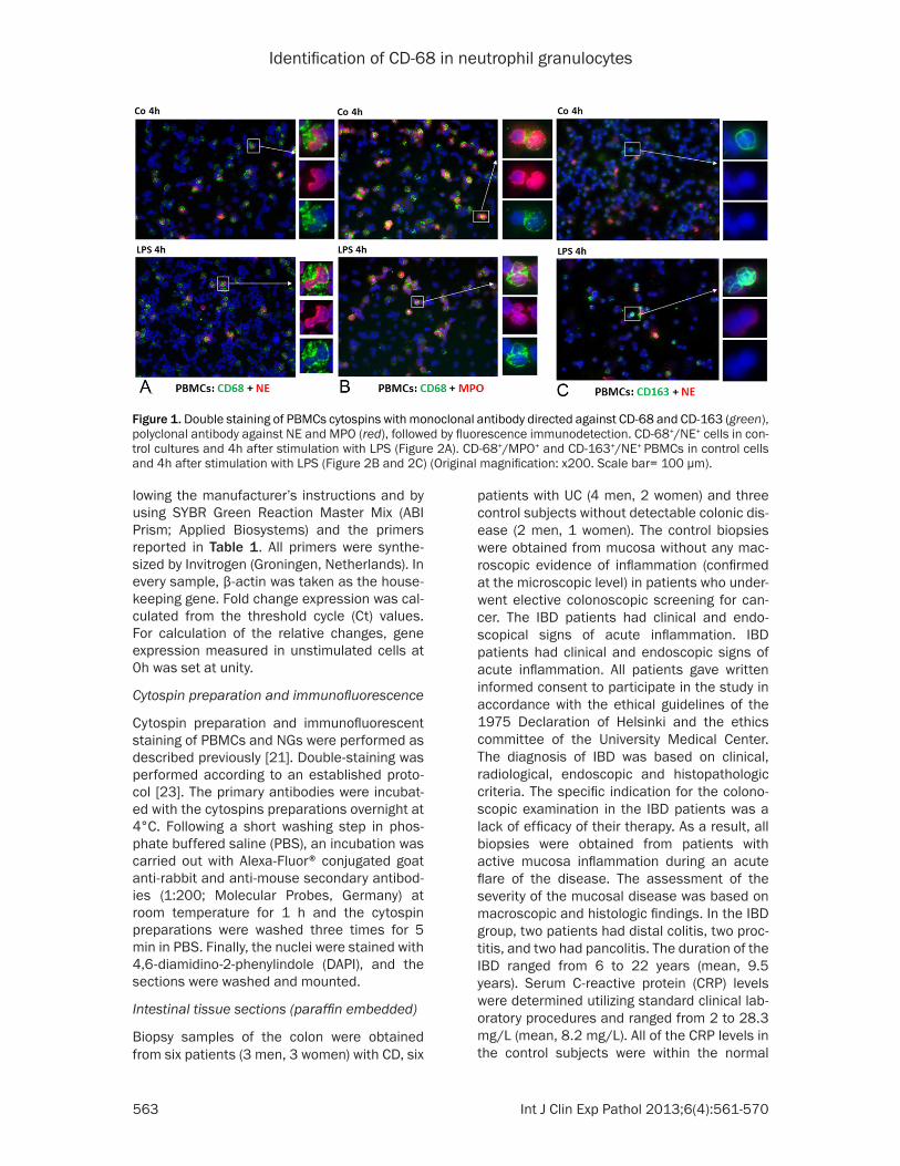
Figure 4. Fold change of mRNA expression of NE, MPO, CD-68 in NGs and PBMCs freshly isolated. Real time PCR was normalized by using β-actin as house-keeping gene. Results represent mean ± SEM value of three experiments (in duplicate).
Identification of CD-68 in neutrophil granulocytes
566 Int J Clin Exp Pathol 2013;6(4):561-570
cells lysates from human NGs with a mouse monoclonal antibody directed against human CD-68 (clone KP1, Abcam). Figure 3B shows that KP-1 detects a band of approximately 110 kDa in stimulated and control NGs, although the expression level did not differ between con-trol and stimulated cells. In order to confirm our results, we used U-937 and THP-1 cell lines as a positive control and we were able to detect the same 110 kDa band in both of them (Figure 3B). This is consistent with mRNA expression data (Figure 4) where we show a constitutive
Identification of CD-68+/MPO+ and CD-68+/NE+ in activated human PBMCs and NG by western blot and immunoprecipitation
To determine the amount of MPO-, NE- and CD-68-protein in human PBMCs and NGs prep-arations, Western blot analyses were carried out. The molecular weight of MPO is 59 kDa. Figure 3A shows a constitutive expression of MPO in human NGs, which decreased after stimulation with LPS. As the next step, we per-formed an immunoprecipitation analysis of
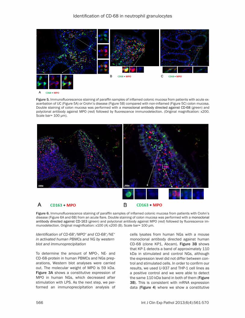
Figure 5. Immunofluorescence staining of paraffin samples of inflamed colonic mucosa from patients with acute ex-acerbation of UC (Figure 5A) or Crohn’s disease (Figure 5B) compared with non-inflamed (Figure 5C) colon mucosa. Double staining of colon mucosa was performed with a monoclonal antibody directed against CD-68 (green) and polyclonal antibody against MPO (red) followed by fluorescence immunodetection. (Original magnification: x200. Scale bar= 100 µm).
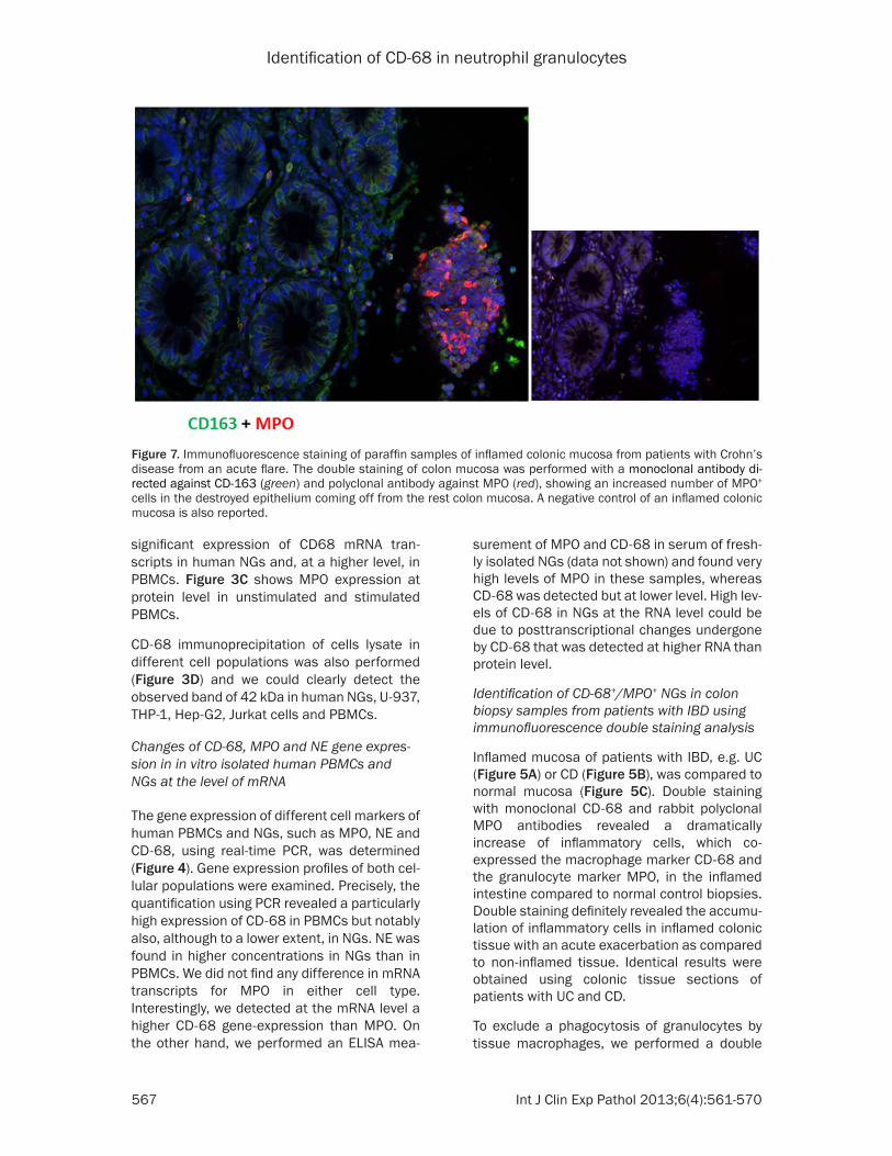
Figure 6. Immunofluorescence staining of paraffin samples of inflamed colonic mucosa from patients with Crohn’s disease (Figure 6A and 6B) from an acute flare. Double staining of colon mucosa was performed with a monoclonal antibody directed against CD-163 (green) and polyclonal antibody against MPO (red) followed by fluorescence im-munodetection. Original magnification: x100 (A) x200 (B). Scale bar= 100 µm.
Identification of CD-68 in neutrophil granulocytes
567 Int J Clin Exp Pathol 2013;6(4):561-570
surement of MPO and CD-68 in serum of fresh-ly isolated NGs (data not shown) and found very high levels of MPO in these samples, whereas CD-68 was detected but at lower level. High lev-els of CD-68 in NGs at the RNA level could be due to posttranscriptional changes undergone by CD-68 that was detected at higher RNA than protein level.
Identification of CD-68+/MPO+ NGs in colon biopsy samples from patients with IBD using immunofluorescence double staining analysis
Inflamed mucosa of patients with IBD, e.g. UC (Figure 5A) or CD (Figure 5B), was compared to normal mucosa (Figure 5C). Double staining with monoclonal CD-68 and rabbit polyclonal MPO antibodies revealed a dramatically increase of inflammatory cells, which co-expressed the macrophage marker CD-68 and the granulocyte marker MPO, in the inflamed intestine compared to normal control biopsies. Double staining definitely revealed the accumu-lation of inflammatory cells in inflamed colonic tissue with an acute exacerbation as compared to non-inflamed tissue. Identical results were obtained using colonic tissue sections of patients with UC and CD.
To exclude a phagocytosis of granulocytes by tissue macrophages, we performed a double
significant expression of CD68 mRNA tran-scripts in human NGs and, at a higher level, in PBMCs. Figure 3C shows MPO expression at protein level in unstimulated and stimulated PBMCs.
CD-68 immunoprecipitation of cells lysate in different cell populations was also performed (Figure 3D) and we could clearly detect the observed band of 42 kDa in human NGs, U-937, THP-1, Hep-G2, Jurkat cells and PBMCs.
Changes of CD-68, MPO and NE gene expres-sion in in vitro isolated human PBMCs and NGs at the level of mRNA
The gene expression of different cell markers of human PBMCs and NGs, such as MPO, NE and CD-68, using real-time PCR, was determined (Figure 4). Gene expression profiles of both cel-lular populations were examined. Precisely, the quantification using PCR revealed a particularly high expression of CD-68 in PBMCs but notably also, although to a lower extent, in NGs. NE was found in higher concentrations in NGs than in PBMCs. We did not find any difference in mRNA transcripts for MPO in either cell type. Interestingly, we detected at the mRNA level a higher CD-68 gene-expression than MPO. On the other hand, we performed an ELISA mea-
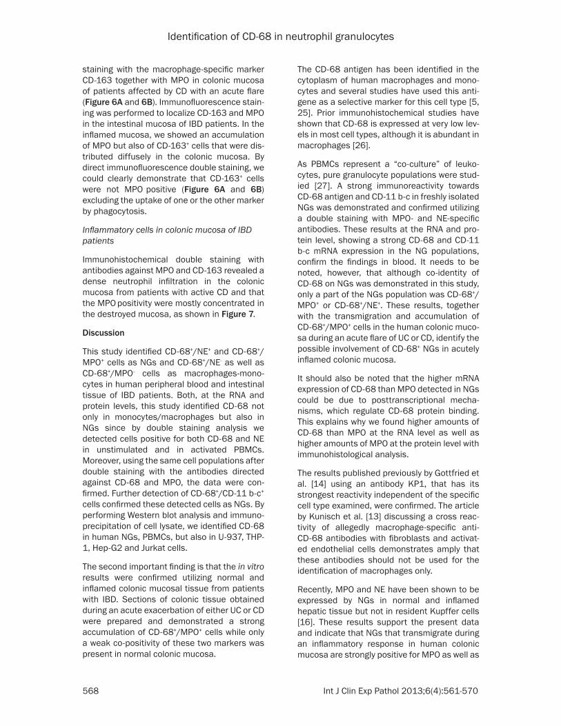
Figure 7. Immunofluorescence staining of paraffin samples of inflamed colonic mucosa from patients with Crohn’s disease from an acute flare. The double staining of colon mucosa was performed with a monoclonal antibody di-rected against CD-163 (green) and polyclonal antibody against MPO (red), showing an increased number of MPO+ cells in the destroyed epithelium coming off from the rest colon mucosa. A negative control of an inflamed colonic mucosa is also reported.
Identification of CD-68 in neutrophil granulocytes
568 Int J Clin Exp Pathol 2013;6(4):561-570
The CD-68 antigen has been identified in the cytoplasm of human macrophages and mono-cytes and several studies have used this anti-gene as a selective marker for this cell type [5, 25]. Prior immunohistochemical studies have shown that CD-68 is expressed at very low lev-els in most cell types, although it is abundant in macrophages [26].
As PBMCs represent a “co-culture” of leuko-cytes, pure granulocyte populations were stud-ied [27]. A strong immunoreactivity towards CD-68 antigen and CD-11 b-c in freshly isolated NGs was demonstrated and confirmed utilizing a double staining with MPO- and NE-specific antibodies. These results at the RNA and pro-tein level, showing a strong CD-68 and CD-11 b-c mRNA expression in the NG populations, confirm the findings in blood. It needs to be noted, however, that although co-identity of CD-68 on NGs was demonstrated in this study, only a part of the NGs population was CD-68+/MPO+ or CD-68+/NE+. These results, together with the transmigration and accumulation of CD-68+/MPO+ cells in the human colonic muco-sa during an acute flare of UC or CD, identify the possible involvement of CD-68+ NGs in acutely inflamed colonic mucosa.
It should also be noted that the higher mRNA expression of CD-68 than MPO detected in NGs could be due to posttranscriptional mecha-nisms, which regulate CD-68 protein binding. This explains why we found higher amounts of CD-68 than MPO at the RNA level as well as higher amounts of MPO at the protein level with immunohistological analysis.
The results published previously by Gottfried et al. [14] using an antibody KP1, that has its strongest reactivity independent of the specific cell type examined, were confirmed. The article by Kunisch et al. [13] discussing a cross reac-tivity of allegedly macrophage-specific anti-CD-68 antibodies with fibroblasts and activat-ed endothelial cells demonstrates amply that these antibodies should not be used for the identification of macrophages only.
Recently, MPO and NE have been shown to be expressed by NGs in normal and inflamed hepatic tissue but not in resident Kupffer cells [16]. These results support the present data and indicate that NGs that transmigrate during an inflammatory response in human colonic mucosa are strongly positive for MPO as well as
staining with the macrophage-specific marker CD-163 together with MPO in colonic mucosa of patients affected by CD with an acute flare (Figure 6A and 6B). Immunofluorescence stain-ing was performed to localize CD-163 and MPO in the intestinal mucosa of IBD patients. In the inflamed mucosa, we showed an accumulation of MPO but also of CD-163+ cells that were dis-tributed diffusely in the colonic mucosa. By direct immunofluorescence double staining, we could clearly demonstrate that CD-163+ cells were not MPO positive (Figure 6A and 6B) excluding the uptake of one or the other marker by phagocytosis.
Inflammatory cells in colonic mucosa of IBD patients
Immunohistochemical double staining with antibodies against MPO and CD-163 revealed a dense neutrophil infiltration in the colonic mucosa from patients with active CD and that the MPO positivity were mostly concentrated in the destroyed mucosa, as shown in Figure 7.
Discussion
This study identified CD-68+/NE+ and CD-68+/MPO+ cells as NGs and CD-68+/NE- as well as CD-68+/MPO- cells as macrophages-mono-cytes in human peripheral blood and intestinal tissue of IBD patients. Both, at the RNA and protein levels, this study identified CD-68 not only in monocytes/macrophages but also in NGs since by double staining analysis we detected cells positive for both CD-68 and NE in unstimulated and in activated PBMCs. Moreover, using the same cell populations after double staining with the antibodies directed against CD-68 and MPO, the data were con-firmed. Further detection of CD-68+/CD-11 b-c+ cells confirmed these detected cells as NGs. By performing Western blot analysis and immuno-precipitation of cell lysate, we identified CD-68 in human NGs, PBMCs, but also in U-937, THP-1, Hep-G2 and Jurkat cells.
The second important finding is that the in vitro results were confirmed utilizing normal and inflamed colonic mucosal tissue from patients with IBD. Sections of colonic tissue obtained during an acute exacerbation of either UC or CD were prepared and demonstrated a strong accumulation of CD-68+/MPO+ cells while only a weak co-positivity of these two markers was present in normal colonic mucosa.
Identification of CD-68 in neutrophil granulocytes
569 Int J Clin Exp Pathol 2013;6(4):561-570
References
[1] Ionescu RM, Vlasak J, Price C and Kirchmeier M. Contribution of variable domains to the sta-bility of humanized IgG1 monoclonal antibod-ies. J Pharm Sci 2008; 97: 1414-1426.
[2] Davey FR, Cordell JL, Erber WN, Pulford KA, Gatter KC and Mason DY. Monoclonal antibody (Y1/82A) with specificity towards peripheral blood monocytes and tissue macrophages. J Clin Pathol 1988; 41: 753-758.
[3] Kelly PM, Bliss E, Morton JA, Burns J and Mc-Gee JO. Monoclonal antibody EBM/11: high cellular specificity for human macrophages. J Clin Pathol 1988; 41: 510-515.
[4] Kreipe H, Radzun HJ, Parwaresch MR, Haislip A and Hansmann ML. Ki-M7 monoclonal anti-body specific for myelomonocytic cell lineage and macrophages in human. J Histochem Cyto-chem 1987; 35: 1117-1126.
[5] Parwaresch MR, Radzun HJ, Kreipe H, Hans-mann ML and Barth J. Monocyte/macrophage-reactive monoclonal antibody Ki-M6 recogniz-es an intracytoplasmic antigen. Am J Pathol 1986; 125: 141-151.
[6] Pulford KA, Rigney EM, Micklem KJ, Jones M, Stross WP, Gatter KC and Mason DY. KP1: a new monoclonal antibody that detects a mono-cyte/macrophage associated antigen in rou-tinely processed tissue sections. J Clin Pathol 1989; 42: 414-421.
[7] Micklem K, Rigney E, Cordell J, Simmons D, Stross P, Turley H, Seed B and Mason D. A hu-man macrophage-associated antigen (CD68) detected by six different monoclonal antibod-ies. Br J Haematol 1989; 73: 6-11.
[8] Matsumoto H, Kumon Y, Watanabe H, Ohnishi T, Shudou M, Ii C, Takahashi H, Imai Y and Tanaka J. Antibodies to CD11b, CD68, and lec-tin label neutrophils rather than microglia in traumatic and ischemic brain lesions. J Neuro-sci Res 2007; 85: 994-1009.
[9] Strobl H, Scheinecker C, Csmarits B, Majdic O and Knapp W. Flow cytometric analysis of in-tracellular CD68 molecule expression in nor-mal and malignant haemopoiesis. Br J Haema-tol 1995; 90: 774-782.
[10] Umino T, Skold CM, Pirruccello SJ, Spurzem JR and Rennard SI. Two-colour flow-cytometric analysis of pulmonary alveolar macrophages from smokers. Eur Respir J 1999; 13: 894-899.
[11] Kempf W, Adams V, Wey N, Moos R, Schmid M, Avitabile E and Campadelli-Fiume G. CD68+ cells of monocyte/macrophage lineage in the environment of AIDS-associated and classic-sporadic Kaposi sarcoma are singly or doubly infected with human herpesviruses 7 and 6B. Proc Natl Acad Sci U S A 1997; 94: 7600-7605.
for CD-68, suggesting that the increased expression of CD-68 in inflamed human colonic tissue is due to the recruitment of MPO-positive NGs that are CD-68+.
Moreover, by direct immunofluorescence dou-ble staining of human PBMCs and NGs, we could clearly demonstrate that CD-163+ cells were not MPO+ excluding the uptake of this marker by phagocytosis. The same results were confirmed in specimens obtained from patients with an acute flare of CD, by performing a dou-ble staining with antibodies against CD-163 and MPO, where we found that CD-163+ and MPO+ cells are two different cell populations excluding the uptake of one or the other marker by phagocytosis.
The present findings need to be considered when immunohistological studies on cryostat or paraffin material from human inflammatory disorders are undertaken, as it is conceivable that a potentially erroneous impression of mac-rophage-derived damage is present [28-30].
The recognition of an increase in newly recruit-ed CD-68+ cells at the site of tissue injury, together with the expression of both CD-68 and CD-11 b-c in pure granulocyte populations, opens a new window on the understanding of inflammatory responses and underscores the important role of these cells during the multi-step process of tissue injury and repair.
In conclusion, this study identifies CD-68 as a marker for NGs and macrophages-monocytes in peripheral blood and acutely inflamed colon-ic mucosa. The observation that different types of non-macrophage-like cells express the “mac-rophage” marker CD-68 in several diseases clearly means that these “macrophage-like” cells have to be more thoroughly identified using other cell type-specific markers and the appropriate technique and fixation.
Acknowledgments
The authors are greatly indebted to Mrs. E. Neumann and Mrs S. Heyroth for their expert technical assistance.
Address correspondence to: Dr. Federico Moriconi, Division of Gastroenterology and Endocrinology, University Medical Center, Göttingen, Germany, Robert-Koch-Straße 40, 37075 Göttingen, Germany. Tel: + 49-551-397474; Fax: + 49-551-398596; E-mail: [email protected]
Identification of CD-68 in neutrophil granulocytes
570 Int J Clin Exp Pathol 2013;6(4):561-570
[22] Moriconi F, Raddatz D, Ho NA, Yeruva S, Dudas J and Ramadori G. Quantitative gene expres-sion of cytokines in peripheral blood leuko-cytes stimulated in vitro: modulation by the anti-tumor nerosis factor-alpha antibody inflix-imab and comparison with the mucosal cyto-kine expression in patients with ulcerative coli-tis. Transl Res 2007; 150: 223-232.
[23] Malik IA, Moriconi F, Sheikh N, Naz N, Khan S, Dudas J, Mansuroglu T, Hess CF, Rave-Frank M, Christiansen H and Ramadori G. Single-dose gamma-irradiation induces up-regulation of chemokine gene expression and recruit-ment of granulocytes into the portal area but not into other regions of rat hepatic tissue. Am J Pathol 2010; 176: 1801-1815.
[24] Nahar-Gohad P, Sultan H, Esteban Y, Stabile A, Ko JL. RACK1 identified as the PCBP1-interact-ing protein with a novel functional role on the regulation of human MOR gene expression. J Neurochem 2013; 124: 466-477.
[25] Gough PJ, Gordon S and Greaves DR. The use of human CD68 transcriptional regulatory se-quences to direct high-level expression of class A scavenger receptor in macrophages in vitro and in vivo. Immunology 2001; 103: 351-361.
[26] Warnke RA, Pulford KA, Pallesen G, Ralfkiaer E, Brown DC, Gatter KC and Mason DY. Diagno-sis of myelomonocytic and macrophage neo-plasms in routinely processed tissue biopsies with monoclonal antibody KP1. Am J Pathol 1989; 135: 1089-1095.
[27] Hodge GL, Flower R and Han P. Optimal stor-age conditions for preserving granulocyte via-bility as monitored by Annexin V binding in whole blood. J Immunol Methods 1999; 225: 27-38.
[28] Antoniades CG, Quaglia A, Taams LS, Mitry RR, Hussain M, Abeles R, Possamai LA, Bruce M, McPhail M, Starling C, Wagner B, Barnardo A, Pomplun S, Auzinger G, Bernal W, Heaton N, Vergani D, Thursz MR and Wendon J. Source and characterization of hepatic macrophages in acetaminophen-induced acute liver failure in humans. Hepatology 2012; 56: 735-746.
[29] Tousson E, Beltagy DM, Gazia MA and Al-Beh-behani B. Expressions of P53 and CD68 in mouse liver with Schistosoma mansoni infec-tion and the protective role of silymarin. Toxicol Ind Health 2012. [Epub ahead of print].
[30] Miura K, Yang L, van RN, Ohnishi H and Seki E. Hepatic recruitment of macrophages promotes nonalcoholic steatohepatitis through CCR2. Am J Physiol Gastrointest Liver Physiol 2012; 302: G1310-G1321.
[12] Hakkinen T, Karkola K and Yla-Herttuala S. Macrophages, smooth muscle cells, endothe-lial cells, and T-cells express CD40 and CD40L in fatty streaks and more advanced human atherosclerotic lesions. Colocalization with epi-topes of oxidized low-density lipoprotein, scav-enger receptor, and CD16 (Fc gammaRIII). Vir-chows Arch 2000; 437: 396-405.
[13] Kunisch E, Fuhrmann R, Roth A, Winter R, Lun-gershausen W and Kinne RW. Macrophage specificity of three anti-CD68 monoclonal anti-bodies (KP1, EBM11, and PGM1) widely used for immunohistochemistry and flow cytometry. Ann Rheum Dis 2004; 63: 774-784.
[14] Gottfried E, Kunz-Schughart LA, Weber A, Rehli M, Peuker A, Muller A, Kastenberger M, Brock-hoff G, Andreesen R and Kreutz M. Expression of CD68 in non-myeloid cell types. Scand J Im-munol 2008; 67: 453-463.
[15] Hameed A, Hruban RH, Gage W, Pettis G and Fox WM III. Immunohistochemical expression of CD68 antigen in human peripheral blood T cells. Hum Pathol 1994; 25: 872-876.
[16] Amanzada A, Malik IA, Nischwitz M, Sultan S, Naz N and Ramadori G. Myeloperoxidase and elastase are only expressed by neutrophils in normal and in inflamed liver. Histochem Cell Biol 2011; 135: 305-315.
[17] Miyazaki K, Sakuma K, Kawamura YI, Izawa M, Ohmori K, Mitsuki M, Yamaji T, Hashimoto Y, Suzuki A, Saito Y, Dohi T and Kannagi R. Co-lonic epithelial cells express specific ligands for mucosal macrophage immunosuppressive receptors siglec-7 and -9. J Immunol 2012; 188: 4690-4700.
[18] Woywodt A, Ludwig D, Neustock P, Kruse A, Schwarting K, Jantschek G, Kirchner H and Stange EF. Mucosal cytokine expression, cel-lular markers and adhesion molecules in in-flammatory bowel disease. Eur J Gastroenterol Hepatol 1999; 11: 267-276.
[19] Bataille F, Klebl F, Rummele P, Schroeder J, Farkas S, Wild PJ, Furst A, Hofstadter F, Schol-merich J, Herfarth H and Rogler G. Morphologi-cal characterisation of Crohn’s disease fistu-lae. Gut 2004; 53: 1314-1321.
[20] Perminow G, Reikvam DH, Lyckander LG, Brandtzaeg P, Vatn MH and Carlsen HS. In-creased number and activation of colonic mac-rophages in pediatric patients with untreated Crohn’s disease. Inflamm Bowel Dis 2009; 15: 1368-1378.
[21] Moriconi F, Malik IA, Amanzada A, Blaschke M, Raddatz D, Khan S and Ramadori G. The anti-TNF-alpha antibody infliximab indirectly regu-lates PECAM-1 gene expression in two models of in vitro blood cell activation. Lab Invest 2012; 92: 166-177.


