1
TEKES [the Finnish Funding Agency for Technology and
Innovation] 5320003/221
Report, 10/08/2013
Lymphatic therapy using negative pressure A clinical study with the LymphaTouch device
Ville-Pekka Vuorinen(1), Jarkko Iivarinen(1, 2), Jukka Jurvelin(2), Olavi Airaksinen(1)
(1) Kuopio University Hospital (2) University of Eastern Finland
Introduction
Lymphedema refers to a condition in which a part of the body becomes swollen due to the
impaired flow of lymph. Lymphedema is a manifestation of lymphatic system insufficiency
and disrupted lymph circulation. Edema as a consequence of tissue damage or surgery is a
common problem worldwide (Bazigou et al. 2013, Hodge et al. 2011, Rockson et al. 2012).
Edema resulting from a burn injury can cause tissue fluid content to increase by 5% in the
skin and by 80% in subcutaneous tissue (Papp et al. 2005). Arm volume can increase by 44%
due to lymphedema, with excess fluid located mainly in the subcutaneous tissue (Brorson et
al. 2006). Women who have had cancer mastectomy constitute a large group of patients
suffering from lymphedema. The swelling in these patients is usually located in the upper
extremity and breast on the operated side (Anttila et al. 2007, Kärki et al. 2009).
Unfortunately, current treatment practices for lymphedema are not effective. The swelling
interferes with patients’ work and everyday functioning, and lowers their quality of life.
Until now, lymphedema has been treated using various combinations of compression therapy
(e.g. with pressure bags, compression bandages or compression sleeves), physical therapy,
guidance and counseling, and manual lymph drainage therapy. There is little evidence of the
efficacy of these treatment practices, however.
The goal of this study was to improve the diagnostics and treatment of edema patients. The
study attempted to demonstrate the benefit and significance of a lymphatic therapy device
(Iivarinen et al., 2013) in the treatment of patients. In particular, the aim was to verify the
physiological effects of LymphaTouch negative pressure therapy in swollen tissue. The study
compared lymphatic therapy administered with a negative pressure device to manual lymph
drainage therapy. The study also sought to establish the safety of lymphatic therapy
administered with the LymphaTouch device.
2
Study hypotheses
The study hypotheses were as follows:
• The negative pressure technique of lymphatic therapy is safe for patients
• The negative pressure technique of lymphatic therapy treats swelling more effectively than
traditional manual lymph drainage therapy
• The following diagnostic measurements indicate superior treatment outcomes:
– Joint mobility measurements (range of motion)
– Grip strength (Jamar)
– Volumetric limb measurement
– Measurement of limb circumference
– MRI measurement of limb volume (Siemens 1.5 tesla)
– Tissue stiffness measurement
– Body composition analysis (InBody)
– Assessment of degree of disability (FACT-B)
– Quality of Life, QoL (DASH)
Materials and methods
Patients
The study included 13 women who had undergone a mastectomy involving removal of the
axillary lymph nodes, and had lymphedema of an upper extremity as a result. The patients
were randomized into two groups: a negative pressure therapy group (n=7, LymphaTouch
device) and a manual lymph drainage therapy group (n=6). The patients had lymphedema in
only one upper extremity (left arm n=8, right arm n=5). Their weight was 86 ± 17 kg (mean ±
variation) and their height was 163 ± 6 cm. Their average age was 62 years (range 46–77
years).
Strict inclusion criteria were applied:
– Female sex
– Lymphedema of an upper extremity following mastectomy
– Minimum of 3 months elapsed since the operation
– Maximum duration of swelling 12 months
3
– No neoplastic disease diagnosed previously, and the breast cancer must not have spread to
other tissues
– Minimum of 1 month since the patient underwent any previous lymphatic therapy
– No other diseases that cause significant swelling
Materials and methods
Course of the study
Patient recruitment was conducted by the physician in charge of the study, physical medicine
specialist Ville-Pekka Vuorinen, together with plastic surgery specialist Paula Mustonen.
Patients who met the criteria were interviewed and had a preliminary clinical examination (to
confirm eligibility). The course of the study was explained to the subjects orally and in
writing, and they were asked for written consent to participate in the study. The ethics
committee of Kuopio University Hospital granted a research permit for the study.
Three of the study visits per protocol were scheduled with patients at the first study visit:
before the treatment period, after the treatment period, and one month after the end of the
treatment period. Measurements for the study were performed at the physical medicine
outpatient clinic of Kuopio University Hospital, and all of the study-related measurements at
all study visits were performed by Ville-Pekka Vuorinen, the physician in charge of the
study. The only exception to this was magnetic resonance imaging (MRI), which was
performed at the Kuopio University Hospital radiology department by Petri Jokiranta,
radiologist.
There were ten treatment visits, which took place on every business day of two consecutive
weeks. All patient treatments were administered by the same lymphatic therapist, Tuija
Nikula (Axis Fysio, Turku), who is trained in the Vodder method. Each treatment visit lasted
approximately 90 minutes, during which subjects received the following treatment per
protocol: 60 minutes of lymphatic therapy, arm measurements, and compression bandaging.
The only difference in the treatment of the patient groups was the type of lymphatic therapy
administered: either manual lymph drainage therapy or LymphaTouch negative pressure
therapy (Figure 1).

4
Mobility of arm joints
Joint mobility of the upper extremities was tested using a goniometer to measure seven
movements of the arm. The tests were performed before and after the treatment periods, and
at the follow-up visit after one month. This was to investigate the effect of the treatments on
the function of the upper extremities.
Figure 1. The negative pressure device used in the study (LymphaTouch, HLD Healthy Life
Devices Ltd, Espoo, Finland).
Grip strength of the hand
Grip strength measurements were used to investigate the effect of the treatments on the
function of the upper extremities (JamarR dynamometer, Figure 2). The measurements were
taken before and after the treatment periods, and at the follow-up visit after one month.
Figure 2. JamarRhand dynamometer
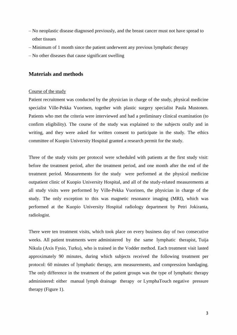
Volumetric limb measurement
We took volumetric measurements to investigate the effect of the treatments on total volume
of the upper extremity. Figure 3 illustrates the technique used. The volumetric measurements
were performed on both arms before and after the treatment period, and at the follow-up visit
after one month.
5
Figure 3. Volumetric limb measurement
Limb circumference
Limb circumferences were measured before and after treatments. The measurement sites
were the knuckles, palm, wrist (0 cm), and at 4-cm intervals along the arm (52 cm maximum).
These measurements were taken in order to investigate the effect of the treatments on arm
size.
MRI measurement of limb volume
A 1.5-tesla MRI scanner at the radiology department of Kuopio University Hospital was used
for imaging of the study patients’ swollen upper extremities. The MRI scans of the affected
extremities were performed before the treatment period and after the treatment period. The
scans were evaluated by radiologist Petri Jokiranta, who measured the total arm area and the
area of muscle compartments in cross-sections of the upper extremities. This provided an
estimate of the treatment effect on subcutaneous tissue area, and thus on any changes in
edema.
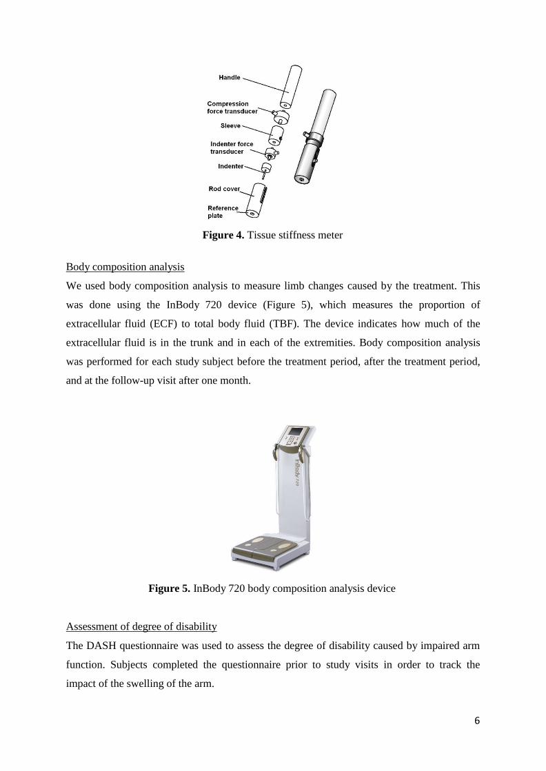
Tissue stiffness measurement
We measured tissue stiffness in order to investigate whether the treatment caused changes in
skin elasticity. Skin elasticity (tissue stiffness) was measured using an indentation device
developed for this purpose (Figure 4) (Arokoski et al. 2005, Iivarinen et al. 2011). The tissue
stiffness measurements were performed on both upper extremities before and after the
treatment period. The measurement sites were the back of the hand, the midpoint of the
forearm (lateral and medial) and the midpoint of the upper arm (lateral).
6
Figure 4. Tissue stiffness meter
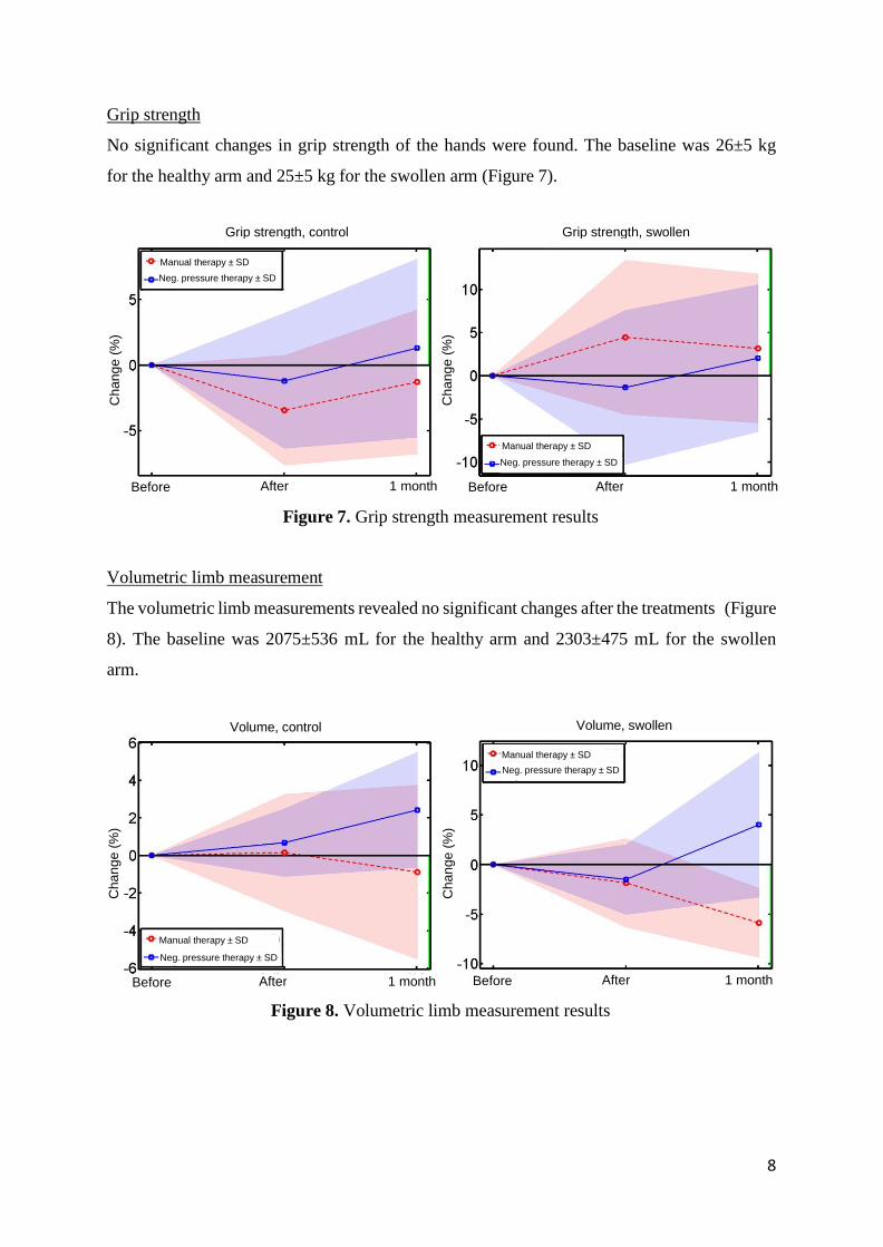
Body composition analysis
We used body composition analysis to measure limb changes caused by the treatment. This
was done using the InBody 720 device (Figure 5), which measures the proportion of
extracellular fluid (ECF) to total body fluid (TBF). The device indicates how much of the
extracellular fluid is in the trunk and in each of the extremities. Body composition analysis
was performed for each study subject before the treatment period, after the treatment period,
and at the follow-up visit after one month.
Figure 5. InBody 720 body composition analysis device
Assessment of degree of disability
The DASH questionnaire was used to assess the degree of disability caused by impaired arm
function. Subjects completed the questionnaire prior to study visits in order to track the
impact of the swelling of the arm.

7
Quality of Life
The FACT-B questionnaire is used to monitor the quality of life of breast-cancer patients.
Subjects completed the questionnaire prior to study visits.
Results
Patient safety
A total of 9 patients were treated with LymphaTouch negative pressure therapy during the
study and in pilot treatments conducted earlier, for a total of 90 treatments. The study patients
reported no adverse effects at all from the LymphaTouch treatments administered. Very mild
discomfort, associated mainly with skin symptoms caused by compression bandages, was
reported by 3 patients. None of the study patients discontinued the study prematurely.
Mobility of arm joints
The baseline for an individual test ranged from 0 to 155 degrees. The joint mobility
measurements and grip strength measurements revealed no significant changes after the
treatments. The results were similar for negative pressure therapy and manual lymph drainage
therapy (Figure 6).
Figure 6. Change in average mobility of arm joints in the healthy (control) arm and the
swollen arm during the study. The vertical green line (arrow) indicates positive treatment
outcomes.
Mobility, control Mobility, swollen MLD therapy ± SD
Neg. pressure therapy ± SD
Manual therapy ± SD
Neg. pressure therapy ± SD
After 1 month Before After 1 month
Cha
ng
e (
%)
Cha
ng
e (
%)
8
Grip strength, control Grip strength, swollen
Manual therapy ± SD
Neg. pressure therapy ± SD
Manual therapy ± SD
Neg. pressure therapy ± SD
Before After 1 month Before After 1 month
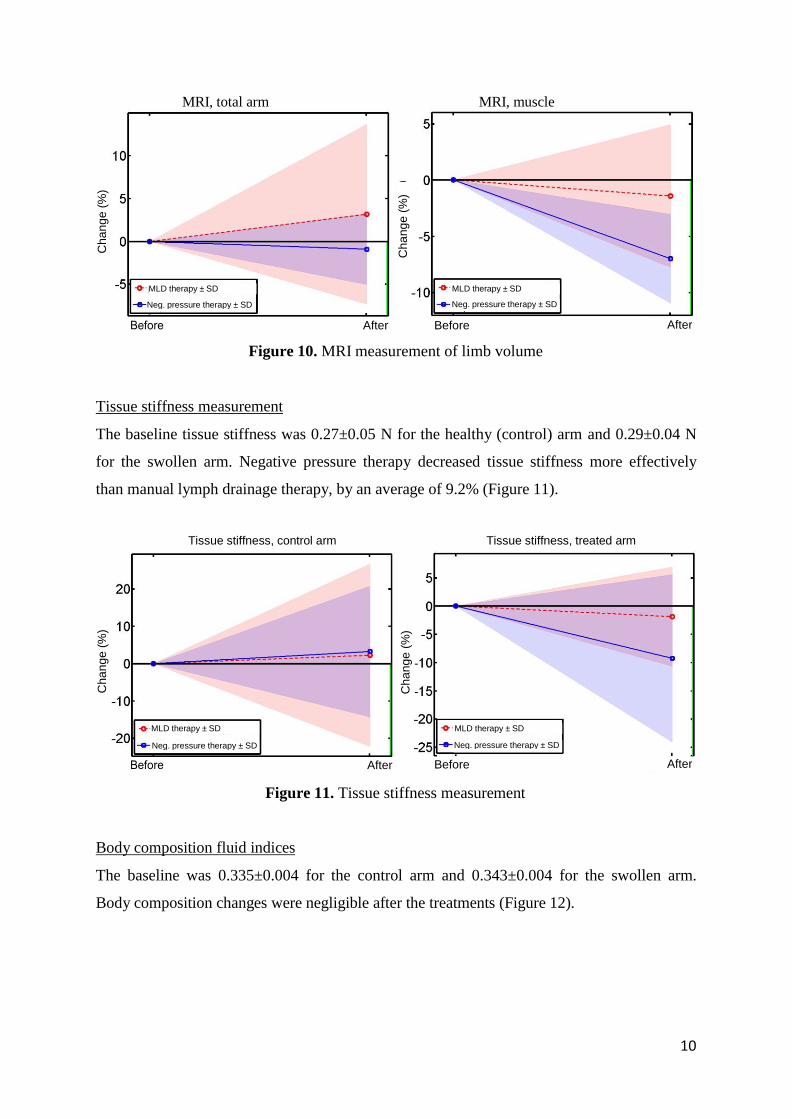
Grip strength
No significant changes in grip strength of the hands were found. The baseline was 26±5 kg
for the healthy arm and 25±5 kg for the swollen arm (Figure 7).
Figure 7. Grip strength measurement results
Volumetric limb measurement
The volumetric limb measurements revealed no significant changes after the treatments (Figure
8). The baseline was 2075±536 mL for the healthy arm and 2303±475 mL for the swollen
arm.
Figure 8. Volumetric limb measurement results
Volume, control Volume, swollen
Manual therapy ± SD
Neg. pressure therapy ± SD
Manual therapy ± SD
Neg. pressure therapy ± SD
Before After 1 month Before After 1 month
Cha
ng
e (
%)
Ch
an
ge
(%
)
Ch
an
ge
(%
) C
ha
ng
e (
%)

9
Circumference, control arm Circumference, treated arm
MLD therapy ± SD
Neg. pressure therapy ± SD
MLD therapy ± SD
Neg. pressure therapy ± SD
Before After Before After
Circumference, healthy arm, before -> after Circumference, swollen arm, before -> after
MLD therapy ± SD
Neg. pressure therapy ± SD
MLD therapy ± SD
Neg. pressure therapy ± SD
Measurement site (cm) [0 - wrist, K - knuckles, palm - P] Measurement site (cm) [0 - wrist, K - knuckles, palm - P]
Limb circumference
The baseline was 17.7–38.0 cm for the healthy (control) arm, depending on the measurement
site. The circumference of the swollen arm was 0.2–3.2 cm greater. Limb circumference
decreased after treatments, and the results of negative pressure therapy and manual lymph
drainage therapy were about the same (Figure 9).
Figure 9. Limb circumference
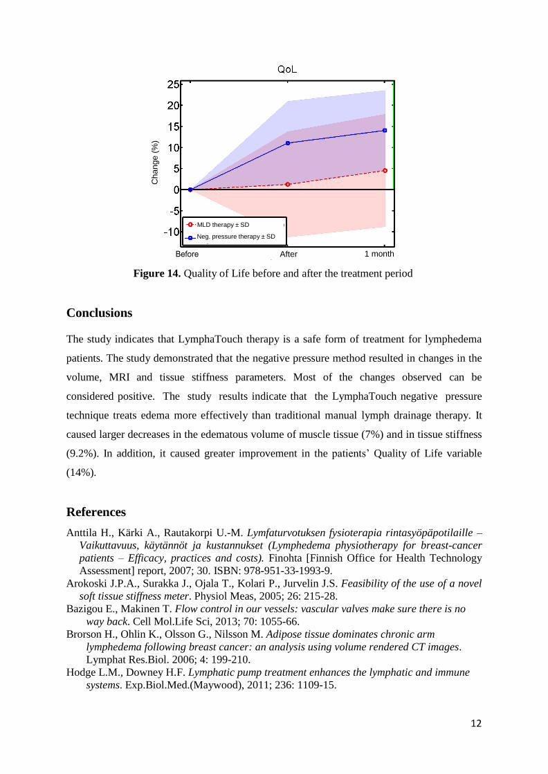
MRI measurement of limb volume
The baseline was 10,109±2470 mm² for the swollen arm and 3002±406 mm² for the arm
muscles. The total arm volume as measured by MRI did not change, but the volume of
muscle tissue decreased by an average of 7.0% following negative pressure therapy (Figure
10).
Cha
ng
e (
%)
Ch
an
ge
(%
)
Cha
ng
e (
%)
Ch
an
ge
(%
)
10
Tissue stiffness, control arm Tissue stiffness, treated arm
MLD therapy ± SD
Neg. pressure therapy ± SD
MLD therapy ± SD
Neg. pressure therapy ± SD
After Before After
Figure 10. MRI measurement of limb volume
Tissue stiffness measurement
The baseline tissue stiffness was 0.27±0.05 N for the healthy (control) arm and 0.29±0.04 N
for the swollen arm. Negative pressure therapy decreased tissue stiffness more effectively
than manual lymph drainage therapy, by an average of 9.2% (Figure 11).
Figure 11. Tissue stiffness measurement
Body composition fluid indices
The baseline was 0.335±0.004 for the control arm and 0.343±0.004 for the swollen arm.
Body composition changes were negligible after the treatments (Figure 12).
MRI, total arm MRI, muscle
MLD therapy ± SD
Neg. pressure therapy ± SD
MLD therapy ± SD
Neg. pressure therapy ± SD
After Before After
Cha
ng
e (
%)
Ch
an
ge
(%
)
Ch
an
ge
(%
) C
ha
ng
e (
%)

11
Degree of disability
MLD therapy ± SD
Neg. pressure therapy ± SD
Before After 1 month
Figure 12. Body composition analysis
Assessment of degree of disability
The baseline was 21±12%. The degree of disability decreased by an average of 30.2% after
negative pressure therapy (Figure 13).
Figure 13. Degree of disability experienced by the patient before and after the treatment
period
Quality of Life
Quality of Life was 102±14 at baseline and improved by an average of 14.0% after negative
pressure therapy (Figure 14).
ECF/TBF, control ECF/TBF, swollen
MLD therapy ± SD
Neg. pressure therapy ± SD
MLD therapy ± SD
Neg. pressure therapy ± SD
Before After 1 month Before After 1 month
Ch
an
ge
(%
)
Cha
ng
e (
%)
Ch
an
ge
(%
)
12
Neg. pressure therapy ± SD
Figure 14. Quality of Life before and after the treatment period
Conclusions
The study indicates that LymphaTouch therapy is a safe form of treatment for lymphedema
patients. The study demonstrated that the negative pressure method resulted in changes in the
volume, MRI and tissue stiffness parameters. Most of the changes observed can be
considered positive. The study results indicate that the LymphaTouch negative pressure
technique treats edema more effectively than traditional manual lymph drainage therapy. It
caused larger decreases in the edematous volume of muscle tissue (7%) and in tissue stiffness
(9.2%). In addition, it caused greater improvement in the patients’ Quality of Life variable
(14%).
References
Anttila H., Kärki A., Rautakorpi U.-M. Lymfaturvotuksen fysioterapia rintasyöpäpotilaille –
Vaikuttavuus, käytännöt ja kustannukset (Lymphedema physiotherapy for breast-cancer
patients – Efficacy, practices and costs). Finohta [Finnish Office for Health Technology
Assessment] report, 2007; 30. ISBN: 978-951-33-1993-9.
Arokoski J.P.A., Surakka J., Ojala T., Kolari P., Jurvelin J.S. Feasibility of the use of a novel
soft tissue stiffness meter. Physiol Meas, 2005; 26: 215-28.
Bazigou E., Makinen T. Flow control in our vessels: vascular valves make sure there is no
way back. Cell Mol.Life Sci, 2013; 70: 1055-66.
Brorson H., Ohlin K., Olsson G., Nilsson M. Adipose tissue dominates chronic arm
lymphedema following breast cancer: an analysis using volume rendered CT images.
Lymphat Res.Biol. 2006; 4: 199-210.
Hodge L.M., Downey H.F. Lymphatic pump treatment enhances the lymphatic and immune
systems. Exp.Biol.Med.(Maywood), 2011; 236: 1109-15.
MLD therapy ± SD
Neg. pressure therapy ± SD
After 1 month
Ch
an
ge
(%
)

13
Iivarinen J.T., Korhonen R.K., Julkunen P., Jurvelin J.S. Experimental and computational
analysis of soft tissue stiffness in forearm using a manual indentation device. Med Eng &
Phys, 2011; 33: 1245-53.
Iivarinen J.T., Korhonen R.K., Julkunen P., Jurvelin J.S. Experimental and computational
analysis of soft tissue mechanical response under negative pressure in forearm. Skin Res
Tech, 2013; 19: e356-65. Epub May 2012.
Kärki A., Anttila H., Tasmuth T., Rautakorpi U.-M. Lymphoedema therapy in breast cancer
patients – a systematic review on effectiveness and a survey of current practices and costs
in Finland. Acta Oncologica, 2009; 48: 850-9.
Papp A., Romppanen E., Lahtinen T., Uusaro A., Harma M., Alhava E. Red blood cell and
tissue water content in experimental thermal injury. Burns, 2005; 31: 1003-6.
Rockson S.G. Update on the biology and treatment of lymphedema. Curr.Treat.Options
Cardiovasc.Med., 2012; 14: 184-92.


