
JOURNAL OF VIROLOGY, June 1996, p. 3645–3658 Vol. 70, No. 60022-538X/96/$04.0010Copyright q 1996, American Society for Microbiology
Inhibitory Activity of the Equine Infectious Anemia VirusMajor 59 Splice Site in the Absence of Rev
WEI TAN,1 MARTIN SCHALLING,2 CUIPING ZHAO,1 MATTIAS LUUKKONEN,1 MIA NILSSON,2
EVA MARIA FENYO,1 GEORGE N. PAVLAKIS,3 AND STEFAN SCHWARTZ1*
Microbiology and Tumorbiology Center, Karolinska Institute, 171 77 Stockholm,1 and Neurogenetics Unit,Department of Molecular Medicine, Karolinska Hospital, 171 76 Stockholm,2 Sweden, and
National Cancer Institute—Frederick Cancer Research and Development Center,ABL-Basic Research Program, Frederick, Maryland 21702-12013
Received 2 January 1996/Accepted 12 March 1996
The major 5* splice site of equine infectious anemia virus (EIAV) conforms to the consensus 5* splice site ineight consecutive positions and is located immediately upstream of the gag AUG. Our results show that thepresence of this 5* splice site on the EIAV gag mRNA decreases Gag production 30- to 60-fold. This is causedby inefficient nuclear mRNA export and inefficient mRNA utilization. Inhibition could be overcome by pro-viding human immunodeficiency virus type 1 Rev/Rev-responsive element, human T-cell leukemia virus type 1Rex/Rex-responsive element, or simian retrovirus type 1 constitutive transport element. In addition, inhibitioncould be abolished by introducing single point mutations in the 5* splice site or by moving the 5* splice siteaway from its natural position immediately upstream of the gag AUG. This demonstrates that both mainte-nance of a perfect consensus 5* splice site and its proper location on the mRNA are important for inhibitoryactivity of the EIAV major 5* splice site.
Studies of the human immunodeficiency virus type 1(HIV-1) Rev protein have revealed that Rev is required forefficient expression of Gag, Pol, and Env proteins (38). Revacts posttranscriptionally by interacting with an RNA sequencenamed the Rev-responsive element (RRE) which is located onthe gag, pol, and env mRNAs (25, 35, 51, 60). In the absence ofRev, the unspliced and singly spliced HIV-1 mRNAs encodingviral structural proteins are sequestered in the nucleus (25, 27,36, 51), are unstable (27, 50), and are not translated intoproteins (5, 20, 45). It has been suggested that unspliced andsingly spliced HIV-1 mRNAs are trapped in the nucleus as aresult of spliceosome formation on unutilized splice sites onthese mRNAs (15).On the basis of the ability of synthetic peptides of Rev to
inhibit splicing of cellular mRNAs in vitro, it has been sug-gested that Rev acts by inhibiting splicing, thereby facilitatingtransport of unspliced and singly spliced mRNAs from thenucleus to the cytoplasm (41). However, HIV-1 mRNAs con-tain sequences that act in cis to inhibit splicing, and thesesequences are active in the absence of Rev (3, 4, 70, 71). As aresult, high levels of unspliced mRNAs are also detected in theabsence of Rev.Results from several laboratories have shown that HIV-1
mRNAs contain cis-acting, intragenic inhibitory sequences (10,18, 49, 54, 60, 63, 65) that inhibit production of viral structuralproteins in the absence of Rev. Such sequences have beenmapped within the gag, pol, and env regions of HIV-1 mRNAs.Inhibitory sequences located within the HIV-1 p17gag codingsequence were inactivated by the introduction of multiplepoint mutations (63, 65). These silent mutations, which did notaffect the amino acid sequence of the HIV-1 Gag protein,resulted in constitutive high Gag production independently ofRev (63), thereby showing that the inhibitory sequences were
directly involved in inhibition of Gag production in the absenceof Rev. These sequences acted at least in part by reducingmRNA stability (63, 65). Taken together, previously publishedresults are consistent with the idea that Rev interacts withRRE-containing mRNAs and redirects these mRNAs frompathways resulting in nuclear retention and degradation me-diated by inhibitory elements. It has been shown that Rev isable to directly promote nuclear export of RRE-containingmRNAs microinjected into Xenopus oocyte nuclei (29). Fur-thermore, conjugates between bovine serum albumin and pep-tides constituting the activation domain of Rev can inhibitRev-mediated nuclear mRNA export in Xenopus oocytes (28).Recently, cellular proteins with homology to nucleoporinswere shown to interact with the activation domain of Rev,supporting a role for Rev in nuclear mRNA export (8, 31).To date, members of the lentivirus family including HIV-1,
HIV-2, simian immunodeficiency virus, visna virus, caprinearthritis encephalitis virus, feline immunodeficiency virus, andequine infectious anemia virus (EIAV) have been shown toencode Rev proteins (19). These Rev proteins act on RNA-responsive elements located within the env gene. The oncoret-roviruses human T-cell leukemia virus types 1 and 2 (HTLV-Iand -II) and bovine leukemia virus (19) encode Rev-like pro-teins termed Rex. These proteins bind to RNA elementsnamed Rex-responsive elements (RxREs) located within theviral long terminal repeat (LTR). Interestingly, the HTLV-IRex protein can replace HIV-1 Rev, but not vice versa (59).Mason-Pfizer monkey virus is a D-type retrovirus which causesimmunodeficiency in newborn rhesus monkeys. It has recentlybeen shown that the 39 untranslated region (UTR) of Mason-Pfizer monkey virus contains a constitutive transport element(CTE) that can substitute for HIV-1 Rev/RRE (9). An elementwith similar function was also found in the 39 UTR of simianretrovirus type 1 (SRV-1) (85). This element can replace Rev/RRE in the context of the whole virus, resulting in Rev-inde-pendent virus expression. These results suggest that eucaryoticcells produce a protein with Rev-like properties and that cel-lular mRNAs may be regulated by similar mechanisms.
* Corresponding author. Mailing address: Microbiology and Tumor-biology Center, Karolinska Institute, P.O. Box 280, 171 77 Stockholm,Sweden. Phone: 468 728 6312. Fax: 468 331 399. Electronic mail ad-dress: [email protected].
3645

To gain further insight into the posttranscriptional mecha-nisms that regulate expression of complex retroviruses, weinvestigated the requirements for efficient expression of theEIAV Gag protein. EIAV is an ungulate lentivirus and theetiologic agent of equine infectious anemia. In general, EIAVshares many biological features with HIV-1, such as lifelongpersistent infection in the natural host, restricted viral expres-sion during an asymptomatic period, and a relatively high rateof antigenic variation and periodic emergence of neutraliza-tion-resistant variants despite the presence of a host immuneresponse (16, 17, 57). As a complex retrovirus, the EIAVgenome contains several short open reading frames (ORFs) inaddition to the gag, pol, and env genes common to all retrovi-ruses (see Fig. 1A). These short ORFs are translated frommultiply spliced mRNAs (6, 12, 56, 61, 74). ORF S1 encodes atranscriptional transactivator named Tat, which acts on a 25-nucleotide (nt) RNA sequence named the Tat-responsive ele-ment (14, 22, 23), located downstream of the transcriptionalstart site on the EIAV LTR (23, 67, 68). The EIAV Tat proteinappears to be functionally and structurally homologous toHIV-1 Tat (13, 21, 22). ORF S2 encodes a protein whosefunction is unknown, and a Rev protein is encoded by ORF S3(52). The EIAV RRE has not been mapped in detail butappears to be a bipartite element located within the env region(52).In the present study, we identified sequences that inhibit
expression of EIAV Gag in the absence of Rev. One inhibitoryRNA sequence is located immediately upstream of the EIAVgag gene and coincides with the major 59 splice site on theEIAV genome. Interestingly, the EIAV 59 splice site has aperfect homology to the consensus 59 splice site, implyingstrong binding to the U1 small nuclear RNA (snRNA) com-ponent of the cellular splicing machinery. Introduction of sin-gle point mutations at positions 22, 21, 11, 12, 13, 14, or15 in the 59 splice site sequence abolished the inhibitory ac-tivity and gave rise to high levels of Gag protein. Inhibitioncould be overcome by providing the HIV-1 Rev/RRE, theHTLV-1 Rex/RxRE, or the SRV-1 CTE in cis. We provideevidence that the unutilized splice site acts by retaining theEIAV gag mRNA in the nucleus and by affecting the efficiencyof utilization of mRNAs containing the 59 splice site.
MATERIALS AND METHODS
Plasmid constructions. To construct pE15, the EIAV matrix p15gag codingsequence (nt 446 to 817) was first PCR amplified from the EIAV plasmid p8 (73)(kindly provided by N. R. Rice) by using oligonucleotide 7227 (59-CAGCGCGCCAAGATGGGAGACCCTTTG-39), which introduced a BssHII site (under-lined) upstream of the translational start codon of gag, and oligonucleotide 7228(59-CTATTAATATTCTTCAGAGGGCTC-39), which introduced a transla-tional stop codon (underlined). The first nucleotide of the EIAV LTR is desig-nated 11 (73). The PCR fragment was subcloned into pNL17 (65) digested withSalI and EcoRI and filled in with Klenow DNA polymerase. This resulted inpE15. To generate pG15R, EIAV sequences between nt 189 and 817 were PCRamplified with oligonucleotides 8921 (59-CAGCGCGCCGCACTCAGATTCTGCGG-39) and 7228 and were subcloned into pBluescript (Stratagene). The p15gag
coding sequence was transferred as an XhoI-EcoRI fragment into pNL17R (65)digested with SalI and EcoRI. pG15 was generated by digestion of pG15R withAsp 718 and then by religation.To generate pE55R, the EIAV p55gag gene (nt 446 to 1906) was first PCR
amplified with oligonucleotides 7227 and 7420 (59-TTACTCCCACAAACTGTCCAGG-39) and subcloned into EcoRV-digested pBluescript (Stratagene), result-ing in pT7E55. The EIAV gag sequence was then transferred as a SalI-EcoRIfragment into pNL17R (65) digested with SalI and EcoRI. pE55 was generatedby partial digestion of pE55R with Asp 718 and by religation. To generatepG55R, EIAV sequences between nt 189 and 1906 were PCR amplified witholigonucleotides 8921 and 7420 and were subcloned into pBluescript (Strat-agene), resulting in pT7G55. The EIAV gag sequence was then transferred as aSalI-EcoRI fragment into pNL17R (65) digested with SalI and EcoRI. pG55 wasgenerated by partial digestion of pG55R with Asp 718 and then by religation. TheHIV-1 59 LTR sequence in plasmid pE55 or pG55 starts at nt 1 and ends at nt
711 (numbering of the HIV-1 sequence refers to the sequence of pNL43) (1). Toconstruct pCE55, a SalI-EcoRI fragment containing the EIAV p55gag gene (nt446 to 1906) from pE55 was first cloned downstream of the human cytomega-lovirus immediate-early promoter. pCE55 was thereafter generated by insertionof a BamHI-BclI fragment containing the simian virus 40 early poly(A) signaldownstream of the EIAV p55gag gene. To generate pCG55, the EIAV sequencesin pCE55 were removed by partial digestion with SalI-EcoRI and were replacedwith a SalI-EcoRI fragment obtained from pG55 (nt 189 to 1906).To construct pG55RxRE, the HTLV-I RxRE was transferred as an EcoRI-
XhoI fragment from pBS-RxRE (80) to pG55 digested with EcoRI and XhoI. Toconstruct pG55CTE, the SRV-1 CTE was PCR amplified with oligonucleotides12745 (59-GCATCAACGCGTGTCGACGGATCCAGACCACCTCCCCTGCGAG-39) and 13827 (59-GCATCAGCGGCCGCCTCGAGTCTAGACAAATCCCTCGGAAGCTGCG-39) (85) and was cloned directly into pG55 digestedwith EcoRI and filled in with Klenow DNA polymerase.Plasmid pD186 was generated by digestion of pG55 with BssHII and SmaI,
filling in of staggered 59 ends by using Klenow DNA polymerase, and religation.pD233 was constructed by partial digestion of pG55 with SalI andMscI, filling in,and religation. pESD(2)G was constructed by first PCR amplifying sequencesupstream of the EIAV gag AUG (nt 189 to 442) by using oligonucleotides 8921and EIAVSD(2) (59-GTCTCCCATTTCGGCGCGCCTCCTGTGTTCTG-39).The oligonucleotide EIAVSD(2) introduced a BssHII restriction site (under-lined), which resulted in four substitutions and insertion of a C nucleotideupstream of the 59 splice site. pESD(2)G was generated by cloning of theBssHII-digested PCR fragment into the BssHII site located immediately up-stream of the EIAV gag gene in plasmid pE55.To construct pG55DE, sequences upstream of the EIAV 59 splice site were
mutated by site-directed mutagenesis on uracil-containing single-stranded DNAof pT7G55 with oligonucleotide DE (59-GTCTCCCATTTCACCTGGGCCCGGAACACCTCC-39) as described elsewhere (48), in which an ApaI site (under-lined) was introduced and a 15-nt sequence immediately upstream of the 59splice site was deleted. The mutated EIAV sequence was transferred as a BssHII-Asp 718 fragment into BssHII-Asp 718-digested pNL17R (65), generating plas-mid pG55DE.To construct plasmids with multiple or single point mutations in the EIAV 59
splice site CAGGTAAG (nt 438 to 445), site-directed mutagenesis on uracil-containing single-stranded DNA of pT7G55 was performed as described else-where (48) with the following oligonucleotides (mutations introduced by theoligonucleotides are indicated by lowercase letters):21C22C23T (59-GTCTCCCATCTTACggaTCCTCCTG-39), 13G14C (59-GTCTCCCATCgcACCTGTCCTCCTG-39), 23T (59-GTCTCCCATCTTACCTaTCCTCCTG-39), 22C (59-GTCTCCCATCTTACCgGTCCTCCTG-39), 21C (59-GTCTCCCATCTTACgTGTCCTCCTG-39), 11A (59-GTCTCCCATCTTAtCTGTCCTCCTG-39), 12A(59-GTCTCCCATCTTtCCTGTCCTCCTG-39), 13G (59-GTCTCCCATCTcACCTGTCCTCCTG-39), 14C (59-GTCTCCCATCgTACCTGTCTTCTTG-39),15A (59-GTCTCCCATtTTACCTGTCCTCCTG-39). Mutants were screened bysequencing, and mutated fragments were transferred as BssHII-ApaI frag-ments into pG55 digested with BssHII and ApaI. This cloning step resulted inthe plasmids pG55-3T-2C-1C, pG5513G14C, pG55-3T, pG55-2C, pG55-1C,pG5511A, pG5512A, pG5513G, pG5514C, and pG5515A, respectively.To construct pESDSD(2)G, a sequence containing the EIAV 59 splice site (nt
189 to 445) was PCR amplified with oligonucleotides 8921 and EAS (59-GGGCGCGCCTTACCTGTCCTCCTGTGTTC-39) and was inserted in theStuI site at nucleotide position 237 in plasmid pESD(2)G. To constructpESD13G, a sequence containing the EIAV 59 splice site was first excised withBssHII and then cloned into BssHII-digested pE55. pESD32G and pESD86G,respectively, were constructed by transferring a BssHII-digested and Klenowfilled-in fragment containing the 59 splice site into MscI- or BssHII-digested andKlenow-filled-in pESD(2)G.The HIV-2 gag coding sequence (nt 535 to 2109) was PCR amplified from the
infectious HIV-2 molecular clone HIV-2ISY (30) by using oligonucleotides 7105(59-CATGCGCGCAGATTGTGGGAGATGGGC-39) and 7422 (59-GAGAAACCTTTGCTGGTCATC-39). A BssHII restriction site (underlined) was intro-duced 12 nt upstream of the HIV-2 gag ATG (underlined) through oligonucle-otide 7105. The PCR fragment was first inserted into EcoRV-digested pBlue-script (Stratagene) and was subsequently transferred as a BssHII-Asp 718fragment to pNL17R (65) partially digested with BssHII and Asp 718, generatingpH2GR. pH2G was generated by digestion of pH2GR with Asp 718 and then byreligation. To construct pESDH2G and pESDH2GR, a BssHII fragment con-taining the EIAV 59 splice site (see above) was subcloned into the BssHII siteimmediately upstream of HIV-2 gag ATG in pH2G and pH2GR, resulting inpESDH2G and pESDH2GR, respectively. To construct pESDM17, a BssHIIfragment containing the EIAV 59 splice site was subcloned into BssHII-digestedplasmid p17M1234 (63). Plasmid pM17ESD was constructed by cloning theEIAV 59 splice site-containing sequence downstream of the p17gag gene inplasmid p17M1234 (63).The HIV-1 Rev expression plasmid pNL14A7 (64), the HIV-1 p37gag expres-
sion plasmid pC37M (63), and the HTLV-I Rex expression plasmid pL3Rex (80)have been described previously.Cells and transfections. HLtat cells (64), a HeLa-derived cell line constitu-
tively producing HIV-1 Tat, were maintained in Dulbecco’s modified Eagle’smedium with 10% fetal calf serum. Transfections were performed by the calcium
3646 TAN ET AL. J. VIROL.

phosphate coprecipitation technique (34) as previously described (77). When thevaccinia virus-T7 RNA polymerase expression system was employed, cells wereinfected with 0.5 3 106 PFU of recombinant vaccinia virus vTF7-3 (33) express-ing T7 RNA polymerase 1 to 2 h prior to transfection. pNLCATW (76), pC37M(63), or pCS1X (76) was included as an internal control in each transfectionexperiment, and alkaline phosphatase activity was determined as previouslydescribed (76).Western blotting (immunoblotting). Immunoblotting was performed as de-
scribed previously (76, 77). Briefly, proteins were electrophoretically separatedon 12 or 15% polyacrylamide-sodium dodecyl sulfate (SDS) gels and then trans-ferred onto nitrocellulose membranes. Blots were blocked for 1 h at 378C with10% nonfat milk in phosphate-buffered saline (PBS) containing 0.3% Tween 20(PBS-T). After being washed three times in PBS-T buffer, the blots were incu-bated for 1 h at 378C with either the HIV-1- or HIV-2-positive patient serum(1:3,000 dilution) (kindly provided by J. Albert and R. Thorstensson) or rabbithyperimmune sera directed against EIAV p15gag or p26gag (37) (1:5,000 dilution)(kindly provided by S. Oroszlan). Horseradish peroxidase (HRP)-conjugateddonkey anti-human immunoglobulin G (IgG) or HRP-conjugated goat anti-rabbit IgG (1:10,000 dilution) was used as a secondary antibody. Specific proteinswere visualized with enhanced chemiluminescence detection reagents (Amer-sham).RNA extractions and Northern (RNA) blotting. Total RNA was prepared from
transfected HLtat cells (64) 24 h posttransfection by the heparin-DNase proce-dure as previously described (77). A 20-mg amount of each RNA sample wasseparated on a 1% denaturing agarose gel and blotted onto nitrocellulose filters.After baking at 808C for 2 h, the membranes were hybridized to a [32P]UTP-labeled riboprobe, generated from pT7G55, at 558C overnight. The filters werewashed three times with 23 SSC (13 SSC is 0.15 M NaCl plus 0.015 M sodiumcitrate)–0.1% SDS and three times with 0.13 SSC–0.1% SDS at 608C. The driedfilters were exposed to X-ray film at 2708C with intensifying screens.Cytoplasmic and nuclear poly(A)1 mRNAs were isolated by Dynabeads Oli-
go(dT)25 (Dynal A.S.) as described (77). Briefly, transfected cells were lysed for5 min on ice in lysis buffer (10 mM Tris-HCl [pH 7.5], 0.14 M NaCl, 5 mM KCl,0.65% Nonidet P-40). Cytoplasmic and nuclear fractions were separated bycentrifugation at 8,000 3 g for 2 min. The pellets containing nuclei were washedonce with lysis buffer and resuspended in lysis buffer lacking Nonidet P-40, andthen an equal volume of lysis buffer containing 1% SDS was added. Afterincubation for 10 min on ice, the samples were frozen on dry ice and then thawedat room temperature and subjected to low-speed centrifugation to remove cel-lular debris. Supernatants obtained from the cytoplasmic and nuclear fractionswere incubated with equal volumes of 23 binding buffer (20 mM Tris-HCl [pH7.5], 1.0 M LiCl, 2 mM EDTA, 0.5% SDS) containing 400 mg of DynabeadsOligo(dT)25 (Dynal A.S.). After being washed three times, poly(A)1 mRNAswere eluted from the beads with elution buffer (2 mM EDTA [pH 7.5]) at 658Cfor 2 to 3 min and stored at 2708C until use.In situ hybridization. In situ RNA hybridization was performed as previously
described (62). Briefly, the 46-mer antisense oligonucleotide probe E46 (59-CGTCACCTTCTCTAACTTCTTGAGCGCCTTGCTCCATGTCAAAGGG-39)complementary to the EIAV p15gag sequence (nt 454 to 499) was end labeled byusing 35S-dATP and terminal deoxynucleotidyltransferase. At 24 h posttransfec-tion, cells in each plate were washed twice with PBS and then hybridized with 107
cpm of probe in 900 ml of hybridization buffer (50% formamide, 43 SSC, 500 mgof salmon sperm DNA per ml, 0.2 M dithiothreitol, 13 Denhardt’s solution, 1%sarcosyl, 10% dextran sulfate) at 428C overnight. The cells were rinsed once with13 SSC at room temperature and four times at 558C. After being washed oncein water for 1 min, the cells were dehydrated for 1 min in 60% ethanol and thenfor 1 min in 90% ethanol. The air-dried cells were covered with NTB2 nuclearemulsion (Kodak, Rochester, N.Y.) and exposed for 2 weeks before develop-ment.RT-PCR. Reverse transcription (RT)-PCR was performed as previously de-
scribed (77). Briefly, cytoplasmic and nuclear poly(A)1 mRNAs were reversetranscribed at 428C for 1 h in a total volume of 30 ml. Reaction without reversetranscriptase was performed in parallel and served as a control for the absenceof plasmid DNA contamination. A 5-ml amount of the cDNA product was PCRamplified in a 100-ml reaction volume with oligonucleotides EP (59-AGGTGACGGTACAAGGGTCTCAGAAA-39) and EW (59-CCCACCATGTTCTTTCAAAGGC-39), which detected cDNA of unspliced EIAV gag mRNA, oligonu-cleotides ACTINS (59-TGAGCTGCGTGTGGCTCC-39) and ACTINA (59-GGCATGGGGGAGGGCATACC-39), which specifically amplify cDNA of splicedactin mRNA, or ACTINS-1 (59-CCAGTGGCTTCCCCAGTG-39) and ACTINA,which detect cDNA of unspliced actin mRNA.Oligonucleotides EW and ACTINA were end labeled with [g-32P]ATP prior
to use. PCR was performed in a total volume of 100 ml for 20 to 25 cycles at 948Cfor 1 min, 558C for 2 min, and 728C for 3 min with a final extension at 728C for10 min. A 10-ml sample from each RT-PCR was analyzed by electrophoresis on5% polyacrylamide gels.
RESULTS
The EIAV 5* UTR inhibits EIAV Gag production. We havepreviously demonstrated that the HIV-1 matrix p17gag coding
region contains inhibitory sequences that act posttranscrip-tionally to repress HIV-1 Gag production in the absence ofRev (63, 65). EIAV is a complex retrovirus but has a relativelysimple genomic organization compared with that of HIV-1(Fig. 1A). In this study, we investigated whether the EIAVp15gag matrix gene contains inhibitory sequences similar tothose found in the HIV-1 p17gag coding sequence. We firstgenerated two plasmids designed to produce the EIAV p15gag
matrix protein. Plasmid pE15 contains the EIAV p15gag codingsequence (nt 446 to 817) (nucleotide numbering of the EIAVsequences follows that used by Stephens et al. [73] and starts atthe first nucleotide of the EIAV LTR), while plasmid pG15contains the complete EIAV 59 UTR (nt 189 to 445) in addi-tion to the p15gag gene (Fig. 1B). Transcription from theseplasmids is under control of the HIV-1 LTR promoter. Toinvestigate whether the p15gag protein was produced fromplasmids pE15 and pG15, these plasmids were separatelytransfected into HLtat cells (64), a HeLa derivative constitu-tively producing HIV-1 Tat protein. A CAT-producing plasmidpNLCATW (76) was included in transfections and served as aninternal control for transfection efficiency and the levels ofCAT protein were measured by capture enzyme-linked immu-nosorbent assaying as previously described (76). Cells wereharvested 24 h posttransfection, and the production of EIAVp15gag protein was assessed by Western immunoblotting. Fig-ure 1C (left panel) shows that high levels of p15gag proteinwere produced from cells transfected with pE15, while p15gag
produced from pG15 was undetectable. These results indicatedthat inhibitory sequences are not contained within the EIAVp15gagmatrix coding sequence and suggested that an inhibitorysequence either overlaps the EIAV 59 UTR and the EIAV gaggene or is located in the EIAV 59 UTR.Since inhibitory sequences were not found in the EIAV
matrix p15gag coding region, we then investigated whether suchsequences were present in other parts of the EIAV p55gag
gene. Therefore, we generated plasmid pE55, which containsthe complete EIAV p55gag gene, and pG55, which contains theEIAV 59 UTR in addition to p55gag coding sequence (Fig. 1B).Western blot analysis revealed that high levels of EIAV p55gag
were produced from cells transfected with pE55 (Fig. 1C, rightpanel), while pG55 did not produce detectable levels of p55gag.These experiments suggested that sequences in the EIAV 59UTR are required for inhibition of Gag production. Westernblot analysis of serial 2-fold dilutions of cell lysates from cellstransfected with pE55 showed that p55gag levels produced frompE55 were 30- to 60-fold higher than those produced frompG55 (Fig. 1D). We also investigated whether the inhibitoryeffect was dependent on the promoter or poly(A) signal used inplasmid pG55. Therefore, the HIV-1 LTR sequences in plas-mids pE55 and pG55 were replaced by human cytomegalovirusimmediate-early promoter and simian virus 40 early poly(A)signal, resulting in plasmids pCE55 and pCG55, respectively(Fig. 1B). As shown in Fig. 1C (right panel), similarly low levelsof p55gag were produced from pCG55 and pG55, while highlevels of Gag were produced from pCE55 and pE55. Theseresults demonstrate that inhibition caused by the presence ofthe EIAV 59 UTR could be reproduced with plasmids withdifferent promoters and poly(A) signals.The inhibitory effect could be overcome by HIV-1 Rev and
RRE, by HTLV-I Rex and RxRE, or by SRV-1 CTE. To deter-mine whether the EIAV inhibitory sequence was important forRev regulation, we investigated whether inhibition could beovercome by providing HIV-1 Rev in trans and RRE in cis. TheHIV-1 RRE sequence was introduced within the transcriptionunit downstream of the EIAV gag coding sequence in plasmidpG15, pE55, or pG55, generating plasmids pG15R, pE55R,
VOL. 70, 1996 INHIBITORY EFFECT OF THE MAJOR EIAV 59 SPLICE SITE 3647

and pG55R, respectively (Fig. 2A). Western blot analysisrevealed that pG15R and pG55R produced undetectablelevels of EIAV Gag in the absence of Rev (Fig. 2B), while inthe presence of Rev, both produced high levels of Gag whichwere similar to those produced from pE15 and pE55 (Fig.2B). Analysis of serial 2-fold dilutions of cell lysates fromcells transfected with pG55R in the presence of Rev revealedthat p55gag levels were ;60-fold higher than in the absence ofRev (Fig. 2C). We did not observe a negative effect of HIV-1RRE such as that which has been described with other testsystems (10, 54). Instead, similar high levels of p55gag wereproduced from pE55 and pE55R and Gag levels were notelevated from pE55R in the presence of Rev (Fig. 2B, rightpanel).It is known that HTLV-I Rex can functionally replace HIV-1
Rev (59). Therefore, the HTLV-I RxRE was introduced down-stream of the EIAV gag gene in pG55, generating plasmidpG55RxRE (Fig. 2A). Only in the presence of Rex didpG55RxRE produce p55gag levels comparable to those pro-duced by pE55 (Fig. 2D, left panel). As expected, providingHIV-1 Rev in trans did not increase EIAV Gag productionfrom pG55RxRE (Fig. 2D, left panel), while the presence ofHTLV-I Rex resulted in elevated Gag production from plas-mid pG55R (Fig. 2D, left panel).The 39 UTRs of Mason-Pfizer monkey virus and SRV-1
contain CTEs that can functionally replace HIV-1 Rev/RREand render expression of HIV-1 structural genes Rev indepen-dent (9, 85). To test if the SRV-1 CTE could function asRev/RRE or Rex/RxRE to relieve inhibition on EIAV Gagexpression, a 172-bp fragment spanning the SRV-1 CTE (85)was introduced into pG55, generating plasmid pG55CTE (Fig.2A). This plasmid produced high levels of p55gag, but not ashigh as those produced from pE55 or from pG55R in thepresence of Rev (Fig. 2D, right panel). Taken together, thesedata demonstrate that inhibition of EIAV Gag expression is aposttranscriptional event, supporting the idea that the pres-ence of inhibitory EIAV sequences is important for Rev reg-ulation.EIAV gag mRNAs containing the 5* UTR sequence are in-
efficiently transported out of the nucleus and are inefficientlyutilized. Since the difference in Gag production from pG55Rin the absence or presence of Rev was ;60-fold, we wished toanalyze RNA levels produced under these experimental con-ditions. Northern blot analysis of total RNA extracted fromcells transfected with pG55R or pG15R in the absence orpresence of Rev revealed that similar mRNA levels were pro-duced in the absence or presence of Rev (Fig. 2E). Therefore,the difference at the protein level could not be accounted forby a similar difference at the RNA level.It has been established that the mRNAs encoding HIV-1
structural proteins are trapped in the nucleus in the absence ofRev and that the presence of Rev protein facilitates transportof these mRNAs from the nucleus to the cytoplasm (25, 27, 36,51). We then investigated whether the inefficient p55gag pro-duction from pG55R in the absence of Rev was due to nuclear
FIG. 1. The EIAV 59 UTR contains sequences that strongly inhibit EIAVGag production. (A) Schematic organization of EIAV genome (not to scale).Black boxes, tat ORFs; stippled boxes, rev ORFs; open boxes, ORFs of gag, pol,env, and S2 genes; arrows, oligonucleotides used for PCR amplification of EIAVgag sequences. Numbers indicate nucleotide positions in the EIAV genome (thefirst nucleotide of the EIAV LTR is designated11) (73). (B) Structures of EIAVp15gag and p55gag expression plasmids. Black boxes, HIV-1 LTR sequences; openboxes, EIAV sequences; dark stippled boxes, human cytomegalovirus (HCMV)immediate-early promoter; light stippled boxes, simian virus 40 (SV40) earlypoly(A) signal. Numbers indicate nucleotide positions in the EIAV genome (thefirst nucleotide of the EIAV LTR is designated 11) (73). Names of the plasmidsare indicated on the left. (C) Western blot analysis of EIAV p15gag and p55gag
production from HLtat cells transfected with Gag expression plasmids as indi-cated. Proteins were separated on 12% polyacrylamide-SDS gels and electro-
phoretically transferred onto nitrocellulose membranes. The EIAV p15gag orp55gag proteins were detected with rabbit hyperimmune sera against EIAV p15gag
or p55gag (1:5,000) and then by incubation with HRP-conjugated goat anti-rabbitimmunoglobulin antiserum (1:10,000). Proteins were visualized with enhancedchemiluminescence detection reagents. The positions of EIAV p15gag and p55gag
are indicated on the left. (D) Western blot analysis of EIAV p55gag productionfrom cells transfected with pG55 or pE55. Cell lysates from pE55-transfectedcells were serially twofold diluted and analyzed by Western immunoblotting asdescribed above. The position of EIAV p55gag is indicated on the left.
3648 TAN ET AL. J. VIROL.
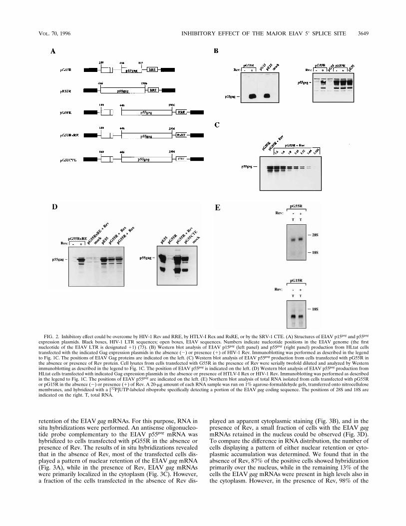
retention of the EIAV gag mRNAs. For this purpose, RNA insitu hybridizations were performed. An antisense oligonucleo-tide probe complementary to the EIAV p55gag mRNA washybridized to cells transfected with pG55R in the absence orpresence of Rev. The results of in situ hybridizations revealedthat in the absence of Rev, most of the transfected cells dis-played a pattern of nuclear retention of the EIAV gag mRNA(Fig. 3A), while in the presence of Rev, EIAV gag mRNAswere primarily localized in the cytoplasm (Fig. 3C). However,a fraction of the cells transfected in the absence of Rev dis-
played an apparent cytoplasmic staining (Fig. 3B), and in thepresence of Rev, a small fraction of cells with the EIAV gagmRNAs retained in the nucleus could be observed (Fig. 3D).To compare the difference in RNA distribution, the number ofcells displaying a pattern of either nuclear retention or cyto-plasmic accumulation was determined. We found that in theabsence of Rev, 87% of the positive cells showed hybridizationprimarily over the nucleus, while in the remaining 13% of thecells the EIAV gag mRNAs were present in high levels also inthe cytoplasm. However, in the presence of Rev, 98% of the
FIG. 2. Inhibitory effect could be overcome by HIV-1 Rev and RRE, by HTLV-I Rex and RxRE, or by the SRV-1 CTE. (A) Structures of EIAV p15gag and p55gag
expression plasmids. Black boxes, HIV-1 LTR sequences; open boxes, EIAV sequences. Numbers indicate nucleotide positions in the EIAV genome (the firstnucleotide of the EIAV LTR is designated 11) (73). (B) Western blot analysis of EIAV p15gag (left panel) and p55gag (right panel) production from HLtat cellstransfected with the indicated Gag expression plasmids in the absence (2) or presence (1) of HIV-1 Rev. Immunoblotting was performed as described in the legendto Fig. 1C. The positions of EIAV Gag proteins are indicated on the left. (C) Western blot analysis of EIAV p55gag production from cells transfected with pG55R inthe absence or presence of Rev protein. Cell lysates from cells transfected with G55R in the presence of Rev were serially twofold diluted and analyzed by Westernimmunoblotting as described in the legend to Fig. 1C. The position of EIAV p55gag is indicated on the left. (D) Western blot analysis of EIAV p55gag production fromHLtat cells transfected with indicated Gag expression plasmids in the absence or presence of HTLV-I Rex or HIV-1 Rev. Immunoblotting was performed as describedin the legend to Fig. 1C. The positions of EIAV p55gag are indicated on the left. (E) Northern blot analysis of total RNA isolated from cells transfected with pG55Ror pG15R in the absence (2) or presence (1) of Rev. A 20-mg amount of each RNA sample was run on 1% agarose-formaldehyde gels, transferred onto nitrocellulosemembranes, and hybridized with a [32P]UTP-labeled riboprobe specifically detecting a portion of the EIAV gag coding sequence. The positions of 28S and 18S areindicated on the right. T, total RNA.
VOL. 70, 1996 INHIBITORY EFFECT OF THE MAJOR EIAV 59 SPLICE SITE 3649

cells showed primarily cytoplasmic accumulation of EIAV gagmRNAs.To confirm the results obtained by in situ hybridizations,
cytoplasmic poly(A)1 mRNA was extracted from cells trans-fected with pG55R in the absence or presence of Rev and wasanalyzed by RT-PCR. The poly(A)1 mRNAs were seriallydiluted 4-fold and were subjected to RT and then to PCRamplification with oligonucleotides specifically amplifying aportion of the EIAV p15gag coding sequence. CytoplasmicEIAV gag poly(A)1 mRNA levels produced from pG55R inthe presence of Rev were approximately 4-fold higher thanthose produced in the absence of Rev (Fig. 4, upper panel),while production of EIAV p55gag from pG55R increased by;60-fold in the presence of Rev (Fig. 2C). Therefore, by in situhybridization and by RT-PCR, we could show that there wasapproximately 4- to 7-fold more cytoplasmic poly(A)1 EIAVgag mRNA in the presence of Rev than in the absence of Rev.To control for the cell fractionation technique, we also ana-lyzed spliced and unspliced actin mRNAs. The results showedthat unspliced actin RNAs were found primarily in the nuclearfractions (Fig. 4, bottom panel), demonstrating that nuclearleakage did not occur, while spliced actin mRNA was found inboth nuclear and cytoplasmic fractions (Fig. 4, bottom panel).We concluded that Rev acted not only by increasing the levels
of EIAV gag mRNAs in the cytoplasm, but also by improvingthe translatability of the gag mRNAs. This supports the ideathat Rev is a multifunctional protein or that the action of Revhas multiple effects.The negative effect on p55gag production could be bypassed
by transcription in the cytoplasm. We then wished to testwhether transcription in the cytoplasm could bypass the in-hibition exerted by the EIAV sequences. For this purpose,we employed the vaccinia virus-T7 RNA polymerase-basedexpression system (33). T7 RNA polymerase expressed fromthe recombinant vaccinia virus vTF7-3 is located in the cyto-plasm, since it lacks a nuclear localization signal (24). As aresult, transcription directed by T7 RNA polymerase occursin the cytoplasm. Two expression plasmids were constructed:pT7G55, which contains the EIAV p55gag gene and upstreamsequence (nt 189 to 1906), and pT7E55, which contains theEIAV p55gag gene (nt 446 to 1906) but lacks the EIAV inhib-itory sequence (Fig. 5A). In both plasmids, the EIAV p55gag
gene is under the control of the bacteriophage T7 promoterspecifically recognized by T7 RNA polymerase. To evaluateEIAV p55gag production, HLtat cells were first infected withrecombinant vaccinia virus vTF7-3 (33) and then transfectedwith pT7G55 and pT7E55. Western blot analysis revealed atwo- to four-fold difference in p55gag production between these
FIG. 3. Intracellular localization of EIAV gagmRNAs detected by in situ hybridization. An antisense oligonucleotide was hybridized to cells transfected with pG55Rin the absence or presence of Rev protein. EIAV gag mRNAs produced from pG55R in the absence of Rev were located primarily in the nucleus (A), althoughcytoplasmic localization of hybridization signals could be observed in a portion of cells (B). EIAV gag mRNAs produced from pG55R in the presence of Rev werepredominantly localized in the cytoplasm (C); however, a small percentage of cells with hybridization signals primarily over the nucleus could be detected (D).
3650 TAN ET AL. J. VIROL.

two plasmids (Fig. 5B and data not shown), while paralleltransfections with pG55 and pE55 showed a great difference inGag production (Fig. 5B). These results demonstrated that thenegative effect exerted by the EIAV inhibitory sequence couldbe partially relieved by transcription in the cytoplasm, indicat-ing that nuclear factors are required for efficient inhibition ofEIAV Gag production. This is in agreement with the negativeeffect on nuclear mRNA export. However, it should be notedthat the 59 ends of the mRNAs produced from pT7G55 andpT7E55 are different, and this may potentially affect the ex-pression levels of the EIAV p55gag protein.Inhibitory activity coincides with the major EIAV 5* splice
site. We then wished to map and characterize the EIAV in-hibitory element. As shown in Fig. 6A, plasmid pG55 contains254 nt upstream of the gag AUG, which are absent in plasmidpE55. Progressive 59 end deletions were introduced in thisregion. A total of 186 or 233 nt was deleted from the 59 end ofthe EIAV sequence in plasmid pG55, generating plasmidpD186 or pD233, respectively (Fig. 6A). Western blot analysisrevealed that similarly low levels of EIAV Gag were producedfrom plasmids pD186, pD233, and pG55, while high levels ofGag were produced from pE55 (Fig. 6B), indicating that theinhibitory sequence is located downstream of nt 421 (Fig. 6A).Since the EIAV major 59 splice site is present in this region, itwas mutated by introduction of multiple point mutations,
changing the EIAV 59 splice site containing sequence fromACAGGTAAG to cgcGccAAG. This altered the highly con-served GT dinucleotide in the 59 splice site to CC. The mutantplasmid, named pESD(2)G (Fig. 6A), produced high levels ofEIAV Gag, comparable to those produced by pE55 (Fig. 6B),demonstrating that the EIAV 59 splice site was required forinhibition of Gag production in the absence of Rev.Inhibitory activity of the EIAV 5* splice site could be abol-
ished by single point mutations.We first wished to exclude thepossibility that sequences upstream of the EIAV 59 splice sitecontributed to inhibition of EIAV Gag expression. Since wehave shown that the EIAV 59 UTR sequence upstream of nt421 is not required for inhibition (Fig. 6A and B), a 15-ntsequence (nt 423 to 437) located immediately upstream of theEIAV 59 splice site was deleted from plasmid pG55, generatingplasmid pG55DE (Fig. 7A). This plasmid did not produceelevated levels of p55gag (Fig. 7B), demonstrating that up-stream of the EIAV gag AUG, the 59 splice site is the onlysequence required for inhibition of Gag expression.Compared with the invariable G and T at positions 11 and
12, positions 23, 22, 21, 13, 14, and 15 on the 59 splice sitemay vary without inactivating the 59 splice site. In fact, manyfunctional 59 splice sites on cellular mRNAs deviate from theconsensus sequence at positions other than 11 and 12 (53).Inspection of this 59 splice site revealed that it has a perfectcomplementarity to the U1 snRNA in eight consecutive nucle-otide positions (Fig. 7A). To investigate if maintenance of aperfect consensus 59 splice site is required for inhibition, wefirst changed the three nucleotides (CAG) at positions 23 to21 immediately upstream of GT in the 59 splice site to TCC bysite-directed mutagenesis, generating plasmid pG55-3T-2C-1C(Fig. 7A). In addition, the 2 nt (AA) at positions 13 and 14immediately downstream of the invariable GT dinucleotidewere mutated to GC, generating plasmid pG5513G14C (Fig.7A). Both pG55-3T-2C-1C and pG5513G14C produced highlevels of Gag, comparable to those produced from pE55 (Fig.
FIG. 4. RT-PCR analysis of cytoplasmic and nuclear poly(A)1 mRNAs. Toppanel, PCR amplification of the cDNA synthesized from serially fourfold-dilutedcytoplasmic poly(A)1 mRNA isolated from cells transfected with pG55R in theabsence (2) or presence (1) of HIV-1 Rev protein; bottom panel, PCR ampli-fication of cDNA from spliced and unspliced cytoplasmic or nuclear poly(A)1
actin mRNAs. The numbers of cycles used for PCR amplification of cDNAs wereadjusted to detect cDNA in the linear range of the assay. N, nuclear RNA; C,cytoplasmic RNA; actin-S, spliced actin mRNA; actin-U, unspliced actin RNA.Oligonucleotides were end labeled with [g-32P]ATP prior to PCR. The PCRproducts were analyzed on 5% polyacrylamide gels and then by autoradiography.Positions of the amplified DNA fragments are indicated on the left.
FIG. 5. Expression of EIAV p55gag with the vaccinia virus-T7 RNA polymer-ase expression system and in vitro translation system. (A) Structures of EIAVp55gag expression plasmids driven by the bacteriophage T7 promoter. Stippledboxes, bacteriophage T7 promoter; open boxes, EIAV sequences. Bent arrowsshow the directions of transcription driven by the T7 promoter. Numbers indi-cate nucleotide positions in the EIAV genome (the first nucleotide of the EIAVLTR is designated 11) (73). (B) Western blot analysis of EIAV p55gag produc-tion from pT7G55- or pT7E55-transfected HLtat cells that are infected withrecombinant vaccinia virus, vTF7-3, producing T7 RNA polymerase (left panel),or from HLtat cells transfected with pG55 or pE55 (right panel), respectively.
VOL. 70, 1996 INHIBITORY EFFECT OF THE MAJOR EIAV 59 SPLICE SITE 3651

7B). These results showed that mutations in the variable posi-tions of the 59 splice site could abolish inhibitory activity evenif the highly conserved GT dinucleotide was left intact, thusdemonstrating that nucleotides at these positions also contrib-ute to inhibition of EIAV Gag expression.To evaluate the importance of each nucleotide position in
the 59 splice site for inhibition, each of the eight nucleotides inthe 59 splice site was mutated one by one as shown in Fig. 7A.The resulting plasmids were named pG55-3T, pG55-2C, pG55-1C, pG5511A, pG5512A, pG5513G, pG5514C, and pG5515A, respectively (Fig. 7A). Western blot analysis revealed thathigh levels of p55gag were produced from all of the mutants,respectively, except for pG55-3T (Fig. 7C). Although theEIAV Gag levels produced from pG55-2C were slightly lowerthan those produced from pE55, they were still substantiallyhigher than those produced from pG55, indicating that nucle-otide A at position 22 in the 59 splice site contributes toinhibition to a lower extent than other positions of nucleotidesin the 59 splice site. Interestingly, among these mutant plas-mids, only pG55-3T did not produce elevated levels of p55gag.The difference in Gag production between pG55-3T andpG5511A was 30- to 60-fold (Fig. 7D), while the difference incytoplasmic EIAV poly(A)1 gag mRNA levels was approxi-
mately 4-fold (data not shown). All mutants produced similarlevels of p55gag when they were expressed with the vacciniavirus T7 RNA polymerase-based expression system (data notshown). Taken together, these experiments established thatthe full activity of the EIAV inhibitory sequence is criticallydependent on the seven nucleotides (AGGTAAG) of the 59splice site.A minimal distance between the EIAV 5* splice site and the
gag AUG is required for inhibitory activity. The natural posi-tion of the EIAV major 59 splice site is immediately upstreamof the EIAV gag translational start codon (Fig. 7A). This or-ganization is unique for EIAV among the retroviruses. Wethen investigated whether the inhibitory effect could be abol-ished by increasing the distance between the EIAV 59 splicesite and the EIAV gag AUG. We generated plasmidspESD13G, pESD32G, pESD86G, and pESDSD(2)G (Fig.8A), in which the 59 splice site was placed 13, 32, 86, and 216nt upstream of the EIAV gag AUG, respectively. Figure 8Bshows that plasmid pESD13G produced similarly low levels ofp55gag as pG55, while high levels of p55gag were produced fromplasmids pESD32G, pESD86G, and pESDSD(2)G, respec-tively. It could be argued that utilization of the splice siteswould result in gag mRNAs lacking the 59 splice site that couldpotentially produce high levels of p55gag. Northern blots oftotal RNA extracted from cells transfected with pE55,pESD13G, or pESD86G showed that similar levels of un-spliced gag mRNAs were produced among them (Fig. 8C). Tofurther verify that the EIAV 59 splice site is not utilized inpESD13G, we analyzed the structures of cytoplasmic poly(A)1
mRNAs produced from pESD13G, pESD32G, or pESD86Gby RT-PCR. DNA bands corresponding in size to unsplicedgag mRNA were detected (Fig. 8D). The appearance of thesebands was dependent on reverse transcriptase. In parallel PCRreactions, the corresponding plasmid DNAs were used asDNA templates. The amplified DNAs obtained from thesereactions had the same sizes as the corresponding DNAs gen-erated by RT-PCR (Fig. 8D), demonstrating that mRNAs pro-duced from pESD13G, pESD32G, and pESD86G were notspliced. In conclusion, these results demonstrated that the neg-ative effect of the EIAV 59 splice site was lost when the dis-tance between the EIAV 59 splice site and the gag AUG in-creased to 32 nt or more, indicating that close proximity of theEIAV 59 splice site to the gag coding sequence was required forstrong inhibition.The EIAV 5* splice site inhibits heterologous gag gene ex-
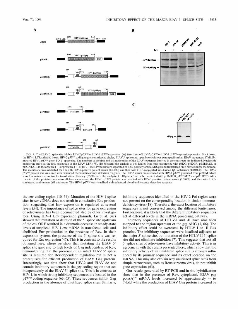
pression. To test if the EIAV 59 splice site could inhibit ex-pression of a heterologous retroviral gag gene, we cloned theEIAV splice site-containing sequence (nt 189 to 445) 21 ntupstream of the HIV-2 gag gene in plasmid pH2G (Fig. 9A),generating plasmid pESDH2G (Fig. 9A). Western blot analysisrevealed that plasmid pH2G produced high levels of HIV-2p55gag (Fig. 9B), while low levels of HIV-2 Gag were producedfrom pESDH2G, demonstrating that the EIAV 59 splice sitecould inhibit HIV-2 Gag production. To test if this inhibitioncould be overcome by the HIV-1 Rev and RRE, RRE wasinserted downstream of the HIV-2 gag gene in plasmidpESDH2G, resulting in plasmid pESDH2GR (Fig. 9A). Sincethe Gag levels produced from pESDH2GR in the presence ofRev were similar to those produced from pH2G (Fig. 9B), theresults demonstrated that the 59 splice site of EIAV couldconfer Rev dependence to a heterologous gag gene. Produc-tion of HIV-2 p55gag was not substantially increased from plas-mid pH2GR in the presence of Rev (Fig. 9B), demonstratingthat the HIV-2 gag gene does not contain endogenous inhibi-tory sequences. The HIV-1 p37gag levels produced from the
FIG. 6. Sequences containing the EIAV 59 splice site inhibit EIAV p55gag
production. (A) Structures of EIAV p55gag expression plasmids with deletionsupstream of the EIAV gagAUG or with mutations within the major 59 splice site.Black boxes, the HIV-1 LTR sequences; open boxes, the EIAV sequences;stippled circles, the major 59 splice site of EIAV; SD, 59 splice site. Numbersindicate nucleotide positions in the EIAV genome (the first nucleotide of theEIAV LTR is designated 11) (73). The parentheses indicate deletions; XX, themutations introduced in the EIAV 59 splice site. The sequences encoding the 59splice site in pG55 or the mutated splice site in pESD(2)G are displayed.Mutated nucleotides in the 59 splice site are indicated by lowercase letters, whilethe nonmutated nucleotides are indicated by capital letters. (B) Western blotanalysis of EIAV p55gag production from cells transfected with pG55, pD186,pD233, pE55, and pESD(2)G. Immunoblotting was performed as described inthe legend to Fig. 1C.
3652 TAN ET AL. J. VIROL.

internal control plasmid pC37M (63) did not vary significantlyamong these samples (Fig. 9B).In contrast to HIV-2 gag, HIV-1 gag contains strong intra-
genic inhibitory sequences (18, 49, 65). One inhibitory se-quence mapped to the HIV-1 p17gag coding region (65). Intro-duction of 28 point mutations inactivated this inhibitoryelement (63). To test if the EIAV 59 splice site could inhibitproduction of HIV-1 p17gag from plasmid p17M1234, in whichthe inhibitory HIV-1 sequences were mutated (Fig. 9A), theEIAV 59 splice site was inserted 18 nt upstream of the mutatedHIV-1 p17gag gene in plasmid p17M1234 (63), generating plas-mid pESDM17 (Fig. 9A). As shown in Fig. 9C, the presence ofthe EIAV splice site upstream of the HIV-1 p17gag AUGresulted in a dramatic decrease in p17gag production, whilepM17ESD (Fig. 9A), in which the EIAV splice site is presentdownstream of the gag sequence, produced high levels ofp17gag, comparable to those produced by p17M1234 (Fig. 9C).These results confirmed that the inhibitory activity of theEIAV 59 splice site is position dependent.
DISCUSSIONIn the present study, we have shown that EIAV mRNAs
contain negative regulatory elements that inhibit production ofEIAV Gag in the absence of Rev. Deletion and mutation
analysis mapped an inhibitory RNA sequence to the EIAVmajor 59 splice site located immediately upstream of the gagAUG.The EIAV 59 splice site is complementary to the U1 snRNA
in eight consecutive nucleotide positions (58). Introduction ofsingle point mutations in the 59 splice site could abolish inhib-itory activity. Mutations at position 23 of the 59 splice site hadno effect on inhibition, demonstrating that this nucleotide po-sition does not contribute to inhibition. This is also the leastconserved position in a 59 splice site (53). It has been shownthat single point mutations within a 59 splice site are welltolerated (2) and that the vast majority of naturally occurring59 splice sites deviate from the consensus 59 splice site in oneor several positions (53), but they remain functionally intact.However, a 59 splice site can be improved by increasing itscomplementarity to U1 snRNA (83), indicating that thestrength of U1 binding is an important determinant of theefficiency of splicing (84). Therefore, one explanation for theloss of inhibitory activity, when point mutations were intro-duced in the EIAV 59 splice site, is that Watson-Crick base-pairing interactions between the EIAVmRNA and U1 snRNAare not maximized. However, cotransfections of pG5515Awith pU114 (84), which contains a U1 snRNA sequence that
FIG. 7. The EIAV inhibitory sequence coincides with the major 59 splice siteon the EIAV genome. (A) Structures of EIAV p55gag expression plasmids. Blackboxes, HIV-1 LTRs; open boxes, EIAV sequences; stippled circle, EIAV 59splice site; SD, 59 splice site. The nucleotide sequence of the EIAV 59 splice siteis shown on the top. Numbers indicate nucleotide positions in the EIAV genome(the first nucleotide of the EIAV LTR is designated 11) (73). Parenthesesindicate sequence which was deleted. Multiple and single point mutations withinthe 59 splice site are displayed. ATG is the translational start codon of the EIAVgag gene. The names of the plasmids are indicated on the right. (B) Western blotanalysis of EIAV p55gag production from mutated plasmids. (C) Western blotanalysis of EIAV p55gag production from plasmids with single point mutations inthe 59 splice site. (D) Quantitative comparison of EIAV p55gag production fromcells transfected with pG55-3T or pG5511A. Cell lysates obtained from cellstransfected with pG5511A were serially twofold diluted and analyzed by West-ern immunoblotting as described in the legend to Fig. 1C.
VOL. 70, 1996 INHIBITORY EFFECT OF THE MAJOR EIAV 59 SPLICE SITE 3653

is complementary to the EIAV 59 splice site mutated to A atposition 15, did not result in inhibition of EIAV Gag produc-tion (data not shown). Although we cannot rule out that theU1 snRNA is involved in inhibition of gene expression, ourdata suggest that additional factors are required for inhibition.For example, it was recently shown that the small nuclear RNAU5 interacts with positions 22 and 23 of the 59 splice site (55,69) and that U6 interacts with positions 14, 15, and 16 in the59 splice site (40, 46). U5 and U6 presumably bind to the 59splice site after release of U1 (43). Therefore, various pointmutations in the 59 splice site may affect different steps inspliceosome assembly. In addition to the snRNPs, other cellu-lar components interact with 59 splice sites. One of them,ASF/SF2, binds specifically to the 59 splice site in vitro, and itwas proposed that it aids in recognition of pre-mRNA 59 splicesites (26, 86). Binding of ASF/SF2 was negatively affected bysingle point mutations in the 59 splice site sequence (86), lend-ing further support to the idea that mutation of one nucleotideposition in the 59 splice site may affect more than one step inspliceosome formation.How a 59 splice site is recognized is not well understood (39).
For example, it has been reported that when splice sites areinserted into different locations in the pre-mRNA, they areoften not used. On the basis of these and other observations,
many investigators concluded that sequences in addition to theconsensus 59 splice site are required for 59 splice site selection.Numerous mRNAs contain purine-rich sequences, termedsplicing enhancers (11, 78, 82), that act in cis to stimulatesplicing. Proteins of the SR family of splicing factors have beenshown to bind to exonic enhancers (44, 72), and in one case theU1 snRNA cross-linked to such a sequence (81). It is believedthat early steps in spliceosome formation are affected (72), andit has been shown that SR proteins that bind to splicing en-hancers act by stimulating binding of U1 small nuclear ribo-nucleoparticle to the 59 splice site (26, 42). Exonic sequencesthat are required for efficient splicing have been identified inthe retroviruses Rous sarcoma virus (32) and HIV-1 (70). Oneexplanation for the position-dependent inhibitory effect of theEIAV 59 splice site is that the p15gag encoding region containsa sequence that is required for efficient recognition of theEIAV 59 splice site by the cellular splicing machinery.It has been shown that insertion of HIV-1 splice sites in a
b-globin mRNA resulted in nuclear entrapment of this mRNA(15). Simultaneous mutations in both the HIV-1 59 and 39splice sites eliminated Rev responsiveness and resulted in ef-ficient transport of the mRNA from the nucleus to the cyto-plasm (15). However, these mRNAs contained only the splicesites from HIV-1 but not the intragenic inhibitory sequences in
FIG. 8. The inhibitory effect of the EIAV 59 splice site was abolished when the distance between the EIAV 59 splice site and the gag AUG increased to 32 nt ormore. (A) Structures of EIAV p55gag expression plasmids, with the EIAV 59 splice site located at various positions upstream of the EIAV gag AUG. Black boxes, theHIV-1 LTRs; open boxes, EIAV sequences; stippled circle, EIAV 59 splice site; SD, 59 splice site. Numbers indicate nucleotide positions in the EIAV genome (thefirst nucleotide of the EIAV LTR is designated 11) (73). XX, mutations introduced in the EIAV 59 splice site (see Fig. 6A). The numbers of nucleotides locatedbetween the EIAV 59 splice site and the gag AUG are indicated on the right. (B) Western blot analysis of Gag production from the EIAV Gag expression plasmidsas indicated. (C) Northern blot analysis of total RNA extracted from cells transfected with pE55, pESD13G, or pESD86G. The EIAV gag RNA was detected asdescribed in the legend to Fig. 3A. (D) RT-PCR analysis of cytoplasmic poly(A)1 mRNAs isolated from cells transfected with pESD13G, pESD32G, or pESD86G.cDNAs were PCR amplified with oligonucleotides specific for the EIAV gag gene. The same set of oligonucleotides were employed in a parallel PCRs with DNAs ofplasmids pESD13G, pESD32G, pESD86G, and pG55 as templates. The amplified products were analyzed on 5% polyacrylamide gels and visualized by ethidiumbromide staining. M, DNA size marker.
3654 TAN ET AL. J. VIROL.
the env coding region (10, 54). Mutation of the HIV-1 splicesites in env cDNAs does not result in constitutive Env produc-tion, suggesting that Env expression is regulated at severallevels (54). The importance of splice sites for gene expressionof retroviruses has been documented also by other investiga-tors. Using HIV-1 Env expression plasmids, Lu et al. (47)showed that mutation or deletion of the 59 splice site upstreamof the env ORF resulted in a dramatic decrease in steady-statelevels of unspliced HIV-1 env mRNA in transfected cells andabolished Env production in the presence of Rev. In theirexpression system, the presence of the 59 splice site was re-quired for Env expression (47). This is in contrast to the resultsobtained here, where we show that mutating the EIAV 59splice site gave rise to high levels of Gag independent of Rev,demonstrating that the presence of an intact EIAV 59 splicesite is required for Rev-dependent regulation but is not aprerequisite for efficient production of EIAV Gag protein.Interestingly, our data show that HIV-2 and EIAV do notcontain inhibitory sequences in the gag coding region that actindependently of the EIAV 59 splice site. This is in contrast toHIV-1, in which strong inhibitory sequences are located in thep17gag coding sequence (63, 65). These sequences inhibit Gagproduction in the absence of unutilized splice sites. Similarly,
inhibitory sequences identified in the HIV-2 Pol region werenot present on the corresponding location in simian immuno-deficiency virus (18). Therefore, the exact location of inhibitorysequences is not conserved among the different lentiviruses.Furthermore, it is likely that the different inhibitory sequencesact at different levels in the mRNA processing pathway.Inhibitory sequences of HTLV-I and -II have also been
mapped to the region upstream of the gag AUG (7, 66). Theinhibitory effect could be overcome by HTLV I or -II Rexproteins. The inhibitory sequences were localized adjacent tothe major 59 splice site, but mutation of the HTLV-II 59 splicesite did not eliminate inhibition (7). This suggests that not all59 splice sites of retroviruses have inhibitory activity. This is inagreement with the results presented here, which show that theinhibitory activity of an unutilized splice site is strongly influ-enced by its primary sequence and its exact location on themRNA. This may also explain why unutilized splice sites fromsimple retroviruses, such as Rous sarcoma virus, do not inhibitgene expression (63).Our results generated by RT-PCR and in situ hybridization
show that in the presence of Rev, cytoplasmic EIAV gagpoly(A)1 mRNA levels increased by approximately 4- to7-fold, while the production of EIAV Gag protein increased by
FIG. 9. The EIAV 59 splice site inhibits HIV-2 p55gag or HIV-1 p17gag expression. (A) Structures of HIV-2 p55gag or HIV-1 p17gag expression plasmids. Black boxes,the HIV-1 LTRs; shaded boxes, HIV-2 p55gag coding sequences; stippled circles, EIAV 59 splice site; open boxes without extra specification, EIAV sequences; 17M1234,mutated HIV-1 p17gag gene; SD, 59 splice site. The numbers of the first and last nucleotides of the EIAV sequences inserted in the constructs are indicated. Nucleotidenumbering starts at the first nucleotide of the EIAV LTR (73). (B) Western blot analysis of cell lysates from cells transfected with pH2G, pH2GR, pESDH2G, orpESDH2GR in the absence (2) or presence (1) of HIV-1 Rev. Proteins were separated on 12% polyacrylamide-SDS gel and transferred onto nitrocellulose membrane.The membrane was incubated for 1 h with HIV-2-positive patient serum (1:1,000) and then with HRP-conjugated anti-human IgG antiserum (1:10,000). The HIV-2p55gag protein was visualized with enhanced chemiluminescence detection reagents. The HIV-2 serum cross-reacted with HIV-1 p37gag produced from pC37M, whichserved as an internal control for transfection efficiency. (C) Western blot analysis of cell lysates from cells transfected with p17M1234, pESDM17, and pM17ESD. Aftertransfer of the proteins onto nitrocellulose membranes, the HIV-1 p17gag protein was detected with HIV-1-positive patient serum (1:3,000) and then with HRP-conjugated anti-human IgG antiserum. The HIV-1 p17gag was visualized with enhanced chemiluminescence detection reagents.
VOL. 70, 1996 INHIBITORY EFFECT OF THE MAJOR EIAV 59 SPLICE SITE 3655

30- to 60-fold. This indicates that the presence of the EIAV 59splice site affects not only subcellular distribution of EIAV gagmRNAs, but also their utilization, indicating that the EIAVinhibitory element may act at different levels after RNA tran-scription. It was recently reported that EIAV gag mRNAs areretained in the nucleus in the absence of Rev (52). InefficientmRNA utilization has been observed for Rev-dependentHIV-1 mRNAs in the absence of Rev (5, 20, 45). The EIAVmRNAs may be exported through a nonproductive pathway ormay be associated with nuclear factors when they enter thecytoplasm. Indeed, spliceosomal proteins can function as re-pressors of translation in vitro (75). Alternatively, the RNAsequence upstream of the EIAV gag AUG may form a sec-ondary structure which may hinder migration of ribosomes onthe EIAV mRNA. However, this is less likely, since high levelsof Gag were produced when the mRNAs were expressed in thecytoplasm, which indicates that inhibition requires nuclear fac-tors. The initiator codon for the EIAV tat ORF is located atnucleotide position 354 upstream of the gag AUG and couldtherefore inhibit translation of the EIAV gag ORF (12). TheEIAV gag levels did not increase when the tat start codon wasdeleted from plasmid pG55 (compare pG55 with pD186). Thisis most likely due to the fact that tat translation initiationoccurs at a non-AUG codon, CUG, which allows a large frac-tion of the ribosomes to continue scanning for a downstreamAUG (12).Inhibitory sequences on retrovirus mRNAs may be impor-
tant for establishment of latent infections, thereby allowing thevirus to escape the immune surveillance of the host. We haverecently proposed that a complex retrovirus lacking inhibitorysequences would not be dependent on Rev (63). Expression ofsuch a viral genome from a promoter regulated solely by cel-lular factors, e.g., the promoter of a simple retrovirus, mayresult in constitutive expression and failure of the virus toescape the immune response. This would constitute a novelapproach for vaccines (79). It has been proposed that the factthat simple retroviruses have not been isolated from humansand ungulates reflects the ability of humans and ungulates tomount a protective immune response to infection with simplerretroviruses but not to that with complex retroviruses (79).Therefore, a simple retrovirus producing HIV-1 Gag, Pol, andEnv may be used as vaccine, either as a live attenuated virus oras an inactivated virus.
ACKNOWLEDGMENTS
We thank M. Jellne for expert technical assistance, N. R. Rice forproviding plasmids containing the EIAV genome, S. Oroszlan forantisera against EIAV p15gag and p26gag, B. K. Felber for materials anddiscussion, B. Moss for recombinant vaccinia virus vTF7-3, and J.Albert and R. Thorstensson for HIV-1- and HIV-2-positive patientsera.Research was sponsored by the Swedish Medical Research Council,
Procordias Research Foundation, the Swedish Medical Society, theKarolinska Institute, the Swedish Cancer Society, the Swedish Societyfor Medical Research, and the National Cancer Institute, DHHS,under contract no. NO1-CO-46000 with ABL.
REFERENCES
1. Adachi, A., H. E. Gendelman, S. Koenig, T. Folks, R. Willey, A. Rabson, andM. A. Martin. 1986. Production of acquired immunodeficiency syndrome-associated retrovirus in human and nonhuman cells transfected with aninfectious molecular clone. J. Virol. 59:284–291.
2. Aebi, M., H. Hornig, R. A. Padgett, J. Reiser, and C. Weissmann. 1986.Sequence requirements for splicing of higher eukaryotic nuclear pre-mRNA.Cell 47:555–565.
3. Amendt, B. A., D. Hesslein, L.-J. Chang, and C. M. Stoltzfus. 1994. Presence
of negative and positive cis-acting RNA splicing elements within and flankingthe first tat coding exon of human immunodeficiency virus type 1. Mol. Cell.Biol. 14:3960–3970.
4. Amendt, B. A., Z.-H. Si, and C. M. Stoltzfus. 1995. Presence of exon splicingsilencers within the human immunodeficiency virus type 1 tat exon 2 andtat-rev exon 3: evidence for inhibition mediated by cellular factors. Mol. Cell.Biol. 15:4606–4615.
5. Arrigo, S. J., and I. S. Y. Chen. 1991. Rev is necessary for translation but notcytoplasmic accumulation of HIV-1 vif, vpr, and env/vpu 2 RNAs. Genes Dev.5:808–819.
6. Beisel, C. E., J. F. Edwards, L. L. Dunn, and N. R. Rice. 1993. Analysis ofmultiple mRNAs from pathogenic equine infectious anemia virus (EIAV) inan acutely infected horse reveals a novel protein, Ttm, derived from thecarboxy terminus of the EIAV transmembrane protein. J. Virol. 67:832–842.
7. Black, A. C., I. S. Y. Chen, S. Arrigo, C. T. Ruland, T. Allogiamento, E. Chin,and J. D. Rosenblatt. 1991. Regulation of HTLV-II gene expression by Rexinvolves positive and negative cis-acting elements in the 59 long terminalrepeat. Virology 181:433–444.
8. Bogerd, H. P., R. A. Fridell, S. Madore, and B. R. Cullen. 1995. Identificationof a novel cellular cofactor for the Rev/Rex class of retroviral regulatoryproteins. Cell 82:485–494.
9. Bray, M., S. Prasad, J. W. Dubay, E. Hunter, K. T. Jeang, D. Rekosh, andM. L. Hammarskjold. 1994. A small element from the Mason-Pfizer monkeyvirus genome makes human immunodeficiency virus type 1 expression andreplication Rev-independent. Proc. Natl. Acad. Sci. USA 91:1256–1260.
10. Brighty, D. W., and M. Rosenberg. 1994. A cis-acting repressive sequencethat overlaps the Rev-responsive element of human immunodeficiency virustype 1 regulates nuclear retention of env mRNAs independently of knownsplice signals. Proc. Natl. Acad. Sci. USA 91:8314–8318.
11. Caputi, M., G. Casari, S. Guenzi, R. Tagliabue, A. Sidoli, C. A. Melo, andF. E. Baralle. 1994. A novel bipartite enhancer modulates the differentialprocessing of the human fibronectin EDA exon. Nucleic Acids Res. 22:1018–1022.
12. Carroll, R., and D. Derse. 1993. Translation of equine infectious anemiavirus bicistronic tat-rev mRNA requires leaky ribosome scanning of the tatCTG initiation codon. J. Virol. 67:1433–1440.
13. Carroll, R., L. Martarano, and D. Derse. 1991. Identification of lentivirusTat functional domains through generation of equine infectious anemiavirus/human immunodeficiency virus type 1 tat gene chimeras. J. Virol.65:3460–3467.
14. Carvalho, M., and D. Derse. 1991. Mutational analysis of the equine infec-tious anemia virus Tat-responsive element. J. Virol. 65:3468–3474.
15. Chang, D. D., and P. A. Sharp. 1989. Regulation by HIV Rev depends uponrecognition of splice sites. Cell 59:789–795.
16. Cheevers, W. P., and T. C. McGuire. 1985. Equine infectious anemia virus:immunopathogenesis and persistence. Rev. Infect. Dis. 7:83–88.
17. Cheevers, W. P., and T. C. McGuire. 1988. The lentiviruses: Maedi/visna,caprine arthritis-encephalitis, and equine infectious anemia. Adv. Virus Res.34:189–215.
18. Cochrane, A. W., K. S. Jones, S. Beidas, P. J. Dillon, A. M. Skalka, and C. A.Rosen. 1991. Identification and characterization of intragenic sequenceswhich repress human immunodeficiency virus structural gene expression. J.Virol. 65:5305–5313.
19. Cullen, B. R. 1992. Mechanism of action of regulatory proteins encoded bycomplex retroviruses. Microbiol. Rev. 56:375–394.
20. D’Agostino, D. M., B. K. Felber, J. E. Harrison, and G. N. Pavlakis. 1992.The Rev protein of human immunodeficiency virus type 1 promotes polyso-mal association and translation of gag/pol and vpu/env mRNAs. Mol. Cell.Biol. 12:1375–1386.
21. Derse, D., M. Carvalho, R. Carroll, and B. M. Peterlin. 1991. A minimallentivirus Tat. J. Virol. 65:7012–7015.
22. Dorn, P., L. DaSilva, L. Martarano, and D. Derse. 1990. Equine infectiousanemia virus tat: insights into the structure, function, and evolution of len-tivirus trans-activator proteins. J. Virol. 64:1616–1624.
23. Dorn, P. L., and D. Derse. 1988. cis- and trans-acting regulation of geneexpression of equine infectious anemia virus. J. Virol. 62:3522–3526.
24. Dunn, J. J., B. Krippl, K. E. Bernstein, H. Westphal, and F. W. Studier.1988. Targeting bacteriophage T7 RNA polymerase to the mammalian cellnucleus. Gene 68:259–266.
25. Emerman, M., R. Vazeux, and K. Peden. 1989. The rev gene product of thehuman immunodeficiency virus affects envelope-specific RNA localization.Cell 57:1155–1165.
26. Eperon, I. C., D. C. Ireland, R. A. Smith, A. Mayeda, and A. R. Krainer.1993. Pathways for selection of 59 splice sites by U1 snRNPs and SF2/ASF.EMBO J. 12:3607–3617.
27. Felber, B. K., M. Hadzopoulou-Cladaras, C. Cladaras, T. Copeland, andG. N. Pavlakis. 1989. Rev protein of human immunodeficiency virus type 1affects the stability and transport of the viral mRNA. Proc. Natl. Acad. Sci.USA 86:1495–1499.
28. Fischer, U., J. Huber, W. C. Boelens, I. W. Mattaj, and R. Luhrmann. 1995.The HIV-1 Rev activation domain is a nuclear export signal that accesses anexport pathway used by specific cellular RNAs. Cell 82:475–483.
3656 TAN ET AL. J. VIROL.

29. Fischer, U., S. Meyer, M. Teufel, C. Heckel, R. Luhrman, and G. Raut-mann. 1994. Evidence that HIV-1 Rev directly promotes the nuclear exportof unspliced RNA. EMBO J. 13:4105–4112.
30. Franchini, G., K. A. Fargnoli, F. Giombini, L. Jagodzinski, A. De Rossi, M.Bosch, G. Biberfeld, E. M. Fenyo, J. Albert, R. C. Gallo, and F. Wong-Staal.1989. Molecular and biological characterization of a replication competenthuman immunodeficiency type 2 (HIV-2) proviral clone. Proc. Natl. Acad.Sci. USA 86:2433–2437.
31. Fritz, C. C., M. L. Zapp, and M. R. Green. 1995. A human nucleoporin-likeprotein that specifically interacts with HIV Rev. Nature (London) 376:530–533.
32. Fu, X.-D., R. A. Katz, A. M. Skalka, and T. Maniatis. 1991. The role of thebranchpoint and 39-exon sequences in the control of balanced splicing ofavian retrovirus RNA. Genes Dev. 5:211–220.
33. Fuerst, T. R., E. G. Niles, F. W. Studier, and B. Moss. 1986. Eukaryotictransient-expression system based on recombinant vaccinia virus that syn-thesizes bacteriophage T7 RNA polymerase. Proc. Natl. Acad. Sci. USA83:8122–8126.
34. Graham, F. J., and A. J. Van der Eb. 1973. A new technique for the assay ofinfectivity of human adenovirus 5 DNA. Virology 52:456–460.
35. Hadzopoulou-Cladaras, M., B. K. Felber, C. Cladaras, A. Athanassopoulos,A. Tse, and G. N. Pavlakis. 1989. The rev (trs/art) protein of human immu-nodeficiency virus type 1 affects viral mRNA and protein expression via acis-acting sequence in the env region. J. Virol. 63:1265–1274.
36. Hammarskjold, M. L., J. Heimer, B. Hammarskjold, I. Sangwan, L. Albert,and D. Rekosh. 1989. Regulation of human immunodeficiency virus envexpression by the rev gene product. J. Virol. 63:1959–1966.
37. Henderson, L. E., R. C. Sowder, G. W. Smythers, and S. Oroszlan. 1987.Chemical and immunological characterization of equine infectious anemiavirus gag-encoded proteins. J. Virol. 61:1116–1124.
38. Hope, T., and R. J. Pomerantz. 1995. The human immunodeficiency virustype 1 Rev protein: a pivotal protein in the viral life cycle, p. 91–105. InI. S. Y. Chen, H. Koprowski, A. Srinivasan, and P. K. Vogt (ed.), Transactingfunctions of human retroviruses, vol. 193. Springer-Verlag, Berlin.
39. Horowitz, D. S., and A. R. Krainer. 1994. Mechanisms for selecting 59 splicesites in mammalian pre-mRNA splicing. Trends Genet. 10:100–106.
40. Kandels-Lewis, S., and B. Seraphin. 1993. Role of U6 snRNA in 59 splicesite selection. Science 262:2035–2039.
41. Kjems, J., M. Brown, D. D. Chang, and P. A. Sharp. 1991. Structural analysisof the interaction between the human immunodeficiency virus Rev proteinand the Rev response element. Proc. Natl. Acad. Sci. USA 88:683–687.
42. Kohtz, J. D., S. F. Jamison, C. L. Will, P. Zuo, R. Luhrmann, M. A. Garcio-Blanco, and J. L. Manley. 1994. Protein-protein interactions and 59 splice-site recognition in mammalian mRNA precursors. Nature (London) 368:119–124.
43. Konforti, B. B., M. J. Koziolkiewicz, and M. M. Konarska. 1993. Disruptionof base pairing between the 59 splice site and the 59 end of U1 snRNA isrequired for spliceosome assembly. Cell 75:863–873.
44. Lavigueur, H. L., H. L. Branche, A. R. Kornblihtt, and B. Chabot. 1993. Asplicing enhancer in the human fibronectin alternate ED1 exon interacts withSR proteins and stimulates U2 snRNP binding. Genes Dev. 7:2405–2417.
45. Lawrence, J., A. Cochrane, C. Johnson, A. Perkins, and C. Rosen. 1991. TheHIV-1 Rev protein: a model system for coupled RNA transport and trans-lation. New Biol. 3:1220–1232.
46. Lesser, C. F., and C. Guthrie. 1993. Mutations in U6 snRNA that alter splicesite specificity: implications for the active site. Science 262:1982–1988.
47. Lu, X., J. Heimer, D. Rekosh, and M.-L. Hammarskjold. 1990. U1 smallnuclear RNA plays a direct role in the formation of a rev-regulated humanimmunodeficiency virus env mRNA that remains unspliced. Proc. Natl.Acad. Sci. USA 87:7598–7602.
48. Luukkonen, B. G. M., W. Tan, and S. Schwartz. 1995. Efficiency of reinitia-tion of translation on human immunodeficiency virus type 1 mRNAs isdetermined by the length of the upstream open reading frame and by inter-cistronic distance. J. Virol. 69:4086–4094.
49. Maldarelli, F., M. A. Martin, and K. Strebel. 1991. Identification of post-transcriptionally active inhibitory sequences in human immunodeficiencyvirus type 1 RNA: novel level of gene regulation. J. Virol. 65:5732–5743.
50. Malim, M. H., and B. R. Cullen. 1993. Rev and the fate of pre-mRNA in thenucleus: implications for the regulation of RNA processing in eukaryotes.Mol. Cell. Biol. 13:6180–6189.
51. Malim, M. H., J. Hauber, S. Le, J. V. Maizel, and B. R. Cullen. 1989. TheHIV-1 rev transactivator acts through a structured target sequence to acti-vate nuclear export of unspliced viral mRNA. Nature (London) 338:254–257.
52. Martarano, L., R. Stephens, N. Rice, and D. Derse. 1994. Equine infectiousanemia virus trans-regulatory protein Rev controls viral mRNA stability,accumulation, and alternative splicing. J. Virol. 68:3102–3111.
53. Mount, S. M. 1982. A catalogue of splice junction sequences. Nucleic AcidsRes. 10:459–472.
54. Nasioulas, G., A. S. Zolotukhin, C. Tabernero, L. Solomin, C. P. Cunning-ham, G. N. Pavlakis, and B. K. Felber. 1994. Elements distinct from humanimmunodeficiency virus type 1 splice site are responsible for the Rev depen-dence of env mRNA. J. Virol. 68:2986–2993.
55. Newman, A. J., and C. Norman. 1992. U5 snRNA interacts with exon se-quences at 59 and 39 splice sites. Cell 68:743–754.
56. Noiman, S., A. Yaniv, T. Tsach, L. Sherman, T. Miki, S. R. Tronick, and A.Gazit. 1991. The tat protein of equine infectious anemia virus is encoded byat least three types of transcripts. Virology 184:521–530.
57. Pedersen, N. C. 1989. Animal virus infections that defy vaccination: equineinfectious anemia, caprine arthritis-encephalitis, maedi-visna, and feline in-fectious peritonitis. Adv. Vet. Sci. Comp. Med. 33:413–428.
58. Reddy, R. 1985. Compilation of small RNA sequences. Nucleic Acids Res.13:155–163.
59. Rimsky, L., J. Hauber, M. Dukovich, M. H. Malim, A. Langlois, B. R. Cullen,and W. C. Greene. 1988. Functional replacement of the HIV-1 rev protein bythe HTLV-1 rex protein. Nature (London) 335:738–740.
60. Rosen, C. A., E. Terwilliger, A. Dayton, J. G. Sodroski, and W. A. Haseltine.1988. Intragenic cis-acting art gene-responsive sequences of the human im-munodeficiency virus. Proc. Natl. Acad. Sci. USA 85:2071–2075.
61. Rosin-Arbesfeld, R., M. Rivlin, S. Noiman, P. Mashiah, A. Yaniv, T. Miki,S. R. Tronick, and A. Gazit. 1993. Structural and functional characterizationof rev-like transcripts of equine infectious anemia virus. J. Virol. 67:5640–5646.
62. Schalling, M., K. Friberg, K. Seroogy, P. Riederer, E. Bird, S. N. Schiffmann,P. Mailleux, J.-J. Vanderhaeghen, S. Kuga, M. Goldstein, K. Kitahama,P. H. Luppi, M. Jouvet, and T. Hokfelt. 1990. Analysis of expression ofcholecystokinin in dopamine cells in the ventral mesencephalon of severalspecies and in humans with schizophrenia. Proc. Natl. Acad. Sci. USA87:8427–8431.
63. Schwartz, S., M. Campbell, G. Nasioulas, J. Harrison, B. K. Felber, andG. N. Pavlakis. 1992. Mutational inactivation of an inhibitory sequence inhuman immunodeficiency virus type 1 results in Rev-independent gag ex-pression. J. Virol. 66:7176–7182.
64. Schwartz, S., B. K. Felber, D. M. Benko, E. M. Fenyo, and G. N. Pavlakis.1990. Cloning and functional analysis of multiply spliced mRNA species ofhuman immunodeficiency virus type 1. J. Virol. 64:2519–2529.
65. Schwartz, S., B. K. Felber, and G. N. Pavlakis. 1992. Distinct RNA se-quences in the gag region of human immunodeficiency virus type 1 decreaseRNA stability and inhibit expression in the absence of Rev protein. J. Virol.66:150–159.
66. Seiki, M., A. Hikikoshi, and M. Yoshida. 1990. The U5 sequence is a cis-acting repressive element for genomic RNA expression of human T cellleukemia virus type 1. Virology 176:81–86.
67. Sherman, L., A. Gazit, A. Yaniv, T. Kawakami, J. E. Dahlberg, and S. R.Tronick. 1988. Localization of sequences responsible for trans-activation ofthe equine infectious anemia virus long terminal repeat. J. Virol. 62:120–126.
68. Sherman, L., A. Yaniv, H. Lichtman-Pleban, S. R. Tronick, and A. Gazit.1989. Analysis of regulatory elements of the equine infectious anemia virusand caprine arthritis-encephalitis virus long terminal repeats. J. Virol. 63:4925–4931.
69. Sontheimer, E. J., and J. A. Steitz. 1993. The U5 and U6 small nuclear RNAsas active site components of the spliceosome. Science 262:1989–1996.
70. Staffa, A., and A. Cochrane. 1995. Identification of positive and negativesplicing regulatory elements within the terminal tat-rev exon of human im-munodeficiency virus type 1. Mol. Cell. Biol. 15:4597–4605.
71. Staffa, A., and A. Cochrane. 1994. The tat/rev intron of human immunode-ficiency virus type 1 is inefficiently spliced because of suboptimal signals inthe 39 splice site. J. Virol. 68:3071–3079.
72. Staknis, D., and R. Reed. 1994. SR proteins promote the first specific rec-ognition of pre-mRNA and are together with the U1 small nuclear ribonu-cleoprotein particle in a general splicing enhancer complex. Mol. Cell. Biol.11:7670–7682.
73. Stephens, R. M., J. W. Casey, and N. R. Rice. 1986. Equine infectious anemiavirus gag and pol genes: relatedness to visna and AIDS virus. Science 231:589–594.
74. Stephens, R. M., D. Derse, and N. R. Rice. 1990. Cloning and characteriza-tion of cDNAs encoding equine infectious anemia virus Tat and putative Revproteins. J. Virol. 64:3716–3725.
75. Stripecke, R., and M. W. Hentze. 1992. Bacteriophage and spliceosomalproteins function as position-dependent cis/trans repressors of mRNA trans-lation in vitro. Nucleic Acids Res. 20:5555–5564.
76. Tan, W., B. K. Felber, A. S. Zolotukhin, G. N. Pavlakis, and S. Schwartz.1995. Efficient expression of the human papillomavirus type 16 L1 protein inepithelial cells by using Rev and the Rev responsive element of humanimmunodeficiency virus or the cis-acting transactivation element of simianretrovirus type 1. J. Virol. 69:5607–5620.
77. Tan, W., and S. Schwartz. 1995. The Rev protein of human immunodefi-ciency virus type 1 counteracts the effect of an AU-rich negative element inthe human papillomavirus type 1 late 39 untranslated region. J. Virol. 69:2932–2945.
78. Tanaka, K., A. Watakabe, and Y. Shimura. 1994. Polypurine sequenceswithin a downstream exon function as a splicing enhancer. Mol. Cell. Biol.14:1347–1354.
79. Temin, H. M. 1993. A proposal for a new approach to a preventive vaccine
VOL. 70, 1996 INHIBITORY EFFECT OF THE MAJOR EIAV 59 SPLICE SITE 3657
against human immunodeficiency virus type 1. Proc. Natl. Acad. Sci. USA90:4419–4420.
80. Unge, D., L. Solomin, M. Mellini, D. Derse, B. K. Felber, and G. N. Pavlakis.1991. The Rex regulatory protein of human T-cell lymphotropic virus type Ibinds specifically to its target within the viral RNA. Proc. Natl. Acad. Sci.USA 88:7145–7149.
81. Watakabe, A., K. Tanaka, and Y. Shimura. 1993. The role of exon sequencesin splice site selection. Genes Dev. 7:407–418.
82. Xu, R., J. Teng, and T. A. Cooper. 1993. The cardiac troponin T alternativeexon contains a novel purine-rich positive splicing element. Mol. Cell. Biol.13:3660–3674.
83. Zhuang, Y., H. Leung, and A. M. Weiner. 1987. The natural 59 splice site of
simian virus 40 large T antigen can be improved by increasing the basecomplementarity to U1 RNA. Mol. Cell. Biol. 7:3018–3020.
84. Zhuang, Y., and A. M. Weiner. 1986. A compensatory base change in U1snRNA suppresses a 59 splice site mutation. Cell 46:827–835.
85. Zolotukhin, A. S., A. Valentin, G. N. Pavlakis, and B. K. Felber. 1994.Continuous propagation of RRE(2) and Rev(2)RRE(2) human immuno-deficiency virus type 1 molecular clones containing a cis-acting element ofsimian retrovirus type 1 in human peripheral blood lymphocytes. J. Virol.68:7944–7952.
86. Zuo, P., and J. L. Manley. 1994. The human splicing factor ASF/SF2 canspecifically recognize pre-mRNA 59 splice sites. Proc. Natl. Acad. Sci. USA91:3363–3367.
3658 TAN ET AL. J. VIROL.


