Download - Hematology review1
Hematology review
Mihaela Mates PGY3 – Internal Medicine
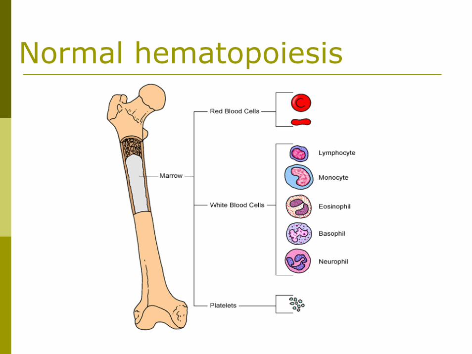
Normal hematopoiesis
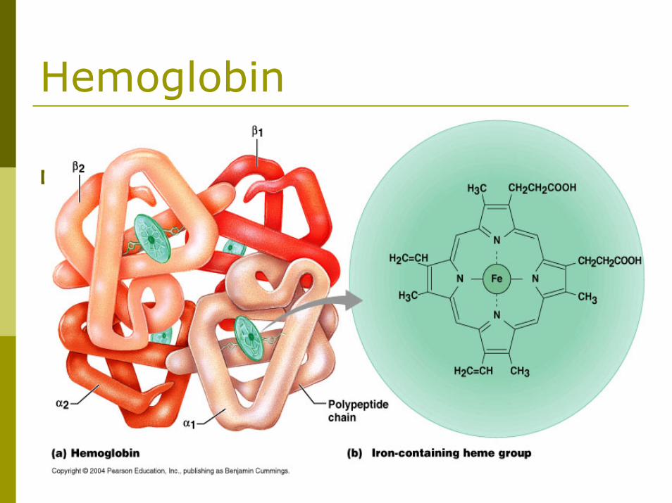
Hemoglobin
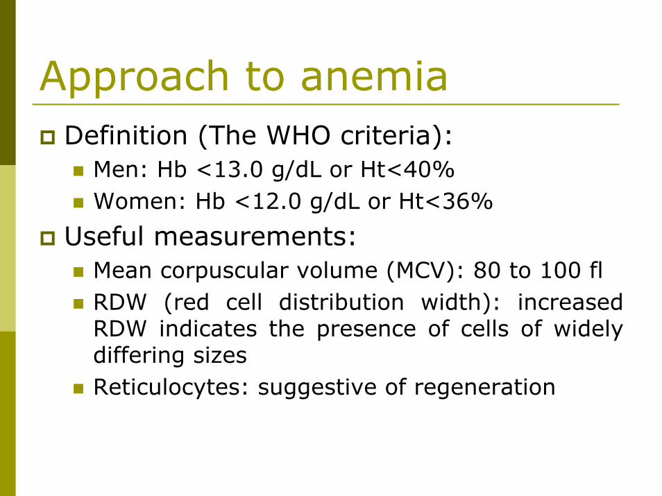
Approach to anemiaDefinition (The WHO criteria):
Men: Hb <13.0 g/dL or Ht<40%Women: Hb <12.0 g/dL or Ht<36%
Useful measurements:Mean corpuscular volume (MCV): 80 to 100 fl RDW (red cell distribution width): increased RDW indicates the presence of cells of widely differing sizesReticulocytes: suggestive of regeneration
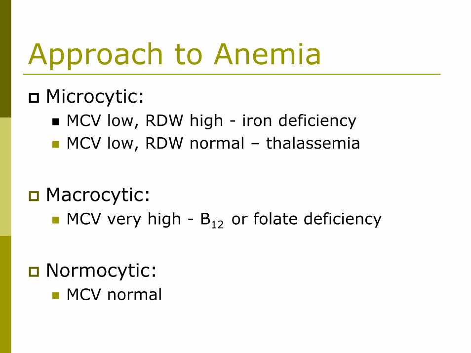
Approach to AnemiaMicrocytic:
MCV low, RDW high - iron deficiencyMCV low, RDW normal – thalassemia
Macrocytic:MCV very high - B12 or folate deficiency
Normocytic:MCV normal
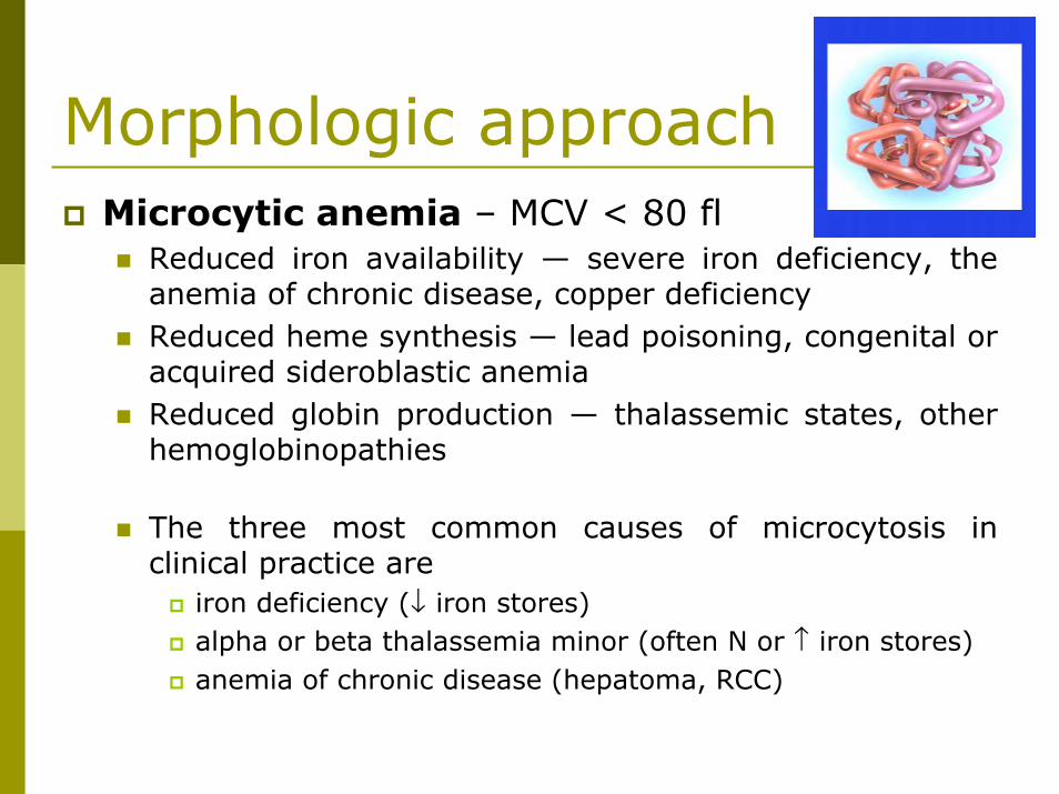
Morphologic approachMicrocytic anemia – MCV < 80 fl
Reduced iron availability — severe iron deficiency, the anemia of chronic disease, copper deficiencyReduced heme synthesis — lead poisoning, congenital or acquired sideroblastic anemiaReduced globin production — thalassemic states, other hemoglobinopathies
The three most common causes of microcytosis in clinical practice are
iron deficiency (↓ iron stores)alpha or beta thalassemia minor (often N or ↑ iron stores)anemia of chronic disease (hepatoma, RCC)
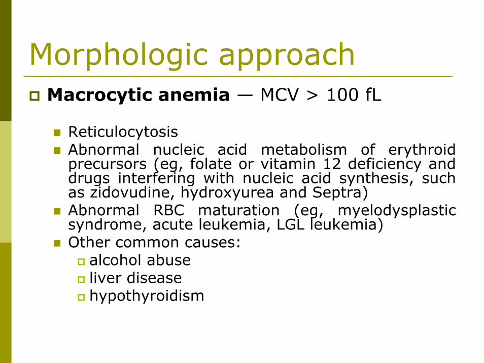
Morphologic approachMacrocytic anemia — MCV > 100 fL
ReticulocytosisAbnormal nucleic acid metabolism of erythroidprecursors (eg, folate or vitamin 12 deficiency and drugs interfering with nucleic acid synthesis, such as zidovudine, hydroxyurea and Septra)Abnormal RBC maturation (eg, myelodysplasticsyndrome, acute leukemia, LGL leukemia)Other common causes:
alcohol abuseliver diseasehypothyroidism
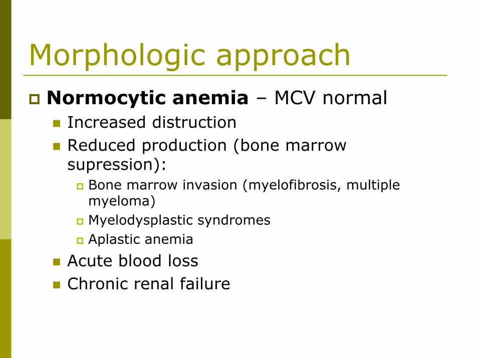
Morphologic approachNormocytic anemia – MCV normal
Increased distructionReduced production (bone marrow supression):
Bone marrow invasion (myelofibrosis, multiple myeloma)Myelodysplastic syndromesAplastic anemia
Acute blood lossChronic renal failure
Iron deficient anemiaDiagnosis:
↓ serum iron↑ TIBC↓ Transferrin saturation <20%↓ Ferritin < 10
Iron replacement requires normalization of the hemoglobin and the body stores
ALWAYS A SYMPTOM – LOOK FOR THE CAUSE
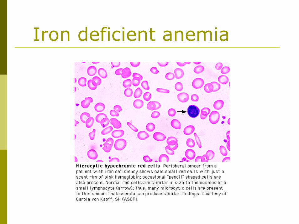
Iron deficient anemia
Anemia of Chronic DiseaseCommon in patients with infection, cancer, inflammatory and rheumatologic diseasesIron can not be remobilized from storageBlunted production of erythropoietin and response to erythropoietinUsually normocytic and normochromic but may be microcytic if severe
TIBC ↓, Iron ↓, Transferrin saturation ↓, Ferritin↑
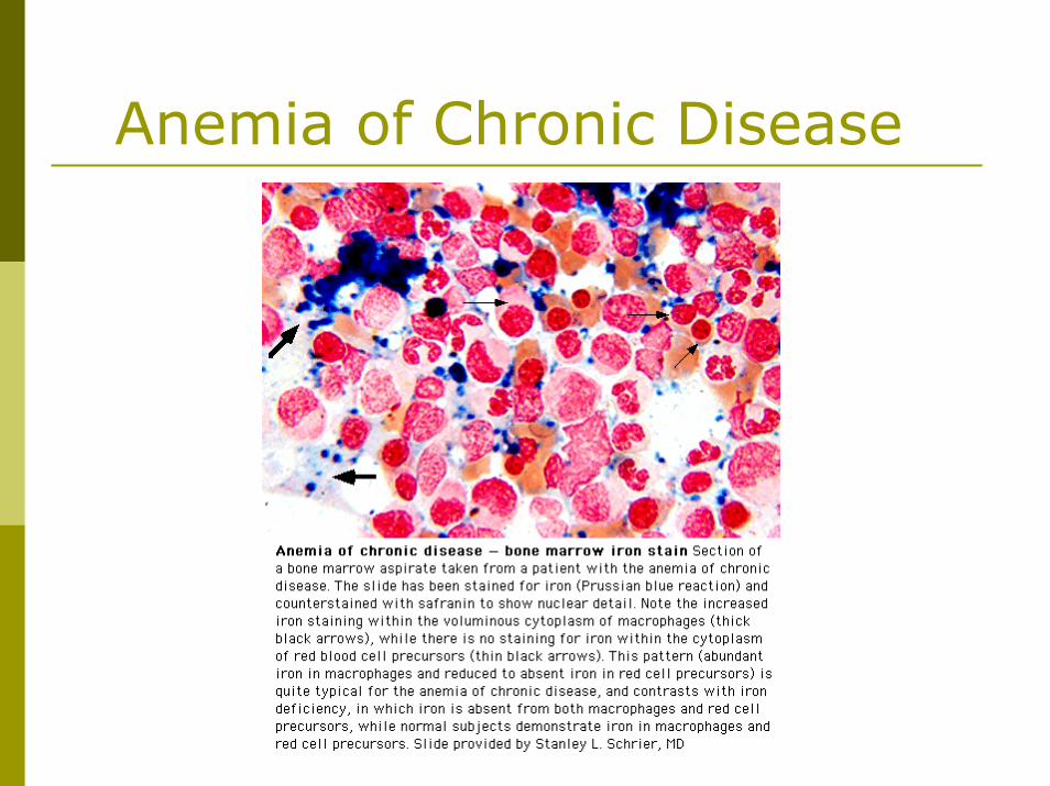
Anemia of Chronic Disease
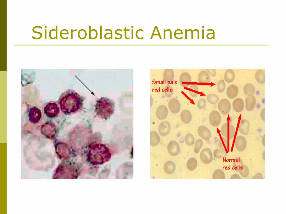
Sideroblastic AnemiaTwo common features:
Ring sideroblasts in the bone marrow (abnormal normoblasts with excessive accumulation of iron in the mitochondria)Impaired heme biosynthesis
Produces a “dimorphic” blood film with microcytes and macrocytesUsually acquired:
Myelodysplastic syndromeDrugs (Ethanol, INH)Toxins (Lead, zinc)Nutritional (Pyridoxine deficiency, copper deficiency)
Sideroblastic Anemia
ThalassemiaAnemia 2º reduced or absent production of one or more globin chains Poikilocytosis (variation in shape) and basophilic stippling may be seen in the blood filmHemoglobin electrophoresis is only diagnostic for beta-thalassemia and may not be diagnostic if iron deficiency is also presentHb H (β-4) prep or DNA analysis is needed to diagnose alpha-thalassemia
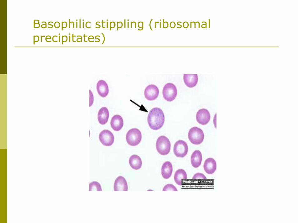
Basophilic stippling (ribosomal precipitates)
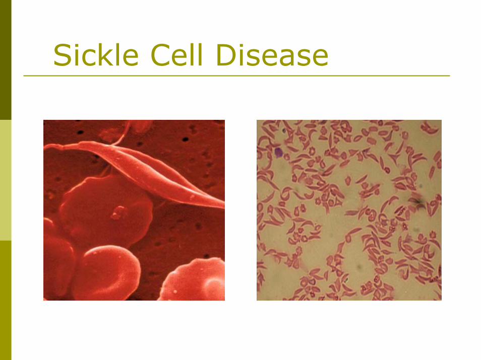
Sickle Cell Disease
Inherited anemia (HbS)Acute crisis management includes fluids, oxygen and pain control +/- transfusionTransfusion therapy, hydroxyurea, magnesium and clotrimazole may reduce the frequency of vaso-occlusive crisesFull vaccination program essential before functional hyposplenism developsTransplant may be curative
Sickle Cell Disease
B12 DeficiencyMegaloblastic anemia (macrocytosis, hypersegmented neutrophils, abnormal megakaryocytes)
Be aware of the neurological complicationsNever treat possible B12 deficiency with folate - the CNS lesions may progressSchilling test distinguishes pernicious anemia from other causes
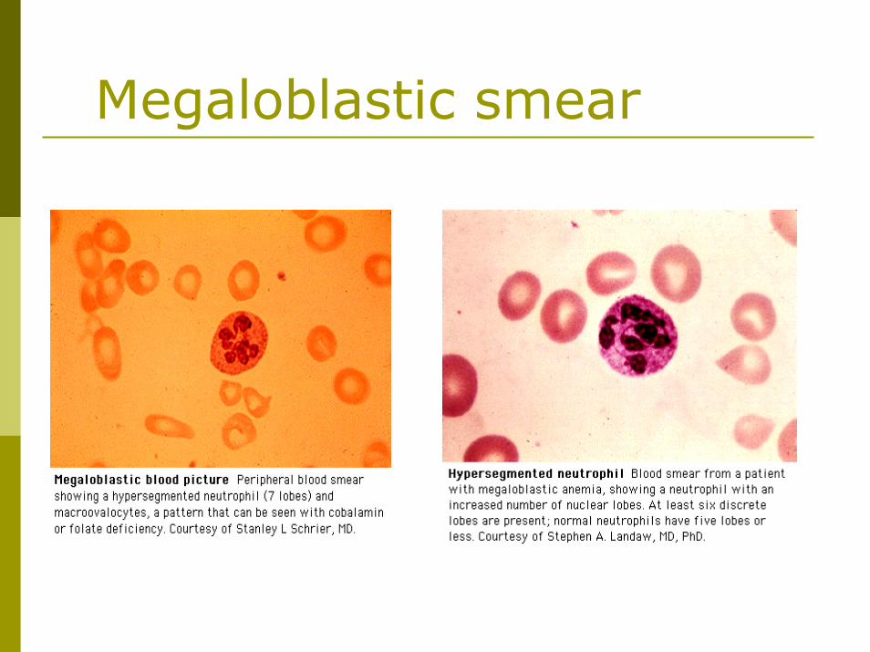
Megaloblastic smear
Folic Acid DeficiencyThe peripheral blood film and bone marrow are identical to B12 deficiencyWomen of childbearing age should take supplemental folate to prevent neural tube defects in their childrenFolate supplementation lowers homocysteine levels leading to less heart disease and stroke
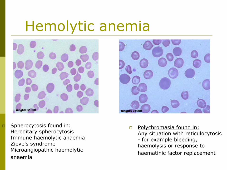
Hemolytic anemiaExtravascular hemolysis
Increased reticulocytesIncreased serum lactate dehydrogenase (LDH) Increased indirect bilirubin concentration
Intravascular hemolysis:RBC fragments, hemoglobinuria, urinary hemosiderin, decreased haptoglobin
Immune (positive direct antiglobulin test)Nonimmune (microangiopathic hemolytic anemia)
Hemolytic Anemia-Intravascular
Inherent RBC Defects:Enzyme defects - G6PD
Acquired Causes: Non-immuneDrowning, burns, infections, PNHRBC fragmentation
DICprosthetic heart valvesvasculitisTTP
Hemolytic Anemia-ExtravascularInherited RBC Defects:
Membrane defects - hereditary spherocytosis and elliptocytosisHemoglobinopathy - Sickle Cell Disease, Thalassemia
Immune causes:Hemolytic transfusion reactionsAIHA- primary or secondaryDrugs eg. penicillinCold agglutinins
Approach to Bruising & BleedingFamily history - bleeding or transfusionDrugs - ASA, NSAIDS and alcohol, steroidsOther diseases - myeloma, renal or liver diseasePattern - lifelong or recent, deep seated bleeds or superficial bruising and petechiaeCheck for the spleen, petechiae, purpura and telangiectasia
Hemostasis InvestigationsPlatelet count and platelet function studiesINR, PTT, fibrinogen, FDP, bleeding timeThrombin time and reptilase timeEuglobulin lysis time Inhibitor studiesFactor assays
made in the liver, vitamin K dependent - 7made in the liver, not vitamin K dependant - 5made in endothelial cells -8
PlateletsAcquired dysfunction is common in ill patients and DDAVP is often a valuable treatmentThe blood film helps differentiate ITP from the early phases of TTP. RBC fragments are present in TTP but not in ITP. Consider a bone marrow if platelet count is very low
Approach to thrombocytopeniaIncrease distruction (ITP, TTP, HIT)Decreased production (amegakaryocyticthrombocytopenia, aplastic anemia, acute leukemia, etc)
Idiopathic (ITP)Secondary:
Drug toxicityConnective tissue diseasesInfections: HIVHypersplenism
Thrombotic thrombocytopenic purpura
Diagnosis:Microangiopathic hemolytic anemia =nonimmune hemolysis (negative direct antiglobulin test) with prominent red cell fragmentation (>1%)ThrombocytopeniaAcute renal insufficiencyNeurological abnormalities (fluctuating)Fever
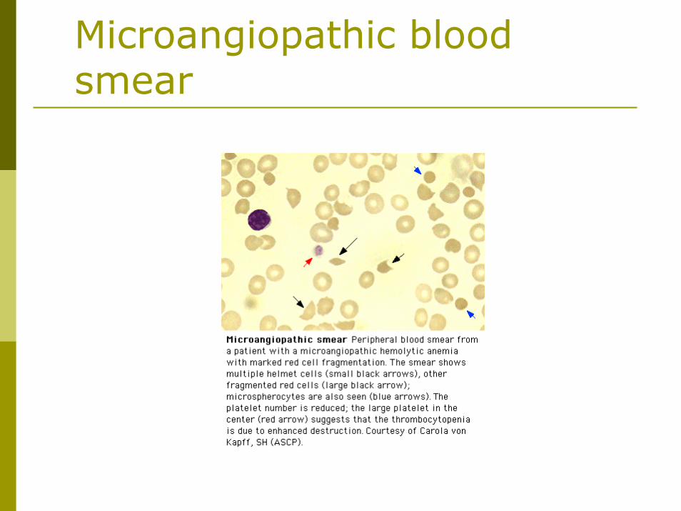
Microangiopathic blood smear
Immune Thrombocytopenic Purpura
Acquired: postviral infections in children (history of infection in the several weeks preceding the illness)Immune/Idiopathic:
Mainstem of treatment: steroids (response in up to 2 weeks)If no response to steroids after 2 weeks consider splenectomyIn severe bleeding treat with IvIg (rapid response)
Disorders of Secondary HemostasisHereditary
Hemophilia A (factor VIII deficiency) and Hemophilia B (factor IX deficiency) are X-linked and produce hemarthroses and hematomas and are treated with recombinant factor concentrates and DDAVP (prolonged PTT)von Willebrand’s disease (prolonged bleeding time and prolonged PTT)
Disorders of Secondary Hemostasis
AcquiredVitamin K deficiency (factors II, VII, IX and X)Liver disease (all factors other than VIII)Circulating anticoagulants (lupus anticoagulant)DIC
DICDIC = uncontrolled THROMBIN and PLASMINExcess thrombin leads to clotting, excess plasmin leads to bleedingNumerous disorders can trigger DIC ie. Sepsis, trauma, cancer, fat or amniotic fluid emboli, acute promyelocytic leukemia
DIC Can be acute or chronic
Produces RBC fragmentation, confusion or coma, focal necrosis in the skin, ARDS, renal failure, bleeding and hypercoagulability
Management of DICTreat the underlying causeGive cautious replacement therapy with FFP, cryoprecipitate and plateletsAvoid products with activated factorsIn exceptional cases use low dose heparin
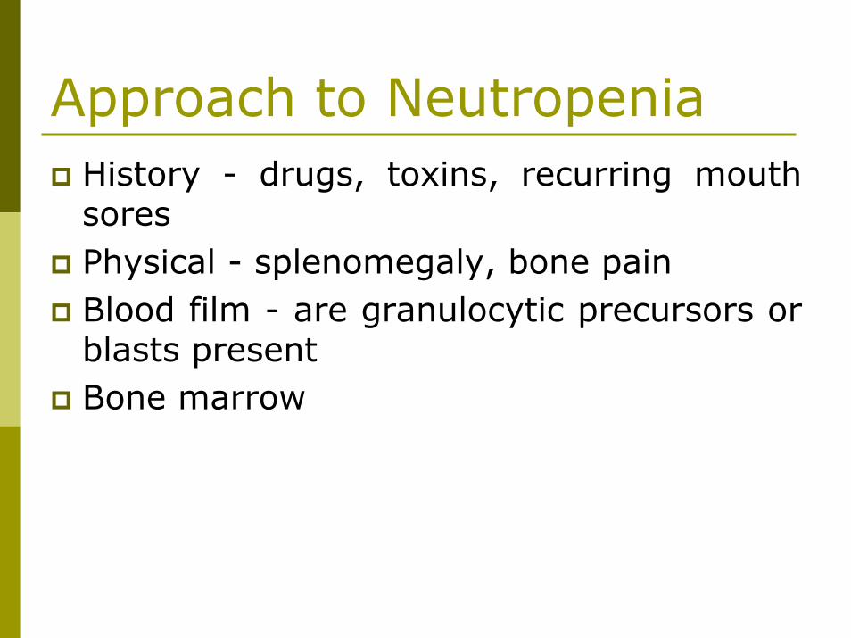
Approach to NeutropeniaHistory - drugs, toxins, recurring mouth soresPhysical - splenomegaly, bone painBlood film - are granulocytic precursors or blasts presentBone marrow
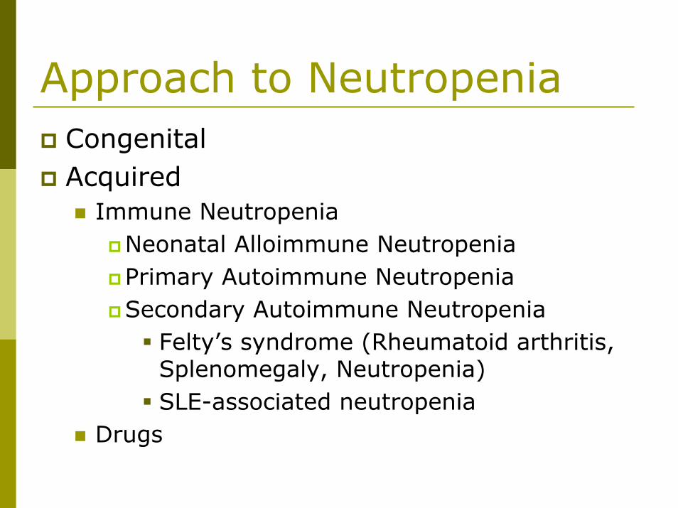
Approach to NeutropeniaCongenitalAcquired
Immune NeutropeniaNeonatal Alloimmune NeutropeniaPrimary Autoimmune NeutropeniaSecondary Autoimmune Neutropenia
Felty’s syndrome (Rheumatoid arthritis, Splenomegaly, Neutropenia)SLE-associated neutropenia
Drugs
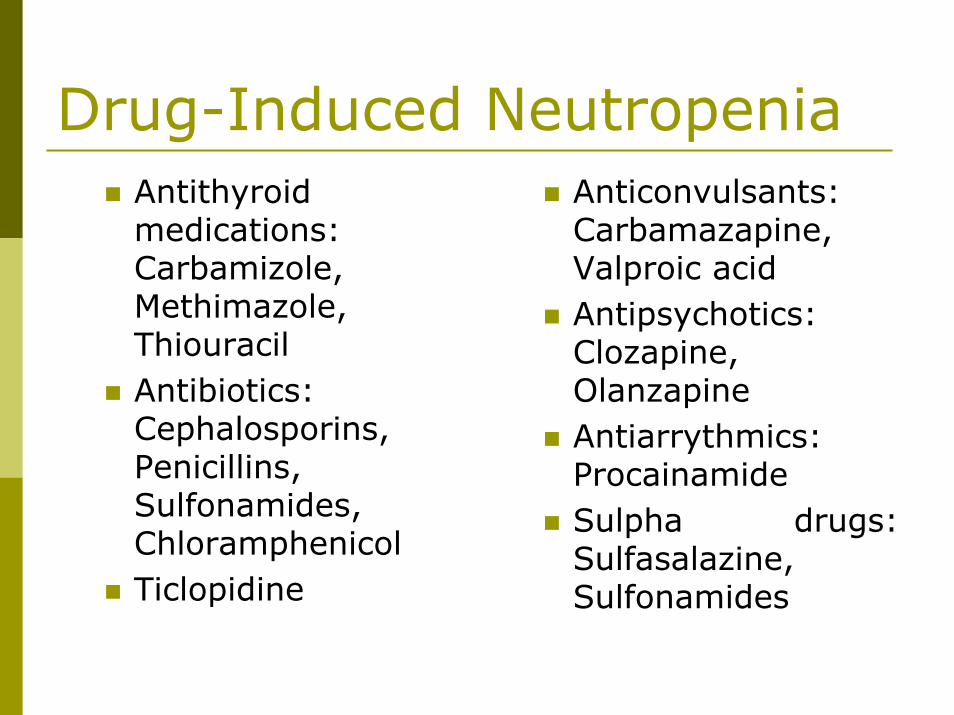
Drug-Induced NeutropeniaAntithyroidmedications: Carbamizole, Methimazole, ThiouracilAntibiotics: Cephalosporins, Penicillins, Sulfonamides, ChloramphenicolTiclopidine
Anticonvulsants: Carbamazapine, Valproic acidAntipsychotics: Clozapine, OlanzapineAntiarrythmics: ProcainamideSulpha drugs: Sulfasalazine, Sulfonamides
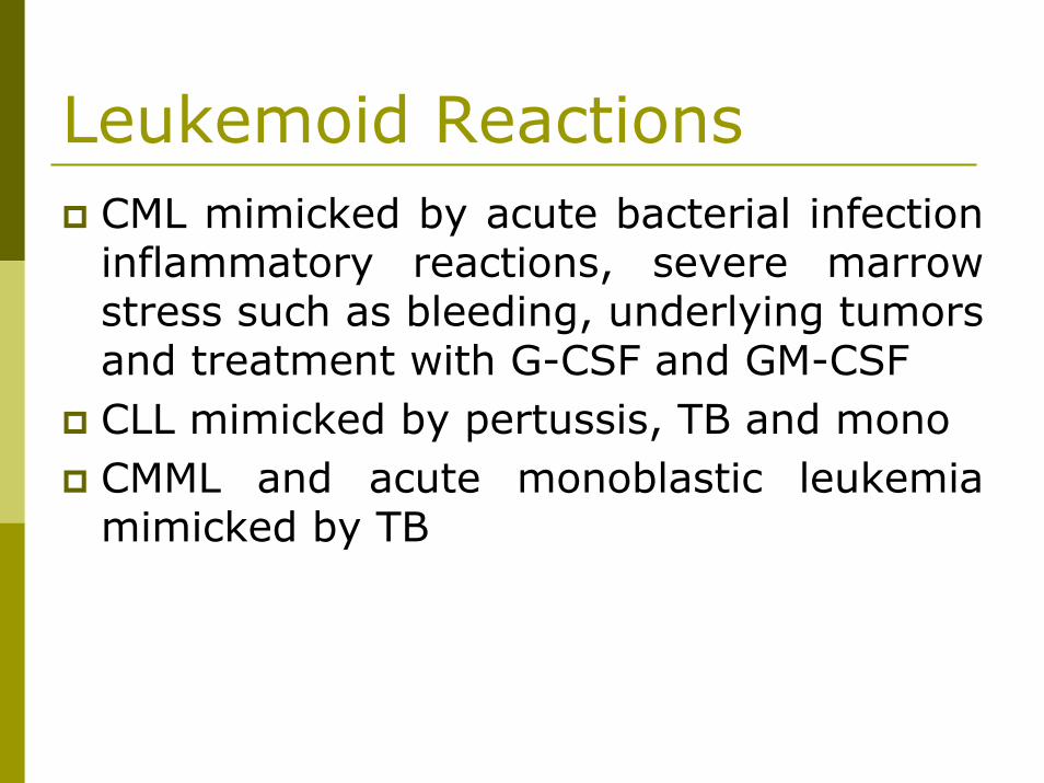
Leukemoid ReactionsCML mimicked by acute bacterial infection inflammatory reactions, severe marrow stress such as bleeding, underlying tumors and treatment with G-CSF and GM-CSFCLL mimicked by pertussis, TB and monoCMML and acute monoblastic leukemia mimicked by TB
Febrile Transfusion ReactionsThe most common reaction, non-immuneWithin 1-6 hours of transfusionMost often due to cytokines in the productBecome more common as the product agesTreatment - Acetaminophen, Demerol and the transfusion of young products, washed products or leukodepleted products. The value of corticosteroids is less clear
Other Transfusion ReactionsUrticaria (soluble plasma substances react with donor IgE) - treat with antihistaminesAnaphylaxis (sec-min) 2o IgA deficiency -HISTORY! – only transfuse washed blood productsAcute hemolytic (ABO mismatch)- recheck blood group and crossmatch - usually due to clerical errorDelayed hemolytic (2-10 days) - mimics AIHACitrate toxicity (hypocalcemia)- give calcium gluconate
Approach to pancytopeniaCentral Peripheral
Central causesEmpty marrow: Aplastic anemia, Hypoplastic MDS, myelofibrosisInfiltration by abnormal cells: leukemia, lymphoma, solid tumors, TBDeranged marrow: MyelodysplasticsyndromeStarved marrow: B12, folic acidDrug-induced: chemotherapy, antibiotics (sulpha), alcohol
Peripheral causesHypersplenismAutoimmuneSevere Sepsis
Aplastic AnemiaPancytopenia with ”empty” marrowMost idiopathic cases are due to abnormal T cell inhibition of hematopoiesisTreatment:
Immunosuppression (Cyclosporin and antithymocyteglobulin) Allogeneic BMT
Give irradiated, CMV negative blood products until CMV status is known
Myeloproliferative DisordersAll are disorders of the pluripotent stem cell
Acute Myelogenous Leukemia and Acute Lymphoblastic LeukemiaChronic Myeloproliferative DisordersMyelodysplastic Syndromes
Acute Myelogenous LeukemiaUsually seen in adultsIntensive, toxic treatment is needed to produce a complete remissionMarrow transplantation may be curativeBlasts are large with abundant cytoplasmAuer rods are diagnostic and granules are common
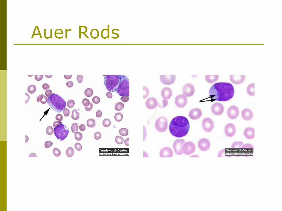
Auer Rods
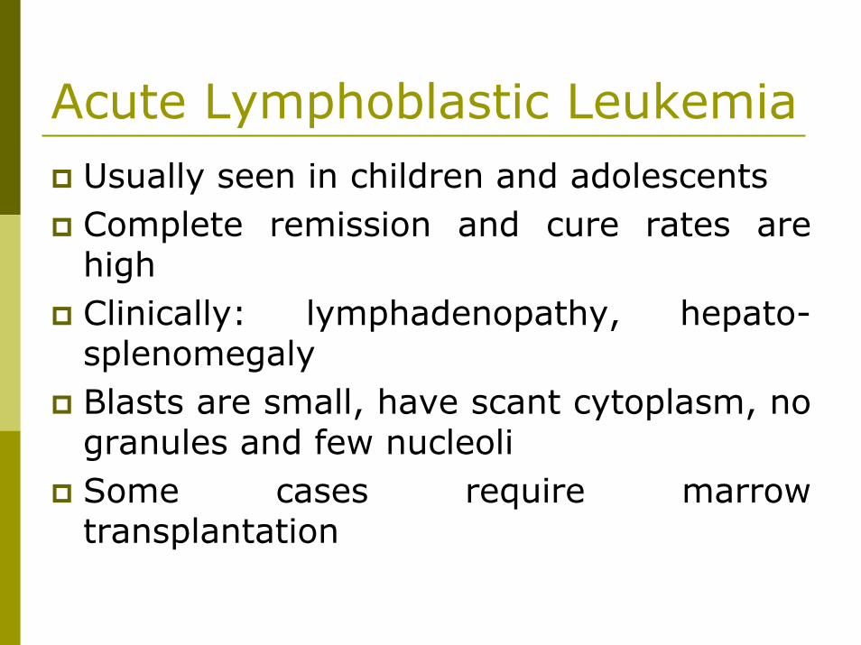
Acute Lymphoblastic LeukemiaUsually seen in children and adolescentsComplete remission and cure rates are highClinically: lymphadenopathy, hepato-splenomegalyBlasts are small, have scant cytoplasm, no granules and few nucleoliSome cases require marrow transplantation
Myelodysplastic SyndromesRefractory anemia Refractory anemia with ring sideroblasts Refractory anemia with excess blasts Refractory anemia with excess blasts in transformation Chronic myelomonocytic leukemia
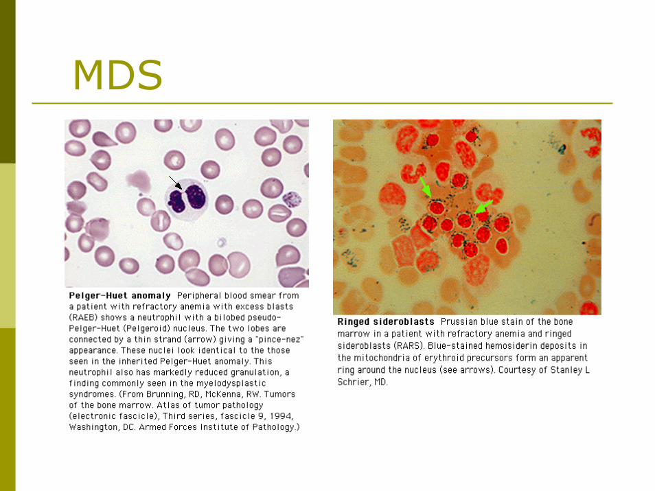
MDS
Chronic Myeloproliferative Disorders
Polycythemia rubra vera (tx: phlebotomy, hydroxyurea)
Chronic myeloid leukemia (tx: Gleevec, BMT)
Idiopathic Myelofibrosis
Essential thrombocythemia
Chronic Lymphocytic LeukemiaCommon in the elderlyThe blood film shows a lymphocytosis that may be extreme and smudge cellsLymphadenopathy and splenomegaly Autoimmune anemia and thrombocytopeniaTreatment is observation, alkylating agents, fludarabine, steroids or radiation
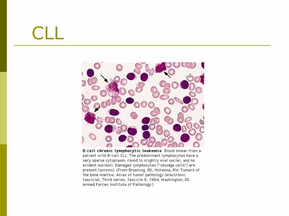
CLL
Multiple MyelomaA monoclonal immunoglobulin in the serum or a single light chain in the urine is found (SPEP, UPEP) and marrow plasmocytosisHypercalcemia and renal failure are frequentLytic bone lesions are classical but osteoporosis is more common (alk phos is normal)The blood film shows rouleauxHigh ESR
MGUSCharacteristics:
Presence of a monoclonal immunoglobulin < 3g/l (in 1% of people)Normal marrow (<10%plasma cells), normal chemistry (no anemia, hypercalcemia, no renal failure) and no lytic lesionsThe M-protein remains stableMay transform to myeloma
MacroglobulinemiaMonoclonal IgM proteinClinically:
LymphadenopathyHepatosplenomegalyHyperviscosity syndrome
ESR may be very lowTreat the hyperviscosity with plasmapheresis
Hodgkin’s DiseaseThe Reed-Sternberg cell is diagnostic
The cure rate is high
Long term complications are heart disease, hypothyroidism and secondary malignancies
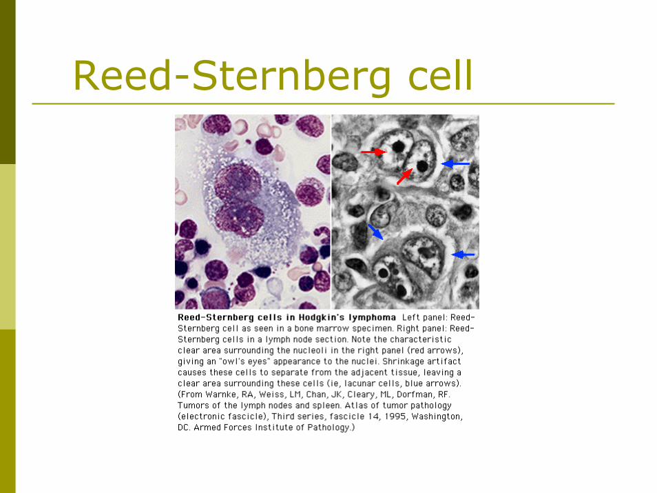
Reed-Sternberg cell
Non-Hodgkin’s LymphomaNumerous classification systemsLow grade: Radiation +/- gentle chemotherapy does not cure but does produce long survivalIntermediate and high grade: Radiation ± chemotherapy is used. Cure is possibleAutoimmune hemolytic anemia and thrombocytopenia are commonHypersplenism is commonImmunosuppression is frequent
never give live vaccines
Deep Vein Thrombosis and Pulmonary Embolism
1859 Rudolph Virchow described the major pathogenic determinants:1) Blood stasis 2) Changes in the vessel wall 3) Hypercoagulability
ThrombophiliaAntiphospholipid antibodies Factor V Leiden G20210A prothrombin gene mutation Deficiency of protein C, protein S, and antithrombin Hyperhomocysteinemia
DVT/PE treatmentStart LMWH (superior to UFH) if no contraindicationsStart Warfarin with LMWHStop LMWH after 2 consecutive days of therapeutic INR (2-3)
Thrombolytics indicated if circulatory collapse (shock)
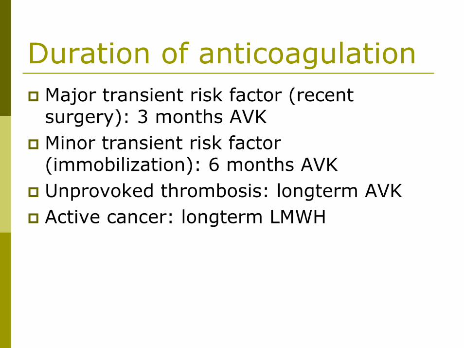
Duration of anticoagulationMajor transient risk factor (recent surgery): 3 months AVKMinor transient risk factor (immobilization): 6 months AVKUnprovoked thrombosis: longterm AVKActive cancer: longterm LMWH
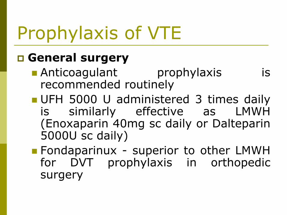
Prophylaxis of VTEGeneral surgery
Anticoagulant prophylaxis is recommended routinelyUFH 5000 U administered 3 times daily is similarly effective as LMWH (Enoxaparin 40mg sc daily or Dalteparin5000U sc daily)Fondaparinux - superior to other LMWH for DVT prophylaxis in orthopedic surgery
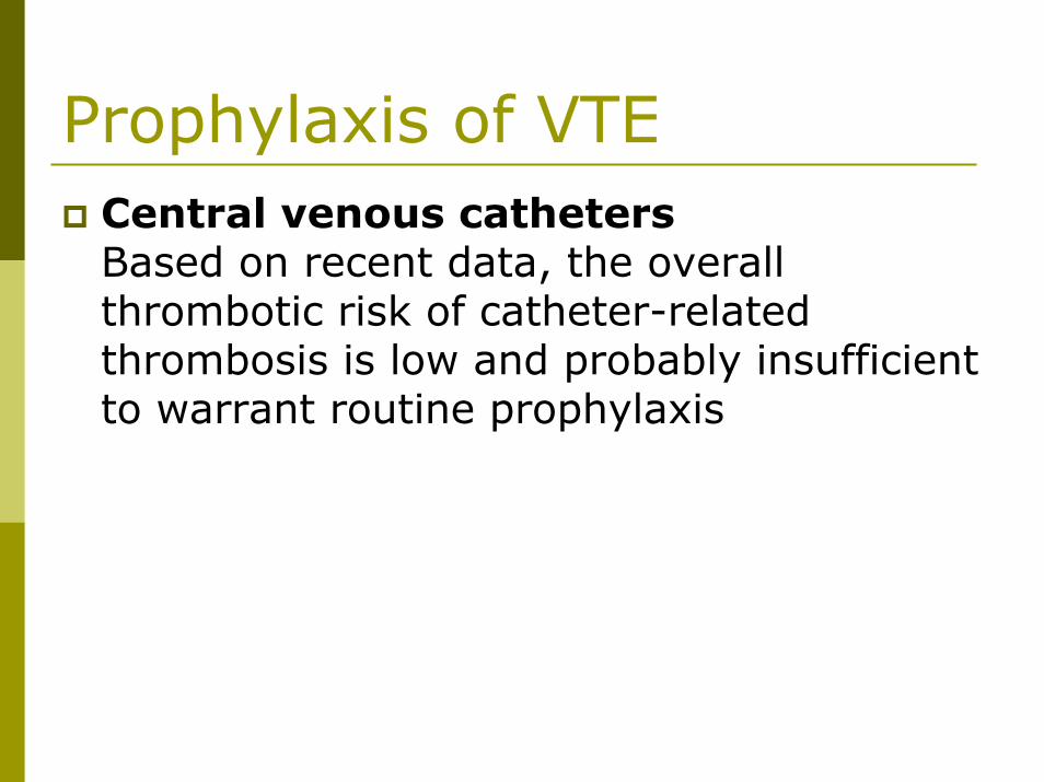
Prophylaxis of VTECentral venous cathetersBased on recent data, the overall thrombotic risk of catheter-related thrombosis is low and probably insufficient to warrant routine prophylaxis
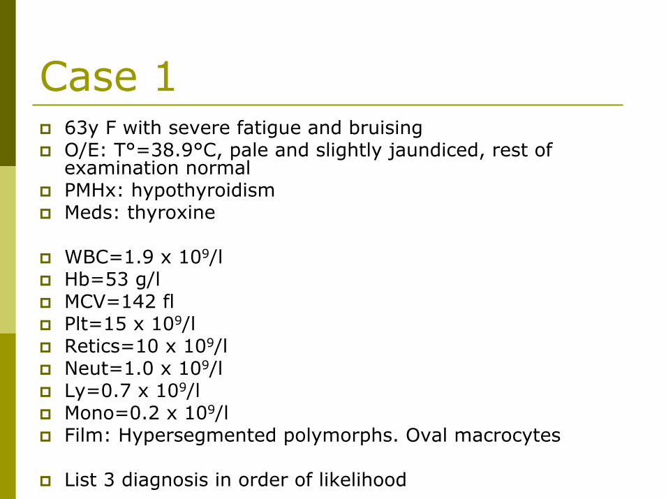
Case 163y F with severe fatigue and bruisingO/E: T°=38.9°C, pale and slightly jaundiced, rest of examination normalPMHx: hypothyroidismMeds: thyroxine
WBC=1.9 x 10⁹/lHb=53 g/lMCV=142 flPlt=15 x 109/lRetics=10 x 109/lNeut=1.0 x 109/lLy=0.7 x 109/lMono=0.2 x 109/lFilm: Hypersegmented polymorphs. Oval macrocytes
List 3 diagnosis in order of likelihood
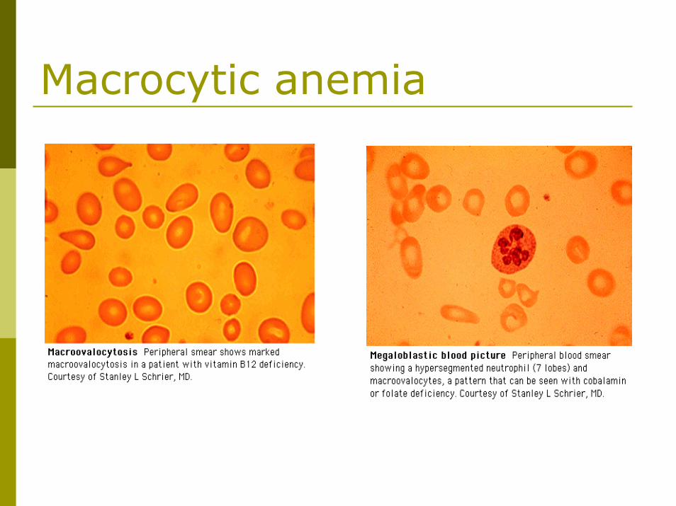
Macrocytic anemia
Case 245y F on routine annual physical examination found to have following blood count (previously healthy, on no meds):
WBC: 7.5Hb=61MCV=58Plt=345Retics=10Neut=4.3Ly=2.7Mono=0.5Film: Hypochromia. Microcytosis.
What is the most likely hematologic diagnosis?List 3 alternative possibilities.What tests would you order?Outline a management plan.
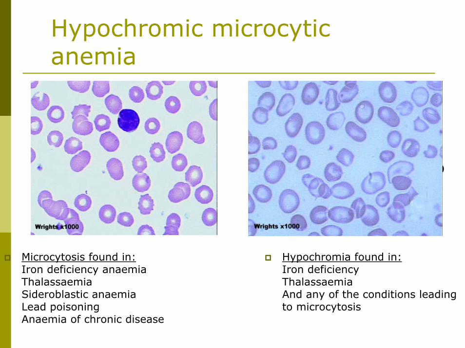
Hypochromic microcytic anemia
Microcytosis found in:Iron deficiency anaemiaThalassaemiaSideroblastic anaemiaLead poisoningAnaemia of chronic disease
Hypochromia found in:Iron deficiencyThalassaemiaAnd any of the conditions leading to microcytosis
Found in:Iron deficiencyThalassaemiaAnd any of the conditions leading to microcytosis
Case 359y F feeling “washed out”. Spleen palpable 4 cm BCM, jaundiced. Blood counts as follows:
WBC=8.5Hb=61MCV=110Retics=560Plt=156Neut=4.5Ly=3.0Mono=0.8Eo=0.2Film: Spherocytes. Polychromasia.
State the two most likely diagnoses, in order of probabilityOutline a plan of investigation and management
Hemolytic anemia
Spherocytosis found in:Hereditary spherocytosisImmune haemolytic anaemiaZieve's syndromeMicroangiopathic haemolyticanaemia
Polychromasia found in:Any situation with reticulocytosis- for example bleeding, haemolysis or response to haematinic factor replacement
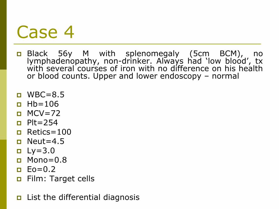
Case 4Black 56y M with splenomegaly (5cm BCM), no lymphadenopathy, non-drinker. Always had ‘low blood’, txwith several courses of iron with no difference on his health or blood counts. Upper and lower endoscopy – normal
WBC=8.5Hb=106MCV=72Plt=254Retics=100Neut=4.5Ly=3.0Mono=0.8Eo=0.2Film: Target cells
List the differential diagnosis
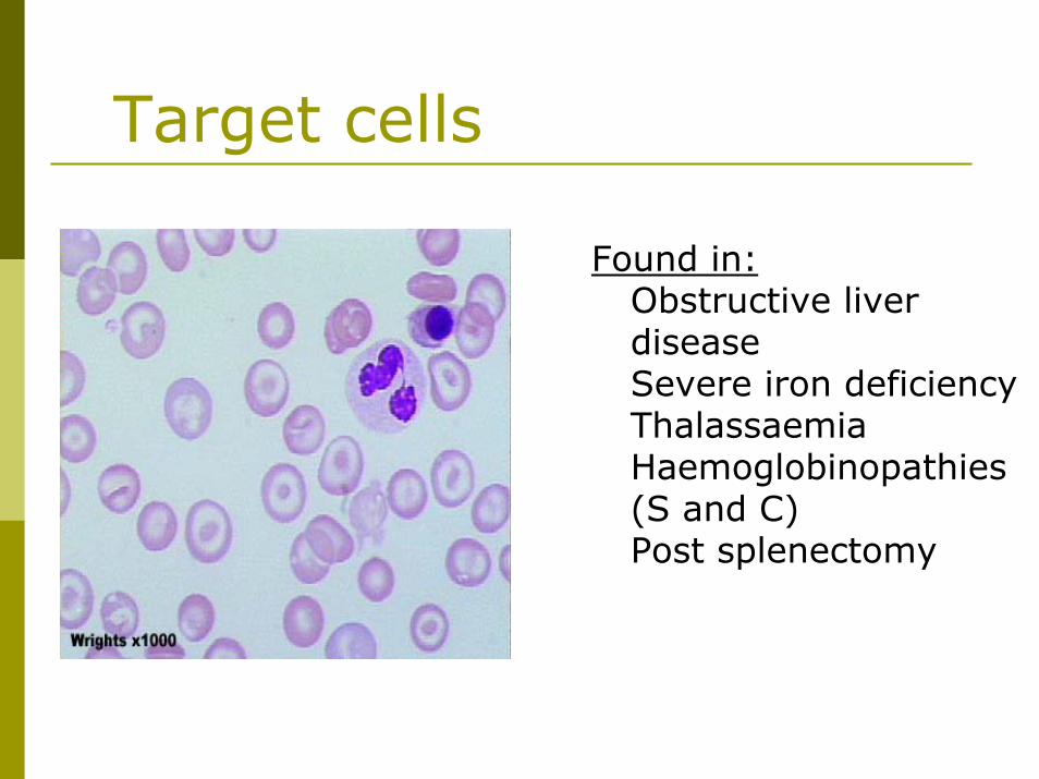
Target cells
Found in:Obstructive liver diseaseSevere iron deficiencyThalassaemiaHaemoglobinopathies(S and C)Post splenectomy
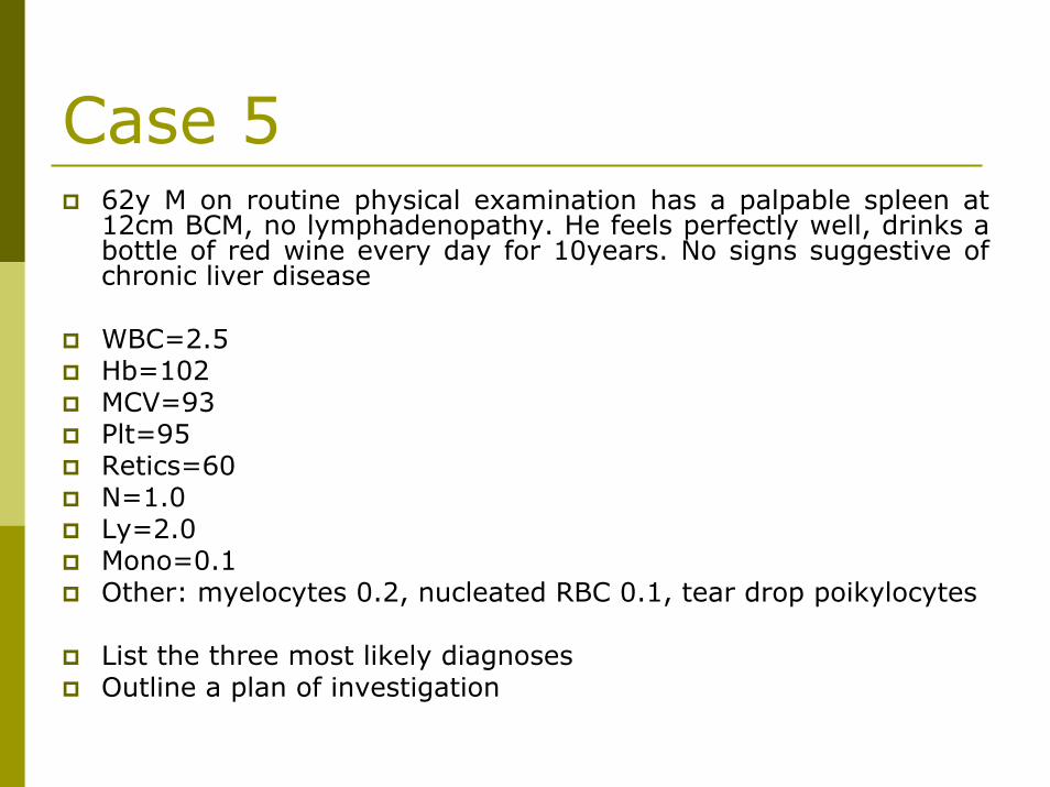
Case 562y M on routine physical examination has a palpable spleen at 12cm BCM, no lymphadenopathy. He feels perfectly well, drinks a bottle of red wine every day for 10years. No signs suggestive ofchronic liver disease
WBC=2.5Hb=102MCV=93Plt=95Retics=60N=1.0Ly=2.0Mono=0.1Other: myelocytes 0.2, nucleated RBC 0.1, tear drop poikylocytes
List the three most likely diagnosesOutline a plan of investigation
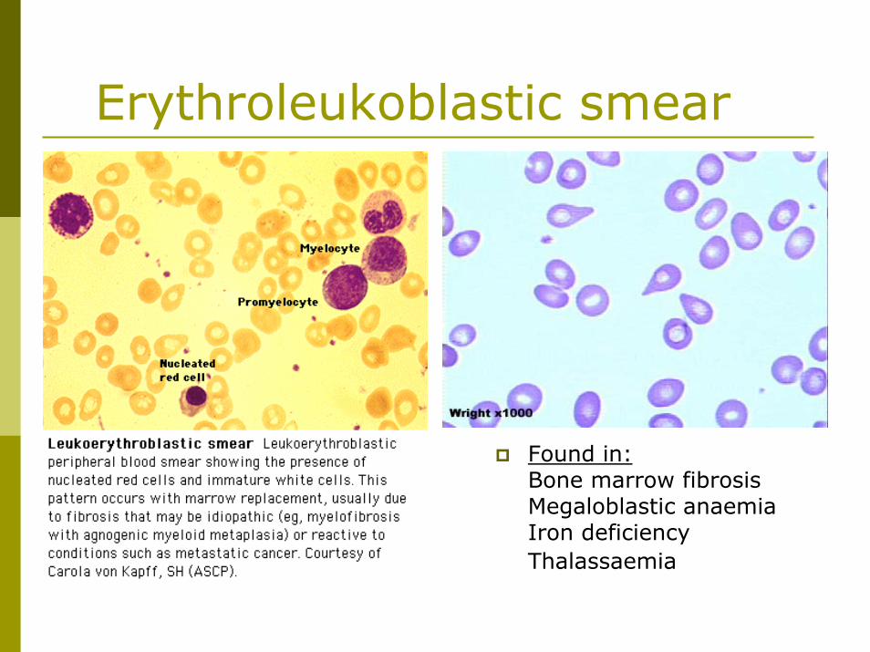
Erythroleukoblastic smear
Found in:Bone marrow fibrosisMegaloblastic anaemiaIron deficiencyThalassaemia
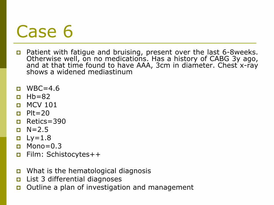
Case 6Patient with fatigue and bruising, present over the last 6-8weeks. Otherwise well, on no medications. Has a history of CABG 3y ago,and at that time found to have AAA, 3cm in diameter. Chest x-ray shows a widened mediastinum
WBC=4.6Hb=82MCV 101Plt=20Retics=390N=2.5Ly=1.8Mono=0.3Film: Schistocytes++
What is the hematological diagnosisList 3 differential diagnosesOutline a plan of investigation and management
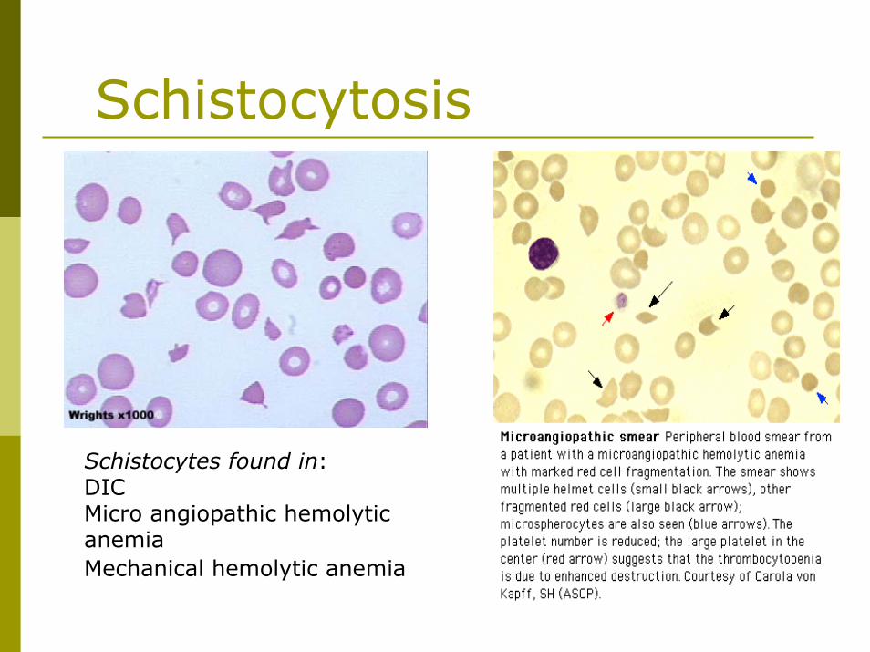
Schistocytosis
Schistocytes found in:DICMicro angiopathic hemolytic anemiaMechanical hemolytic anemia
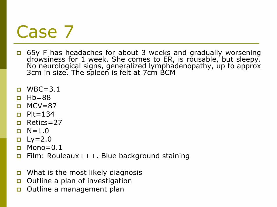
Case 765y F has headaches for about 3 weeks and gradually worsening drowsiness for 1 week. She comes to ER, is rousable, but sleepy. No neurological signs, generalized lymphadenopathy, up to approx 3cm in size. The spleen is felt at 7cm BCM
WBC=3.1Hb=88MCV=87Plt=134Retics=27N=1.0Ly=2.0Mono=0.1Film: Rouleaux+++. Blue background staining
What is the most likely diagnosisOutline a plan of investigationOutline a management plan
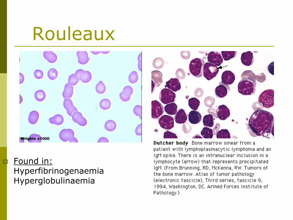
Rouleaux
Found in:HyperfibrinogenaemiaHyperglobulinaemia
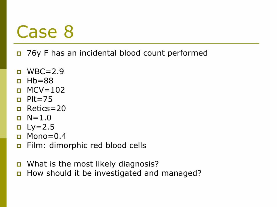
Case 876y F has an incidental blood count performed
WBC=2.9Hb=88MCV=102Plt=75Retics=20N=1.0Ly=2.5Mono=0.4Film: dimorphic red blood cells
What is the most likely diagnosis?How should it be investigated and managed?
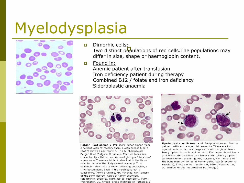
MyelodysplasiaDimorhic cells:Two distinct populations of red cells.The populations may differ in size, shape or haemoglobin content. Found in:Anemic patient after transfusionIron deficiency patient during therapyCombined B12 / folate and iron deficiencySideroblastic anaemia


