Download - Gastric Neoplasms
Gastric Neoplasms
Dr. D.W. DaughertyDepartment of Surgery
Gastric AdenocarcinomaEpidemiology
a. Among top 10 causes of cancer related deaths in theUS.
b. Highest incidence in Japan, where it accounts forapproximately 50% of cancer-related deaths amongmen and 40% of cancer-related deaths among women.
c. Incidence also high in Chile, Costa Rica, Hungary,Portugal, Singapore, and Romania.
d. Migration from these areas appears to decreaseincidence.
e. Most strongly related to early infection with H. pylori.
f. No conclusive evidence of correlation with diet.
Pre-malignant Lesionsa. Highest risk is associated with polyps.
b. Two main categories of gastric polyps: Hyperplasticand Adenomatous.
c. Hyperplastic polypsare considered to have NOneoplastic potential.
d. Adenomatous polyps carry a 10-20% risk for thedevelopment of carcinoma.
Hyperplastic polypsa. Common, occurring in 0.5-1% of general population
and accounting for 70-80% of all gastric polyps.
b. An overgrowth of histologically normal appearinggastric epthelium.
c. Atypia is rare.
d. Considered to have NO neoplastic potential.
e. Most are asymptomatic.
f. Dyspepsia and vague complaints of epigastricdiscomfort are most common.
g. Co-existing gastroduodenal disease is frequentlycommon.
h. Complications are unsual. GI hemorrhage occurs in lessthan 20%.
i. Endoscopic examination with removal is indicated andsufficient for treatment.
Adenomatous polpysa. Distinct risk for development of malignancy.
b. Atypia is common, and risk for development ofcarcinoma is 10-20%. Risk is greatest in polyps over2cm in diameter and with multiple polyps.
c. Symptoms are similar for those of hyperplastic polyps -Dyspepsia and vague complaints of epigastricdiscomfort are most common.
d. Endoscopic examination with removal for thepedunculated polyp is indicated and sufficient ifhistological exam shows no evidence of cancer.
e. Operative excision is recommended for sessile polypslarger than 2cm, for polyps with biopsy-proven invasivecarcinoma, and for polyps complicated by pain and/orbleeding.
f. After removal, routine endoscopic surveillance isindicated.
Gastritisa. Malignancy appears to be increased in patients with
gastritis associated with pernicious anemia.
b. Characterized by fundic mucosal atrophy, loss ofparietal and chief cells, hypochlorhydria, andhypergastrinemia. Is present in 3% of people older than60 years of age.
c. Risk of Gastric CA doubles in patients who have hadpernicious anemia for 5 years or greater.
d. Intestinal metaplasia, presence of intestinal glands inthe gastric mucosa, is also commonly associated withgastritis and gastric cancer.
e. NO direct evidence has been provided to show theevolution from metaplasia to dysplasia to carcinoma toinvasive cancer in gastric cancer.
Helicobacter Pyloria. Associated with inflammatory conditions in the
stomach.
b. Seropositivity increases risk for gastric cancer three-fold.
c. High risk for cancer in the antrum and body; however,NOT a risk factor for cancers at the esophagastricjunction.
d. Postulated that long term gastric inflammation,consequent to childhood acquisition of H. pylori, makesthe gastric mucosa more susceptible to environmentalcarcinogens.
e. Treated with triple therapy: Proton-pump Inhibitor,Amoxicillin, and Clarithromycin.
Gastric Remnant Cancera. Theory that previous gastrectomy increases risk for
subsequent cancer development.
b. Several large, prospective studies show no realincreased risk until after 25 years post-operatively whenthe relative risk is increased three-fold.
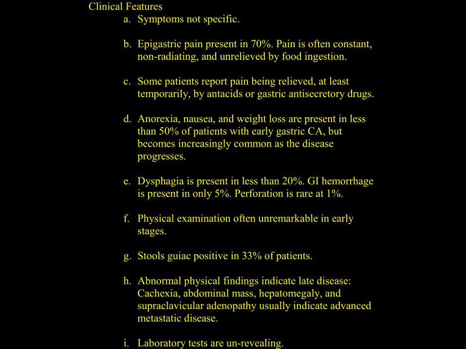
Clinical Featuresa. Symptoms not specific.
b. Epigastric pain present in 70%. Pain is often constant,non-radiating, and unrelieved by food ingestion.
c. Some patients report pain being relieved, at leasttemporarily, by antacids or gastric antisecretory drugs.
d. Anorexia, nausea, and weight loss are present in lessthan 50% of patients with early gastric CA, butbecomes increasingly common as the diseaseprogresses.
e. Dysphagia is present in less than 20%. GI hemorrhageis present in only 5%. Perforation is rare at 1%.
f. Physical examination often unremarkable in earlystages.
g. Stools guiac positive in 33% of patients.
h. Abnormal physical findings indicate late disease:Cachexia, abdominal mass, hepatomegaly, andsupraclavicular adenopathy usually indicate advancedmetastatic disease.
i. Laboratory tests are un-revealing.
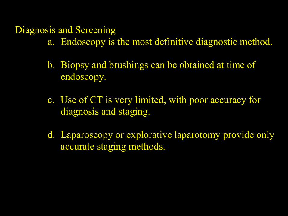
Diagnosis and Screeninga. Endoscopy is the most definitive diagnostic method.
b. Biopsy and brushings can be obtained at time ofendoscopy.
c. Use of CT is very limited, with poor accuracy fordiagnosis and staging.
d. Laparoscopy or explorative laparotomy provide onlyaccurate staging methods.
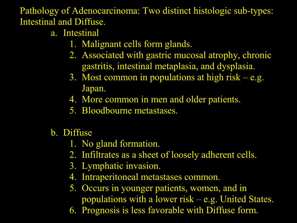
Pathology of Adenocarcinoma: Two distinct histologic sub-types:Intestinal and Diffuse.
a. Intestinal1. Malignant cells form glands.2. Associated with gastric mucosal atrophy, chronic
gastritis, intestinal metaplasia, and dysplasia.3. Most common in populations at high risk – e.g.
Japan.4. More common in men and older patients.5. Bloodbourne metastases.
b. Diffuse1. No gland formation.2. Infiltrates as a sheet of loosely adherent cells.3. Lymphatic invasion.4. Intraperitoneal metastases common.5. Occurs in younger patients, women, and in
populations with a lower risk – e.g. United States.6. Prognosis is less favorable with Diffuse form.
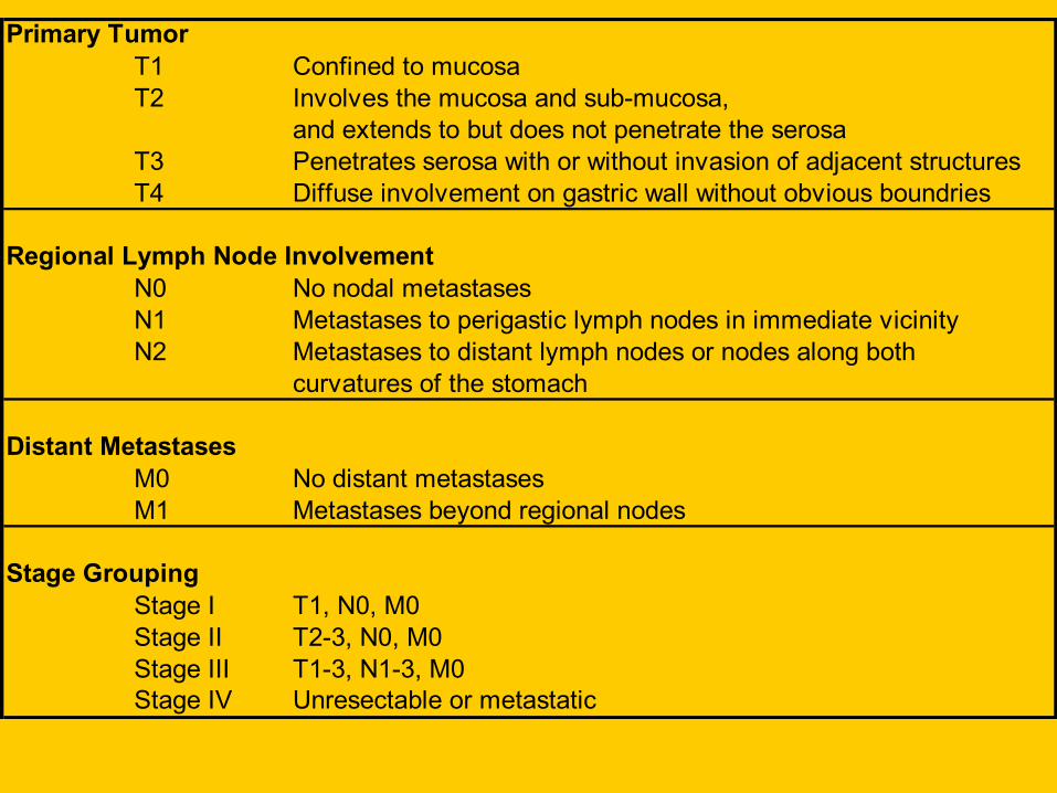
Primary TumorT1 Confined to mucosaT2 Involves the mucosa and sub-mucosa,
and extends to but does not penetrate the serosaT3 Penetrates serosa with or without invasion of adjacent structuresT4 Diffuse involvement on gastric wall without obvious boundries
Regional Lymph Node InvolvementN0 No nodal metastasesN1 Metastases to perigastic lymph nodes in immediate vicinityN2 Metastases to distant lymph nodes or nodes along both
curvatures of the stomach
Distant MetastasesM0 No distant metastasesM1 Metastases beyond regional nodes
Stage GroupingStage I T1, N0, M0Stage II T2-3, N0, M0Stage III T1-3, N1-3, M0Stage IV Unresectable or metastatic
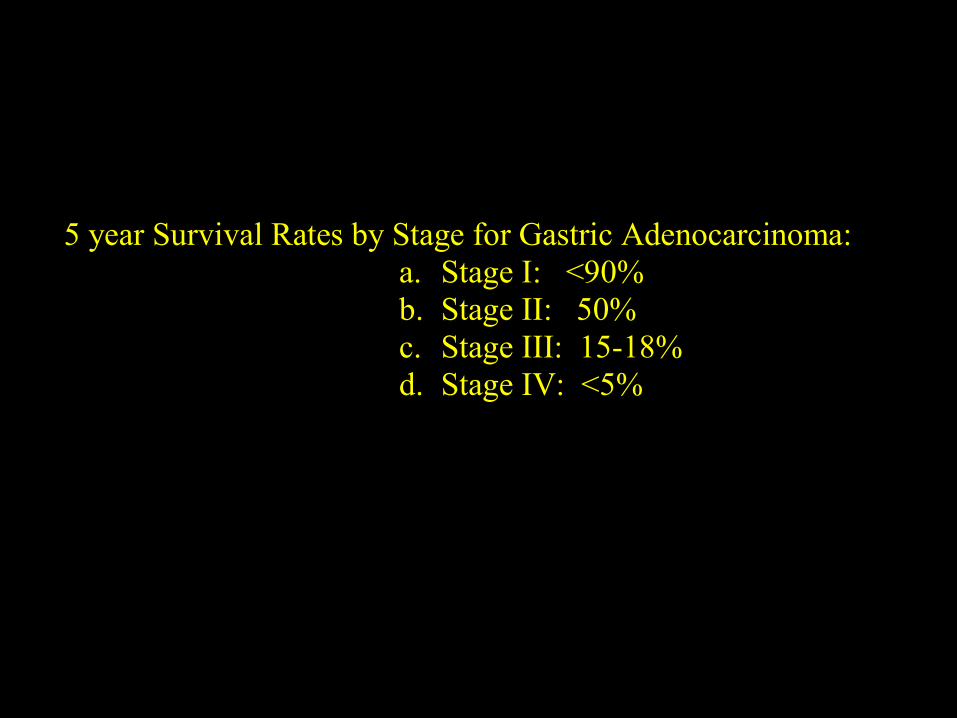
5 year Survival Rates by Stage for Gastric Adenocarcinoma:a. Stage I: <90%b. Stage II: 50%c. Stage III: 15-18%d. Stage IV: <5%
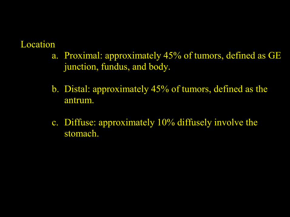
Locationa. Proximal: approximately 45% of tumors, defined as GE
junction, fundus, and body.
b. Distal: approximately 45% of tumors, defined as theantrum.
c. Diffuse: approximately 10% diffusely involve thestomach.
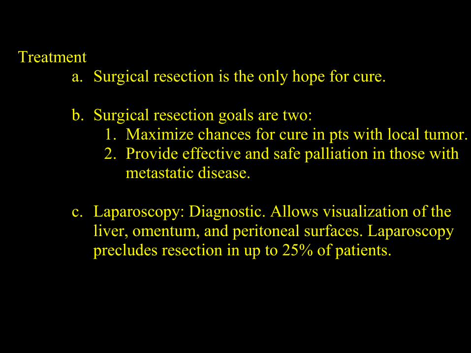
Treatmenta. Surgical resection is the only hope for cure.
b. Surgical resection goals are two:1. Maximize chances for cure in pts with local tumor.2. Provide effective and safe palliation in those with
metastatic disease.
c. Laparoscopy: Diagnostic. Allows visualization of theliver, omentum, and peritoneal surfaces. Laparoscopyprecludes resection in up to 25% of patients.
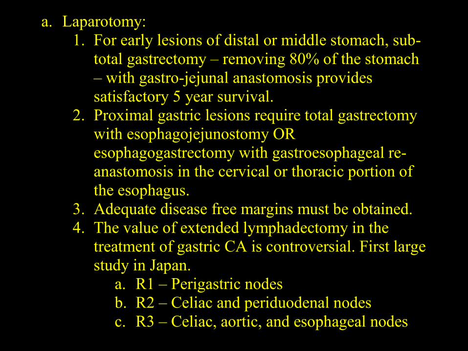
a. Laparotomy:1. For early lesions of distal or middle stomach, sub-
total gastrectomy – removing 80% of the stomach– with gastro-jejunal anastomosis providessatisfactory 5 year survival.
2. Proximal gastric lesions require total gastrectomywith esophagojejunostomy OResophagogastrectomy with gastroesophageal re-anastomosis in the cervical or thoracic portion ofthe esophagus.
3. Adequate disease free margins must be obtained.4. The value of extended lymphadectomy in the
treatment of gastric CA is controversial. First largestudy in Japan.
a. R1 – Perigastric nodesb. R2 – Celiac and periduodenal nodesc. R3 – Celiac, aortic, and esophageal nodes
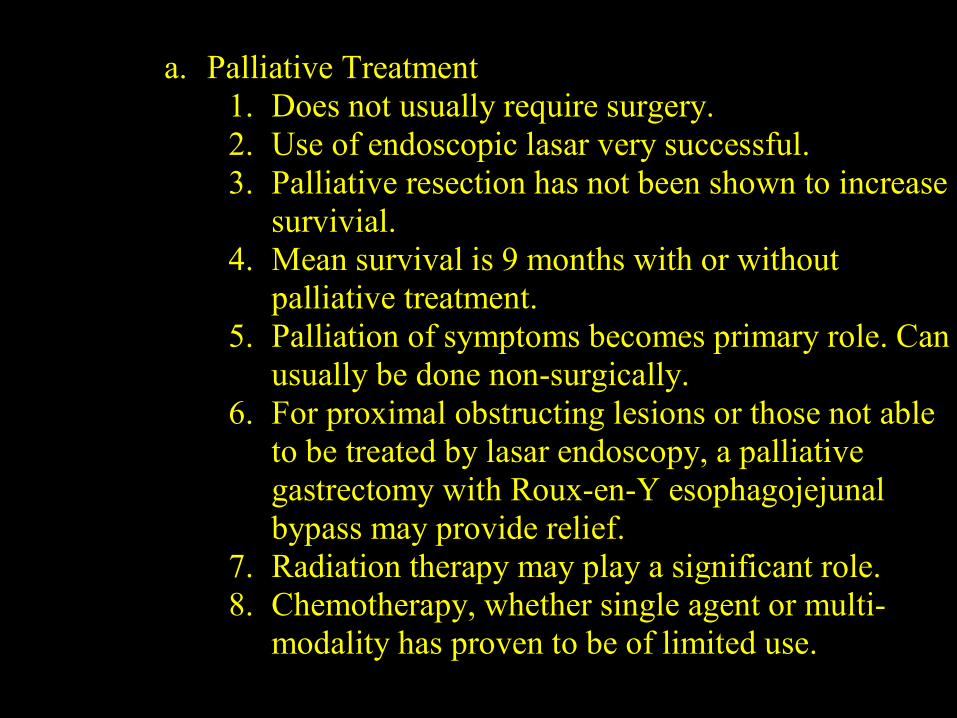
a. Palliative Treatment1. Does not usually require surgery.2. Use of endoscopic lasar very successful.3. Palliative resection has not been shown to increase
survivial.4. Mean survival is 9 months with or without
palliative treatment.5. Palliation of symptoms becomes primary role. Can
usually be done non-surgically.6. For proximal obstructing lesions or those not able
to be treated by lasar endoscopy, a palliativegastrectomy with Roux-en-Y esophagojejunalbypass may provide relief.
7. Radiation therapy may play a significant role.8. Chemotherapy, whether single agent or multi-
modality has proven to be of limited use.
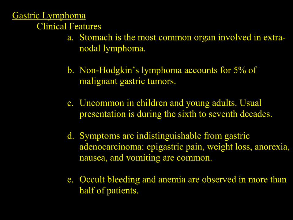
Gastric LymphomaClinical Features
a. Stomach is the most common organ involved in extra-nodal lymphoma.
b. Non-Hodgkin’s lymphoma accounts for 5% ofmalignant gastric tumors.
c. Uncommon in children and young adults. Usualpresentation is during the sixth to seventh decades.
d. Symptoms are indistinguishable from gastricadenocarcinoma: epigastric pain, weight loss, anorexia,nausea, and vomiting are common.
e. Occult bleeding and anemia are observed in more thanhalf of patients.
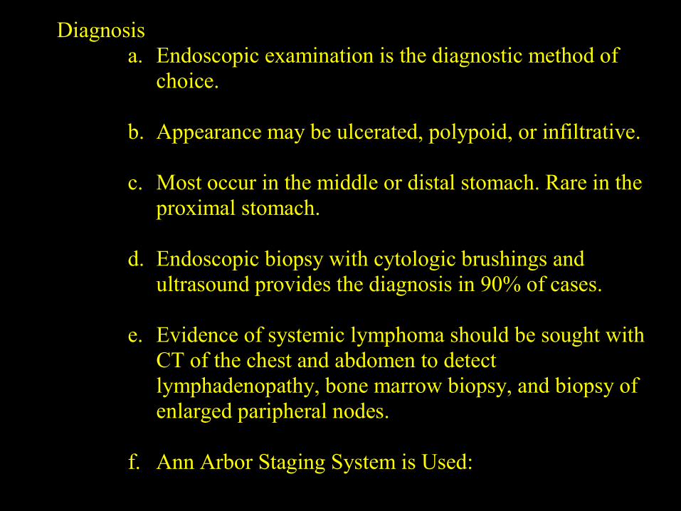
Diagnosisa. Endoscopic examination is the diagnostic method of
choice.
b. Appearance may be ulcerated, polypoid, or infiltrative.
c. Most occur in the middle or distal stomach. Rare in theproximal stomach.
d. Endoscopic biopsy with cytologic brushings andultrasound provides the diagnosis in 90% of cases.
e. Evidence of systemic lymphoma should be sought withCT of the chest and abdomen to detectlymphadenopathy, bone marrow biopsy, and biopsy ofenlarged paripheral nodes.
f. Ann Arbor Staging System is Used:
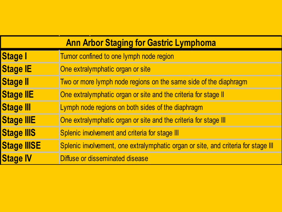
Stage I Tumor confined to one lymph node region
Stage IE One extralymphatic organ or site
Stage II Two or more lymph node regions on the same side of the diaphragm
Stage IIE One extralymphatic organ or site and the criteria for stage II
Stage III Lymph node regions on both sides of the diaphragm
Stage IIIE One extralymphatic organ or site and the criteria for stage III
Stage IIIS Splenic involvement and criteria for stage III
Stage IIISE Splenic involvement, one extralymphatic organ or site, and criteria for stage III
Stage IV Diffuse or disseminated disease
Ann Arbor Staging for Gastric Lymphoma
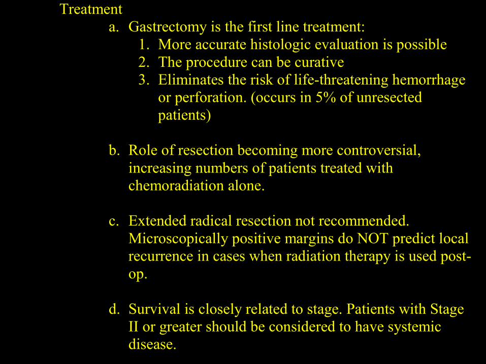
Treatmenta. Gastrectomy is the first line treatment:
1. More accurate histologic evaluation is possible2. The procedure can be curative3. Eliminates the risk of life-threatening hemorrhage
or perforation. (occurs in 5% of unresectedpatients)
b. Role of resection becoming more controversial,increasing numbers of patients treated withchemoradiation alone.
c. Extended radical resection not recommended.Microscopically positive margins do NOT predict localrecurrence in cases when radiation therapy is used post-op.
d. Survival is closely related to stage. Patients with StageII or greater should be considered to have systemicdisease.
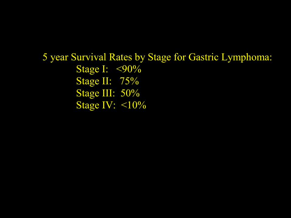
5 year Survival Rates by Stage for Gastric Lymphoma:Stage I: <90%Stage II: 75%Stage III: 50%Stage IV: <10%
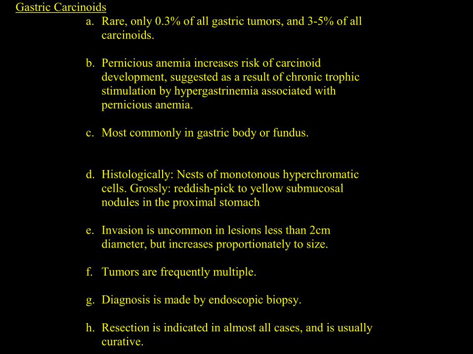
Gastric Carcinoidsa. Rare, only 0.3% of all gastric tumors, and 3-5% of all
carcinoids.
b. Pernicious anemia increases risk of carcinoiddevelopment, suggested as a result of chronic trophicstimulation by hypergastrinemia associated withpernicious anemia.
c. Most commonly in gastric body or fundus.
d. Histologically: Nests of monotonous hyperchromaticcells. Grossly: reddish-pick to yellow submucosalnodules in the proximal stomach
e. Invasion is uncommon in lesions less than 2cmdiameter, but increases proportionately to size.
f. Tumors are frequently multiple.
g. Diagnosis is made by endoscopic biopsy.
h. Resection is indicated in almost all cases, and is usuallycurative.
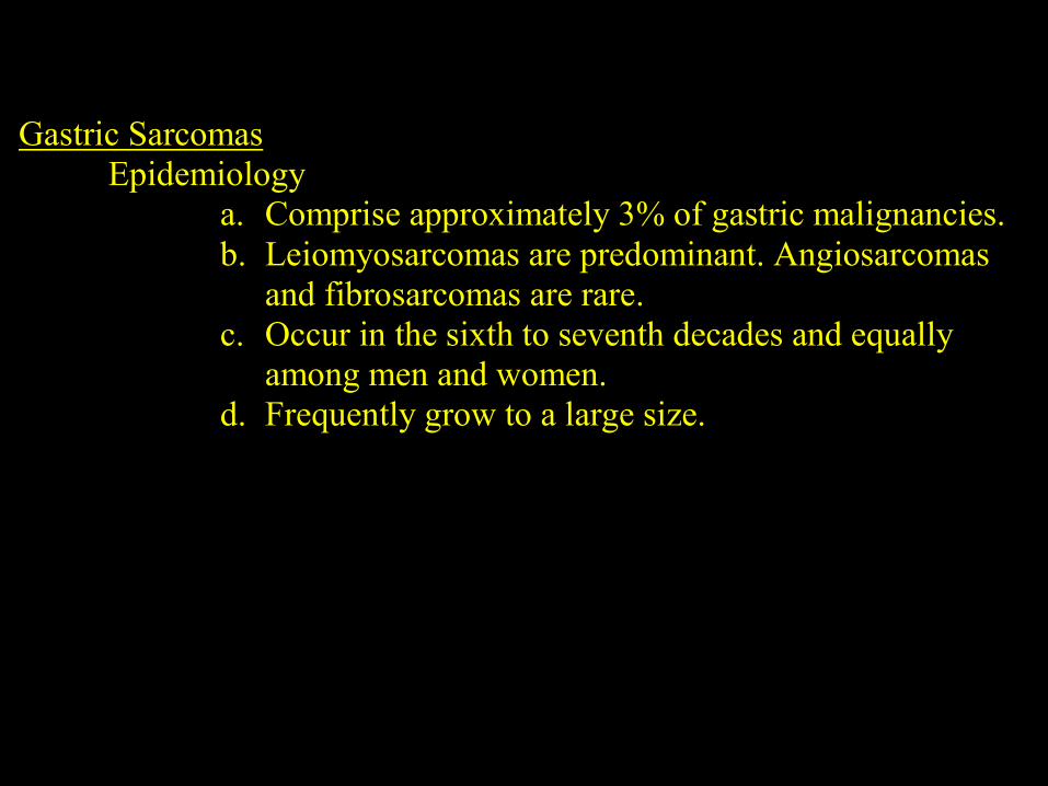
Gastric SarcomasEpidemiology
a. Comprise approximately 3% of gastric malignancies.b. Leiomyosarcomas are predominant. Angiosarcomas
and fibrosarcomas are rare.c. Occur in the sixth to seventh decades and equally
among men and women.d. Frequently grow to a large size.
Clinical Featuresa. Symptoms identical to those of adenocarcinoma.
Occasionally, with large lesions, an epigastric massmay be palpated.
b. Symptoms usually related to mass effects withcompression of adjacent structures. GI hemorrhage mayoccur secondary to overlying mucosal necrosis.
Diagnosisa. Endoscopic exam and biopsy is the diagnostic method
of choice. But can be negative if the major componentof growth is extraluminal. But, usually the tumorsappear as grey-white masses with a psuedo-capsuleoften separating it from surrounding healthy tissue.
b. Graded histologically with the frequency of mitoticspindles as the prime indicator of tumor aggressiveness.5-10 mitosis per high power field demonstrateincreased metastasis. In benign disease mitoses areusually rare or absent.
Treatmenta. Intraperitoneal sarcomatosis is frequent as a local
recurrence after resection. Metastasis occurs viahematogenous spread. Hepatic involvement is common.
b. Do not respond to radiation or chemotherapy.
c. Surgical resection is the treatment of choice. En blocresection should be attempted. Lymphadenectomy isnot indicated due to low frequency of lymphatic spread.
Overall survival is approximately 50% at 5 years,

Questions ??


