Download - Eyeball BMJ

MOB TCD
Professor Emeritus Moira O’Brien
FRCPI, FFSEM, FFSEM (UK), FTCD
Trinity College
Dublin
Eyeball
Eyeball
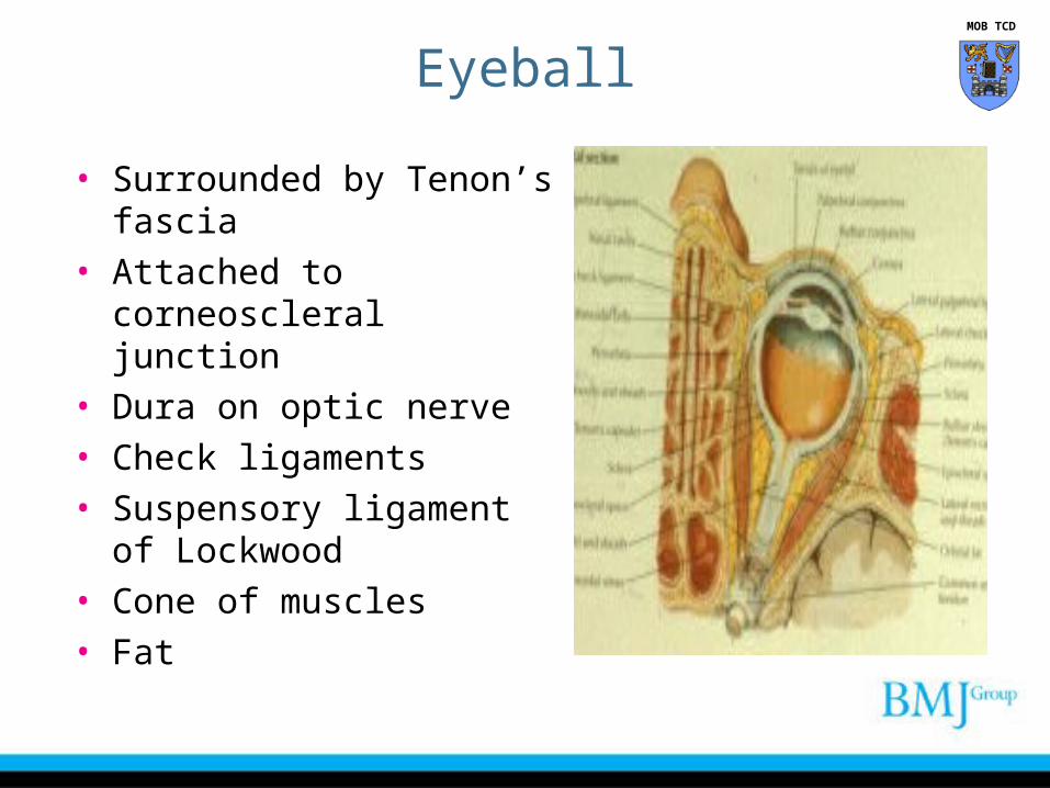
• Surrounded by Tenon’s fascia
• Attached to corneoscleral junction
• Dura on optic nerve• Check ligaments • Suspensory ligament of
Lockwood• Cone of muscles • Fat
MOB TCD
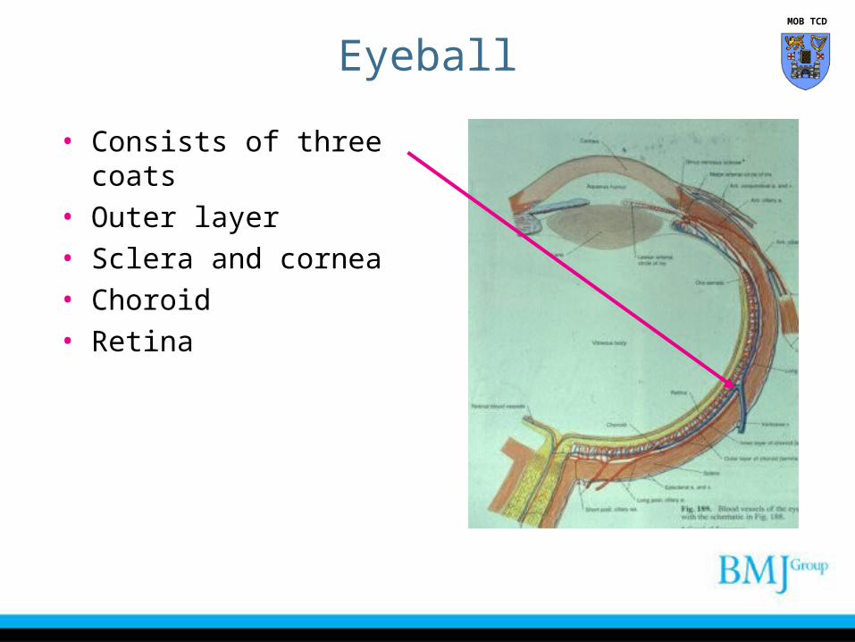
• Consists of three coats• Outer layer• Sclera and cornea• Choroid• Retina
Eyeball MOB TCD
Cornea
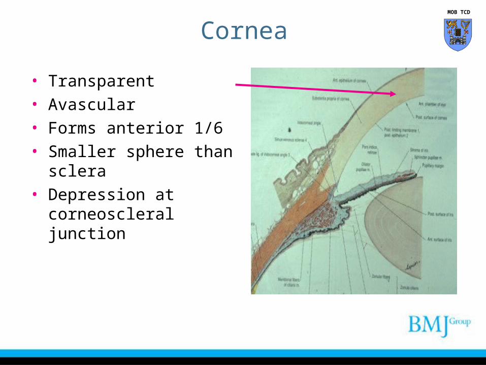
• Transparent• Avascular • Forms anterior 1/6 • Smaller sphere than sclera• Depression at
corneoscleral junction
MOB TCD
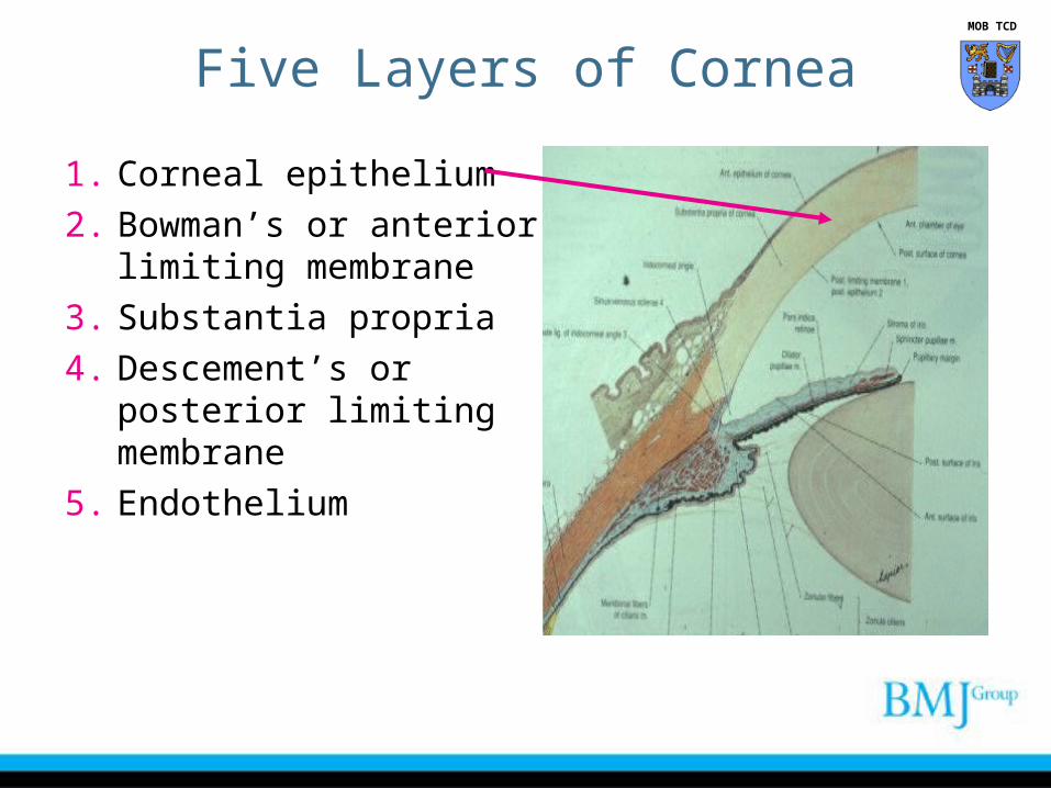
Five Layers of Cornea
1. Corneal epithelium
2. Bowman’s or anterior limiting membrane
3. Substantia propria
4. Descement’s or posterior limiting membrane
5. Endothelium
MOB TCD
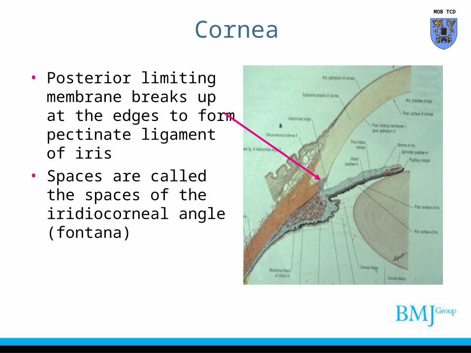
• Posterior limiting membrane breaks up at the edges to form pectinate ligament of iris
• Spaces are called the spaces of the iridiocorneal angle (fontana)
Cornea MOB TCD
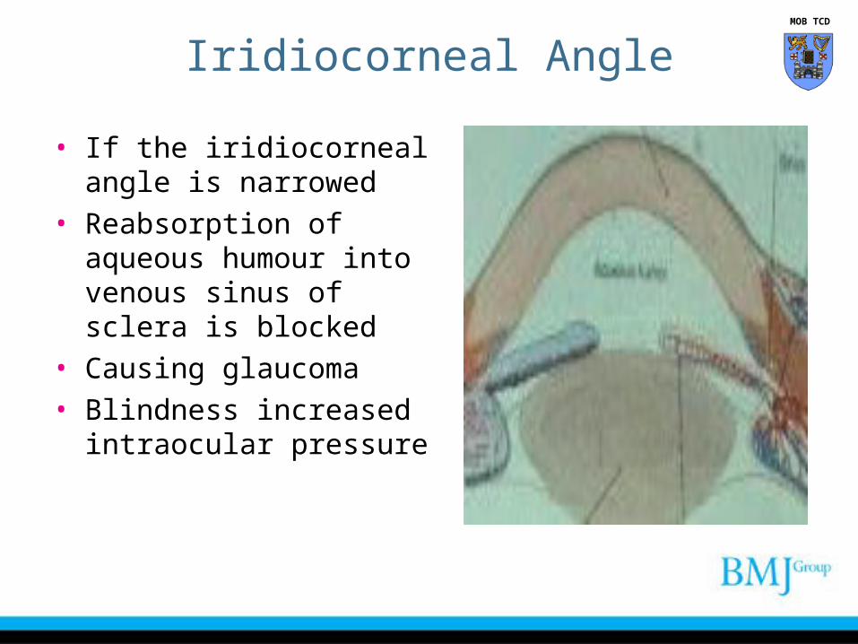
Iridiocorneal Angle
• If the iridiocorneal angle is narrowed
• Reabsorption of aqueous humour into venous sinus of sclera is blocked
• Causing glaucoma• Blindness increased
intraocular pressure
MOB TCD
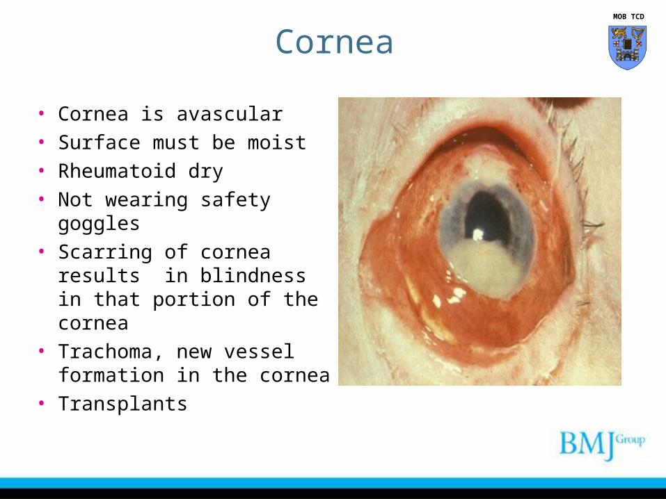
• Cornea is avascular• Surface must be moist • Rheumatoid dry• Not wearing safety goggles• Scarring of cornea results
in blindness in that portion of the cornea
• Trachoma, new vessel formation in the cornea
• Transplants
Cornea MOB TCD
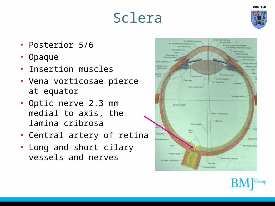
Sclera
• Posterior 5/6 • Opaque • Insertion muscles• Vena vorticosae pierce at
equator• Optic nerve 2.3 mm medial to
axis, the lamina cribrosa• Central artery of retina • Long and short cilary vessels
and nerves
MOB TCD
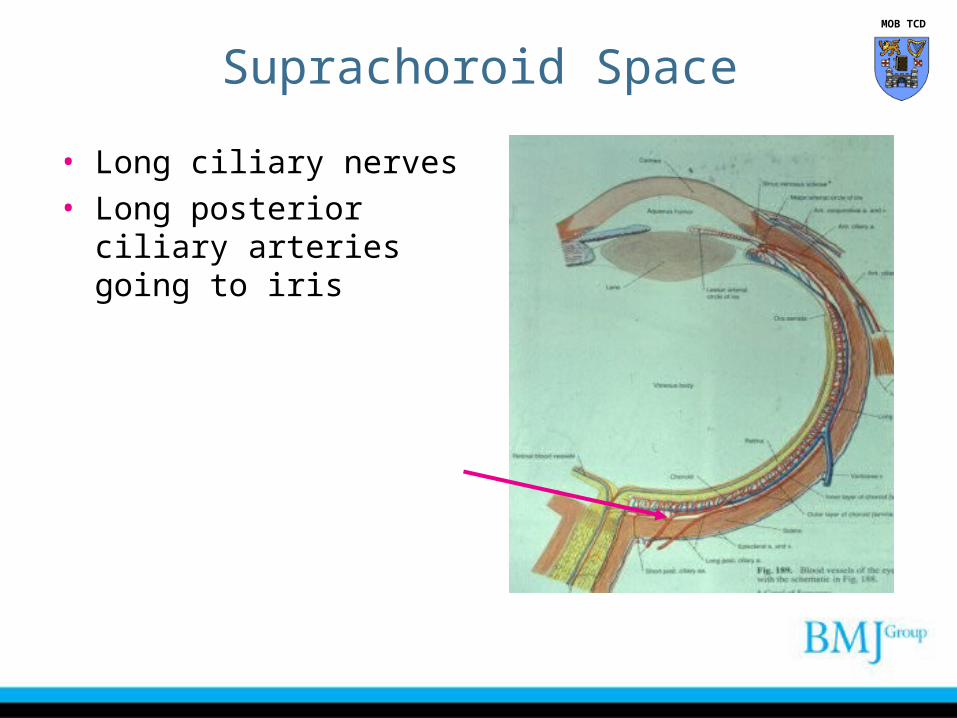
Suprachoroid Space
• Long ciliary nerves• Long posterior ciliary
arteries going to iris
MOB TCD
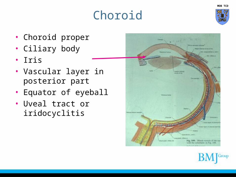
Choroid
• Choroid proper• Ciliary body• Iris• Vascular layer in posterior
part• Equator of eyeball• Uveal tract or iridocyclitis
MOB TCD
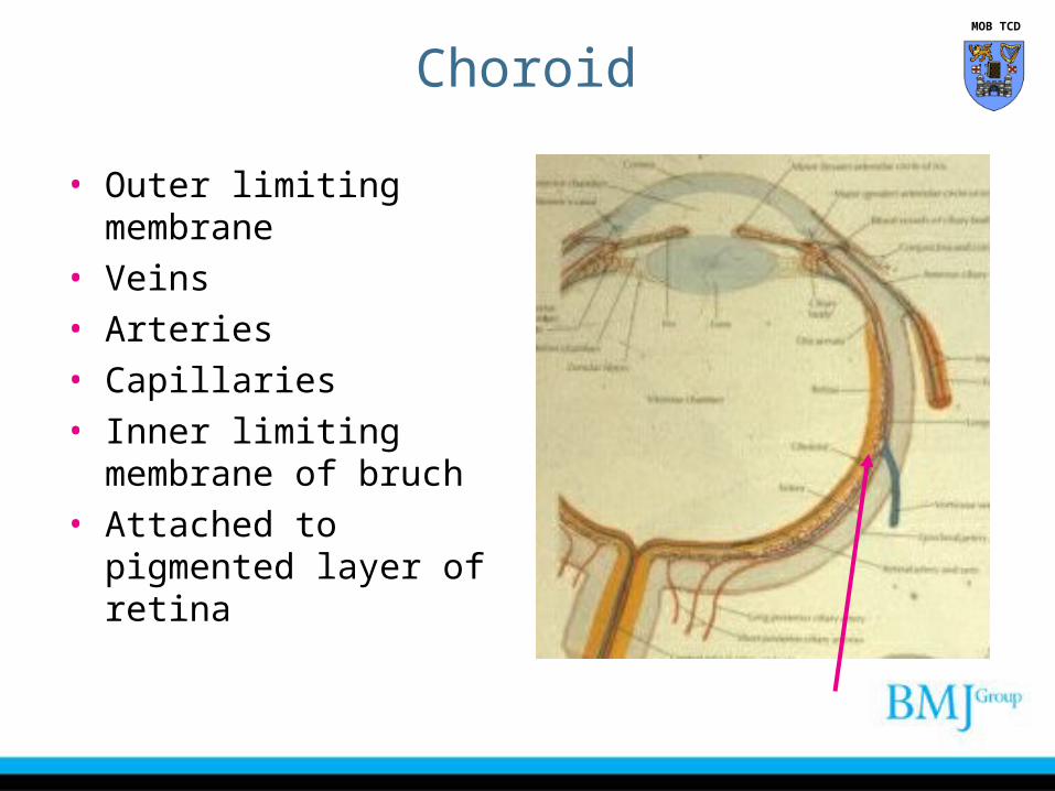
• Outer limiting membrane• Veins• Arteries• Capillaries• Inner limiting membrane of
bruch • Attached to pigmented
layer of retina
Choroid MOB TCD
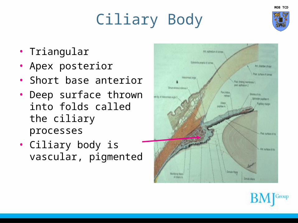
Ciliary Body
• Triangular• Apex posterior• Short base anterior• Deep surface thrown into
folds called the ciliary processes
• Ciliary body is vascular, pigmented
MOB TCD
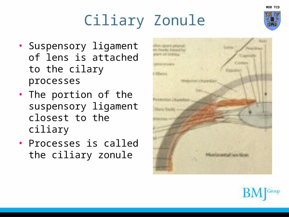
Ciliary Zonule
• Suspensory ligament of lens is attached to the cilary processes
• The portion of the suspensory ligament closest to the ciliary
• Processes is called the ciliary zonule
MOB TCD
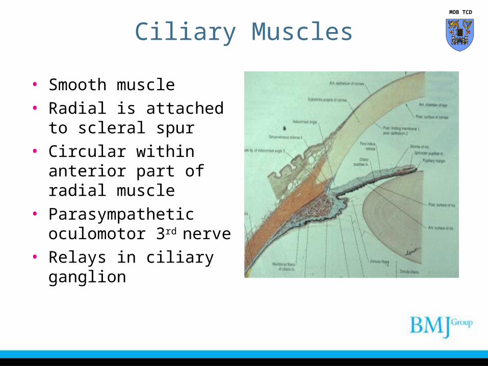
Ciliary Muscles
• Smooth muscle• Radial is attached to
scleral spur• Circular within anterior
part of radial muscle• Parasympathetic
oculomotor 3rd nerve• Relays in ciliary ganglion
MOB TCD
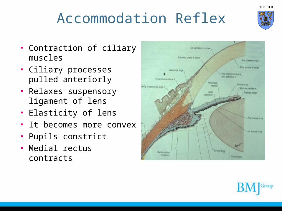
Accommodation Reflex
• Contraction of ciliary muscles
• Ciliary processes pulled anteriorly
• Relaxes suspensory ligament of lens
• Elasticity of lens • It becomes more convex• Pupils constrict• Medial rectus contracts
MOB TCD
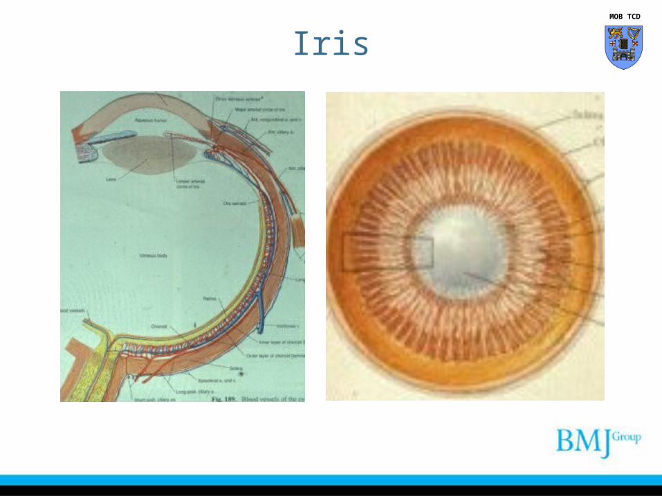
Iris MOB TCD
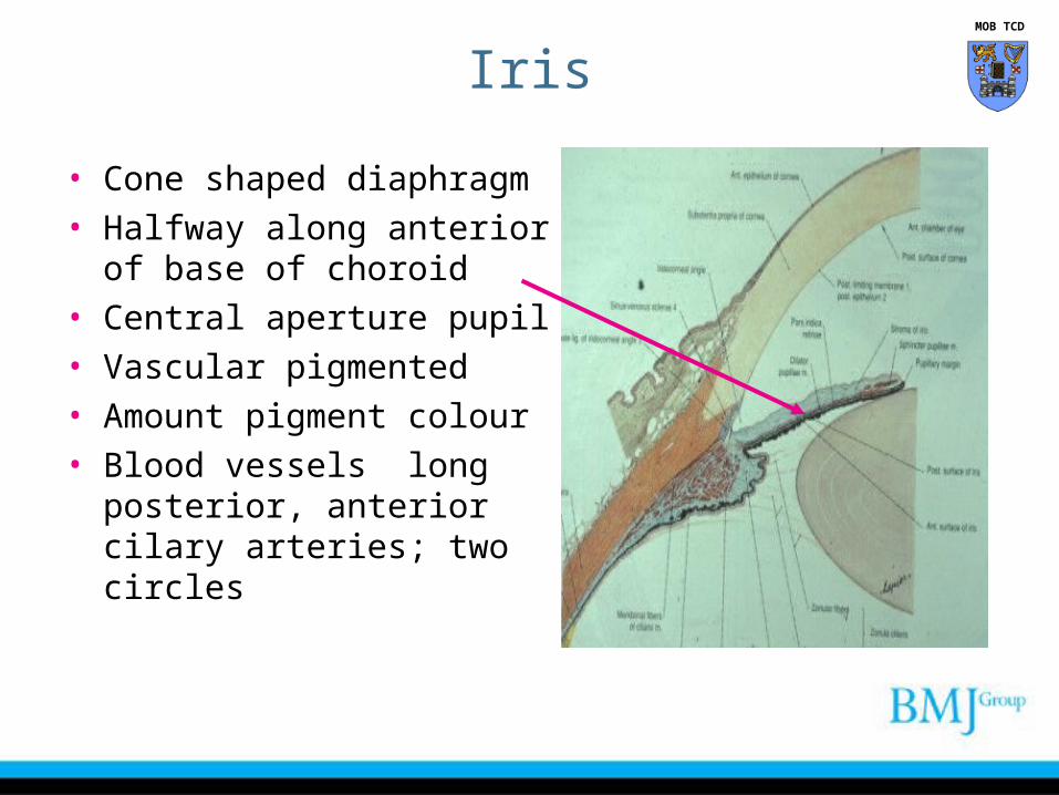
• Cone shaped diaphragm• Halfway along anterior of
base of choroid• Central aperture pupil• Vascular pigmented• Amount pigment colour• Blood vessels long
posterior, anterior cilary arteries; two circles
Iris MOB TCD
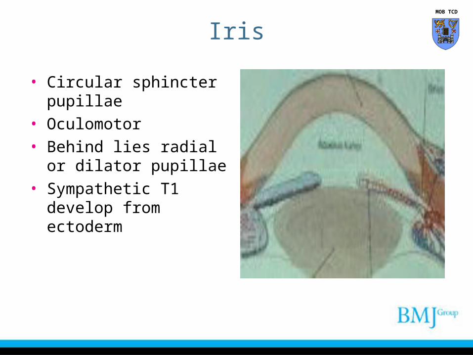
• Circular sphincter pupillae • Oculomotor• Behind lies radial or dilator
pupillae• Sympathetic T1 develop
from ectoderm
Iris MOB TCD
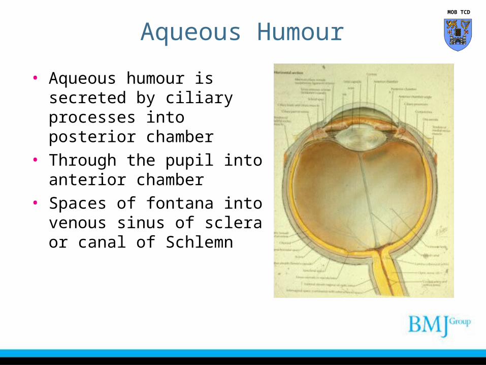
Aqueous Humour
• Aqueous humour is secreted by ciliary processes into posterior chamber
• Through the pupil into anterior chamber
• Spaces of fontana into venous sinus of sclera or canal of Schlemn
MOB TCD
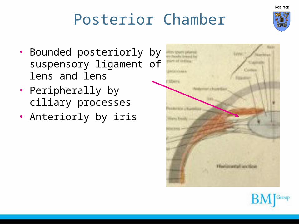
Posterior Chamber
• Bounded posteriorly by suspensory ligament of lens and lens
• Peripherally by ciliary processes
• Anteriorly by iris
MOB TCD
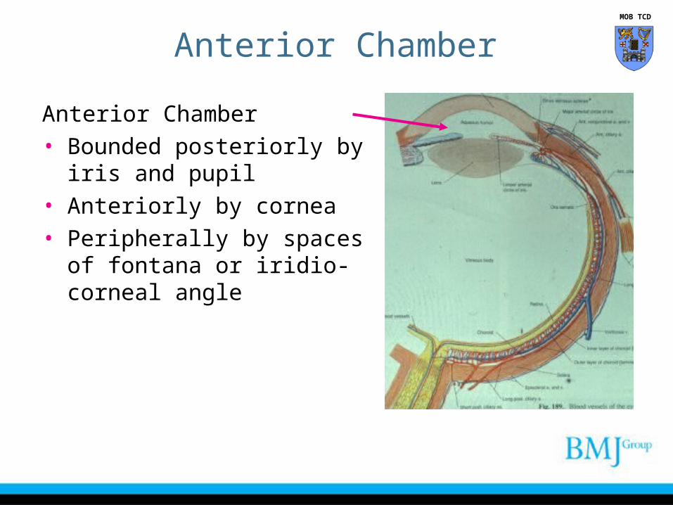
Anterior Chamber
Anterior Chamber• Bounded posteriorly by iris and
pupil• Anteriorly by cornea• Peripherally by spaces of
fontana or iridio-corneal angle
MOB TCD
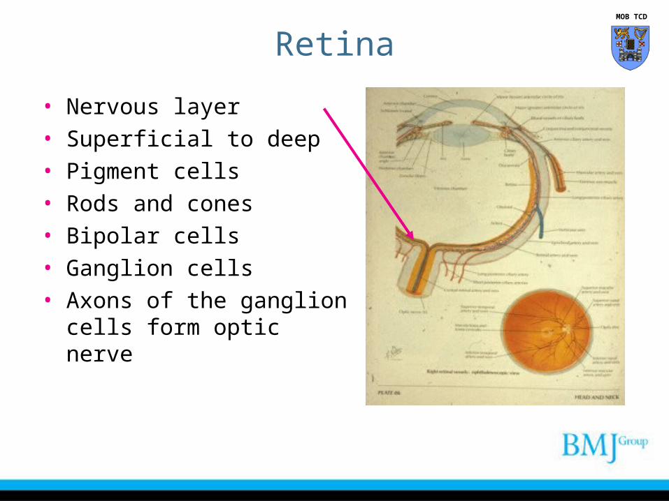
Retina
• Nervous layer• Superficial to deep• Pigment cells• Rods and cones• Bipolar cells• Ganglion cells• Axons of the ganglion cells
form optic nerve
MOB TCD
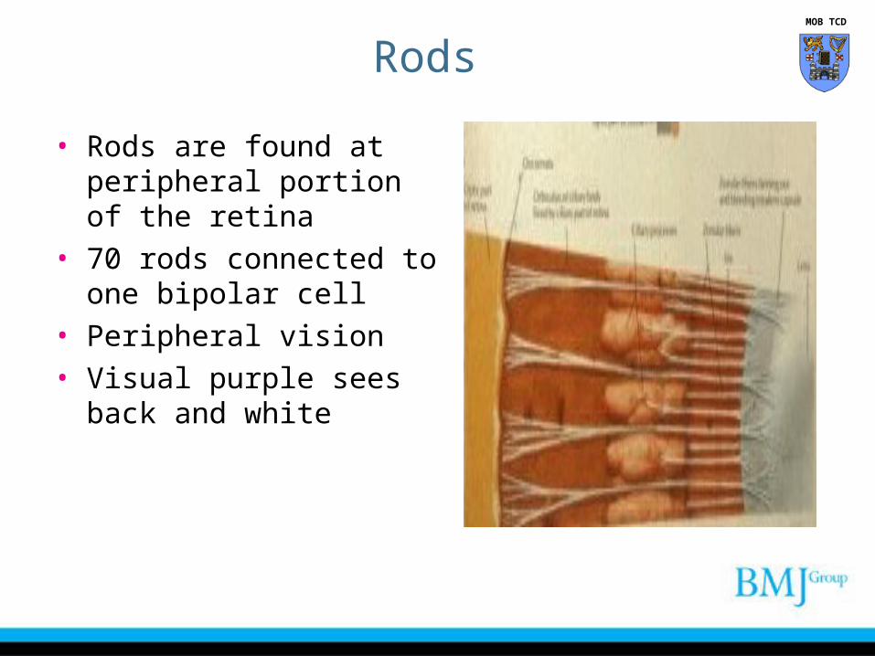
Rods
• Rods are found at peripheral portion of the retina
• 70 rods connected to one bipolar cell
• Peripheral vision• Visual purple sees back and
white
MOB TCD
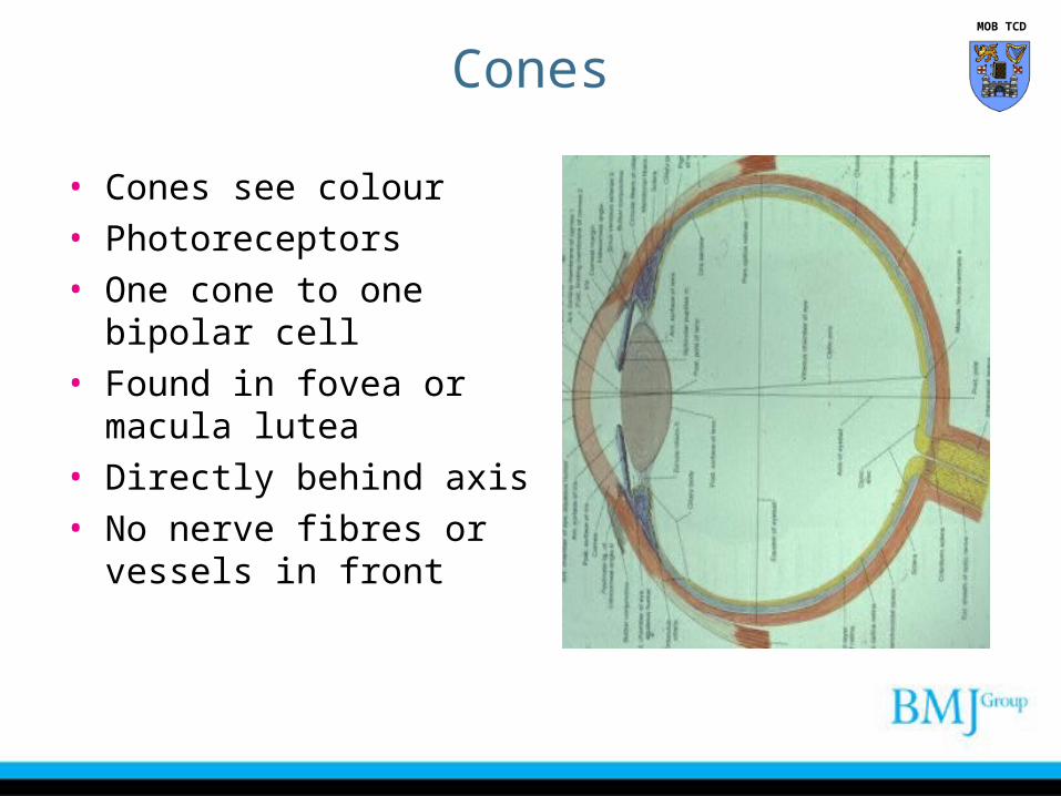
Cones
• Cones see colour • Photoreceptors• One cone to one bipolar cell• Found in fovea or macula
lutea• Directly behind axis• No nerve fibres or vessels
in front
MOB TCD
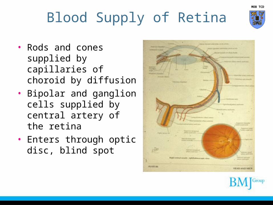
Blood Supply of Retina
• Rods and cones supplied by capillaries of choroid by diffusion
• Bipolar and ganglion cells supplied by central artery of the retina
• Enters through optic disc, blind spot
MOB TCD
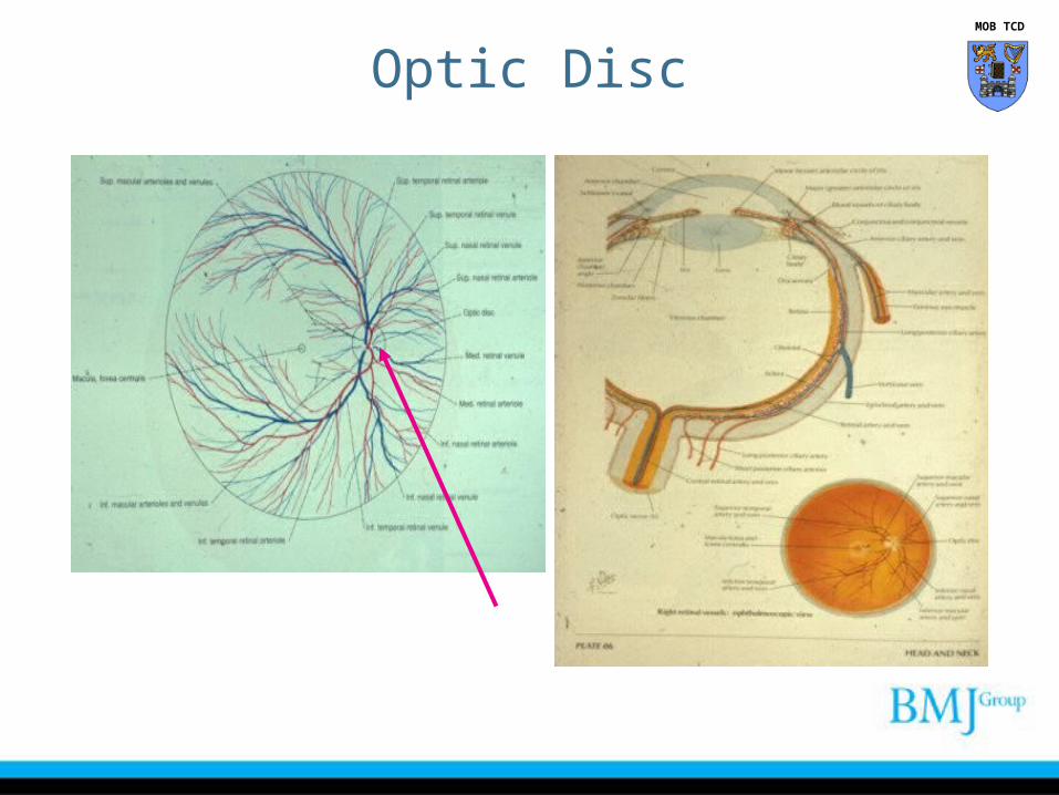
Optic Disc MOB TCD
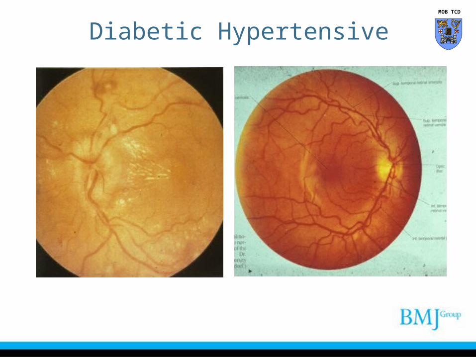
Diabetic Hypertensive MOB TCD
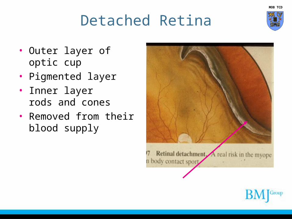
Detached Retina
• Outer layer of optic cup• Pigmented layer• Inner layer
rods and cones• Removed from their blood
supply
MOB TCD
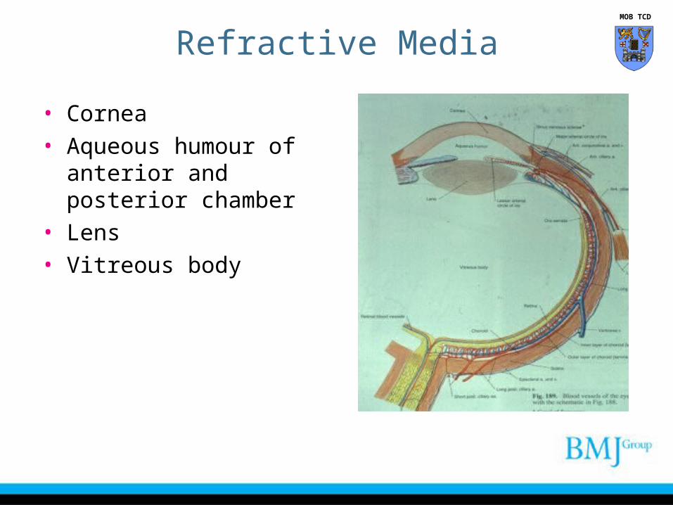
Refractive Media
• Cornea• Aqueous humour of anterior
and posterior chamber• Lens• Vitreous body
MOB TCD
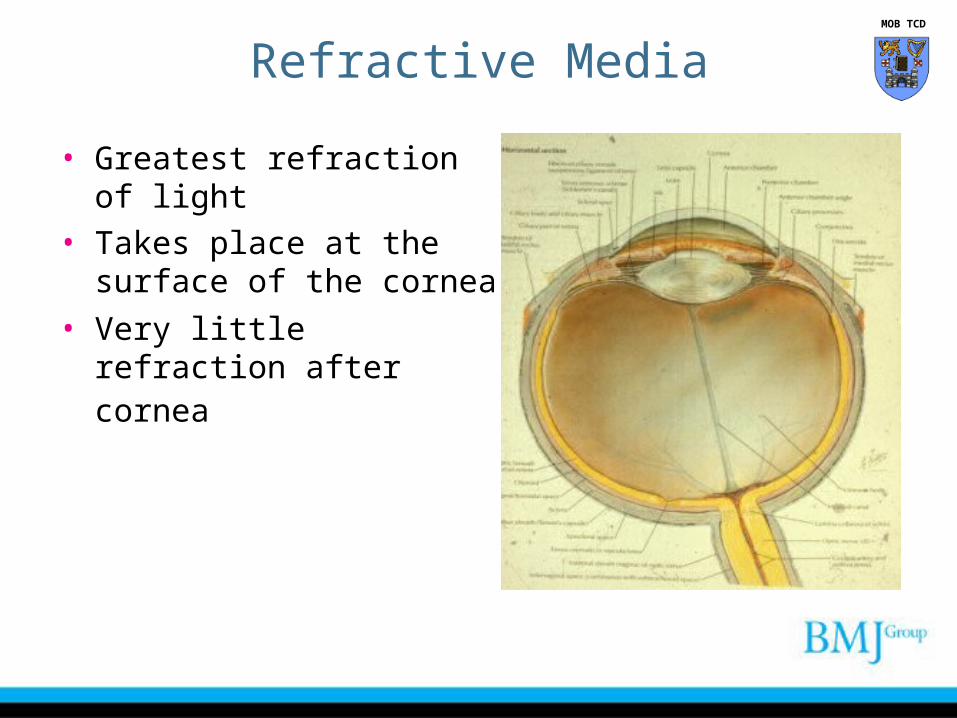
• Greatest refraction of light • Takes place at the surface
of the cornea
• Very little refraction after cornea
Refractive Media MOB TCD

Lens
• Crystalline• Translucent• Avascular structure• Lies in hyloid fossa• Posterior surface is highly
convex
• Does not alter its shape
MOB TCD

• Suspensory ligament is attached to periphery of the lens
• Cataract opaque lens• Nutrition from aqueous
humour
Lens MOB TCD
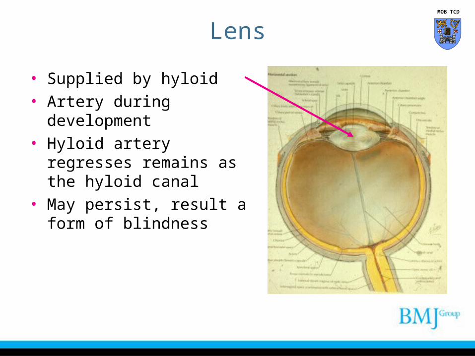
• Supplied by hyloid • Artery during development• Hyloid artery regresses
remains as the hyloid canal• May persist, result a form of
blindness
Lens MOB TCD
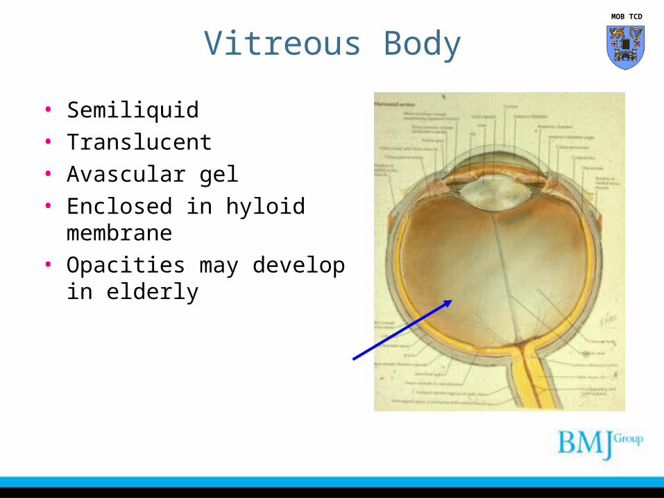
Vitreous Body
• Semiliquid• Translucent• Avascular gel• Enclosed in hyloid membrane• Opacities may develop in
elderly
MOB TCD
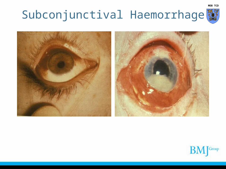
Subconjunctival Haemorrhage MOB TCD

“BMJ Publishing Group Limited (“BMJ Group”) 2012. All rights reserved.”


