Eval
cop
intr
Shinyoung An, D
This research was supported by the(NRF-2013R1A1A1008441).
aClinical Assistant Professor, AdvancbAssistant Professor, Department ofcGraduate student, Department of PdAssociate Professor, Department ofeProfessor, Department of Prosthodo
An et al
uating the marginal fit of zirconia
ings with digital impressions with an
aoral digital scanner
DS, MS,a Sungtae Kim, DDS, PhD,b
Hyunmin Choi, BDS,c Jae-Hoon Lee, DDS, PhD,d andHong-Seok Moon, DDS, PhDe
Yonsei University College of Dentistry, Seoul, Korea; Seoul NationalUniversity School of Dentistry, Seoul, Korea
Statement of problem. Digital impression systems have been developed to overcome the disadvantages associated withconventional impression methods.
Purpose. The purpose of this study was to compare the marginal fit of zirconia copings designed with the use of an iTerodigital scanner with those designed by the conventional impression technique.
Material and methods. Thirty identical cast, base-metal dies from 1 maxillary central incisor prepared for a ceramiccrown restoration were fabricated. For the conventional impression group (CI), base metal dies (n¼10) were replicated as stonedies by means of a conventional impression technique with polyvinyl siloxane material. For the iTero with polyurethane group (iP),base metal dies (n¼10) were replicated as polyurethane dies with the iTero digital impression system. For the iTero with no diesgroup (iNo), base metal dies (n¼10) were scanned with the iTero digital impression system, but no dies were fabricated. For eachgroup, 10 zirconia copings were fabricated based on the stone dies (CI group), polyurethane dies (iP group), or stereolithographyfiles (iNo group). The marginal gap of each specimen was measured with a light microscope at 50� magnification. One-wayanalysis of variance and the Tukey honestly significant difference test were used for statistical analysis (a¼.05).
Results. Statistically significant differences were found between the CI group and iP group (P<.05) and between the CI groupand iNo group (P<.05).
Conclusions. The marginal gap between the restoration and definitive cast base metal die was greater in the groups that usedthe digital impression method than in the group that used the conventional impression method. However, the marginaldiscrepancies of all of the groups were clinically acceptable. (J Prosthet Dent 2014;-:---)
Clinical Implications
Impressions made with the conventional method are more accurate thandigital impressions, but iTero digital impressions are an acceptablealternative impression technique.
One of the most critical steps in thefabrication of fixed prostheses is thecapture of an accurate impression.1,2
Precise replication should be madefor the restoration, with a clinically
Basic Scie
ed EducatPeriodontrosthodonProsthodntics, Yon
acceptable marginal gap.3-7 Tradition-ally, an accurate negative impression hasbeen used to transfer the necessary in-formation from the patient’s oral cavityto the dental technician’s laboratory.
nce Research Program of the National Research
ion in General Dentistry, Yonsei University Colology, Dental Research Institute, Seoul Nationtics, Yonsei University College of Dentistry.ontics, Yonsei University College of Dentistry.sei University College of Dentistry.
From this negative, the technician canfabricate accurate gypsum casts thatduplicate the original intraoral structure.However, conventional impression pro-cedures have disadvantages, such as
Foundation of Korea
lege of Dentistry.al University School of Dentistry.
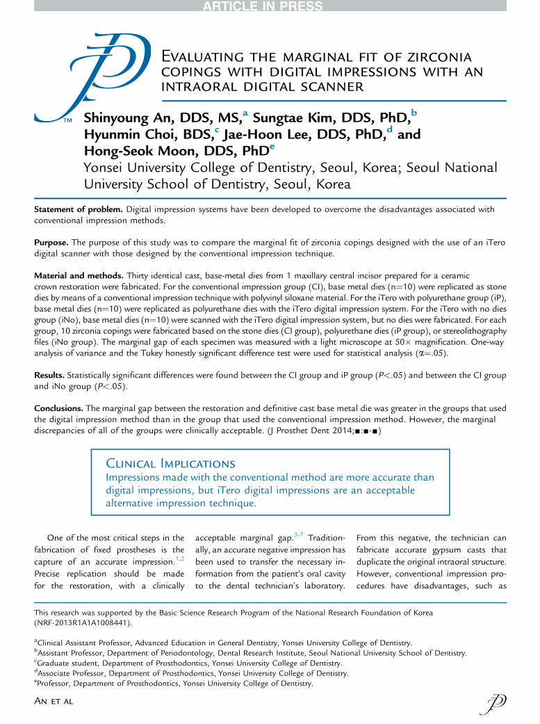
1 Cast metal definitive die fabrication.
2 Volume - Issue -
patient discomfort (gagging; objection-able odor or taste), the need for disin-fection and storage of the materials andtrays, and distortion due to mixing ofthe impression materials and variabletemperatures and humidity.8
Recent advances in digital impressiontechnologies have eliminated many of theproblems associated with conventionalimpression techniques.9,10 Some digitalimpression systems, for example, CEREC(Sirona Dental Systems) and E4D (D4DTechnologies), capture virtual 3D imagesof prepared teeth, from which restora-tions can be fabricated directly.11-14
Other systems, for example, iTero(Cadent), CEREC, and Lava COS (3MESPE), can fabricate restorations indi-rectly, on the basis of accurate definitivecasts.15,16 Digital impressions eliminateseveral time-consuming steps in thedental office, including tray selection,dispensing and setting of materials, andshipment of impressions to the labora-tory.17 Dental laboratories save timebecause they do not have to fabricate ormount casts on the articulator. Patientcomfort, treatment acceptance, no needfor disinfection, cost-effectiveness, andthe ability to make immediate correc-tions during preparation are added ben-efits. Moreover, digital scans can bestored on a computer, whereas conven-tional casts, which may chip or break,take up space in the dental office. Thedisadvantages of digital impression sys-tems are the need for initial instructionson how to use the device and the highcost of the equipment.
With the iTero system, the definitiverestorations are produced in the labora-tory but are fabricated on polyurethanecasts createdon the basis of data from thedigital scans, as opposed to gypsum castsmade from conventional impressions.Definitive restorations may also be madedirectly on the basis of digital scan data,without the need for polyurethane casts.Although studies on the accuracy of digi-tal impressions have reported that digitalimpressions are suitable for clinical ap-plications,14,15 further studies are requiredregarding the marginal fit of restorations.
Marginal fit is a critical factor insuccessful prosthodontic treatment.18,19
The Journal of Prosthetic Dentis
Ill-fitting margins can cause hypersensi-tivity, dental caries, plaque accumula-tion, and gingivitis, as well asperiodontitis and alveolar bone loss thatmay lead to tooth loss.20-23 When themarginal gap is large, the surface of thecement is exposed, which causes disso-lution of the cement.24,25 Improving thefidelity of restorations and reducing thethickness of cement film is importantbecause this can influence marginalleakage.26-28 Holmes et al29 definedvarious types of measurements betweenthe casting surface and the tooth so thatthe marginal gap could be determinedand described in a standardizedmanner. The angular combination of themarginal gap and the extension error(overextension or underextension) iscalled the absolute marginal discrep-ancy.29 The present study used theconcept of absolute marginal discrep-ancy to measure the marginal fit ofcopings. However, to date, a clinicallyacceptable range of marginal discrep-ancy has not been defined.30 McLean31
reported that for a good long-termprognosis, the clinically acceptablemarginal gap for a crown is approxi-mately 120 mm. Most authors agree thata marginal discrepancy in the range of100 to 120 mm is clinically acceptablewith regard to longevity.30-32
Many reports on digital impressionsystems have been published,33,34 butonly a few have used the iTero digitalscanner. The purpose of this study wasto compare the marginal fit of zirconiacopings fabricated on the basis of data
try
from the iTero digital scanner withthose fabricated on the basis of con-ventional impression techniques. Themarginal fit of zirconia copings madedirectly from the digital scan data wasalso compared. The null hypothesis wasthat no difference would be found inthe marginal fit between zirconia cop-ings designed with the iTero digitalscanner and those designed with theconventional impression technique.
MATERIAL AND METHODS
An ivorine (A5A-500; Nissin DentalProducts) left maxillary central incisorwas prepared for a ceramic crown.An incisal reduction of 2.0 mm andan axial reduction of approximately1.0 mm were prepared with a high-speed handpiece. The tooth was fin-ished with a milling machine (D-F 44;HarnischþRieth), which resulted in achamfer margin of 1.0 mm in depthwith 6 degrees of convergence.35
Impressions of the prepared typo-dont tooth were made with polyvinylsiloxane (Exafine; GC Corp). Theimpression was poured in acrylic resin(Pattern resin; GC Corp), invested, andcast in Ni-Cr alloy (Verabond 2; Aal-badent) to produce a definitive die(Fig. 1). This definitive die was made toprevent wear or damage to the typo-dont tooth.
For the conventional impression(CI) group, impressions of the definitivecast were made with polyvinyl siloxane(Exafine; GC Corp), and dies made of
An et al
- 2014 3
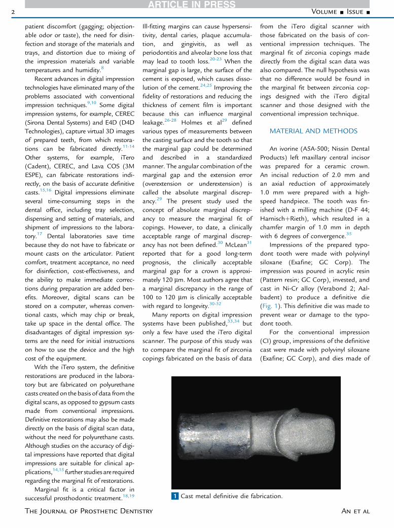
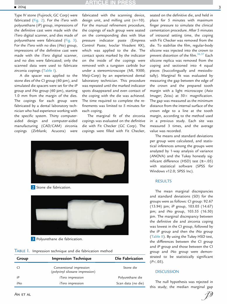
Type IV stone (Fujirock; GC Corp) werefabricated (Fig. 2). For the iTero withpolyurethane (iP) group, impressions ofthe definitive cast were made with theiTero digital scanner, and dies made ofpolyurethane were fabricated (Fig. 3).For the iTero with no dies (iNo) group,impressions of the definitive cast weremade with the iTero digital scanner,and no dies were fabricated; only thescanned data were used to fabricatezirconia copings (Table I).
A die spacer was applied to thestone dies of the CI group (60 mm), andsimulated die spacers were set for the iPgroup and iNo group (60 mm), starting1.0 mm from the margin of the dies.The copings for each group werefabricated by a dental laboratory tech-nician who had experience working withthe specific system. Thirty computer-aided design and computer-aidedmanufacturing (CAD/CAM) zirconiacopings (Zirblank; Acucera) were
2 Stone die fabrication.
3 Polyurethane die fabrication
Table I. Impression technique and die
Group Impression Tech
CI Conventional impre(polyvinyl siloxane im
iP iTero impressio
iNo iTero impressio
An et al
fabricated with the scanning device,design unit, and milling unit (n¼10).For the manual refinement procedure,the copings of each group were seatedon the corresponding dies with bluepressure indicator paste (EmpressControl Paste; Ivoclar Vivadent KK),which was applied to the die. Thecontact spots marked by the indicatoron the inside of the copings wereremoved with a tungsten carbide burunder a stereomicroscope (ML 9300;Meiji-Corp) by an experienced dentallaboratory technician. This procedurewas repeated until the marked indicatorspots disappeared and even contact ofthe coping with the die was achieved.The time required to complete the re-finements was limited to 5 minutes foreach coping.
The marginal fit of the zirconiacopings was evaluated on the definitivedie with Fit Checker (GC Corp). Thecopings were filled with Fit Checker,
.
fabrication method
nique Die Fabrication
ssionpression)
Stone die
n Polyurethane die
n Scan data (no die)
seated on the definitive die, and held inplace for 5 minutes with maximumfinger pressure to simulate the clinicalcementation procedure. After 5 minutesof intraoral setting time, the copingwith Fit Checker was removed from thedie. To stabilize the film, regular-bodysilicone was injected into the crown toprevent distortion of the film.36,37 Eachsilicone replica was removed from thecoping and sectioned into 4 equalpieces (buccolingually and mesiodis-tally). Marginal fit was evaluated bymeasuring the gap between the edge ofthe crown and the prepared toothmargin with a light microscope (AxioImager; Zeiss) at 50� magnification.The gap was measured as the minimumdistance from the internal surface of thecrown edge to a line at the toothmargin, according to the method usedin a previous study. Each site wasmeasured 3 times, and the averagevalue was recorded.
The means and standard deviationsper group were calculated, and statis-tical inferences among the groups wereanalyzed by 1-way analysis of variance(ANOVA) and the Tukey honestly sig-nificant difference (HSD) test (a¼.05)with statistical software (SPSS forWindows v12.0; SPSS Inc).
RESULTS
The mean marginal discrepanciesand standard deviations (SD) for thegroups were as follows: CI group: 92.67(13.94) mm; iP group, 103.05 (14.67)mm; and iNo group, 103.55 (16.50)mm. The marginal discrepancy betweenthe definitive die and zirconia copingwas lowest in the CI group, followed bythe iP group and then the iNo group(Table II). By using the Tukey HSD test,the differences between the CI groupand iP group and those between the CIgroup and iNo group were demon-strated to be statistically significant(P<.05).
DISCUSSION
The null hypothesis was rejected inthis study; the median marginal gap
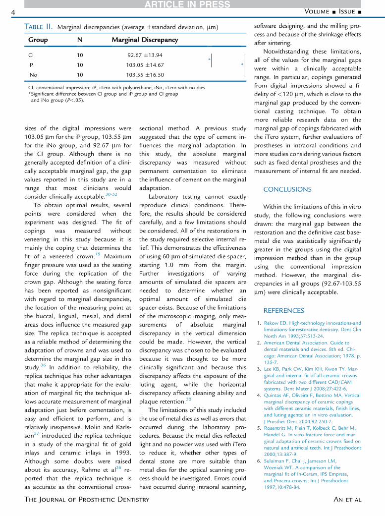
Table II. Marginal discrepancies (average �standard deviation, mm)
Group N Marginal Discrepancy
CI 10 92.67 �13.94*
*iP 10 103.05 �14.67
iNo 10 103.55 �16.50
CI, conventional impression; iP, iTero with polyurethane; iNo, iTero with no dies.*Significant difference between CI group and iP group and CI groupand iNo group (P<.05).
4 Volume - Issue -
sizes of the digital impressions were103.05 mm for the iP group, 103.55 mmfor the iNo group, and 92.67 mm forthe CI group. Although there is nogenerally accepted definition of a clini-cally acceptable marginal gap, the gapvalues reported in this study are in arange that most clinicians wouldconsider clinically acceptable.30-32
To obtain optimal results, severalpoints were considered when theexperiment was designed. The fit ofcopings was measured withoutveneering in this study because it ismainly the coping that determines thefit of a veneered crown.19 Maximumfinger pressure was used as the seatingforce during the replication of thecrown gap. Although the seating forcehas been reported as nonsignificantwith regard to marginal discrepancies,the location of the measuring point atthe buccal, lingual, mesial, and distalareas does influence the measured gapsize. The replica technique is acceptedas a reliable method of determining theadaptation of crowns and was used todetermine the marginal gap size in thisstudy.36 In addition to reliability, thereplica technique has other advantagesthat make it appropriate for the evalu-ation of marginal fit; the technique al-lows accurate measurement of marginaladaptation just before cementation, iseasy and efficient to perform, and isrelatively inexpensive. Molin and Karls-son37 introduced the replica techniquein a study of the marginal fit of goldinlays and ceramic inlays in 1993.Although some doubts were raisedabout its accuracy, Rahme et al36 re-ported that the replica technique isas accurate as the conventional cross-
The Journal of Prosthetic Dentis
sectional method. A previous studysuggested that the type of cement in-fluences the marginal adaptation. Inthis study, the absolute marginaldiscrepancy was measured withoutpermanent cementation to eliminatethe influence of cement on the marginaladaptation.
Laboratory testing cannot exactlyreproduce clinical conditions. There-fore, the results should be consideredcarefully, and a few limitations shouldbe considered. All of the restorations inthe study required selective internal re-lief. This demonstrates the effectivenessof using 60 mm of simulated die spacer,starting 1.0 mm from the margin.Further investigations of varyingamounts of simulated die spacers areneeded to determine whether anoptimal amount of simulated diespacer exists. Because of the limitationsof the microscopic imaging, only mea-surements of absolute marginaldiscrepancy in the vertical dimensioncould be made. However, the verticaldiscrepancy was chosen to be evaluatedbecause it was thought to be moreclinically significant and because thisdiscrepancy affects the exposure of theluting agent, while the horizontaldiscrepancy affects cleaning ability andplaque retention.30
The limitations of this study includedthe use of metal dies as well as errors thatoccurred during the laboratory pro-cedures. Because the metal dies reflectedlight and no powder was used with iTeroto reduce it, whether other types ofdental stone are more suitable thanmetal dies for the optical scanning pro-cess should be investigated. Errors couldhave occurred during intraoral scanning,
try
software designing, and the milling pro-cess and because of the shrinkage effectsafter sintering.
Notwithstanding these limitations,all of the values for the marginal gapswere within a clinically acceptablerange. In particular, copings generatedfrom digital impressions showed a fi-delity of <120 mm, which is close to themarginal gap produced by the conven-tional casting technique. To obtainmore reliable research data on themarginal gap of copings fabricated withthe iTero system, further evaluations ofprostheses in intraoral conditions andmore studies considering various factorssuch as fixed dental prostheses and themeasurement of internal fit are needed.
CONCLUSIONS
Within the limitations of this in vitrostudy, the following conclusions weredrawn: the marginal gap between therestoration and the definitive cast base-metal die was statistically significantlygreater in the groups using the digitalimpression method than in the groupusing the conventional impressionmethod. However, the marginal dis-crepancies in all groups (92.67-103.55mm) were clinically acceptable.
REFERENCES
1. Rekow ED. High-technology innovations-andlimitations-for restorative dentistry. Dent ClinNorth Am 1993;37:513-24.
2. American Dental Association. Guide todental materials and devices. 8th ed. Chi-cago: American Dental Association; 1978. p.135-7.
3. Lee KB, Park CW, Kim KH, Kwon TY. Mar-ginal and internal fit of all-ceramic crownsfabricated with two different CAD/CAMsystems. Dent Mater J 2008;27:422-6.
4. Quintas AF, Oliveira F, Bottino MA. Verticalmarginal discrepancy of ceramic copingswith different ceramic materials, finish lines,and luting agents: an in vitro evaluation.J Prosthet Dent 2004;92:250-7.
5. Rosentritt M, Plein T, Kolbeck C, Behr M,Handel G. In vitro fracture force and mar-ginal adaptation of ceramic crowns fixed onnatural and artificial teeth. Int J Prosthodont2000;13:387-9.
6. Sulaiman F, Chai J, Jameson LM,Wozniak WT. A comparison of themarginal fit of In-Ceram, IPS Empress,and Procera crowns. Int J Prosthodont1997;10:478-84.
An et al
- 2014 5
7. Hung SH, Hung KS, Eick JD, Chappell RP.Marginal fit of porcelain-fused-to-metal andtwo types of ceramic crown. J Prosthet Dent1990;63:26-31.
8. Wassell RW, Barker D, Walls AW. Crownsand other extra-coronal restorations:impression materials and technique. Br DentJ 2002;192:679-84, 87-90.
9. Bayne SC, Heymann HO. CAD/CAM indentistry: present and future applications.Quintessence Int 1996;27:431-3.
10. Sturdevant JR, Bayne SC, Heymann HO.Margin gap size of ceramic inlays usingsecond-generation CAD/CAM equipment.J Esthet Dent 1999;11:206-14.
11. Reich SM, Peltz ID, Wichmann M,Estafan DJ. A comparative study of twoCEREC software systems in evaluatingmanufacturing time and accuracy of resto-rations. Gen Dent 2005;53:195-8.
12. Filser F, Kocher P, Weibel F, Luthy H,Scharer P, Gauckler LJ. Reliability andstrength of all-ceramic dental restorationsfabricated by direct ceramic machining(DCM). Int J Comput Dent 2001;4:89-106.
13. Nakamura T, Dei N, Kojima T,Wakabayashi K. Marginal and internal fit ofCerec 3 CAD/CAM all-ceramic crowns. Int JProsthodont 2003;16:244-8.
14. Tinschert J, Natt G, Mautsch W,Spiekermann H, Anusavice KJ. Marginal fit ofalumina- and zirconia-based fixed partialdentures produced by a CAD/CAM system.Oper Dent 2001;26:367-74.
15. Reich S, Wichmann M, Nkenke E,Proeschel P. Clinical fit of all-ceramic three-unit fixed partial dentures, generated withthree different CAD/CAM systems. Eur J OralSci 2005;113:174-9.
16. Suttor D, Bunke K, Hoescheler S,Hauptmann H, Hertlein G. LAVA-the systemfor all-ceramic ZrO2 crown and bridgeframeworks. Int J Comput Dent 2001;4:195-206.
17. Andersson M, Razzoog ME, Oden A,Hegenbarth EA, Lang BR. Procera: a new wayto achieve an all-ceramic crown. Quintes-sence Int 1998;29:285-96.
An et al
18. Denissen H, Dozic A, van der Zel J, vanWaas M. Marginal fit and short-term clinicalperformance of porcelain-veneered CICERO,CEREC, and Procera onlays. J Prosthet Dent2000;84:506-13.
19. Christensen GJ. Marginal fit of gold inlaycastings. J Prosthet Dent 1966;16:297-305.
20. Palomo F, Peden J. Periodontal consider-ations of restorative procedures. J ProsthetDent 1976;36:387-94.
21. Lang NP, Kiel RA, Anderhalden K. Clinicaland microbiological effects of subgingivalrestorations with overhanging or clinicallyperfect margins. J Clin Periodontol 1983;10:563-78.
22. Valderhaug J, Heloe LA. Oral hygiene in agroup of supervised patients with fixedprostheses. J Periodontol 1977;48:221-4.
23. Sorensen SE, Larsen IB, Jorgensen KD.Gingival and alveolar bone reaction to mar-ginal fit of subgingival crown margins. ScandJ Dent Res 1986;94:109-14.
24. Felton DA, Kanoy BE, Bayne SC,Wirthman GP. Effect of in vivo crown margindiscrepancies on periodontal health.J Prosthet Dent 1991;65:357-64.
25. Gonzalo E, Suárez MJ, Serrano B, Lozano JF.A comparison of the marginal vertical dis-crepancies of zirconium and metal ceramicposterior fixed dental prostheses before andafter cementation. J Prosthet Dent 2009;102:378-84.
26. Albert FE, El-Mowafy OM. Marginal adap-tation and microleakage of Procera AllCeramcrowns with four cements. Int J Prosthodont2004;17:529-35.
27. Kern M, Schaller HG, Strub JR. Marginal fitof restorations before and after cementa-tion in vivo. Int J Prosthodont 1993;6:585-91.
28. Rinke S, Hüls A, Jahn L. Marginal accuracyand fracture strength of conventionaland copy-milled all-ceramic crowns.Int J Prosthodont 1995;8:303-10.
29. Holmes JR, Bayne SC, Holland GA,Sulik WD. Considerations in measurementof marginal fit. J Prosthet Dent 1989;62:405-8.
30. Sorensen JA. A standardized method fordetermination of crown margin fidelity.J Prosthet Dent 1990;64:18-24.
31. McLean JW. Polycarboxylate cements. Fiveyears’ experience in general practice. Br DentJ 1972;132:9-15.
32. McLean JW, von Fraunhofer JA. The esti-mation of cement film thickness by anin vivo technique. Br Dent J 1971;131:107-11.
33. Akbar JH, Petrie CS, Walker MP, Williams K,Eick JD. Marginal adaptation of Cerec 3CAD/CAM composite crowns using twodifferent finish line preparation designs.J Prosthodont 2006;15:155-63.
34. May KB, Russell MM, Razzoog ME,Lang BR. Precision of fit: the Procera All-Ceram crown. J Prosthet Dent 1998;80:394-404.
35. Bindl A, Mormann WH. Marginal and inter-nal fit of all-ceramic CAD/CAM crown-copings on chamfer preparations. J OralRehabil 2005;32:441-7.
36. Rahme HY, Tehini GE, Adib SM, Ardo AS,Rifai KT. In vitro evaluation of the “replicatechnique” in the measurement of the fit ofProcera crowns. J Contemp Dent Pract2008;9:25-32.
37. Molin M, Karlsson S. The fit of gold inlaysand three ceramic inlay systems. A clinicaland in vitro study. Acta Odontol Scand1993;51:201-6.
Corresponding author:Dr Hong-Seok MoonDepartment of ProsthodonticsYonsei University College of Dentistry50 Yonsei-ro, Seodaemun-gu, Seoul 120-752KOREAE-mail: [email protected]
AcknowledgmentThe authors thank The Acucera Center, Korea forceramic materials.
Copyright ª 2014 by the Editorial Council forThe Journal of Prosthetic Dentistry.


