
page 1 of 21
Diagnosis and treatment of vascular compression syndromes of the abdomen based on the anatomical features of man and gender-specific characteristics after puberty
Content Preface .........................................................................................................................................................1 Anatomical introduction ................................................................................................................................2 Nutcracker syndrome and nutcracker phenomenon .........................................................................................3 Pelvic congestion syndrome ...........................................................................................................................9 May-Thurner-constellation. ..........................................................................................................................12 Midline (congestion) syndrome ....................................................................................................................13 Celiac ganglion compression syndrome (Celiac trunk compression syndrome, (Celiac artery compression
syndrome, Dunbar-Syndrome, Median arcuate ligament syndrome (MALS)). .........................................15 Mesenteric artery compression syndrome (Wilkie syndrome) .........................................................................19
Important Note: The symptoms and explanations presented here cannot and should not replace a doctor's visit. In general: all the symptoms are ambiguous and can have very different causes, of course, also those which are not shown here. The author assumes no liability for diagnostics and therapies which are performed or omitted despite or because of these explanations. You should always consult with confidence to your doctor concerning all your complaints! Preface Abdominal complaints often have a simple cause, which can be clarified easily, especially if they occur suddenly or show clearly recognizable correlations with other symptoms (e.g. diarrhea, vomiting, and fever). However, if abdominal pain, lasts for a long time, over weeks or months, elaborate tests are often required in order to clarify their cause. Not rarely, the origin of the pain may remain enigmatic, so that psychological causes and specific living conditions are considered as the main cause or trigger of the complaints. And yet, hardly anyone is aware that a number of patients suffer from compression of blood vessels or compression of organs by abdominal blood vessels, the so-called vascular compression syndromes. Since these diseases may give rise to many, seemingly unrelated symptoms in addition to pain, they are explained here for a broader audience. So-called vegetative symptoms play a significant role in the suffering of patients with vascular compression syndromes. These very annoying symptoms as nausea, dizziness, respiratory disabilities (primarily inhalation), loss of appetite and rapid satiety, (nearly) fainting on exertion and episodes of diarrhea are found very frequently. They emerge in addition to pain or even isolated. Left-sided flank pain and abdominal pain are the predominant pain localizations but from there the pain may radiate to the back or the chest, sometimes to the left thigh. Headaches are not rarely found and may be exaggerated during physical exercises, may be throbbing and emerge preferentially in the nape. We often receive inquiries from patients from abroad, looking for advice and a comprehensible explanation of their complaints as well as the anatomical and functional implications of vascular compression syndromes. Therefore, this article is written to ease the understanding of these conditions. It is written in simple, mainly non-medical terms and directed to lay people. If there remain questions concerning specific circumstances of your own condition - then simply write by email to:
page 2 of 21
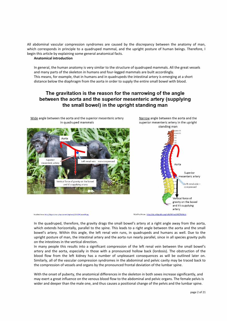
All abdominal vascular compression syndromes are caused by the discrepancy between the anatomy of man, which corresponds in principle to a quadruped mammal, and the upright posture of human beings. Therefore, I begin this article by explaining some general anatomical facts.
Anatomical introduction
In general, the human anatomy is very similar to the structure of quadruped mammals. All the great vessels and many parts of the skeleton in humans and four-legged mammals are built accordingly. This means, for example, that in humans and in quadrupeds the intestinal artery is emerging at a short distance below the diaphragm from the aorta in order to supply the entire small bowel with blood.
In the quadruped, therefore, the gravity drags the small bowel’s artery at a right angle away from the aorta, which extends horizontally, parallel to the spine. This leads to a right angle between the aorta and the small bowel’s artery. Within this angle, the left renal vein runs, in quadrupeds and humans as well. Due to the upright posture of man, the intestinal artery and the aorta run nearly parallel, since in all species gravity pulls on the intestines in the vertical direction. In many people this results into a significant compression of the left renal vein between the small bowel’s artery and the aorta, especially in those with a pronounced hollow back (lordosis). The obstruction of the blood flow from the left kidney has a number of unpleasant consequences as will be outlined later on. Similarly, all of the vascular compression syndromes in the abdominal and pelvic cavity may be traced back to the compression of vessels and organs by the pronounced frontal deviation of the lumbar spine.
With the onset of puberty, the anatomical differences in the skeleton in both sexes increase significantly, and may exert a great influence on the venous blood flow to the abdominal and pelvic organs. The female pelvis is wider and deeper than the male one, and thus causes a positional change of the pelvis and the lumbar spine.

page 3 of 21
male pelvis and spine, smaller distance between the thighbones resulting in less lordosis
female pelvis and spine, wider distance between the thighbones resulting in stronger lordosis and
hollow back
In humans, the entire body weight is balanced on an imaginary vertical line so that a standing balance can be maintained. The greater depth (fronto-dorsal distance) of the female pelvis shifts the anchor-point of the spine and the pelvis (the sacroiliac-joints) further back. In women, the frontal parts of the body have to be shifted frontally to maintain the standing position since the deeper pelvis dislodges the sacroiliac-joints backwards forcing the upper parts of the body anteriorly, thus leading to a hollow back.
This increased lordosis of the lumbar spine can cause numerous problems for those blood vessels that need to cross the spine from left to right, but also for other anatomic structures lying in front of the spine.
Nutcracker syndrome and nutcracker phenomenon
As Nutcracker syndrome a situation is referred to, when bloody urine, a compression of the left renal vein and left flank pain are observed at the same time, whereas the nutcracker phenomenon is the description of the left renal vein compression with its many sequelae except bloody urine – hematuria. If the blood is visible by

page 4 of 21
the naked eye the symptom is called macrohematuria, if the blood can be detected only by means of special devices this is called microhematuria.
Frequent symptoms:
1. abdominal pain: often above the navel 2. left-sided flank pain 3. abdominal pain (often left-sided) 4. headaches 5. deterioration of complaints during physical exertion
The Nutcracker phenomenon is an entrapment of the left renal vein between the aorta and the upper intestinal artery (superior mesenteric artery, which is feeding the entire small bowel) In the nutcracker phenomenon, the aorta is dislodged frontally by the lumbar spine. This can be observed very frequently in young girls, young and slender women, pregnant women, people with soft connective tissue and overweight people. Within this angle between the aorta and the upper intestinal artery runs the left renal vein (see image below: "left renal vein") and the duodenum. Both rather soft organs may be compressed by the so called nutcracker, whose branches are formed by the aorta behind and the upper intestinal artery in front of the left renal vein. The blood flow in the left renal vein becomes disturbed, accelerated and pulsating. The obstruction of the left renal vein blocks the outflow from the left kidney. Its blood is then forced into tributaries that normally bring blood from their organs towards the left renal vein. This sets these organs under pressure, they swell, their vessels become engorged and the walls of these vessels react with an inflammation. These so called collateral vessels enlarge and go baggy, become varicose veins, which are painful.
Postgraduate Medical Journal (June 1983) 59, 376-379

page 5 of 21
In humans, contrasting to quadrupeds, the upper intestinal artery lies quite close to the aorta. This is a consequence of the bipedal gait. The upright posture of humans leads to a perpendicular position of the bowel so that the feeding artery of the small bowel runs nearly parallel to the aorta. In quadrupeds this angle is wide, nearly rectangular, since the bowel hangs downwards, following the gravitational force. The aorta runs horizontally thus leaving abundant space for the left renal vein. The curvature of the spine in quadrupeds differs from the human one. Humans develop a double-s-shaped spine after starting to walk as children whereas quadrupeds miss this swinging spinal shape. In animals the blood from the left kidney can pass easily across the midline of the body to reach the inferior vena cava. This is the major vein of the lower body collecting blood from the legs, the pelvis and the abdominal organs. (see image below from Mosby's Medical Dictionary, 8th edition. © 2009, Elsevier).
page 6 of 21
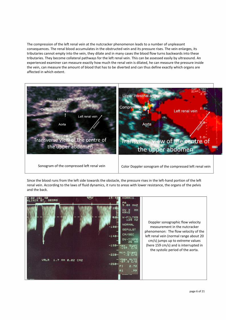
The compression of the left renal vein at the nutcracker phenomenon leads to a number of unpleasant consequences. The renal blood accumulates in the obstructed vein and its pressure rises. The vein enlarges, its tributaries cannot empty into the vein, they dilate and in many cases the blood flow turns backwards into these tributaries. They become collateral pathways for the left renal vein. This can be assessed easily by ultrasound. An experienced examiner can measure exactly how much the renal vein is dilated, he can measure the pressure inside the vein, can measure the amount of blood that has to be diverted and can thus define exactly which organs are affected in which extent.
Sonogram of the compressed left renal vein
Color Doppler sonogram of the compressed left renal vein
Since the blood runs from the left side towards the obstacle, the pressure rises in the left-hand portion of the left renal vein. According to the laws of fluid dynamics, it runs to areas with lower resistance, the organs of the pelvis and the back.
Doppler sonographic flow velocity measurement in the nutcracker
phenomenon: The flow velocity of the left renal vein (normal range about 20
cm/s) jumps up to extreme values (here 159 cm/s) and is interrupted in
the systolic period of the aorta.

page 7 of 21
The accumulated blood must now flow through other veins that have connection to the left renal vein. Normally, the left renal vein is the goal of the blood of other adjacent organs, which dock their veins to the left renal vein as a larger vessel. Under normal circumstances, the blood runs from these organs toward the renal vein. If the pressure in the left renal vein rises, it may happen that the blood is pushed back and flow changes its direction. In the last consequence, the blood that is meant to flow from the left renal vein to the inferior vena cava is diverted via so-called collateral vessels, which then have to take the congested renal vein blood.
Schematic representation of the
connections of the left renal vein.
Accumulated blood within the partially
obstructed left renal vein can use these pathways to finally
reach the inferior or superior vena cava on the right side of the spine. These large
veins finally pour the blood into the right atrium of the heart.
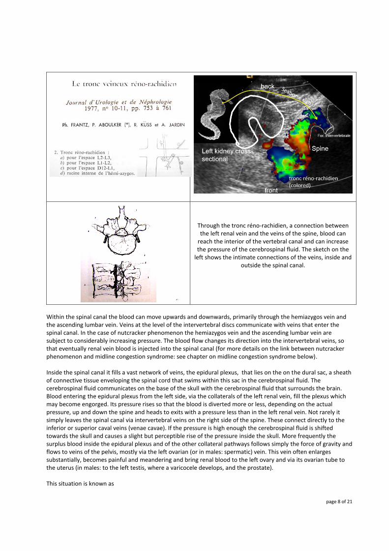
For the assessment of the situation that emerges then, it is important to know that the kidney is the most vascularized, blood filled organ of the body, second only to the brain. Even at minor flow impediments a substantial amount of blood has to be carried away from the left kidney across the above mentioned collaterals. These collateral vessels are actually the feeder to the renal vein and thus have a much smaller and weaker caliber. In addition, the organs drained by are significantly less perfused compared to the kidney. The backflow of renal blood to these organs produces a high pressure within these vessels since the congestion pressure from the left renal vein is directly transferred to these organs due to their open communication as in communicating tubes. An important but not well known collateral vessel, which exists in many people, is a short but large vein to the spinal column, called the réno-rachidien. Other bypasses are the left ovarian or spermatic vein, the ascending lumbar vein, the veins of the ureter, the hemiazygos vein and other veins of the retroperitoneum.
page 8 of 21
Through the tronc réno-rachidien, a connection between the left renal vein and the veins of the spine, blood can
reach the interior of the vertebral canal and can increase the pressure of the cerebrospinal fluid. The sketch on the
left shows the intimate connections of the veins, inside and outside the spinal canal.
Within the spinal canal the blood can move upwards and downwards, primarily through the hemiazygos vein and the ascending lumbar vein. Veins at the level of the intervertebral discs communicate with veins that enter the spinal canal. In the case of nutcracker phenomenon the hemiazygos vein and the ascending lumbar vein are subject to considerably increasing pressure. The blood flow changes its direction into the intervertebral veins, so that eventually renal vein blood is injected into the spinal canal (for more details on the link between nutcracker phenomenon and midline congestion syndrome: see chapter on midline congestion syndrome below). Inside the spinal canal it fills a vast network of veins, the epidural plexus, that lies on the on the dural sac, a sheath of connective tissue enveloping the spinal cord that swims within this sac in the cerebrospinal fluid. The cerebrospinal fluid communicates on the base of the skull with the cerebrospinal fluid that surrounds the brain. Blood entering the epidural plexus from the left side, via the collaterals of the left renal vein, fill the plexus which may become engorged. Its pressure rises so that the blood is diverted more or less, depending on the actual pressure, up and down the spine and heads to exits with a pressure less than in the left renal vein. Not rarely it simply leaves the spinal canal via intervertebral veins on the right side of the spine. These connect directly to the inferior or superior caval veins (venae cavae). If the pressure is high enough the cerebrospinal fluid is shifted towards the skull and causes a slight but perceptible rise of the pressure inside the skull. More frequently the surplus blood inside the epidural plexus and of the other collateral pathways follows simply the force of gravity and flows to veins of the pelvis, mostly via the left ovarian (or in males: spermatic) vein. This vein often enlarges substantially, becomes painful and meandering and bring renal blood to the left ovary and via its ovarian tube to the uterus (in males: to the left testis, where a varicocele develops, and the prostate). This situation is known as
page 9 of 21
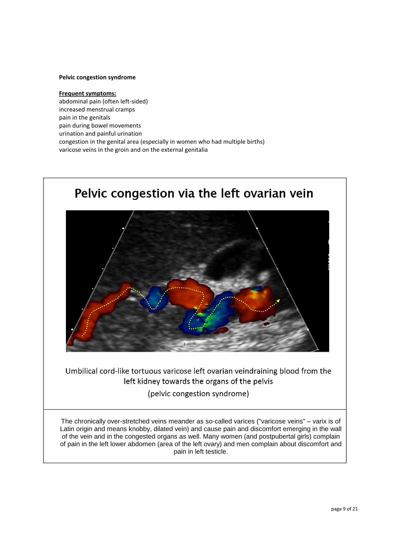
Pelvic congestion syndrome
Frequent symptoms: abdominal pain (often left-sided) increased menstrual cramps pain in the genitals pain during bowel movements urination and painful urination congestion in the genital area (especially in women who had multiple births) varicose veins in the groin and on the external genitalia
The chronically over-stretched veins meander as so-called varices ("varicose veins" – varix is of Latin origin and means knobby, dilated vein) and cause pain and discomfort emerging in the wall of the vein and in the congested organs as well. Many women (and postpubertal girls) complain of pain in the left lower abdomen (area of the left ovary) and men complain about discomfort and
pain in left testicle.
page 10 of 21
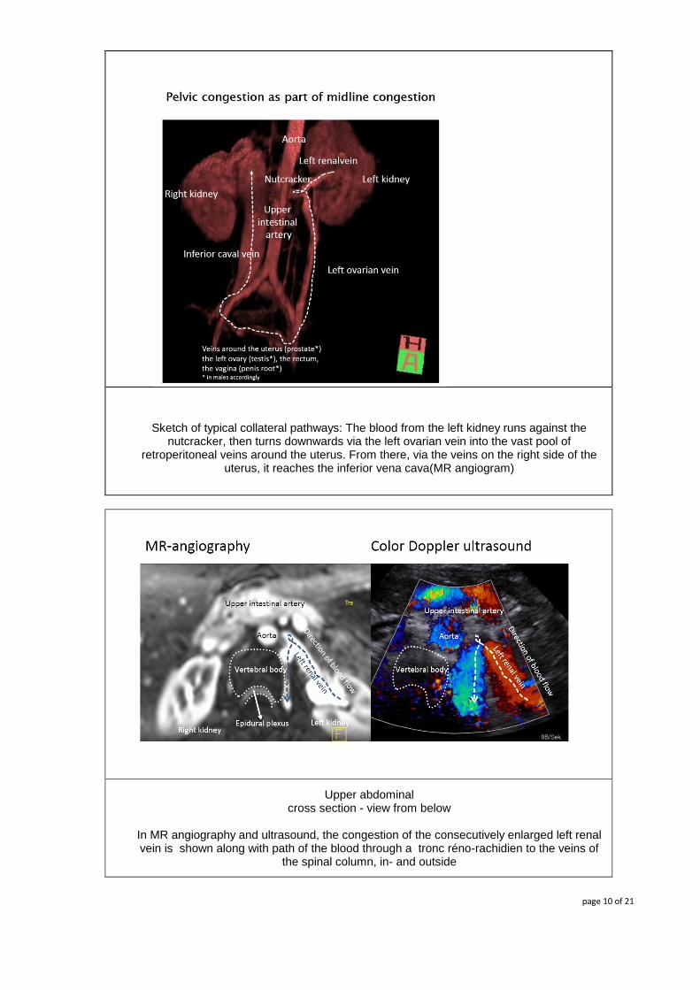
Sketch of typical collateral pathways: The blood from the left kidney runs against the
nutcracker, then turns downwards via the left ovarian vein into the vast pool of retroperitoneal veins around the uterus. From there, via the veins on the right side of the
uterus, it reaches the inferior vena cava(MR angiogram)
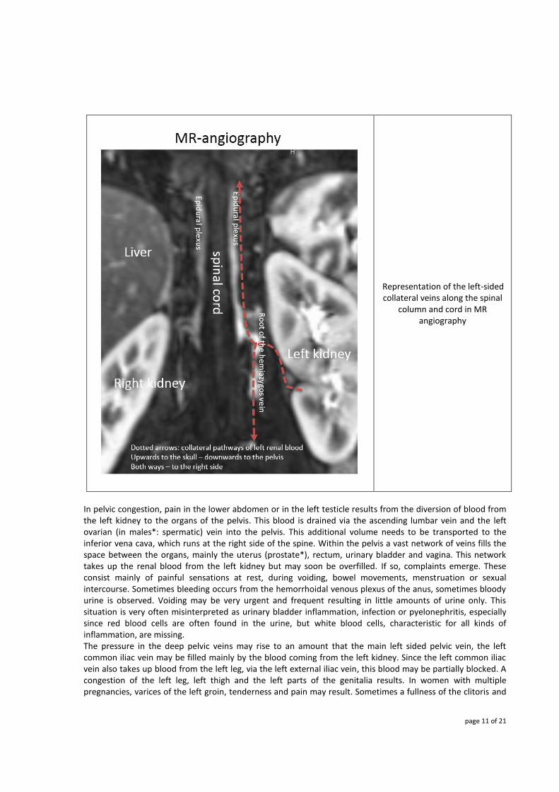
Upper abdominal cross section - view from below
In MR angiography and ultrasound, the congestion of the consecutively enlarged left renal vein is shown along with path of the blood through a tronc réno-rachidien to the veins of
the spinal column, in- and outside
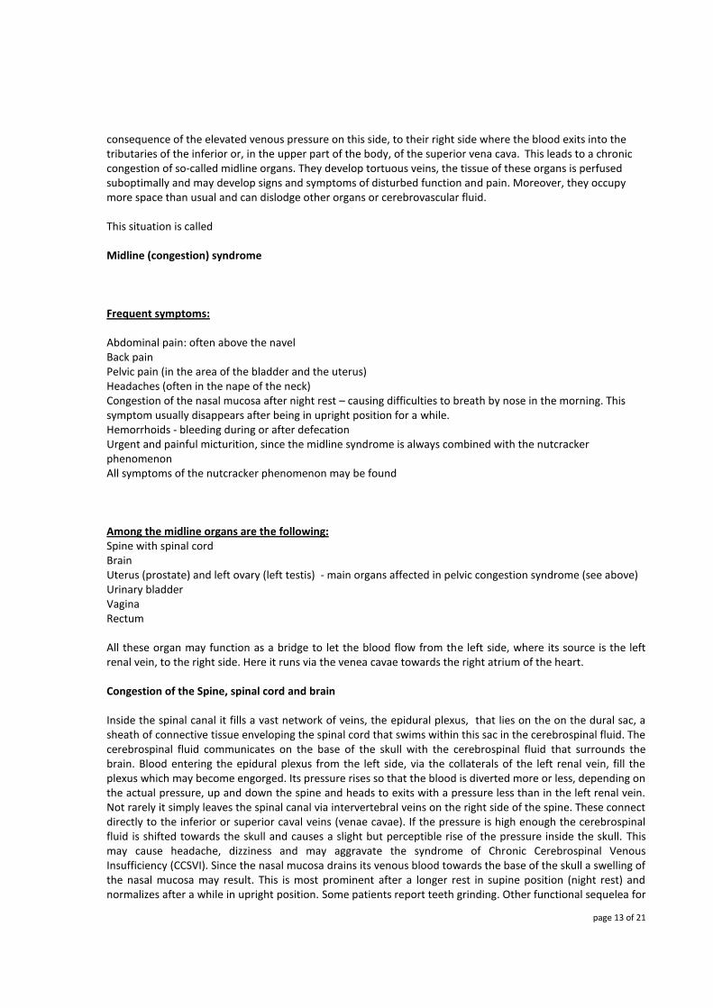
page 11 of 21
Representation of the left-sided collateral veins along the spinal
column and cord in MR angiography
In pelvic congestion, pain in the lower abdomen or in the left testicle results from the diversion of blood from the left kidney to the organs of the pelvis. This blood is drained via the ascending lumbar vein and the left ovarian (in males*: spermatic) vein into the pelvis. This additional volume needs to be transported to the inferior vena cava, which runs at the right side of the spine. Within the pelvis a vast network of veins fills the space between the organs, mainly the uterus (prostate*), rectum, urinary bladder and vagina. This network takes up the renal blood from the left kidney but may soon be overfilled. If so, complaints emerge. These consist mainly of painful sensations at rest, during voiding, bowel movements, menstruation or sexual intercourse. Sometimes bleeding occurs from the hemorrhoidal venous plexus of the anus, sometimes bloody urine is observed. Voiding may be very urgent and frequent resulting in little amounts of urine only. This situation is very often misinterpreted as urinary bladder inflammation, infection or pyelonephritis, especially since red blood cells are often found in the urine, but white blood cells, characteristic for all kinds of inflammation, are missing. The pressure in the deep pelvic veins may rise to an amount that the main left sided pelvic vein, the left common iliac vein may be filled mainly by the blood coming from the left kidney. Since the left common iliac vein also takes up blood from the left leg, via the left external iliac vein, this blood may be partially blocked. A congestion of the left leg, left thigh and the left parts of the genitalia results. In women with multiple pregnancies, varices of the left groin, tenderness and pain may result. Sometimes a fullness of the clitoris and

page 12 of 21
the labia majora and minora with unsolicited sexual arousal occurs and may be bothering. Thrombosis of the deep veins of the left leg, mainly of the calf, and varices of the left leg may develop. To get access to the inferior vena cava, left renal blood is thus pressed partly across deep veins of the pelvic plexus, partly across the left common iliac vein. But this large vessel may also be blocked in persons who have a strong lumbar lordosis. In this case the promontory is especially prominent and sticks out frontally into the pelvis from behind. This compression of the left common iliac vein by the overlying right common iliac artery against the promontory is known, after its first description by two Swiss physicians, as
May-Thurner-constellation.
Frequent symptoms: Abdominal pain (often left-sided) which may radiate into the left thigh left-sided flank pain Swelling of the left leg Tendency to thrombosis and varicose veins in the left leg
This constellation means that the blood, which was initially blocked by the nutcracker (see above), is now stopped at a second obstacle and has to find another collateral circulation to reach the right side of the body and there the inferior vena cava. Frequently, the blood flow turns backwards from the obstacle into the left internal iliac vein and thus seeks to cross the midline of the body via the already mentioned deep pelvic plexus.
Compression of the left common iliac
vein (blue) between the right common
iliac artery (red) and the promontory is
called May-Thurner-constellation
The overfilling of the deep pelvic collateral circulation causes significant discomfort for many patients since it causes a chronic venous congestion of the pelvic organs. Among them, those lying at the midline of the body, have a special function. They can take up blood from veins of their left side and can transport this, as a
page 13 of 21
consequence of the elevated venous pressure on this side, to their right side where the blood exits into the tributaries of the inferior or, in the upper part of the body, of the superior vena cava. This leads to a chronic congestion of so-called midline organs. They develop tortuous veins, the tissue of these organs is perfused suboptimally and may develop signs and symptoms of disturbed function and pain. Moreover, they occupy more space than usual and can dislodge other organs or cerebrovascular fluid.
This situation is called
Midline (congestion) syndrome
Frequent symptoms:
Abdominal pain: often above the navel Back pain Pelvic pain (in the area of the bladder and the uterus) Headaches (often in the nape of the neck) Congestion of the nasal mucosa after night rest – causing difficulties to breath by nose in the morning. This symptom usually disappears after being in upright position for a while. Hemorrhoids - bleeding during or after defecation Urgent and painful micturition, since the midline syndrome is always combined with the nutcracker phenomenon All symptoms of the nutcracker phenomenon may be found
Among the midline organs are the following: Spine with spinal cord Brain Uterus (prostate) and left ovary (left testis) - main organs affected in pelvic congestion syndrome (see above) Urinary bladder Vagina Rectum
All these organ may function as a bridge to let the blood flow from the left side, where its source is the left renal vein, to the right side. Here it runs via the venea cavae towards the right atrium of the heart.
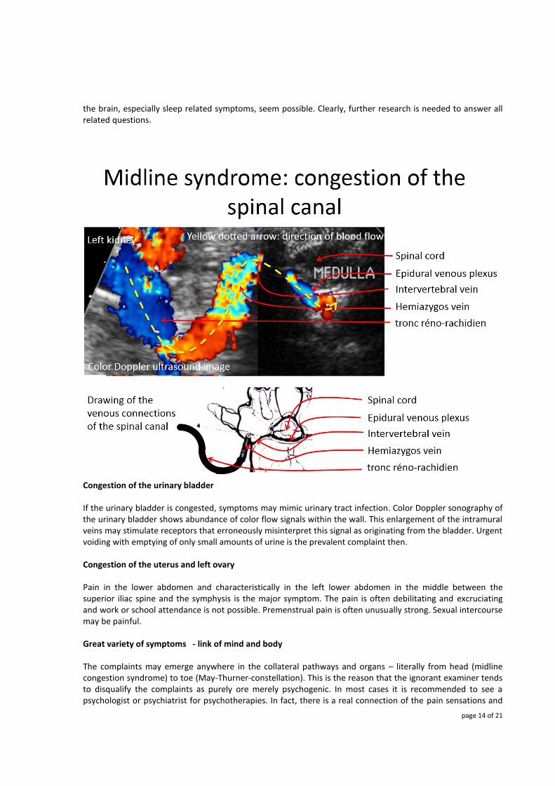
Congestion of the Spine, spinal cord and brain
Inside the spinal canal it fills a vast network of veins, the epidural plexus, that lies on the on the dural sac, a sheath of connective tissue enveloping the spinal cord that swims within this sac in the cerebrospinal fluid. The cerebrospinal fluid communicates on the base of the skull with the cerebrospinal fluid that surrounds the brain. Blood entering the epidural plexus from the left side, via the collaterals of the left renal vein, fill the plexus which may become engorged. Its pressure rises so that the blood is diverted more or less, depending on the actual pressure, up and down the spine and heads to exits with a pressure less than in the left renal vein. Not rarely it simply leaves the spinal canal via intervertebral veins on the right side of the spine. These connect directly to the inferior or superior caval veins (venae cavae). If the pressure is high enough the cerebrospinal fluid is shifted towards the skull and causes a slight but perceptible rise of the pressure inside the skull. This may cause headache, dizziness and may aggravate the syndrome of Chronic Cerebrospinal Venous Insufficiency (CCSVI). Since the nasal mucosa drains its venous blood towards the base of the skull a swelling of the nasal mucosa may result. This is most prominent after a longer rest in supine position (night rest) and normalizes after a while in upright position. Some patients report teeth grinding. Other functional sequelea for
page 14 of 21
the brain, especially sleep related symptoms, seem possible. Clearly, further research is needed to answer all related questions.
Congestion of the urinary bladder
If the urinary bladder is congested, symptoms may mimic urinary tract infection. Color Doppler sonography of the urinary bladder shows abundance of color flow signals within the wall. This enlargement of the intramural veins may stimulate receptors that erroneously misinterpret this signal as originating from the bladder. Urgent voiding with emptying of only small amounts of urine is the prevalent complaint then.
Congestion of the uterus and left ovary
Pain in the lower abdomen and characteristically in the left lower abdomen in the middle between the superior iliac spine and the symphysis is the major symptom. The pain is often debilitating and excruciating and work or school attendance is not possible. Premenstrual pain is often unusually strong. Sexual intercourse may be painful.
Great variety of symptoms - link of mind and body
The complaints may emerge anywhere in the collateral pathways and organs – literally from head (midline congestion syndrome) to toe (May-Thurner-constellation). This is the reason that the ignorant examiner tends to disqualify the complaints as purely ore merely psychogenic. In most cases it is recommended to see a psychologist or psychiatrist for psychotherapies. In fact, there is a real connection of the pain sensations and
page 15 of 21
psychological or mental sources: in psychological excitement as well as in physical exercise the heart rate is increased. This is due to the general activation of the sympathetic part of the autonomous nervous system. This sympathetic activation is the evolutionary response pattern for situations where the body needs to mobilize reserves and is prone to flee or fight. That’s why it is no wonder, that the venous congestion is aggravated in phases of stress – psychological or physical (if one still wants to separate these parts of existence). The heart pumps more blood which is trapped before the compression site of the respective vein and the pressure rises, aggravating all above mentioned complaints. The correct conclusion is therefore not to send the patient simply away to see a specialist for mental affairs but to do a thorough, quantitative, dynamic color Doppler sonographic examination with measurements of the venous pressure and flow volumes in the different parts of the collateral circulation by an experienced investigator.
From my experience with many hundreds of patients with abdominal vascular compression syndromes I can recommend to differentiate the many possible causes to choose the correct therapy – mostly by medical treatment and in rare cases by a minimally invasive operation.
Since these entities are not well known, even among many doctors, it is always useful to seek a second opinion.
Another frequent vascular compression syndrome is he
Celiac ganglion compression syndrome (Celiac trunk compression syndrome, (Celiac artery compression syndrome, Dunbar-Syndrome, Median arcuate ligament syndrome (MALS)).
Frequent symptoms:
Abdominal pain: often right below the sternum, sometimes radiating like a belt or into the chest Blocked inspiration – pronounced in physical activities Loss of appetite Early satiation Weight loss Fainting or near-fainting Bouts of sweating Tachycardia Short lasting episodes of diarrhea Symptoms are not always but may be linked to food intake
This disease comes from the irritation of the celiac ganglion (often referred to as the solar plexus), a web of nerves in the upper abdomen that is located immediately below the diaphragm. The dorsal edge of the diaphragm, arching over the aorta, is the arcuate ligament. If lordosis is prominent or the distance of this ligament to the solar plexus, overriding the celiac trunk, is too short, the movement of the diaphragm during in- and expiration may irritate this sympathetic nervous tissue leading to a bunch of vegetative symptoms and pain.
This constellation develops with the growth in puberty, most pronounced in - but not limited to - tall and slender girls. The female pelvis grows deeper and wider in puberty than in males. This has two important effect onto the female spine – both of them result in a pronounced lordosis. The widening of the pelvis exerts tension on the spine by both psoas muscles. Since the spine is nearly incompressible, the only possible consequence of this pull is a pronounced lordotic curvature of the lumbar spine. The deepening of the female pelvis pushes the anchoring joints for the spine, the sacroiliac joints, backwards, thus the distance to the hip joints enlarges. To maintain an upright position of the body while standing, women are forced to bend their spine to shift the thorax anteriorly to keep the balance along the vertical. This

page 16 of 21
is only possible with an increase of lumbar lordosis, which is in part responsible for the typical female body silhouette.
This pronounced lumbar lordosis tightens the anchoring muscle bundles of the diaphragm, the crura diaphragmatica, like the tendon of a bow, so that their connection, the arcuate ligament, presses onto the celiac ganglion (the solar plexus). Those affected, feel numerous vegetative symptoms and pain, which are caused by the irritation of the autonomic nervous ganglion.
Based on: http://bodybuilding.k21vek.com/anatomy/abdominal/2-6.htm
The blood vessels in front of the spine are also compressed. The compression can be shown precisely in color Doppler ultrasound and affects preeminently two vessels: 1. the left renal vein and 2. the celiac trunk. Concerning the left renal vein compression see the paragraph on nutcracker phenomenon. The compression of the celiac trunk is an indirect sign of the compression of the overriding celiac ganglion, which cannot be shown directly with standard imaging procedures.
The ganglion is highlighted with asterisks. View from the right side from below. From: Xiao Ming Zhang, Qiong Hui Zhao, Nan Lin Zeng, Chang Ping Cai, Xing Guo Xie, Cheng Jun Li, Jun Liu, Ji Yong Zhou: The Celiac Ganglia: Anatomic Study Using MRI in Cadavers AJR 2006; 186:1520–1523

page 17 of 21
To understand the complaints, it is helpful to know that the ganglion exerts certain so called vegetative functions that cannot be steered intentionally. These are functions primarily related to food intake, such as peristalsis, the secretion of digestive juices and the distribution of the blood after a meal. In the fasting state, the resting bowel needs much less blood than after ingestion. The injection blood after a meal is controlled by the celiac ganglion. In addition, the stimulation of the nerve fibers causes pain in the epigastric angle (the region between the rib cage and the sternum). This pain often radiates toward the heart, or belt-like to both sides of the upper abdomen. Because irritation of the ganglion is a mechanical one, exerted by the moving diaphragm, it is understandable that activities that need deeper breathing, will increase the discomfort. So, many patients complain of an increase of their complaints during physical exertion but also in other conditions, in which they must breathe heavier, for example, psychological stress. Typically, pain and circulatory problems as well as blocked inspiration are pronounced in sports and psychological stress. These breathing problems are sometimes confused with bronchial asthma. But then the expiration is impeded. This can be distinguished easily if asked for and helps to differentiate both conditions. The vegetative symptoms can be very diverse, and include nausea, loss of appetite, and bloating, impaired digestion after food intake, as well as short recurrent bouts of diarrhea, dizziness, sweating and volatile skin redness. The diagnosis can be made by means of a thorough color Doppler ultrasound exam. The sonographic diagnosis is based on the detection variable narrowing and displacement of the celiac trunk during respiration.
Original ultrasound image of the spatial relationship of relevant structures in celiac ganglion compression syndrome: here in a midposition between inspiration and expiration.
Ultrasound image of the spatial relationship of colorfully highlighted relevant structures in celiac ganglion compression syndrome: here in a midposition between inspiration and expiration.
It can be seen quite clearly that in a midposition between inspiration and expiration the arcuate ligament, the lower border of the diaphragm, compresses the celiac truck that is pushed downwards (to the right image border) and deformed hook-like. Its origin is severely compressed.
page 18 of 21
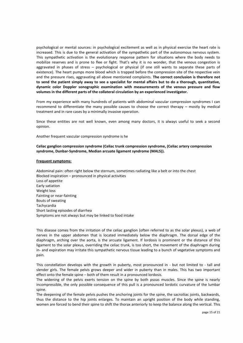
Original ultrasound image of the spatial relationship of relevant structures in celiac ganglion compression
syndrome: here in inspiration.
Ultrasound image of the spatial relationship of
colorfully highlighted relevant structures in celiac ganglion compression syndrome: here in
inspiration.
It can be seen quite clearly that in inspiration the arcuate ligament, the lower border of the diaphragm, retracts and gives the celiac truck free. This vessel expands at its origin and stands erect and runs in a straight fashion off
the aorta. The celiac trunk runs now parallel to the upper intestinal artery.
It must be remembered that the celiac ganglion itself cannot be detected reliably by means of sonography. Therefore, the imaging diagnosis relies on the evidence of a displacement and external compression of the trunk. In rare cases, despite the lack of evidence for the narrowing of the trunk, a celiac ganglion compression syndrome might be present. Then, the standard diagnostic procedure might fail. In such cases, the experience of the examiner is of uppermost relevance in order to associate the present symptoms with the respective findings. Then, even more than in other straight forward cases, an extensive differential diagnosis of other causes of abdominal pain and vegetative symptoms is crucial. Most important is the assessment of the vessel in different respiratory phases, its changing angle with the aorta and the meticulous measurement of the angle-corrected blood flow velocities. Here, apart from the absolute blood measurement of flow velocities and their acceleration it is fundamental to correlate all data to those of the aorta.
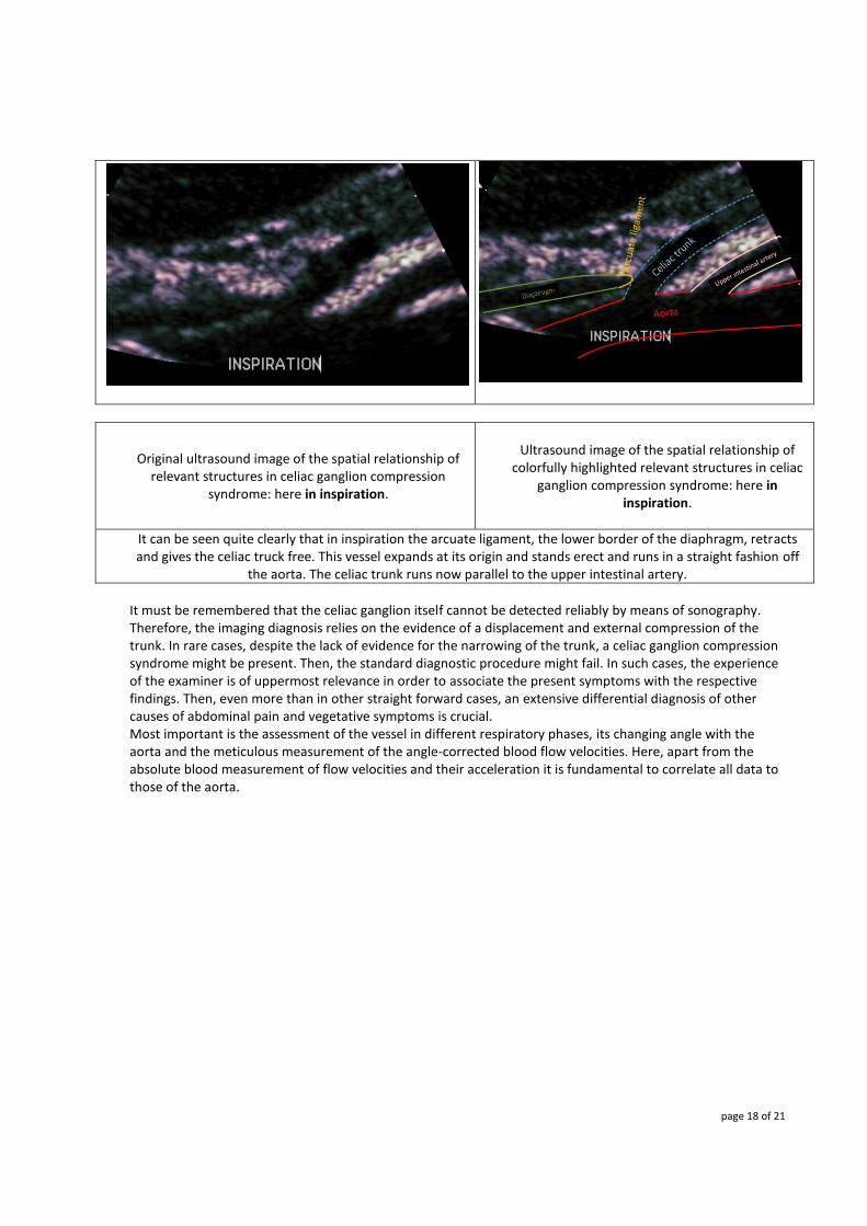
page 19 of 21
Comparison of the changes in flow velocity and location of the celiac trunk in different respiratory phases. It
can be seen clearly that flow velocity changes significantly with breathing: decrease in inspiration, increase in expiration. The flow velocity in the aorta is much lower.
If the diagnosis is made relevant differential diagnoses are ruled out and the symptoms may be associated with the disease, then the treatment can be planned. It consists of a laparoscopic surgery that leaves behind only very small scars. In almost all cases an immediate and usually permanent relief of symptoms will follow. Occasionally, however, scars and sprouting of nervous buds from the operated celiac ganglion may lead to irritation again. Then, rarely a second operation is required to remove these tissue outgrowths. Finally, another vascular compression syndrome should be mentioned that is known as
Mesenteric artery compression syndrome (Wilkie syndrome)
Frequent symptoms:
Abdominal pain: in the middle between the sternum and the navel Severe ache shortly (5-10 min) after the beginning of the meal Fear to eat because of strict time-relation to pain Severe weight loss
page 20 of 21
Sensation that the upper abdomen bloats after eating Sensation that the food is stuck Alleviation of symptoms in left side laying position with lifted pelvis Often concomitant diseases of the spine like malformations, scoliosis, which is not rarely came to existence due to cerebral palsy of infancy or muscle diseases or immobilization
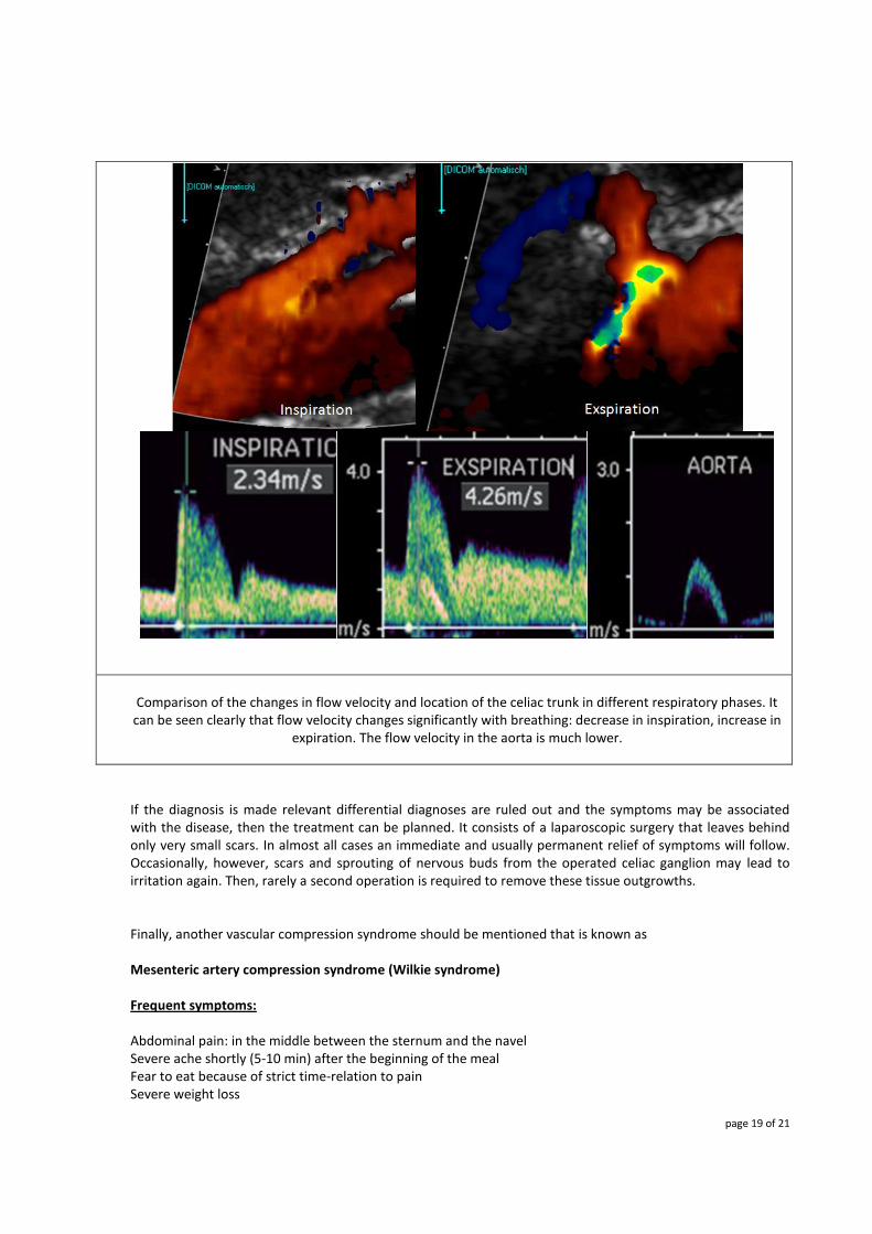
Again, this disease develops on the basis of an increased lordosis of the lumbar spine. In these patients the duodenum, which crosses the spine in the same vascular angle like the left renal vein (the “nutcracker” – see paragraph on nutcracker phenomenon above) is strangulated in the fork of the superior mesenteric artery and the aorta.
Schematic diagram of the compression of the duodenum by the upper intestinal artery
(superior mesenteric artery)
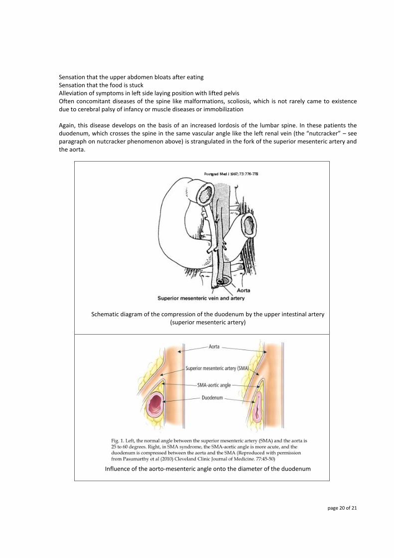
Influence of the aorto-mesenteric angle onto the diameter of the duodenum

page 21 of 21
Since both the left renal vein, coming from the left, and the duodenum, coming from the right, must pass the aorto-mesenteric angle, also called the “nutcracker”, both the nutcracker phenomenon and the superior mesenteric artery syndrome can occur simultaneously. Due to the compression of the duodenum, the patients experience a failure of food transport a short time after starting the food intake. The chyme backs up at the compression site, a few centimeters after leaving the stomach. Frequently, patients report sensations of loud peristalsis, bubbling, and sudden change of symptoms “as if something slips through”. Nausea and vomiting often terrify the affected ones and they try to prevent these ordeals by reducing food intake, which in turn, over time, intensifies the suffering since in meagre persons the aorto-mesenteric angle tends to become even narrower. The diagnosis needs a high grade of suspicion and a precise anamnesis but can then be made sonographically. A functional examination is required to demonstrate the duodenal obstruction and the accumulation of chyme in the first centimeters of the duodenum. Other diagnostic modalities are endoscopy and computed tomography or MR tomography of the upper abdomen after swallowing of a contrast medium. Again, a causal therapy by various operations is possible. This brief description of the different vascular compression syndromes for lay people should make clear, how many and apparently unrelated symptoms might be caused by the imperfect human maladaptation to the bipedal gait.
Fortunately, a straight forward diagnosis is possible in all cases. It relies basically on a subtle sonographic and color Doppler sonographic examination in the hands of an experienced examiner. Here, measurements of flow velocities and blood flow volumes should be included, since the purely morphological description of the phenomenon, as in static imaging techniques (MRI, CT, x-ray) is rarely sufficient and often misguiding. If there are more questions or a need for a second opinion or if you want to make an appointment please make contact by e-mail via: [email protected]
Please provide a telephone number to call you back.
Prof. Dr. med. habil. Thomas Scholbach Leipzig, Germany
updated:2014/07/31


