Protocol
Development and validation of a simple, sensitive, second antibody
format enzyme immunoassay for LH determination in plasma
B.S. Prakash*, Vijay Paul, N. Anandlaxmi
Division of Dairy Cattle Physiology, National Dairy Research Institute, Karnal, Haryana 132001, India
Received 25 July 2002; accepted 25 July 2002
Abstract
The objective of this study was to develop and validate a direct simple and highly sensitive enzyme immunoassay (EIA) for
luteinizing hormone (LH) determination in buffalo plasma on microtiter plates using the biotin–streptavidin amplification
system and the second antibody coating. Biotin was coupled to LH and used to bridge between streptavidin-peroxidase and
immobilized antiserum in competitive assay. The EIA was carried out directly in 20 Al buffalo plasma. The LH standards
ranging from 6.25 to 200 pg/well/20 Al were prepared in hormone free plasma collected from a buffalo on day 4 post-calving.
The sensitivity of EIA procedure was 6.25 pg/well LH, which corresponds to 0.31 ng/ml plasma; the 50% relative binding
sensitivity was seen at 50 pg/well/20 Al. Plasma volumes for the EIA, viz. 10 and 20 Al, did not influence the shape of standard
curve even though a slight drop in the OD450 was seen with higher plasma volumes. A parallelism was carried out to compare
the endogenous buffalo plasma LH with bovine LH standards. For the biological validation of assay, 10 Murrah buffaloes were
used. These were administered (10 Ag im) with a synthetic analogue of gonadotropin-releasing hormone (GnRH) and blood
samples were collected at 15-min interval using indwelling jugular catheter beginning just prior to GnRH injection till 6 h and
thereafter 2-h interval for another 18 h. In all animals, sharp increases in LH concentrations were recorded post-GnRH
administration, which confirms the biological validation of the EIA. To record the LH peak during periestrus in a cycling
buffalo, the blood samples were collected at 2-h intervals from onset of behavioral estrus signs till ovulation. The LH peak was
observed after the initial behavioural estrus signs followed by the gradual decline in the levels towards the ovulation.
D 2002 Elsevier Science B.V. All rights reserved.
Keywords: Buffalo endocrinology; LH; EIA; Plasma
1. Background
The hypophysial hormone LH plays an important
role in ovulation and luteinization in females. The
control of ovulation is brought about by the interactions
between the pituitary gonadotropins, FSH and luteiniz-
ing hormone (LH) and intraovarian factors such as
steroids, cytokines and other growth factors (Findley et
al., 1996).Measurement of LH in peripheral circulation
of buffaloes is important for understanding the phe-
0022-1759/02/$ - see front matter D 2002 Elsevier Science B.V. All rights reserved.
PII: S0022 -1759 (02 )00301 -0
Abbreviations: LH, luteinizing hormone; EIA, enzyme immuno-
assay; PBS, phosphate-buffered saline; BSA, bovine serum
albumin; GnRH, gonadotropin-releasing hormone.
* Corresponding author. Fax: +91-184-250042.
E-mail address: [email protected] (B.S. Prakash).
www.elsevier.com/locate/jim
Journal of Immunological Methods 270 (2002) 281–290
nomena limiting its fertility. LH measurements in
buffalo plasma are currently being carried out by
sensitive radioimmunoassay (RIA) procedures which
were established several years ago using 125I as the
label (Heranjal et al., 1976; Kaker et al., 1980; Galhotra
et al., 1981; Arora and Pandey, 1982; Rao and Pandey,
1983; Kanai and Shimizu, 1984; Avenell et al., 1985;
Singh, 1998). Although these methods are reliable and
accurate, they suffer from the problems associated with
the use of radioisotopes, which restricts their use to
specialized laboratories. The RIA procedure also suf-
fers from the disadvantage of using 125I as the label,
which has a short half-life. While enzyme immuno-
assay (EIA) procedures have been developed for
bovine LH (Mutayoba et al., 1990), GH (Hennies and
Holtz, 1993) and FSH (Prakash et al., 1999), no EIA
has so been established for buffalo LH. Hence, we
decided to develop a sensitive and convenient second
antibody EIA for LH determination in buffalo plasma
using the biotin–streptavidin-peroxidase amplification
system.
2. Type of research
1. Standardization and determination of LH in buffalo
plasma by EIA.
2. Biological validation of enzyme immunoassay by
measuring LH level after administration of gona-
dotropin-releasing hormone (GnRH) analogue.
3. Measurement of plasma LH during estrus in buffalo.
3. Time required
(1) Preparation of biotinyl–LH conjugate: 2 days.
(2) First coating of microtiter plates with goat
antirabbit IgG overnight.
(3) Second coating with 1% BSA in PBS 40 to 50 min.
(4) Immune reaction between antigen and antibody
overnight.
(5) Addition of biotinyl–LH conjugate and streptavi-
din-peroxidase 30 min for each step.
(6) Substrate reaction: 40 min.
(7) Addition of 4 N H2SO4 and reading of optical
density in Microtiter plate reader: 5 min.
(8) Microtiter plate washing (Four times each for 15
min).
4. Materials
4.1. Preparation of biotinyl–LH conjugate
Special equipment:
� Dialysis sack (250-7U, Sigma, USA).� Dialysis assembly (2 l beaker filled with PBS and
having a magnetic stirrer).
Chemicals and reagents:
� Bovine LH (USDA-bLH-B-6, Beltsville, USA).� PBS pH 7.4 (50 mM NaPO4, 0.15 M NaCl, pH
adjusted with 5 N HCl).� Biotinamidocaproate-N-hydroxysuccinimideester
(Biotin; Sigma, Germany).� 1 M NH4Cl solution in distilled water.� 1% bovine serum albumin solution in PBS (BSA;
Sigma, Germany).� Glycerol (Hi Media, India).
4.2. Preparation of affinity purified goat IgG anti-
rabbit IgG
� See Anandlaxmi and Prakash (2001).
4.3. EIA procedure: first coating with goat IgG anti-
rabbit IgG and second coating with 1% BSA
Special equipments:
� Microtiterplate shaker (Titertek, Flow Laboratories,
Germany).� Microtiter plates (Greiner, Labortechnik, Ger-
many).� Digital multichannel pipette (Flow TitertekR, Fin-
land).
Chemical and reagents:
� Goat IgG antirabbit IgG (Anandlaxmi and Prakash,
2001).� Coating buffer pH 9.6 (15 mM Na2 CO3, 35 mM
NaHCO3).� 1% bovine serum albumin in PBS (BSA, Sigma,
Germany).
B.S. Prakash et al. / Journal of Immunological Methods 270 (2002) 281–290282
4.4. Washing of coated microtiter plates
Special equipments:
� Automated microtiter plate washer (Model: EL50x8MS, USA).
Chemical and reagents:
� Washing solution (0.05% polyoxyethylenesorbitan
monolaurate, Tween 20 in distilled water, Sigma,
Germany).
4.5. Assay protocol
Special equipments:
� Dilutor dispenser (Hamilton, MicrolabR 500
series, Switzerland).� Digital multichannel pipette (Flow TitertekR).� Automated microtiter plate washer (Model: EL50x
8MS).
Chemical and reagents:
� Bovine LH standards (USDA-bLH-B-6).� Hormone free buffalo plasma having LH concen-
trations lower than the measurable limit collected
on day 4 of the parturition.� Rabbit polyclonal anti bovineLHantiserum(USDA-
309-684P, Beltsville, USA); very specific for LH
(USDA-bLH-B-6), as provided by the USDA the
cross-reactivity of the bLH antisera (USDA-309-
684P) with USDA-bFSH-B-1, USDA-bTSH-I-1,
USDA-bGH-B-1, and USDA-bPRL-B-1 was less
than 0.7%.� Assay buffer pH 7.4 (50 mM NaPO4, 0.15 M
NaCl, 0.02% thimerosal; pH7.4 adjusted with 5 N
HCl).� Biotinyl–LH conjugate.� Streptavidin-peroxidase (Sigma, Germany).
4.6. Substrate reaction
Special equipments:
� Microtiter plate reader (Model: ECIL, Microscan,
India).
� Graph pad PRISMR 2.01, software package.
Chemical and reagents:
� Substrate buffer pH 4.0 (0.05 M citric acid, 0.11 M
Na2HPO4, 0.05% ureum peroxide; pH4.0 adjusted
with 5 N HCl).� Substrate solution: 17 ml substrate buffer plus 340
Al 3,3V,5,5V-tetramethyl benzidene (Sigma, Ger-
many); 12.5 mg/ml dimethyl sulfoxide (Sigma,
Germany).� 4 N H2SO4 solution.
4.7. Biological validation of the buffalo plasma LH
enzyme immunoassay
Special equipment:
� Refrigerated centrifuge (IEC, India).
Chemical and reagents:
� Buserelin-Acetate (ReceptalR, Intervet, India).� Local anesthesia (XylocaineR 2%, Astra Zeneca,
India) and antibiotic (OxytetracyclineR, SarabhaiZydus, India) given during catheterization.
� Indwelling jugular catheter (60 cm surgical tubing
No. 51, Romsons, India; Three way valve with
stopper and sterilized 14-gauge stainless steel
needle).� 10 and 5 ml disposable syringes (Dispo vanR,
India).� 18-gauge stainless steel needle.� Heparin sodium salt (SRL, India) solutions con-
taining 200 IU/100 Al and 200 IU/ml in normal
saline.� 15 ml polypropylene tubes (Chemtron, India).� 2 ml Storage vials (Tarson, India).
5. Detailed procedure
5.1. Preparation of biotinyl–LH conjugate
(i) Add 40 Ag bovine LH (USDA-bLH-B-6)
dissolved in 200 Al of phosphate buffered saline
solution (PBS: pH7.4), 12 Al biotinamidocap-
B.S. Prakash et al. / Journal of Immunological Methods 270 (2002) 281–290 283
roate-N-hydroxysuccinimideester dissolved in
dimethyl sulfoxide (1 mg/ml) and immediately
vortex the mixture and incubate further for 3 h at
room temperature under constant agitation.
(ii) Stop the coupling reaction by the addition of 20
Al NH4Cl (1 M) and incubate the reaction
mixture further for 30 min before addition of 2
ml of a solution of 1% BSA in PBS pH7.4.
(iii) Biotin–LH conjugate is isolated by dialysis of
mixture in dialysis sack overnight at 4 jC with
four changes in PBS.
(iv) After dialysis, the conjugate is mixed with an
equal volume of glycerol to prevent freezing and
preserved at � 20 jC in 1 ml aliquots.
5.2. Preparation of affinity purified goat IgG anti-
rabbit IgG
For preparation of affinity purified goat IgG anti-
rabbit IgG see Anandlaxmi and Prakash (2001). The
brief procedure is detailed below:
(i) About 40 ml plasma from a goat immunized
against rabbit IgG containing 20 IU heparin/ml of
blood is vortexed with rabbit IgG agarose and
loaded onto a small column.
(ii) First non-specific proteins are eluted with PBS
(10 mM NaPO4, 0.5 M NaCl, pH7.2) buffer.
(iii) Proteins bound specifically are eluted with 15 ml
of 0.1 M glycine–HCl (pH2.0).
(iv) All steps are performed at room temperature.
(v) The eluted fractions (3 ml each) are collected in
vials containing 0.2 ml of 1 M Tris–HCl (pH
8.0).
(vi) The eluted IgG is dialyzed overnight against PBS
and the protein content determined by measuring
the absorbance spectrophtometrically at 260 nm
and 280 nm, and extrapolated from a normo-
graph.
5.3. EIA procedure
a. First coating with goat IgG antirabbit IgG:
(i) The first coating is performed by adding 0.63 Agof goat IgG dissolved in 100 Al of coating buffer
(pH 9.6) per well of the microtiterplate. The
plates are subsequently incubated overnight at 4
jC.
b. Second coating with 1% BSA:
(ii) For blocking the remaining binding sites, add 300
Al of 1% BSA in PBS to all the wells and
incubate for 40 to 50 min at room temperature
under constant shaking.
c. Washing of coated microtiter plates:
(iii) The coated plates are washed twice with 350 Al/well of washing solution (0.05% Tween 20) using
an automated microtiterplate washer.
5.4. Assay protocol
(i) Duplicate of 20 Al of unknown buffalo plasma
sample or bovine LH standards (USDA-bLH-B-
6; prepared in hormone free plasma collected on
day 4 of parturition) ranging from 6.25 to 400 pg/
20 Al/well are simultaneously pipetted into
respective wells along with 100 Al of LH anti-
body diluted 1:160,000 in assay buffer (pH7.4)
with the aid of a dilutor dispenser.
(ii) Thereafter, the plates are incubated overnight at
room temperature after 30 min constant agitation.
(iii) The next day plates are decanted and washed
twice with washing solution before addition of
100 Al of biotinyl–LH conjugate diluted 1:400 in
assay buffer.
(iv) The plates are further incubated for 30 min with
constant agitation, decanted and washed four
times with washing solution.
(v) Then add 20 ng streptavidin-peroxidase in 100 Alof assay buffer to all the wells and wrap the plates
in aluminum foils and incubate further for 30 min
under constant agitation.
(vi) All steps are performed at room temperature.
5.5. Substrate reaction
(i) The plates are then washed five times with
washing solution and incubated further in the
dark for 40 min after addition of 150 Al of
substrate solution per well.
(ii) Stop the reaction by the addition of 50 Al 4 N
H2SO4 and measure the colour produced at 450
nm with a 12-channel microtiter plate reader.
(iii) Calculate LH concentration in buffalo plasma
samples from the graph; plotted LH concen-
tration against absorbance at 450 nm by using
Graph pad PRISMR 2.01, software package.
B.S. Prakash et al. / Journal of Immunological Methods 270 (2002) 281–290284

5.6. Biological validation of the buffalo plasma LH
enzyme immunoassay
(i) For the biological validation of the assay, 10 non-
lactating cycling Murrah buffaloes maintained at
the National Dairy Research Institute farm are
used.
(ii) These are administered (10 Ag im) with a
synthetic analogue of GnRH (Buserelin-Acetate)
and blood samples (4 ml) are collected at 15-min
interval using indwelling jugular catheter begin-
ning just prior to GnRH injection till 6 h and
thereafter 2-h interval for another 18 h.
(iii) All experimental protocols and animal care met
IACUC regulations. Before catheterization, the
local anesthesia (XylocaineR) is given and after
removal of catheter the animal is treated with
antibiotic (Oxytetracycline) for 3 days.
(iv) In another experiment designed to measure the
levels of LH during periestrus, blood samples are
collected at 2-h intervals by jugular venipuncture
from the onset of behavioural estrus till 2 h of the
ovulation.
(v) The blood samples are collected in heparinized
polypropylene tubes and immediately kept in ice-
box (4 jC) and then centrifuged at 3000 rpm for
20 min at 4 jC, plasma separated out is stored at
� 20 jC till assayed for LH.
6. Results
6.1. Standardization of enzyme immunoassay for
buffalo plasma LH determination
Titration of biotinyl–LH antiserum: A two dimen-
sional titer determination for the optimum dilution of
LH label and the antiserum was carried out. Antibody
dilutions ranging from 1:5000 to 1:640,000 and the
biotinyl–LH dilutions of 1:100 to 1:1600 were tested.
The antibody titer of 1:160,000 and the biotinyl–LH
conjugate titer of 1:400 were found to be the most
suitable and achieved an OD450 of around 1.5.
6.2. Assay validation
6.2.1. Assay interference and sensitivity
To determine the possible interference of plasma
with the assay sensitivity, bovine LH standards in va-
rious amounts of plasma (10, 20 and 40 Al) were run in
Fig. 1. Influence of different volumes of 10, 20 and 40 Al of buffalo plasma on optical density displacement in LH standard curve. Along with
different volumes of plasma, the standards were also prepared in assay buffer. Optical density was measured at 450 nm.
B.S. Prakash et al. / Journal of Immunological Methods 270 (2002) 281–290 285
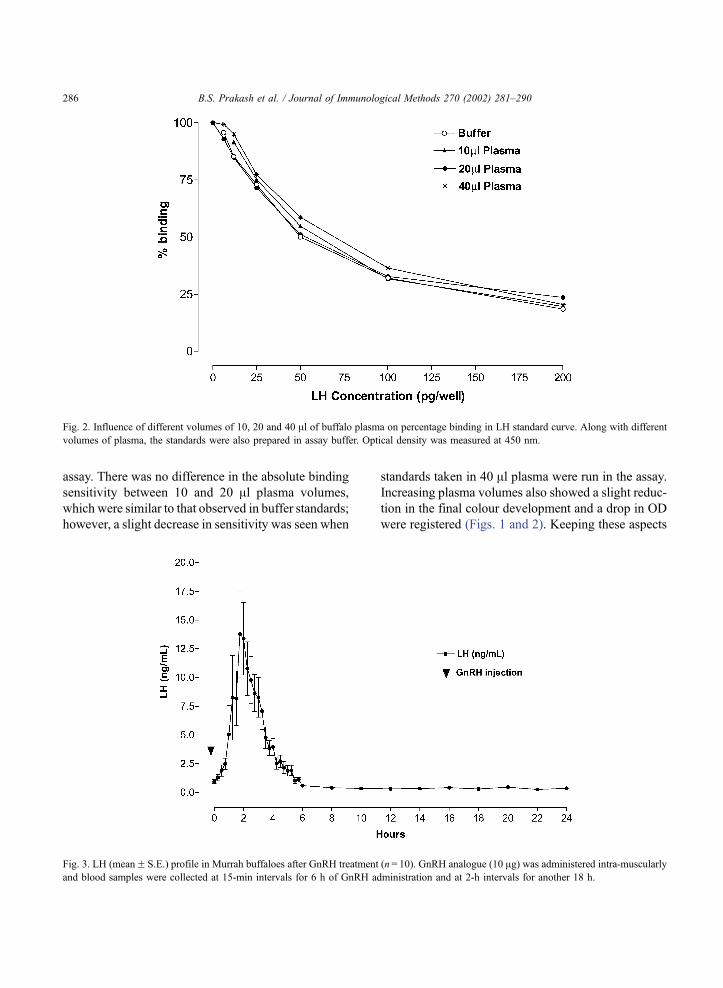
assay. There was no difference in the absolute binding
sensitivity between 10 and 20 Al plasma volumes,
which were similar to that observed in buffer standards;
however, a slight decrease in sensitivity was seen when
standards taken in 40 Al plasma were run in the assay.
Increasing plasma volumes also showed a slight reduc-
tion in the final colour development and a drop in OD
were registered (Figs. 1 and 2). Keeping these aspects
Fig. 2. Influence of different volumes of 10, 20 and 40 Al of buffalo plasma on percentage binding in LH standard curve. Along with different
volumes of plasma, the standards were also prepared in assay buffer. Optical density was measured at 450 nm.
Fig. 3. LH (meanF S.E.) profile in Murrah buffaloes after GnRH treatment (n= 10). GnRH analogue (10 Ag) was administered intra-muscularly
and blood samples were collected at 15-min intervals for 6 h of GnRH administration and at 2-h intervals for another 18 h.
B.S. Prakash et al. / Journal of Immunological Methods 270 (2002) 281–290286
in view, standards were subsequently prepared in
hormone free plasma and run along with the unknowns
in the test. The nonspecific binding using both the
volumes of plasma was low (OD450) ranging from
0.114 to 0.145. All assays were hence conducted taking
20 Al of unknown plasma samples and standards per
well in duplicates. The lowest LH detection limit
significantly from zero concentration was 6.25 pg/20
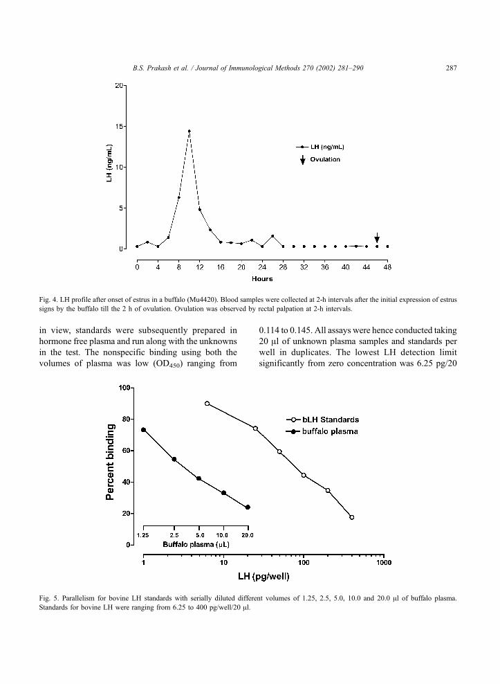
Fig. 4. LH profile after onset of estrus in a buffalo (Mu4420). Blood samples were collected at 2-h intervals after the initial expression of estrus
signs by the buffalo till the 2 h of ovulation. Ovulation was observed by rectal palpation at 2-h intervals.
Fig. 5. Parallelism for bovine LH standards with serially diluted different volumes of 1.25, 2.5, 5.0, 10.0 and 20.0 Al of buffalo plasma.
Standards for bovine LH were ranging from 6.25 to 400 pg/well/20 Al.
B.S. Prakash et al. / Journal of Immunological Methods 270 (2002) 281–290 287

Al plasma which corresponded to 0.31 ng/ml plasma.
The 50% relative binding (B/B0) sensitivity was 50 pg/
20 Al plasma/well which corresponded to 2.5 ng/ml
plasma.
6.2.2. Intra- and inter-assay precision
Intra- and inter-assay coefficients of variations
determined using pooled plasma containing 0.62 and
5.0 ng/ml in 11 assays were 7.73% and 16.0% and
3.21% and 10.89%, respectively.
6.3. Biological validation
Mean LH concentrations in blood samples col-
lected from the 10 non-lactating cycling Murrah
buffaloes after GnRH injection are presented in (Fig.
3). The LH concentration rose sharply to a peak mean
value of 13.78F 3.74 ng/ml after 2 h of the GnRH
analogue administration. Subsequently, the hormone
concentration declined to reach basal levels (0.31 ng/
ml) at about 6 h post-treatment and remained basal
thereafter. An LH peak was also recorded in a buffalo
post-estrus with the peak value of 14.9 ng/ml, which
was followed by ovulation (Fig. 4).
6.4. bLH parallelism with buffalo plasma
The homology between bovine LH standards used
and endogenous LH in buffalo plasma was assessed
by conducting parallelism. To serve the purpose,
buffalo plasma sample containing high level of endog-
enous LH was serially diluted (containing 20, 10, 5,
2.5 and 1.25 Al buffalo plasma sample size) and run
along with the bovine LH standards (in buffer) in an
assay. When plotted with the increasing plasma vol-
umes and increasing standard concentrations a parallel
drop in relative percent binding was observed (Fig. 5).
Both curves, i.e. for increasing plasma volumes and
bovine LH standards, were lying almost parallel to
each other, thereby, confirming the actual LH estima-
tion in buffalo plasma.
7. Discussion
The method described here is the first report using
the second antibody technique and the LH–biotin–
streptavidin system for buffalo plasma LH EIA. The
use of the second antibody for coating the wells instead
of the hormone specific antibody is preferred as it
reduces assay variability associated with uneven bind-
ing of the latter antibody to the wells and further
reduces the amount of hormone specific antibody
needed in the EIA (Meyer, 1986).
The high degree of parallelism in the concentrations
plotted from hormone values obtained from serial
dilutions of a blood sample from buffalo containing
high LH, and the standard curve of bovine LH (Fig. 5)
indicates considerable homology between buffalo LH
and bovine LH used in the assay.
To obtain a high degree of sensitivity in direct EIA,
less sample volume is desirable to reduce the non-
specific binding and plasma matrix effects (Mutayoba
et al., 1990). This requires the use of highly specific
antibody, a very efficient amplification system and
optimum ligand antibody dilutions at suitable incuba-
tion temperature. In our EIA there was a decrease in
optical density with increasing plasma volumes
although the sensitivities and the relative binding
percentage did not change when 10 and 20 Al plasma
were taken along with standards (Figs. 1 and 2). In
order to compensate for this effect, it is necessary to
use the same plasma volumes for standards and
unknowns. A high assay sensitivity of 6.25 pg/well
LH was obtained when 20 Al of plasma was taken for
estimation. This was sufficient to determine the low
physiological baseline LH concentrations as well as
distinctly observe the LH release in 10 cycling Murrah
buffaloes after GnRH analogue injection. The appli-
cation of short period of incubation in EIA for
samples with expected high LH levels (>10 ng/ml)
is a helpful tool for rapid confirmation of estrus where
LH surge is the dominating indicator of heat and
ovulation in cattle and buffaloes, as well as in several
biomedical reproductive studies modifying the func-
tion of hypothalamo-hypophyseal axis. The assay
described here require less expensive instrumentation
and reagents when compared to RIA, and can be
adopted in developing countries where financial con-
straints limit the adoption of RIA. Highly purified LH
preparations from cattle and other species of animals
are available, and biotinylation of LH is not difficult
as compared to iodination procedures. Biotin and
streptavidin-peroxidase of good quality are also com-
mercially available at rather cheaper costs than 125I
preparations.
B.S. Prakash et al. / Journal of Immunological Methods 270 (2002) 281–290288
8. Quick procedure
8.1. Enzyme immunoassay procedure
(i) First coating with 0.63 Ag of goat antirabbit IgG
dissolved in 100 Al of coating buffer per well of
the microtiter plate and incubate overnight at 4
jC.(ii) Second coating or blocking the remaining sites
with 300 Al of 1% BSA in PBS per well and
incubate at room temperature for 40 to 50 min
under constant shaking.
(iii) Wash the coated plates twice with 350 Al per wellof washing solution.
8.2. Assay protocol
(i) Pipette out duplicates of 20 Al buffalo plasma
samples or bovine LH standards along with 100
Al LH antibody diluted 1:160,000 in assay buffer
in respective wells of coated microtiter plates.
(ii) Incubate overnight at room temperature after 30
min constant agitation.
(iii) Decant and wash twice with washing solution
before addition of 100 Al biotinyl LH conjugate
(1:400 in assay buffer).
(iv) Incubate further for 30 min with constant shaking
and then decant and wash four times with
washing solution.
(v) Add 20 ng streptavidin-peroxidase in 100 Al assaybuffer per well and wrap the plate in aluminum
foil and incubate for 30 min with constant
shaking.
8.3. Substrate reaction
(i) Wash the plate five times and then add 150 Alsubstrate solution per well and incubate further in
dark for 40 min.
(ii) Stop the reaction with addition of 50 Al 4 N
H2SO4 per well.
(iii) Measure the absorbance of colour developed at
450 nm.
(iv) Plot the standard curve; plotting LH concentration
against absorbance obtained and calculates con-
centration of LH in buffalo plasma samples by
using Graph pad PRISMR 2.01 software.
Acknowledgements
The authors wish to thank the United States
Department of Agriculture, Animal Hormone Program,
Beltsville, USA for the generous gift of reference LH
standards and bovine LH antiserum. The funds
provided by National Agricultural Technology Project
PSR No. 47 for this study is duly acknowledged. We
are also grateful to Director, NDRI, Karnal for pro-
viding all necessary facilities during the course of this
study.
References
Anandlaxmi, N., Prakash, B.S., 2001. Production and purification
of goat antirabbit IgG. Indian J. Dairy Sci. 54, 332–334.
Arora, R.C., Pandey, R.S., 1982. Pattern of plasma progesterone,
oestradiol-17h, luteinizing hormone and androgen in non-
pregnant buffalo (Bubalus bubalis). Acta Endocrinol. 100,
279–284.
Avenell, J.A., Saepudin, Y., Fletcher, I.C., 1985. Concentration of
LH, estradiol-17h and progesterone in the peripheral plasma of
Swamp buffalo. J. Reprod. Fertil. 74, 419–424.
Findley, J.K., Drummond, A.E., Fry, R.C., 1996. Intragonadal re-
gulation of follicular development and ovulation. Anim. Reprod.
Sci. 42, 321–331.
Galhotra, M.M., Kaker, M.L., Razdan, M.N., 1981. Serum LH le-
vels during pre- and post-puberty, pregnancy and lactation in
Murrah buffaloes. Theriogenology 16, 477–481.
Hennies, M., Holtz, W., 1993. Enzyme immunoassay for the deter-
mination of bovine growth hormone using avidin–biotin-perox-
idase complexes. J. Immunol. Methods 157, 149–157.
Heranjal, D.D., Sheth, A.R., Moodbidri, S.B., Desai, R., Rao, S.S.,
1976. A note on luteinizing hormone during the estrus cycle and
early pregnancy in Indian buffaloes. Indian J. Dairy Sci. 32,
247–249.
Kaker, M.L., Razdan, M.N., Galhotra, M.M., 1980. Serum LH con-
centrations in cyclic buffaloes (Bubalus bubalis). J. Reprod.
Fertil. 60, 419–424.
Kanai, Y., Shimizu, H., 1984. Plasma concentrations of LH, pro-
gesterone and estradiol during the estrous cycle in Swamp buf-
faloes (Bubalus bubalis). J. Reprod. Fertil. 70, 507–510.
Meyer, H.H.D., 1986. Possibilities to improve enzyme immunoas-
say (EIA) techniques and their application in animal production.
Proceedings of International Symposium on the Use of Nuclear
Techniques in Studies of Animal Production and Health in Dif-
ferent Environments. IAEA, Vienna, pp. 255–262.
Mutayoba, B.H., Meyer, H.H.D., Schams, D., Schallenberger, E.,
1990. Development of a sensitive enzyme immunoassay for LH
determination in bovine plasma using the streptavidin–biotin
technique. Acta Endocrinol. (Copenh.) 122, 227–232.
Prakash, B.S., Wallenhorst, S., Metten, M., Holtz, W., Wuttke, W.,
1999. Development of a sensitive enzyme immunoassay (EIA)
B.S. Prakash et al. / Journal of Immunological Methods 270 (2002) 281–290 289

for FSH determination in bovine plasma. Anim. Reprod. Sci. 55,
183–192.
Rao, L.V., Pandey, R.S., 1983. Seasonal variations in oestradiol-17hand luteinizing hormone in the blood of buffalo cows (Bubalus
bubalis). J. Endocrinol. 98, 251–255.
Singh, J.K., 1998. Seasonal effects on physiological and endocrine
changes during estrous cycle in buffaloes. PhD Thesis submit-
ted to National Dairy Research Institute, Karnal, Haryana, In-
dia.
B.S. Prakash et al. / Journal of Immunological Methods 270 (2002) 281–290290


