
Design and Conduct of Preclinical and Clinical Trials in Opthalmology
Faraza JavedPhD Pharmacology

What is ophthalmology? Opthalmos=eye Logos=word, thought, discourseThe science of eyes is opthalmology

The branch of medicine concerned with the eyes Anatomy Function Disease

Procedures involved in clinical trials
Eye diseases are among the most devastating disorders leading to retinal ganglion cell (RGC) degeneration, visual field loss and, potentially, blindness. The pathophysiologic mechanisms underlying optic neuropathies are not fully understood, although research in this area is substantial.

Animal models are required in order to investigate the mechanism of each disease in detail, to improve our knowledge of how and why optic nerve axons die, and to test new treatment modalities. Developing an animal model for a disease, however, is usually complicated and challenging, and most experimental models are remote from the human disease.

There are several animal species that can be developed as suitable models for human disease. The advantage of using a monkey is its close phylogeny and high homology with humans.
Monkeys have retinal and optic nerve anatomy that is nearly identical to human eyes.

The validity of the experimental model, however, depends upon the degree to which it emulates the human condition, and a primate model is considered as having the closest compatibility for conducting research with the goal of understanding human diseases.
Indeed, testing new treatment modalities in a monkey model is usually the last step before embarking upon clinical trials in humans. Unfortunately, monkeys are very expensive, their availability is limited, and they are difficult to handle. Experiments in monkeys require highly experienced teams and special housing facilities, making them beyond the reach of many research laboratories.

Thus, rats and mice are commonly used to investigate optic nerve diseases. There is great conservation between rats, mice, and human genomes, which allows them to be widely used for research on human optic nerve disease.

While rats are commonly used to study optic nerve injuries, the availability of a mouse model confers unique advantages. Genetic studies in mice are proving to be a potent means for learning about genes and pathologic mechanisms that cause disease. In addition, the genome of the mouse can be altered by adding transgenes or altering endogenous genes.

Laboratory mice live for approximately 2 years and often develop diseases that take decades to develop in humans. Models of optic nerve disease in the mouse can enable us to study the mechanism of RGC degeneration and potential new therapies using genetic manipulation in various transgenic and knockout mice that are not available in rats.

Experimental glaucoma in monkeys
While the most useful model for experimental glaucoma in monkeys is produced by argon laser photocoagulation treatment to the trabecular meshwork. The model was first suggested by Gaasterland and Kupfer and later refined by Quigley and Hohman. In order to induce elevated IOP, monkeys are sedated and the laser beam is focused as precisely as possible on the centre of the mid-trabecular meshwork.
The recommended laser setting is a 50 m spot diameter, 1–1.5 W, and a 0.5-s duration. In this model, the treated eyes develop sustained, moderately elevated IOP with decreased outflow facility and optic nerve cupping such as that in human glaucoma.
IOP can be measured under sedation and topical anaesthesia using the Goldman applanation tonometer. Optic nerve damage can be evaluated by stereoscopic ophthalmoscopy, fundus photographs, nerve fibre layer photographs, axonal counting by image analysis system, and RGC loss.

A second model for producing chronic experimental glaucoma in primates was developed by Quigley and Addicks. In this model, extended IOP elevation was produced by injection of autologous, fixed red blood cells (RBC), and ghost red cells into the anterior chamber. Filling of the anterior chamber with blood, however, leads to the disadvantage of poor visibility on the optic nerve head and retina.
Experimental glaucoma in rats
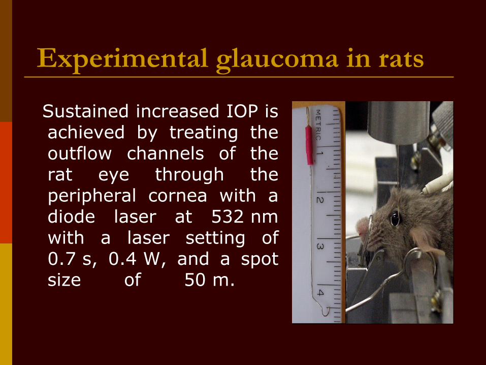
Sustained increased IOP is achieved by treating the outflow channels of the rat eye through the peripheral cornea with a diode laser at 532 nm with a laser setting of 0.7 s, 0.4 W, and a spot size of 50 m.

The laser beam is delivered from a slit–lamp system without additional lenses and with the animal under general anaesthesia.
The rat eye is rotated manually to allow the laser beam to be directed in a sharp angle to the trabecular meshwork (TM).

Treatment takes approximately 3 min per eye by an experienced researcher, therefore many rats can be treated in 1 day.

The laser treatment is given unilaterally and is repeated after a week if the IOP difference between the treated eye and the fellow eye is less than 6 mmHg. IOP is measured with the Tonopen XL in both eyes before and immediately after laser treatment, every 3–4 days in the first 2 weeks, and weekly thereafter. In all, 10 measurements are obtained for each eye from which the mean is calculated.
There are three additional currently available models for experimental glaucoma in rats, but all of them are more time consuming and technically more difficult to reproduce than the laser photocoagulation model.
Morrison and co-workers increased rat IOP by hyperosmotic saline microinjection into the episcleral veins of Brown–Norway rats in order to increase the outflow resistance.
This was sufficient for about 50% of rats to develop elevated IOP, even though most animals needed repeated injections. The advantage of using Brown–Norway rats is that IOP can be measured by a Tonopen –XL on awake animals. General anaesthetics were shown to have resulted in a rapid and substantial decrease in IOP in all eyes, and so measurements of IOP in awake animals provide the most accurate documentation of IOP history for rat glaucoma model studies.

Another rodent model for chronic glaucoma was developed by Ueda et al. They injected India ink into the anterior chamber 1 week prior to laser photocoagulation treatment. Increased IOP was achieved in all rats within 4 weeks and optic nerve damage developed as well.
With recent advances in genetic manipulation, the development of experimental glaucoma in mice became a matter of high priority. One of the main obstacles was measuring IOP in the mouse eye. Several devices were recently developed to measure IOP in a noninvasive way, all claiming to have the ability to measure IOP accurately in mice.

Some use the Tonopen without the ocufilm cover, while others use a modified Goldmann tonometer, both with reliable results.

Another model that has become well established over the past few years was developed by John et al. It consists of the mouse strain DBA/2J that develops glaucoma subsequent to anterior chamber changes. The glaucoma in the DBA/2J mice shares similarities with pigmentary dispersion glaucoma in humans.

These mice develop pigment dispersion, iris transillumination, iris atrophy, anterior synechia, and elevated IOP. The prevalence and severity increase with age, followed by RGC death, and optic nerve degeneration. Two chromosomal regions, one on chromosome 6 and one on chromosome 4, contribute to this glaucoma model.

Recently, John and his group generated and studied mouse models with mutations in genes that are responsible for anterior segment dysgenesis anomalies in humans. Developing these mouse models supports the search for mutations in specific human candidate genes. Although these models are specific for unique types of glaucoma, they can be used to test the mechanism of RGC death in glaucoma as well as to evaluate new treatment modalities.
Optic nerve transection
In rats, optic nerve transection is usually performed using an intraorbital approach. Using a binocular operating microscope, the superior conjunctiva is incised, the muscles and connective tissue are separated, and the intraorbital optic nerve is exposed.

A blade knife is used to transect the optic nerve behind the globe, taking care not to interfere with the blood supply and sparing the meningeal sheaths. At the end of the procedure, the retinas should be examined ophthalmoscopically to assure blood vessel patency.

RGCs degenerate rapidly after optic nerve transection. One–half of them will degenerate 4 days after optic nerve axotomy, and it rises to about 70% in 1 week and to more than 95% in 2 weeks.
In monkeys, the optic nerve is transected by a lateral approach through the skin and the bone of the orbit approximately 6 mm posterior to the globe. The rate of RGC degeneration in this model is usually slower, with about a 70% loss at 1 month and a 90% loss at 2 months.

Ischaemic optic neuropathy Recently, an animal model for anterior ischaemic
optic neuropathy (AION) in rats and mice was developed by Bernstein et al. In this model, a photosensitizing agent is injected intravenously into anaesthetized rats.

Using a custom-designed fundus contact lens, a laser beam directly activates the dye within the small vessels perfusing the optic nerve while sparing the large calibre vessels perfusing the inner retina. Pale oedema of the optic nerve appears shortly thereafter. Electrophysiologically, there is a decrease in amplitude and, histologically, there is impairment in axonal transport. The mean RGC loss by 39 days after the treatment is 40%. A similar model was developed for use in mice.

Clinical Trials

Inclusion Criteria for Clinical Trials1.Subjects must be capable of providing informed
consent.2.Subjects must be able to comply with the
protocol.3.Disease severity must be sufficient to
demonstrate a statistically significant and clinically meaningful effect of therapy.
4.Specific diagnostic criteria must be defined to ensure homogeneity of disease status, which can lead to a more precise study.
5.Subjects must be capable of responding to the proposed mechanism of action of the intervention to be studied.
Exclusion Criteria for Clinical Trials1.Subjects have concurrent disease that could
confound the response to therapy.2.Subjects are unlikely to comply with the protocol
or likely to be lost to follow-up.3.Subjects have known hypersensitivity or
intolerance to the proposed therapy.4.Subjects use concomitant therapy that affects
either tear function or ocular surface integrity.5.Subjects have had surgical or other manipulation
of the eye that could confound the outcome parameters or interfere with the mechanism of action of the proposed intervention to be studied.

FACILITATE MULTICENTER AND INTERNATIONAL COLLABORATIVE CLINICAL TRIALS
The Subcommittee recommends the development of criteria to be used in multinational venues. Important aspects to consider for such international trials are the use of uniform terminology. This may require that terms are translated and back-translated for clarity and accuracy It is necessary to resolve cultural or ethnic connotations or implications in terminology. There should be uniform interpretation of outcome variables with standardized protocols for measurement and recording of data.

Testing procedures should be uniform, with use of standardized reagents, standardized protocols, and consistent recording of results. It is necessary to maintain skill levels of data collectors and observers, including certification of investigators and research coordinators and technicians. Attempts should be made to reduce biases related to population differences (race, ethnicity, climatic).

Thanks


