
1756 | Mol. BioSyst., 2016, 12, 1756--1759 This journal is©The Royal Society of Chemistry 2016
Cite this:Mol. BioSyst., 2016,
12, 1756
Comprehensive mapping of O-GlcNAcmodification sites using a chemicallycleavable tag†
Matthew E. Griffin,a Elizabeth H. Jensen,a Daniel E. Mason,b Courtney L. Jenkins,a
Shannon E. Stone,a Eric C. Petersb and Linda C. Hsieh-Wilson*a
The post-translational modification of serine or threonine residues
of proteins with a single N-acetylglucosamine monosaccharide
(O-GlcNAcylation) is essential for cell survival and function. However,
relatively few O-GlcNAc modification sites have been mapped due to
the difficulty of enriching and detecting O-GlcNAcylated peptides
from complex samples. Here we describe an improved approach to
quantitatively label and enrich O-GlcNAcylated proteins for site
identification. Chemoenzymatic labelling followed by copper(I)-
catalysed azide–alkyne cycloaddition (CuAAC) installs a new mass
spectrometry (MS)-compatible linker designed for facile purification
of O-GlcNAcylated proteins from cell lysates. The linker also allows
subsequent quantitative release of O-GlcNAcylated proteins for
downstream MS analysis. We validate the approach by unambiguously
identifying several established O-GlcNAc sites on the proteins a-crystallin
and O-GlcNAc transferase (OGT), as well as discovering new, previously
unreported sites on OGT. Notably, these novel sites on OGT lie in
key functional domains of the protein, underscoring how this site
identification method may reveal important biological insights into
protein activity and regulation.
The dynamic and reversible post-translational modification of intra-cellular proteins by b-linked O-GlcNAc, known as O-GlcNAcylation,is necessary for the regulation of numerous cellular processes,including transcription, translation, protein homeostasis, and meta-bolism.1–6 Alterations in O-GlcNAcylation are associated with humandiseases such as cancer, diabetes, and neurodegeneration.6–8 How-ever, understanding the roles of O-GlcNAcylation in specific physio-logical contexts will require a more comprehensive characterizationof the O-GlcNAc proteome and the modification sites on proteins.Notably, although thousands of proteins have been putatively shownto be O-GlcNAcylated,9–15 relatively few glycosylation sites have beenmapped. Mass spectrometric (MS) identification of O-GlcNAcylatedpeptides from complex mixtures has been challenging due to the
substoichiometric nature of O-GlcNAcylation and is furtherexacerbated by suppression of O-GlcNAc peptide ionization inthe presence of the unmodified peptide.16 Thus, improvedmethods to enrich O-GlcNAcylated peptides or proteins aremuch needed, particularly approaches that can be directly usedin conjunction with MS/MS sequencing to achieve a morecomprehensive understanding of O-GlcNAc modification sites.
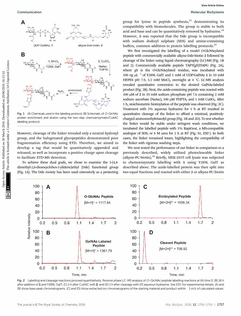
Robust enrichment of O-GlcNAcylated proteins can be accom-plished using a two-step chemoenzymatic approach.11,17 First, theO-GlcNAc moiety is tagged with a non-natural azide group bytreatment of cell lysates with UDP-GalNAz 1 and a mutant galacto-syltransferase (Y289L GalT)18 that specifically recognizes terminalGlcNAc moieties (Fig. 1). Next, a biotin group is attached viacopper(I)-catalysed azide–alkyne cycloaddition (CuAAC),19 whichallows for affinity purification. Although a limited set of alkyne-biotin linkers are commercially available, many existing linkers arenot ideal for mapping O-GlcNAc modification sites. In particular,harsh conditions are usually required to disrupt the femtomolarbiotin–streptavidin interaction,20 which may hydrolyse the labileO-GlcNAc moiety. Additionally, many linkers contain a largespacer between the biotin group and the alkyne functionality,which appends a relatively large mass to the glycopeptide andcan preclude its sequencing by mass spectrometry.20 Therefore, afacile method to release the labelled peptides and proteins withminimal added mass would greatly facilitate downstream analysis.
Several cleavable linkers have been previously developed forthe enrichment of O-GlcNAcylated proteins.13,15,16,21 However,each suffers from significant drawbacks for site identification. Forexample, a photocleavable linker was employed in conjunctionwith UDP-GalNAz 1 and Y289L GalT to sequence modified peptidesfrom mouse brain lysate.15,16 Importantly, the moiety retained aftercleavage provided a positively-charged amine group, which increasedthe overall peptide charge and facilitated ionization by electron-transfer dissociation (ETD), the most successful MS/MS methodfor O-GlcNAc peptide sequencing.12,22 Unfortunately, cleavageof the linker was found to be incomplete.16 In a recent report, adibromine-containing, acid-cleavable linker was employed toidentify various glycan modifications including O-GlcNAc.21
a Department of Chemistry and Chemical Engineering, California Institute of Technology,
Pasadena, CA 91125, USA. E-mail: [email protected] Genomics Institute of the Novartis Research Foundation, San Diego, CA 92121, USA
† Electronic supplementary information (ESI) available. See DOI: 10.1039/c6mb00138f
Received 22nd February 2016,Accepted 25th March 2016
DOI: 10.1039/c6mb00138f
www.rsc.org/molecularbiosystems
MolecularBioSystems
COMMUNICATION
Ope
n A
cces
s A
rtic
le. P
ublis
hed
on 3
0 M
arch
201
6. D
ownl
oade
d on
20/
06/2
016
16:1
5:52
. T
his
artic
le is
lice
nsed
und
er a
Cre
ativ
e C
omm
ons
Attr
ibut
ion
3.0
Unp
orte
d L
icen
ce.
View Article OnlineView Journal | View Issue
This journal is©The Royal Society of Chemistry 2016 Mol. BioSyst., 2016, 12, 1756--1759 | 1757
However, cleavage of the linker revealed only a neutral hydroxylgroup, and the halogenated glycopeptides demonstrated poorfragmentation efficiency using ETD. Therefore, we aimed todevelop a tag that would be quantitatively appended andreleased, as well as incorporate a positive charge upon cleavageto facilitate ETD-MS detection.
To achieve these dual goals, we chose to examine the 1-(4,4-dimethyl-2,6-dioxocyclohex-1-ylidene)ethyl (Dde) functional group(Fig. 1A). The Dde moiety has been used extensively as a protecting
group for lysine in peptide synthesis,23 demonstrating itscompatibility with biomolecules. The group is stable to bothacid and base and can be quantitatively removed by hydrazine.24
However, it was reported that the Dde group is incompatiblewith sodium dodecyl sulphate (SDS) and amine-containingbuffers, common additives to protein labelling protocols.25
We first investigated the labelling of a model O-GlcNAcylatedpeptide with commercially available alkyne-Dde-biotin 2 followed bycleavage of the linker using liquid chromatography (LC)-MS (Fig. 1Band 2). Commercially available peptide TAPT(gS)TIAPG (Fig. 2A),where gS is the O-GlcNAcylated residue, was incubated with100 ng mL�1 of Y289L GalT and 1 mM of UDP-GalNAz 1 in 10 mMHEPES pH 7.9, 5.5 mM MnCl2 overnight at 4 1C. LC-MS analysisrevealed quantitative conversion to the desired GalNAz-labeledproduct (Fig. 2B). Next, the azide-containing peptide was reacted with100 mM of 2 in 10 mM sodium phosphate pH 7.6 containing 2 mMsodium ascorbate (NaAsc), 100 mM THPTA, and 1 mM CuSO4. After1 h, stoichiometric biotinylation of the peptide was observed (Fig. 2C).Treatment with 2% aqueous hydrazine for 1 h at RT resulted inquantitative cleavage of the linker to afford a minimal, positively-charged aminomethyltriazolyl group (Fig. 1B and 2D). To test whetherthe linker would be stable under stringent wash conditions, weincubated the labelled peptide with 1% RapiGest, a MS-compatibleanalogue of SDS, or 6 M urea for 1 h at RT (Fig. S1, ESI†). In bothcases, the linker remained intact, highlighting the compatibility ofthe linker with rigorous washing steps.
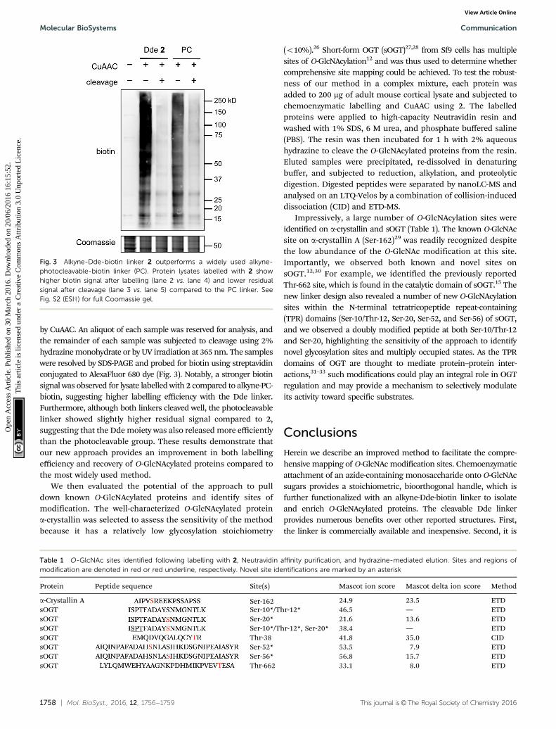
We next tested the performance of our linker in comparison to apreviously described, widely utilized photocleavable linker(alkyne-PC-biotin).16 Briefly, HEK-293T cell lysate was subjectedto chemoenzymatic labelling with 1 using Y289L GalT asdescribed above. The azide-labelled protein was then split intotwo equal fractions and reacted with either 2 or alkyne-PC-biotin
Fig. 1 (A) Chemicals used in the labelling protocol. (B) Schematic of O-GlcNAcprotein enrichment and elution using the two-step chemoenzymatic/CuAAClabelling protocol.
Fig. 2 Labelling and cleavage reactions proceed quantitatively. Reverse phase LC-MS analysis of O-GlcNAc peptide labelling reactions at (A) time 0, (B) 16 hafter addition of 1 and Y289L GalT, (C) 1 h after CuAAC with 2, and (D) 1 h after cleavage with 2% aqueous hydrazine. See ESI† for experimental details. (A) and(B) show base peak chromatograms. (C) and (D) show extracted ion chromatograms of the starting material and product within �1 m/z of calculated values.
Communication Molecular BioSystems
Ope
n A
cces
s A
rtic
le. P
ublis
hed
on 3
0 M
arch
201
6. D
ownl
oade
d on
20/
06/2
016
16:1
5:52
. T
his
artic
le is
lice
nsed
und
er a
Cre
ativ
e C
omm
ons
Attr
ibut
ion
3.0
Unp
orte
d L
icen
ce.
View Article Online
1758 | Mol. BioSyst., 2016, 12, 1756--1759 This journal is©The Royal Society of Chemistry 2016
by CuAAC. An aliquot of each sample was reserved for analysis, andthe remainder of each sample was subjected to cleavage using 2%hydrazine monohydrate or by UV irradiation at 365 nm. The sampleswere resolved by SDS-PAGE and probed for biotin using streptavidinconjugated to AlexaFluor 680 dye (Fig. 3). Notably, a stronger biotinsignal was observed for lysate labelled with 2 compared to alkyne-PC-biotin, suggesting higher labelling efficiency with the Dde linker.Furthermore, although both linkers cleaved well, the photocleavablelinker showed slightly higher residual signal compared to 2,suggesting that the Dde moiety was also released more efficientlythan the photocleavable group. These results demonstrate thatour new approach provides an improvement in both labellingefficiency and recovery of O-GlcNAcylated proteins compared tothe most widely used method.
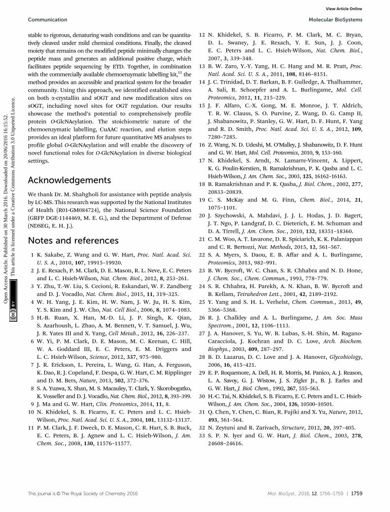
We then evaluated the potential of the approach to pulldown known O-GlcNAcylated proteins and identify sites ofmodification. The well-characterized O-GlcNAcylated proteina-crystallin was selected to assess the sensitivity of the methodbecause it has a relatively low glycosylation stoichiometry
(o10%).26 Short-form OGT (sOGT)27,28 from Sf9 cells has multiplesites of O-GlcNAcylation12 and was thus used to determine whethercomprehensive site mapping could be achieved. To test the robust-ness of our method in a complex mixture, each protein wasadded to 200 mg of adult mouse cortical lysate and subjected tochemoenzymatic labelling and CuAAC using 2. The labelledproteins were applied to high-capacity Neutravidin resin andwashed with 1% SDS, 6 M urea, and phosphate buffered saline(PBS). The resin was then incubated for 1 h with 2% aqueoushydrazine to cleave the O-GlcNAcylated proteins from the resin.Eluted samples were precipitated, re-dissolved in denaturingbuffer, and subjected to reduction, alkylation, and proteolyticdigestion. Digested peptides were separated by nanoLC-MS andanalysed on an LTQ-Velos by a combination of collision-induceddissociation (CID) and ETD-MS.
Impressively, a large number of O-GlcNAcylation sites wereidentified on a-crystallin and sOGT (Table 1). The known O-GlcNAcsite on a-crystallin A (Ser-162)29 was readily recognized despitethe low abundance of the O-GlcNAc modification at this site.Importantly, we observed both known and novel sites onsOGT.12,30 For example, we identified the previously reportedThr-662 site, which is found in the catalytic domain of sOGT.15 Thenew linker design also revealed a number of new O-GlcNAcylationsites within the N-terminal tetratricopeptide repeat-containing(TPR) domains (Ser-10/Thr-12, Ser-20, Ser-52, and Ser-56) of sOGT,and we observed a doubly modified peptide at both Ser-10/Thr-12and Ser-20, highlighting the sensitivity of the approach to identifynovel glycosylation sites and multiply occupied states. As the TPRdomains of OGT are thought to mediate protein–protein inter-actions,31–33 such modifications could play an integral role in OGTregulation and may provide a mechanism to selectively modulateits activity toward specific substrates.
Conclusions
Herein we describe an improved method to facilitate the compre-hensive mapping of O-GlcNAc modification sites. Chemoenzymaticattachment of an azide-containing monosaccharide onto O-GlcNAcsugars provides a stoichiometric, bioorthogonal handle, which isfurther functionalized with an alkyne-Dde-biotin linker to isolateand enrich O-GlcNAcylated proteins. The cleavable Dde linkerprovides numerous benefits over other reported structures. First,the linker is commercially available and inexpensive. Second, it is
Fig. 3 Alkyne-Dde-biotin linker 2 outperforms a widely used alkyne-photocleavable-biotin linker (PC). Protein lysates labelled with 2 showhigher biotin signal after labelling (lane 2 vs. lane 4) and lower residualsignal after cleavage (lane 3 vs. lane 5) compared to the PC linker. SeeFig. S2 (ESI†) for full Coomassie gel.
Table 1 O-GlcNAc sites identified following labelling with 2, Neutravidin affinity purification, and hydrazine-mediated elution. Sites and regions ofmodification are denoted in red or red underline, respectively. Novel site identifications are marked by an asterisk
Protein Peptide sequence Site(s) Mascot ion score Mascot delta ion score Method
a-Crystallin A Ser-162 24.9 23.5 ETDsOGT Ser-10*/Thr-12* 46.5 — ETDsOGT Ser-20* 21.6 13.6 ETDsOGT Ser-10*/Thr-12*, Ser-20* 38.4 — ETDsOGT Thr-38 41.8 35.0 CIDsOGT Ser-52* 53.5 7.9 ETDsOGT Ser-56* 56.8 15.7 ETDsOGT Thr-662 33.1 8.0 ETD
Molecular BioSystems Communication
Ope
n A
cces
s A
rtic
le. P
ublis
hed
on 3
0 M
arch
201
6. D
ownl
oade
d on
20/
06/2
016
16:1
5:52
. T
his
artic
le is
lice
nsed
und
er a
Cre
ativ
e C
omm
ons
Attr
ibut
ion
3.0
Unp
orte
d L
icen
ce.
View Article Online
This journal is©The Royal Society of Chemistry 2016 Mol. BioSyst., 2016, 12, 1756--1759 | 1759
stable to rigorous, denaturing wash conditions and can be quantita-tively cleaved under mild chemical conditions. Finally, the cleavedmoiety that remains on the modified peptide minimally changes thepeptide mass and generates an additional positive charge, whichfacilitates peptide sequencing by ETD. Together, in combinationwith the commercially available chemoenzymatic labelling kit,11 themethod provides an accessible and practical system for the broadercommunity. Using this approach, we identified established siteson both a-crystallin and sOGT and new modification sites onsOGT, including novel sites for OGT regulation. Our resultsshowcase the method’s potential to comprehensively profileprotein O-GlcNAcylation. The stoichiometric nature of thechemoenzymatic labelling, CuAAC reaction, and elution stepsprovides an ideal platform for future quantitative MS analyses toprofile global O-GlcNAcylation and will enable the discovery ofnovel functional roles for O-GlcNAcylation in diverse biologicalsettings.
Acknowledgements
We thank Dr. M. Shahgholi for assistance with peptide analysisby LC-MS. This research was supported by the National Institutesof Health (R01-GM084724), the National Science Foundation(GRFP DGE-1144469, M. E. G.), and the Department of Defense(NDSEG, E. H. J.).
Notes and references
1 K. Sakabe, Z. Wang and G. W. Hart, Proc. Natl. Acad. Sci.U. S. A., 2010, 107, 19915–19920.
2 J. E. Rexach, P. M. Clark, D. E. Mason, R. L. Neve, E. C. Petersand L. C. Hsieh-Wilson, Nat. Chem. Biol., 2012, 8, 253–261.
3 Y. Zhu, T.-W. Liu, S. Cecioni, R. Eskandari, W. F. Zandbergand D. J. Vocadlo, Nat. Chem. Biol., 2015, 11, 319–325.
4 W. H. Yang, J. E. Kim, H. W. Nam, J. W. Ju, H. S. Kim,Y. S. Kim and J. W. Cho, Nat. Cell Biol., 2006, 8, 1074–1083.
5 H.-B. Ruan, X. Han, M.-D. Li, J. P. Singh, K. Qian,S. Azarhoush, L. Zhao, A. M. Bennett, V. T. Samuel, J. Wu,J. R. Yates III and X. Yang, Cell Metab., 2012, 16, 226–237.
6 W. Yi, P. M. Clark, D. E. Mason, M. C. Keenan, C. Hill,W. A. Goddard III, E. C. Peters, E. M. Driggers andL. C. Hsieh-Wilson, Science, 2012, 337, 975–980.
7 J. R. Erickson, L. Pereira, L. Wang, G. Han, A. Ferguson,K. Dao, R. J. Copeland, F. Despa, G. W. Hart, C. M. Ripplingerand D. M. Bers, Nature, 2013, 502, 372–376.
8 S. A. Yuzwa, X. Shan, M. S. Macauley, T. Clark, Y. Skorobogatko,K. Vosseller and D. J. Vocadlo, Nat. Chem. Biol., 2012, 8, 393–399.
9 J. Ma and G. W. Hart, Clin. Proteomics, 2014, 11, 8.10 N. Khidekel, S. B. Ficarro, E. C. Peters and L. C. Hsieh-
Wilson, Proc. Natl. Acad. Sci. U. S. A., 2004, 101, 13132–13137.11 P. M. Clark, J. F. Dweck, D. E. Mason, C. R. Hart, S. B. Buck,
E. C. Peters, B. J. Agnew and L. C. Hsieh-Wilson, J. Am.Chem. Soc., 2008, 130, 11576–11577.
12 N. Khidekel, S. B. Ficarro, P. M. Clark, M. C. Bryan,D. L. Swaney, J. E. Rexach, Y. E. Sun, J. J. Coon,E. C. Peters and L. C. Hsieh-Wilson, Nat. Chem. Biol.,2007, 3, 339–348.
13 B. W. Zaro, Y.-Y. Yang, H. C. Hang and M. R. Pratt, Proc.Natl. Acad. Sci. U. S. A., 2011, 108, 8146–8151.
14 J. C. Trinidad, D. T. Barkan, B. F. Gulledge, A. Thalhammer,A. Sali, R. Schoepfer and A. L. Burlingame, Mol. Cell.Proteomics, 2012, 11, 215–229.
15 J. F. Alfaro, C.-X. Gong, M. E. Monroe, J. T. Aldrich,T. R. W. Clauss, S. O. Purvine, Z. Wang, D. G. Camp II,J. Shabanowitz, P. Stanley, G. W. Hart, D. F. Hunt, F. Yangand R. D. Smith, Proc. Natl. Acad. Sci. U. S. A., 2012, 109,7280–7285.
16 Z. Wang, N. D. Udeshi, M. O’Malley, J. Shabanowitz, D. F. Huntand G. W. Hart, Mol. Cell. Proteomics, 2010, 9, 153–160.
17 N. Khidekel, S. Arndt, N. Lamarre-Vincent, A. Lippert,K. G. Poulin-Kerstien, B. Ramakrishnan, P. K. Qasba and L. C.Hsieh-Wilson, J. Am. Chem. Soc., 2003, 125, 16162–16163.
18 B. Ramakrishnan and P. K. Qasba, J. Biol. Chem., 2002, 277,20833–20839.
19 C. S. McKay and M. G. Finn, Chem. Biol., 2014, 21,1075–1101.
20 J. Szychowski, A. Mahdavi, J. J. L. Hodas, J. D. Bagert,J. T. Ngo, P. Landgraf, D. C. Dieterich, E. M. Schuman andD. A. Tirrell, J. Am. Chem. Soc., 2010, 132, 18351–18360.
21 C. M. Woo, A. T. Iavarone, D. R. Spiciarich, K. K. Palaniappanand C. R. Bertozzi, Nat. Methods, 2015, 12, 561–567.
22 S. A. Myers, S. Daou, E. B. Affar and A. L. Burlingame,Proteomics, 2013, 982–991.
23 B. W. Bycroft, W. C. Chan, S. R. Chhabra and N. D. Hone,J. Chem. Soc., Chem. Commun., 1993, 778–779.
24 S. R. Chhabra, H. Parekh, A. N. Khan, B. W. Bycroft andB. Kellam, Tetrahedron Lett., 2001, 42, 2189–2192.
25 Y. Yang and S. H. L. Verhelst, Chem. Commun., 2013, 49,5366–5368.
26 R. J. Chalkley and A. L. Burlingame, J. Am. Soc. MassSpectrom., 2001, 12, 1106–1113.
27 J. A. Hanover, S. Yu, W. B. Lubas, S.-H. Shin, M. Ragano-Caracciola, J. Kochran and D. C. Love, Arch. Biochem.Biophys., 2003, 409, 287–297.
28 B. D. Lazarus, D. C. Love and J. A. Hanover, Glycobiology,2006, 16, 415–421.
29 E. P. Roquemore, A. Dell, H. R. Morris, M. Panico, A. J. Reason,L. A. Savoy, G. J. Wistow, J. S. Zigler Jr., B. J. Earles andG. W. Hart, J. Biol. Chem., 1992, 267, 555–563.
30 H.-C. Tai, N. Khidekel, S. B. Ficarro, E. C. Peters and L. C. Hsieh-Wilson, J. Am. Chem. Soc., 2004, 126, 10500–10501.
31 Q. Chen, Y. Chen, C. Bian, R. Fujiki and X. Yu, Nature, 2012,493, 561–564.
32 N. Zeytuni and R. Zarivach, Structure, 2012, 20, 397–405.33 S. P. N. Iyer and G. W. Hart, J. Biol. Chem., 2003, 278,
24608–24616.
Communication Molecular BioSystems
Ope
n A
cces
s A
rtic
le. P
ublis
hed
on 3
0 M
arch
201
6. D
ownl
oade
d on
20/
06/2
016
16:1
5:52
. T
his
artic
le is
lice
nsed
und
er a
Cre
ativ
e C
omm
ons
Attr
ibut
ion
3.0
Unp
orte
d L
icen
ce.
View Article Online


