
J Ayub Med Coll Abbottabad 2014;26(4)
http://www.ayubmed.edu.pk/JAMC/26-4/Amjad.pdf 611
CASE REPORT PILOMYXOID ASTROCYTOMA PRESENTING AS DIENCEPHALIC
SYNDROME
Amjad Ali Khan, Ahmed Kamal El-Borai Department of Histopathology, Prince Salman Armed Forces Hospital, Tabuk, Kingdom of Saudi Arabia
We report the case of a child who presented with diencephalic syndrome. During diagnostic work-up, he was found to have a supra-sellar hypothalamic tumour. Histopathological examination of the tumour revealed it to be pilomyxoid astrocytoma, which is a WHO grade-II tumour, previously considered to be part of the spectrum of WHO grade-I pilocytic astrocytomas. However, because of its characteristic histopathology and behaviour, it was later segregated from pilocytic astrocytomas. In this case report, we discuss the cytological and histopathological features of this tumor with the aim of increasing awareness of this tumour amongst general histopathologists, to highlight the importance of its differentiation from pilocytic astrocytomas in view of its different behaviour, treatment and prognosis and that it should be included in the differential diagnosis of diencephalic syndrome. Keywords: Diencephalic syndrome, Pilocytic astrocytomas, supra-sellar hypothalamic tumour, pilocytic astrocytoma
J Ayub Med Coll Abbottabad 2014;26(4):611–5
INTRODUCTION Pilomyxoid astrocytoma (PMA) is a rare paediatric brain tumor.1 This entity was initially considered a variant of pilocytic astrocytoma (PA). But later, it was realized that in spite of many similarities and overlaps, it differs from pilocytic astrocytoma in terms of its morphology and behaviour. Pilomyxoid astrocytoma has a monomorphic histological appearance, more myxoid background, more aggressive disease course and a higher recurrence and CNS dissemination rate than that of pilocytic astrocytoma.2 Therefore it was recognized as a distinct tumour entity in the 2007 edition of WHO classification of central nervous system tumours and was designated grade-II astrocytoma (in the four tiered grading system), thus segregating it from the typical grade-I pilocytic astrocytoma.
In this case report, the cytological (intraoperative smears) and histopathological (intraoperative frozen and permanent section) features are discussed. So that on one hand, the busy general histopathologist should be aware of this entity and make an appropriate diagnosis bearing in mind that pilocytic and pilomyxoid astrocytomas are two distinct entities with different behaviour, treatment and prognosis; while on the other hand, the pediatrician should be aware of this entity to be included in the differential diagnosis of diencephalic syndrome. CASE REPORT We report a case of 18 months old boy brought to the paediatrics clinic because of bilateral abnormal eye movements, failure to thrive and delayed
milestones. According to the patient’s mother, the child started to have abnormal right eye movement at the age of 6 months, followed 2 weeks later by similar movements in the contra-lateral eye. This was associated with vomiting, decrease in appetite and weight loss that gradually improved with symptomatic medication. The patient’s birth history was unremarkable; his birth weight was 2 kg with no detectable abnormalities.
On examination, the child was severely emaciated having considerably low growth parameters; 5.2 kg body weight, 70 cm length and 46cm head circumference (all parameters <3rd centile). He was able to follow objects in all ocular directions with abnormal eye movements in the form of nystagmus but visual acuity though difficult to assess, was bilateral perception of light. The laboratory investigations showed low levels of plasma cortisol, thyroid stimulating hormone (TSH) and free T4 (Table-1). Therefore, provisional clinical diagnosis of Diencephalic syndrome of Russell was made.
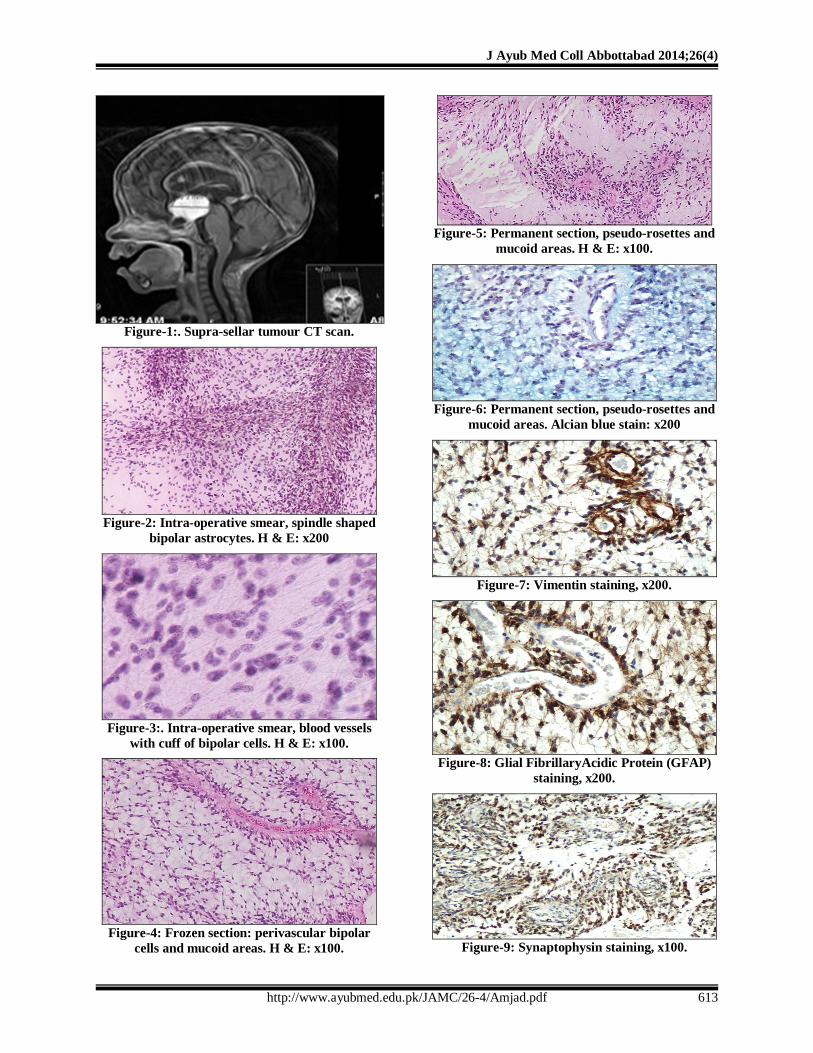
Brain ultrasound and MRI showed a well-defined, homogenously hyper-echoic supra-sellar hypothalamic mass (Figure-1). High T2 signal intensity showed the mass to be lobulated, measuring 4.2×4.2 cm, with hydrocephalic changes in the 3rdand lateral ventricles. There was also lepto-meningeal enhancement at the brainstem and upper cervical spine. Neurosurgical biopsy and limited partial resection of this highly vascular tumour were planned. Intra-operative CSF cytology showed absence of circulating malignant cells.

J Ayub Med Coll Abbottabad 2014;26(4)
http://www.ayubmed.edu.pk/JAMC/26-4/Amjad.pdf 612
Intra-operatively small soft gelatinous biopsy material was received in the laboratory for intra-operative consultation. Smears were prepared which showed many bipolar (spindle shaped) piloid cells imparting fibrillary appearance within myxoid background without Rosenthal fibbers (Figure-2); these cells were concentrated around blood vessels, with long axis of their spindle shaped nuclei perpendicular to the blood vessels (Figure-3).The frozen sections showed in addition, pseudo-rosettes, i.e., perivascular radial arrangement of cells seen in transverse section of blood vessels (Figure-4). The differential diagnosis at frozen sections included pilocytic astrocytoma, ependymoma and astroblastoma.
The pilocytic astrocytoma was favoured by the presence of bipolar cells and piloid background though pseudo-rosettes and myxoid background were against such a diagnosis. Ependymomas entered the differential diagnosis due to the presence of pseudo-rosettes, but prominent piloid background, presence of bipolar cells and absence of true rosettes were not favouring such a diagnosis.
Astroblastoma was also part of differential diagnosis, but it is characterized by plump spindle shaped cells having unipolar short stout processes attaching the cells to the blood vessel walls (pseudo-rosettes). In view of these considerations, intra-operative diagnosis of low grade glioma was rendered.
The permanent sections showed moderately dense tumour composed of monotonous population of bipolar (spindle shaped) piloid cells set in fibrillary background admixed with moderate amount of myxoid material. These cells showed only occasional mitoses and were arranged around frequently found capillaries forming pseudo-rosettes (Figure-5).
No necrosis, Rosenthal fibbers or eosinophilic granular bodies were present. The myxoid material was positive for Alcian blue (Figure-6) and negative for PAS. The piloid cells were positive for Vimentin (Figure-7), GFAP (Figure-8), Synaptophysin (Figure-9), and Ki67+ (3%). Therefore, a diagnosis of pilomyxoid astrocytoma, WHO grade-II was made.
Patient’s age at the time of diagnosis was less than 3 years, so systemic chemotherapy was started after calculating the doses according to his body weight. Later, it was repeated when the tumour started progressing.
Concomitantly, nutritional supportive program and hormone replacement therapy (Hydrocortisone 3mg am, 1.5 mg pm and L-thyroxin 25 µgm O.D) were administered together
with other supportive measures for chemotherapy related toxicities. Cytotoxic doses were modified with 25% reduction in the latest 3 cycles because of poor patient’s tolerance.
Re-evaluation MRI at mid chemotherapy cycles revealed more than 50% reduction of the tumour volume. The symptomatic improvement was also noted in terms of improving appetite, developing physical and mental milestones, and relatively improved growth parameters (weight: 8.7 kg, height: 78 cm and head circumference: 48 cm approaching 10th centile). Mildly improved vision was also noticed in the form of bilateral hand movement visualization at 50 cm and absence of other neurological manifestations apart from persisting nystagmus.
The potential role of a second look operation and/or adjuvant radiotherapy was further discussed at the end of chemotherapy. However further intervention was not recommended due to expected high risk of complications and morbidities in light of; surgically inaccessible tumour site, poor patient performance and age restrictions.
The tumour size was static for two and a half years controlled by repeated cycles of chemotherapy; however, it started increasing in size with surrounding infiltration and poor response to chemotherapy leading to patient’s death after four years of follow-up
Table-1: Patient’s haematological and biochemical profile
Variable Level Range Values WBC 5.3 4.8–10.8x10⁹/L HB 11.6 14–18 g/dL Platelets 345 130–400 x109/L Na 127 135–145 mmol/L K 4.8 3.2–5 mmol/L Ca 2.18 2.12–2.52mmol/L Creatinine 10 50–120 µmol/L ALT 48 30–65 U/L AST 27 15–37 U/L Bilirubin 03 0–14 µmol/L FBS 05 4.1 5.9 mmol/L Cortisol 02 4–15 µg/dl TSH 0.1 0.5–5mIU/ml FT4 6.6 10.2-20.9 pmol/L
Table-2: Typical Pilomyxoid and Pilocytic Histologic Features8
Feature Pilomyxoid Pilocytic Architecture Monophasic Biphasic Protoplasmic cells Rare Present Rosenthal fibers Absent Present Myxoid background Predominant Infrequent Calcification Uncommon Occasional Eosinophilic granular bodies Absent Present Angiocentric pattern Frequent Rare
J Ayub Med Coll Abbottabad 2014;26(4)
http://www.ayubmed.edu.pk/JAMC/26-4/Amjad.pdf 613
Figure-1:. Supra-sellar tumour CT scan.
Figure-2: Intra-operative smear, spindle shaped
bipolar astrocytes. H & E: x200
Figure-3:. Intra-operative smear, blood vessels
with cuff of bipolar cells. H & E: x100.
Figure-4: Frozen section: perivascular bipolar
cells and mucoid areas. H & E: x100.
Figure-5: Permanent section, pseudo-rosettes and
mucoid areas. H & E: x100.
Figure-6: Permanent section, pseudo-rosettes and
mucoid areas. Alcian blue stain: x200
Figure-7: Vimentin staining, x200.
Figure-8: Glial FibrillaryAcidic Protein (GFAP)
staining, x200.
Figure-9: Synaptophysin staining, x100.

J Ayub Med Coll Abbottabad 2014;26(4)
http://www.ayubmed.edu.pk/JAMC/26-4/Amjad.pdf 614
DISCUSSION Pilomyxoid astrocytomas occur during the first two decades of life especially in infants and young children with no gender predilection; however, the exact incidence of this tumour is unknown.3 The reported mean age for diagnosis is around 18 months with special predilection for the hypothalamic/chiasmic region and cerebellum; however, it can occur anywhere along the neuraxis. Tumours occurring in the hypothalamic/chiasmic region can cause diencephalic syndrome. Its true cell of origin is not known, but it is mostly believed to be of astrocytic origin; however, because of its ultrastructural similarity to the periventricular tanycyte, Fuller et al suggested a tanycytic origin for these tumors.4 Pilomyxoid astrocytomas have been associated with neurofibromatosis type11, pilocytic astrocytomas and spontaneous intratumoral hemorrhages.5 Polymerase chain reaction has shown KIAA1549:BRAF fusions in 33% of pilomyxoid astrocytomas.6
Diencephalic syndrome first described by Russell in 1951is characterized by severe emaciation, preserved linear growth, nystagmus and vomiting, it is a rare but potentially lethal disease of infants and young children that can occur with or without a tumour of hypothalamic or chiasmatic region.7 The usual tumours having predilection for hypothalamic/chiasmic region (thus causing diencephalic syndrome) include pilocytic astrocytoma, pilomyxoid astrocytoma, gangliocytomas, pleomorphic xanthoastrocytoma, astroblastoma, dysembryoplastic neuroepithelial tumor and choroid plexus tumors.
Pilocytic astrocytomas are low grade astrocytomas with excellent prognosis and indolent course. Pilomyxoid astrocytoma was initially considered a morphological variant of pilocytic astrocytoma because the affected age group and the preferred site of affection is the same for both the tumors. However, it was observed that a subgroup of pilocytic astrocytomas had an aggressive behaviour, high mortality, early recurrence and high CNS dissemination rates.2 Contrary to PA showing 20-year survival rates of 70% to 80%,8 PMA tends to behave more aggressively with a decreased disease free survival and higher mortality rates. Tumour related death rate of 33% was reported in patients with PMA as compared to 17% only with classical PA.9 Comparing outcome of 21 patients with hypothalamic PMAs to 42 patients with PAs of same location and equal degrees of gross total resection, the former group had a higher rate of local recurrence (76%and 50%, respectively) with a mean follow up duration of 26 months.9 Also, progression free
survival was significantly shorter in the PMA than PA patients (26 months and 147 months respectively, p<0.001) in addition to a decreased overall survival among the former group as well (mean duration 63 and 213 months respectively, p<0.001).9
Pilomyxoid astrocytomas have soft, gelatinous or mucoid gross consistency. The intra-operative smears prepared are moderately cellular having monomorphic appearance, being composed of spindle shaped cells having long hair like processes at both ends (bipolar, seekh kebab on skewer appearance, Figure-2). The stroma is mucoid or myxoid with many traversing piloid (hair-like) processes and capillary sized blood vessels with perivascular arrangement of tumor cells. The permanent sections also show the same microscopic picture without detectable eosinophilic granular bodies or Rosenthal fibers.3
The location, piloid background and spindle cell population raises differential diagnosis with pilocytic astrocytoma. The latter is a WHO grade-I tumour having biphasic microscopic appearance i.e., areas of moderate and low cellularity. The areas of moderate cellularity are composed of bipolar bland spindle shaped cells having long fibbers imparting fibrillary background with scattered thick refractile Rosenthal fibers, whereas the areas of low cellularity are fibril poor, composed of bland protoplasmic multipolar piloid cells, granular eosinophilic bodies, only focal myxoid change and microcysts. There is no perivascular neoplastic cellular arrangement (pseudo-rosettes) or frequent mucoid background (Table-2).
The perivascular arrangement of cells (pseudo-rosettes) and myxoid/mucoidbackground raise the differential diagnosis of ependymoma. However, ependymomas are characterized by true ependymal rosettes with central canals/lumens and perivascular pseudo-rosettes having anuclear dense fibrillary perivascular zones.
Astroblastomas are also characterized by pseudo-rosettes; however, the constituents cells arranged around hyalinized and sclerotic blood vessels are plump having unipolar stout processes through which these cells are attached to the blood vessels. Also, the background is usually non-fibrillary.
Angiocentric glioma, as the name suggests is characterized by perivascular arrangement of the neoplastic cells. It is an infiltrative tumour with sub-pialpalisading; composed of monophasic population of spindle shaped cells having bipolar fibrillary processes with focal tiny schwannoma-like nodules. The neoplastic cells may have small cytoplasmic eosinophilic inclusions, the stroma is fibrillary and minimally myxoid. However, this tumor affects

J Ayub Med Coll Abbottabad 2014;26(4)
http://www.ayubmed.edu.pk/JAMC/26-4/Amjad.pdf 615
patients with mean age of 17 years at presentation. It mostly presents with seizures and affects the superficial cerebral cortex.
Being a recently identified tumor entity, no specific standard of care for PMA is yet known. However, like other paediatric low-grade astrocytomas, surgery is the primary treatment for PMA and the degree of gross total resection depends on the tumour site. Because of the young age of patients at diagnosis, chemotherapy is most often used which can delay the need for radiation. Adjuvant therapy should be immediately started after surgery without waiting for tumour growth or recurrence.
Adjuvant radiation therapy in low-grade astrocytomas is generally limited to patients older than 3 or 5 years of age whose disease progresses after an initial resection. However, for older children, some authors concluded that radiotherapy concomitantly administered with chemotherapy may prove to be a more effective method of treatment for PMA despite its toxicity which may be severe in some cases.
In summary pilomyxoid astrocytoma is a WHO grade-II tumour distinct from indolent pilocytic astrocytoma categorized as WHO grade I tumour. These two tumour types have different morphology, behaviour, treatment and prognosis. These tumours should be suspected in cases of diencephalic syndrome. Chemotherapy appears to be an effective part of multimodal treatment approach in pilomyxoid astrocytoma after total or subtotal resection of the tumour.
ACKNOWLEDGEMENT We are grateful to Miss Rowena Castillo for proof reading the manuscript and providing valuable suggestions.
REFERENCES 1. Linscott LL, Osborn AG, Blaser S, Castillo M, Hewlett RH,
Wieselthaler N, et al. Pilomyxoid Astrocytoma: expanding the imaging spectrum. AJNR Am J Neuroradiol 2008; 29:1861–6.
2. Tihan T, Fisher PG, Kepner JL, Godfraind C, McComb RD, Goldthwaite PT, et al. Pediatric astrocytomas with monomorphous pilomyxoid features and a less favorable outcome. J Neuropathol Exp Neurol 1999;58:1061–8.
3. Louis DN, Ohgaki H, Wiestler OD, Cavenee WK, Burger PC, Jouvet A, et al. World Health Organization Classification of Tumours of the Central Nervous System. Acta Neuropathol 2007;114(2):97–109.
4. Petito CK. Suprasellar monomorphous pilomyxoid gliomas. AJNR Am J Neuradiol 2003,24:1931–2.
5. Gottfried ON, Fults DW, Townsend JJ, Couldwell WT. Spontaneous hemorrhage associated with a pilomyxoid astrocytoma. Case report. J Neurosurg 2003;99(2):416–20.
6. Lin A, Rodriguez FJ, Karajannis MA, Williams SC, Legault G, Zagzag D, et al. BRAF alterations in primary glial and glioneuronal neoplasms of the central nervous system with identification of 2 novel KIAA1549: BRAF fusion variants. J Neuropathol Exp Neurol 2012;71(1):66-72.
7. Poussaint TY, Barnes PD, Nichols K, Anthony DC, Cohen L, Tarbell NJ, et al. Diencephalic Syndrome: clinical features and imaging findings. AJNR Am J Neuroradiol;18:1499–505.
8. Komotar RJ, Burger PC, Carson BS, Brem H, Olivi A, Goldthwaite PT, et al. Pilocytic and pilomyxoid hypothalamic/chiasmatic astrocytomas. Neurosurgery 2004;54:72–9.
9. Komotar RJ, Mocco J, Jones JE, Zacharia BE, Tihan T, Feldstein NA, et al. Pilomyxoid astrocytoma: diagnosis, prognosis, and management. Neurosurg Focus 2005;18(6a):E7.
Address for Correspondence: Dr. Amjad Ali Khan, Department of Histopathology, Prince Salman Armed Forces Hospital, Tabuk, Kingdom of Saudi Arabia. Cell: +966-144411088-85225 Email: [email protected]


