Cartilage Oligomeric Matrix Protein in IdiopathicPulmonary FibrosisLouis J. Vuga1*., Jadranka Milosevic1., Kusum Pandit1, Ahmi Ben-Yehudah3, Yanxia Chu1,
Thomas Richards1, Joshua Sciurba1, Michael Myerburg1, Yingze Zhang1, Anil V. Parwani2,
Kevin F. Gibson1, Naftali Kaminski4
1Dorothy P and Richard P Simmons Center for Interstitial Lung Diseases, Division of Pulmonary, Allergy and Critical Care Medicine, University of Pittsburgh, School of
Medicine, Pittsburgh, Pennsylvania, United States of America, 2Department of Pathology, University of Pittsburgh, School of Medicine, Pittsburgh, Pennsylvania, United
States of America, 3 Pittsburgh Development Center, Magee-Women’s Research Institute and Foundation, University of Pittsburgh, School of Medicine, Pittsburgh,
Pennsylvania, United States of America, 4 Pulmonary, Critical Care and Sleep Medicine, Yale School of Medicine, New Haven, Connecticut, United States of America
Abstract
Idiopathic pulmonary fibrosis (IPF) is a progressive and life threatening disease with median survival of 2.5–3 years. The IPFlung is characterized by abnormal lung remodeling, epithelial cell hyperplasia, myofibroblast foci formation, andextracellular matrix deposition. Analysis of gene expression microarray data revealed that cartilage oligomeric matrixprotein (COMP), a non-collagenous extracellular matrix protein is among the most significantly up-regulated genes (Foldchange 13, p-value ,0.05) in IPF lungs. This finding was confirmed at the mRNA level by nCounterH expression analysis inadditional 115 IPF lungs and 154 control lungs as well as at the protein level by western blot analysis. Immunohistochemicalanalysis revealed that COMP was expressed in dense fibrotic regions of IPF lungs and co-localized with vimentin and aroundpSMAD3 expressing cells. Stimulation of normal human lung fibroblasts with TGF-b1 induced an increase in COMP mRNAand protein expression. Silencing COMP in normal human lung fibroblasts significantly inhibited cell proliferation andnegatively impacted the effects of TGF-b1 on COL1A1 and PAI1. COMP protein concentration measured by ELISA assay wassignificantly increased in serum of IPF patients compared to controls. Analysis of serum COMP concentrations in 23 patientswho had prospective blood draws revealed that COMP levels increased in a time dependent fashion and correlated withdeclines in force vital capacity (FVC). Taken together, our results should encourage more research into the potential use ofCOMP as a biomarker for disease activity and TGF-b1 activity in patients with IPF. Hence, studies that explore modalities thataffect COMP expression, alleviate extracellular matrix rigidity and lung restriction in IPF and interfere with the amplificationof TGF-b1 signaling should be persuaded.
Citation: Vuga LJ, Milosevic J, Pandit K, Ben-Yehudah A, Chu Y, et al. (2013) Cartilage Oligomeric Matrix Protein in Idiopathic Pulmonary Fibrosis. PLoS ONE 8(12):e83120. doi:10.1371/journal.pone.0083120
Editor: Min Wu, University of North Dakota, United States of America
Received June 12, 2013; Accepted October 30, 2013; Published December 20, 2013
Copyright: � 2013 Vuga et al. This is an open-access article distributed under the terms of the Creative Commons Attribution License, which permitsunrestricted use, distribution, and reproduction in any medium, provided the original author and source are credited.
Funding: Support for this research was provided by the Grants funded by National Institutes of Health (NIH) through the National Heart, Lung, and BloodInstitute (NHLBI): HL108869, HL073745, R01HL095397, RC2HL101715, U01HL108642, and P50HL0894932 as well as funded by the Dorothy P. and Richard P.Simmons Endowed Chair for Pulmonary Research, a generous donation from the Dorothy P. and Richard P. Simmons Family. The funders had no role in studydesign, data collection and analysis, decision to publish, or preparation of the manuscript.
Competing Interests: The authors have declared that no competing interests exist.
* E-mail: [email protected]
. These authors contributed equally to this work.
Introduction
Idiopathic pulmonary fibrosis is a chronic and devastating
disease without a known etiology [1]. To date, IPF remains
incurable with a median survival of 2.5 to 3 years [2] and it has the
worst prognosis among interstitial lung diseases [3]. The prevailing
hypothesis of disease pathogenesis suggests the disease begins as an
alveolar epithelial injury with aberrant alveolar re-epithelialization
[4]. What is believed to follow is a cascade of events including local
changes in epithelial cell phenotypes, fibroblast-myofibroblast
transformation, macrophage activation, epithelial cell apoptosis,
release of a variety of cytokines, chemokines, and growth factors,
including transforming growth factor b1 (TGF-b1). TGF-b1 is
probably the most studied among them, because of its wide known
roles in extracellular matrix deposition, as well as extensive effects
on fibroblast and epithelial cell phenotypes [5–7]. While the
relative contribution of these events is unclear, the end result is
extensive lung remodeling, uncontrolled extracellular matrix
deposition and formation of myofibroblast foci.
We and others have applied genome scale transcript profiling
techniques of human IPF lungs to better understand the disease,
identify novel targets for therapeutic interventions as well as new
biomarkers [8–13]. These studies have led to generation of
expression profiles, and usually focused on one or two target
molecules [8,10,14–16], but they still contain a wealth of
information and should be mined for more. Recently, re-analyzing
the datasets, we discovered that the cartilage oligomeric matrix
protein (COMP), a protein never studied in the context of IPF, is
among the top up-regulated genes in IPF lungs in published
datasets [17].
Cartilage oligomeric matrix protein (COMP) is an extracellular
matrix protein that is mainly localized to tendon, cartilage, and
pericartilage tissues [18]. COMP has four epidermal growth factor
PLOS ONE | www.plosone.org 1 December 2013 | Volume 8 | Issue 12 | e83120
binding domains, 8 TSP-3 repeats, and a thrombospondin C-
terminal domain, which together are responsible for binding
interactions with other proteins and extracellular matrix compo-
nents such as TGF-b1 [19,20]. COMP interacts with multiple
matrix components, including collagens type I, II, and IX,
proteoglycans, non-collagenous matrix proteins such as fibronectin
and matrilins [21–23]. Most importantly COMP functions as
matrix assembling facilitator and plays a role in the stability of the
collagen network. COMP binds and brings five collagen molecules
close to each other and promotes collagen fibril formation [24].
However, COMP doesn’t bind to the formed collagen fibrils;
instead it works as a catalyst to arrange the collagen molecules for
early and abnormal fibril formation and thus may contribute to
matrix rigidity.
Increases in COMP have been reported in several diseases [25–
27]. In rheumatoid arthritis and osteoarthritis injury to chondro-
cytes leads to increased secretion of COMP [25] and interaction of
COMP with rheumatoid arthritis synovial fibroblasts through
integrins has been reported [28,29] COMP secretion from skin
fibroblasts has been reported in affected skin of keloids [30] and
systemic sclerosis patients [31–33]. Elevations of COMP have also
been reported in vascular atherosclerosis [34], systemic lupus
erythematosus (SLE) [35], renal fibrosis [36], degenerating acinar
cells of chronic pancreatitis [37], and liver cirrhosis [38]. While
increases in COMP have not been reported in lung fibrosis, we
noticed that COMP was increased in some of our microarray
datasets.
Based on these observations, we decided to investigate the role
of COMP in IPF. We analyzed the expression of COMP in a
larger set of lungs, localized its protein over-expression in IPF
lungs and determined its regulation and effects on normal human
lung fibroblasts and determined the relationship between elevated
COMP serum levels and measures of disease severity in IPF.
Materials and Methods
Gene Expression MicroarrayThe IPF and control lung tissues were obtained from University
of Pittsburgh Health Sciences Tissue Bank (Pittsburgh, PA). The
experimental materials, procedures, samples collection, IRB, and
statistical analysis have been previously described by us [17,39].
The gene expression microarray was performed on 15 controls
and 23 UIP samples. The data is available at Gene Expression
Omnibus (GSE-10667).
nCounterH Gene Expression AnalysisWe extracted total RNA from 154 Control lungs and 115 IPF
lung tissues obtained from the Lung Tissue Research Consortium
(LTRC) for nCounterH Analysis System (Nanostring, Seattle, WA)
validation of COMP mRNA expression. Details about the samples
are available at the LGRC (Lung Genomics Research consortium)
website (https://www.lung-genomics.org). 500 ng of total RNA
were hybridized to a 39 biotinylated capture probe and a 59
reporter probe tagged to a fluorescent barcode. Following
overnight hybridization at 65uC, the samples were transferred to
the nCounterH Prep Station, excess probes were washed out, and
the probe-RNA complexes were bound and immobilized on
streptavidin-coated cartridges. The cartridges were scanned in the
nCounterH Digital Analyzer using 1155 fields of vision. The data
was normalized using GUSB as the housekeeping gene.
Cell Culture and TransfectionEarly passages (1–3) of primary normal human lung fibroblast
(NHLF) (Lonza Ltd, Basel Switzerland) were cultured in a
humidified atmosphere containing 5% of CO2 in incubator
(Kendro Lab, New Town, CT) at 37uC and maintained as per the
supplier’s instructions. For hypoxic conditions, cells were placed in
hypoxic conditions with 1% of oxygen for 24 hours. All cells were
grown until 70–80% confluence. Whenever indicated, cells were
stimulated with recombinant TGF-b1 (R&D, Minneapolis, MN)
and/or transfected with 50 nM siCOMP and their corresponding
negative controls (Thermo Scientific Dharmacon, Lafayette, CO)
using Lipofectamine 2000 (Invitrogen, Carlsbad, CA) according to
the manufacturer’s instructions.
RNA Extraction and Real-time RT-PCR AnalysisTotal RNA were extracted from NHLF. All reagents (including
primers for COMP) and analysis software used for qRT-PCR
experiment were obtained from ABI, (Foster City, CA) and
performed according to the vendor recommendation as previously
described by us [16].
Protein Isolation and Western Blot AnalysisLung tissues and NHLF were lysed, harvested following the
manufacturers’ protocol (Thermo Fisher ScientificTM, Rockford,
IL). The concentrations of protein were measured by using
Pierce’s Bicinchoninic acid (BCA) (Pierce, Rockford, IL). For
Western blot analysis, equal amounts of cellular extracts (10 mg)were separated on 10% SDS-PAGE gels and transferred to PVDF-
Plus membranes (GE Osmonics, Trevose, PA). Western blots were
performed with antibodies against COMP (1:1,000; Lifespan
Bioscience, Inc., Seattle, WA), b-actin (1:10,000; Sigma – Aldrich,
St. Louis, MO), P-SMAD3 (1:1,000; Cell Signaling Technology,
Inc. Beverly, MA). After incubation with the respective secondary
antibodies, specific bands were visualized by autoradiography
using enhanced chemiluminescence according to the manufactur-
er’s instructions (PerkinElmer Life Sciences, Boston, MA).
Densitometry was performed using the shareware, ImageJ
(http://rsbweb.nih.gov/ij/).
ImmunohistochemistryParaffin embedded IPF and control lungs were obtained from
University of Pittsburgh Health Sciences Tissue Bank (Pittsburgh,
PA). Tissue slides were deparaffinized in serials: 100%, 90%, 80%,
Table 1. Demographic and the clinical information of thepatients in the longitudinal study.
Variables Characteristics IPF (N=23)
Gender Male 16
Female 7
Race Caucasian 23
Smoking Smokers 15
Non smokers 8
Age Mean6SD 68.168.6
Male 67.868.1
Female 68.9610.4
Baseline PFTs FVC%(predicted) 68.6617.9
DLCO%(predicted) 48.3616.5
CPI 50.7611.4
FVC: Force Vital Capacity, DLCO: Diffusing Capacity of Lung for CarbonMonoxide,CPI: Composite Physiological Index.doi:10.1371/journal.pone.0083120.t001
COMP in IPF
PLOS ONE | www.plosone.org 2 December 2013 | Volume 8 | Issue 12 | e83120
70%, 60% and 50% ethanol and rehydrated three times in PBS
each time for 10 minutes. Slides were incubated for 45 minutes
with 5% donkey serum in Tris-buffered saline (TBS) pH 7.4
containing 3% bovine serum albumin (Sigma-Aldrich) and
incubated for 4 hours with primary antibody. After five washes
with 0.5% BSA in TBS for 5 minutes each time, slides were
incubated in a biotinylated donkey anti-rat secondary antibody for
30 minutes. After 2 washes with 0.5% BSA in TBS for 10 minutes
each, the slides and arrays were then incubated with streptavidin-
linked alkaline-phosphatase (Jackson Immuno Research, West
Grove, PA). Slides were washed again and incubated for 15
minutes in Fast Red substrate (DakoCytomation, Carpinteria, CA)
to detect the activity of alkaline-phosphatase. Briefly, tissue
sections and arrays were washed for five minutes in water and
counterstained using Mayer’s Hematoxylin (DakoCytomation,
Carpinteria, CA). The images were visualized with OlympusTM
microscope, PROVIS (Olympus America Inc., Melville, NY).
Confocal ImagingFrozen IPF lung slides were fixed in 2% Paraformaldehyde
(Sigma-Aldrich, St. Louis, MO) for 20 minutes and permeabilized
using 0.1% Triton X in PBS for 15 minutes, followed by
rehydration in PBS, washes with 0.5% BSA in PBS and blocking
with 5% donkey serum (Sigma) in 3% BSA in PBS for 45 minutes.
Slides were incubated with anti-COMP (Accurate Chemical and
scientific corporation, Westbury, NY), Vimentin (Abcam, Cam-
bridge, MA), or pSMAD3 (Lifespan Bioscience, Inc., Seattle, WA)
antibodies in a blocking solution overnight at 4uC. Secondary
antibodies, nucleus staining (DAPI) and confocal imagining were
performed as previously described by us [16,40].
Longitudinal Study PopulationAll patients were evaluated at the University of Pittsburgh
Medical Center, Pittsburgh, PA and studies were approved by
the Institutional Review Board (IRB) at the University of
Pittsburgh. The diagnosis of IPF was established on the basis of
American Thoracic Society (ATS) and European Respiratory
Society (ERS) Criteria [41] and surgical lung biopsy when
clinically indicated. Clinical data were available through the
Simmons Center Database at the University of Pittsburgh.
Smoking status was defined as previously described [42]. All
patients signed informed consent to participate in the study.
Subjects enrolled in the study were followed at intervals of 3 to
4 months according to usual care practices at the Dorothy P
and Richard P Simmons Center for Interstitial Lung Diseases.
Physiologic data (Pulmonary Function Tests [PFT] and oxygen
desaturation studies) and physician assessments were performed
at all visits. Radiographic studies (X-rays or Computed
Tomography Scans) were performed when clinically indicated
and blood samples were collected and pulmonary function tests
(PFT) were examined in intervals of 3 to 4 months. The
demographic and the clinical information of the patients in the
longitudinal study are shown in the Table 1.
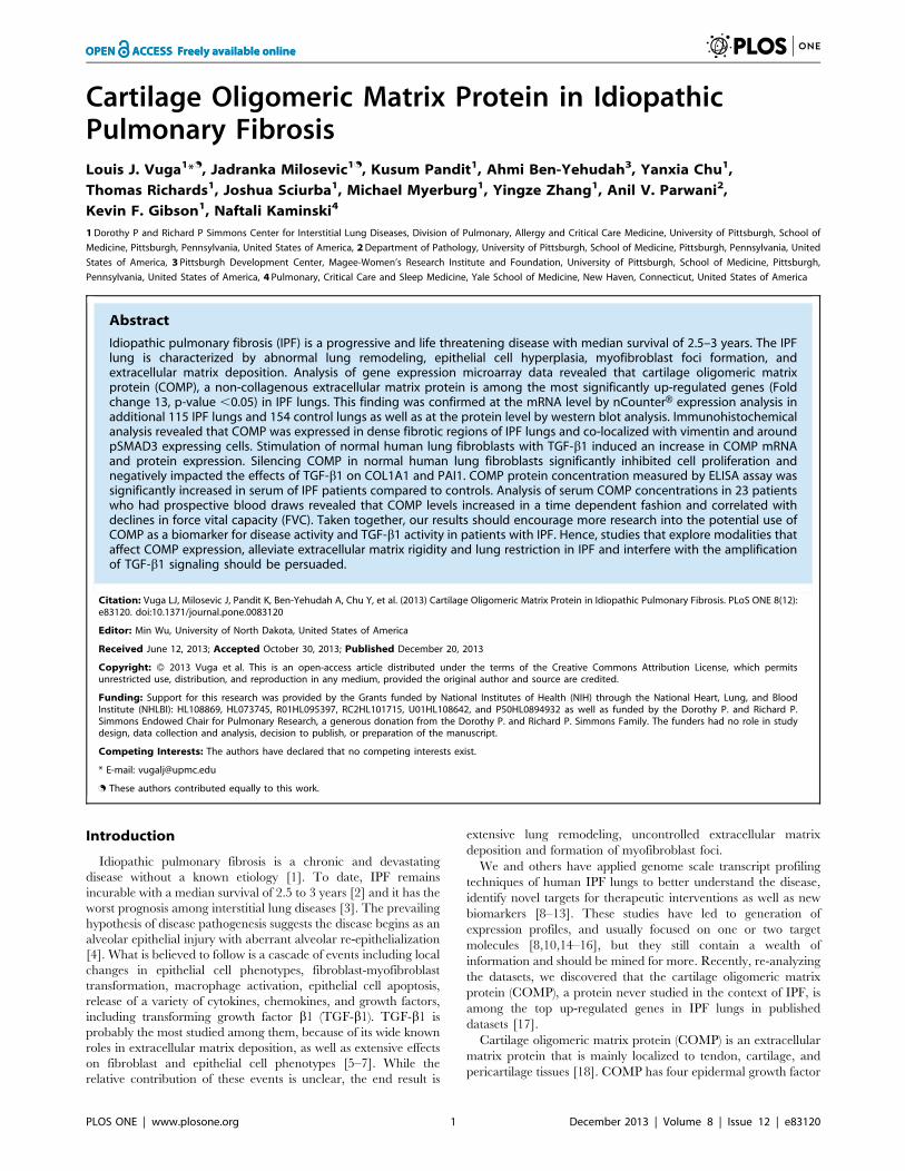
Figure 1. COMP gene and protein levels are increased in IPF lungs. (A) Microarrays analysis revealed an increased COMP gene expression in23 IPF lungs compared to 15 control lungs (A) and verified in 13 IPF lungs and 13 control lungs by using qRT-PCR (B). (C) COMP mRNA levels weredetermined by nCounterH in 115 IPF lungs and 154 control lungs. (D–E) Protein levels of COMP were determined in IPF lungs (n = 6) and control lungs(n = 6) by using western blot and quantified by using ImageJ (p,0.05). b- Actin was used as loading control and the western blot shown is arepresentative of three repeated experiments.doi:10.1371/journal.pone.0083120.g001
COMP in IPF
PLOS ONE | www.plosone.org 3 December 2013 | Volume 8 | Issue 12 | e83120
Enzyme-Linked Immunosorbent Assay (ELISA)The IPF patients were recruited in longitudinal study as
described in ‘‘Study Population’’. Participants were followed up
for 2.5 years while their blood was drawn and PFT were
examined. We measured COMP level in serum of IPF patients
and controls using COMP ELISA kit (AnnaMar Medical AB,
Goeteborg, Sweden). The experimental procedures were followed
as recommended by the vendor and data were analyzed by
utilizing Delta soft 111 version 2.243 (Bio-Rad, Hercules, CA).
Statistical Analysis of DataAll values were presented as mean 6 SD. Group comparisons
were made using an unpaired, two-tailed Student’s t-test for
normally distributed data. A level of p,0.05 was considered
statistically significant. Longitudinal study of COMP and all the
PFT for 23 patients within 120 days of any blood draw were used
for correlation analysis of COMP level to FVC %. A data set with
one record per PFT occasion, associating each PFT with COMP
level from the blood draw nearest it, was generated for all PFT of
23 patients within 120 days of any blood draw. A generalized
estimating equation was used to fit to the data, based on the
reference of Yan et al [43] with FVC % predicted as dependent
and COMP as independent variable, and accounting for the
subject effect by assuming an exchangeable within-subject
correlation structure.
Results
COMP Gene and Protein Expression is Higher in IPFLungsCOMP was one of the most significantly increased genes in our
previously published microarray data [17] and qRT-PCR
confirmed its up-regulation in the same tissues (Figure 1A–B).
We also verified the array result in a separate, larger cohort
consisting of 115 IPF lung samples and 154 normal histology
controls, by using nCounterH expression analysis and demonstrat-
ed that COMP mRNA was significantly increased in IPF lungs
(8.8 Fold change, P- value =,0.05 compared to normal histology
lungs) (Figure 1C). To compare COMP protein levels in IPF lungs
to those in control lungs, we performed western blot analysis. We
found a significant increase of COMP protein levels in IPF lungs
(Figure 1D–E). In order to localize COMP in IPF lungs, we
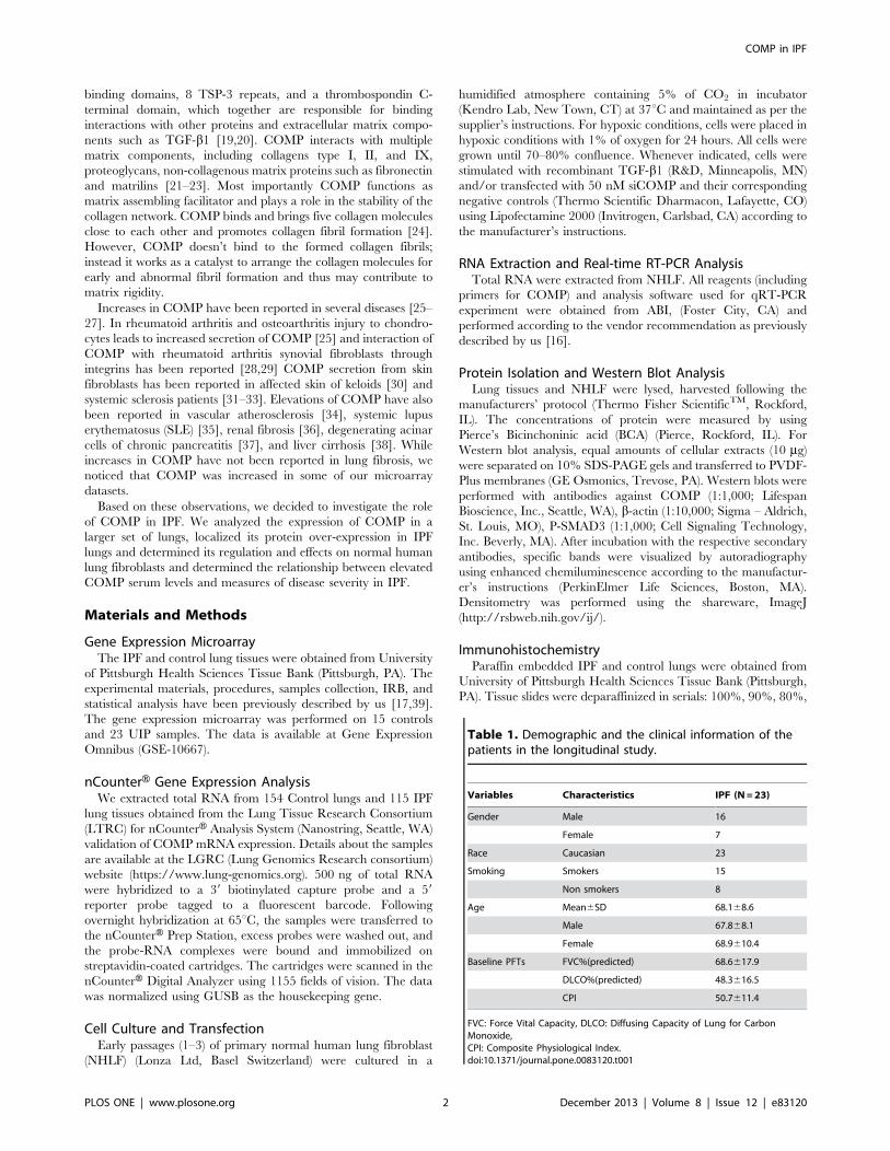
performed Immunohistochemistry (IHC) analysis. In normal
histology lungs, COMP was mainly located in the cartilaginous
areas of the large airways (Figure 2B). The IPF lung exhibited
expression of COMP (red in color) in areas of dense fibrosis and
myofibroblast foci (Figure 2C). To identify the types of cell that
secrete COMP protein in the lungs, we performed immunofluo-
rescence stains on frozen IPF and control lungs. COMP (green)
and Vimentin (red) were co-localized in IPF lungs (Figure 2G)
suggesting that COMP was secreted mainly by mesenchymal cells,
most probably fibroblasts in IPF lungs.
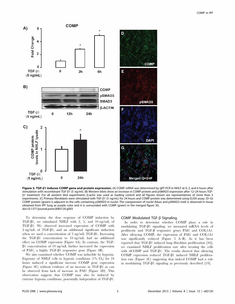
COMP is a TGF-b1 and Hypoxia Inducible MoleculeAfter co-localizing COMP in fibroblasts of IPF lungs, we
wanted to determine whether TGF-b1 regulates COMP expres-
sion. Stimulation of NHLF with TGF-b1 (5 ng/ml) induced
increase in mRNA and protein levels of COMP in a time
dependent manner as determined by qRT-PCR, western blot and
ELISA (Figure 3A–C). To determine whether COMP expression
in the fibrotic lungs could be associated with TGF-b1 effects, we
co-localized phosphorylated SMAD3 (pSMAD3) and COMP. We
found that COMP proteins were localized adjacent to cells
containing pSMAD3 in the nuclei (Figure 3G) suggesting that this
indeed was the case.
Figure 2. COMP is localized in fibrotic regions of idiopathic pulmonary fibrosis lungs. (A–B) Localization of COMP protein (red) in tissueobtained from control lung: a healthy lung parenchyma without COMP (A) and pericartilage airway region with COMP accumulation (B). (C) COMPprotein (red) is located in fibrotic region of IPF Lungs. (D–G) Co-localization of COMP and vimentin in tissue obtained from IPF lung. The greenfluorescence represents COMP and the red fluorescence shows fibroblasts marker; vimentin. Nuclei were counterstained with 4, 6- diamidino-2-phenylindole (blue). Yellow represents co-expression of COMP (green) and vimentin (red) in IPF lung as a yellow tinge. All figures shown arerepresentatives of more than 3 experiments and magnification of 406.doi:10.1371/journal.pone.0083120.g002
COMP in IPF
PLOS ONE | www.plosone.org 4 December 2013 | Volume 8 | Issue 12 | e83120
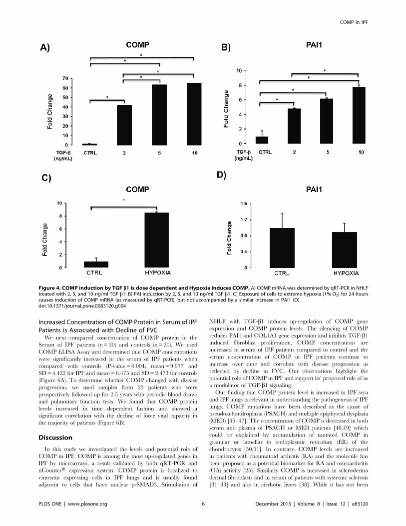
To determine the dose response of COMP induction by
TGF-b1, we stimulated NHLF with 2, 5, and 10 ng/mL of
TGF-b1. We observed increased expression of COMP with
2 ng/mL of TGF-b1, and an additional significant induction
when we used a concentration of 5 ng/mL TGF-b1. Increasingthe TGF-b1 concentration to 10 ng/mL had no additional
effect on COMP expression (Figure 4A). In contrast, the TGF-
b1 concentration of 10 ng/mL further increased the expression
of PAI1, a highly TGF-b1 responsive gene (Figure 4B).
We also examined whether COMP was inducible by hypoxia.
Exposure of NHLF cells to hypoxic conditions (1% O2) for 24
hours induced a significant increase COMP gene expression
(Figure 4C) without evidence of an increase in TGF-b1 as can
be observed from lack of increase in PAI1 (Figure 4D). This
observation suggests that COMP may also be induced by
extreme hypoxia conditions, potentially independent of TGF-b1.
COMP Modulated TGF-b SignalingIn order to determine whether COMP plays a role in
modulating TGF-b1 signaling, we measured mRNA levels of
profibrotic and TGF-b responsive genes PAI1 and COL1A1.
After silencing COMP, the expression of PAI1 and COL1A1
was significantly reduced (Figure 5 A–B). As it has been
reported that TGF-b1 induced lung fibroblast proliferation [44],
we examined NHLF proliferation rate after treating the cells
with siCOMP and TGF-b1. The results showed that silencing
COMP expression reduced TGF-b1 induced NHLF prolifera-
tion rate (Figure 5C) suggesting that indeed COMP had a role
in modulating TGF-b1 signaling as previously described [19].
Figure 3. TGF-b1 induces COMP gene and protein expression. (A) COMP mRNA was determined by qRT-PCR in NHLF at 0, 2, and 6 hours afterstimulation with recombinant TGF-b1 (5 ng/ml). (B) Western blots show an increase in COMP protein and pSMAD3 expression after 12–24 hours TGF-b1 treatment. For all western blot experiments b-actin was used as loading control and all figures shown are representatives of more than 3experiments. (C) Primary fibroblasts were stimulated with TGF-b1 (5 ng/mL) for 24 hours and COMP protein was determined using ELISA assay. (D–G)COMP protein (green) is adjacent to the cells containing pSMAD3 in nuclei. The coexpression of nuclei (blue) and pSMAD3 (red) is observed in tissueobtained from IPF lung as purple color and it is surrounded with COMP (green) in the merged figure 3G.doi:10.1371/journal.pone.0083120.g003
COMP in IPF
PLOS ONE | www.plosone.org 5 December 2013 | Volume 8 | Issue 12 | e83120
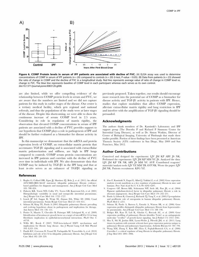
Increased Concentration of COMP Protein in Serum of IPFPatients is Associated with Decline of FVCWe next compared concentration of COMP protein in the
Serum of IPF patients (n = 20) and controls (n = 20). We used
COMP ELISA Assay and determined that COMP concentrations
were significantly increased in the serum of IPF patients when
compared with controls (P-value = 0.004, mean= 9.977 and
SD=4.422 for IPF and mean=6.475 and SD=2.473 for controls
(Figure 6A). To determine whether COMP changed with disease
progression, we used samples from 23 patients who were
prospectively followed up for 2.5 years with periodic blood draws
and pulmonary function tests. We found that COMP protein
levels increased in time dependent fashion and showed a
significant correlation with the decline of force vital capacity in
the majority of patients (Figure 6B).
Discussion
In this study we investigated the levels and potential role of
COMP in IPF. COMP is among the most up-regulated genes in
IPF by microarrays, a result validated by both qRT-PCR and
nCounterH expression system. COMP protein is localized to
vimentin expressing cells in IPF lungs and is usually found
adjacent to cells that have nuclear p-SMAD3. Stimulation of
NHLF with TGF-b1 induces up-regulation of COMP gene
expression and COMP protein levels. The silencing of COMP
reduces PAI1 and COL1A1 gene expression and inhibits TGF-b1induced fibroblast proliferation. COMP concentrations are
increased in serum of IPF patients compared to control and the
serum concentration of COMP in IPF patients continue to
increase over time and correlate with disease progression as
reflected by decline in FVC. Our observations highlight the
potential role of COMP in IPF and support its’ proposed role of as
a modulator of TGF-b1 signaling.
Our finding that COMP protein level is increased in IPF sera
and IPF lungs is relevant in understanding the pathogenesis of IPF
lungs. COMP mutations have been described as the cause of
pseudoachondroplasia (PSACH) and multiple epiphyseal dysplasia
(MED) [45–47]. The concentration of COMP is decreased in both
serum and plasma of PSACH or MED patients [48,49] which
could be explained by accumulation of mutated COMP in
granular or lamellar in endoplasmic reticulum (ER) of the
chondrocytes [50,51]. In contrary, COMP levels are increased
in patients with rheumatoid arthritis (RA) and the molecule has
been proposed as a potential biomarker for RA and osteoarthritis
(OA) activity [25]. Similarly COMP is increased in scleroderma
dermal fibroblasts and in serum of patients with systemic sclerosis
[31–33] and also in cirrhotic livers [38]. While it has not been
Figure 4. COMP induction by TGF b1 is dose dependent and Hypoxia induces COMP. A) COMP mRNA was determined by qRT-PCR in NHLFtreated with 2, 5, and 10 ng/ml TGF b1. B) PAI induction by 2, 5, and 10 ng/ml TGF b1. C) Exposure of cells to extreme hypoxia (1% O2) for 24 hourscauses induction of COMP mRNA (as measured by qRT-PCR), but not accompanied by a similar increase in PAI1 (D).doi:10.1371/journal.pone.0083120.g004
COMP in IPF
PLOS ONE | www.plosone.org 6 December 2013 | Volume 8 | Issue 12 | e83120

studied directly in the lung, a recent longitudinal study in patients
with systemic scleroderma with lung involvement suggested that
elevated COMP concentrations in serum were predictive of
mortality [52]. While we did not assess mortality, we did show an
increase in COMP proteins in the lungs and bloods of patients
with IPF and demonstrated that the increase was associated with
decline of FVC over time, concurrent with previous results in
other disease and suggesting that COMP should be added to
repertoire of proteins evaluated as potential peripheral blood
biomarkers in IPF.
While we could not directly demonstrate COMP induction by
TGF-b1 in human IPF lungs, we demonstrated that COMP
mRNA and protein levels were increased after stimulation of
human lung fibroblasts with TGF-b1. This is consistent with other
reports that demonstrated induction of COMP after TGF-b1stimulation in keloid and dermal fibroblast [30,33]. Consistent
with that, we have shown that COMP expression in the IPF lung is
generally distributed around cells that express vimentin and that
have abundant phosphorylated nuclear SMAD3, indicative of
TGF-b1 stimulation. This is of particular interest because it had
been described that COMP directly binds to members of TGF-b1family and enhances their signal transduction activities [19]. When
we examined the impacts of TGF-b1 on NHLF after the silencing
of COMP, we found reduction in TGF-b1 target genes as well as
significant reduction in TGF-b1 induced fibroblast proliferation,
suggesting that indeed COMP does enhance TGF-b1 signaling.
While it is difficult to establish experimentally a similar relation in
the human IPF lung, our results that demonstrate co-localization
of COMP to areas where there is evidence of TGF-b1 activities,
add to these observations and suggest the possibility of a positive
feedback loop between COMP and TGF-b1 activities in the IPF
lung.
The fibrotic lung is characterized by the intensive accumulation
of extracellular matrix (ECM). The pathologic ECM depositions in
fibrotic lungs include collagens (I, III, V, VI and VII), fibronectin,
elastin, and cartilage related proteins [53]. Interestingly, we found
COMP was increased in dense fibrotic areas of lung parenchyma
in IPF. We also localized COMP in normal lung but only around
cartilage possessing airway (Figure 2A–B). In the case of COMP
expression in chronic diseases such as pseudoachondroplasia
(PSACH) and multiple epiphyseal dysplasia (MED), rheumatoid
arthritis, systemic sclerosis and pathological would healing
(Keloid), it was reported that COMP inhibits normal collagen
fibrils formation and destabilize normal extracellular matrix
formation in areas where COMP molecules are excessively higher
in the relationship to collagen [30,54]. It was suggested that this
inhibition cause increased lung rigidity by the lost elasticity typical
of normal ECM. This may be of particular interest, because of the
evidence that abnormal ECM rigidity plays a significant role in the
pathogenesis of fibrosis [55]. In this context, it is of interest to note
that other cartilage related proteins such as osteopontin [10,56],
periostin [57], and YKL-40 [58] have been described as being
significantly increased in IPF.
It is critically important to acknowledge few limitations of our
study. First, we provide evidence on COMP regulation through
TGF-b1 and its involvement in TGF-b1 signaling cascade in
NHLF, but we do not provide similar data in-vivo. We feel that
the co-localization of vimentin, pSMAD3 and COMP in fibrotic
foci in human IPF lungs suggests that TGF-b1 induces COMP
secretion mostly in fibrotic regions of IPF lungs and to some extent
more informative than using a limited model of lung fibrosis. To
that extent we did observe that in-vitro exposing NHLF to extreme
hypoxia in-vitro did also induce COMP, but it is unclear whether
this mechanism would be relevant in-vivo. Clinically, our findings
Figure 5. COMP modulates TGF-b signaling. NHLF were trans-fected with 70 nM of siCOMP or scrambled (SCR), treated for 6 hourswith TGF-b1 (5 ng/mL) and RNA extracted after 24 hours. (A–B) qRT-PCRwas used to determine mRNA levels of PAI1 and COL1A1 in NHLF. (C)The effect of COMP inhibition using siRNA on TGF-b1 induced NHLFproliferation.doi:10.1371/journal.pone.0083120.g005
COMP in IPF
PLOS ONE | www.plosone.org 7 December 2013 | Volume 8 | Issue 12 | e83120
are also limited, while we offer compelling evidence of the
relationship between COMP protein levels in serum and FVC, we
are aware that the numbers are limited and we did not capture
patients for this study in earlier stages of the disease. Our center is
a tertiary medical facility, which gets regional and national
referrals, and thus the populations of the study were at later stages
of the disease. Despite this shortcoming, we were able to show the
continuous increase of serum COMP level in 2.5 years.
Considering its role in regulation of matrix rigidity, the
observation that elevated COMP concentrations in serum of IPF
patients are associated with a decline of FVC provides support to
our hypothesis that COMP plays a role in pathogenesis of IPF and
should be further evaluated as a biomarker for disease activity in
IPF.
In this manuscript we demonstrate that the mRNA and protein
expression levels of COMP, an extracellular matrix protein that
accentuates TGF-b1 signaling and is associated with extracellular
matrix polymerization and stiffness, are high in IPF lungs
compared to controls. COMP serum protein concentrations are
increased in IPF patients and correlate with the decline of FVC
over time in individuals with IPF. We also demonstrate data that
COMP may be induced by TGF-b1 in the IPF lung and that at
least in-vitro serves as an enhancer of TGFb1 signaling as
previously proposed. Taken together, our results should encourage
more research into the potential use of COMP as a biomarker for
disease activity and TGF-b1 activity in patients with IPF. Hence,
studies that explore modalities that affect COMP expression,
alleviate extracellular matrix rigidity and lung restriction in IPF
and interfere with the amplification of TGF-b1 signaling should be
persuaded.
Acknowledgments
The authors thank members of Dr. Kaminski’s Laboratory and IPF
support group (The Dorothy P and Richard P Simmons Center for
Interstitial Lung Diseases), as well as Dr. Simon Watkins, Director of
Center of Biological Imaging, University of Pittsburgh that made these
studies possible. Portion of these findings have been presented at American
Thoracic Society (ATS) conference in San Diego, May 2009 and San
Francisco, May 2012.
Author Contributions
Conceived and designed the experiments: LJV JM KP ABY JS NK.
Performed the experiments: LJV JM KP MM YC JS. Analyzed the data:
LJV JM KP TR NK ABY JS MM YC AVP. Contributed reagents/
materials/analysis tools: LJV YZ MMTR AVP NK. Wrote the paper: LJV
JM NK. Patients recruitment: KFG YZ.
References
1. Raghu G, Collard HR, Egan JJ, Martinez FJ, Behr J, et al. (2011) An official
ATS/ERS/JRS/ALAT statement: idiopathic pulmonary fibrosis: evidence-
based guidelines for diagnosis and management. Am J Respir Crit Care Med
183: 788–824.
2. Flaherty KR, Travis WD, Colby TV, Toews GB, Kazerooni EA, et al. (2001)
Histopathologic variability in usual and nonspecific interstitial pneumonias.
Am J Respir Crit Care Med 164: 1722–1727.
3. Lynch JP 3rd, Saggar R, Weigt SS, Zisman DA, White ES (2006) Usual
interstitial pneumonia. Semin Respir Crit Care Med 27: 634–651.
4. Selman M, King TE, Pardo A (2001) Idiopathic pulmonary fibrosis: prevailing
and evolving hypotheses about its pathogenesis and implications for therapy.
Ann Intern Med 134: 136–151.
5. Pottier N, Maurin T, Chevalier B, Puissegur MP, Lebrigand K, et al. (2009)
Identification of keratinocyte growth factor as a target of microRNA-155 in lung
fibroblasts: implication in epithelial-mesenchymal interactions. PLoS One 4:
e6718.
6. Willis BC, Borok Z (2007) TGF-beta-induced EMT: mechanisms and
implications for fibrotic lung disease. Am J Physiol Lung Cell Mol Physiol
293: L525–534.
7. Pandit KV, Corcoran D, Yousef H, Yarlagadda M, Tzouvelekis A, et al. (2010)
Inhibition and role of let-7d in idiopathic pulmonary fibrosis. Am J Respir Crit
Care Med 182: 220–229.
8. Zuo F, Kaminski N, Eugui E, Allard J, Yakhini Z, et al. (2002) Gene expression
analysis reveals matrilysin as a key regulator of pulmonary fibrosis in mice and
humans. Proc Natl Acad Sci U S A 99: 6292–6297.
9. Cosgrove GP, Brown KK, Schiemann WP, Serls AE, Parr JE, et al. (2004)
Pigment epithelium-derived factor in idiopathic pulmonary fibrosis: a role in
aberrant angiogenesis. Am J Respir Crit Care Med 170: 242–251.
10. Pardo A, Gibson K, Cisneros J, Richards TJ, Yang Y, et al. (2005) Up-regulation
and profibrotic role of osteopontin in human idiopathic pulmonary fibrosis.
PLoS Med 2: e251.
11. Selman M, Pardo A, Barrera L, Estrada A, Watson SR, et al. (2006) Gene
expression profiles distinguish idiopathic pulmonary fibrosis from hypersensitiv-
ity pneumonitis. Am J Respir Crit Care Med 173: 188–198.
12. Bridges RS, Kass D, Loh K, Glackin C, Borczuk AC, et al. (2009) Gene
expression profiling of pulmonary fibrosis identifies Twist1 as an antiapoptotic
molecular ‘‘rectifier’’ of growth factor signaling. Am J Pathol 175: 2351–2361.
13. Hsu E, Shi H, Jordan RM, Lyons-Weiler J, Pilewski JM, et al. (2011) Lung
tissues in patients with systemic sclerosis have gene expression patterns unique to
pulmonary fibrosis and pulmonary hypertension. Arthritis Rheum 63: 783–794.
14. Wang XM, Zhang Y, Kim HP, Zhou Z, Feghali-Bostwick CA, et al. (2006)
Caveolin-1: a critical regulator of lung fibrosis in idiopathic pulmonary fibrosis.
J Exp Med 203: 2895–2906.
Figure 6. COMP Protein levels in serum of IPF patients are associated with decline of FVC. (A) ELISA assay was used to determineconcentrations of COMP in serum of IPF patients (n = 20) compared to controls (n = 20) (t-test, P-value ,0.05). (B) Data from patients (n = 23) showedthe ratio of change in COMP and the decline of FVC in a longitudinal study. Red line represents average value of ratio of change in COMP level vs.change in FVC. The blue line represents baseline of COMP level in each participant whereas each serves as its own control.doi:10.1371/journal.pone.0083120.g006
COMP in IPF
PLOS ONE | www.plosone.org 8 December 2013 | Volume 8 | Issue 12 | e83120
15. Englert JM, Hanford LE, Kaminski N, Tobolewski JM, Tan RJ, et al. (2008) A
role for the receptor for advanced glycation end products in idiopathicpulmonary fibrosis. Am J Pathol 172: 583–591.
16. Vuga LJ, Ben-Yehudah A, Kovkarova-Naumovski E, Oriss T, Gibson KF, et al.
(2009) WNT5A is a regulator of fibroblast proliferation and resistance toapoptosis. Am J Respir Cell Mol Biol 41: 583–589.
17. Konishi K, Gibson KF, Lindell KO, Richards TJ, Zhang Y, et al. (2009) Geneexpression profiles of acute exacerbations of idiopathic pulmonary fibrosis.
Am J Respir Crit Care Med 180: 167–175.
18. DiCesare PE, Morgelin M, Carlson CS, Pasumarti S, Paulsson M (1995)Cartilage oligomeric matrix protein: isolation and characterization from human
articular cartilage. J Orthop Res 13: 422–428.19. Haudenschild DR, Hong E, Yik JH, Chromy B, Morgelin M, et al. (2011)
Enhanced activity of transforming growth factor beta1 (TGF-beta1) bound tocartilage oligomeric matrix protein. J Biol Chem 286: 43250–43258.
20. Newton G, Weremowicz S, Morton CC, Copeland NG, Gilbert DJ, et al. (1994)
Characterization of human and mouse cartilage oligomeric matrix protein.Genomics 24: 435–439.
21. Thur J, Rosenberg K, Nitsche DP, Pihlajamaa T, Ala-Kokko L, et al. (2001)Mutations in cartilage oligomeric matrix protein causing pseudoachondroplasia
and multiple epiphyseal dysplasia affect binding of calcium and collagen I, II,
and IX. J Biol Chem 276: 6083–6092.22. Mann HH, Ozbek S, Engel J, Paulsson M, Wagener R (2004) Interactions
between the cartilage oligomeric matrix protein and matrilins. Implications formatrix assembly and the pathogenesis of chondrodysplasias. J Biol Chem 279:
25294–25298.23. Holden P, Meadows RS, Chapman KL, Grant ME, Kadler KE, et al. (2001)
Cartilage oligomeric matrix protein interacts with type IX collagen, and
disruptions to these interactions identify a pathogenetic mechanism in a bonedysplasia family. J Biol Chem 276: 6046–6055.
24. Halasz K, Kassner A, Morgelin M, Heinegard D (2007) COMP acts as a catalystin collagen fibrillogenesis. J Biol Chem 282: 31166–31173.
25. Chaganti RK, Kelman A, Lui L, Yao W, Javaid MK, et al. (2008) Change in
serum measurements of cartilage oligomeric matrix protein and association withthe development and worsening of radiographic hip osteoarthritis. Osteoarthritis
Cartilage 16: 566–571.26. Smith RK, Zunino L, Webbon PM, Heinegard D (1997) The distribution of
cartilage oligomeric matrix protein (COMP) in tendon and its variation withtendon site, age and load. Matrix Biol 16: 255–271.
27. Hesselstrand R, Kassner A, Heinegard D, Saxne T (2008) COMP: a candidate
molecule in the pathogenesis of systemic sclerosis with a potential as a diseasemarker. Ann Rheum Dis 67: 1242–1248.
28. Neidhart M, Zaucke F, von Knoch R, Jungel A, Michel BA, et al. (2005)Galectin-3 is induced in rheumatoid arthritis synovial fibroblasts after adhesion
to cartilage oligomeric matrix protein. Ann Rheum Dis 64: 419–424.
29. Chen FH, Thomas AO, Hecht JT, Goldring MB, Lawler J (2005) Cartilageoligomeric matrix protein/thrombospondin 5 supports chondrocyte attachment
through interaction with integrins. J Biol Chem 280: 32655–32661.30. Inui S, Shono F, Nakajima T, Hosokawa K, Itami S (2011) Identification and
characterization of cartilage oligomeric matrix protein as a novel pathogenicfactor in keloids. Am J Pathol 179: 1951–1960.
31. Yamamoto M, Takahashi H, Suzuki C, Naishiro Y, Yamamoto H, et al. (2007)
Cartilage oligomeric matrix protein in systemic sclerosis. Rheumatology(Oxford) 46: 1858–1859.
32. Farina G, Lemaire R, Korn JH, Widom RL (2006) Cartilage oligomeric matrixprotein is overexpressed by scleroderma dermal fibroblasts. Matrix Biol 25: 213–
222.
33. Farina G, Lemaire R, Pancari P, Bayle J, Widom RL, et al. (2009) Cartilageoligomeric matrix protein expression in systemic sclerosis reveals heterogeneity
of dermal fibroblast responses to transforming growth factor beta. Ann RheumDis 68: 435–441.
34. Canfield AE, Farrington C, Dziobon MD, Boot-Handford RP, Heagerty AM, et
al. (2002) The involvement of matrix glycoproteins in vascular calcification andfibrosis: an immunohistochemical study. J Pathol 196: 228–234.
35. Wislowska M, Jablonska B (2005) Cartilage oligomeric matrix protein in serumin systemic lupus erythematosus and knee osteoarthritis. Preliminary commu-
nication. Rheumatol Int 25: 373–378.36. Kim W, Moon SO, Lee SY, Jang KY, Cho CH, et al. (2006) COMP-
angiopoietin-1 ameliorates renal fibrosis in a unilateral ureteral obstruction
model. J Am Soc Nephrol 17: 2474–2483.
37. Liao Q, Kleeff J, Xiao Y, Di Cesare PE, Korc M, et al. (2003) COMP is
selectively up-regulated in degenerating acinar cells in chronic pancreatitis andin chronic-pancreatitis-like lesions in pancreatic cancer. Scand J Gastroenterol
38: 207–215.
38. Xiao Y, Kleeff J, Guo J, Gazdhar A, Liao Q, et al. (2004) Cartilage oligomericmatrix protein expression in hepatocellular carcinoma and the cirrhotic liver.
J Gastroenterol Hepatol 19: 296–302.39. Milosevic J, Pandit K, Magister M, Rabinovich E, Ellwanger DC, et al. (2012)
Profibrotic role of miR-154 in pulmonary fibrosis. Am J Respir Cell Mol Biol 47:
879–887.40. Gilani SR, Vuga LJ, Lindell KO, Gibson KF, Xue J, et al. (2010) CD28 down-
regulation on circulating CD4 T-cells is associated with poor prognoses ofpatients with idiopathic pulmonary fibrosis. PLoS One 5: e8959.
41. Demedts M, Costabel U (2002) ATS/ERS international multidisciplinaryconsensus classification of the idiopathic interstitial pneumonias. Eur Respir J 19:
794–796.
42. King TE, Jr., Tooze JA, Schwarz MI, Brown KR, Cherniack RM (2001)Predicting survival in idiopathic pulmonary fibrosis: scoring system and survival
model. Am J Respir Crit Care Med 164: 1171–1181.43. Yan J, Fine J (2004) Estimating equations for association structures. Stat Med 23:
859–874; discussion 875–857,879–880.
44. Khalil N, Xu YD, O’Connor R, Duronio V (2005) Proliferation of pulmonaryinterstitial fibroblasts is mediated by transforming growth factor-beta1-induced
release of extracellular fibroblast growth factor-2 and phosphorylation of p38MAPK and JNK. J Biol Chem 280: 43000–43009.
45. Ikegawa S, Ohashi H, Nishimura G, Kim KC, Sannohe A, et al. (1998) Noveland recurrent COMP (cartilage oligomeric matrix protein) mutations in
pseudoachondroplasia and multiple epiphyseal dysplasia. Hum Genet 103:
633–638.46. Deere M, Sanford T, Ferguson HL, Daniels K, Hecht JT (1998) Identification of
twelve mutations in cartilage oligomeric matrix protein (COMP) in patients withpseudoachondroplasia. Am J Med Genet 80: 510–513.
47. Briggs MD, Hoffman SM, King LM, Olsen AS, Mohrenweiser H, et al. (1995)
Pseudoachondroplasia and multiple epiphyseal dysplasia due to mutations in thecartilage oligomeric matrix protein gene. Nat Genet 10: 330–336.
48. Chen HC, Shah SH, Li YJ, Stabler TV, Jordan JM, et al. (2008) Inverseassociation of general joint hypermobility with hand and knee osteoarthritis and
serum cartilage oligomeric matrix protein levels. Arthritis Rheum 58: 3854–3864.
49. Tufan AC, Satiroglu-Tufan NL, Jackson GC, Semerci CN, Solak S, et al. (2007)
Serum or plasma cartilage oligomeric matrix protein concentration as adiagnostic marker in pseudoachondroplasia: differential diagnosis of a family.
Eur J Hum Genet 15: 1023–1028.50. Stanescu R, Stanescu V, Muriel MP, Maroteaux P (1993) Multiple epiphyseal
dysplasia, Fairbank type: morphologic and biochemical study of cartilage.
Am J Med Genet 45: 501–507.51. Hansen U, Platz N, Becker A, Bruckner P, Paulsson M, et al. (2011) A secreted
variant of cartilage oligomeric matrix protein carrying a chondrodysplasia-causing mutation (p.H587R) disrupts collagen fibrillogenesis. Arthritis Rheum
63: 159–167.52. Hesselstrand R, Andreasson K, Wuttge DM, Bozovic G, Scheja A, et al. (2012)
Increased serum COMP predicts mortality in SSc: results from a longitudinal
study of interstitial lung disease. Rheumatology (Oxford) 51: 915–920.53. Pardo A, Selman M, Kaminski N (2008) Approaching the degradome in
idiopathic pulmonary fibrosis. Int J Biochem Cell Biol 40: 1141–1155.54. Agarwal P, Schulz JN, Blumbach K, Andreasson K, Heinegard D, et al. (2013)
Enhanced deposition of cartilage oligomeric matrix protein is a common feature
in fibrotic skin pathologies. Matrix Biol 32: 325–331.55. Zhou Y, Huang X, Hecker L, Kurundkar D, Kurundkar A, et al. (2013)
Inhibition of mechanosensitive signaling in myofibroblasts ameliorates experi-mental pulmonary fibrosis. J Clin Invest 123: 1096–1108.
56. Kadota J, Mizunoe S, Mito K, Mukae H, Yoshioka S, et al. (2005) High plasma
concentrations of osteopontin in patients with interstitial pneumonia. RespirMed 99: 111–117.
57. Okamoto M, Hoshino T, Kitasato Y, Sakazaki Y, Kawayama T, et al. (2011)Periostin, a matrix protein, is a novel biomarker for idiopathic interstitial
pneumonias. Eur Respir J 37: 1119–1127.58. Korthagen NM, van Moorsel CH, Barlo NP, Ruven HJ, Kruit A, et al. (2011)
Serum and BALF YKL-40 levels are predictors of survival in idiopathic
pulmonary fibrosis. Respir Med 105: 106–113.
COMP in IPF
PLOS ONE | www.plosone.org 9 December 2013 | Volume 8 | Issue 12 | e83120


