Saint Petersburg State University Faculty of Physics
Department of Quantum Magnetic Phenomena
International Symposium and Summer School in Saint Petersburg
Nuclear Magnetic Resonance in Condensed Matter
10th meeting: “NMR in Life Sciences” July 8 – 12 2013
Book of Abstracts
Saint Petersburg, Russia 2013
an AMPERE event
International Symposium and Summer School in Saint Petersburg
Nuclear Magnetic Resonance in Condensed Matter
10th meeting: “NMR in Life Sciences” July 8 – 12 2013
an
AMPERE event
ББК В334.2, Г512 М43 Department of Quantum Magnetic Phenomena
Faculty of Physics Saint Petersburg State University Saint Petersburg, 198504, Russia
http://nmr.phys.spbu.ru/nmrcm/
M43 Nuclear Magnetic Resonance in Condensed Matter: Abstracts of the International Symposium and Summer School, 10th meeting: “NMR in Life Sciences” – Saint Petersburg: “Solo” Publisher, 2013. – 128 p. ISBN
Symposium and Summer School are supported by: • Saint Petersburg State University • German-Russian Interdisciplinary Science Center • Russian Foundation for Basic Research • Dynasty Foundation • Bruker Biospin Russia
International Advisory Board V. Balevicius (Vilnius, Lithuania)
V. I. Chizhik (Saint Petersburg, Russia) J. Fraissard (Paris, France) H. Haranczyk (Kraków, Poland) S. Jurga (Poznań, Poland) O. B. Lapina (Novosibirsk, Russia) D. Michel (Leipzig, Germany)
V. I. Minkin (Rostov-on-Don, Russia) K. V. Ramanathan (Bangalore, India) R. Z. Sagdeev (Novosibirsk, Russia) K. M. Salikhov (Kazan, Russia) A. V. Skripov (Ekaterinburg, Russia) N. R. Skrynnikov (Purdue, USA) M. S. Tagirov (Kazan, Russia)
Organizing Committee Members:
S. F. Boureiko A. V. Donets V. V. Frolov V. V. Matveev S. M. Sukharzhevskii P. M. Tolstoy
Layout of Abstracts Book: A. A. Levantovsky
Co-Chairmen: V. I. Chizhik R. Z. Sagdeev (Novosibirsk)
Vice-Chairmen: A. V. Egorov M. G. Shelyapina
Registered names, trademarks, etc. used in this book, even without specific indication thereof, are not to be considered unprotected by law.
ISBN ББК В334.2, Г512 © Organizing Committee NMRCM 2013, Saint Petersburg, 2013.
© “Solo” Publisher, Saint Petersburg, 2013. Printed in Russian Federation.
– 3 – NMRCM 2013, Saint Petersburg, Russia, July 8 – 12, 2013
Contents
I. Lectures ................................................................................................................ 11
E. V. Charnaya, C. Tien, M. K. Lee, D. Nefedov, Y. A. Kumzerov, J. Haase, D. Michel
NMR studies of nanostructured sodium and sodium-potassium alloy ............................................................ 13
Denis Grebenkov
Theoretical and numerical methods for DWMRI .............................................................................................. 14
Uwe Eichhoff
Recent developments in animal MRI. A personal review of the 21th annual meeting of ISMRM 2013
in Salt Lake City ................................................................................................................................................. 15
Jacques Fraissard
A “pot-pourri” of 129Xe NMR technique ............................................................................................................ 16
Sergio Fuentes
Inorganic-Fullerenes based on MS2 dichalcogenides (M=Mo, W, Ta, Nb) and TiO2 ......................................... 17
Valerij Kiselev
Tracking neuronal fibers using diffusion-weighted MRI ................................................................................... 18
Boris B. Kharkov, Sergey V. Dvinskikh
Fine art of packing: Solid State NMR study of surfactants at solid interfaces .................................................. 19
Olga Lapina, Eugine Papulovsky, Dzhalil Khabibulin, and Alexandre Shubin
Multinuclear SSNMR/DFT GIPAW for molecular structure and reactivity relationships for supported oxide
catalysts ............................................................................................................................................................. 20
Dieter Michel
Proton and deuteron ordering and dynamics in solids and in molecules adsorbed in porous materials ........ 21
Dmitry Novikov
Characterizing tissue microstructure with time-dependent diffusion ............................................................. 22
Rita G. Nunes
Diffusion-weighted imaging pulse sequences: Echo planar imaging and alternative methods ....................... 23
A. F. Privalov
Recent developments in Field Cycling NMR ..................................................................................................... 24
A. V. Skripov
Nuclear magnetic resonance studies of atomic motion in borohydride-based hydrogen storage materials .. 25
Murat Tagirov
Fullerene like nanoparticles of PrF3: from creations to medical applications .................................................. 26
Sergey Vasiliev, Jarno Järvinen, Denis Zvezdov, Janne Ahokas, Sergey Sheludyakov, Otto Vainio,
Takao Mizusaki, Yutaka Fujii, Seitaro Mitsudo, Minchan Gwak, SangGap Lee
Dynamic nuclear polarization at ultralow temperatures .................................................................................. 27
Vitaly I. Volkov
Pulsed Field Gradient NMR for biological membranes and polymeric electrolyte investigations ................... 28
Matthias Weigel
Extended Phase Graphs: What they mean and how to include diffusion ........................................................ 29
NMRCM 2013, Saint Petersburg, Russia, July 8 – 12, 2013 – 4 –
II. Oral Reports ......................................................................................................... 31
Nikolay V. Anisimov, Ekaterina I. Shalamova, Svetlana S. Batova, Andrey A. Samoylenko
Use of MRI database for analysis of evolution of magnetic field of superconducting magnet ........................ 34
Vyacheslav A. Chertkov, Dmitriy A. Cheshkov, Tatiana A. Ganina, Sergey S. Nechausov, Kirill F. Sheberstov,
Alla K. Shestakova
Novel high resolution NMR techniques for elucidation of molecular structure and dynamics ....................... 35
Rinat G. Dzhambulatov, Alexey V. Donets
Hydration properties of carboxylic acid functional groups in aqueous solutions ............................................ 36
Jan Gabriel, Michael Vogel
7Li NMR field-cycling relaxometry as tool to investigate lithium ion dynamics in solid-state electrolytes ...... 37
Oksana Ilina
Concomitant gradient terms in low-field MRI .................................................................................................. 38
Vadim V. Kachala, Elena A. Khokhlova and Valentine P. Ananikov
High-resolution NMR studies in ionic liqiuds .................................................................................................... 39
Kerstin Kämpf, Beke Kremmling, Michael Vogel
Temperature dependence of internal protein backbone dynamics studied by 2H NMR and random-walk
simulations ........................................................................................................................................................ 40
Boris B. Kharkov
Phase behavior of CTAB bilayer intercalated into magadiite ........................................................................... 41
Ilya A. Khodov, Mikhail Yu. Nikiforov, Gennady A. Alper, Sergey V. Efimov, Vladimer V. Klochkov,
Luís A. E. Batista de Carvalho
Conformation preference of ibuprofen in chloroform by 2D-3D NMR and GIAO quantum chemistry
calculation ......................................................................................................................................................... 42
Alexandr A. Khrapichev
Chemical Exchange Saturation Transfer (CEST) for intracellular pH-sensitive Magnetic Resonance Imaging
(MRI) .................................................................................................................................................................. 43
K. Klyukin, M. G. Shelyapina, D. Fruchart
Hydrogen diffusion in various structures of MgHx: a DFT study ....................................................................... 44
Anastasia Y. Kultaeva, Artem I. Antipov, Stanislav M. Sukharzhevskii
Effect radiation annealing on the kinetics of accumulation of electron-hole centers in
paleontological objects ..................................................................................................................................... 45
Vyacheslav V. Kuzmin, M. E. Hayden, G. Tastevin, P.-J. Nacher
New insight on nonlinear spin dynamics in highly polarized liquids ................................................................ 46
Vladimir Matveev, Denis Markelov, Vladimir Chizhik, Petri Ingman, Erkki Lähderanta
13C and 1H NMR Relaxation and segmental mobility in [emim]CH3COO Ionic Liquid ....................................... 47
Ivan V. Pleshakov, Nikolay S. Klekhta
NMR signal operation in magnetic substances by the external field pulses .................................................... 48
M. Podrecca, G. Grolet, V. Delsinne & J. M. Colet
Regulation of trophoblast migration: from a fingerprinting view to a mechanistic prospect
in bioassay design ............................................................................................................................................. 49
Sevastyan O. Rabdano, Alexey V. Donets
Hydrophobic and hydrophilic effects in aqueous solutions of organic molecules ........................................... 50
– 5 – NMRCM 2013, Saint Petersburg, Russia, July 8 – 12, 2013
V. A. Ryzhov, I. V. Pleshakov, A. A. Nechitailov, N. V. Glebova, E. N. Pyatyshev, A. V. Malkova, I. A. Kiselev,
V. V. Matveev
Magnetic study of nanostructural composite material based on cobalt compounds and porous silicon ....... 51
Ago Samoson
Hot spinning ...................................................................................................................................................... 52
M. A. Shevtsov, L. Yu. Yakovleva, B. P. Nikolaev, Ya. Yu. Marchenko, A. V. Dobrodumov, K. V. Onokhin,
A. L. Mikhrina, I. V. Guzhova, M. G. Martynova, O. A. Bystrova, A. M. Ischenko, B. A. Margulis
Magnetic Resonance Study of SPIONs conjugated with Hsp70 in C6 glioma intracranial model ..................... 53
Timur A. Sibgatullin, Frank J. Vergeldt, Henk Van As
Spin-spin relaxation-diffusion correlation analysis for estimation of distribution of membrane permeability
and cell size in plant tissues .............................................................................................................................. 54
Nikolai Skrynnikov
Ensemble-restrained MD simulations: accurate structure leads to accurate dynamics .................................. 55
Sergey G. Vasil’ev, Vitaly I. Volkov, Elena A. Tatarinova, Aziz M. Muzafarov
A solid-state NMR investigation of MQ silicone copolymers ............................................................................ 56
Anna V. Vyvodtceva, Marina G. Shelyapina, Alexey Privalov, Daniel Fruchart
1H NMR study of hydrogen diffusion in disordered Ti-V-Cr alloys .................................................................... 57
III. Poster Session ..................................................................................................... 59
E. M. Alakshin, R. R. Gazizulin, A. V. Klochkov, K. Kono, S. L. Korableva, V. V. Kuzmin, A. M. Sabitova,
T. R. Safin, K. R. Safiullin, M. S. Tagirov
Magnetic coupling in system 3He – PrF3 nanoparticles ..................................................................................... 61
Nikolay V. Anisimov, Olga V. Isaeva, Svetlana S. Batova
Simplification of MRI contrast by inversion recovery method ......................................................................... 62
Hike Asatryan, Darya Kramushchenko, Yulia Uspenskaya
Registration of “antisite” defects in Y3Al5O12 by electron paramagnetic resonance ........................................ 63
Mariia I. Averina, Andrei V. Egorov
Concentration effect on cations solvation in Ca(NO3)2 – LiNO3 – H2O ternary system at normal conditions.
A molecular dynamics simulation study ........................................................................................................... 64
Eduard Baibekov
To the possibility of the high-temperature spin-photon coherence in a microwave cavity ............................ 65
A. G. Bazir, O. S. Vezo, B. P. Nikolaev, Ya. Yu. Marchenko, L. Yu. Yakovleva, V. I. Rolich, V. V. Vojtylov
NMR and Optical Properties of Dextran coated magnetic Nanosols ................................................................ 66
Eduard V. Bocharov, Konstantin S. Mineev, Dmitry M. Lesovoy, Sergey A. Goncharuk, Olga V. Bocharova,
Pavel K. Kuzmichev, Pavel E. Volynski, Alexander S. Arseniev
Structural and thermodynamic aspects of specific interactions of transmembrane domains of bitopic
proteins ............................................................................................................................................................. 67
Olga V. Bocharova, Anatatoly S. Urban, Ilia S. Chaplygin, Kirill D. Nadezhdin, Pavel E. Volynski,
Eduard V. Bocharov, Alexander S. Arseniev
Structural insight on the dimerization of transmembrane domain of amyloid precursor protein .................. 68
Yury V. Bogachev, Julia S. Chernenco, Kamil G. Gareev, Irina E. Kononova, Lev B. Matyushkin,
Vyacheslav A. Moshnikov
Study of NMR relaxation in suspensions of magnetite-silica nanocomposite .................................................. 69
NMRCM 2013, Saint Petersburg, Russia, July 8 – 12, 2013 – 6 –
Yu. Bogachev, Yu. Chernenko, L. Grunin, V. Drapkin, M. Knyazev, Ya. Marchenko, A. Naumova
Compact combined EPR/NMR/DNP Equipment ............................................................................................... 70
Inna G. Borodkina, Anatolii S. Burlov, Gasan M. Abakarov, Konstantin A. Lyssenko, Igor S. Vasilchenko,
Ali I. Uraev, Gennadii S. Borodkin, Dmitryi A. Garnovskii, Pavel B. Chepurnoy, Yuriy F. Mal’tsev,
Mikhail Yu. Antipin, Vladimir I. Minkin
Structure of Te-containing salen-type complexes with transition metals in solution and solid state ............. 71
Inna G. Borodkina, Vera A. Anisimova, Ivan E. Tolpygin, Gennadii S. Borodkin, Igor S. Vasilchenko,
Pavel B. Chepurnoy, Yuriy F. Mal’tsev, Vladimir I. Minkin
Prototropic tautomerism 1(10)H-2,3,4,10-tetrahydropyrimido ....................................................................... 72
Eugenia A. Burilova, Anna B. Ziyatdinova, Rustem R. Amirov
Paramagnetic NMR-probing of polymeric solutions ......................................................................................... 73
Anna S. Dmitruk, S. G. Vasil’ev, E. A. Tatarinova
Synthesis and characterization of Hexa(trimethylsiloxy)disiloxane using 1H, 13C and 29Si NMR spectroscopy 74
Andrei V. Egorov, Mariia I. Averina
Multinuclear NMR relaxation as a tool of investigating local structure and dynamics of ternary aqueous
solutions of nitrate salts .................................................................................................................................... 75
Galina N. Fedyukina, Boris V. Sakharov, Pavel Ch. Kopilov, Sergey A. Ivanov, Andrey P. Anisimov
H1-NMR relaxation study of an amorphous state of the freeze-dried powders ............................................... 76
Dmitry Furman
Detection of foreign bodies in the object using magnetic resonance imaging ................................................ 77
Ramil R. Gainov, Vera V. Klekovkina, Farit G. Vagizov, Alexander V. Dooglav, Vladimir A. Golovanevskiy,
Ivan N. Pen’kov
First-principles calculations of CuFeS2: characterization based on the nuclear-resonance
spectroscopy data ............................................................................................................................................. 78
Egor Gerts, Andrei V. Komolkin, Vladimir A. Burmistrov, Victor V. Alexandriysky, Sergey V. Dvinskikh
Influence of hydrogen bonding on macro- and microscopic parameters of cyanobiphenyls.
The comparative study ...................................................................................................................................... 79
H. Harańczyk, E. Baran, M. Florek-Wojciechowska, P. Nowak, J. Nizioł , T. Okuda, K. Strzałka, S. Knutelski,
J. Tarasiuk
Residual moisture behavior in extremophilic Polypedilum vanderplanki monitored by 1H-NMR .................... 80
H. Harańczyk, P. Nowak, M. Florek-Wojciechowska, E. Baran and M. A. Olech
Magnetisation transfer in Cetraria aculeata (Schreb.) Fr. thalli observed by 1H-NMR .................................... 81
A. V. Ievlev, Y. S. Chernyshev
Molecular mobility in multi-component solutions ........................................................................................... 82
Valeria Ievleva, Alexei Baryshev, Elena Kurenkova, Alexander Ievlev, Sergey Lavrov, Marina Shelyapina,
Anait Alexanyan
1H NMR study of hydrides of disordered binary Ti1-xNbx alloys (with x = 0.5, 0.6 and 0.3) .............................. 83
A. U. Karseev, V. V. Davidov, V. I. Dudkin
Nutation nuclear-magnetic magnetometer ...................................................................................................... 85
Boris B. Kharkov
Sign-sensitive measurement of heteronuclear dipolar couplings to spin-1 ..................................................... 86
– 7 – NMRCM 2013, Saint Petersburg, Russia, July 8 – 12, 2013
Ilya A. Khodov, Mikhail Yu. Nikiforov, Gennady A. Alper, Nugzar Zh. Mamardashvili
Structural analysis of porphyrin complex of Sn(IV)porphyrinatesin with 4-imidazolylphenol in benzene
by 2D ROESY and 2D DOSY NMR spectroscopy ................................................................................................ 87
Nikolay S. Klekhta, Ivan V. Pleshakov
NMR in domain walls of magnetically ordered material: the information from the field pulses effect .......... 88
Pavel A. Kupriyanov, Vladimir I. Chizhik
NMR in the Earth magnetic field: Pre-polarization of Nuclei with Alternating Magnetic Field ........................ 89
G. S. Kupriyanova, V. V. Molchanov, E. A. Severin, I. G. Mershiev
The method of signal detection of nuclear magnetic resonance in inhomogeneous magnetic field by using of
composite pulses............................................................................................................................................... 90
Sergey E. Kurnikov, Alexey V. Donets
Features of the 23Na quadrupole NMR-relaxation in aqueous solutions .......................................................... 91
Vyacheslav V. Kuzmin, K. R. Safiullin, P.-J. Nacher
Signal feedback in NMR and MRI ...................................................................................................................... 92
V. Loskutov, S. Zhakov, Y. Dolomansky
Determination of the effective magnetic field gradient in a porous medium by CPMG sequence ................. 93
Katerina Makarova, Ekaterina V. Rokhina, Elena Golovina and Henk Van As
DFT calculations for identification of FDMPO radical adducts from fast isotropic Electron Spin Resonance and
F19 Nuclear Magnetic Resonance spectra ......................................................................................................... 94
Denis A. Markelov, Maria V. Popova
The temperature effects in different surfactant systems by means 13C NMR .................................................. 95
Denis A. Markelov, Mariya E. Mikhailova, Stanislav G. Falkovich, Igor M. Neelov, Vladimir V. Matveev,
Anatolii A. Darinskii
Hydrodynamic and aggregation properties of 4th-generation poly-L-lysine dendrimers in aqueous NaCl
solution by 1H NMR, light scattering, and molecular dynamics simulation ...................................................... 96
Thomas Meier, Jürgen Haase
Ultra-High Pressure NMR Investigation on Elemental Liquid Gallium up to 25 kbar ....................................... 97
Ivan Mershiev, Galina Kupriyanova
Broadband composite pulses for Spin=1NQR in powders ................................................................................ 98
B. P. Nikolaev, Ya. Yu. Marchenko, L. Y. Yakovleva
A novel approach of antidote drug “Ferrocin” for clinical MR imaging of digestive system ............................ 99
K. Paradowska, O. Stefaniak, I. Wawer
Solid state NMR study of cell wall materials ................................................................................................... 100
Victoria Parshina, Svetlana Ievleva
Preparation of agar phantoms which are designed mimic human tissues and theirs comparing with real
tissues in medical MR images ......................................................................................................................... 101
Maria V. Popova, Philip S. Bubnov, Anna A. Vorontsova
NMR study of Sodium Lauroyl Sarcosinate in Solution and Binary Mixtures ................................................. 104
Filipp Riabchun, Vyatcheslav Frolov
Field-shifting method for Magnetization Transfer Contrast in low-field (6.8 mT) Magnetic Resonance
Imaging ............................................................................................................................................................ 105
NMRCM 2013, Saint Petersburg, Russia, July 8 – 12, 2013 – 8 –
S. A. Ruzheva, A. V. Bogaychuk
The use of two-dimensional COSY and NOESY methods for the identification of the NMR signals of organic
dyes ................................................................................................................................................................. 106
Ivan A. Rykov, Marina G. Shelyapina, Vladimir I. Chizhik
Proton magnetic resonance in multiple metal hydrides ................................................................................. 107
Boris V. Sakharov, Tatiana Kornushina, Sergey N. Viryasov, Mikhail Khramov
Effect of water on molecular mobility in protein and peptone powders studied by pulsed 1H NMR ............ 108
Andrey V. Savinkov, Alexander V. Dooglav, Boris Z. Malkin, Murat S. Tagirov, Dmitry P. Pavlov,
Vera V. Klekovkina
Magnetic properties of the rare-earth trifluorides RF3 (R = Tm, Eu) studied by 19F NMR .............................. 109
M. Schäfer, A. F. Privalov, F. Fujara, I. V. Murin, V. N. Postnov, D. V. Postnov
1H diffusion in chemically functionalized fullerene doped Nafion proton electrolyte membranes measured
by SFG-NMR .................................................................................................................................................... 110
Kirill F. Sheberstov, Dmitriy A. Cheshkov, Vyacheslav A. Chertkov
Orientation effects of aromatic compounds in isotropic media as studied by total-line-shape analysis of 1H
NMR spectra .................................................................................................................................................... 111
Sergey Sheludiakov, Janne Ahokas, Sergey Vasiliev, Jarno Järvinen, Vladimir Khmelenko, Shun Mao and
David Lee
Magnetic resonance study of atomic hydrogen and deuterium in solid H2 and D2 matrixes below 1K ......... 112
Alla K. Shestakova, Vyacheslav A. Chertkov
A comparison of solid state and solution structure for a complex between hydrated lanthanum triflate and
[221]cryptand .................................................................................................................................................. 113
M. A. Shevzov, B. P. Nikolaev, Ya. Yu. Marchenko, L. Yu. Yakovleva, V. T. Lebedev
Magnetic Resonance Imaging of Rat Glioma Model enhanced by using water-soluble Gadolinium Fullerene
......................................................................................................................................................................... 114
A. V. Soloninin, A. V. Skripov, D. B. Ravnsbæk, T. R. Jensen, Y. Filinchuk
NMR study of hydrogen dynamics in mixed-metal borohydride LiZn2(BH4)5 ................................................. 115
Elena Tupikina, Sergev Smirnov, Peter Tolstoy
A weak hydrogen bond donors: geometry and NMR properties of complexes of 1,1-dinitroethane ........... 116
Alina A. Uskova, Andrei V. Komolkin
Site-site potential in coarse-grain model of the benzene ............................................................................... 117
Tatyana А. Vasilenko, Andrey K. Kirillov, Alexander N. Molchanov, Grigoriy A. Troitsky,
Andrey V. Vyshnyakov, Igor G. Kostenko, Tatyana V. Pichka
Emission of methane from fossil coal in the condition of enhanced moisture contents ............................... 118
Vladimir Y. Volkov
Restore FID using modified CPMG pulse sequence ........................................................................................ 119
A. I. Zhernovoy, S. V. Dyachenko, M. A. Vaseshenkova
Investigation of dependence the Curie constant in colloidal solution from induction of magnetic field of
superparamagnetic nanoparticles .................................................................................................................. 120
Author Index .......................................................................................................... 121
List of Participants ................................................................................................. 125

– 9 – NMRCM 2013, Saint Petersburg, Russia, July 8 – 12, 2013


Part I
Lectures

– 13 – NMRCM 2013, Saint Petersburg, Russia, July 8 – 12, 2013
Experimental data of NMR studies on metallic sodium and sodium-potassium alloy embedded into nanoporous matrices are presented. Size-effects and influence of nanoconfinement on the Knight shift, melting and freezing phase transitions,
and spin relaxation in liquid and solid sodium and its alloy are revealed. Alterations in atomic mobility were evaluated within the framework of theoretical models of spin relaxation in solid and liquid metals.
NMR studies of nanostructured sodium and sodium-potassium alloy E. V. Charnaya, C. Tien1, M. K. Lee1, D. Nefedov, Y. A. Kumzerov2, J. Haase3, D. Michel3
Physics Department, St. Petersburg State University, St. Petersburg 198504, Russia
E-mail: [email protected] 1Physics Department, National Cheng Kung University,Tainan 70101, Taiwan 2Ioffe Physiko-Technical Institute RAS, St. Petersburg 194021 Russia 3Faculty of Sciences and Geosciences, Leipzig University, Leipzig D-04103, Germany
NMRCM 2013, Saint Petersburg, Russia, July 8 – 12, 2013 – 14 –
Diffusion Weighted Magnetic Resonance Imaging is a widespread experimental technique that relies on encoding of the random trajectories of diffusing nuclei by inhomogeneous magnetic fields. The non-invasive character of DWMRI made this technique the gold standard in material sciences, neurosciences and medicine. A geometrical confinement considerably affects the diffusive motion of the nuclei and the consequent signal attenuation under inhomogeneous
magnetic fields. In this lecture, we focus on theoretical and numerical aspects of resticted diffusion in NMR. We will present probabilistic, PDE and spectral approaches to describe restricted diffusion and the consequent signal formation. These approaches provide complementary views onto DWMRI and suggest efficient numerical techniques for simulating DWMRI in artificial or image-reconstructed porous media.
Theoretical and numerical methods for DWMRI Denis Grebenkov
Laboratoire de Physique de la Matiere Condensee, CNRS – Ecole Polytechnique
F-91128 Palaiseau Cedex France
http://pmc.polytechnique.fr/pagespeso/dg
E-mail: [email protected]
– 15 – NMRCM 2013, Saint Petersburg, Russia, July 8 – 12, 2013
1. Introduction The 21th meeting of the ISMRM (International Society of
Magnetic Resonance in Medicine) took place 20-26 April 2013 in Salt Lake City. The society brings together leading physicists, chemists, biologists and medical doctors in a highly successful interdisciplinary approach. Besides an extensive teaching program, 883 oral lectures, 1983 traditional posters and 1708 electronic posters have been presented. There were participants from all major countries all over the world (for example 600 from China) with one exception: the Russian Federation. It seems, that an extremely interesting and important field of science is almost completely neglected by the Russian scientists.
2. Intention of the review The intention of this review is to pinpoint the importance
of this field of science mainly to the Russian audience, to outline the for my personal opinion most important directions of development. As well the up to date state of hardware developments at Bruker BioSpin MRI GmbH will be discussed.
3. Important directions of animal MIRI 3.1. Brain mapping
During the last years atlases of the animal and human brain with sub-millimeter resolution have been created by various MRI methods such as functional MRI, resting state functional MRI and diffusion tensor imaging.
Functional MRI (fMRI) relies on signal intensity changes in the brain images resulting from changes in blood oxygenation due to the impact of external stimuli (visual, acoustic, mechanical) and/or performance of designated tasks. This allows to assign special functions to designated brain areas.
Resting State Functional fMRI (rs-fMRI) analyses the frequencies of signal intensity fluctuations in the resting brain and find connectivities between the voxels. This allows to obtain information about connections between different brain areas.
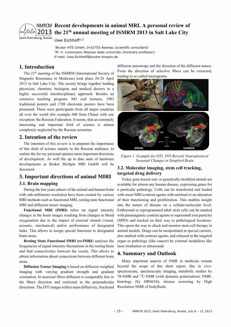
Diffusion Tensor Imaging is based on diffusion weighted imaging with varying gradient strength and gradient orientation. In neuronal fibres diffusion is comparably free in the fibres direction and restricted in the perpendicular directions. The DTI images reflect main diffusivity, fractional
diffusion anisotropy and the direction of the diffusion tensor. From the direction of selective fibres can be extracted, leading to so called tractograms.
Figure 1. Example for DTI: DTI Reveals Neuroplastical
Seasonal Changes in Songbird Brain
3.2. Molecular imaging, stem cell tracking, targeted drug delivery
Today gene-knock-out- or genetically modified animal are available for almost any human disease, expressing genes for a particular pathology. Cells can be transfected and loaded with smart MRI-contrast agents with minimal or no alteration of their functioning and proliferation. This enables insight into the nature of disease on a cellular/molecular level. Embryonal or reprogrammed adult stem cells can be marked with paramagnetic contrast agents or supersmall iron particles (SPIO) and tracked on their way to pathological locations. This opens the way to check and monitor stem cell therapy in animal models. Drugs can be incapsulated in special carriers, also marked with contract agents, and released in the targeted organ or pathology (like cancer) by external modalities like laser irradiation or ultrasound.
4. Summary and Outlook Many important aspects of NMR in medicine remain
beyond the scope of this short report, like in vivo-spectroscopy, spectroscopic imaging, metabolic studies by 1H-NMR and 13C-NMR (with dynamic polarization), NMR-histology (by HRMAS), disease screening by High Resolution NMR of bodyfluids.
Recent developments in animal MRI. A personal review of the 21th annual meeting of ISMRM 2013 in Salt Lake City Uwe Eichhoff1,2
1Bruker HTS GmbH, D-63755 Alzenau (scientific consultant) 2M. V. Lomonosov Moscow state university (honorary professor)
E-mail: [email protected]
NMRCM 2013, Saint Petersburg, Russia, July 8 – 12, 2013 – 16 –
The 129Xe NMR technique introduced in 1980 allows the determination of pore size, location and charge of compensating cations, structural defects, distribution of adsorbed species, etc. It is applied for the characterization of a lot of solids: mesoporous silica, clays, liquid crystals, metal-organic framework compounds (mainly their elasticity), carbons, polymers, diffusion in porous structures and even in archaeology.
Since 2000, this technique has taken a new turn with the advent of laser-hyperpolarized xenon (HP-Xe) in the characterization of materials and organisms. The use of HP-Xe increases the sensitivity for the detection of xenon by several orders of magnitude. The range of its applications becomes wider each day. Now this monotonic probe allows
for remarkable explorations ranging from intricate experiments on single-crystal surfaces to the study of the complex nature of gas exchange in mammalian lungs. For example, encaged in a cryptophane cage bearing a ligand, xenon is a very sensitive sensor for detecting biomedically relevant protein targets or metal cations involved in many pathological and physiological processes. Medical applications increase each day, such as: xenon dissolved in the blood for the measurement of the rate of blood in arteries and veins, xenon imaging in brain or human lungs collected in vivo.
We will give some applications of this universal probe for material and biological characterizations.
A “pot-pourri” of 129Xe NMR technique Jacques Fraissard
Université Pierre et Marie Curie Laboratoire de Physique Quantique, Paris, France
E-mail: [email protected]
– 17 – NMRCM 2013, Saint Petersburg, Russia, July 8 – 12, 2013
Transition metal dichalcogenides MS2 (M= Mo, W, Ta, Nb) have been extensively used as catalysts and lubricants due to their anisotropic behavior related with their layer structure where basal planes are very stable and border planes very reactive. Indeed, MS2 closed structures have been synthesized as multilayer nanotubes, ribbons and onion-like nanostructures and have named the Inorganic Fullerenes (IF) by Tenne [1, 2]. Some of these materials such as the hollow-nanoparticles of WS2 present entirely new properties for lubrication. On the other hand, titania nanotubes, can be considered other type of inorganic fullerenes which have large potential for applications in environmental protection and health care [3] due to their photocatalytic and nanostructural properties.
In this work, the synthesis methods, the physicochemical properties and the nanostructure characterization of several types of IFs will be presented. The application to areas of catalysis [4], lubrication, health care, impact protection will also be discussed.
References [1] Y. Rapoport, Y. Bilik, M. Feldman, S.R. Homyonfer, R.
Cohen and R. Tenne, Nature, 387, (1997) 791-793. [2] R. Tenne and G. Seifert, Annu. Rev. Mater. Res., 39
(2009) 387–413. [3] Batur Ercan, Erik Taylor, Ece Alpaslan, Thomas J
Webster, Nanotechnology, 29 (2011)22. [4] A. Olivas, S. Fuentes, A. Camacho and M.J. Yacaman, J.
Materials Research, vol 19, 2176-2184, (2004).
Inorganic-Fullerenes based on MS2 dichalcogenides (M=Mo, W, Ta, Nb) and TiO2 Sergio Fuentes
Center of Nanosciences and Nanotechnology of UNAM,
Apartado Postal 2681, Ensenada, Baja California, México
E-mail: [email protected]
NMRCM 2013, Saint Petersburg, Russia, July 8 – 12, 2013 – 18 –
Neuronal fibers forming brain white matter realize connections between different cortical areas and other parts of the brain. In vivo knowledge of the individual-specific configuration of neuronal fibers would be of great value for the diagnostics, for the surgical planning, and for the fundamental neuroscience. There has been a significant progress towards this goal during last decade inspired by the development of diffusion-weighted magnetic resonance imaging (dMRI) of the brain. This technique exploits the fact that diffusion of water molecules is strongly anisotropic within the neuronal fiber bundles.
An inherent problem of dMRI is its relatively coarse resolution of a few millimeters, often resulting in a number of fiber bundles placed within one voxel of an MRI image. This puts more weight on the biophysical modeling of the MRI
signal in order to resolve the fiber content of each voxel, and to reconstruct the global fiber connectivity map.
We address this challenge by utilizing the self-consistency conditions resulted from the global nature of neuronal tracts stretching across many voxels. This strategy is pursued in the algorithm which we call “Global fiber tracking”, inspired by the parallels with statistical physics of disordered systems. At its core, our algorithm employs the global “energy” minimization for the interacting segments with a tendency to polymerize into long fibers, placed in an external “field” locally orienting them in accord with the measured diffusion anisotropy. The resulting fiber configuration is a “ground state” into which such interacting segments freeze. This algorithm has been successfully validated by reconstructing fiber configuration in an artificial phantom, and demonstrates good performance on clinical dMRI data.
Tracking neuronal fibers using diffusion-weighted MRI Valerij Kiselev
Dpt. of Radiology, Medical Physics, University Medical Center Freiburg
E-mail: [email protected]
– 19 – NMRCM 2013, Saint Petersburg, Russia, July 8 – 12, 2013
Mesostructured materials possess unique surface, structural, and bulk properties that lead to important practical applications. Theoretical and experimental studies of hydrocarbons confined in, intercalated between or adsorbed to inorganic surfaces report significant differences in structure and dynamics compared to those in the bulk. Understanding the molecular origin of confinement-induced changes is of fundamental interest to a wide range of applications.
In our work, new characterization tool for molecular structure and dynamics of organic component in mesostructural materials is developed. Aim is to provide general approach in term that broad array of systems with wide range of mobility can be characterized within single experimental technique. It should also provide quantitative information without relying on model assumptions.
In the present talk, we focus on surfactant-containing mesostructural organic-inorganic composites. Due to amphiphilic molecular properties, surfactants exhibit strong tendency to self-assemble in solution as well as to adsorb onto various interfaces such as solid surfaces. Surfactants are also widely used in design of organic-inorganic composites and in synthesis of mesoporous materials.
We apply solid state NMR spectroscopy to quantitatively characterize the surfactant conformational dynamics. Magic-
angle-spinning dipolar recoupling technique is used to study surfactant conformational motion in a wide dynamic range from essentially immobilized rigid state to highly flexible state with significant degree of conformational and rotational freedom. Dipolar recoupling combined with two-dimensional 13C separated local field spectroscopy allows for direct estimations of C–H bond order parameter profiles in hydrocarbon chains. Such approach does not require assumptions and adjustable parameters and reflects the changes in conformational dynamics without relying on specific motional model.
Dynamics of surfactant molecules adsorbed at solid surfaces, confined in ordered mesopores, and intercalated into inorganic layered structures is compared. The study shows that in surfactant bilayers intercalated in aluminophosphate layered structure molecules undergo fast rotation around molecules axis while chain is highly ordered and adopts trans-conformation. Both in cylindrical pores of hexagonal mesoporous silica and in adsorbed layer on silica nanoparticles the order parameter is gradually decreasing towards the end of the chain. Experimental order parameter profiles are also compared to those in surfactants aggregated in concentrated aqueous solution, in hexagonal and lamellar mesophases.
Fine art of packing: Solid State NMR study of surfactants at solid interfaces Boris B. Kharkov1,2, Sergey V. Dvinskikh1
1Department of Chemistry and Industrial NMR Centre, Royal Institute of Technology - KTH,
SE-10044 Stockholm, Sweden 2Department of Quantum Magnetic Phenomena, St. Petersburg State University - SPbGU, 198504,
St. Petersburg, Russia
E-mail: [email protected]
NMRCM 2013, Saint Petersburg, Russia, July 8 – 12, 2013 – 20 –
Supported vanadia catalysts belong to a large class of catalytic materials that are technologically indispensable in the 21st century: environmental control, production of energy, petrochemicals and pharmaceuticals, and the destruction of toxic agents. The wide range of catalytic applications reflects the versatile reactivity/selectivity characteristics of supported vanadia catalysts that have their origins in the ability to tune the molecular/electronic structures of the catalytic active vanadia nano-domains formed on the surfaces of oxide substrates (surface functional diversity).
In this lecture we are going to present multinuclear SSNMR/DFT GIPAW data of the molecular structure and reactivity/selectivity relationships for supported vanadium oxide catalysts. Step by step characterization of molecular structure of the catalytic vanadia nano-domains formed in supported binary VOx/MOx systems and in multilayered VOx/M1Ox/M2Ox catalysts, where M1, M2 – Si, Al, Ti, Zr, Nb, will be given. Special attention will be paid to solid-state NMR in its modern applications (High magnetic Field – HF and High Speed- HS MAS). According to these data the versatile types of vanadia domains could be formed, their structural characteristics are determined by a number of factors (vanadium content, nature of the support, structure of the support surface, nature of the layers in multilayered materials, structure of the layers, sequence of deposition, treatment conditions and so on). It is clear that it is rather difficult or even impossible to realize all the types of vanadia sites and to estimate their catalytic characteristics experimentally, especially due to the simultaneous formation of several vanadia species. However, this problem could be solved by quantum chemical simulations. Using the combination of finite cluster and periodic DFT quantum mechanical calculations of structural/spectral properties, the adequate structures for the most basic systems were obtained. Reactivity of the chosen adequate sites was also estimated.
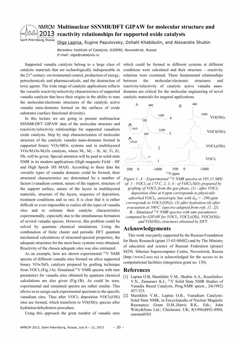
As an example, here are shown experimental 51V NMR spectra of different vanadia sites formed on silica supported binary VOx/SiO2 catalysts prepared by grafting technique from VOCl3 (Fig.1A). Simulated 51V NMR spectra with nmr parameters for vanadia sites obtained by quantum chemical calculations are also given (Fig.1B). As could be seen, experimental and simulated spectra are rather similar. This allows us to assign each experimental spectrum to the specific vanadium sites. Thus after VOCl3 deposition VOCl2(OSi) sites are formed, which transform to VO(OSi)3 species after hydration/dehydration procedure.
Using this approach the great number of vanadia sites
which could be formed in different systems at different conditions were calculated and their structure – reactivity relations were exemined. These fundamental relationships between the molecular/electronic structures and reactivity/selectivity of catalytic active vanadia nano-domains are critical for the molecular engineering of novel catalytic materials for targeted applications.
Figure 1. A – Experimental 51V NMR spectra at 105.15 MHz
of: 1 - VOCl3 at 173C, 2, 3, 4 - of VOCl3/SiO2 prepared by
grafting of VOCl3 from the gas phase, (1) - after VOCl3
deposition (line at 6 ppm corresponds to physically
adsorbed VOCl3, anisotropic line with δiso = -290 ppm
corresponds to VOCl2(OSi)); (3) after hydration;(4) after
evacuation at 100C. (spectra adapted from refs. [1, 2]).
B – Simulated 51V NMR spectra with nmr parameters
computed by GIPAW for VOCl3, VOCl2(OSi), VOCl(OSi)2
and VO(OSi)3 structures obtained by DFT.
Acknowledgements This work was partly supported by the Russian Foundation
for Basic Research (grant 13-03-00482) and by The Ministry of education and science of Russian Federation (project 8429). Siberian Supercomputer Centre, Novosirirsk, Russia (http://www2.sscc.ru) is acknowledged for the access to its computational facilities (integration grant no. 130).
References [1] Lapina O.B, Mastikhin V.M., Shubin A.A., Krasilnikov
V.N., Zamaraev K.I., 51V Solid State NMR Studies of Vanadia Based Catalysts, Prog.NMR spectr., 24(1992) 457-525.
[2] Mastikhin V.M., Lapina O.B., Vanadium Catalysts: Solid State NMR, in Encyclopedia of Nuclear Magnetic Resonance; Grant D.M.,Harris R.K., Eds.; John Wiley&Sons Ltd.; Chichester, UK, 8(1996)4892-4904, emrstm0585
Multinuclear SSNMR/DFT GIPAW for molecular structure and reactivity relationships for supported oxide catalysts Olga Lapina, Eugine Papulovsky, Dzhalil Khabibulin, and Alexandre Shubin
Boreskov Institute of Catalysis, 630090, Novosibirsk, Russia
E-mail: [email protected]
500 0 -1000 500 0 -1000
51V/ppm
VOCl3
VOCl2(OSi)
VOCl(OSi)2
VO(OSi)3
A B
1
2
3
4
x4
460
1100
460
1000
290
1200
360
+H2O
-H2O
– 21 – NMRCM 2013, Saint Petersburg, Russia, July 8 – 12, 2013
Proton ordering and exchange dynamics play an important role in hydrogen bonded solids and are also important for molecules adsorbed in porous materials. In the lectures previous and recent results are discussed which are related to the application of spin relaxation studies for adsorbed water, two-dimensional deuteron-exchange spectroscopy and order-disorder phenomena in solids.
As an introduction to the lecture, former results are characterized which are concerned with proton exchange in adsorbed water, proton mobility in systems with strong acidic sites obtained by means of proton spin relaxation.
Then proton mobility is investigated with respect to the high ionic conductivity in crystals with hydrogen bonds. This is interesting, on the one hand, due to the practical applications of these crystals and, on the other hand, it gives the possibility to contribute in general to the understanding of the microscopic mechanism of electric conductivity. From this point of view, a particular interest is devoted to crystals with the structural peculiarity of quasi one-dimensional
chains of atomic groups bound by hydrogen bonds showing a high conductivity. Such crystals serve as very appropriate model objects in order to study the proton ordering and to verify various assumptions about the microscopic mechanisms of proton conductivity. Substances with a surprisingly high ionic (protonic) conductivity are, among others, single crystals of ammonium hydrogen selenate NH4HSeO4 (AHSe), betaine phoshate (BP), betaine phoshite (BPI) and mixed crystals if the type BP1-xBPIx, containing quasi one-dimensional chains of SeO4-, PO4-, and PO3- tetrahedra connected by hydrogen bonds. The role of structural defects for the conductivity process is discussed.
In a third part, NMR spectroscopy is applied to investigate proton ordering in various crystals in relation to structural phase transitions.
Comments and support of my former colleagues, in particular professor Rolf Boettcher and professor Georg Voelkel, are greatly acknowledged.
Proton and deuteron ordering and dynamics in solids and in molecules adsorbed in porous materials Dieter Michel
Universität Leipzig, Fakultät für Physik und Geowissenschaften,
Linnéstr. 5, 04103 Leipzig, Germany
E-mail: [email protected]
NMRCM 2013, Saint Petersburg, Russia, July 8 – 12, 2013 – 22 –
Molecular diffusion measurements are widely used to probe microstructure in materials and living organisms noninvasively. The precise relation of diffusion metrics to microstructure remains a major challenge: In complex samples, it is often unclear which structural features are most relevant and can be quantified. Here we classify the structural complexity in terms of the long time tail exponent in the molecular velocity autocorrelation function. The specific
values of the dynamical exponent let us identify the relevant tissue microanatomy affecting water diffusion measured with MRI in muscles and in brain, and the microstructural changes in ischemic stroke.
Our framework presents a systematic way to identify the most relevant part of structural complexity using transport measured with a variety of techniques.
Characterizing tissue microstructure with time-dependent diffusion Dmitry Novikov
New York University Medical School, New York
E-mail: [email protected]
– 23 – NMRCM 2013, Saint Petersburg, Russia, July 8 – 12, 2013
1. Introduction The simplest diffusion-weighting module consists of a pair
of diffusion gradients separated by a 180º refocusing pulse. In the presence of motion, phase accrued while the first gradient is applied is not perfectly cancelled by the second gradient. Incoherent motion associated to diffusion therefore leads to a distribution of phases within each voxel resulting in signal loss.
Diffusion-Weighted (DW) images need to be sensitive to the microscopic motion of water molecules (of the order of a few microns) which renders them extremely sensitive to bulk motion. Rigid-body motion can lead to global phase shifts and spatial phase gradients [1], while pulsatile brain motion [2] produces non-linear phase patterns.
2. Echo Planar Imaging Single-shot echo planar imaging (ssEPI) is the most used
sequence for DWI as it samples the whole of k-space following a single excitation, avoiding phase inconsistencies between different data points. Unfortunately, the long readout window means ssEPI is very sensitive to field inhomogeneities with images displaying spatial distortions and signal loss particularly at tissue-air interfaces. Geometric distortions due to eddy currents generated by the diffusion module are also problematic [3]. Strategies to minimize and correct for these effects will be discussed.
Image resolution attained with ssEPI is limited due to relaxation and so multi-shot approaches are needed for high-resolution imaging. In this case, it is essential to correct for phase inconsistencies between shots, which can be measured using navigators. To increase efficiency, self-navigated sequences based on EPI, radial or spiral trajectories have been suggested.
3. Alternative Methods 3.1 Fast Spin Echo
Fast Spin Echo (FSE) techniques are robust to field inhomogeneities, but require precise control of the signal phase at the start of the refocusing pulse train (CPMG condition [4]). This is particularly challenging to achieve with DW. The sequence can be modified so that the FSE signal becomes phase insensitive but at the cost of a 50% signal reduction [5]. A different strategy consists in correcting for motion-induced phase patterns ensuring that the CPMG condition is satisfied following the diffusion sensitisation
module. Norris et al. demonstrated real time correction for linear phase patterns induced by rigid-body motion [6]. To correct also for the residual non-linear patterns, tailored RF excitation pulses can be used [7].
3.2 Steady-State Free Precession Diffusion contrast is achieved by sacrificing signal-to-
noise (SNR). Steady-State Free Precession (SSFP) sequences are very promising as they use very short repetition times and take advantage of the contribution from multiple coherence pathways to achieve high SNR per unit time. As will be discussed in the presentation, the downside is that signal depends also on the flip angle and relaxation times, making diffusion quantification more complicated. A review of DW-SSFP methods is presented in [8].
Acknowledgements Portuguese Foundation for Science and Technology (PEst-
OE/SAU/UI0645/2011).
References [1] Anderson AW, Gore JC. Analysis and correction of
motion artifacts in diffusion weighted imaging – Magn
Reson Med, 32, 379-387 (1994). [2] Miller KL, Pauly JM. Nonlinear phase correction for
navigated diffusion imaging. – Magn Reson Med, 50, 343-353 (2003).
[3] Jezzard P, Barnett AS, Pierpaoli C. Characterization of and correction for eddy current artifacts in echo planar diffusion imaging. – Magn Reson Med, 39, 801-812 (1998).
[4] Meiboom S, Gill D. Modified Spin-Echo Method for Measuring Nuclear Relaxation Times. – Rev Sci Instrum, 29, 688-691 (1958).
[5] Alsop DC. Phase insensitive preparation of single-shot RARE: application to diffusion imaging in humans. – Magn Reson Med, 38, 527-533 (1997)
[6] Norris DG, Driesel W. Online motion correction for diffusion-weighted imaging using navigator echoes: Application to RARE imaging without sensitivity loss. –
Magn Reson Med, 45, 729-733 (2001). [7] R.G. Nunes, S.J. Malik, J.V. Hajnal. Single shot Fast
Spin Echo diffusion imaging with correction for non-linear phase errors using tailored RF pulses. – Magn
Reson Med, In Press, (2013). [8] J.A. McNab, K.L. Miller. Steady-state diffusion-
weighted imaging: theory, acquisition and analysis. – NMR Biomed, 21, 783–793 (2010).
Diffusion-weighted imaging pulse sequences: Echo planar imaging and alternative methods Rita G. Nunes
Institute of Biophysics and Biomedical Engineering, Faculty of Sciences, University of Lisbon,
Lisbon, Portugal
E-mail: [email protected]
NMRCM 2013, Saint Petersburg, Russia, July 8 – 12, 2013 – 24 –
1. Introduction Recent developments of Field Cycling (FC) NMR have
opened new possibilities for the studies of condensed matter. Together with the availability of commercial relaxometers [1, 2] it renders the FC technique increasingly attractive for scientists. The main advantage of FC is the possibility to investigate the evolution of a spin system in a broad frequency range using a magnetic field which can be quickly switched (cycled). The goal of this contribution is to present some new developments of our group in the instrumentation and to give an overview of new techniques thereby further developing FC NMR.
2. Instrumentation A typical FC experiment is best understood by considering
the basic three steps of the magnetic field setting: First, the “polarization” of the sample in a magnetic field Bpol; second, the “evolution” of the spin system in an adjustable field Bev in which the nuclear spin polarization relaxes towards its new equilibrium state with a corresponding rate T1
-1(Bev); third, “detection” of the evolving polarization in a field Bdet. This experiment is repeated in cycles as a function of the hold time tev of the evolution field Bev.
For the reliability of an FC relaxometer the following parameters are of importance: a) the value of Bpol and Bdet should be as high as possible, b) the range of Bev should as broad as possible, c) the switching time should be as short as possible, d) the stability of the Bdet should be good and e) the magnetic field homogeneity should be high. Depending on the experimental requirements not all of these conditions have necessarily to be fulfilled. In any case the accuracy of field regulation after the switching from Bpol to Bev, especially if Bev is in the range of µT or even below, is of a great importance. The field regulation over six orders of magnitude is a challenging problem [3]. Nevertheless, if signal averaging is required the stability of Bdet during the NMR detection phase is a challenge. Compensation of magnetic field instabilities in FC NMR by reference deconvolution is a promising solution [4]. All these points will be discussed explicitly.
3. Experimental techniques Results of several types of FC experiments on various
nuclei will be presented and discussed: 1. Analysis of the T1 dispersion in the solid state over a broad
temperature range: In the case of an exponential relaxation the nuclear polarization will approach its new equilibrium with a corresponding rate T1
-1(Bev) which can be related to the spectral density of local magnetic field fluctuations, J(ω), which in turn is related to the correlation function of the dynamic processes. This method allows tracing correlation times over a very broad range from 10-3 to 10-10 s [5].
2. Double NMR-NQR FC: With this method one can detect 14N quadrupolar transitions in various systems. Drugs and explosives can be detected with a sensitivity much higher as with pure NQR [6].
3. Investigation of slow molecular dynamics in polymers in ultralow magnetic fields: FC NMR can be applied for Bev down to about 10-6 T corresponding to Larmor frequencies down to several tens of Hz [7].
4. Rotational resonance in the laboratory system: Using FC is possible to mechanically rotate samples with NMR frequency corresponding to Bev [8].
4. References [1] http://www.stelar.it/ [2] http://www.spinscope.com/ [3] B. Kresse, A. F. Privalov, F. Fujara, Solid State NMR 40,
134, 2011 [4] S. Reutter, A. Privalov, Appl. Magn. Res. 44, 55, 2013 [5] A. F. Privalov, O. Lips, F. Fujara, J. Phys.Cond. Matter,
14, 4515, 2002 [6] M. Nolte, A. Privalov, J. Altmann, V. Anferov, F. Fujara,
J. Phys. D, 35, 939, 2002 [7] A. Herrmann, B. Kresse, J. Gmeiner, A.F. Privalov, D.
Kruk, F. Fujara, E. A. Rössler, Macromolecules 45, 1408, 2012
[8] S. Reuter, A. Privalov, G. Buntkowsky, F. Fujara. Solid State NMR, 41, 74, 2012
Recent developments in Field Cycling NMR A. F. Privalov
Institut für Festkörperphysik, TU Darmstadt, Hochschulstr. 6, 64289 Darmstadt, Germany
E-mail: [email protected]
– 25 – NMRCM 2013, Saint Petersburg, Russia, July 8 – 12, 2013
Development of new sustainable and environment-friendly energy systems requires safe and efficient ways of energy storage. Renewable energy can be stored directly as electricity in batteries or indirectly as hydrogen in solid-state hydrides. Metal borohydrides have received recent attention as promising hydrogen-storage materials due to their high hydrogen densities. However, practical use of the known metal borohydrides is hindered by their stability with respect to thermal decomposition and the slow hydrogen sorption kinetics. Elucidation of the complex structures and hydrogen dynamics in these materials may give a key to improving their
hydrogen-storage properties. This lecture presents a review of the dynamical properties of borohydrides and the related compounds. It is based mainly on recent experimental results obtained by the NMR group at the Institute of Metal Physics (Ekaterinburg). We will discuss the relations between the motional parameters derived from NMR experiments and the structural features of the borohydrides. A special emphasis will be made on novel borohydride-based systems showing both fast reorientational motion of BH4 groups and fast translational diffusion of ions.
Nuclear magnetic resonance studies of atomic motion in borohydride-based hydrogen storage materials A. V. Skripov
Institute of Metal Physics, Ural Branch of the Russian Academy of Sciences,
Ekaterinburg, Russia
E-mail: [email protected]
NMRCM 2013, Saint Petersburg, Russia, July 8 – 12, 2013 – 26 –
Recently, the interest in nanoparticles has been steadily increasing owing to their unique physical and chemical properties. Deposition from colloid solutions is a well elaborated method, which makes it possible to obtain nanosized samples of double and triple rare-earth fluorides [1]. A modification of this technology with the use of microwave radiation is described in [2], where it was shown that there are internal cavities in the synthesized particles (suchnanoparticles are called fullerene like nanoparticles).This technology was used to synthesize a series of crystalline fullerene like PrF3 nanoparticles [3, 4]. Since the hydrothermal synthesis is performed in an aqueous solution, it is possible to suppose that water is located in the internal cavities of such nanoparticles. According our results
of experiments this hypothesis was demonstrated by using nuclear magnetic resonance cryoporometry and high-resolution transmission electron microscopy methods [5].
Finally some possible application for medicine will be presented.
[1] X. Wang and Y. D. Li, Angew. Chem. Int. Ed. 42, 3497 (2003).
[2] L. Ma, W. Chen, Y. Zheng, et al., Mater. Lett. 61, 2765 (2007).
[3] M. S. Tagirov, E. M. Alakshin, R. R. Gazizulin, et al.,J. Low Temp. Phys. 162, 645 (2011)
[4] M. S. Tagirov, E. M. Alakshin, R. R. Gazizulin, et al., J. Low Temp. Phys. 162, 645 (2011).
[5] E. M. Alakshina, D. S. Blokhina, A. M. Sabitova, et al., JETP Letters, 2012, Vol. 96, No. 3, pp. 181–183.
Fullerene like nanoparticles of PrF3: from creations to medical applications Murat Tagirov
Kazan Federal University
Kazan, Russia
E-mail: [email protected]
– 27 – NMRCM 2013, Saint Petersburg, Russia, July 8 – 12, 2013
1. Introduction Dynamic nuclear polarization (DNP) is an important tool
in magnetic resonance, which provides versatile control of the nuclear polarization and substantial enhancement of the sensitivity of NMR. Very interesting systems where the effects of DNP can be studied are shallow donors (P, As, Bi) in Si. Magnetic resonance studies of these samples have a long history starting from pioneering work of Feher [1]. Recently the interest to this system has been raised by the proposal of Kane [2] to utilize these impurity atoms for quantum computing with the nuclear spin serving as a memory qubit and electron spin for readout.
In this work we report on the first experimental study of DNP of 31P donors in silicon performed in strong magnetic field and temperatures below 1K. At these conditions donor electron spins are fully polarized, electron and nuclear relaxation times are extremely long. All these factors substantially change the efficiency of DNP: pumping with extremely low RF powers (<1 µW) for reasonably short time (≈1 hour) very high values of DNP of 31P were reached.
2. Samples and methods We performed experiments with the samples of silicon of
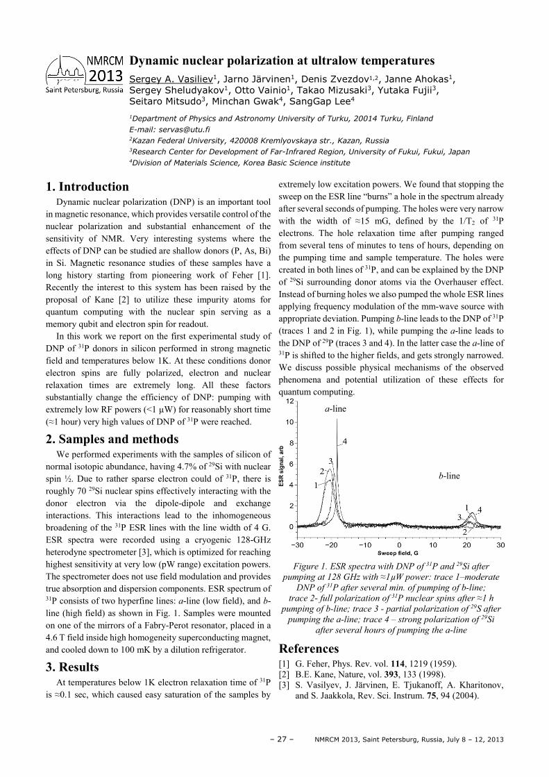
normal isotopic abundance, having 4.7% of 29Si with nuclear spin ½. Due to rather sparse electron could of 31P, there is roughly 70 29Si nuclear spins effectively interacting with the donor electron via the dipole-dipole and exchange interactions. This interactions lead to the inhomogeneous broadening of the 31P ESR lines with the line width of 4 G. ESR spectra were recorded using a cryogenic 128-GHz heterodyne spectrometer [3], which is optimized for reaching highest sensitivity at very low (pW range) excitation powers. The spectrometer does not use field modulation and provides true absorption and dispersion components. ESR spectrum of 31P consists of two hyperfine lines: a-line (low field), and b-line (high field) as shown in Fig. 1. Samples were mounted on one of the mirrors of a Fabry-Perot resonator, placed in a 4.6 T field inside high homogeneity superconducting magnet, and cooled down to 100 mK by a dilution refrigerator.
3. Results At temperatures below 1K electron relaxation time of 31P
is ≈0.1 sec, which caused easy saturation of the samples by
extremely low excitation powers. We found that stopping the sweep on the ESR line “burns” a hole in the spectrum already after several seconds of pumping. The holes were very narrow with the width of ≈15 mG, defined by the 1/T2 of 31P electrons. The hole relaxation time after pumping ranged from several tens of minutes to tens of hours, depending on the pumping time and sample temperature. The holes were created in both lines of 31P, and can be explained by the DNP of 29Si surrounding donor atoms via the Overhauser effect. Instead of burning holes we also pumped the whole ESR lines applying frequency modulation of the mm-wave source with appropriate deviation. Pumping b-line leads to the DNP of 31P (traces 1 and 2 in Fig. 1), while pumping the a-line leads to the DNP of 29P (traces 3 and 4). In the latter case the a-line of 31P is shifted to the higher fields, and gets strongly narrowed. We discuss possible physical mechanisms of the observed phenomena and potential utilization of these effects for quantum computing.
Figure 1. ESR spectra with DNP of 31P and 29Si after
pumping at 128 GHz with ≈1µW power: trace 1–moderate
DNP of 31P after several min. of pumping of b-line;
trace 2- full polarization of 31P nuclear spins after ≈1 h
pumping of b-line; trace 3 - partial polarization of 29S after
pumping the a-line; trace 4 – strong polarization of 29Si
after several hours of pumping the a-line
References [1] G. Feher, Phys. Rev. vol. 114, 1219 (1959). [2] B.E. Kane, Nature, vol. 393, 133 (1998). [3] S. Vasilyev, J. Järvinen, E. Tjukanoff, A. Kharitonov,
and S. Jaakkola, Rev. Sci. Instrum. 75, 94 (2004).
Dynamic nuclear polarization at ultralow temperatures Sergey A. Vasiliev1, Jarno Järvinen1, Denis Zvezdov1,2, Janne Ahokas1, Sergey Sheludyakov1, Otto Vainio1, Takao Mizusaki3, Yutaka Fujii3, Seitaro Mitsudo3, Minchan Gwak4, SangGap Lee4
1Department of Physics and Astronomy University of Turku, 20014 Turku, Finland
E-mail: [email protected] 2Kazan Federal University, 420008 Kremlyovskaya str., Kazan, Russia 3Research Center for Development of Far-Infrared Region, University of Fukui, Fukui, Japan 4Division of Materials Science, Korea Basic Science institute
a-line
b-line
NMRCM 2013, Saint Petersburg, Russia, July 8 – 12, 2013 – 28 –
1. Introduction The self-diffusion measurements, especially the
techniques using the pulsed field gradient NMR following by Fourier transforms, are the unique methods for direct structural and dynamic studies in systems with the fast ionic and molecular transport. For diffusional water permeability in biological systems, pulsed field gradient NMR (PFG-NMR) spectroscopy has become the method of choice due to its remarkable sensitivity to molecular displacements in the range of 10nm–100 mm and to its non-invasive character.
In order to interpret the experimental data correctly, the
model investigations are necessary.
The results were obtained at the Laboratory of Membrane Processes, Karpov Institute of Physical Chemistry, Moscow, Russia; Laboratory of NMR, Institute of Problems of Chemical Physics, Russian Academy of Sciences, Chernogolovka, Moscow Region Russia and at Laboratory of Food and Biomaterial Science and Engineering, Graduated School of Life Science and Biotechnology, Korea University, Seoul, Korea. This presentation devotes to investigations of ionic and water transport in biological cells (chlorella, yeast and erythrocytes), in cation-exchange membranes and polymeric electrolytes for the lithium cells. Well known perfluorinated cation-exchange memdranes, sulfocation-exchange styrene bivinyl benzene resin CU-2 with different amount of crosslinked agent and aromatic bi sulfocontaining polyamides were stidied. Two polyamide isomer compositions iso (µPA) and tere (πPA) were investigated. Lithium and solvating molecules self-diffusion processes in polymeric electrolytes on the basis of polyester di-acrylate, lithium perchlorate and ethylene carbonate were investigated. The investigation of the polymeric electrolyte systems on the basis of LiBF4 diacrylatenic with ionic liquid 1- butyl-3-methyl imidazole tetra fluorine borate (BMIBF4) was carried out.
2. Polymeric electrolytes Sulfo-, carboxyl-, aminogroups containing ion exchange
membranes were investigated as model systems. The hydration of fixed groups and alkaline and alkaline – earth ions were studied in details in perfluorinated Nafion membranes. The mechanism of charge group – counter ion or water molecule interactions were understood from high resolution hetero nuclear NMR data. Microscopic ionic and water molecule mobilities were determined by NMR relaxations. Self-diffusion coefficients of protonic molecules and lithium and fluorine counter ions in different spatial scales were measured directly by PFG NMR. It was concluded that the macroscopic electro – mass transfer is controlled by local ion and molecule jumps between adjacent charge groups. The interconnection between ionogenic channel structure, mobile ion or molecule-charge groups
binding and translational ionic and molecular mobility was determined. On the basic of this knowledge, the main particularities of water behaviour in proteins and gels have been understood. It was shown that hydrogen bond is very important for proton and water molecules motions in biological ionic channels. The degree of lithium perchlorate dissociation was estimated from the comparison of calculated and measured conductivity values. The Li+ cation solvation processes in polymeric electrolytes (PE) on the basis of polyester di-acrylate, lithium perchlorate and ethylene carbonate (EC) were investigated. It was revealed that the high ionic conductivity was realized when the amount of EC molecules per Li+ more than 4 and the lithium cations were solvated of EC molecules. At these conditions the dissociation degree of LiClO4 molecules (Li+ concentration) strongly increses and the cation translation transfer is occured together with EC molecules. The fast exchange between the ions and the neighboring solvation sphere molecules following by high ionic mobility occurs at the same time [1].
3. Biological cell membranes Water self-diffusion in cells of chlorella, yeast and red
blood cell was investigated. These cells were selected as model systems with different cell membrane permeabilities. The apparent self-diffusion coefficients of intracellular and extracellular water were measured dependent on diffusion time. The regions of restricted diffusion and hindered diffusion were observed. Scaling approach and two compartment exchange model were applied to calculate cell sizes and permeabilities [2, 3]. The values of permeability calculated by these two ways are very close to each other. The correctness of these theoretical interpretations was also demonstrated by good agreement of cell sizes obtained from PFG NMR and electron microscopic data. The permeabilities are 3.10-6, 6.10-6 and about 10-4 m/s for chlorella, yeast and red blood cells, respectively, depending on cell growing conditions and physical chemistry treating. The average cell sizes are varied from 2 to 4 microns. The water exchange mechanism in biological cells is discussed.
Acknowledgements The investigation was supported by Russian Basic
Research Foundation, grant 13-03-00698-a.
References [1] V.I. Volkov, A.A. Marinin Russian Chemical Reviews
82(3) 248-272 (2013) [2] Suh K.J., Hong Y.S., Volkov V.I., Skirda V.D. et.al
Biophys. Chem. 104, 121-130, (2003). [3] Cho J.H., Hong Y.S., Volkov V.I., Skirda V.D. et. al.
Magnetic Resonance Imaging 21,1009-1017 (2003).
Pulsed Field Gradient NMR for biological membranes and polymeric electrolyte investigations Vitaly I. Volkov
Institute of Problems of Chemical Physics RAS, Chernogolovka, Moscow Region, 142432, Russia
E-mail: [email protected]
– 29 – NMRCM 2013, Saint Petersburg, Russia, July 8 – 12, 2013
The essence of phase graphing is to predict when echoes are generated in and after a sequence of multiple gradients and RF pulses, and what the corresponding echo intensities are. In the modern interpretation magnetization is depicted in Fourier space and the action of the RF pulses on these Fourier
components is characterized. If care is taken, further effects of diffusion can be included and quantified.
My talk will shed some light on the meaning and interpretation of extended phase graphs and what prerequisites are necessary for adding diffusion effects.
Extended Phase Graphs: What they mean and how to include diffusion Matthias Weigel
University Medical Center Freiburg, Dept. of Radiology, Medical Physics
Freiburg, Germany
www.matthias-weigel.net
E-mail: [email protected]


Part II
Oral Reports

NMRCM 2013, Saint Petersburg, Russia, July 8 – 12, 2013 – 34 –
1. Introduction The field strength of a horizontal bore magnet depends
both on the current through the solenoid and the Earth magnetic field. It is useful to trace the evolution of the Larmor frequency to estimate the dynamics of both fields.
2. Materials and Methods Materials of a database of MRI studies within 14 years
were used for analysis of evolution of the Larmor frequency of a 0.5T MR-scanner (Bruker Tomikon S50) with a horizontal bore (north-east orientation) superconducting (SC) magnet (Magnex Ltd). This database [1] contents not only images but also pulse sequences parameters and results of automatic definition of operating frequency (F) of the MR scanner. This operation is made in the beginning of each MRI study to determine isocenter frequency. To do this the free induction signal is registered and position of the maximal peak is determined by Fourier analysis. Typical NMR spectrum of human organ contents two strong peaks from water and fat divided by 3.5 ppm. Value F depends on a water/fat proportion in organ under study as organ specific receiver coils are used and their locations are adapted to the investigated organs.
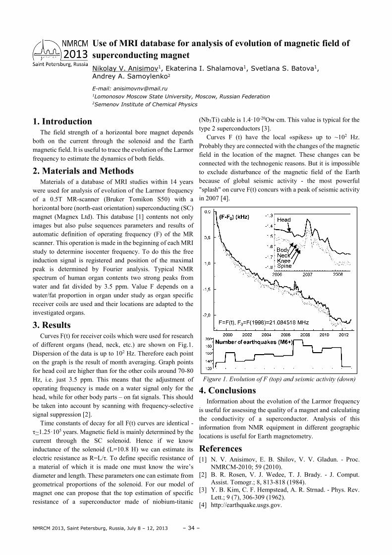
3. Results Curves F(t) for receiver coils which were used for research
of different organs (head, neck, etc.) are shown on Fig.1. Dispersion of the data is up to 102 Hz. Therefore each point on the graph is the result of month averaging. Graph points for head coil are higher than for the other coils around 70-80 Hz, i.e. just 3.5 ppm. This means that the adjustment of operating frequency is made on a water signal only for the head, while for other body parts – on fat signals. This should be taken into account by scanning with frequency-selective signal suppression [2].
Time constants of decay for all F(t) curves are identical - τ~1.25·105 years. Magnetic field is mainly determined by the current through the SC solenoid. Hence if we know inductance of the solenoid (L=10.8 H) we can estimate its electric resistance as R=L/τ. To define specific resistance of a material of which it is made one must know the wire’s diameter and length. These parameters one can estimate from geometrical proportions of the solenoid. For our model of magnet one can propose that the top estimation of specific resistance of a superconductor made of niobium-titanic
(Nb3Ti) cable is 1.4·10-20Ом·cm. This value is typical for the type 2 superconductors [3].
Curves F (t) have the local «spikes» up to ~102 Hz. Probably they are connected with the changes of the magnetic field in the location of the magnet. These changes can be connected with the technogenic reasons. But it is impossible to exclude disturbance of the magnetic field of the Earth because of global seismic activity - the most powerful "splash" on curve F(t) concurs with a peak of seismic activity in 2007 [4].
Figure 1. Evolution of F (top) and seismic activity (down)
4. Conclusions Information about the evolution of the Larmor frequency
is useful for assessing the quality of a magnet and calculating the conductivity of a superconductor. Analysis of this information from NMR equipment in different geographic locations is useful for Earth magnetometry.
References [1] N. V. Anisimov, E. B. Shilov, V. V. Gladun. - Proc.
NMRCM-2010; 59 (2010). [2] B. R. Rosen, V. J. Wedee, T. J. Brady. - J. Comput.
Assist. Tomogr.; 8, 813-818 (1984). [3] Y. B. Kim, C. F. Hempstead, A. R. Strnad. - Phys. Rev.
Lett.; 9 (7), 306-309 (1962). [4] http://earthquake.usgs.gov.
Use of MRI database for analysis of evolution of magnetic field of superconducting magnet Nikolay V. Anisimov1, Ekaterina I. Shalamova1, Svetlana S. Batova1, Andrey A. Samoylenko2
E-mail: [email protected] 1Lomonosov Moscow State University, Moscow, Russian Federation 2Semenov Institute of Chemical Physics
– 35 – NMRCM 2013, Saint Petersburg, Russia, July 8 – 12, 2013
1. Introduction Spin-spin coupling constants (SSCC) are the key NMR
parameters for structure determination nowadays. However, direct measurement of these parameters is difficult in many cases due to peaks overlapping, short relaxation times and/or second order effects (see [1-2]). Theoretical description for SSCC needs also to be developed in practical aspects. We showed recently [3], that dynamic behavior of many important molecular systems can be described in terms of vibrations with large amplitude. Accurate structure studies of saturated four- and five-membered cycles imply solving specific problem of quantitative description of dynamic processes with very low barriers in them. Here we present application of few new effective techniques for extraction information on the dynamic structure [3, 4] via the high precision analysis of NMR multiplets and theoretical description of the NMR parameters.
2. Methods We developed a practical method for evaluation of the
parameters of conformational dynamics in terms of vibrations with large amplitude. The method based on: (i) the results of complete analysis of high resolution NMR spectra, (ii) ab’initio calculations of a reaction path and surfaces of potential energy and spin-spin coupling constants, (iii) a numerical solution of corresponding vibration problem and (iv) refinement for the parameters of the energy surface based on the best fit of experimental (see e.g. [1-2]) and calculated spin-spin couplings.
As a starting point, the undistorted potential energy surface (PES) of inner rotation for the compounds studied was built by applying the scanning technique to skeletal dihedral angles [3]. This allows us to get a trial “reaction path” for the pseudorotation process. Conformational dependencies for spin-spin coupling constants (SSCC) for principal points on the reaction path were calculated using FP DFT technique [5]. 1H NMR spectra were recorded for a series of solvents and “Bruker AV-600” spectrometer at room temperature, and were treated using total lineshape analysis technique (program VALISA [1]) which allows to get very accurate estimates of experimental SSCC values. Finally, the reverse spectral problem was solved to adjust experimental and calculated data and build up the “true” potential of pseudorotation. We developed REVIBR program [3, 6],
which solves numerically corresponding vibration problem and models the dynamic averaging using the technique of convolution of the spin-spin coupling surfaces using the whole set of vibration energies and eigenvectors (normally, 200 lowest ones). Convolution criterion used in REVIBR program allows to get calculated SSCC for given temperature. Nonlinear optimization (Levenberg-Marquart techniques) of the estimated parameters for the “true” pseudorotation PES (modeling difference of ground states of main conformers ∆E and heights for the conformational barriers ∆E≠) used to get best fit of experimental and calculated SSCC values.
3. Results Advantages of the technique developed demonstrated on a
series of monosubstituted cyclobutanes, trans-1,2-dihalocyclopentanes, tetrahydrofuran [3, 6], tetrahydro-thiophene, tetrahydrothiophene-1-oxide, pyrrolidine, proline and ribonucleosides. The data obtained shows, that the pseudorotation process in every four- and five-membered system under study is carrying out by the mechanism with high amplitude of vibration. Major conformations of tetrahydrofuran and terahydrothiophene are twists 4Т5 and 5Т4, for pyrrolidine – envelope Е1 with equatorial NH-bond, for terahydrothiophene-1-oxyde – envelopes Е3 and with axial oxygen and for proline – envelope Е5 with axial СООН-group. Method used also for characterization of internal rotation in acyclic systems: styrene, substituted cis- and trans-azobenzenes, cinnamic aldehyde as well as in natural endogenic hormones noradrenaline and adrenaline.
References [1] S.V. Zubkov, S.S. Golotvin, V.A. Chertkov. – Russ.
Chem. Bull., 51, 1222-1230 (2002). [2] S.V. Zubkov, V.A. Chertkov – IJMS, 4, 107-118 (2003). [3] A.V. Chertkov, O.I. Pokrovsky, A.K. Shestakova, V.А.
Chertkov – Chem. Heterocycl. Comp., 44, 782-784 (2008).
[4] J.W. Blanchard, M.P. Ledbetter, T. Theis, M.C. Budker, D. Budker, A. Pines, J. Am. Chem. Soc., 135, 3607-3612 (2013).
[5] W. Deng, J.R. Cheeseman, M.J. Frisch, J. Chem. Theory
and Comput., 2, 1028-37 (2006). [6] A.V. Chertkov, A.K. Shestakova, V.A. Chertkov, Chem.
Heterocycl. Comp., 48, 412 – 421 (2012).
Novel high resolution NMR techniques for elucidation of molecular structure and dynamics Vyacheslav A. Chertkov1, Dmitriy A. Cheshkov1,2, Tatiana A. Ganina1, Sergey S. Nechausov1, Kirill F. Sheberstov1,3, Alla K. Shestakova2
1Department of Chemistry, Moscow State University, 1 Leninskie Gory, Moscow, 119992, Russia 2State Research Institute of Chemistry and Technology of Organoelement Compounds, 38 Shosse
Entuziastov, Moscow, 111123, Russia 3Faculty of Materials Science, Moscow State University, 1 Leninskie Gory, Moscow, 119992, Russia
E-mail: [email protected]
NMRCM 2013, Saint Petersburg, Russia, July 8 – 12, 2013 – 36 –
1. Introduction This work is directed on the solution one of the most
important problem of biochemistry - development of method for studying the hydration properties of organic molecules. Analysis of concentration and temperature dependences of spin-lattice relaxation rates of solvent nuclei allows us to solve this problem. Firstly this method was developed for the investigation the electrolyte solutions [1] and later was used for the solution of acetic acid [2] and the solutions of amino acids [3]. In the present work the method was developed for aqueous solutions of carboxylic and dicarboxylic acids. For the first time the hydration properties of different functional groups of acids were separated.
2. Experiment and methods The samples of solution of acetic, propionic, butyric,
malonic and succinic acids in heavy water D2O were prepared. Measurements of the spin-lattice times of deuterium nuclei of solvent were carried out on Bruker SPX 4-100 or Bruker Avance 500 MHz by “inversion-recovery” method.
The water forms ordered structure around acid molecules. Water molecules are characterised by the certain dynamics and structure of hydrogen bonds near each functional group of organic molecule. In the case of the fast exchange of solvent molecules in the solution the relaxation rate of the solvent nuclei is given by the formula [1].
1 1
1 11 1 10
1 1 11 ,
N Ni i
i ii
mn mn
T M T M T
− −
= =
= + −
∑ ∑
where m is the molality, M is the number of moles in 1000 grams of solvent, T1i is the solvent nuclei relaxation time in the structure number i, T10 is the relaxation time of free solvent, ni is the number of molecules in the i - structure (coordination number).
The formula predicts the linear concentration dependence, but distinct deviations from linearity have been observed. The bends may be explained by the change in solution structure. The first bend corresponds to the disappearance of free solvent structure. The position of the bend gives number of the water molecules in the hydration shell of acid molecule and coordination number of methylene group. The quantum chemical calculations confirmed these parameters. The linear approximation of experimental dependences and detailed analysis of slopes allow to determine the relaxation rates of the 2H nuclei of water near each functional group. The
activation energies in the different substructures of the solutions can be determined from the temperature dependences of relaxation rate.
3. Results The analysis of concentration and temperature
dependences of spin-lattice relaxation rates of the 2H nuclei of solvent molecule gives the following significant characteristics of hydration properties of carboxylic acids:
• An acetic, propionic and butyric acids molecules coordinate 20, 27 and 34 molecules of D2O respectively.
• The coordination number of the methylene group is 7. This result agrees with result for amino acids obtained by NMR-relaxation data and quantum chemical calculations [2].
• The deuteron relaxation rates of water molecules in hydration shells of the methylene, methyl and carboxylic group were determined at different temperatures.
• The mobility of the water molecules near the hydrophobic methylene and methyl groups are less than in the pure water at different temperatures.
• The deuteron relaxation rate of solvent near the hydrophilic carboxylic group is similar with rate in pure water at low temperature T=276 K.
• Activation energies of water mobility for acetic and propionic acids solutions were determined.
The obtained parameters give information about structure and dynamic features of water near each functional group of organic molecules.
Acknowledgements NMR data were obtained in department of Quantum
Magnetic Phenomena and Magnetic Resonance Center of SPbSU.
This work is supported by the Russian Foundation for Basic Research (grant 10-02-01043-a). The authors acknowledge Saint-Petersburg State University for a research grant.
References [1] V. I. Chizhik, NMR relaxation and microstructure of
aqueous electrolyte solutions, Mol. Phys., 1997, Vol. 90, No. 4, p. 653-659
[2] M. A. Vovk, M. S. Pavlova, V. I. Chizhik, A. A. Vorontsova. Models of acetate ion hydration shells according to nuclear magnetic relaxation and quantum chemical calculations. Russian Journal of Physical Chamistry, 2011, V. 85(9), 1715-1720.
[3] Rabdano S., Bachelor work, 2012.
Hydration properties of carboxylic acid functional groups in aqueous solutions Rinat G. Dzhambulatov, Alexey V. Donets
Department of Physics, St. Petersburg State University, St. Petersburg, Russia
E-mail: [email protected]
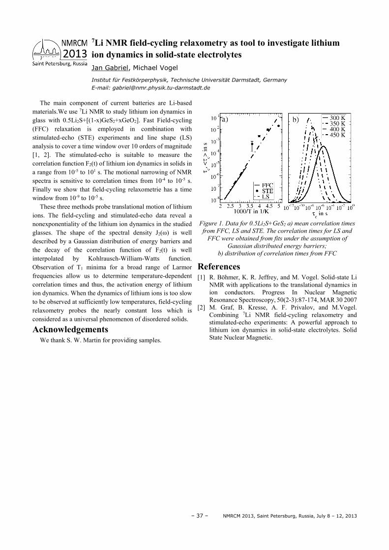
– 37 – NMRCM 2013, Saint Petersburg, Russia, July 8 – 12, 2013
The main component of current batteries are Li-based materials.We use 7Li NMR to study lithium ion dynamics in glass with 0.5Li2S+[(1-x)GeS2+xGeO2]. Fast Field-cycling (FFC) relaxation is employed in combination with stimulated-echo (STE) experiments and line shape (LS) analysis to cover a time window over 10 orders of magnitude [1, 2]. The stimulated-echo is suitable to measure the correlation function F2(t) of lithium ion dynamics in solids in a range from 10-5 to 101 s. The motional narrowing of NMR spectra is sensitive to correlation times from 10-4 to 10-5 s. Finally we show that field-cycling relaxometrie has a time window from 10-9 to 10-5 s.
These three methods probe translational motion of lithium ions. The field-cycling and stimulated-echo data reveal a nonexponentiality of the lithium ion dynamics in the studied glasses. The shape of the spectral density J2(ω) is well described by a Gaussian distribution of energy barriers and the decay of the correlation function of F2(t) is well interpolated by Kohlrausch-William-Watts function. Observation of T1 minima for a broad range of Larmor frequencies allow us to determine temperature-dependent correlation times and thus, the activation energy of lithium ion dynamics. When the dynamics of lithium ions is too slow to be observed at sufficiently low temperatures, field-cycling relaxometry probes the nearly constant loss which is considered as a universal phenomenon of disordered solids.
Acknowledgements We thank S. W. Martin for providing samples.
Figure 1. Data for 0.5Li2S+GeS2 a) mean correlation times
from FFC, LS and STE. The correlation times for LS and
FFC were obtained from fits under the assumption of
Gaussian distributed energy barriers;
b) distribution of correlation times from FFC
References [1] R. Böhmer, K. R. Jeffrey, and M. Vogel. Solid-state Li
NMR with applications to the translational dynamics in ion conductors. Progress In Nuclear Magnetic Resonance Spectroscopy, 50(2-3):87-174, MAR 30 2007
[2] M. Graf, B. Kresse, A. F. Privalov, and M.Vogel. Combining 7Li NMR field-cycling relaxometry and stimulated-echo experiments: A powerful approach to lithium ion dynamics in solid-state electrolytes. Solid State Nuclear Magnetic.
7Li NMR field-cycling relaxometry as tool to investigate lithium ion dynamics in solid-state electrolytes Jan Gabriel, Michael Vogel
Institut für Festkörperphysik, Technische Universität Darmstadt, Germany
E-mail: [email protected]
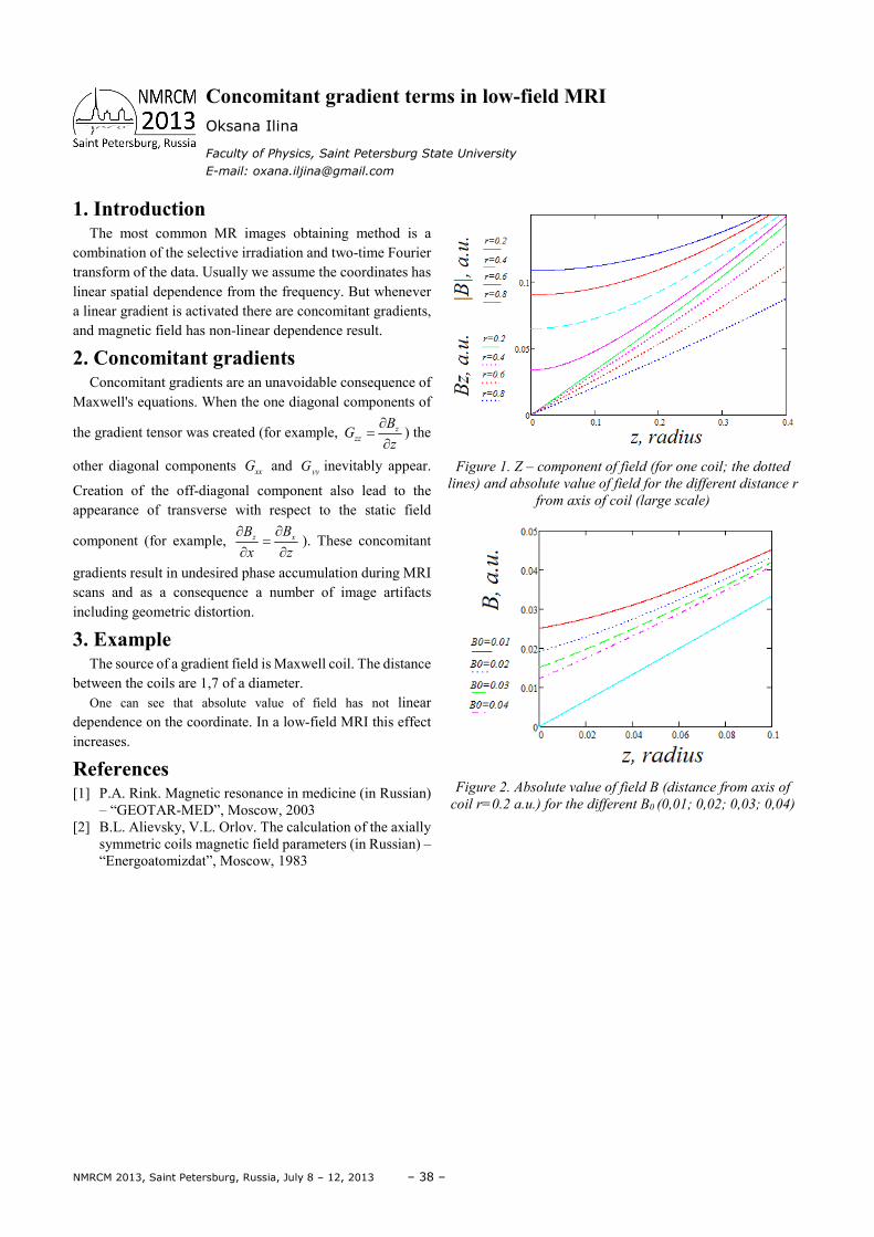
NMRCM 2013, Saint Petersburg, Russia, July 8 – 12, 2013 – 38 –
1. Introduction The most common MR images obtaining method is a
combination of the selective irradiation and two-time Fourier transform of the data. Usually we assume the coordinates has linear spatial dependence from the frequency. But whenever a linear gradient is activated there are concomitant gradients, and magnetic field has non-linear dependence result.
2. Concomitant gradients Concomitant gradients are an unavoidable consequence of
Maxwell's equations. When the one diagonal components of
the gradient tensor was created (for example, z
zz
BG
z
∂=
∂) the
other diagonal components xx
G and yyG inevitably appear.
Creation of the off-diagonal component also lead to the appearance of transverse with respect to the static field
component (for example, xzBB
x z
∂∂=
∂ ∂). These concomitant
gradients result in undesired phase accumulation during MRI scans and as a consequence a number of image artifacts including geometric distortion.
3. Example The source of a gradient field is Maxwell coil. The distance
between the coils are 1,7 of a diameter. One can see that absolute value of field has not linear
dependence on the coordinate. In a low-field MRI this effect increases.
References [1] P.A. Rink. Magnetic resonance in medicine (in Russian)
– “GEOTAR-MED”, Moscow, 2003 [2] B.L. Alievsky, V.L. Orlov. The calculation of the axially
symmetric coils magnetic field parameters (in Russian) – “Energoatomizdat”, Moscow, 1983
Figure 1. Z – component of field (for one coil; the dotted
lines) and absolute value of field for the different distance r
from axis of coil (large scale)
Figure 2. Absolute value of field B (distance from axis of
coil r=0.2 a.u.) for the different B0 (0,01; 0,02; 0,03; 0,04)
Concomitant gradient terms in low-field MRI Oksana Ilina
Faculty of Physics, Saint Petersburg State University
E-mail: [email protected]
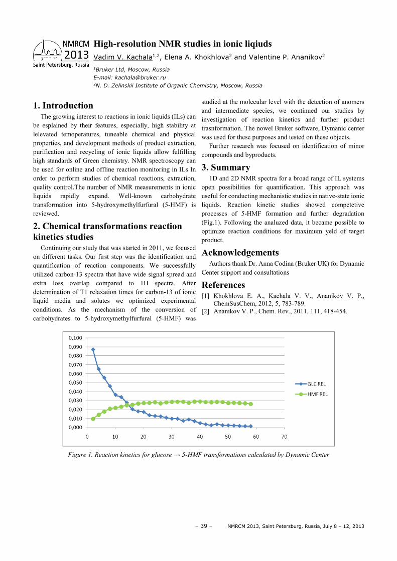
– 39 – NMRCM 2013, Saint Petersburg, Russia, July 8 – 12, 2013
1. Introduction The growing interest to reactions in ionic liquids (ILs) can
be esplained by their features, especially, high stability at lelevated temoperatures, tuneable chemical and physical properties, and development methods of product extraction, purification and recycling of ionic liquids allow fulfilling high standards of Green chemistry. NMR spectroscopy can be used for online and offline reaction monitoring in ILs In order to perform studies of chemical reactions, extraction, quality control.The number of NMR measurements in ionic liquids rapidly expand. Well-known carbohydrate transformation into 5-hydroxymethylfurfural (5-HMF) is reviewed.
2. Chemical transformations reaction kinetics studies
Continuing our study that was started in 2011, we focused on different tasks. Our first step was the identification and quantification of reaction components. We successfully utilized carbon-13 spectra that have wide signal spread and extra loss overlap compared to 1H spectra. After determination of T1 relaxation times for carbon-13 of ionic liquid media and solutes we optimized experimental conditions. As the mechanism of the conversion of carbohydrates to 5-hydroxymethylfurfural (5-HMF) was
studied at the molecular level with the detection of anomers and intermediate species, we continued our studies by investigation of reaction kinetics and further product trasnformation. The nowel Bruker software, Dymanic center was used for these purposes and tested on these objects.
Further research was focused on identification of minor compounds and byproducts.
3. Summary 1D and 2D NMR spectra for a broad range of IL systems
open possibilities for quantification. This approach was useful for conducting mechanistic studies in native-state ionic liquids. Reaction kinetic studies showed competetive processes of 5-HMF formation and further degradation (Fig.1). Following the analuzed data, it became possible to optimize reaction conditions for maximum yeld of target product.
Acknowledgements Authors thank Dr. Anna Codina (Bruker UK) for Dynamic
Center support and consultations
References [1] Khokhlova E. A., Kachala V. V., Ananikov V. P.,
ChemSusChem, 2012, 5, 783-789. [2] Ananikov V. P., Chem. Rev., 2011, 111, 418-454.
Figure 1. Reaction kinetics for glucose → 5-HMF transformations calculated by Dynamic Center
High-resolution NMR studies in ionic liqiuds Vadim V. Kachala1,2, Elena A. Khokhlova2 and Valentine P. Ananikov2
1Bruker Ltd, Moscow, Russia
E-mail: [email protected] 2N. D. Zelinskii Institute of Organic Chemistry, Moscow, Russia
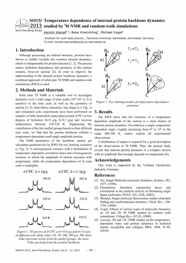
NMRCM 2013, Saint Petersburg, Russia, July 8 – 12, 2013 – 40 –
1. Introduction Although possessing an ordered structure, proteins have
shown to exhibit versatile but common internal dynamics, which is indispensable for protein function [1, 2]. The precise nature, hydration dependence and geometry of this motion remains, however unclear [3]. In order to improve the understanding of the internal protein backbone dynamics, a combined approach of solid state 2H NMR and random-walk simulations (RWS) is used.
2. Methods and Materials Solid state 2H NMR is a valuable tool to investigate
dynamics over a wide range of time scales (10-9-101 s). It is sensitive to the time scale as well as the geometry of motion [4, 5]. Spin-lattice relaxation, line shape (c.f. Fig. 1) and stimulated echo experiments have been performed on samples of fully deuterated cyano phycocyanin (CPC) at two degrees of hydration (h=0 g/g, h=0.3 g/g) and varyious temperatures between 150-310 K. Suppressing the contribution of the fast methyl groups based on their different time scale, we find that the protein backbone exhibits a temperature-dependent small angle amplitude motion.
The NMR parameters of the backbone motion are calculated quantitatively by RWS for two limiting scenarios (s. Fig. 2): A heterogeneous scenario with a distribution of temperature dependent correlation times and a homogeneous scenario, in which the amplitude of motion increases with temperature, while the temperature dependence of its time scale is negligible.
Figure 1. 2H spectra of d-CPC at h=0.0 g/g and h=0.3 g/g
at different echo delay times (20, 50, 100, 200 µs). The inner
Pake spectrum results from the methyl groups, the outer
Pake spectrum from the protein backbone
Figure 2. Two limiting models of temperature dependence
of motion
3. Results The RWS show that the existence of a temperature
dependent amplitude of the motion is a main feature of internal protein dynamics. Nevertheless a single temperature dependent angle, roughly increasing from 0° to 15° in the range 200-300 K, cannot explain all experimental observations
A distribution of angles is required for a good description of the observations in 2H NMR. Thus, the present study reveals that internal protein dynamics is a complex motion with an amplitude that strongly depends on temperature [6].
Acknowledgements This work is supported by the Verband Chemischer
Industrie, Germany.
References [1] Xie, Single Molecule enzymatic dynamics, Science, 282,
1877, (1998). [2] Flomenbom, Stretched exponential decay and
correlations in the catalytic activity of fluctuating single lipase molecules, PNAS, 102, 2368, (2005).
[3] Michael, Single-molecule fluorescence studies of protein folding and conformational dynamics, Chem. Rev., 106, 1785, (2006).
[4] Vogel, Effects of various types of molecular dynamics on 1D and 2D 2H NMR studied by random walk simulations, J.Magn.Res.,147,43, (2000).
[5] Lusceac, 2H and 13C NMR studies on the temperature-dependent water and protein dynamics in hydrated elastin, myoglobin and collagen, BBA, 1804, 41-48, (2010).
Temperature dependence of internal protein backbone dynamics studied by 2H NMR and random-walk simulations Kerstin Kämpf1,2, Beke Kremmling1, Michael Vogel1
1Institute for solid state physics , Technical University Darmstadt, Darmstadt, Germany 2E-mail: [email protected]
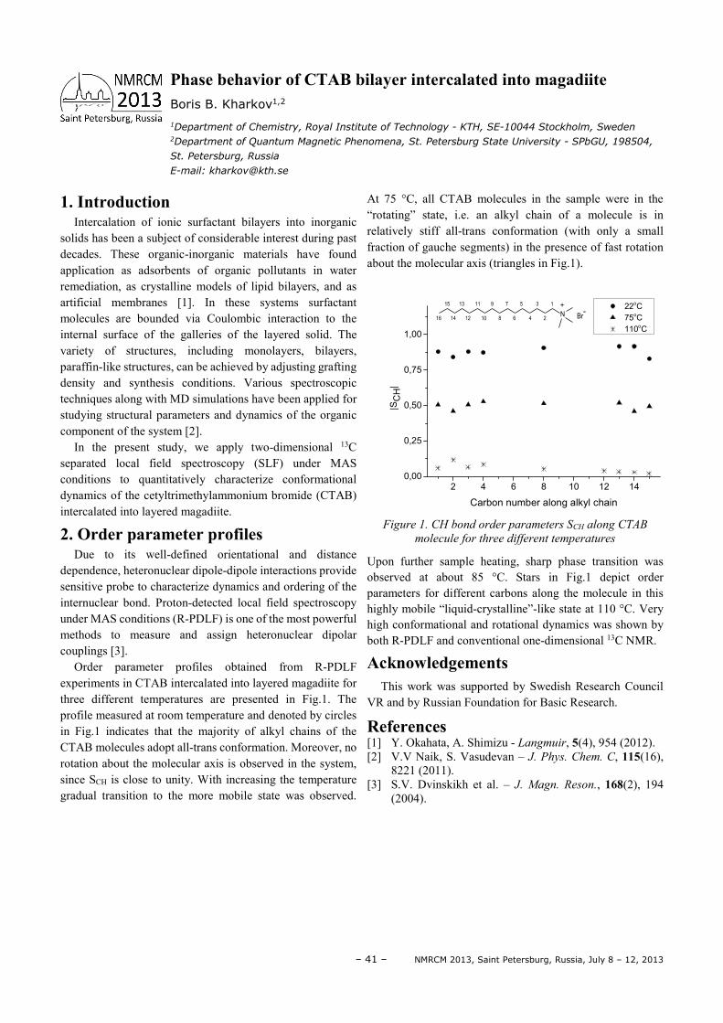
– 41 – NMRCM 2013, Saint Petersburg, Russia, July 8 – 12, 2013
1. Introduction Intercalation of ionic surfactant bilayers into inorganic
solids has been a subject of considerable interest during past decades. These organic-inorganic materials have found application as adsorbents of organic pollutants in water remediation, as crystalline models of lipid bilayers, and as artificial membranes [1]. In these systems surfactant molecules are bounded via Coulombic interaction to the internal surface of the galleries of the layered solid. The variety of structures, including monolayers, bilayers, paraffin-like structures, can be achieved by adjusting grafting density and synthesis conditions. Various spectroscopic techniques along with MD simulations have been applied for studying structural parameters and dynamics of the organic component of the system [2].
In the present study, we apply two-dimensional 13C separated local field spectroscopy (SLF) under MAS conditions to quantitatively characterize conformational dynamics of the cetyltrimethylammonium bromide (CTAB) intercalated into layered magadiite.
2. Order parameter profiles Due to its well-defined orientational and distance
dependence, heteronuclear dipole-dipole interactions provide sensitive probe to characterize dynamics and ordering of the internuclear bond. Proton-detected local field spectroscopy under MAS conditions (R-PDLF) is one of the most powerful methods to measure and assign heteronuclear dipolar couplings [3].
Order parameter profiles obtained from R-PDLF experiments in CTAB intercalated into layered magadiite for three different temperatures are presented in Fig.1. The profile measured at room temperature and denoted by circles in Fig.1 indicates that the majority of alkyl chains of the CTAB molecules adopt all-trans conformation. Moreover, no rotation about the molecular axis is observed in the system, since SCH is close to unity. With increasing the temperature gradual transition to the more mobile state was observed.
At 75 °C, all CTAB molecules in the sample were in the “rotating” state, i.e. an alkyl chain of a molecule is in relatively stiff all-trans conformation (with only a small fraction of gauche segments) in the presence of fast rotation about the molecular axis (triangles in Fig.1).
2 4 6 8 10 12 140,00
0,25
0,50
0,75
1,00
16 14 12 10 8 6 4 2
15 13 11 9 7 5 3 1
N+
Br-
22oC
75oC
110oC
|SC
H|
Carbon number along alkyl chain
Figure 1. CH bond order parameters SCH along CTAB
molecule for three different temperatures
Upon further sample heating, sharp phase transition was observed at about 85 °C. Stars in Fig.1 depict order parameters for different carbons along the molecule in this highly mobile “liquid-crystalline”-like state at 110 °C. Very high conformational and rotational dynamics was shown by both R-PDLF and conventional one-dimensional 13C NMR.
Acknowledgements This work was supported by Swedish Research Council
VR and by Russian Foundation for Basic Research.
References [1] Y. Okahata, A. Shimizu - Langmuir, 5(4), 954 (2012). [2] V.V Naik, S. Vasudevan – J. Phys. Chem. C, 115(16),
8221 (2011). [3] S.V. Dvinskikh et al. – J. Magn. Reson., 168(2), 194
(2004).
Phase behavior of CTAB bilayer intercalated into magadiite Boris B. Kharkov1,2
1Department of Chemistry, Royal Institute of Technology - KTH, SE-10044 Stockholm, Sweden 2Department of Quantum Magnetic Phenomena, St. Petersburg State University - SPbGU, 198504,
St. Petersburg, Russia
E-mail: [email protected]
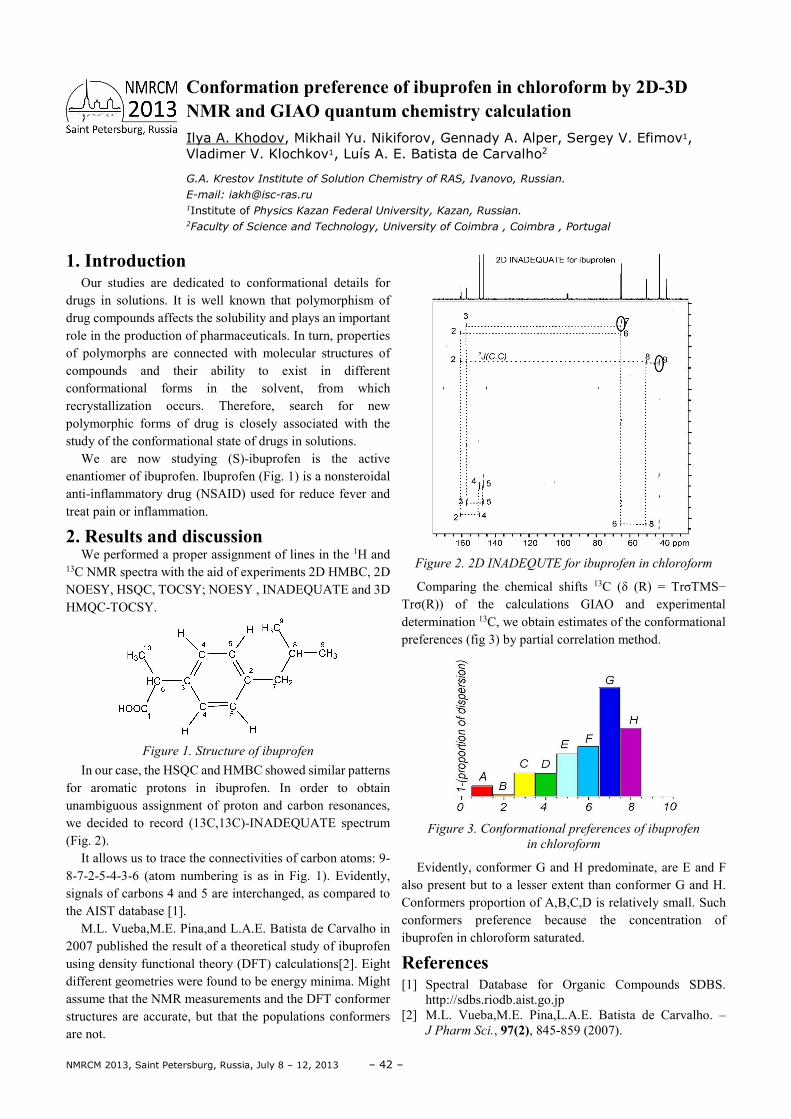
NMRCM 2013, Saint Petersburg, Russia, July 8 – 12, 2013 – 42 –
1. Introduction Our studies are dedicated to conformational details for
drugs in solutions. It is well known that polymorphism of drug compounds affects the solubility and plays an important role in the production of pharmaceuticals. In turn, properties of polymorphs are connected with molecular structures of compounds and their ability to exist in different conformational forms in the solvent, from which recrystallization occurs. Therefore, search for new polymorphic forms of drug is closely associated with the study of the conformational state of drugs in solutions.
We are now studying (S)-ibuprofen is the active enantiomer of ibuprofen. Ibuprofen (Fig. 1) is a nonsteroidal anti-inflammatory drug (NSAID) used for reduce fever and treat pain or inflammation.
2. Results and discussion We performed a proper assignment of lines in the 1H and
13C NMR spectra with the aid of experiments 2D HMBC, 2D NOESY, HSQC, TOCSY; NOESY , INADEQUATE and 3D HMQC-TOCSY.
Figure 1. Structure of ibuprofen
In our case, the HSQC and HMBC showed similar patterns for aromatic protons in ibuprofen. In order to obtain unambiguous assignment of proton and carbon resonances, we decided to record (13C,13C)-INADEQUATE spectrum (Fig. 2).
It allows us to trace the connectivities of carbon atoms: 9-8-7-2-5-4-3-6 (atom numbering is as in Fig. 1). Evidently, signals of carbons 4 and 5 are interchanged, as compared to the AIST database [1].
M.L. Vueba,M.E. Pina,and L.A.E. Batista de Carvalho in 2007 published the result of a theoretical study of ibuprofen using density functional theory (DFT) calculations[2]. Eight different geometries were found to be energy minima. Might assume that the NMR measurements and the DFT conformer structures are accurate, but that the populations conformers are not.
Figure 2. 2D INADEQUTE for ibuprofen in chloroform
Comparing the chemical shifts 13C (δ (R) = TrσTMS− Trσ(R)) of the calculations GIAO and experimental determination 13C, we obtain estimates of the conformational preferences (fig 3) by partial correlation method.
Figure 3. Conformational preferences of ibuprofen
in chloroform
Evidently, conformer G and H predominate, are E and F also present but to a lesser extent than conformer G and H. Conformers proportion of A,B,C,D is relatively small. Such conformers preference because the concentration of ibuprofen in chloroform saturated.
References [1] Spectral Database for Organic Compounds SDBS.
http://sdbs.riodb.aist.go.jp [2] M.L. Vueba,M.E. Pina,L.A.E. Batista de Carvalho. –
J Pharm Sci., 97(2), 845-859 (2007).
Conformation preference of ibuprofen in chloroform by 2D-3D NMR and GIAO quantum chemistry calculation Ilya A. Khodov, Mikhail Yu. Nikiforov, Gennady A. Alper, Sergey V. Efimov1, Vladimer V. Klochkov1, Luís A. E. Batista de Carvalho2
G.A. Krestov Institute of Solution Chemistry of RAS, Ivanovo, Russian.
E-mail: [email protected] 1Institute of Physics Kazan Federal University, Kazan, Russian. 2Faculty of Science and Technology, University of Coimbra , Coimbra , Portugal
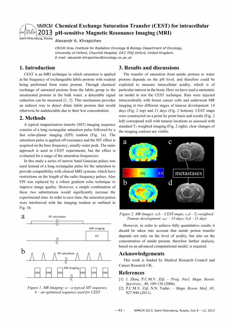
– 43 – NMRCM 2013, Saint Petersburg, Russia, July 8 – 12, 2013
1. Introduction CEST is an MRI technique in which saturation is applied
at the frequency of exchangeable labile protons with readout being performed from water protons. Through chemical exchange of saturated protons from the labile group to the unsaturated protons in the bulk water, a detectable signal reduction can be measured [1, 2]. This mechanism provides an indirect way to detect dilute labile protons that would otherwise be undetectable due to their low concentration.
2. Methods A typical magnetization transfer (MT) imaging sequence
consists of a long rectangular saturation pulse followed by a fast echo-planar imaging (EPI) readout (Fig. 1a). The saturation pulse is applied off-resonance and the MT effect is acquired on the base frequency, usually water peak. The same approach is used in CEST experiments, but the effect is evaluated for a range of the saturation frequencies.
In this study a series of narrow band Gaussian pulses was used instead of a long rectangular pulse for the saturation to provide compatibility with clinical MRI systems, which have restrictions on the length of the radio frequency pulses. Also EPI was replaced by a robust gradient echo technique to improve image quality. However, a simple combination of these two substitutions would significantly increase the experimental time. In order to save time, the saturation pulses were interleaved with the imaging readout as outlined in Fig. 1b.
Figure 1. MR Imaging: a – a typical MT sequence,
b – an optimised sequence used for CEST
3. Results and discussions The transfer of saturation from amide protons to water
protons depends on the pH level, and therefore could be exploited to measure intracellular acidity, which is of particular interest in the brain. Here we have used a metastatic rat model to test the CEST technique. Rats were injected intracerebrally with breast cancer cells and underwent MR imaging at two different stages of tumour development: 14 days (Fig. 2 top) and 21 days (Fig. 2 bottom). CEST maps were constructed on a point by point basis and results (Fig. 2 left) correspond well with tumour locations as assessed with standard T2 weighted imaging (Fig. 2 right), clear changes of the imaging contrast are visible.
Figure 2. MR Images: a,b – CEST-maps; c,d – T2-weighted.
Tumour development: a,c – 14 days; b,d – 21 days
However, in order to achieve fully quantitative results it should be taken into account that amide proton transfer depends not only on the level of acidity, but also on the concentration of amide present, therefore further analysis, based on an advanced computational model, is required.
Acknowledgements This work is funded by Medical Research Council and
Cancer Research UK.
References [1] J. Zhou, P.C.M.V. Zijl. – Prog. Nucl. Magn. Reson.
Spectrosc., 48, 109-136 (2006). [2] P.C.M.V. Zijl, N.N. Yadav. – Magn. Reson. Med., 65,
927-948 (2011).
Chemical Exchange Saturation Transfer (CEST) for intracellular pH-sensitive Magnetic Resonance Imaging (MRI) Alexandr A. Khrapichev
CR/UK Gray Institute for Radiation Oncology & Biology Department of Oncology,
University of Oxford, Churchill Hospital, OX3 7DQ Oxford, United Kingdom.
E-mail: [email protected]
NMRCM 2013, Saint Petersburg, Russia, July 8 – 12, 2013 – 44 –
1. Introduction Magnesium is one of the most promising candidates for
hydrogen storage, mainly because of its high hydrogen storage capacity (up to 7.6 w%), low cost, large reserves and high reversibility of the hydrogen sorption reaction. Unfortunately, a rather high temperature of the hydrogen release, very low hydrogenation/dehydrogenation rates and a high reactivity toward air and oxygen essentially limit its application for practical uses.
Nevertheless, it was found that the limited dissociation rate of hydrogen on the metal surface may be improved by additions of transition metal catalysts (TM). Investigations by means of in situ X-ray [1] and neutron [2] diffraction of Mg/Nb thin films and nanocrystallites showed that hydrogen atoms penetrate inside Mg through the so-called Nb “gates”. However, up-to-now there is no complete description of such a phenomenon on the microscopic level.
In our previous works [3, 4] we proposed one of the possible explanations of such hydrogen kinetic improvements in magnesium. We found that Nb additives stabilize the Mg bcc structure on interface border that could exhibit a much better hydrogen diffusion rate reference to other possible structures (hcp, fcc, rutile) of Mg.
In this contribution we report on the results of our theoretical studies of hydrogen diffusion in different structures of magnesium. To calculate diffusion coefficients and energy activation barriers we considered various possible diffusion paths in bulk Mg. Zero point energy (ZPE) contributions to energy barriers were also calculated.
2. Method of calculations Calculations were performed within the framework of
density functional theory (DFT) using the plane-wave pseudopotential method and the Perdew–Burke–Ernzerhof GGA exchange and correlation potential as implemented in Quantum Espresso package [5]. For all calculated structures we optimized the lattice parameters and the atomic positions. The kinetic energy/charge cut-off was equal to 70/260 Ry, the 12×12×12 k-point mesh was used. To estimate ZPE contributions we performed phonon calculations using the linear-response method. The Nudged Elastic Band (NEP) method was used to locate the minimum energy path for H-
diffusion. As the initial and final hydrogen states the tetrahedral and octahedral sites were selected.
3. Results and discussion The calculations of the activation energy barriers for hcp,
fcc and bcc structure of MgHx at low hydrogen concentration are still in progress. But preliminary results have shown that bcc-MgHx has the minimal activation barriers, wheras hcp-MgHx has the biggest ones. Such theoretical results were experimentally confirmed by faster hydrogen diffusion through the bcc lattice observed experimentally in Mg1-xNbxH2 [6].
Also using the scheme presented in Ref. [7] we calculated diffusion coefficient for all magnesium structures. The ZPE contributions to diffusion coefficient proved to be rather insignificant.
Our early calculations [4] showed that in the bcc-MgHx structure hydrogen atoms tend to be randomly distributed over the interstitial sites. We assume that this may reduce the so-called “blocking” hydride layer effect. Our present calculations revealed that the bcc-MgHx structure also has the smallest activation energy barriers. From our point of view these two factors may be responsible for faster hydrogen diffusion through the bcc lattice as observed experimentally in Mg1-xNbxH2 [6] and may explain the catalytic effect of TM additions in magnesium.
References [1] J.F. Pelletier, J. Huot, M. Sutton, R. Schulz, A.R.
Sandy,L.B. Lurio and S.G.J. Mochrie, Phys. Rev. B, 63, (2001) 052103.
[2] P. de Rango, A. Chaise, J. Charbonnier, D. Fruchart, M. Jehan, Ph. Marty, S. Miraglia, S. Rivoirard, N. Skryabina, J. Alloys Compd., 445-447 (2007) 52.
[3] K. Klyukin, M.G. Shelyapina, D. Fruchart, Solid State Phenomena, 170 (2011) 298.
[4] K Klyukin, MG Shelyapina, D Fruchart, Journal of Alloys and Compounds (2013), http://dx.doi.org/10.1016/j.jallcom.2013.02.089
[5] Giannozzi, Paolo, et al., Journal of Physics: Condensed Matter 21.39 (2009): 395502.
[6] X.H. Tan , L. Wang , C.M.B. Holt, B. Zahiri , M.H. Eikerling and D. Mitlin, Phys. Chem. Chem. Phys.,14 (2012) 10904.
[7] K. W. Kehr, in Hydrogen in Metals I, edited by G. Alefeld and J. Völkl (Springer, Berlin, 1978), p. 197
Hydrogen diffusion in various structures of MgHx: a DFT study K. Klyukin1, M. G. Shelyapina1, D. Fruchart2
1St.Peterburg State University, 3, Ulyanovskaya, Petrodvorets, 198504, St.Peterburg, Russia 2Institut Néel, CNRS, 25 avenue des Martyrs, BP 166, 38042 Grenoble cedex 9, France
E-mail: [email protected]
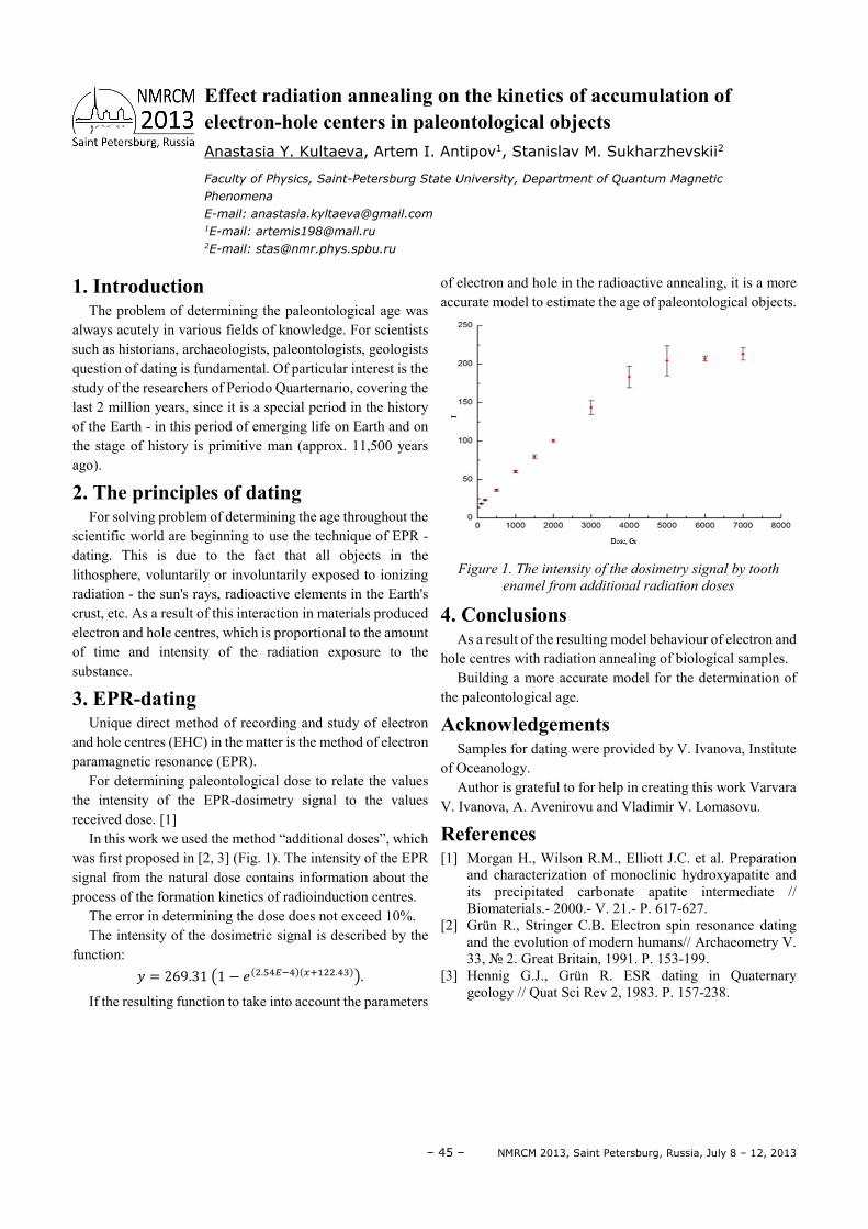
– 45 – NMRCM 2013, Saint Petersburg, Russia, July 8 – 12, 2013
1. Introduction The problem of determining the paleontological age was
always acutely in various fields of knowledge. For scientists such as historians, archaeologists, paleontologists, geologists question of dating is fundamental. Of particular interest is the study of the researchers of Periodo Quarternario, covering the last 2 million years, since it is a special period in the history of the Earth - in this period of emerging life on Earth and on the stage of history is primitive man (approx. 11,500 years ago).
2. The principles of dating For solving problem of determining the age throughout the
scientific world are beginning to use the technique of EPR - dating. This is due to the fact that all objects in the lithosphere, voluntarily or involuntarily exposed to ionizing radiation - the sun's rays, radioactive elements in the Earth's crust, etc. As a result of this interaction in materials produced electron and hole centres, which is proportional to the amount of time and intensity of the radiation exposure to the substance.
3. EPR-dating Unique direct method of recording and study of electron
and hole centres (EHC) in the matter is the method of electron paramagnetic resonance (EPR).
For determining paleontological dose to relate the values the intensity of the EPR-dosimetry signal to the values received dose. [1]
In this work we used the method “additional doses”, which was first proposed in [2, 3] (Fig. 1). The intensity of the EPR signal from the natural dose contains information about the process of the formation kinetics of radioinduction centres.
The error in determining the dose does not exceed 10%. The intensity of the dosimetric signal is described by the
function: = 269.31 1 − . ..
If the resulting function to take into account the parameters
of electron and hole in the radioactive annealing, it is a more accurate model to estimate the age of paleontological objects.
Figure 1. The intensity of the dosimetry signal by tooth
enamel from additional radiation doses
4. Conclusions As a result of the resulting model behaviour of electron and
hole centres with radiation annealing of biological samples. Building a more accurate model for the determination of
the paleontological age.
Acknowledgements Samples for dating were provided by V. Ivanova, Institute
of Oceanology. Author is grateful to for help in creating this work Varvara
V. Ivanova, A. Avenirovu and Vladimir V. Lomasovu.
References [1] Morgan H., Wilson R.M., Elliott J.C. et al. Preparation
and characterization of monoclinic hydroxyapatite and its precipitated carbonate apatite intermediate // Biomaterials.- 2000.- V. 21.- P. 617-627.
[2] Grün R., Stringer C.B. Electron spin resonance dating and the evolution of modern humans// Archaeometry V. 33, 2. Great Britain, 1991. P. 153-199.
[3] Hennig G.J., Grün R. ESR dating in Quaternary geology // Quat Sci Rev 2, 1983. P. 157-238.
Effect radiation annealing on the kinetics of accumulation of electron-hole centers in paleontological objects Anastasia Y. Kultaeva, Artem I. Antipov1, Stanislav M. Sukharzhevskii2
Faculty of Physics, Saint-Petersburg State University, Department of Quantum Magnetic
Phenomena
E-mail: [email protected] 1E-mail: [email protected] 2E-mail: [email protected]
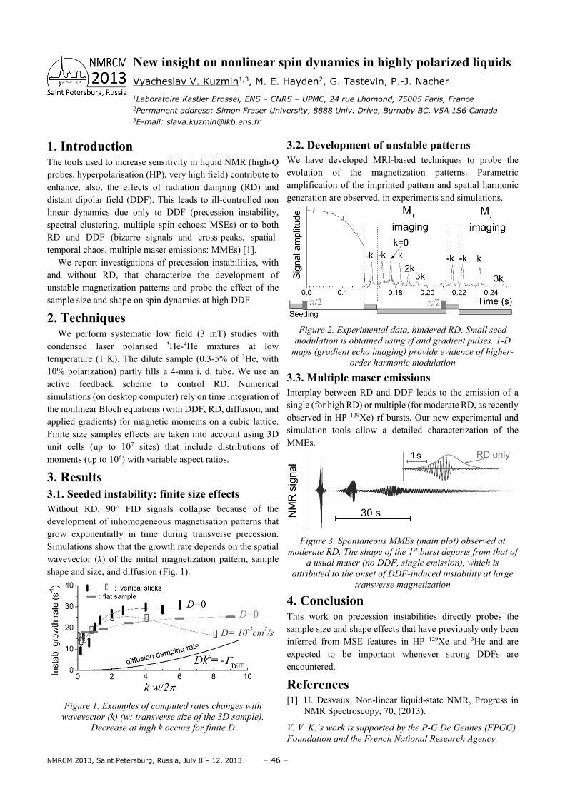
NMRCM 2013, Saint Petersburg, Russia, July 8 – 12, 2013 – 46 –
1. Introduction The tools used to increase sensitivity in liquid NMR (high-Q probes, hyperpolarisation (HP), very high field) contribute to enhance, also, the effects of radiation damping (RD) and distant dipolar field (DDF). This leads to ill-controlled non linear dynamics due only to DDF (precession instability, spectral clustering, multiple spin echoes: MSEs) or to both RD and DDF (bizarre signals and cross-peaks, spatial-temporal chaos, multiple maser emissions: MMEs) [1].
We report investigations of precession instabilities, with and without RD, that characterize the development of unstable magnetization patterns and probe the effect of the sample size and shape on spin dynamics at high DDF.
2. Techniques We perform systematic low field (3 mT) studies with
condensed laser polarised 3He-4He mixtures at low temperature (1 K). The dilute sample (0.3-5% of 3He, with 10% polarization) partly fills a 4-mm i. d. tube. We use an active feedback scheme to control RD. Numerical simulations (on desktop computer) rely on time integration of the nonlinear Bloch equations (with DDF, RD, diffusion, and applied gradients) for magnetic moments on a cubic lattice. Finite size samples effects are taken into account using 3D unit cells (up to 107 sites) that include distributions of moments (up to 106) with variable aspect ratios.
3. Results 3.1. Seeded instability: finite size effects Without RD, 90° FID signals collapse because of the development of inhomogeneous magnetisation patterns that grow exponentially in time during transverse precession. Simulations show that the growth rate depends on the spatial wavevector (k) of the initial magnetization pattern, sample shape and size, and diffusion (Fig. 1).
Figure 1. Examples of computed rates changes with
wavevector (k) (w: transverse size of the 3D sample).
Decrease at high k occurs for finite D
3.2. Development of unstable patterns We have developed MRI-based techniques to probe the evolution of the magnetization patterns. Parametric amplification of the imprinted pattern and spatial harmonic generation are observed, in experiments and simulations.
Figure 2. Experimental data, hindered RD. Small seed
modulation is obtained using rf and gradient pulses. 1-D
maps (gradient echo imaging) provide evidence of higher-
order harmonic modulation
3.3. Multiple maser emissions Interplay between RD and DDF leads to the emission of a single (for high RD) or multiple (for moderate RD, as recently observed in HP 129Xe) rf bursts. Our new experimental and simulation tools allow a detailed characterization of the MMEs.
Figure 3. Spontaneous MMEs (main plot) observed at
moderate RD. The shape of the 1st burst departs from that of
a usual maser (no DDF, single emission), which is
attributed to the onset of DDF-induced instability at large
transverse magnetization
4. Conclusion This work on precession instabilities directly probes the sample size and shape effects that have previously only been inferred from MSE features in HP 129Xe and 3He and are expected to be important whenever strong DDFs are encountered.
References [1] H. Desvaux, Non-linear liquid-state NMR, Progress in
NMR Spectroscopy, 70, (2013).
V. V. K.’s work is supported by the P-G De Gennes (FPGG)
Foundation and the French National Research Agency.
New insight on nonlinear spin dynamics in highly polarized liquids Vyacheslav V. Kuzmin1,3, M. E. Hayden2, G. Tastevin, P.-J. Nacher
1Laboratoire Kastler Brossel, ENS – CNRS – UPMC, 24 rue Lhomond, 75005 Paris, France 2Permanent address: Simon Fraser University, 8888 Univ. Drive, Burnaby BC, V5A 1S6 Canada 3E-mail: [email protected]
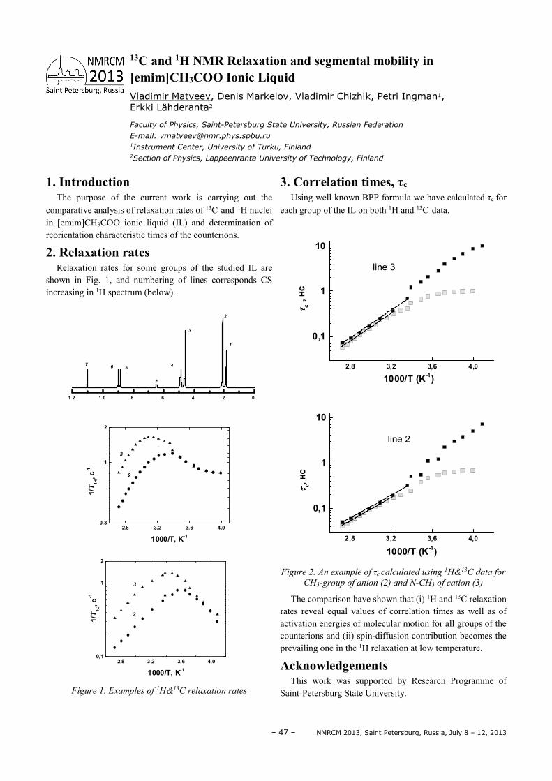
– 47 – NMRCM 2013, Saint Petersburg, Russia, July 8 – 12, 2013
1. Introduction The purpose of the current work is carrying out the
comparative analysis of relaxation rates of 13C and 1H nuclei in [emim]CH3COO ionic liquid (IL) and determination of reorientation characteristic times of the counterions.
2. Relaxation rates Relaxation rates for some groups of the studied IL are
shown in Fig. 1, and numbering of lines corresponds CS increasing in 1H spectrum (below).
1 2 1 0 8 6 4 2 0
76 5
*
4
3
2
1
2.8 3.2 3.6 4.00.3
1
2
2
1/T
1H, c
-1
1000/T, K-1
3
2,8 3,2 3,6 4,00,1
1
2
1/T
1C, c
-1
2
1000/T, K-1
3
Figure 1. Examples of 1H&13C relaxation rates
3. Correlation times, τc Using well known BPP formula we have calculated τc for
each group of the IL on both 1H and 13C data.
2,8 3,2 3,6 4,0
0,1
1
10
1000/T (K-1)
ττ ττ c ,
нс
line 3
2,8 3,2 3,6 4,0
0,1
1
10
ττ ττ c, н
с
1000/T (K-1)
line 2
Figure 2. An example of τc calculated using 1H&13C data for
CH3-group of anion (2) and N-CH3 of cation (3)
The comparison have shown that (i) 1H and 13C relaxation rates reveal equal values of correlation times as well as of activation energies of molecular motion for all groups of the counterions and (ii) spin-diffusion contribution becomes the prevailing one in the 1H relaxation at low temperature.
Acknowledgements This work was supported by Research Programme of
Saint-Petersburg State University.
13C and 1H NMR Relaxation and segmental mobility in [emim]CH3COO Ionic Liquid Vladimir Matveev, Denis Markelov, Vladimir Chizhik, Petri Ingman1, Erkki Lähderanta2
Faculty of Physics, Saint-Petersburg State University, Russian Federation
E-mail: [email protected] 1Instrument Center, University of Turku, Finland 2Section of Physics, Lappeenranta University of Technology, Finland

NMRCM 2013, Saint Petersburg, Russia, July 8 – 12, 2013 – 48 –
1. Introduction The problem of nuclear spin echo control attracts an
attention because of growing interest to the feasibilities to operate with NMR signal by external actions. It is especially important for the magnetically ordered materials, where the signal forms in the domain walls and it is possible to manipulate with them by application of magnetic field pulses, [1-2]. This report is devoted to the principles of such an operation and its potential for the obtaining of information on the properties of substance.
2. Experiment The situation, when the echo signal, excited in magnetic
sample (Li0.425Fe2.425Zn0.15O4) by a standard non steady-state NMR technique, using radio frequency (rf) pulses, and subjected also by an additional sequence of magnetic (video) pulses, is under discussion. The different variants of video pulses effect are examined: i) the action of single pulse; ii) the action of two unidirectional pulse sequence; iii) the action of the sequence of two pulses with different sign. It is taken into account that video pulses can be applied both in the interval between rf pulses and simultaneously with them.
3. Results It was observed that, as in [3], the single video pulse
depresses the nuclear spin system response because of the displacement of domain walls, which was experimentally proved by the obtaining of the constant field dependence of the echo intensity. It was shown that the phenomenon can be described by so-called ‘optical model’, where the individual spin isochromates are considered as optical beams, and the depression as their ‘defocusing’. An example of two-pulse and stimulated echo signals (with amplitudes Ie and Ise correspondingly) quenching by the single video pulse with the input time tv, is demonstrated in Fig. 1 (here the delay time between the first and second rf pulses is 185 µs, and between the second and third ones is 790 µs).
The model implies a number of other effects, for instance, the restoring of the depressed two-pulse echo under the action of the sequence of two video pulses. It is illustrated by the Fig. 2, where an increasing of restoration effect is shown, when the durations of video pulses (τv1, τv2) become equal.
It was also revealed, that in the case of the coincidence of video and rf pulses the spin system can be excited in different places of sample, which means that it is possible to scan the material near the domain wall positioning.
Figure 1. Echo-signals depression by single video pulse
Figure 2. Echo-signal restoring by two video pulses
Acknowledgements The work was supported by the Presidium of Russian
Academy of Sciences, Program P-03.
References [1] L.A. Rassvetalov, A.B. Levitski. – Sov. Solid State Phys.,
23, 3354-3359 (1981) [2] G.I. Mamniashvili, T.O. Gegechkori; A.M. Akhalkatsi,
C.A. Gavasheli. – Low Temperature Physics, 38, 466-472 (2012)
[3] I.V. Pleshakov, N.S. Klekhta, Yu.I. Kuzmin. – Technical
Physics Letters, 38, 853-855 (2012)
NMR signal operation in magnetic substances by the external field pulses Ivan V. Pleshakov1,2, Nikolay S. Klekhta2
1Ioffe Physical-Technical Institute of the Russian Academy of Sciences,
194021 St. Petersburg, Russia
E-mail: [email protected] 2Saint-Petersburg State Polytechnic University, 195251 St. Petersburg, Russia
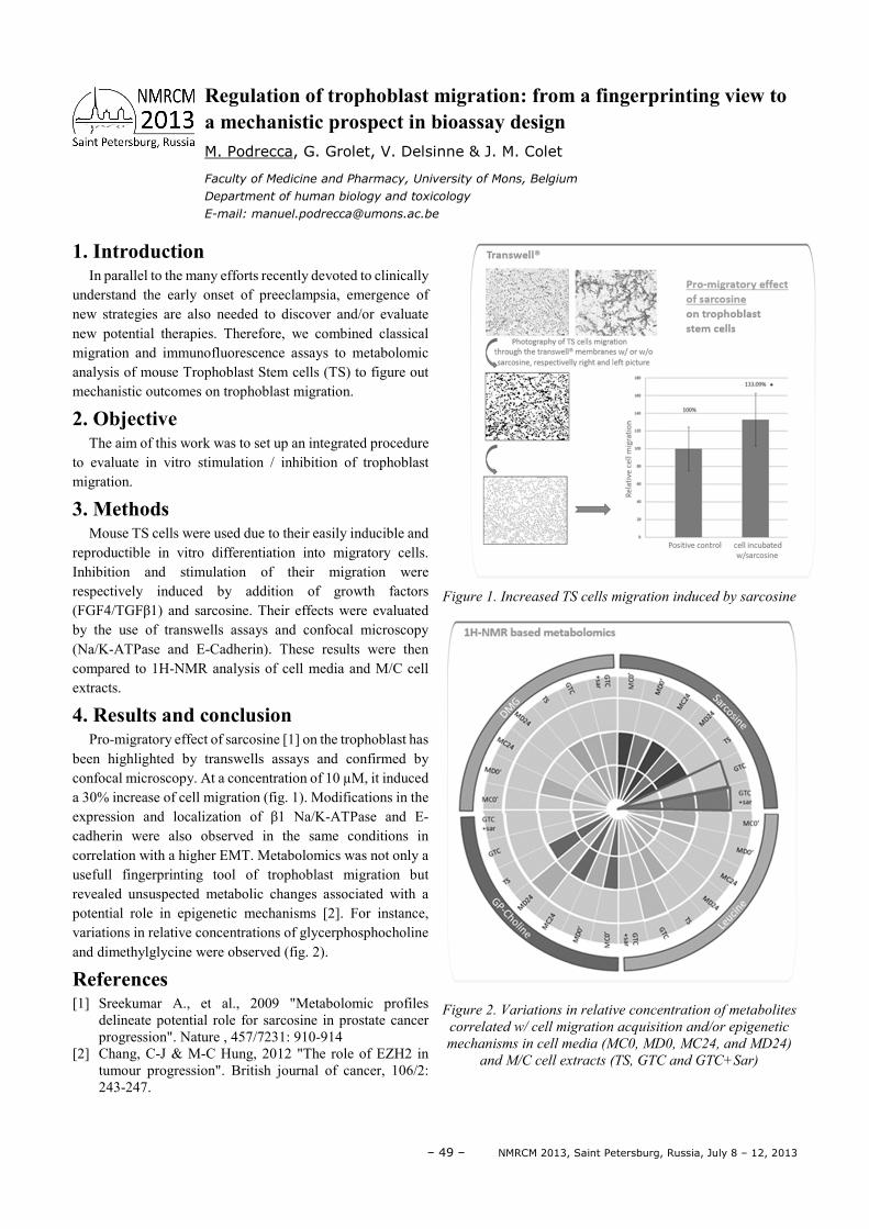
– 49 – NMRCM 2013, Saint Petersburg, Russia, July 8 – 12, 2013
1. Introduction In parallel to the many efforts recently devoted to clinically
understand the early onset of preeclampsia, emergence of new strategies are also needed to discover and/or evaluate new potential therapies. Therefore, we combined classical migration and immunofluorescence assays to metabolomic analysis of mouse Trophoblast Stem cells (TS) to figure out mechanistic outcomes on trophoblast migration.
2. Objective The aim of this work was to set up an integrated procedure
to evaluate in vitro stimulation / inhibition of trophoblast migration.
3. Methods Mouse TS cells were used due to their easily inducible and
reproductible in vitro differentiation into migratory cells. Inhibition and stimulation of their migration were respectively induced by addition of growth factors (FGF4/TGFβ1) and sarcosine. Their effects were evaluated by the use of transwells assays and confocal microscopy (Na/K-ATPase and E-Cadherin). These results were then compared to 1H-NMR analysis of cell media and M/C cell extracts.
4. Results and conclusion Pro-migratory effect of sarcosine [1] on the trophoblast has
been highlighted by transwells assays and confirmed by confocal microscopy. At a concentration of 10 µM, it induced a 30% increase of cell migration (fig. 1). Modifications in the expression and localization of β1 Na/K-ATPase and E-cadherin were also observed in the same conditions in correlation with a higher EMT. Metabolomics was not only a usefull fingerprinting tool of trophoblast migration but revealed unsuspected metabolic changes associated with a potential role in epigenetic mechanisms [2]. For instance, variations in relative concentrations of glycerphosphocholine and dimethylglycine were observed (fig. 2).
References [1] Sreekumar A., et al., 2009 "Metabolomic profiles
delineate potential role for sarcosine in prostate cancer progression". Nature , 457/7231: 910-914
[2] Chang, C-J & M-C Hung, 2012 "The role of EZH2 in tumour progression". British journal of cancer, 106/2: 243-247.
Figure 1. Increased TS cells migration induced by sarcosine
Figure 2. Variations in relative concentration of metabolites
correlated w/ cell migration acquisition and/or epigenetic
mechanisms in cell media (MC0, MD0, MC24, and MD24)
and M/C cell extracts (TS, GTC and GTC+Sar)
Regulation of trophoblast migration: from a fingerprinting view to a mechanistic prospect in bioassay design M. Podrecca, G. Grolet, V. Delsinne & J. M. Colet
Faculty of Medicine and Pharmacy, University of Mons, Belgium
Department of human biology and toxicology
E-mail: [email protected]
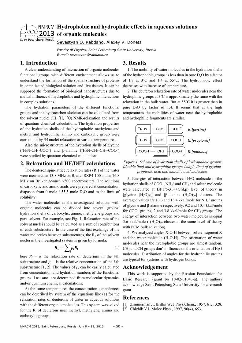
NMRCM 2013, Saint Petersburg, Russia, July 8 – 12, 2013 – 50 –
1. Introduction А clear understanding of interaction of organic molecules
functional groups with different environment allows us to understand the formation of the spatial structure of proteins in complicated biological solution and live tissues. It can be supposed the formation of biological nanostructures due to mutual influence of hydrophobic and hydrophilic interactions in complex solutions.
The hydration parameters of the different functional groups and the hydrocarbon skeleton can be calculated from the solvent nuclei (1H, 2H, 17O) NMR-relaxation and results of quantum chemical calculations. The hydration properties of the hydration shells of the hydrophobic methylene and methyl and hydrophilic amino and carboxylic group were carried out by 2H nuclei relaxation at various temperatures.
Also the microstructure of the hydration shells of glycine (+H3N-CH2-COO–) and β-alanine (+H3N-CH2-CH2-COO–) were studied by quantum chemical calculations.
2. Relaxation and HF/DFT calculations The deuteron spin-lattice relaxation rates (R1) of the water
were measured at 13.8 MHz on Bruker SXP4-100 and at 76.8 MHz on Bruker AvanceIII/500 spectrometers. The solutions of carboxylic and amino acids were prepared at concentration diapason from 0 mole / 55.5 mole D2O and to the limit of solubility.
The water molecules in the investigated solutions with organic molecules can be divided into several groups: hydration shells of carboxylic, amino, methylene groups and pure solvent. For example, see Fig. 1. Relaxation rate of the solvent nuclei should be calculated as a sum of contributions of each substructure. In the case of the fast exchange of the water molecules between substructures, the R1 of the solvent nuclei in the investigated system is given by formula:
(1)
here Ri – is the relaxation rate of deuterium in the i-th substructure and pi – is the relative concentration of the i-th substructure [1, 2]. The values of pi can be easily calculated from concentration and hydration numbers of the functional groups. Last ones are determined from molecular dynamics and/or quantum chemical calculations.
At the same temperatures the concentration dependences can be described by system of the equations like (1) for the relaxation rates of deuterons of water in aqueous solutions with the different organic molecules. This system was solved for the Ri of deuterons near methyl, methylene, amino and carboxylic groups.
3. Results 1. The mobility of water molecules in the hydration shells
of the hydrophobic groups is less than in pure D2O by a factor of 1.7 at 3˚C and 1.4 at 55˚C. The hydrophobic effect decreases with increase of temperature.
2. The deuteron relaxation rate of water molecules near the hydrophilic groups at 3˚C is approximately the same with the relaxation in the bulk water. But at 55˚C it is greater than in pure D2O by factor of 1.4. It seems that at the high temperatures the mobilities of water near the hydrophobic and hydrophilic fragments are similar.
Figure 1. Scheme of hydration shells of hydrophobic groups
(double line) and hydrophilic groups (single line) of glycine,
propionic acid and malonic acid molecules
3. Energies of interaction between H2O molecule in the hydration shells of COO–, NH3
+ and CH2 and solute molecule were calculated at DFT/6-31++G(d,p) level of theory in [glycine (H2O)57] and [β-alanine (H2O)70] clusters. The averaged values are 13.3 and 13.4 kkal/mole for NH3
+ groups of glycine and β-alanine respectively, 9.2 and 10.4 kkal/mole for COO– groups, 2 and 3.8 kkal/mole for CH2 groups. The energy of interaction between two water molecules is equal 5.6 kkal/mole ( (H2O)60 cluster at the same level of theory with PCM bulk solvation).
4. We analyzed angles X-O-H between solute fragment X and the water molecule (H-O-H). The orientation of water molecules near the hydrophobic groups are almost random. CH2 and CH groups don’t influence on the orientation of H2O molecules. Distribution of angles for the hydrophilic groups are typical for systems with hydrogen bonds.
Acknowledgement This work is supported by the Russian Foundation for
Basic Research (grant 10-02-01043-a). The authors acknowledge Saint-Petersburg State University for a research grant.
References [1] Zimmerman J., Brittin W. J.Phys.Chem., 1957, 61, 1328. [2] Chizhik V.I. Molec.Phys., 1997, 90(4), 653.
Hydrophobic and hydrophilic effects in aqueous solutions of organic molecules Sevastyan O. Rabdano, Alexey V. Donets
Faculty of Physics, Saint-Petersburg State University, Russia
E-mail: [email protected]
– 51 – NMRCM 2013, Saint Petersburg, Russia, July 8 – 12, 2013
Heterogeneous system, consisted of a semiconductor and magnetically ordered substance, is one of topical problem of solid state physics, which attracts an attention of experts because the magnetic parameters of matter may be transformed by the confinement and interplay of cluster surface with supporting body [1,2]. This work presents the results of investigation of a composite material obtained by the filling of cylindrical cavities of porous silicon, by an initial Co-containing compound (CoCl2·6H2O) followed by its reduction to a magnetic phase via chemical reaction with sodium tetraborohydride (NaBH4) (por-Si). This process can lead to the formation of two main phases, metallic cobalt and cobalt boride (Co2B) [3], the latter also possessing ferromagnetic (F) properties [4]. The tasks of this work were: (i) to confirm an origination of magnetic nanoparticles in pores; (ii) to specify what kinds of substances are formed in the pores. Expecting that a state of magnetic material in the pores is mainly determined by described above chemical reactions we repeated them in a glass test tube instead of Si matrix (Ref-sample) to obtain a larger amount of material for study. It was very lightweight (that indicates a porous character of product), easy electrifiable insulator powder. Comparative study of magnetic properties of por-Si, Ref-sample and a submicron bit of metal Co (Co-bulk) was carried out.
Registration of second harmonic of magnetization (M2) generation in the parallel steady (H) and harmonic magnetic fields h(t) (f=15.7 MHz) [5] as well as X-range electron magnetic resonance (EMR) spectrometer, in which the recording signal is proportional to off-diagonal element of the magnetic susceptibility tensor, My(ω) =χyx(ω)hx(ω), [6] were employed for study of the magnetic properties. Symmetry properties of M2 allows one to detect the appearance of a remanent magnetization, Mrem, in a sample at arising H-
hysteresis (symmetric relative to the point H=0 H-scan was used).
The obtained results of comparative study of the por-Si, Ref-sample, and Co-bulk reveal the following features. The M2 signals of all three samples exhibit the H-hysteresis that suggests the presence of F ordered regions with Mrem in all the samples. The Im(Re)M2(H) curves for por-Si and Ref-sample
reveal a close H-form and a close M2(H) dependence on T (at least, in ReM2 component) and on frequency of H-scan (Fscan=0.01÷10Hz). The latter implies a necessity of using a conception of blocking temperature to explain the F-dependence of the responses. The decreasing of field hysteresis in the M2 response at heating (at fixed Fscan) and at decreasing of Fscan (at fixed temperature) evidences a single domain regime of the formed F clusters in these samples. Some difference in T-behavior of ImM2(H) can be mainly referred to pore surface effect on a magnetic dynamics of a material, forming inside the pores. The found temperature dependences of “coercivity”, HC, in ReM2 component of the por-Si and Ref-sample responses at lowest Fscan = 0.01 Hz allow us to estimate the values of blocking temperatures for these compounds as TBp~ 473K and TBR~ 471K accordingly. The essentially larger H-hysteresis and “coercivity”, HC, as well as much weaker dependences on T and Fscan are found in metallic Co-bulk. Besides, the huge difference in EMR signals of Co-bulk and Ref-sample (a reproductible EMR signal is not found for por-Si) is also detected. The obtained results let us to conclude that in the composite as well as in the reference substance ferromagnetic clusters of predominantly insulating compounds of cobalt and boron (Co2B, TC ≈ 440 K [4] or Co3B, TC ≈ 760 K [7]), appear as a result of chemical reaction. In the former sample they form on the inner surface of matrix and for both cases the single domain state of particles is realized. The closeness of the estimated values of blocking temperature values together with the other experimental results obtained for por-Si composite and Ref-sample provides an evidence of the similarity of the physical properties of clusters in both investigated materials.
References [1] J. P. Chen et al. – J. Appl. Phys., 76, 6316 (1994). [2] N.V. Glebova et al. – Tech. Phys. Lett., 36, 878 (2010). [3] G. N. Glavee et al. – Inorg. Chem., 32, 474 (1993). [4] C. Petit and M. P. Pileni. – JMMM, 116, 82 (1997). [5] A.V. Lazuta et al.- Sov. Phys. JETP, 73, 1086 (1991). [6] V.A. Ryzhov et al. – Tech. Phys., 40, 71 (1995). [7] L. Yiping et al. – JMMM, 79, 321 (1989).
Magnetic study of nanostructural composite material based on cobalt compounds and porous silicon V. A. Ryzhov1, I. V. Pleshakov2, A. A. Nechitailov2, N. V. Glebova2, E. N. Pyatyshev3, A. V. Malkova4, I. A. Kiselev1, V. V. Matveev4
1Petersburg Nuclear Physics Institute, NRC Kurchatov Institute, Gatchina, 188300, Russia
E-mail: [email protected] 2Ioffe Physical Technical Institute, Russian Academy of Sciences, St. Petersburg, 194021 Russia
E-mail: [email protected] 3St. Petersburg State Polytechnical University, St. Petersburg, 194021 Russia
E-mail: [email protected] 4Faculty of Physics, St. Petersburg State University, St. Petersburg, 199164 Russia
E-mail: [email protected]
NMRCM 2013, Saint Petersburg, Russia, July 8 – 12, 2013 – 52 –
Study of biological polymers in water solution has arguably become the raison d'être of NMR. Sadly however, the technical progress is leading this activity to thermal death. While increase in magnetic fields requires shorter motional correlation times, the research logic gravitates to larger and with that slower molecular systems. We shall argue that coherent mechanical spinning is beginning to substitute random thermal motion and solid state experiments will adopt experimental hierarchy and protocols of “high resolution”. With a new generation of µ−rotor, bio-VT probe
technology [1] we have observed breakdown of manyspin systems at 100kHz rates, leading to resolution, adequate for the assignement of ca 100 residue proteins within minutes.
New technology offers also convenient solutions for sample heating, some examples of a reaction monitoring by natural abundance 13C through-flow at 400C are given.
References [1] www.nmri.eu
Hot spinning Ago Samoson
NMR Institute and Tallinn University of Technology, Estonia
E-mail: [email protected]

– 53 – NMRCM 2013, Saint Petersburg, Russia, July 8 – 12, 2013
1. Introduction The superparamagnetic iron oxide nanoparticles (SPIONs)
have the ability to function as a platform for development of theranostic agents. For therapeutic potency and specific delivery to the tumor site of SPIONs we conjugated the latter with recombinant heat shock protein Hsp70. The possibility for the MRI monitoring of targeted delivery of the magnetic conjugate of Hsp70 was analyzed in rat C6 glioma model.
2. Materials and methods The dextran-coated SPIONs were prepared by co-
precipitation from Fe+2, Fe+3 salt solutions as was described earlier [1]. Recombinant human heat shock protein Hsp70 was prepared from E. coli transformed with a pMSHsp70 plasmid. Magnetic conjugates were formed from SPIONs and Hsp70 by coupling COO protein groups to carbodiimide activated surface dextran. The biological activity of Hsp70 in the conjugate was assessed by the chaperone ELISA-assay. Magnetic characteristics of conjugate were analysized by proton relaxometry. Proton magnetic relaxation times T2 were measured with the help of the NMR-spectrometer (CXP-300, Bruker) in magnetic field 7.1 T. The in vitro distribution of SPIONs was assessed on the C6 glioma cell culture with the help of confocal and electron microscopy. The in vivo traffic was analyzed in the rat model of intracranial C6 glioma. MR images (gradient echo (FLASH), T1- and T2-weighted, multi-sc and multi-echo (MSME T2-map) of rat glioma were obtained by Bruker Avance II NMR spectrometer 11 T.
3. Results The measured coefficients of the relaxation efficiency
R1=0.37 mM-1s-1, R2=113 mM-1s-1, R2*=230 mM-1s-1 of
SPIONs and its conjugates correspond to values of negative contrast agents. The results suggest the strong relaxation efficiency of magnetic Hsp70 conjugates in aqueous dispersions. The magnitude of magnetic relaxation rate of Hsp70 conjugates matches well with their appreciable contrast manifestation in phantom study made by MRI. In in vitro studies Hsp70 conjugates sedimented onto C6 cells and passed through the plasma membrane in the endosome-like structures. Visual examination of the confocal and electron microscopy images demonstrated more endosome-filled or not included into the endosomes nanoparticles in case of the Hsp70 conjugates. MRI study of experimental animals confirms the contrast effect of iron oxide nanoparticles conjugated with Hsp70 in the procedure of targeted administration. After magnetic nanoparticles infusion the most significant change in contrast was observed in the tumor
on T2-weighted images. Following intravenous infusion of the SPIONs after 24 h there was a decrease of the T2 relaxivity time in comparison to the control animals (P<0.05). Application of the Hsp70-SPIONs further reduced the relaxivity time in the T2-weighted regimen and numerous zones of conjugate dispersion could be observed as the areas of signal drop (especially in the FLASH regimen) (P<0.001) (Figure 1). On the subsequent postmortem fluorescence images of the brain sections in the reflecting-laser light scanning on confocal microscope we could confirm the accumulation of the Hsp70-SPIONs in the glioma tumor. The study demonstrated that Hsp70-SPION conjugates being intravenously injected in the tumor model accumulate in the glioma and enhance the contrast of their MRI images.
Figure 1. MR T2-weighted images of control and animal treated
with intravenous injection of Hsp70-SPIONs. Presented the axial
sections of the brain. The tumor in control animal is pointed by
white solid arrow. Red arrows point the zones that correspond to
the nanoparticles accumulation
4. Conclusions The contrast efficiency of SPIONs conjugated with Hsp70
in MRI diagnostics is confirmed in a rat model of grafted intracranial glioma tumor. Magnetic Hsp70 conjugate intravenously injected in C6 glioma rat model accumulates in the tumor and exhibit as strong T2 relaxation agent.
Acknowledgements This study was supported by the grant of the Program of
Russian Academy of Sciences (RAS) “Molecular and Cell Biology” and grant of Russian Fund for Basic Research 10-0401049.
References [1] B.P. Nikolaev, Y.Y. Marchenko, L.Y. Yakovleva et al.
Magnetic epidermal growth factor for targeted delivery to grafted tumor in mouse model – IEEE Transactions of magnetic, 49, 1-7 (2013).
Magnetic Resonance Study of SPIONs conjugated with Hsp70 in C6 glioma intracranial model M. A. Shevtsov1, L. Yu. Yakovleva2, B. P. Nikolaev2, Ya. Yu. Marchenko2, A. V. Dobrodumov3, K. V. Onokhin1, A. L. Mikhrina4, I. V. Guzhova1, M. G. Martynova1, O. A. Bystrova1, A. M. Ischenko2, B. A. Margulis1
1Institute of Cytology of the Russian Academy of Sciences (RAS), St. Petersburg, Russia 2Research Institute of Highly Pure Biopreparations, St.Petersburg, Russia 3Institute of Macromolecular Compounds of the Russian Academy of Sciences (RAS),
St. Petersburg, Russia 4I.M. Sechenov institute of evolutionary physiology and biochemistry of the Russian Academy of
Sciences (RAS), St. Petersburg, Russia
E-mail: [email protected]
NMRCM 2013, Saint Petersburg, Russia, July 8 – 12, 2013 – 54 –
1. Introduction The measurements of membrane permeability of plant and
animal cells are essential for solving a wide range of fundamental and applied problems. The most of methods of water membrane permeability measurements used up to date are restricted as they measure either a characteristic of only a single cell or an averaged over the sample parameter. But a study of physiological processes has to be performed preferably on a living intact organism – an object with complex and heterogeneous structure.
A new method is suggested for estimate of the distribution of cell size and membrane permeability of cells in heterogeneous plant tissues. This method is based on the analysis of correlation between rates of spin-spin relaxation of magnetization of water protons and apparent self-diffusion coefficient of water.
2. Materials and Methods Measurements were performed on the 1H NMR
spectrometer operating at 30 MHz. T2-D correlation (DRCOSY) was measured using a modified stimulated echo (STE) pulse sequence with a train of 180o RF-pulses for acquisition of the relaxation signal decay following the diffusion encoding of signal. A maximum pulsed field gradient strength was 1 T/m. T2-D correlation maps were obtained using 2D Fast Laplas Inversion of measured 2D dataset of signal attenuation as a function of b-value (diffusion encoding) and time for transverse relaxation during a train of 180o RF-pulses. For demonstrative purposes the inner part of apple parenchyma tissue was chosen as an object of experiments. Temperature of the sample was kept constant at T = 25oC.
3. Theory An alteration of permeability (P) of membrane of plant cell
leads other things being equal to i) an alteration of apparent spin-spin relaxation time of water protons and ii) an alteration of time dependence of apparent mean diffusion coefficient of water in the long-time limit in biological tissues. Both, T2 and D, can be measured by NMR method. However, they are also sensitive to the variation of cell size R. As a result, measuring either T2-relaxation or diffusion alone doesn’t allow the
evaluation of the distribution of membrane permeability in heterogeneous biological objects.
In present work a new approach offered for analysis of T2-D correlation at short diffusion labelling time (t) when the displacement of water molecules is much less compared to the cell size. In approximation of spherical geometry of a cell a following equation can be derived
0
2 2 0
1 ( ( ) )1 1 94app bulk
D t DH
T T D t
π −− = ,
where H – magnetization sink strength H P∝ ; 2bulkT and
2appT – spin-spin relaxation time of water protons measured in
pure water and in plant cell respectively; D0 – selfdiffusion coefficient of bulk water. If the relaxation time in the extracellular space is infinitely short, H equals the true membrane permeability P.
This equation shows that at given membrane permeability
2 2
1 1app bulk
T T− is linearly correlate with 0
0
1 ( ( ) )D t D
D t
−.
Permeability of membrane of pant cell can be determined even if cell size is unknown.
4. Results Analysis of T2-D correlation maps measured at diffusion
labelling time t = 12, 25 and 50 ms shows that the distribution of membrane permeability is narrow in apple parenchyma tissue. This finding is well agreed with the role of the apple parenchyma as a homogenous storage tissue. As far as magnetization sink strength H is determined the cell size a distribution can be derived from T2-D correlation. A measured cell size distribution is in good agreement with microscopy of apple parenchyma.
Finally, applications of given approach range from a fundamental research on the field of adaptive strategies of plants to the environmental stress (for example, drought) to applied purposes like a quality control of foodstuffs.
Acknowledgements The reported study was partially supported by RFBR,
research projects No. 12-04-31677, 13-04-01203.
Spin-spin relaxation-diffusion correlation analysis for estimation of distribution of membrane permeability and cell size in plant tissues Timur A. Sibgatullin1, Frank J. Vergeldt2, Henk Van As2
1Kazan Institute of Biochemistry and Biophysics, 420111, str. Lobachevsky 2, Kazan, Russia.
E-mail: [email protected] 2Wageningen NMR Centre, Wageningen University, 6703 HA, Dreijenlaan 3, Wageningen,
The Netherlands
E-mail: [email protected]

– 55 – NMRCM 2013, Saint Petersburg, Russia, July 8 – 12, 2013
Currently, the best existing MD force fields cannot accurately reproduce the global free-energy minimum which realizes the experimental protein structure. As a result, long MD trajectories tend to drift away from the starting coordinates (e.g. crystallographic structures). To address this problem, we have devised a new simulation strategy aimed at protein crystals. An MD simulation of protein crystal is essentially an ensemble simulation involving multiple protein molecules in a crystal unit cell (or a block of unit cells). To ensure that average protein coordinates remain correct during the simulation, we introduced crystallography-based restraints into the MD protocol. Since these restraints are aimed at the ensemble-average structure they have only minimal impact on conformational dynamics of the individual protein molecules. So long as the average structure remains reasonable, the proteins move in a native-like fashion
as dictated by the original force field. To validate this approach we have used the data from solid-state NMR spectroscopy, which is the orthogonal experimental technique uniquely sensitive to protein local dynamics. The new method has been tested on human ubiquitin and SH3 domain from chicken α-spectrin. The ensemble-restrained MD simulations produced lower crystallographic R factors than conventional simulations; they also led to more accurate predictions for solid-state chemical shifts and backbone order parameters. Taken together, these results suggest that the presented trajectories may be among the most realistic protein MD simulations ever reported. In this context the ensemble restraints based on high-resolution crystallographic data can be viewed as protein-specific empirical corrections to the standard force fields.
Ensemble-restrained MD simulations: accurate structure leads to accurate dynamics Nikolai Skrynnikov
Department of Chemistry, Purdue University, USA
E-mail: [email protected]
NMRCM 2013, Saint Petersburg, Russia, July 8 – 12, 2013 – 56 –
1. Introduction Silicones are compounds of silicon that possesses at least
one silicon-carbon bond and have a siloxane linkage. The designation M, D, T and Q, respectively, are used for mono-, di-, tri-, and quaternary coordination of oxygen around silicon in silicones. MQ silicone copolymers (also called the MQ resins) are the best examples of commercial hybrid organic–inorganic materials with nanoscale dimensions [1]. These siloxane copolymers are composed of clusters of densely linked quadrifunctional silicate Q (SiO4/2) groups end-capped with monofunctional trimethylsiloxy M (Me3SiO1/2) groups. Most silicone resins will contain residual silanol (SiOH), which allows them to react further at some point.
MQ resins are used in a large variety of silicone products ranging from cosmetics components to composite materials. Silicone resins are used extensively in coating applications, most prominent among these are pressure sensitive adhesives, plastic hardcoats and paper release coatings [2].
2. Characterization of MQ resins The composition of MQ copolymers greatly affects the
physical properties of the final product, such as appearance, density, transmittance, viscosity, softening point and tackability. The structure of an MQ resin molecule is defined by three characterization parameters: M/Q ratio, molecular weight, and %OH. Silicon 29 solid-state NMR has proved to be a powerful tool in determination of mono-, di-, tri- and tetrafunctional cross-linked groups and various substituents in polysiloxanes. Cross-polarization (CP) and direct-polarization (DP) excitation methods in concert with magic spinning (MAS) allows to obtain qualitative and quantitative information on composition, surface and bulk properties, local molecular environment of diverse silicon containing materials such as silica gels, organosilicon polymers and resins, mesoporous materials, hybrid inorganic-organic materials.
3. Experimental The samples of MQ copolymers were obtained by the
hydrolytic polycondensation of the alkoxysilanes in the presence of acetic acid as an active medium [2].
Solid state NMR experiments were performed on Bruker Avance III spectrometer with a 9.4 T widebore superconducting magnet. The spectrometer operated at 400.22 MHz for 1H, 100.64 MHz for 13C and 79.51 MHz for 29Si. Samples were placed in 3.2 mm rotor in diameter for spinning at magic angle.
The direct polarization is the straightest way to obtain quantitative spectra, but this technique is time-consuming method especially in the case of 29Si nuclei. By the reason of long repetition time requirements for quantitative analysis the only limited number of scans could be acquired in acceptable time of experiment, so DP spectra have low signal to noise ratio which affects the resulting accuracy. Cross polarization allows to obtain the more intense signal with respect to DP spectra (the CP gain of signal is proportional to the ratio of gyromagnetic ratios of nuclei pair, which in the case of 29Si and 1H is about 5 ) in the more shorter time (the required repetition time depends on H
1T rather than Si1T ). The
intensities of the lines in CP spectra are affected by the cross-polarization dynamics. The variable contact time experiments were performed to restore the intensities.
Spin-lattice relaxation times T1 measurements of 29Si nuclei and analysis of 29Si1H variable contact time signal intensities allowed to obtain quantitative data on the relative content of different sites in MQ copolymers.
Acknowledgements This work is supported by the Russian Foundation of Basic
Research (Project No. 13-03-00698-a).
References [1] Arkles, B. MRS Bulletin. 2001, 26, 402-408. [2] Egorova, E.V., Vasilenko, N.G., Demchenko, N.V.,
Tatarinova, E.A., Muzafarov, A.M., Dokl. Chem., 424, 15 (2009).
A solid-state NMR investigation of MQ silicone copolymers Sergey G. Vasil’ev1, Vitaly I. Volkov1, Elena A. Tatarinova2, Aziz M. Muzafarov2
1Institute of Problems of Chemical Physics, Academician Semenov Avenue 1, Chernogolovka
142432, Russian Federation 2Institute of Synthetic Polymeric Materials, Profsoyuznaya street 70, Moscow 117393, Russian
Federation
E-mail: [email protected]
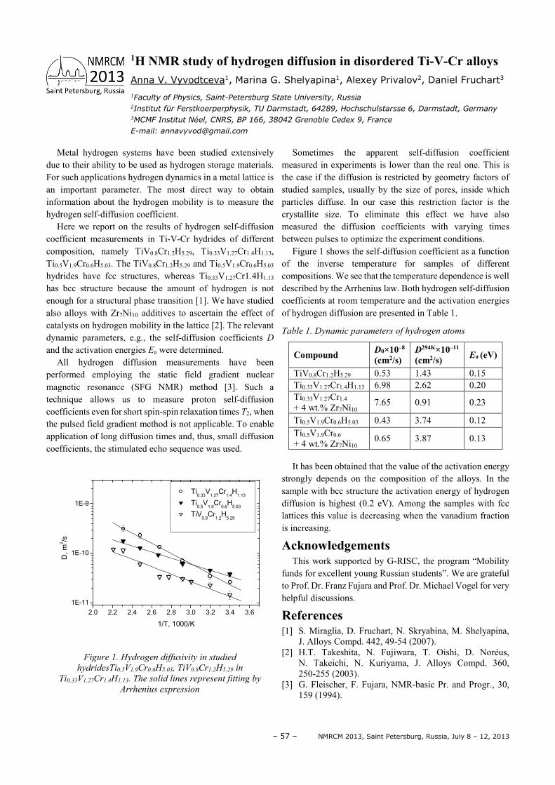
– 57 – NMRCM 2013, Saint Petersburg, Russia, July 8 – 12, 2013
Metal hydrogen systems have been studied extensively due to their ability to be used as hydrogen storage materials. For such applications hydrogen dynamics in a metal lattice is an important parameter. The most direct way to obtain information about the hydrogen mobility is to measure the hydrogen self-diffusion coefficient.
Here we report on the results of hydrogen self-diffusion coefficient measurements in Ti-V-Cr hydrides of different composition, namely TiV0.8Cr1.2H5.29, Ti0.33V1.27Cr1.4H1.13, Ti0.5V1.9Cr0.6H5.03. The TiV0.8Cr1.2H5.29 and Ti0.5V1.9Cr0.6H5.03 hydrides have fcc structures, whereas Ti0.33V1.27Cr1.4H1.13 has bcc structure because the amount of hydrogen is not enough for a structural phase transition [1]. We have studied also alloys with Zr7Ni10 additives to ascertain the effect of catalysts on hydrogen mobility in the lattice [2]. The relevant dynamic parameters, e.g., the self-diffusion coefficients D and the activation energies Ea were determined.
All hydrogen diffusion measurements have been performed employing the static field gradient nuclear magnetic resonance (SFG NMR) method [3]. Such a technique allows us to measure proton self-diffusion coefficients even for short spin-spin relaxation times T2, when the pulsed field gradient method is not applicable. To enable application of long diffusion times and, thus, small diffusion coefficients, the stimulated echo sequence was used.
2.0 2.2 2.4 2.6 2.8 3.0 3.2 3.4 3.6
1E-11
1E-10
1E-9
D,
m2/s
1/T, 1000/K
Ti0.33
V1.27
Cr1.4
H1.13
Ti0.5
V1.9
Cr0.6
H5.03
TiV0.8
Cr1.2
H5.29
Figure 1. Hydrogen diffusivity in studied
hydridesTi0.5V1.9Cr0.6H5.03, TiV0.8Cr1.2H5.29 in
Ti0.33V1.27Cr1.4H1.13. The solid lines represent fitting by
Arrhenius expression
Sometimes the apparent self-diffusion coefficient measured in experiments is lower than the real one. This is the case if the diffusion is restricted by geometry factors of studied samples, usually by the size of pores, inside which particles diffuse. In our case this restriction factor is the crystallite size. To eliminate this effect we have also measured the diffusion coefficients with varying times between pulses to optimize the experiment conditions.
Figure 1 shows the self-diffusion coefficient as a function of the inverse temperature for samples of different compositions. We see that the temperature dependence is well described by the Arrhenius law. Both hydrogen self-diffusion coefficients at room temperature and the activation energies of hydrogen diffusion are presented in Table 1.
Table 1. Dynamic parameters of hydrogen atoms
Compound D0×10–8
(cm2/s) D294K×10–11
(cm2/s) Ea (eV)
TiV0.8Cr1.2H5.29 0.53 1.43 0.15 Ti0.33V1.27Cr1.4H1.13 6.98 2.62 0.20 Ti0.33V1.27Cr1.4
+ 4 wt.% Zr7Ni10 7.65 0.91 0.23
Ti0.5V1.9Cr0.6H5.03 0.43 3.74 0.12 Ti0.5V1.9Cr0.6 + 4 wt.% Zr7Ni10
0.65 3.87 0.13
It has been obtained that the value of the activation energy strongly depends on the composition of the alloys. In the sample with bcc structure the activation energy of hydrogen diffusion is highest (0.2 eV). Among the samples with fcc lattices this value is decreasing when the vanadium fraction is increasing.
Acknowledgements This work supported by G-RISC, the program “Mobility
funds for excellent young Russian students”. We are grateful to Prof. Dr. Franz Fujara and Prof. Dr. Michael Vogel for very helpful discussions.
References [1] S. Miraglia, D. Fruchart, N. Skryabina, M. Shelyapina,
J. Alloys Compd. 442, 49-54 (2007). [2] H.T. Takeshita, N. Fujiwara, T. Oishi, D. Noréus,
N. Takeichi, N. Kuriyama, J. Alloys Compd. 360, 250-255 (2003).
[3] G. Fleischer, F. Fujara, NMR-basic Pr. and Progr., 30, 159 (1994).
1H NMR study of hydrogen diffusion in disordered Ti-V-Cr alloys Anna V. Vyvodtceva1, Marina G. Shelyapina1, Alexey Privalov2, Daniel Fruchart3
1Faculty of Physics, Saint-Petersburg State University, Russia 2Institut für Ferstkoerperphysik, TU Darmstadt, 64289, Hochschulstarsse 6, Darmstadt, Germany
3MCMF Institut Néel, CNRS, BP 166, 38042 Grenoble Cedex 9, France
E-mail: [email protected]


Part III
Poster Session

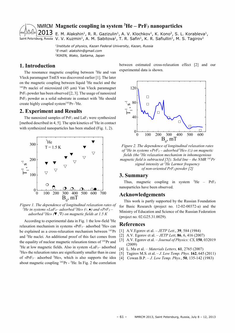
– 61 – NMRCM 2013, Saint Petersburg, Russia, July 8 – 12, 2013
1. Introduction The resonance magnetic coupling between 3He and van
Vleck paramagnet TmES was discovered earlier [1]. The later on the magnetic coupling between liquid 3He nuclei and the 141Pr nuclei of microsized (45 µm) Van Vleck paramagnet PrF3 powder has been observed [2, 3]. The usage of nanosized PrF3 powder as a solid substrate in contact with 3He should create highly coupled system141Pr–3He.
2. Experiment and Results The nanosized samples of PrF3 and LaF3 were synthesized
[method described in 4, 5]. The spin kinetics of 3He in contact with synthesized nanoparticles has been studied (Fig. 1, 2).
0 100 200 300 400 500 600 7000
100
200
300
T-1 1, s
-1
B0, mT
3He T = 1.5 K
Figure 1. The dependence of longitudinal relaxation rates of 3He in systems «LaF3– adsorbed 3Не» (,) and «PrF3 –
adsorbed 3Не» (, ) on magnetic fields at 1.5 К
According to experimental data in Fig. 1 the low-field 3He relaxation mechanism in systems «PrF3– adsorbed 3Не» can be explained as a cross-relaxation mechanism between 141Pr and 3He nuclei. An additional proof of this fact comes from the equality of nuclear magnetic relaxation times of 141Pr and 3He at low magnetic fields. Also in system «LaF3– adsorbed 3Не» the relaxation rates are significantly smaller than in case of «PrF3– adsorbed 3Не», which is also supports the idea about magnetic coupling 141Pr - 3He. In Fig. 2 the correlation
between estimated cross-relaxation effect [2] and our experimental data is shown.
0 100 200 300 400 500 6000
40
80
120
B0, mT
T-1 1, s
-1
Figure 2. The dependence of longitudinal relaxation rates
of 3He in systems «PrF3 – adsorbed 3Не» () on magnetic
fields (the 3Не relaxation mechanism in inhomogenious
magnetic field is subtracted [5]). Solid line – the NMR 141Pr
signal intensity at 3Не Larmor frequency
of non-oriented PrF3 powder [2]
3. Summary Thus, magnetic coupling in system 3He – PrF3
nanoparticles have been observed.
Acknowledgements This work is partly supported by the Russian Foundation
for Basic Research (project no. 12-02-00372-a) and the Ministry of Education and Science of the Russian Federation (project no. 02.G25.31.0029).
References [1] A.V.Egorov et al. – JETP Lett., 39, 584 (1984) [2] A.V. Egorov et al. – JETP Lett, 86, 6, 416 (2007) [3] A.V. Egorov et al. – Journal of Physics: CS, 150, 032019
(2009) [4] L. Ma et al. – Materials Letters, 61, 2765 (2007) [5] Tagirov M.S. et al. – J. Low Temp. Phys. 162, 645 (2011) [4] Cowan B.P. – J. Low Temp. Phys., 50, 135-142 (1983)
Magnetic coupling in system 3He – PrF3 nanoparticles E. M. Alakshin1, R. R. Gazizulin1, A. V. Klochkov1, K. Kono2, S. L. Korableva1, V. V. Kuzmin1, A. M. Sabitova1, T. R. Safin1, K. R. Safiullin1, M. S. Tagirov1
1Institute of physics, Kazan Federal University, Kazan, Russia 1E-mail: [email protected] 2RIKEN, Wako, Saitama, Japan
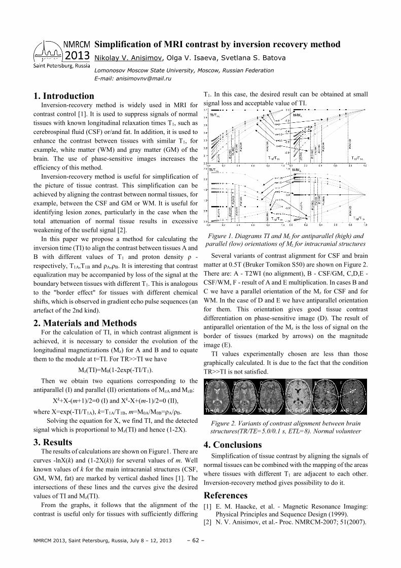
NMRCM 2013, Saint Petersburg, Russia, July 8 – 12, 2013 – 62 –
1. Introduction Inversion-recovery method is widely used in MRI for
contrast control [1]. It is used to suppress signals of normal tissues with known longitudinal relaxation times T1, such as cerebrospinal fluid (CSF) or/and fat. In addition, it is used to enhance the contrast between tissues with similar T1, for example, white matter (WM) and gray matter (GM) of the brain. The use of phase-sensitive images increases the efficiency of this method.
Inversion-recovery method is useful for simplification of the picture of tissue contrast. This simplification can be achieved by aligning the contrast between normal tissues, for example, between the CSF and GM or WM. It is useful for identifying lesion zones, particularly in the case when the total attenuation of normal tissue results in excessive weakening of the useful signal [2].
In this paper we propose a method for calculating the inversion time (TI) to align the contrast between tissues A and B with different values of T1 and proton density ρ - respectively, T1A,T1B and ρA,ρB. It is interesting that contrast equalization may be accompanied by loss of the signal at the boundary between tissues with different T1. This is analogous to the "border effect" for tissues with different chemical shifts, which is observed in gradient echo pulse sequences (an artefact of the 2nd kind).
2. Materials and Methods For the calculation of TI, in which contrast alignment is
achieved, it is necessary to consider the evolution of the longitudinal magnetizations (Mz) for A and B and to equate them to the module at t=TI. For TR>>TI we have
Mz(TI)=M0(1-2exp(-TI/T1).
Then we obtain two equations corresponding to the antiparallel (I) and parallel (II) orientations of MzA and MzB:
Xk+X-(m+1)/2=0 (I) and Xk-X+(m-1)/2=0 (II),
where X=exp(-TI/T1A), k=T1A/T1B, m=M0A/M0B=ρA/ρB. Solving the equation for X, we find TI, and the detected
signal which is proportional to Mz(TI) and hence (1-2X).
3. Results The results of calculations are shown on Figure1. There are
curves -lnX(k) and (1-2X(k)) for several values of m. Well known values of k for the main intracranial structures (CSF, GM, WM, fat) are marked by vertical dashed lines [1]. The intersections of these lines and the curves give the desired values of TI and Mz(TI).
From the graphs, it follows that the alignment of the contrast is useful only for tissues with sufficiently differing
T1. In this case, the desired result can be obtained at small signal loss and acceptable value of TI.
Figure 1. Diagrams TI and Mz for antiparallel (high) and
parallel (low) orientations of Mz for intracranial structures
Several variants of contrast alignment for CSF and brain matter at 0.5T (Bruker Tomikon S50) are shown on Figure 2. There are: A - T2WI (no alignment), B - CSF/GM, C,D,E - CSF/WM, F - result of A and E multiplication. In cases B and C we have a parallel orientation of the Mz for CSF and for WM. In the case of D and E we have antiparallel orientation for them. This orientation gives good tissue contrast differentiation on phase-sensitive image (D). The result of antiparallel orientation of the Mz is the loss of signal on the border of tissues (marked by arrows) on the magnitude image (E).
TI values experimentally chosen are less than those graphically calculated. It is due to the fact that the condition TR>>TI is not satisfied.
Figure 2. Variants of contrast alignment between brain
structures(TR/TE=5.0/0.1 s, ETL=8). Normal volunteer
4. Conclusions Simplification of tissue contrast by aligning the signals of
normal tissues can be combined with the mapping of the areas where tissues with different T1 are adjacent to each other. Inversion-recovery method gives possibility to do it.
References [1] E. M. Haacke, et al. - Magnetic Resonance Imaging:
Physical Principles and Sequence Design (1999). [2] N. V. Anisimov, et al.- Proc. NMRCM-2007; 51(2007).
Simplification of MRI contrast by inversion recovery method Nikolay V. Anisimov, Olga V. Isaeva, Svetlana S. Batova
Lomonosov Moscow State University, Moscow, Russian Federation
E-mail: [email protected]
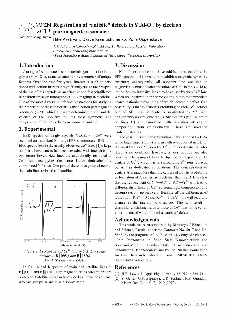
– 63 – NMRCM 2013, Saint Petersburg, Russia, July 8 – 12, 2013
1. Introduction Among of solid-state laser materials yttrium aluminum
garnet (Y3Al5O12), attracted attention by a number of unique features. Over the past few years, interest in such objects, doped with cerium increased significantly due to the prospect of the use of this crystals, as an effective and fast scintillators in positron emission tomography (PET-imaging) in medicine. One of the most direct and informative methods for studying the properties of these materials is the electron paramagnetic resonance (EPR), which allows to determine the spin and the valence of the impurity ion, its local symmetry and composition of the immediate environment, and etc.
2. Experimental EPR spectra of single crystals Y3Al5O12 : Ce3+ were
recorded on a standard X - range EPR-spectrometer JEOL. In EPR spectra beside the usually observed Ce3+ lines [1] a large number of resonances has been revealed with intensities by two orders lower. New lines are undoubtedly attributed to Ce3+ ions occupying the same lattice dodecahedrally coordinated Y3+ sites. One part of these lines grouped near to the main lines referred as “satellite”.
Figure 1. EPR spectra of Ce3+ ions in Y3Al5O12 single
crystals at B[001] and B[110],
T = 4.2K and ν = 9.35GHz
In fig. 1a and b spectra of main and satellite lines at B[001] and B[110] (high magnetic field) orientations are presented. Satellite lines can be divided by intensities at least into two groups, A and B as it shown in fig. 1.
3. Discussion Natural cerium does not have odd isotopes, therefore the
EPR spectra of this ions do not exhibit a magnetic hyperfine structure; consequently, all apparent line are due to magnetically nonequivalent positions of Ce3+ in the Y3Al5O12 lattice. So low intensity lines may be caused by such Ce3+ ions which are localized in the same c-sites, but in the immediate nearest cationic surrounding of which located a defect. One possibility is that in nearest surrounding of such Ce3+ centres one of Al3+ ions in a-site is substituted by Y3+ with considerably greater ionic radius. Such centres (fig. 1a, group of lines B) are associated with deviation of crystal composition from stoichiometrye. These are so-called “antisite” defects.
The possibility of such substitution in the range of 1 - 1.5% in the high temperature crystal growth was reported in [2]. On the substitution of Y3+ ions by Al3+ in the dodecahedral sites there is no evidence, however, in our opinion are also possible. The group of lines A (fig. 1a) corresponds to the centers of Ce3+, which has in surrounding Y3+ ions replaced by Al3+ in dodecahedral positions. The concentration of centers A is much less than the centers of B. The probability of formation of A centers is much less than the B. It is clear that the replacement of Y3+→Al3+ or Al3+→Y3+ will lead to different distortions of Ce3+ surroundings: compression and decompression, respectively. Because of the differences of ionic radii (RAl
3+ = 0.53Ǻ, RY3+ = 1.02Ǻ), this will lead to a
change in the interatomic distances. This will result in dissimilar crystalline fields to those of Ce3+ ions in the cation environment of which formed a “antisite” defect.
Acknowledgements This work has been supported by Ministry of Education
and Science, Russia, under the Contracts No. 8017 and No. 8568; by the programs of the Russian Academy of Sciences: “Spin Phenomena in Solid State Nanostructures and Spintronics” and “Fundamentals of nanostructure and nanomaterial technologies” and by the Russian Foundation for Basic Research under Grant nos. 12-02-01011, 13-02-00821 and 13-02-00802.
References [1] H.R. Lewis. J. Appl. Phys., 1966, v.37, N 2, p.739-741. [2] S. Geller, G.P. Espinoza, L.D. Fullmer, P.B. Grandall.
Mater. Res. Bull. V. 7, 1219 (1972).
Registration of “antisite” defects in Y3Al5O12 by electron paramagnetic resonance Hike Asatryan, Darya Kramushchenko, Yulia Uspenskaya1
A.F. Ioffe physical-technical institute, St. Petersburg, Russian Federation
E-mail: [email protected] 1Saint Petersburg State Institute of Technology (Technical University)
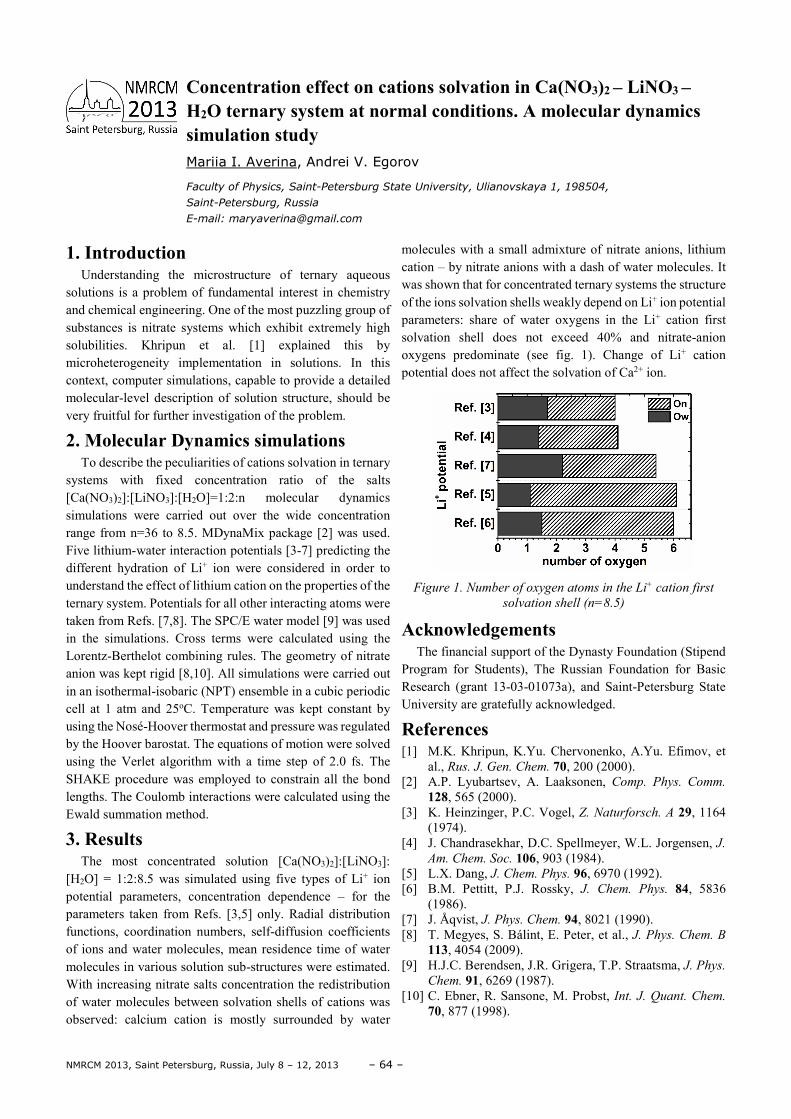
NMRCM 2013, Saint Petersburg, Russia, July 8 – 12, 2013 – 64 –
1. Introduction Understanding the microstructure of ternary aqueous
solutions is a problem of fundamental interest in chemistry and chemical engineering. One of the most puzzling group of substances is nitrate systems which exhibit extremely high solubilities. Khripun et al. [1] explained this by microheterogeneity implementation in solutions. In this context, computer simulations, capable to provide a detailed molecular-level description of solution structure, should be very fruitful for further investigation of the problem.
2. Molecular Dynamics simulations To describe the peculiarities of cations solvation in ternary
systems with fixed concentration ratio of the salts [Ca(NO3)2]:[LiNO3]:[H2O]=1:2:n molecular dynamics simulations were carried out over the wide concentration range from n=36 to 8.5. MDynaMix package [2] was used. Five lithium-water interaction potentials [3-7] predicting the different hydration of Li+ ion were considered in order to understand the effect of lithium cation on the properties of the ternary system. Potentials for all other interacting atoms were taken from Refs. [7,8]. The SPC/E water model [9] was used in the simulations. Cross terms were calculated using the Lorentz-Berthelot combining rules. The geometry of nitrate anion was kept rigid [8,10]. All simulations were carried out in an isothermal-isobaric (NPT) ensemble in a cubic periodic cell at 1 atm and 25oC. Temperature was kept constant by using the Nosé-Hoover thermostat and pressure was regulated by the Hoover barostat. The equations of motion were solved using the Verlet algorithm with a time step of 2.0 fs. The SHAKE procedure was employed to constrain all the bond lengths. The Coulomb interactions were calculated using the Ewald summation method.
3. Results The most concentrated solution [Ca(NO3)2]:[LiNO3]:
[H2O] = 1:2:8.5 was simulated using five types of Li+ ion potential parameters, concentration dependence – for the parameters taken from Refs. [3,5] only. Radial distribution functions, coordination numbers, self-diffusion coefficients of ions and water molecules, mean residence time of water molecules in various solution sub-structures were estimated. With increasing nitrate salts concentration the redistribution of water molecules between solvation shells of cations was observed: calcium cation is mostly surrounded by water
molecules with a small admixture of nitrate anions, lithium cation – by nitrate anions with a dash of water molecules. It was shown that for concentrated ternary systems the structure of the ions solvation shells weakly depend on Li+ ion potential parameters: share of water oxygens in the Li+ cation first solvation shell does not exceed 40% and nitrate-anion oxygens predominate (see fig. 1). Change of Li+ cation potential does not affect the solvation of Ca2+ ion.
Figure 1. Number of oxygen atoms in the Li+ cation first
solvation shell (n=8.5)
Acknowledgements The financial support of the Dynasty Foundation (Stipend
Program for Students), The Russian Foundation for Basic Research (grant 13-03-01073a), and Saint-Petersburg State University are gratefully acknowledged.
References [1] M.K. Khripun, K.Yu. Chervonenko, A.Yu. Efimov, et
al., Rus. J. Gen. Chem. 70, 200 (2000). [2] A.P. Lyubartsev, A. Laaksonen, Comp. Phys. Comm.
128, 565 (2000). [3] K. Heinzinger, P.C. Vogel, Z. Naturforsch. A 29, 1164
(1974). [4] J. Chandrasekhar, D.C. Spellmeyer, W.L. Jorgensen, J.
Am. Chem. Soc. 106, 903 (1984). [5] L.X. Dang, J. Chem. Phys. 96, 6970 (1992). [6] B.M. Pettitt, P.J. Rossky, J. Chem. Phys. 84, 5836
(1986). [7] J. Åqvist, J. Phys. Chem. 94, 8021 (1990). [8] T. Megyes, S. Bálint, E. Peter, et al., J. Phys. Chem. B
113, 4054 (2009). [9] H.J.C. Berendsen, J.R. Grigera, T.P. Straatsma, J. Phys.
Chem. 91, 6269 (1987). [10] C. Ebner, R. Sansone, M. Probst, Int. J. Quant. Chem.
70, 877 (1998).
Concentration effect on cations solvation in Ca(NO3)2 – LiNO3 – H2O ternary system at normal conditions. A molecular dynamics simulation study Mariia I. Averina, Andrei V. Egorov
Faculty of Physics, Saint-Petersburg State University, Ulianovskaya 1, 198504,
Saint-Petersburg, Russia
E-mail: [email protected]
– 65 – NMRCM 2013, Saint Petersburg, Russia, July 8 – 12, 2013
1. Introduction Quantum coherence is a fundamental concept of modern
physics that reveals itself in wide range of phenomena either of matter or light origin. In cavity quantum electrodynamics, the photons in a reflective cavity are coupled to the atoms or spins [1] thus providing an opportunity to combine and observe both light and matter coherence. Recently, several experimental groups reported the observations of magnetic-type coupling that involved interactions of light with the particles bearing electron spin [2, 3]. The coupling with the cavity mode was achieved with standard electron paramagnetic resonance instrumentation. Well-resolved splitting of the cavity mode was an indication of the strong coupling regime based on the coherence of the spin and photon states. Since each cavity photon interacts simultaneously with all the spins in the cavity, there is an opportunity to transfer information coherently from the photons to the spin ensemble as a whole, with obvious applications in quantum computing and quantum holography [4].
2. Tavis-Cummings model Let us consider an ensemble of 1N ≫ identical
noninteracting spins-1/2 coupled with the single-mode radiation field of a lossless microwave cavity. We adopt Tavis-Cummings model [5] and write the system Hamiltonian as
( )0ˆ ˆ ˆˆ ˆ ˆ
s zH a a S g aS a Sω ω+ +
+ −= + + +
where 0ω is the photon frequency, s
ω is the spin excitation
energy, a+ and a are the photonic creation and annihilation
operators. The last term represents the spin-photon interaction. The coupling constant g is the same for all spins
in the host sample in case when the sample dimensions are much smaller than the field wavelength. Supposing that the number of photons in the cavity is << N, one can obtain the corresponding energy spectra either numerically (for
~ 10N ) or analytically. In the latter case, the spin ensemble must be very close to its highest polarization state, i.e.
2z
S N= − . If the spins are under thermal equilibrium, this
corresponds to zero temperature ( 0τ = ). The emission spectrum of the cavity at resonance ( 0 s
ω ω= ) consists of a
single line at frequency 0ω split by a gap of 2g N (vacuum-
field Rabi splitting) [6].
3. Calculation of the cavity emission spectrum at high temperatures
If the spins belong to paramagnetic atoms distributed in a solid, the zero-temperature approximation is inadequate even for τ of a few Kelvin. Utilizing Holstein-Primakoff transformation in the basis of Dicke states [7], we calculate the cavity emission spectrum for low polarization states and
obtain the generalized Rabi splitting 2 2 zg S∆ = , where
… denotes thermal averaging. The lineshapes of the split
lines are Gaussian with the standard deviation of
2
42
z
N
NS
∆− . We show that the splitting is observable
under the condition 0 s Nτ ω≤ ≪ . This means that the spin-
photon coherence in a microwave cavity can be partially preserved even if the spins are randomly directed. For X-band cavity with 0 2 10ω π = GHz and at room temperature the
splitting is observable if 610N > . The further increase of τ diminishes the Rabi splitting and broadens both of the split lines in the emission spectrum that indicates the temperature-induced loss of coherence.
Acknowledgements This work was supported by RFBR (grant no. 12-02-
31336) and by Dynasty Foundation. The author thanks B. Z. Malkin for advice and useful discussions and B. Barbara whose suggestions had motivated this work.
References [1] H. Walther, B.T.H. Varcoe, B.-G. Englert, T. Becker. –
Rep. Prog. Phys., 69, 1325-1382 (2006). [2] E. Abe, H. Wu, A. Ardavan, J.J.L. Morton. – Appl. Phys.
Lett., 98, 251108(1-3) (2011). [3] I. Chiorescu, N. Groll, S. Bertaina, T. Mori, S. Miyashita.
– Phys. Rev. B, 82, 024413(1-7) (2010). [4] H. Wu, R.E. George, J.H. Wesenberg, K.Mølmer, D.I.
Schuster, R.J. Schoelkopf, K.M. Itoh, A.Ardavan, J.J.L. Morton, G.A.D. Briggs. Phys. Rev. Lett. – 105, 140503(1-4) (2010).
[5] M. Tavis, F.W. Cummings. Phys. Rev. – 170, 379-384 (1968).
[6] J.J. Sanchez-Mondragon, N.B. Narozhny, J.H. Eberly. Phys. Rev. Lett. – 51, 550-553 (1983).
[7] R.H. Dicke. Phys. Rev. – 93, 99-110 (1954).
To the possibility of the high-temperature spin-photon coherence in a microwave cavity Eduard Baibekov
Kazan Federal University, 420008 Kazan, Russian Federation
E-mail: [email protected]
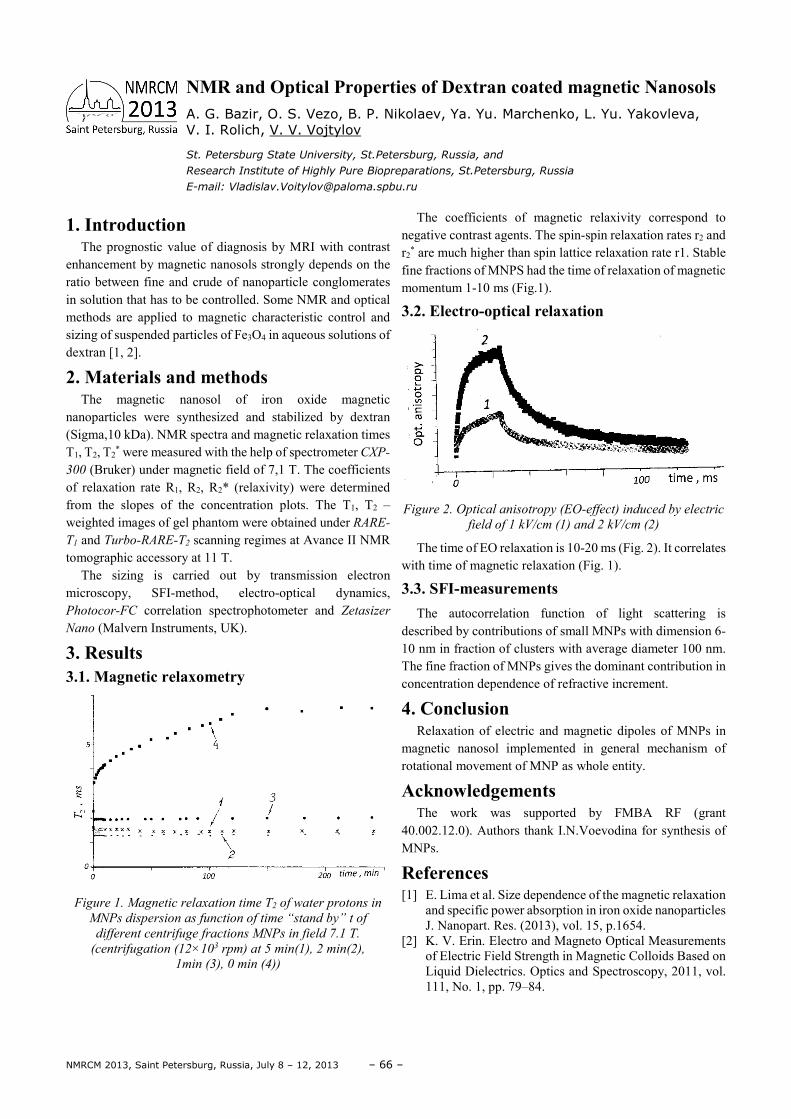
NMRCM 2013, Saint Petersburg, Russia, July 8 – 12, 2013 – 66 –
1. Introduction The prognostic value of diagnosis by MRI with contrast
enhancement by magnetic nanosols strongly depends on the ratio between fine and crude of nanoparticle conglomerates in solution that has to be controlled. Some NMR and optical methods are applied to magnetic characteristic control and sizing of suspended particles of Fe3O4 in aqueous solutions of dextran [1, 2]. 2. Materials and methods
The magnetic nanosol of iron oxide magnetic nanoparticles were synthesized and stabilized by dextran (Sigma,10 kDa). NMR spectra and magnetic relaxation times T1, T2, T2
* were measured with the help of spectrometer CXP-
300 (Bruker) under magnetic field of 7,1 T. The coefficients of relaxation rate R1, R2, R2* (relaxivity) were determined from the slopes of the concentration plots. The T1, T2 – weighted images of gel phantom were obtained under RARE-
T1 and Turbo-RARE-T2 scanning regimes at Avance ΙΙ NMR tomographic accessory at 11 T.
The sizing is carried out by transmission electron microscopy, SFI-method, electro-optical dynamics, Photocor-FC correlation spectrophotometer and Zetasizer
Nano (Malvern Instruments, UK).
3. Results 3.1. Magnetic relaxometry
Figure 1. Magnetic relaxation time T2 of water protons in
MNPs dispersion as function of time “stand by” t of
different centrifuge fractions MNPs in field 7.1 T.
(centrifugation (12×103 rpm) at 5 min(1), 2 min(2),
1min (3), 0 min (4))
The coefficients of magnetic relaxivity correspond to negative contrast agents. The spin-spin relaxation rates r2 and r2
* are much higher than spin lattice relaxation rate r1. Stable fine fractions of MNPS had the time of relaxation of magnetic momentum 1-10 ms (Fig.1).
3.2. Electro-optical relaxation
Figure 2. Optical anisotropy (EO-effect) induced by electric
field of 1 kV/cm (1) and 2 kV/cm (2)
The time of EO relaxation is 10-20 ms (Fig. 2). It correlates with time of magnetic relaxation (Fig. 1).
3.3. SFI-measurements
The autocorrelation function of light scattering is described by contributions of small MNPs with dimension 6-10 nm in fraction of clusters with average diameter 100 nm. The fine fraction of MNPs gives the dominant contribution in concentration dependence of refractive increment.
4. Conclusion Relaxation of electric and magnetic dipoles of MNPs in
magnetic nanosol implemented in general mechanism of rotational movement of MNP as whole entity.
Acknowledgements The work was supported by FMBA RF (grant
40.002.12.0). Authors thank I.N.Voevodina for synthesis of MNPs.
References [1] E. Lima et al. Size dependence of the magnetic relaxation
and specific power absorption in iron oxide nanoparticles J. Nanopart. Res. (2013), vol. 15, p.1654.
[2] K. V. Erin. Electro and Magneto Optical Measurements of Electric Field Strength in Magnetic Colloids Based on Liquid Dielectrics. Optics and Spectroscopy, 2011, vol. 111, No. 1, pp. 79–84.
NMR and Optical Properties of Dextran coated magnetic Nanosols A. G. Bazir, O. S. Vezo, B. P. Nikolaev, Ya. Yu. Marchenko, L. Yu. Yakovleva, V. I. Rolich, V. V. Vojtylov
St. Petersburg State University, St.Petersburg, Russia, and
Research Institute of Highly Pure Biopreparations, St.Petersburg, Russia
E-mail: [email protected]
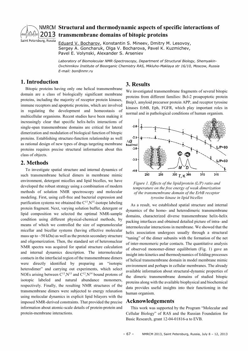
– 67 – NMRCM 2013, Saint Petersburg, Russia, July 8 – 12, 2013
1. Introduction Bitopic proteins having only one helical transmembrane
domain are a class of biologically significant membrane proteins, including the majority of receptor protein kinases, immune receptors and apoptotic proteins, which are involved in regulating the development and homeostasis of multicellular organisms. Recent studies have been making it increasingly clear that specific helix-helix interactions of single-span transmembrane domains are critical for lateral dimerization and modulation of biological function of bitopic proteins. Establishing structure-function relationship as well as rational design of new types of drugs targeting membrane proteins requires precise structural information about this class of objects.
2. Methods To investigate spatial structure and internal dynamics of
such transmembrane helical dimers in membrane mimic environment, detergent micelles and lipid bicelles, we have developed the robust strategy using a combination of modern methods of solution NMR spectroscopy and molecular modeling. First, using cell-free and bacterial expression and purification systems we obtained the C13,N15-isotope labeling protein fragment. Next, varying solution buffer, detergent or lipid composition we selected the optimal NMR-sample condition using different physical-chemical methods, by means of which we controlled the size of supramolecular micellar and bicellar systems (having effective molecular mass up to ~50 kDa) as well as the protein secondary structure and oligomerization. Then, the standard set of heteronuclear NMR spectra was acquired for spatial structure calculation and internal dynamics description. The intermolecular contacts in the interfacial region of the transmembrane dimers were directly identified by preparing an “isotopic heterodimer” and carrying out experiments, which select NOEs arising between C13,N15 and C12,N14 bound protons of isotopic labeled and natural abundance monomers, respectively. Finally, the resulting NMR structures of the transmembrane dimers were subjected to energy relaxation using molecular dynamics in explicit lipid bilayers with the imposed NMR-derived constraints. That provided the precise information about atomic-scale details of protein-protein and protein-membrane interactions.
3. Results We investigated transmembrane fragments of several bitopic proteins from different families: Bcl-2 proapoptotic protein Bnip3, amyloid precursor protein APP, and receptor tyrosine kinases ErbB, Eph, FGFR, which play important roles in normal and in pathological conditions of human organism.
Figure 1. Effects of the lipid/protein (L/P) ratio and
temperature on the free energy of weak dimerization
of the transmembrane domain of the ErbB receptor
tyrosine kinase in lipid bicelles
As a result, we established spatial structure and internal dynamics of the homo- and heterodimeric transmembrane domains, characterized diverse transmembrane helix-helix packing interfaces and obtained detailed picture of intra- and intermolecular interactions in membrane. We showed that the helix association undergoes usually through a structural “tuning” of the dimer subunits with the formation of the net of inter-monomeric polar contacts. The quantitative analysis of observed monomer-dimer equilibrium (Fig. 1) gave an insight into kinetics and thermodynamics of folding processes of helical transmembrane domain in model membrane mimic environment and perhaps in cellular membranes. The already available information about structural-dynamic properties of the dimeric transmembrane domains of studied bitopic proteins along with the available biophysical and biochemical data provides useful insights into their functioning in the human organism.
Acknowledgements This work was supported by the Program “Molecular and
Cellular Biology” of RAS and the Russian Foundation for Basic Research, grant 12-04-01816-a to EVB.
Structural and thermodynamic aspects of specific interactions of transmembrane domains of bitopic proteins Eduard V. Bocharov, Konstantin S. Mineev, Dmitry M. Lesovoy, Sergey A. Goncharuk, Olga V. Bocharova, Pavel K. Kuzmichev, Pavel E. Volynski, Alexander S. Arseniev
Laboratory of Biomolecular NMR-Spectroscopy, Department of Structural Biology, Shemyakin-
Ovchinnikov Institute of Bioorganic Chemistry RAS, Mikluho-Maklaya str 16/10, Moscow, Russia
E-mail: [email protected]
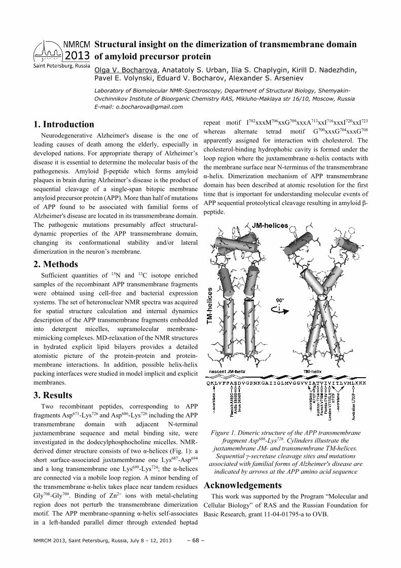
NMRCM 2013, Saint Petersburg, Russia, July 8 – 12, 2013 – 68 –
1. Introduction Neurodegenerative Alzheimer's disease is the one of
leading causes of death among the elderly, especially in developed nations. For appropriate therapy of Alzheimer’s disease it is essential to determine the molecular basis of the pathogenesis. Amyloid β-peptide which forms amyloid plaques in brain during Alzheimer’s disease is the product of sequential cleavage of a single-span bitopic membrane amyloid precursor protein (APP). More than half of mutations of APP found to be associated with familial forms of Alzheimer's disease are located in its transmembrane domain. The pathogenic mutations presumably affect structural-dynamic properties of the APP transmembrane domain, changing its conformational stability and/or lateral dimerization in the neuron’s membrane.
2. Methods Sufficient quantities of 15N and 13C isotope enriched
samples of the recombinant APP transmembrane fragments were obtained using cell-free and bacterial expression systems. The set of heteronuclear NMR spectra was acquired for spatial structure calculation and internal dynamics description of the APP transmembrane fragments embedded into detergent micelles, supramolecular membrane-mimicking complexes. MD-relaxation of the NMR structures in hydrated explicit lipid bilayers provides a detailed atomistic picture of the protein-protein and protein-membrane interactions. In addition, possible helix-helix packing interfaces were studied in model implicit and explicit membranes.
3. Results Two recombinant peptides, corresponding to APP
fragments Asp671-Lys726 and Asp686-Lys726 including the APP transmembrane domain with adjacent N-terminal juxtamembrane sequence and metal binding site, were investigated in the dodecylphosphocholine micelles. NMR-derived dimer structure consists of two α-helices (Fig. 1): a short surface-associated juxtamembrane one Lys687-Asp694 and a long transmembrane one Lys699-Lys724; the α-helices are connected via a mobile loop region. A minor bending of the transmembrane α-helix takes place near tandem residues Gly708-Gly709. Binding of Zn2+ ions with metal-chelating region does not perturb the transmembrane dimerization motif. The APP membrane-spanning α-helix self-associates in a left-handed parallel dimer through extended heptad
repeat motif I702хххM706ххG709хххA713ххI716хххI720ххI723 whereas alternate tetrad motif G700xxxG704xxxG708 apparently assigned for interaction with cholesterol. The cholesterol-binding hydrophobic cavity is formed under the loop region where the juxtamembrane α-helix contacts with the membrane surface near N-terminus of the transmembrane α-helix. Dimerization mechanism of APP transmembrane domain has been described at atomic resolution for the first time that is important for understanding molecular events of APP sequential proteolytical cleavage resulting in amyloid β-peptide.
Figure 1. Dimeric structure of the APP transmembrane
fragment Asp686-Lys726. Cylinders illustrate the
juxtamembrane JM- and transmembrane TM-helices.
Sequential γ-secretase cleavage sites and mutations
associated with familial forms of Alzheimer's disease are
indicated by arrows at the APP amino acid sequence
Acknowledgements This work was supported by the Program “Molecular and
Cellular Biology” of RAS and the Russian Foundation for Basic Research, grant 11-04-01795-a to OVB.
Structural insight on the dimerization of transmembrane domain of amyloid precursor protein Olga V. Bocharova, Anatatoly S. Urban, Ilia S. Chaplygin, Kirill D. Nadezhdin, Pavel E. Volynski, Eduard V. Bocharov, Alexander S. Arseniev
Laboratory of Biomolecular NMR-Spectroscopy, Department of Structural Biology, Shemyakin-
Ovchinnikov Institute of Bioorganic Chemistry RAS, Mikluho-Maklaya str 16/10, Moscow, Russia
E-mail: [email protected]
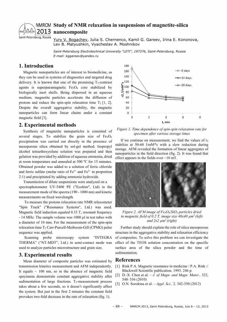
– 69 – NMRCM 2013, Saint Petersburg, Russia, July 8 – 12, 2013
1. Introduction Magnetic nanoparticles are of interest to biomedicine, as
they can be used in systems of diagnostics and targeted drug delivery. It is known that one of the promising T2-contrast agents is superparamagnetic Fe3O4 core stabilized by biologically inert shells. Being dispersed in an aqueous medium, magnetite particles accelerate the diffusion of protons and reduce the spin-spin relaxation time T2 [1, 2]. Despite the overall aggregative stability, the magnetic nanoparticles can form linear chains under a constant magnetic field [3].
2. Experimental methods Synthesis of magnetite nanoparticles is consisted of
several stages. To stabilize the grain size of Fe3O4 precipitation was carried out directly in the presence of mesoporous silica obtained by sol-gel method. Isopropyl alcohol tetraethoxysilane solution was prepared and then gelation was provided by addition of aqueous ammonia, dried at room temperature and annealed at 300 ºC for 15 minutes. Obtained powder was added to a solution of ferric chloride and ferric sulfate (molar ratio of Fe3+ and Fe2+ in proportion 2:1) and precipitated by adding ammonia hydroxide.
Transmission of dilute suspensions were analyzed on a spectrophotometer UV-5400 PE ("Ecohim", Ltd) in the measurement mode of the spectra (190 - 1000 nm) and kinetic measurements on fixed wavelength.
To measure the protons relaxation rate NMR relaxometer "Spin Track" ("Resonance Systems", Ltd.) was used. Magnetic field induction equaled 0.33 T, resonant frequency - 14 MHz. The sample volume was 1000 µl in test tubes with a diameter of 10 mm. For the measurement of the spin-spin relaxation time T2 Carr-Purcell-Meiboom-Gill (CPMG) pulse sequence was applied.
Scanning probe microscopy system “INTEGRA THERMA” (“NT-MDT”, Ltd.) in semi-contact mode was used to analyze particles microstructure and grain size.
3. Experimental results Mean diameter of composite particles was estimated by
transmission kinetics measurement and AFM independently. It equals ~ 100 nm, so in the absence of magnetic field specimens demonstrate constant aggregative stability after sedimentation of large fractions. T2-measurement process takes about a few seconds, so it doesn’t significantly affect the system. But just in the first 2 minutes the constant field provokes two-fold decrease in the rate of relaxation (fig. 1).
Figure 1. Time dependence of spin-spin relaxation rate for
specimen after various storage times
If we continue on measurement, we find the values of r2 stabilize at 50-60 l/mM*s with a slow reduction during storage. AFM revealed the formation of linear aggregates of nanoparticles in the field direction (fig. 2). It was found that effect appears in the fields over ~10 mT.
Figure 2. AFM image of Fe3O4/SiO2 particles dried
in magnetic field of 0.2 T, image size 40x40 µm2 (left)
and 2x2 µm2 (right)
Further study should explain the role of silica mesoporous structure in the aggregative stability and relaxation efficiency of composites. To solve this problem we can investigate the effect of the TEOS solution concentration on the specific surface area of the silica powder and the time of sedimentation.
References [1] Rink P.A. Magnetic resonance in medicine / P.A. Rink //
Blackwell Scientific publication. 1993. 246 p. [2] D.-X. Chen et al. – J. of Magn. and Magn. Mater., 322,
548–556 (2010) [3] O.N. Sorokina et al. – Appl. Sci., 2, 342-350 (2012)
Study of NMR relaxation in suspensions of magnetite-silica nanocomposite Yury V. Bogachev, Julia S. Chernenco, Kamil G. Gareev, Irina E. Kononova, Lev B. Matyushkin, Vyacheslav A. Moshnikov
Saint-Petersburg Electrotechnical University “LETI”, 197376, Saint-Petersburg, Russia
E-mail: [email protected]

NMRCM 2013, Saint Petersburg, Russia, July 8 – 12, 2013 – 70 –
1. Introduction New compact combined EPR/NMR/DNP equipment is developed both for research and for routine applications. The described instrumentation can be characterized by low cost, reliability and simplicity, small size and high level of measurement automation, the wide selection of functional modules and units. The hardware features are based on the latest developments in electronics, computer technology, original design and schematic solutions.
2. Specifications of the EPR part of equipment
Parameter Value
Sensitivity 2x10¹⁰ spin/G
Operating frequency (9.0…9.6) GHz
Maximum microwave power 0,15 W
Induction of magnetic field 0.05…0.7 T
Maximum ampoule diameter 11 mm
3. Specifications of the NMR part of equipment
Parameter Value
Working frequency 5…80 MHz
RX sensitivity Better than 1 µV (when S/N ratio = 3)
Adjustable TX output power Up to 500 W
Three selectable bandwidths of analog filter
From 100 Hz up to 1 MHz
Pulse sequence length Up to 64K events with resolution 100 ns
Probe tube diameter Up to 40 mm
4. Original/Custom instrumentation Among original instrumentation developments there are:
• Compact high-stability microwave units of homodyne and threshold type, low-noise Gunn oscillator being used as a microwave oscillator;
• Handheld magnet systems based on electromagnets and permanent magnets with high uniformity of magnetic field;
• Digital systems for recording and processing the EPR and NMR data.
For DNP measurements at 0.35 T an electromagnet and special cylindrical microwave cavity with TE 011 mode have been used. Also the portable DNP system based on Halbach type permanent magnet has been developed.
5. Applications Various modifications of the described compact combined
EPR/NMR instrumentation are successfully used not only in stationary research or educational laboratories but also in mobile applied laboratories as well as in industrial conditions to control continuous technological processes.
Compact combined EPR/NMR/DNP Equipment Yu. Bogachev1, Yu. Chernenko1, L. Grunin2, V. Drapkin1, M. Knyazev1, Ya. Marchenko1, A. Naumova1
1Department of Physics, Saint-Petersburg Electrotechnical University, 5, prof. Popova st.,
Saint-Petersburg, 197376, Russia; 2Department of Physics, Volga State University of Technology, 3, Lenin sq.,
Yoshkar-Ola, Republic of Mari El, 424000, Russia
E-mail: [email protected], [email protected]
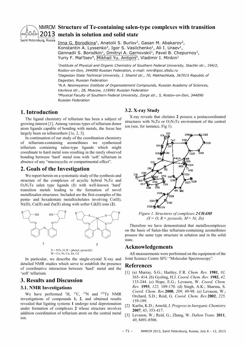
– 71 – NMRCM 2013, Saint Petersburg, Russia, July 8 – 12, 2013
1. Introduction The ligand chemistry of tellurium has been a subject of
growing interest [1]. Among various types of tellurium donor atom ligands capable of bonding with metals, the focus has largely been on telluroethers [1e, 2, 3].
In continuation of our study of the coordination chemistry of tellurium-containing azomethines we synthesized tellurium containing salen-type ligands which might coordinate to hard metal ions resulting in the rarely observed bonding between ‘hard’ metal ions with ‘soft’ tellurium in absence of any “macrocyclic or compartmental effect”.
2. Goals of the Investigation We report herein on a systematic study of the synthesis and
structure of the complexes of acyclic hybrid N4Te and O2N2Te salen type ligands (1) with well-known ‘hard’ transition metals leading to the formation of novel metallosalen structures. Included are the first examples of the penta- and hexadentate metallochelates involving Co(II), Ni(II), Cu(II) and Zn(II) along with softer Cd(II) ions (2).
XH
N
Te
N
HXR R
X
N
Te
N
XR R
M
1 2
X = NTs, O; R = phenyl, pyrazolyl;M = Co, Ni, Cu, Zn, Cd
In particular, we describe the single-crystal X-ray and detailed NMR studies which serve to establish the presence of coordinative interaction between ‘hard’ metal and the ‘soft’ tellurium.
3. Results and Discussion 3.1. NMR Investigations
We have performed 1H, 13C, 15N and 125Te NMR investigations of compounds 1, 2, and obtained results revealed that ligating systems 1 undergo total deprotonation under formation of complexes 2 whose structure involves addition coordination of tellurium atom on the central metal ion.
3.2. X-ray Study X-ray reveals that chelates 2 possess a pentacoordinated
structures with N4Te or O2N2Te environment of the central ion (see, for instance, Fig 1).
Figure 1. Structures of complexes 2.CH3OH
(X = O, R = pyrazole, M = Ni, Zn)
Therefore we have demonstrated that metallocomplexes on the basis of Salen-like tellurium-containing azomethines possess the same type structure in solution and in the solid state.
Acknowledgements All measurements were performed on the equipment of the
Joint Science Centre SFU “Molecular Spectroscopy”.
References [1] (a) Murray, S.G.; Hartley, F.R. Chem. Rev. 1981, 81,
365- 414. (b) Gysling, H.J. Coord. Chem. Rev. 1982, 42, 133-244. (c) Hope, E.G.; Levason, W. Coord. Chem.
Rev. 1993, 122, 109-170. (d) Singh, A.K.; Sharma, S. Coord. Chem. Rev.2000, 209, 49-98. (e) Levason, W.; Orchard, S.D.; Reid, G. Coord. Chem. Rev.2002, 225, 159-199.
[2] Karlin, K.D.; Arnold, J. Progress in Inorganic Chemistry
2007, 43, 353-417. [3] Levason, W.; Reid, G.; Zhang, W. Dalton Trans. 2011,
40, 8491-8506.
Structure of Te-containing salen-type complexes with transition metals in solution and solid state Inna G. Borodkina1, Anatolii S. Burlov1, Gasan M. Abakarov2, Konstantin A. Lyssenko3, Igor S. Vasilchenko1, Ali I. Uraev1, Gennadii S. Borodkin1, Dmitryi A. Garnovskii1, Pavel B. Chepurnoy1, Yuriy F. Mal’tsev4, Mikhail Yu. Antipin3, Vladimir I. Minkin1
1Institute of Physical and Organic Chemistry of Southern Federal University, Stachki str., 194/2,
Rostov-on-Don, 344090 Russian Federation, e-mail: [email protected] 2Dagestan State Technical University, I. Shamil str., 70, Makhachkala, 367015 Republic of
Dagestan, Russian Federation 3N.A. Nesmeyanov Institute of Organoelement Compounds, Russian Academy of Sciences,
Vavilova str., 28, Moscow, 119991 Russian Federation 4Physical Faculty of Southern Federal University, Zorge str., 5, Rostov-on-Don, 344090
Russian Federation
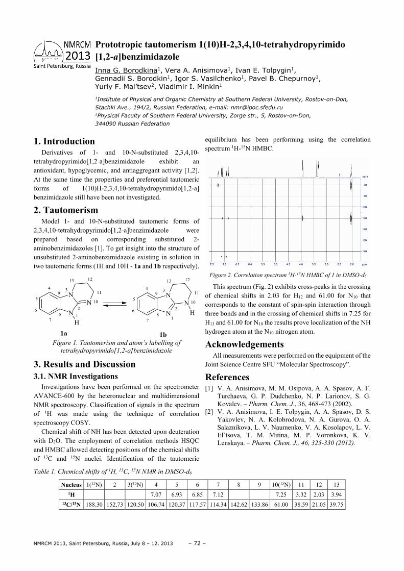
NMRCM 2013, Saint Petersburg, Russia, July 8 – 12, 2013 – 72 –
1. Introduction Derivatives of 1- and 10-N-substituted 2,3,4,10-
tetrahydropyrimido[1,2-a]benzimidazole exhibit an antioxidant, hypoglycemic, and antiaggregant activity [1,2]. At the same time the properties and preferential tautomeric forms of 1(10)H-2,3,4,10-tetrahydropyrimido[1,2-a] benzimidazole still have been not investigated.
2. Tautomerism Model 1- and 10-N-substituted tautomeric forms of
2,3,4,10-tetrahydropyrimido[1,2-a]benzimidazole were prepared based on corresponding substituted 2-aminobenzimidazoles [1]. To get insight into the structure of unsubstituted 2-aminobenzimidazole existing in solution in two tautomeric forms (1H and 10Н - 1a and 1b respectively).
N
NN
H12
34
5
6
78
9
10
11
1213
N
NN
H1
2
34
5
6
78
9
10
11
1213
1a 1bFigure 1. Tautomerism and atom’s labelling of
tetrahydropyrimido[1,2-a]benzimidazole
3. Results and Discussion 3.1. NMR Investigations
Investigations have been performed on the spectrometer AVANCE-600 by the heteronuclear and multidimensional NMR spectroscopy. Classification of signals in the spectrum of 1H was made using the technique of correlation spectroscopy COSY.
Chemical shift of NH has been detected upon deuteration with D2O. The employment of correlation methods HSQC and HMBC allowed detecting positions of the chemical shifts of 13С and 15N nuclei. Identification of the tautomeric
equilibrium has been performing using the correlation spectrum 1H-15N HMBC.
Figure 2. Correlation spectrum 1H-15N HMBC of 1 in DMSO-d6
This spectrum (Fig. 2) exhibits cross-peaks in the crossing of chemical shifts in 2.03 for Н12 and 61.00 for N10 that corresponds to the constant of spin-spin interaction through three bonds and in the crossing of chemical shifts in 7.25 for Н13 and 61.00 for N10 the results prove localization of the NH hydrogen atom at the N10 nitrogen atom.
Acknowledgements All measurements were performed on the equipment of the
Joint Science Centre SFU “Molecular Spectroscopy”.
References [1] V. A. Anisimova, M. M. Osipova, A. A. Spasov, A. F.
Turchaeva, G. P. Dudchenko, N. P. Larionov, S. G. Kovalev. – Pharm. Chem. J., 36, 468-473 (2002).
[2] V. A. Anisimova, I. E. Tolpygin, A. A. Spasov, D. S. Yakovlev, N. A. Kolobrodova, N. A. Gurova, O. A. Salaznikova, L. V. Naumenko, V. A. Kosolapov, L. V. El’tsova, T. M. Mitina, M. P. Voronkova, K. V. Lenskaya. – Pharm. Chem. J., 46, 325-330 (2012).
Table 1. Chemical shifts of 1H, 13C, 15N NMR in DMSO-d6
Nucleus 1(15N) 2 3(15N) 4 5 6 7 8 9 10(15N) 11 12 13 1H 7.07 6.93 6.85 7.12 7.25 3.32 2.03 3.94
13C/15N 188.30 152,73 120.50 106.74 120.37 117.57 114.34 142.62 133.86 61.00 38.59 21.05 39.75
Prototropic tautomerism 1(10)H-2,3,4,10-tetrahydropyrimido [1,2-a]benzimidazole Inna G. Borodkina1, Vera A. Anisimova1, Ivan E. Tolpygin1, Gennadii S. Borodkin1, Igor S. Vasilchenko1, Pavel B. Chepurnoy1, Yuriy F. Mal’tsev2, Vladimir I. Minkin1
1Institute of Physical and Organic Chemistry at Southern Federal University, Rostov-on-Don,
Stachki Ave., 194/2, Russian Federation, e-mail: [email protected] 2Physical Faculty of Southern Federal University, Zorge str., 5, Rostov-on-Don,
344090 Russian Federation
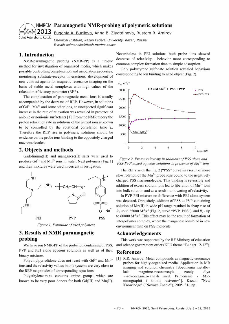
– 73 – NMRCM 2013, Saint Petersburg, Russia, July 8 – 12, 2013
1. Introduction NMR-paramagnetic probing (NMR-PP) is a unique
method for investigation of organized media, which makes possible controlling complexation and association processes, monitoring substrate-receptor interactions, development of new contrast agents for magnetic resonance imaging on the basis of stable metal complexes with high values of the relaxation efficiency parameter (REP).
The complexation of paramagnetic metal ions is usually accompanied by the decrease of REP. However, in solutions of Gd3+, Mn2+ and some other ions, an unexpected significant increase in the rate of relaxation was revealed in presence of anionic or nonionic surfactants [1]. From the NMR theory the proton relaxation rate in solutions of the named ions is known to be controlled by the rotational correlation time τr. Therefore the REP rise in polymeric solutions should be evidence on the probe ions binding to the oppositely charged macromolecules.
2. Objects and methods Gadolinium(III) and manganese(II) salts were used to
produce Gd3+ and Mn2+ ions in water. Next polymers (Fig. 1) and their mixtures were used in current investigation.
PEI PVP PSS
Figure 1. Formulae of used polymers
3. Results of NMR paramagnetic probing
We have run NMR-PP of the probe ion containing of PSS, PVP and PEI alone aqueous solutions as well as of their binary mixtures.
Polyvinylpyrrolidone does not react with Gd3+ and Mn2+ ions and the relaxivity values in this systems are very close to the REP magnitudes of corresponding aqua-ions.
Polyethyleneimine contains amino groups which are known to be very poor donors for both Gd(III) and Mn(II).
Nevertheless in PEI solutions both probe ions showed decrease of relaxivity - behavior more corresponding to common complex formation than to simple adsorption.
Only polystyrene sulfonate solution revealed behaviour corresponding to ion binding to nano object (Fig. 2).
Figure 2. Proton relaxivity in solutions of PSS alone and
PSS-PVP mixed aqueous solutions in presence of Mn2+ ions
The REP rise on the Fig. 2 (“PSS” curve) is a result of more slow rotation of the Mn2+ probe ions bound to the negatively charged PSS macromolecule. This binding is reversible and addition of excess sodium ions led to liberation of Mn2+ ions into bulk solution and as a result - to lowering of relaxivity.
In PVP-PEI mixture no difference with PEI alone system was detected. Oppositely, addition of PSS to PVP-containing solution of Mn(II) in wide pH range resulted in sharp rise of R1 up to 25000 M-1s-1 (Fig. 2, curve “PVP+PSS”), and R2 - up to 60000 M-1s-1. This effect may be the result of formation of interpolymer complex, where the manganese ions bind in new environment than on PSS molecule.
Acknowledgements This work was supported by the RF Ministry of education
and science government order (KFU theme “Budget 12-12”).
References [1] R.R. Amirov. Metal compounds as magnetic-resonance
probes for highly-organized media. Application in MR imaging and solution chemistry [Soedinenia metallov kak magnitno-resonansnyie zondy dlya vysokoorganizovannyh sred. Primenenie v MR-tomographii i khimii rastvorov"]. Kazan: "New Knowledge" ("Novoye Znanie"), 2005. 316 pp.
NH
N
NH
NH2 CH
2CH
N O
* *n
**
SO O
O Na+
n
0
5000
10000
15000
20000
25000
30000
0 2 4 6 8 10CPSS, mM
R 1, M-1s-1
PSS
PVP+PSS
0.2 mM Mn2+ + PSS + PVP
Mn(H2O)62+
Paramagnetic NMR-probing of polymeric solutions Eugenia A. Burilova, Anna B. Ziyatdinova, Rustem R. Amirov
Chemical Institute, Kazan Federal University, Kazan, Russia
E-mail: [email protected]
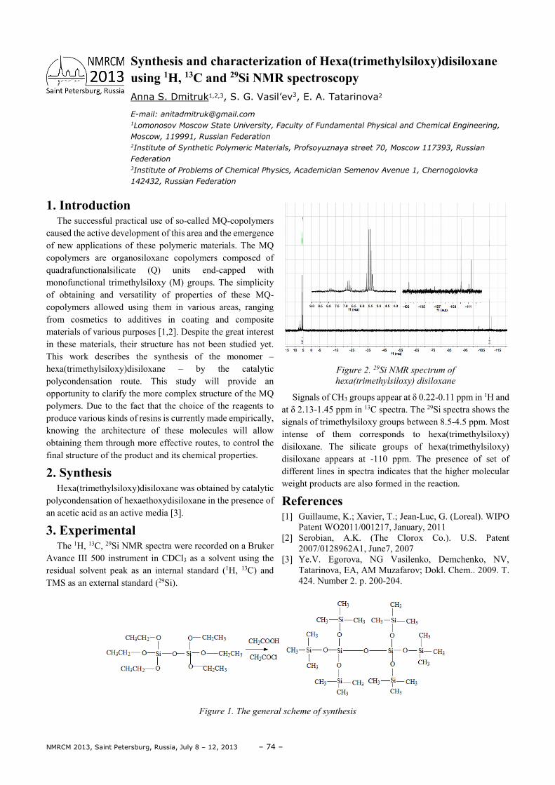
NMRCM 2013, Saint Petersburg, Russia, July 8 – 12, 2013 – 74 –
1. Introduction The successful practical use of so-called MQ-copolymers
caused the active development of this area and the emergence of new applications of these polymeric materials. The MQ copolymers are organosiloxane copolymers composed of quadrafunctionalsilicate (Q) units end-capped with monofunctional trimethylsiloxy (M) groups. The simplicity of obtaining and versatility of properties of these MQ-copolymers allowed using them in various areas, ranging from cosmetics to additives in coating and composite materials of various purposes [1,2]. Despite the great interest in these materials, their structure has not been studied yet. This work describes the synthesis of the monomer – hexa(trimethylsiloxy)disiloxane – by the catalytic polycondensation route. This study will provide an opportunity to clarify the more complex structure of the MQ polymers. Due to the fact that the choice of the reagents to produce various kinds of resins is currently made empirically, knowing the architecture of these molecules will allow obtaining them through more effective routes, to control the final structure of the product and its chemical properties.
2. Synthesis Hexa(trimethylsiloxy)disiloxane was obtained by catalytic
polycondensation of hexaethoxydisiloxane in the presence of an acetic acid as an active media [3].
3. Experimental The 1Н, 13С, 29Si NMR spectra were recorded on a Bruker
Avance III 500 instrument in CDCl3 as a solvent using the residual solvent peak as an internal standard (1H, 13C) and TMS as an external standard (29Si).
Figure 2. 29Si NMR spectrum of
hexa(trimethylsiloxy) disiloxane
Signals of CH3 groups appear at δ 0.22-0.11 ppm in 1H and at δ 2.13-1.45 ppm in 13C spectra. The 29Si spectra shows the signals of trimethylsiloxy groups between 8.5-4.5 ppm. Most intense of them corresponds to hexa(trimethylsiloxy) disiloxane. The silicate groups of hexa(trimethylsiloxy) disiloxane appears at -110 ppm. The presence of set of different lines in spectra indicates that the higher molecular weight products are also formed in the reaction.
References [1] Guillaume, K.; Xavier, Т.; Jean-Luc, G. (Loreal). WIPO
Patent WO2011/001217, January, 2011 [2] Serobian, A.K. (The Clorox Co.). U.S. Patent
2007/0128962A1, June7, 2007 [3] Ye.V. Egorova, NG Vasilenko, Demchenko, NV,
Tatarinova, EA, AM Muzafarov; Dokl. Chem.. 2009. T. 424. Number 2. p. 200-204.
Figure 1. The general scheme of synthesis
Synthesis and characterization of Hexa(trimethylsiloxy)disiloxane using 1H, 13C and 29Si NMR spectroscopy Anna S. Dmitruk1,2,3, S. G. Vasil’ev3, E. A. Tatarinova2
E-mail: [email protected] 1Lomonosov Moscow State University, Faculty of Fundamental Physical and Chemical Engineering,
Moscow, 119991, Russian Federation
2Institute of Synthetic Polymeric Materials, Profsoyuznaya street 70, Moscow 117393, Russian
Federation 3Institute of Problems of Chemical Physics, Academician Semenov Avenue 1, Chernogolovka
142432, Russian Federation
– 75 – NMRCM 2013, Saint Petersburg, Russia, July 8 – 12, 2013
1. Introduction Describing the microstructure and dynamics of ternary
aqueous solutions at the molecular level is of importance to the understanding of the solvation phenomenon. Complexity of the problem requires a variety of methods to be employed. NMR relaxation is one of an important means of investigating the properties of binary solutions (see [1] and reference wherein). In order to ascertain whether or not relaxation measurements may be effectively used to describe concentrated ternary systems the study of ternary aqueous solutions of nitrate salts Ca(NO3)2−LiNO3−H2O was carried out.
2. Experimental The nitrate salts (LiNO3 from Sigma-Aldrich and
Ca(NO3)2·4H2O from Fluka) were preliminary dried at 1600C. Solutions were prepared by weighing out appropriate amounts of the salts and diluting volumetrically with heavy water (99.97% of deuterium). Concentrations of the components were determined by NMR quantification using the external concentration standards. The list of the systems studied is given in Table 1. All NMR measurements were performed on a Bruker Avance 500 SB spectrometer. The temperature was maintained at 25.0±0.10C. Spin-lattice relaxation times (T1) were measured by using the inversion-recovery technique. The error in relaxation times measurements does not exceed 5 %.
Table 1. Solutions studied
Solution Ca(NO3)2 conc. mol/l
LiNO3 conc. mol/l
1 3.0 2.8
2 3.6 3.1
3 4.0 3.7
4 4.8 4.3
5 5.0 4.9
6 5.0 5.3
7 5.0 6.1
3. Results The spin-lattice relaxation times of the 43Ca, 7Li, and 14N
nuclei are collected in Table 2. To obtain structural of dynamical parameters from experimental measurements it is necessary to make assumptions on the local structure of the ions environment. Such assumptions highly influence the interpretation of the relaxation data. The results indicate that NMR relaxation technique should be used as a complement to the results obtained by other methods. In particular, it can be a validation method for Molecular dynamics computer simulations.
Table 2. Relaxation times of the nuclei studied (250C)
Solution T1, sec 43Ca 7Li 14N
1 0.066 10.1 0.0085
2 0.049 7.7 0.0062
3 0.033 5.3 0.0040
4 0.022 3.6 0.0026
5 0.013 2.5 0.0016
6 0.010 1.9 0.0011
7 0.005 1.1 0.0005
Acknowledgements We are indebted to Dr. Sergey Dvinskikh, Royal Institute
of Technology, Sweden for his help with experiments. The financial support of The Russian Foundation for Basic Research (grant 13-03-01073a), and Saint-Petersburg State University are gratefully acknowledged.
References [1] V.I. Chizhik, Mol. Phys. 90, 653 (1997).
Multinuclear NMR relaxation as a tool of investigating local structure and dynamics of ternary aqueous solutions of nitrate salts Andrei V. Egorov, Mariia I. Averina
Faculty of Physics, Saint-Petersburg State University, Ulianovskaya 1, 198504,
Saint-Petersburg, Russia
E-mail: [email protected]
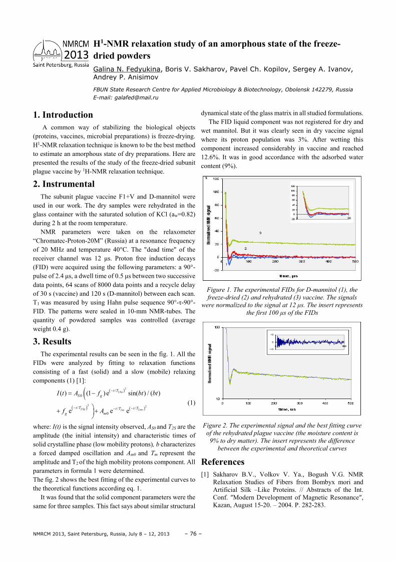
NMRCM 2013, Saint Petersburg, Russia, July 8 – 12, 2013 – 76 –
1. Introduction A common way of stabilizing the biological objects
(proteins, vaccines, microbial preparations) is freeze-drying. H1-NMR relaxation technique is known to be the best method to estimate an amorphous state of dry preparations. Here are presented the results of the study of the freeze-dried subunit plague vaccine by 1H-NMR relaxation technique.
2. Instrumental The subunit plague vaccine F1+V and D-mannitol were
used in our work. The dry samples were rehydrated in the glass container with the saturated solution of KCl (aw=0.82) during 2 h at the room temperature.
NMR parameters were taken on the relaxometer “Chromatec-Proton-20M” (Russia) at a resonance frequency of 20 MHz and temperature 40°C. The "dead time" of the receiver channel was 12 µs. Proton free induction decays (FID) were acquired using the following parameters: a 90°-pulse of 2.4 µs, a dwell time of 0.5 µs between two successive data points, 64 scans of 8000 data points and a recycle delay of 30 s (vaccine) and 120 s (D-mannitol) between each scan. T1 was measured by using Hahn pulse sequence 90°-τ-90°-FID. The patterns were sealed in 10-mm NMR-tubes. The quantity of powdered samples was controlled (average weight 0.4 g).
3. Results The experimental results can be seen in the fig. 1. All the
FIDs were analyzed by fitting to relaxation functions consisting of a fast (solid) and a slow (mobile) relaxing components (1) [1]:
( )(( ) ( )
22
2 22 22
/0
/ //0
( ) (1 )e sin( ) / ( )
e e e
Sa
Sg mvm
t T
S g
t T t Tt T
g m
I t A f bt bt
f A
−
− −−
= −
+ +
(1)
where: I(t) is the signal intensity observed, AS0 and T2S are the amplitude (the initial intensity) and characteristic times of solid crystalline phase (low mobility protons), b characterizes a forced damped oscillation and Am0 and Tm represent the
amplitude and T2 of the high mobility protons component. All parameters in formula 1 were determined. The fig. 2 shows the best fitting of the experimental curves to the theoretical functions according eq. 1.
It was found that the solid component parameters were the same for three samples. This fact says about similar structural
dynamical state of the glass matrix in all studied formulations. The FID liquid component was not registered for dry and
wet mannitol. But it was clearly seen in dry vaccine signal where its proton population was 3%. After wetting this component increased considerably in vaccine and reached 12.6%. It was in good accordance with the adsorbed water content (9%).
Figure 1. The experimental FIDs for D-mannitol (1), the
freeze-dried (2) and rehydrated (3) vaccine. The signals
were normalized to the signal at 12 µs. The insert represents
the first 100 µs of the FIDs
Figure 2. The experimental signal and the best fitting curve
of the rehydrated plague vaccine (the moisture content is
9% to dry matter). The insert represents the difference
between the experimental and theoretical curves
References [1] Sakharov B.V., Volkov V. Ya., Bogush V.G. NMR
Relaxation Studies of Fibers from Bombyx mori and Artificial Silk –Like Proteins. // Abstracts of the Int. Conf. ″Modern Development of Magnetic Resonance″, Kazan, August 15-20. – 2004. P. 282-283.
H1-NMR relaxation study of an amorphous state of the freeze-dried powders Galina N. Fedyukina, Boris V. Sakharov, Pavel Ch. Kopilov, Sergey A. Ivanov, Andrey P. Anisimov
FBUN State Research Centre for Applied Microbiology & Biotechnology, Obolensk 142279, Russia
E-mail: [email protected]
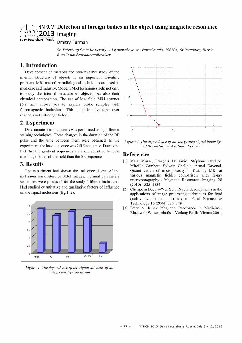
– 77 – NMRCM 2013, Saint Petersburg, Russia, July 8 – 12, 2013
1. Introduction Development of methods for non-invasive study of the
internal structure of objects is an important scientific problem. MRI and other radiological techniques are used in medicine and industry. Modern MRI techniques help not only to study the internal structure of objects, but also their chemical composition. The use of low field MRI scanner (6.8 mT) allows you to explore protic samples with ferromagnetic inclusions. This is their advantage over scanners with stronger fields.
2. Experiment Determination of inclusions was performed using different
staining techniques. There changes in the duration of the RF pulse and the time between them were obtained. In the experiment, the base sequence was GRE-sequence. Due to the fact that the gradient sequences are more sensitive to local inhomogeneities of the field than the SE sequence.
3. Results The experiment had shown the influence degree of the
inclusions parameters on MRI images. Optimal parameters sequences were produced for the study different inclusions. Had studied quantitative and qualitative factors of influence on the signal inclusions (fig.1, 2).
Figure 1. The dependence of the signal intensity of the
integrated type inclusion
Figure 2. The dependence of the integrated signal intensity
of the inclusion of volume. For iron
References [1] Maja Musse, François De Guio, Stéphane Quellec,
Mireille Cambert, Sylvain Challois, Armel Davenel. Quantification of microporosity in fruit by MRI at various magnetic fields: comparison with X-ray microtomography.- Magnetic Resonance Imaging 28 (2010) 1525–1534
[2] Cheng-Jin Du, Da-Wen Sun. Recent developments in the applications of image processing techniques for food quality evaluation. - Trends in Food Science & Technology 15 (2004) 230–249
[3] Peter A. Rinck Magnetic Resonance in Medicine.-Blackwell Wissenschafts – Verlang Berlin Vienna 2001.
Detection of foreign bodies in the object using magnetic resonance imaging Dmitry Furman
St. Peterburg State University, 1 Ulyanovskaya st., Petrodvorets, 198504, St.Peterburg, Russia
E-mail: [email protected]
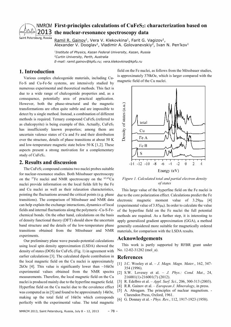
NMRCM 2013, Saint Petersburg, Russia, July 8 – 12, 2013 – 78 –
1. Introduction Various complex chalcogenide materials, including Cu-
Fe-S and Cu-Fe-Se systems, are intensively studied by numerous experimental and theoretical methods. This fact is due to a wide range of chalcogenide properties and, as a consequence, potentially area of practical application. However, both the phase-structural and the magnetic transformations are often quite subtle and are impossible to detect by a single method. Instead, a combination of different methods is required. Ternary compound CuFeS2 (referred to as chalcopyrite) is being example of this. Actually, CuFeS2 has insufficiently known properties; among them are uncertain valence states of Cu and Fe and their distribution over the structure, details of phase transitions at about 50 K and low-temperature magnetic state below 50 K [1,2]. These aspects present a strong motivation for a complementary study of CuFeS2.
2. Results and discussion The CuFeS2 compound contains two nuclei probes suitable
for nuclear-resonance studies. Both Mössbauer spectroscopy on the 57Fe nuclei and NMR spectroscopy on the 63,65Cu nuclei provide information on the local fields felt by the Fe and Cu nuclei as well as their relaxation characteristics, pointing the fluctuations around the critical points (e.g. phase transitions). The comparison of Mössbauer and NMR data can help explain the exchange interactions, dynamics of local fields and internal fluctuations along the polymeric -Cu-S-Fe- chemical bonds. On the other hand, calculations on the basis of density functional theory (DFT) should show the uncertain band structure and the details of the low-temperature phase transitions obtained from the Mössbauer and NMR experiments.
Our preliminary plane wave pseudo-potential calculations using local spin density approximation (LSDA) showed the density of states (DOS) for CuFeS2 (Fig. 1) in agreement with earlier calculations [3]. The calculated dipole contribution in the local magnetic field on the Cu nuclei is approximately 2kOe [4]. This value is significantly lower than ~16kOe experimental values obtained from the NMR spectra measurements. Therefore, the local magnetic field on the Cu nuclei is produced mainly due to the hyperfine magnetic field. Hyperfine field on the Cu nuclei due to the covalence effect was computed as in [5] and found to be approximately 14kOe, making up the total field of 16kOe which corresponds perfectly with the experimental value. The total magnetic
field on the Fe nuclei, as follows from the Mössbauer studies, is approximately 370kOe, which is larger compared with the magnetic field of the Cu nuclei.
Figure 1. Calculated total and partial electron density
of states
This large value of the hyperfine field on the Fe nuclei is due to the core polarization effect. Calculations predict the Fe electronic magnetic moment value of 3.29µB [4] (experimental value of 3.85µB). In order to calculate the value of the hyperfine field on the Fe nuclei the full potential methods are required. As a further step, it is interesting to apply generalized gradient approximation (GGA), a method generally considered more suitable for magnetically ordered materials, for comparison with the LSDA results.
Acknowledgements This work is partly supported by RFBR grant under
No. 12-02-31282 (mol_a).
References [1] J.C. Wooley et al. – J. Magn. Magn. Mater., 162, 347-
354 (1996). [2] S.W. Lovesey et al. – J. Phys.: Cond. Mat., 24,
216001(1)-216001(7) (2012). [3] R. Edelbro et al. – Appl. Surf. Sci., 206, 300-313 (2003). [4] R.R. Gainov et al. – European J. Mineralogy, in press. [5] A. Abragam. The principles of nuclear magnetism. -
Clarendon Press, Oxford, 1961. [6] G. Donnay et al. - Phys. Rev., 112, 1917-1923 (1958).
First-principles calculations of CuFeS2: characterization based on the nuclear-resonance spectroscopy data Ramil R. Gainov1, Vera V. Klekovkina1, Farit G. Vagizov1, Alexander V. Dooglav1, Vladimir A. Golovanevskiy2, Ivan N. Pen’kov1
1Institute of Physics, Kazan Federal University, Kazan, Russia 2Curtin University, Perth, Australia
E-mail: [email protected]; [email protected]
– 79 – NMRCM 2013, Saint Petersburg, Russia, July 8 – 12, 2013
1. Introduction The interest in materials with non-covalent interactions is
greatly increased for the last few years. In particular hydrogen-bonded systems are of great practical importance as they influence the mobility and ordering of the material. Two liquid crystals (LCs) of cyanobiphenyl series (HO6OCB, also referred as HOC6B or H6CBP, and 7OCB (Fig. 1)) have been studied by virtue of NMR and molecular dynamics simulations methods. Both LCs have approximately the same length, however HO6OCB can form hydrogen bonds between terminal groups, while 7OCB can not. That influences drastically on the properties of LCs.
Figure 1. ω-hydroxy-4-n-hexiloxy-4’-cyanobiphenyl
(HO6OCB, upper) and 4-n-heptyloxy-4'-cyanobiphenyl
(7OCB, lower)
Dew to the temperature dependence of chemical shift spectral lines correlation should be done for the spectra in liquid crystalline phase. Method described in the paper of Guo and Fung [1] has been applied for that purpose.
2. Experiment Experimental part was carried out at Royal Institute of
Technology, Stockholm, Sweden by prof. S. V. Dvinskikh.
1H and 13C NMR spectra were obtained using PDLF technique on a Bruker Avance III 500MHz spectrometer. Proton NMR with pulsed-field-gradient has been applied to obtain self-diffusion coefficents [2].
3. Simulation details Fully atomistic model has been chosen to carry out a
reliable study of hydrogen bonding. MuScaT method [3], which allows “on the fly” transition between more and less detailed models (fully atomistic and united atoms models in our case), has been applied to speed up the equilibration. 784 molecules have been placed in cubic cells with periodic boundary conditions for both LCs. OPLS and OPLS-AA force fields and the canonical (NVT) ensemble have been used. Temperature has been kept at 0.98TNI (the temperature of nematic to isotropic transition) for each LC. After the equilibration the analysis was performed on trajectories of 10 ns for both systems. More information on the simulation details could be found in our poster.
4. Results and discussion The purpose of this work is to determine the influence of
hydrogen-bonding on several macro- and microscopic parameters, such as order parameters, self-diffusion coefficients and conformations of the above mentioned LCs, which will be discussed in details in our poster.
References [1] Guo, W., Fung, B. M. (1991). The Journal of chemical
physics, 95, 3917. [2] Dvinskikh, S. V. (2013). Applied Magnetic Resonance,
44(1-2), 169-180. [3] Neverov, V. S., Komolkin, A. V. (2012). The Journal of
Chemical Physics, 136, 094102.
Influence of hydrogen bonding on macro- and microscopic parameters of cyanobiphenyls. The comparative study Egor Gerts1,4, Andrei V. Komolkin1, Vladimir A. Burmistrov2, Victor V. Alexandriysky3, Sergey V. Dvinskikh1
1Faculty of Physics, Saint-Petersburg State University, Russian Federation
2Ivanovo State University of Chemical Technology, Ivanovo, Russian Federation
3Institute of Solution Chemistry, Russian Academy of Sciences, Ivanovo, Russian Federation
4E-mail: [email protected]
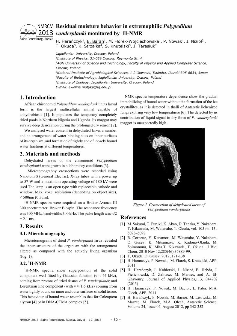
NMRCM 2013, Saint Petersburg, Russia, July 8 – 12, 2013 – 80 –
1. Introduction African chironomid Polypedilum vanderplanki in its larval
form is the largest multicellular animal capable of anhydrobiosis [1]. It populates the temporary completely dried pools in Northern Nigeria and Uganda. Its maggot may survive deep desiccation during the prolonged dry season [2].
We analysed water content in dehydrated larva, a number and an arrangement of water binding sites on inner surfaces of its organism, and formation of tightly and of loosely bound water fractions at different temperatures.
2. Materials and methods
Dehydrated larvas of the chironomid Polypedilum
vanderplanki were grown in a laboratory conditions [3]. Microtomography crossections were recorded using
Nanotom S (General Electric). X-ray tubes with a power up to 57 W and a maximum operating voltage of 180 kV were used.The lamp is an open type with replaceable cathode and window. Max. voxel resolution (depending on object size), < 500nm (0.5µm).
1H-NMR spectra were acquired on a Bruker Avance III 300 spectrometer, Bruker Biospin. The resonance frequency was 300 MHz, bandwidths 300 kHz. The pulse length was π/2 = 2.1 ms.
3. Results 3.1. Microtomography
Microtomograms of dried P. vanderplanki larva revealed the inner structure of the organism with the arrangement altered as compared with the actively living organism (Fig. 1).
3.2. 1H-NMR 1H-NMR spectra show superposition of the solid
component well fitted by Gaussian function (ν ≈ 44 kHz), coming from protons of dried tissues of P. vanderplanki; and Lorentzian line component (with ν ≈ 1.6 kHz) coming from water tightly bound on inner and outer surfaces of solid tissue. This behaviour of bound water resembles that for Coleoptera alytron [4] or in DNA-CTMA complex [5].
NMR spectra temperature dependence show the gradual immobilizing of bound water without the formation of the ice crystallites, as it is detected in thalli of Antarctic lichenized fungi expiring very low temperatuere [6]. The detected by us contribution of liquid signal in dry form of P. vanderplanki maggot is unexpectedly high.
Figure 1. Crossection of dehydrated larva of
Polypedilum vanderplanki
References [1] M. Sakurai, T. Furuki, K. Akao, D. Tanaka, Y. Nakahara,
T. Kikawada, M. Watanabe, T. Okuda, vol. 105 no. 13 , 5093–5098.
[2] R. Cornette, Y. Kanamori, M. Watanabe, Y. Nakahara, O. Gusev, K. Mitsumasu, K. Kadono-Okuda, M. Shimomura, K. Mita,T. Kikawada, T. Okuda., J Biol Chem. 2010 Nov 12;285(46):35889-99.
[3] T. Okuda. O. Gusev, 2012, 121-138 [4] H. Harańczyk, P. Nowak, , M. Florek, S. Knutelski, APP,
2011 [5] H. Harańczyk, J. Kobierski, J. Nizioł, E. Hebda, J.
Pielichowski, D. Zalitacz, M. Marzec, and A. El-Ghayoury, Journal of Applied Physics,113, 044702 (2013)
[6] H. Harańczyk, P. Nowak, M. Bacior, Ł. Pater, M.A. Olech, APP, 2011
[7] H. Harańczyk, P. Nowak, M. Bacior, M. Lisowska, M. Marzec, M. Florek, M.A. Olech, Antarctic Science, Volume 24, Issue 04, August 2012, pp 342-352
Residual moisture behavior in extremophilic Polypedilum vanderplanki monitored by 1H-NMR H. Harańczyk1, E. Baran1, M. Florek-Wojciechowska1, P. Nowak1, J. Nizioł2 , T. Okuda3, K. Strzałka4, S. Knutelski5, J. Tarasiuk2
Jagiellonian University, Cracow, Poland 1Institute of Physics, 31-059 Cracow, Reymonta St. 4
2AGH University of Science and Technology, Faculty of Physics and Applied Computer Science,
Cracow, Poland 3National Institute of Agrobiological Sciences, 1-2 Ohwashi, Tsukuba, Ibaraki 305-8634, Japan 4Faculty of Biotechnology, Jagiellonian University, Cracow, Poland 5Institute of Zoology, Jagiellonian University, Cracow, Poland
E-mail: [email protected]
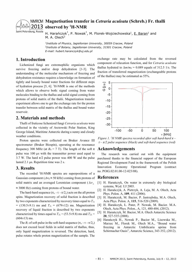
– 81 – NMRCM 2013, Saint Petersburg, Russia, July 8 – 12, 2013
1. Introduction Lichenized fungi are extremophilic organisms which
survive freezing and/or deep dehydration [1-3]. The understanding of the molecular mechanism of freezing and dehydration resistance requires a knowledge on formation of tightly and loosely bound water fractions for different steps of hydration process [5, 6]. 1H-NMR is one of the methods which allows to observe both: signal coming from water molecules binding to the thallus and solid signal coming from protons of solid matrix of the thalii. Magnetization transfer experiment allows one to get the exchange rate for the proton transfer between solid matrix of the thallus and bound water reservoir.
2. Materials and methods Thalli of fruticose lichenized fungi Cetraria aculeata were
collected in the vicinity of Arctowski Polar Station, King George Island, Maritime Antarctic during a sunny and cloudy weather conditions.
Proton spectra were collected on Bruker Avance III spectrometer (Bruker Biospin), operating at the resonance frequency 300 MHz (at B0 = 7 T). The length of the soft π pulse was 100 µs with the transmitter power used equal to 3.7 W. The hard π/2 pulse power was 400 W and the pulse lasted 2.1 µs. Repetition time was 2 s.
3. Results The recorded 1H-NMR spectra are superpositions of a
Gaussian component (∆νG ≈ 50 kHz) coming from protons of solid matrix and an averaged Lorentzian component ( Lν∆
≈ 3000 Hz) coming from protons of bound water. The hard-hard sequence (πx - τ - π/2y) acts on the all proton
spins. Magnetization recovery of solid fraction is described by two exponents characterized by recovery times equal to T1s = (130.5±9.1) ms and T2s = (679±12) ms. Magnetization recovery of liquid fraction is described by two exponents characterized by times equal to T1L = (53.5±9.8) ms and T2L = (604±13) ms.
The B1 of soft pulse in the soft-hard sequence (πx - τ - π/2y) does not exceed local fields in solid matrix of thallus, thus, only liquid magnetization is reversed. The detection, hard, pulse rotates whole proton magnetization of the sample. The
exchange rate may be calculated from the reversed component of relaxation function, and for Cetraria aculeata thallus hydrated to ∆m/m0 = 0.089 equals of 312.5 1/s. The fraction of transferred magnetization (exchangeable protons of the thallus) may be estimated as 55%.
-100 -50 0 50 100
-30
-25
-20
-15
-10
-5
0
5
G [
j.u
.]
ν [kHz]
Figure 1. 1H NMR spectra recorded after soft hard-hard π –
τ – π/2 pulse sequence (black) and soft-hard sequence (red)
Acknowledgements The research was carried out with the equipment
purchased thanks to the financial support of the European Regional Development Fund in the framework of the Polish Innovation Economy Operational Program (contract no. POIG.02.01.00-12-023/08).
References [1] H. Harańczyk, On water in extremely dry biological
systems, Wyd. UJ 2003. [2] H. Harańczyk, A. Pietrzyk, A. Leja, M. A. Olech, Acta
Phys. Polon. A, 109, 411 (2006). [3] H. Harańczyk, M. Bacior, P. Jastrzębska, M.A. Olech,
Acta Phys. Polon. A, 115, 516-520 (2009). [4] H. Harańczyk, Ł. Pater, P. Nowak, M. Bacior. M.A.
Olech, Acta Phys. Polon., A, 121, 480-484, (2012). [5] H. Harańczyk, M. Bacior, M.A. Olech Antarctic Science
20, 527-535 (2008). [6] Harańczyk H., Nowak P., Bacior M., Lisowska M.,
Marzec M., Florek M., Olech M.A., “Bound water freezing in Antarctic Umbilicaria aprina from Schirmacher Oasis”, Antarctic Science, 343-352, (2012).
Magnetisation transfer in Cetraria aculeata (Schreb.) Fr. thalli observed by 1H-NMR H. Harańczyk
1, P. Nowak
1, M. Florek-Wojciechowska1, E. Baran1 and
M. A. Olech2
1Institute of Physics, Jagiellonian University, 30059 Cracow, Poland 2Insitiute of Botany, Jagiellonian University, 31501 Cracow, Poland
E-mail: [email protected]
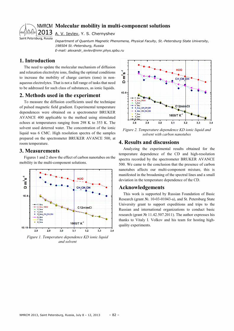
NMRCM 2013, Saint Petersburg, Russia, July 8 – 12, 2013 – 82 –
1. Introduction The need to update the molecular mechanism of diffusion
and relaxation electrolyte ions, finding the optimal conditions to increase the mobility of charge carriers (ions) in non-aqueous electrolytes. That is not a full range of tasks that need to be addressed for such class of substances, as ionic liquids.
2. Methods used in the experiment To measure the diffusion coefficients used the technique
of pulsed magnetic field gradient. Experimental temperature dependences were obtained on a spectrometer BRUKER AVANCE 400 applicable to the method using stimulated echoes at temperatures ranging from 298 K to 353 K. The solvent used deterred water. The concentration of the ionic liquid was 6 CMC. High resolution spectra of the samples prepared on the spectrometer BRUKER AVANCE 500, at room temperature.
3. Measurements Figures 1 and 2 show the effect of carbon nanotubes on the
mobility in the multi-component solutions.
Figure 1. Temperature dependence KD ionic liquid
and solvent
Figure 2. Temperature dependence KD ionic liquid and
solvent with carbon nanotubes
4. Results and discussions Analyzing the experimental results obtained for the
temperature dependence of the CD and high-resolution spectra recorded by the spectrometer BRUKER AVANCE 500. We came to the conclusion that the presence of carbon nanotubes affects our multi-component mixture, this is manifested in the broadening of the spectral lines and a small deviation in the temperature dependence of the CD.
Acknowledgements This work is supported by Russian Foundation of Basic
Research (grant . 10-03-01043-а), and St. Petersburg State University grant to support expeditions and trips to the Russian and international organizations to conduct basic research (grant 11.42.507.2011). The author expresses his thanks to Vitaly I. Volkov and his team for hosting high-quality experiments.
Molecular mobility in multi-component solutions A. V. Ievlev, Y. S. Chernyshev
Department of Quantum Magnetic Phenomena, Physical Faculty, St.-Petersburg State University,
198504 St.-Petersburg, Russia
E-mail: [email protected]
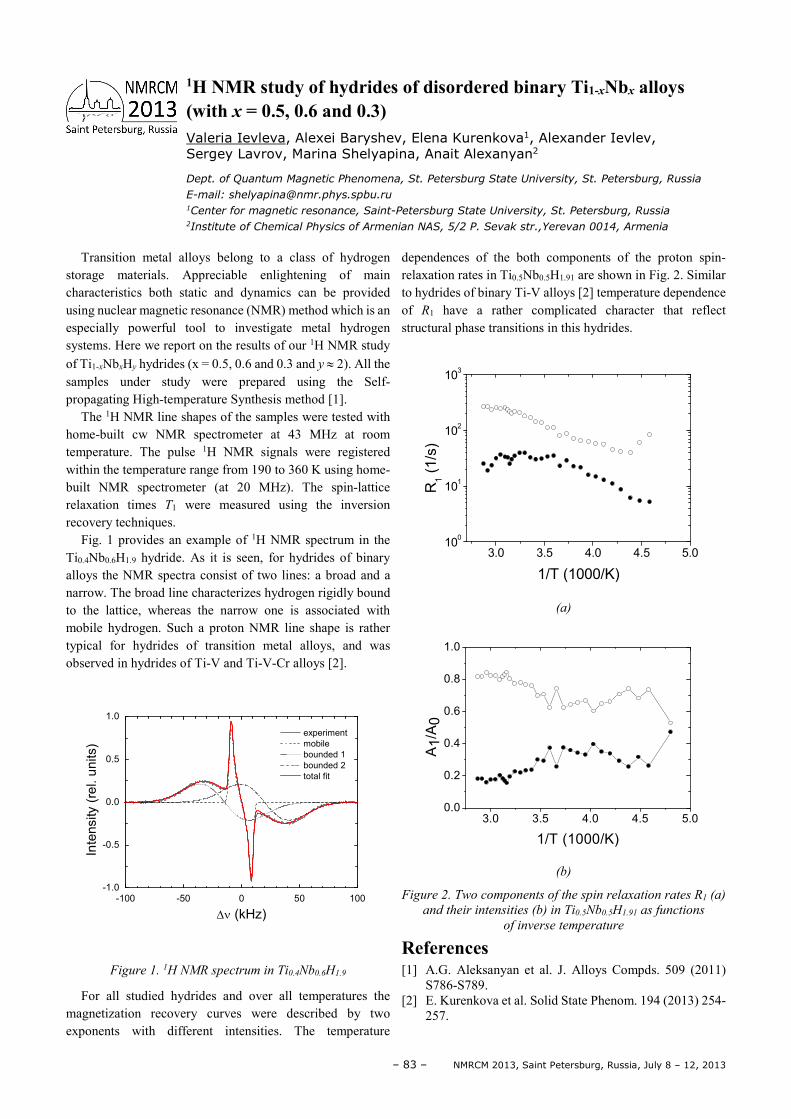
– 83 – NMRCM 2013, Saint Petersburg, Russia, July 8 – 12, 2013
Transition metal alloys belong to a class of hydrogen storage materials. Appreciable enlightening of main characteristics both static and dynamics can be provided using nuclear magnetic resonance (NMR) method which is an especially powerful tool to investigate metal hydrogen systems. Here we report on the results of our 1H NMR study of Ti1-xNbxHy hydrides (x = 0.5, 0.6 and 0.3 and y ≈ 2). All the samples under study were prepared using the Self-propagating High-temperature Synthesis method [1].
The 1H NMR line shapes of the samples were tested with home-built cw NMR spectrometer at 43 MHz at room temperature. The pulse 1H NMR signals were registered within the temperature range from 190 to 360 K using home-built NMR spectrometer (at 20 MHz). The spin-lattice relaxation times T1 were measured using the inversion recovery techniques.
Fig. 1 provides an example of 1H NMR spectrum in the Ti0.4Nb0.6H1.9 hydride. As it is seen, for hydrides of binary alloys the NMR spectra consist of two lines: a broad and a narrow. The broad line characterizes hydrogen rigidly bound to the lattice, whereas the narrow one is associated with mobile hydrogen. Such a proton NMR line shape is rather typical for hydrides of transition metal alloys, and was observed in hydrides of Ti-V and Ti-V-Cr alloys [2].
-100 -50 0 50 100-1.0
-0.5
0.0
0.5
1.0
experiment
mobile
bounded 1
bounded 2
total fit
Inte
nsity (
rel. u
nits)
∆ν (kHz)
Figure 1. 1H NMR spectrum in Ti0.4Nb0.6H1.9
For all studied hydrides and over all temperatures the magnetization recovery curves were described by two exponents with different intensities. The temperature
dependences of the both components of the proton spin-relaxation rates in Ti0.5Nb0.5H1.91 are shown in Fig. 2. Similar to hydrides of binary Ti-V alloys [2] temperature dependence of R1 have a rather complicated character that reflect structural phase transitions in this hydrides.
3.0 3.5 4.0 4.5 5.010
0
101
102
103
R1 (
1/s
)
1/T (1000/K)
(a)
3.0 3.5 4.0 4.5 5.00.0
0.2
0.4
0.6
0.8
1.0
A1/A
0
1/T (1000/K)
(b)
Figure 2. Two components of the spin relaxation rates R1 (a)
and their intensities (b) in Ti0.5Nb0.5H1.91 as functions
of inverse temperature
References [1] A.G. Aleksanyan et al. J. Alloys Compds. 509 (2011)
S786-S789. [2] E. Kurenkova et al. Solid State Phenom. 194 (2013) 254-
257.
1H NMR study of hydrides of disordered binary Ti1-xNbx alloys (with x = 0.5, 0.6 and 0.3) Valeria Ievleva, Alexei Baryshev, Elena Kurenkova1, Alexander Ievlev, Sergey Lavrov, Marina Shelyapina, Anait Alexanyan2
Dept. of Quantum Magnetic Phenomena, St. Petersburg State University, St. Petersburg, Russia
E-mail: [email protected] 1Center for magnetic resonance, Saint-Petersburg State University, St. Petersburg, Russia 2Institute of Chemical Physics of Armenian NAS, 5/2 P. Sevak str.,Yerevan 0014, Armenia
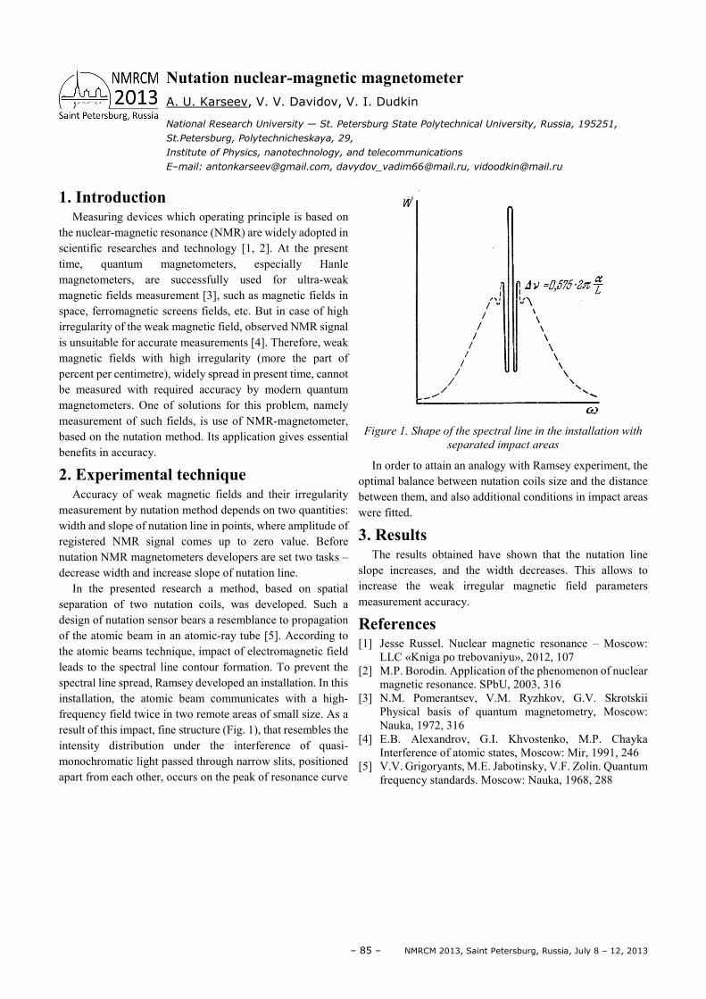
– 85 – NMRCM 2013, Saint Petersburg, Russia, July 8 – 12, 2013
1. Introduction
Measuring devices which operating principle is based on the nuclear-magnetic resonance (NMR) are widely adopted in scientific researches and technology [1, 2]. At the present time, quantum magnetometers, especially Hanle magnetometers, are successfully used for ultra-weak magnetic fields measurement [3], such as magnetic fields in space, ferromagnetic screens fields, etc. But in case of high irregularity of the weak magnetic field, observed NMR signal is unsuitable for accurate measurements [4]. Therefore, weak magnetic fields with high irregularity (more the part of percent per centimetre), widely spread in present time, cannot be measured with required accuracy by modern quantum magnetometers. One of solutions for this problem, namely measurement of such fields, is use of NMR-magnetometer, based on the nutation method. Its application gives essential benefits in accuracy.
2. Experimental technique
Accuracy of weak magnetic fields and their irregularity measurement by nutation method depends on two quantities: width and slope of nutation line in points, where amplitude of registered NMR signal comes up to zero value. Before nutation NMR magnetometers developers are set two tasks – decrease width and increase slope of nutation line.
In the presented research a method, based on spatial separation of two nutation coils, was developed. Such a design of nutation sensor bears a resemblance to propagation of the atomic beam in an atomic-ray tube [5]. According to the atomic beams technique, impact of electromagnetic field leads to the spectral line contour formation. To prevent the spectral line spread, Ramsey developed an installation. In this installation, the atomic beam communicates with a high-frequency field twice in two remote areas of small size. As a result of this impact, fine structure (Fig. 1), that resembles the intensity distribution under the interference of quasi-monochromatic light passed through narrow slits, positioned apart from each other, occurs on the peak of resonance curve
Figure 1. Shape of the spectral line in the installation with
separated impact areas
In order to attain an analogy with Ramsey experiment, the optimal balance between nutation coils size and the distance between them, and also additional conditions in impact areas were fitted.
3. Results
The results obtained have shown that the nutation line slope increases, and the width decreases. This allows to increase the weak irregular magnetic field parameters measurement accuracy.
References [1] Jesse Russel. Nuclear magnetic resonance – Moscow:
LLC «Kniga po trebovaniyu», 2012, 107 [2] M.P. Borodin. Application of the phenomenon of nuclear
magnetic resonance. SPbU, 2003, 316 [3] N.M. Pomerantsev, V.M. Ryzhkov, G.V. Skrotskii
Physical basis of quantum magnetometry, Moscow: Nauka, 1972, 316
[4] E.B. Alexandrov, G.I. Khvostenko, M.P. Chayka Interference of atomic states, Moscow: Mir, 1991, 246
[5] V.V. Grigoryants, M.E. Jabotinsky, V.F. Zolin. Quantum frequency standards. Moscow: Nauka, 1968, 288
Nutation nuclear-magnetic magnetometer
A. U. Karseev, V. V. Davidov, V. I. Dudkin
National Research University — St. Petersburg State Polytechnical University, Russia, 195251,
St.Petersburg, Polytechnicheskaya, 29,
Institute of Physics, nanotechnology, and telecommunications
E–mail: [email protected], [email protected], [email protected]
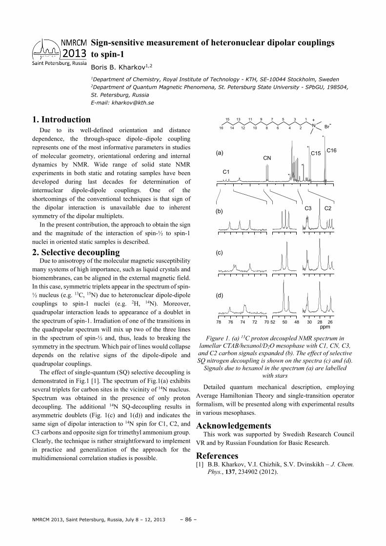
NMRCM 2013, Saint Petersburg, Russia, July 8 – 12, 2013 – 86 –
1. Introduction Due to its well-defined orientation and distance
dependence, the through-space dipole–dipole coupling represents one of the most informative parameters in studies of molecular geometry, orientational ordering and internal dynamics by NMR. Wide range of solid state NMR experiments in both static and rotating samples have been developed during last decades for determination of internuclear dipole-dipole couplings. One of the shortcomings of the conventional techniques is that sign of the dipolar interaction is unavailable due to inherent symmetry of the dipolar multiplets.
In the present contribution, the approach to obtain the sign and the magnitude of the interaction of spin-½ to spin-1 nuclei in oriented static samples is described.
2. Selective decoupling Due to anisotropy of the molecular magnetic susceptibility
many systems of high importance, such as liquid crystals and biomembranes, can be aligned in the external magnetic field. In this case, symmetric triplets appear in the spectrum of spin-½ nucleus (e.g. 13C, 15N) due to heteronuclear dipole-dipole couplings to spin-1 nuclei (e.g. 2H, 14N). Moreover, quadrupolar interaction leads to appearance of a doublet in the spectrum of spin-1. Irradiation of one of the transitions in the quadrupolar spectrum will mix up two of the three lines in the spectrum of spin-½ and, thus, leads to breaking the symmetry in the spectrum. Which pair of lines would collapse depends on the relative signs of the dipole-dipole and quadrupolar couplings.
The effect of single-quantum (SQ) selective decoupling is demonstrated in Fig.1 [1]. The spectrum of Fig.1(a) exhibits several triplets for carbon sites in the vicinity of 14N nucleus. Spectrum was obtained in the presence of only proton decoupling. The additional 14N SQ-decoupling results in asymmetric doublets (Fig. 1(c) and 1(d)) and indicates the same sign of dipolar interaction to 14N spin for C1, C2, and C3 carbons and opposite sign for trimethyl ammonium group. Clearly, the technique is rather straightforward to implement in practice and generalization of the approach for the multidimensional correlation studies is possible.
78 76 74 72 70 52 50 48 30 28 26
(b)C3 C2
(c)
(d)
16 14 12 10 8 6 4 2
15 13 11 9 7 5 3 1
N+
Br-
ppm
C16C15
*
*
*
**
C1
CN(a)
Figure 1. (a) 13C proton decoupled NMR spectrum in
lamellar CTAB/hexanol/D2O mesophase with C1, CN, C3,
and C2 carbon signals expanded (b). The effect of selective
SQ nitrogen decoupling is shown on the spectra (c) and (d).
Signals due to hexanol in the spectrum (a) are labelled
with stars
Detailed quantum mechanical description, employing Average Hamiltonian Theory and single-transition operator formalism, will be presented along with experimental results in various mesophases.
Acknowledgements This work was supported by Swedish Research Council
VR and by Russian Foundation for Basic Research.
References [1] B.B. Kharkov, V.I. Chizhik, S.V. Dvinskikh – J. Chem.
Phys., 137, 234902 (2012).
Sign-sensitive measurement of heteronuclear dipolar couplings to spin-1 Boris B. Kharkov1,2
1Department of Chemistry, Royal Institute of Technology - KTH, SE-10044 Stockholm, Sweden 2Department of Quantum Magnetic Phenomena, St. Petersburg State University - SPbGU, 198504,
St. Petersburg, Russia
E-mail: [email protected]
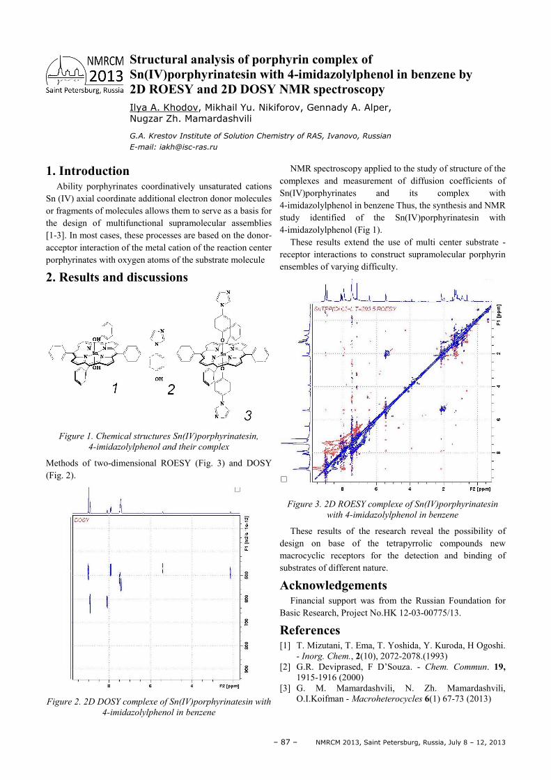
– 87 – NMRCM 2013, Saint Petersburg, Russia, July 8 – 12, 2013
1. Introduction Ability porphyrinates coordinatively unsaturated cations
Sn (IV) axial coordinate additional electron donor molecules or fragments of molecules allows them to serve as a basis for the design of multifunctional supramolecular assemblies [1-3]. In most cases, these processes are based on the donor-acceptor interaction of the metal cation of the reaction center porphyrinates with oxygen atoms of the substrate molecule
2. Results and discussions
Figure 1. Chemical structures Sn(IV)porphyrinatesin,
4-imidazolylphenol and their complex
Methods of two-dimensional ROESY (Fig. 3) and DOSY (Fig. 2).
Figure 2. 2D DOSY complexe of Sn(IV)porphyrinatesin with
4-imidazolylphenol in benzene
NMR spectroscopy applied to the study of structure of the complexes and measurement of diffusion coefficients of Sn(IV)porphyrinates and its complex with 4-imidazolylphenol in benzene Thus, the synthesis and NMR study identified of the Sn(IV)porphyrinatesin with 4-imidazolylphenol (Fig 1).
These results extend the use of multi center substrate - receptor interactions to construct supramolecular porphyrin ensembles of varying difficulty.
Figure 3. 2D ROESY complexe of Sn(IV)porphyrinatesin
with 4-imidazolylphenol in benzene
These results of the research reveal the possibility of design on base of the tetrapyrrolic compounds new macrocyclic receptors for the detection and binding of substrates of different nature.
Acknowledgements Financial support was from the Russian Foundation for
Basic Research, Project No.HK 12-03-00775/13.
References [1] Т. Mizutani, Т. Еmа, Т. Yoshida, Y. Kuroda, H Ogoshi.
- Inorg. Chem., 2(10), 2072-2078.(1993) [2] G.R. Deviprased, F D’Souza. - Chem. Commun. 19,
1915-1916 (2000) [3] G. М. Mamardashvili, N. Zh. Mamardashvili,
O.I.Koifman - Macroheterocycles 6(1) 67-73 (2013)
Structural analysis of porphyrin complex of Sn(IV)porphyrinatesin with 4-imidazolylphenol in benzene by 2D ROESY and 2D DOSY NMR spectroscopy Ilya A. Khodov, Mikhail Yu. Nikiforov, Gennady A. Alper, Nugzar Zh. Mamardashvili
G.A. Krestov Institute of Solution Chemistry of RAS, Ivanovo, Russian
E-mail: [email protected]
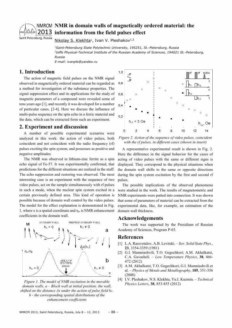
NMRCM 2013, Saint Petersburg, Russia, July 8 – 12, 2013 – 88 –
1. Introduction The action of magnetic field pulses on the NMR signal
observed in magnetically ordered material can be regarded as a method for investigation of the substance properties. The signal suppression effect and its applications for the study of magnetic parameters of a compound were revealed some of tens years ago [1], and recently it was developed for a number of particular cases, [2-4]. Here we discuss the influence of multi-pulse sequence on the spin echo in a ferric material and the data, which can be extracted form such an experiment.
2. Experiment and discussion A number of possible experimental scenarios were
analyzed in this work: the action of video pulses, both coincident and not coincident with the radio frequency (rf) pulses exciting the spin system, and possesses as positive and negative amplitudes.
The NMR was observed in lithium-zinc ferrite as a spin echo signal of Fe-57. It was experimentally confirmed, that predictions for the different situations are realized in the stuff. The echo suppression and restoring was observed. The most interesting case is an experiment with the sequence of two video pulses, act on the sample simultaneously with rf pulses in such a mode, when the nuclear spin system excited in a certain previously defined area. This kind of operation is possible because of domain wall control by the video pulses. The model for the effect explanation is demonstrated in Fig. 1, where x is a spatial coordinate and ηw is NMR enhancement coefficients in the domain wall.
Figure 1. The model of NMR excitation in the movable
domain walls. a – Bloch wall at initial position; the wall,
shifted on the distance ∆x under the action of pulse field hV.
b - the corresponding spatial distributions of the
enhancement coefficients
Figure 2. Action of the sequence of video pulses, coincident
with the rf pulses, in different cases (shown in insert)
A representative experimental result is shown in Fig. 2. Here the difference in the signal behavior for the cases of acting of video pulses with the same or different signs is displayed. They correspond to the physical situations when the domain wall shifts to the same or opposite directions during the spin system excitation by the first and second rf pulses.
The possible implications of the observed phenomena were studied in the work. The results of magnetometric and NMR experiments were putted into connection. It was shown that some of parameters of material can be extracted from the experimental data, like, for example, an estimation of the domain wall thickness.
Acknowledgements The work was supported by the Presidium of Russian
Academy of Sciences, Program P-03.
References [1] L.A. Rassvetalov, A.B. Levitski. – Sov. Solid State Phys.,
23, 3354-3359 (1981) [2] G.I. Mamniashvili, T.O. Gegechkori; A.M. Akhalkatsi,
C.A. Gavasheli. – Low Temperature Physics, 38, 466-472 (2012)
[3] A.M. Akhalkatsi, T.O. Gegechkori, G.I. Mamniashvili et al. – Physics of Metals and Metallography, 105, 351-356 (2008)
[4] I.V. Pleshakov, N.S. Klekhta, Yu.I. Kuzmin. – Technical
Physics Letters, 38, 853-855 (2012)
NMR in domain walls of magnetically ordered material: the information from the field pulses effect Nikolay S. Klekhta1, Ivan V. Pleshakov1,2
1Saint-Petersburg State Polytechnic University, 195251, St.-Petersburg, Russia 2Ioffe Physical-Technical Institute of the Russian Academy of Sciences, 194021 St.-Petersburg,
Russia
E-mail: [email protected]
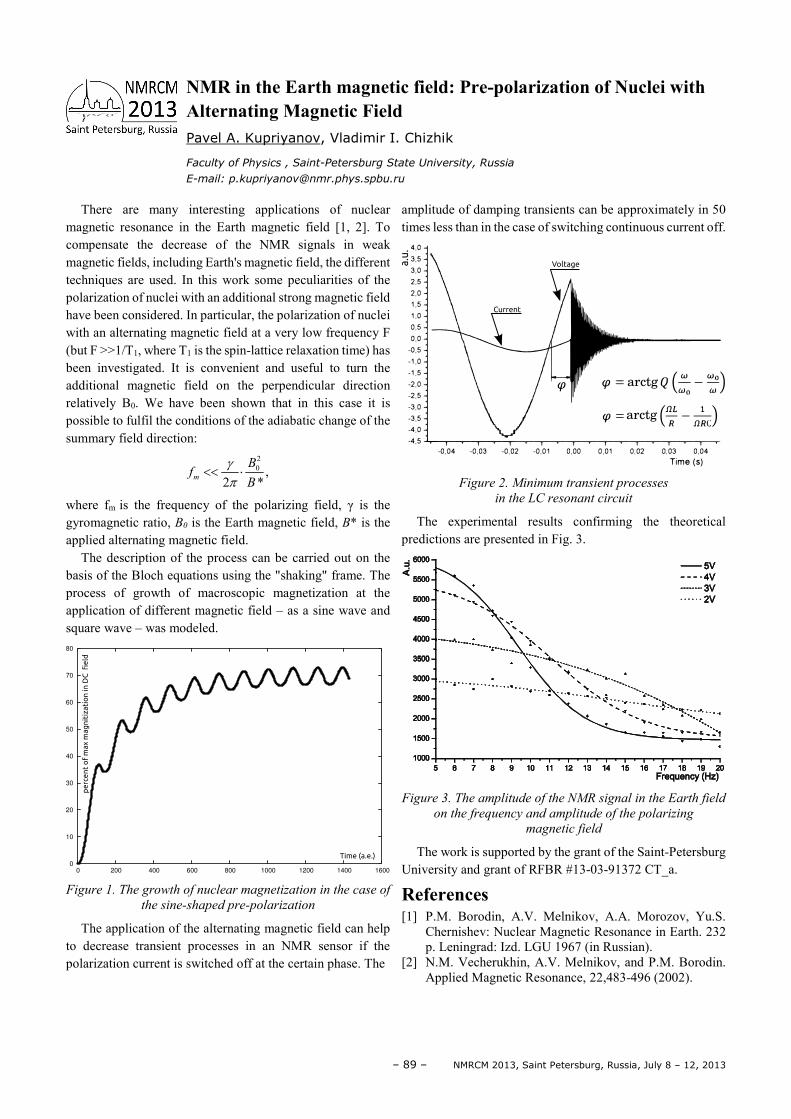
– 89 – NMRCM 2013, Saint Petersburg, Russia, July 8 – 12, 2013
There are many interesting applications of nuclear magnetic resonance in the Earth magnetic field [1, 2]. To compensate the decrease of the NMR signals in weak magnetic fields, including Earth's magnetic field, the different techniques are used. In this work some peculiarities of the polarization of nuclei with an additional strong magnetic field have been considered. In particular, the polarization of nuclei with an alternating magnetic field at a very low frequency F (but F >>1/T1, where T1 is the spin-lattice relaxation time) has been investigated. It is convenient and useful to turn the additional magnetic field on the perpendicular direction relatively B0. We have been shown that in this case it is possible to fulfil the conditions of the adiabatic change of the summary field direction:
20 ,
2 *m
Bf
B
γπ
<< ⋅
where fm is the frequency of the polarizing field, γ is the gyromagnetic ratio, B0 is the Earth magnetic field, B* is the applied alternating magnetic field.
The description of the process can be carried out on the basis of the Bloch equations using the "shaking" frame. The process of growth of macroscopic magnetization at the application of different magnetic field – as a sine wave and square wave – was modeled.
Figure 1. The growth of nuclear magnetization in the case of
the sine-shaped pre-polarization
The application of the alternating magnetic field can help to decrease transient processes in an NMR sensor if the polarization current is switched off at the certain phase. The
amplitude of damping transients can be approximately in 50 times less than in the case of switching continuous current off.
Figure 2. Minimum transient processes
in the LC resonant circuit
The experimental results confirming the theoretical predictions are presented in Fig. 3.
Figure 3. The amplitude of the NMR signal in the Earth field
on the frequency and amplitude of the polarizing
magnetic field
The work is supported by the grant of the Saint-Petersburg University and grant of RFBR #13-03-91372 CT_a.
References [1] P.M. Borodin, A.V. Melnikov, A.A. Morozov, Yu.S.
Chernishev: Nuclear Magnetic Resonance in Earth. 232 p. Leningrad: Izd. LGU 1967 (in Russian).
[2] N.M. Vecherukhin, A.V. Melnikov, and P.M. Borodin. Applied Magnetic Resonance, 22,483-496 (2002).
NMR in the Earth magnetic field: Pre-polarization of Nuclei with Alternating Magnetic Field Pavel A. Kupriyanov, Vladimir I. Chizhik
Faculty of Physics , Saint-Petersburg State University, Russia
E-mail: [email protected]
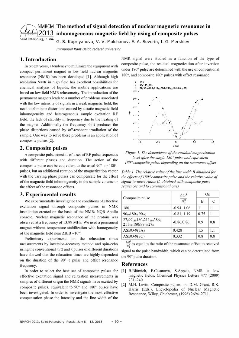
NMRCM 2013, Saint Petersburg, Russia, July 8 – 12, 2013 – 90 –
1. Introduction In recent years, a tendency to minimize the equipment with
compact permanent magnet in low field nuclear magnetic resonance (NMR) has been developed [1]. Although high resolution NMR in high field has excellent possibilities for chemical analysis of liquids, the mobile applications are based on low field NMR relaxometry. The introduction of the permanent magnets leads to a number of problems associated with the low intensity of signals in a weak magnetic field, the need to eliminate distortions caused by a static magnetic field inhomogeneity and heterogeneous sample excitation RF field, the lack of stability in frequency due to the heating of the magnet. Additionally the frequency shift produces the phase distortions caused by off-resonant irradiation of the sample. One way to solve these problems is an application of composite pulses [2].
2. Composite pulses A composite pulse consists of a set of RF pulse sequences
with different phases and duration. The action of the composite pulse can be equivalent to the usual 90°- or 180°- pulses, but an additional rotation of the magnetization vector with the varying phase pulses can compensate for the effect of the magnetic field inhomogeneity in the sample volume or the effect of the resonance offsets.
3. Experimental results We experimentally investigated the conditions of effective
excitation signal through composite pulses in NMR installation created on the basis of the NMR/ NQR Apollo console. Nuclear magnetic resonance of the protons was observed at a frequency of 13.99 MHz. We used a permanent magnet without temperature stabilization with homogeneity of the magnetic field near ∆B/B ∼10-4.
Preliminary experiments on the relaxation times measurements by inversion-recovery method and spin-echo using the conventional π / 2 and π pulses of different durations have showed that the relaxation times are highly dependent on the duration of the 90° τ pulse and offset resonance frequency.
In order to select the best set of composite pulses for effective excitation signal and relaxation measurements in samples of different origin the NMR signals have excited by composite pulses, equivalent to 90° and 180° pulses have been investigated. In order to investigate the most effective compensation phase the intensity and the line width of the
NMR signal were studied as a function of the type of composite pulse, the residual magnetization after inversion under 180° pulse are determined with the use of conventional 180°, and composite 180° pulses with offset resonance.
Figure 1. The dependence of the residual magnetization
level after the single 180° pulse and equivalent
180° composite pulse, depending on the resonance offset
Table 1. The relative value of the line width B obtained for
the effects of 180° composite pulse and the relative value of
signal-to-noise ratios C, obtained with composite pulse
sequences and to conventional ones
Composite pulse ∆
Oil
B C
180 -0.94, 1,06 1 1 9090180
0 90 90 -0.81, 1.19 0.75 1
2709918018002111803860
211180180099180270 -0.86,0.86 0.9 0.8
ASBO-9(7A) 0.428 1.5 1.1 ASBO-9(7C) 0.332 0.8 0.8
∆ !" is equal to the ratio of the resonance offset to received
signal to the pulse bandwidth, which can be determined from the 90° pulse duration.
References [1] B.Blümich, F.Casanova, S.Appelt, NMR at low
magnetic fields, Chemical Physics Letters 477 (2009) 231–240
[2] M.H. Levitt, Composite pulses, in: D.M. Grant, R.K. Harris (Eds.), Encyclopedia of Nuclear Magnetic Resonance, Wiley, Chichester, (1996) 2694–2711.
The method of signal detection of nuclear magnetic resonance in inhomogeneous magnetic field by using of composite pulses G. S. Kupriyanova, V. V. Molchanov, E. A. Severin, I. G. Mershiev
Immanuel Kant Baltic federal university
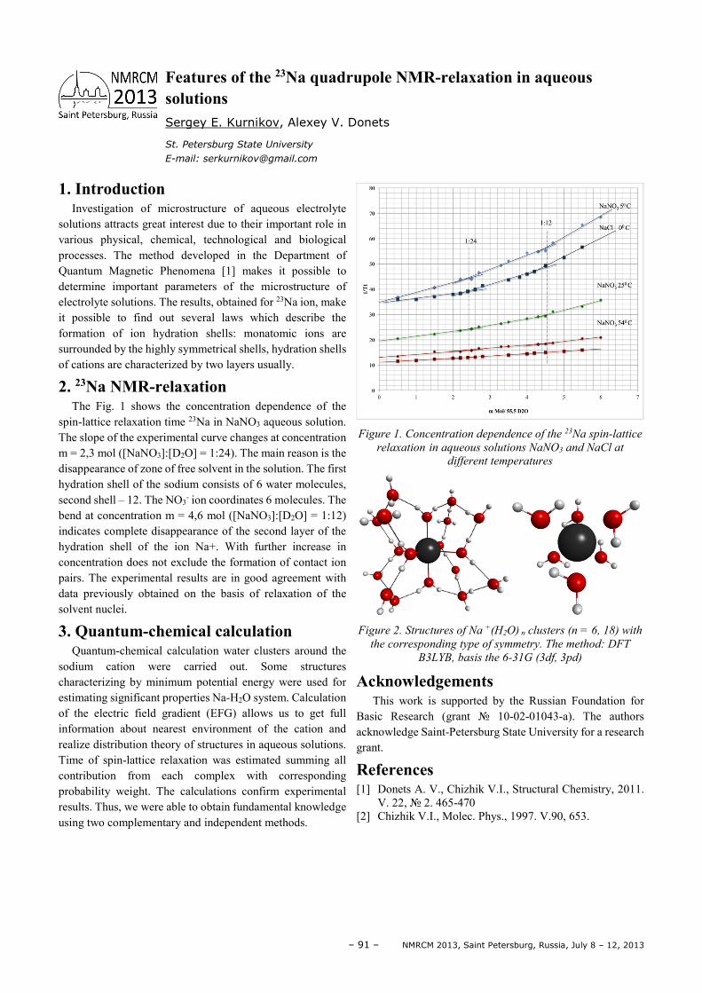
– 91 – NMRCM 2013, Saint Petersburg, Russia, July 8 – 12, 2013
1. Introduction Investigation of microstructure of aqueous electrolyte
solutions attracts great interest due to their important role in various physical, chemical, technological and biological processes. The method developed in the Department of Quantum Magnetic Phenomena [1] makes it possible to determine important parameters of the microstructure of electrolyte solutions. The results, obtained for 23Na ion, make it possible to find out several laws which describe the formation of ion hydration shells: monatomic ions are surrounded by the highly symmetrical shells, hydration shells of cations are characterized by two layers usually.
2. 23Na NMR-relaxation The Fig. 1 shows the concentration dependence of the
spin-lattice relaxation time 23Na in NaNO3 aqueous solution. The slope of the experimental curve changes at concentration m = 2,3 mol ([NaNO3]:[D2O] = 1:24). The main reason is the disappearance of zone of free solvent in the solution. The first hydration shell of the sodium consists of 6 water molecules, second shell – 12. The NO3
- ion coordinates 6 molecules. The bend at concentration m = 4,6 mol ([NaNO3]:[D2O] = 1:12) indicates complete disappearance of the second layer of the hydration shell of the ion Na+. With further increase in concentration does not exclude the formation of contact ion pairs. The experimental results are in good agreement with data previously obtained on the basis of relaxation of the solvent nuclei.
3. Quantum-chemical calculation Quantum-chemical calculation water clusters around the
sodium cation were carried out. Some structures characterizing by minimum potential energy were used for estimating significant properties Na-H2O system. Calculation of the electric field gradient (EFG) allows us to get full information about nearest environment of the cation and realize distribution theory of structures in aqueous solutions. Time of spin-lattice relaxation was estimated summing all contribution from each complex with corresponding probability weight. The calculations confirm experimental results. Thus, we were able to obtain fundamental knowledge using two complementary and independent methods.
Figure 1. Concentration dependence of the 23Na spin-lattice
relaxation in aqueous solutions NaNO3 and NaCl at
different temperatures
Figure 2. Structures of Na +(H2O) n clusters (n = 6, 18) with
the corresponding type of symmetry. The method: DFT
B3LYB, basis the 6-31G (3df, 3pd)
Acknowledgements This work is supported by the Russian Foundation for
Basic Research (grant 10-02-01043-a). The authors acknowledge Saint-Petersburg State University for a research grant.
References [1] Donets A. V., Chizhik V.I., Structural Chemistry, 2011.
V. 22, 2. 465-470 [2] Chizhik V.I., Molec. Phys., 1997. V.90, 653.
Features of the 23Na quadrupole NMR-relaxation in aqueous solutions Sergey E. Kurnikov, Alexey V. Donets
St. Petersburg State University
E-mail: [email protected]
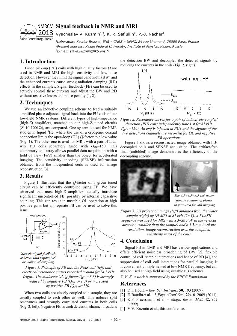
NMRCM 2013, Saint Petersburg, Russia, July 8 – 12, 2013 – 92 –
1. Introduction Tuned pick-up (PU) coils with high quality factors Q are
used in NMR and MRI for high-sensitivity and low-noise detection. However they limit the signal bandwidth (BW) and the enhanced currents cause strong radiation damping (RD) effects in the samples. Signal feedback (FB) can be used to actively control these currents and adjust the BW and RD without resistive losses and noise penalty [1, 2].
2. Techniques We use an inductive coupling scheme to feed a suitably
amplified phase-adjusted signal back into the PU coils of our low-field NMR systems. Different types of high-impedance (high-Z) amplifiers, matched to our high-Z tuned circuits (Z~10-100kΩ), are compared. One system is used for NMR studies in liquid 3He, where the use of a cryogenic coaxial connection limits the open-loop (OL) Q-factor to a low value (Fig. 1). The other one is used for MRI, with a pair of Litz-wire PU coils separately tuned with QOL~150. This elementary coil-array allows parallel data acquisition with a field of view (FoV) smaller than the object for accelerated imaging. The sensitivity encoding (SENSE) information obtained from the independent coils is used for image reconstruction [3].
3. Results Figure 1 illustrates that the Q-factor of a given tuned
circuit can be efficiently controlled using FB. We have observed that most high-Z amplifiers actually introduce significant uncontrolled FB, possibly by internal capacitive coupling. This can result in unstable OL operation at high positive gain, but appropriate FB can be used to solve this issue.
Generic signal feedback
scheme, with capacitive1
or inductive2 coupling
Figure 1. Principle of FB into the NMR coil (left) and
electrical resonance curves recorded around f0=74.7 kHz
(right). The moderate OL Q-factor (QOL=8.8) is strongly
reduced by negative FB (QFB<0=1.3) or increased
by positive FB (QFB>0=110)
When two coils are closely coupled to a sample, they are usually coupled to each other as well. This induces split resonances and strongly correlated currents in both coils (Fig. 2, left). Negative FB in each detection channel broadens
the detection BW and decouples the detected signals by reducing the currents in the coils (Fig. 2, right).
Figure 2. Resonance curves for a pair of inductively coupled
detection (PU) coils independently tuned at f0=87 kHz
(QOL=150). An emf is injected in PU1 and the signals of the
two detections channels are recorded for OL and negative
FB operations
Figure 3 shows a reconstructed image obtained with FB-decoupled coils and SENSE acquisition. The artifact-free final (unfolded) image demonstrates the efficiency of the decoupling scheme.
The 4.5×4.5×3.5 cm3 water
sample containing plastic
shapes used for MR imaging
Figure 3. 2D projection image (left) obtained from the water
sample (right) by 1H MRI at 87 kHz (2mT). A FLASH
sequence was used for MRI with a 3-cm FoV in the vertical
direction (smaller than the sample) and a 1.5-mm in-plane
resolution. Image reconstruction uses the computed
sensitivity maps of the coils
4. Conclusion Signal FB in NMR and MRI has various applications and
offers efficient noiseless broadening of BW [2], flexible control of coil−sample interactions and hence of RD [4], and suppression of coil−coil interactions for parallel imaging. It is conveniently implemented at low NMR frequency, but can also be used at high field using suitable FB schemes. V. V. K.’s work is supported by the FPGG Foundation.
References [1] D.I. Hoult. – Rev. Sci. Instrum., 50, 193 (2009). [2] E. Baudin et al. – J. Phys.: Conf. Ser., 294, 012009 (2011). [3] K.P. Pruessmann et al. – Magn. Reson. Med, 42, 952
(1999). [4] V.V. Kuzmin et al., this conference.
Signal feedback in NMR and MRI Vyacheslav V. Kuzmin1,3, K. R. Safiullin2, P.-J. Nacher1
1Laboratoire Kastler Brossel, ENS – CNRS – UPMC, 24 rue Lhomond, 75005 Paris, France 2Present address: Kazan Federal University, Institute of Physics, Kazan, Russia. 3E-mail: [email protected]
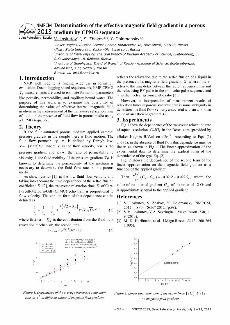
– 93 – NMRCM 2013, Saint Petersburg, Russia, July 8 – 12, 2013
1. Introduction NMR well logging is finding wide use in formation
evaluation. Due to logging speed requirements, NMR CPMG 2T measurements are used to estimate formation parameters
like porosity, permeability, and capillary bound water. The purpose of this work is to examine the possibility of determining the value of effective internal magnetic field gradient in the measurement of the transverse relaxation time of liquid in the presence of fluid flow in porous media using a CPMG sequence.
2. Theory If the fluid-saturated porous medium applied external
pressure gradient in the sample there is fluid motion. The fluid flow permeability, κ , is defined by Darcy's law:
( )/v pκ η= − ∇ where v is the flow velocity, p∇ is the
pressure gradient and /κ η , the ratio of permeability to viscosity, is the fluid mobility. If the pressure gradient p∇ is known, to determine the permeability of the medium is necessary to determine the fluid flow rate in this porous media.
As shown earlier [1], at the low fluid flow velocity and taking into account the time dependence of the self-diffusion coefficient D [2], the transverse relaxation time 2T of Carr-Purcell-Meiborn-Gill (CPMG) echo train is proportional to flow velocity. The explicit form of this dependence can be defined as
( ) 2 2 5/2
2 2 2
4 2 0.51 1 135B D
G DvT T T
γ τ−
= + + , (1)
where first term 2BT is the contribution from the fluid bulk relaxation mechanism, the second term
2 2 221/ /12DT G Dγ τ= (2)
reflects the relaxation due to the self-diffusion of a liquid in the presence of a magnetic field gradient, G, where time τ refers to the time delay between the radio frequency pulse and the refocusing RF pulse in the spin echo pulse sequence and γ is the nuclear gyromagnetic ratio [3].
However, at interpretation of measurement results of relaxation times in porous systems there is some ambiguity in definition of a fluid flow velocity associated with an unknown value of an efficient gradient G .
3. Experiments Fig.1 show the dependence of the transverse relaxation rate
of aqueous solution 4CuSO in the Berea core (provided by
«Baker Hughes B.V.») on ( )22τ . According to Eqs. (1) and (2), in the absence of fluid flow this dependence must be linear, as shown in Fig.1. The linear approximation of the experimental data to determine the explicit form of the dependence of the type Eq. (2).
Fig. 2 shows the dependence of the second term of the linear approximation on the magnetic field gradient as a function of the applied gradient.
Then ( )2
0 int 00.0265 0.02212
DG G G
γ+ = − + , where the
value of the internal gradient intG of the order of 12 Gs and is approximately equal to the applied gradient.
References [1] V. Loskutov, S. Zhakov, Y, Dolomansky. NMRCM,
2012. – SPb., “Solo” 2012. (p.98). [2] V.V. Loskutov, V.A. Sevriugin. J.Magn.Reson. 230, 1–
9 (2013). [3] M. D. Hurlimann at al. J.Magn.Reson. A113, 260-264
(1995).
0 2 4 6 8 10 12 14
2
4
6Gr=11,5 Gs: 1/<T2>=1,261+0,227(2τ)
2
Gr=10 Gs: 1/<T2>=1,251+0,19405(2τ)2
Gr=15 Gs: 1/<T2>=1,319+0,302(2τ)2
Gr=13 Gs
Gr=15 Gs
Gr=10 Gs
Gr=11.5 Gs
1/<T
2>
(2τ)2, ms2
Gr=13 Gs: 1/<T2>=1,209+0,269(2τ)2
Figure 1. Dependence of the average transverse relaxation
rate on 2τ at different values of magnetic field gradient
0 1 2 3 4 5 6 7 8 9 10 11 12 13 14 15
-0,05
0,00
0,05
0,10
0,15
0,20
0,25
0,30
0,35
-0,0265+0,022*Gr
D(g
G)2
/12
Gr, Gs
Figure 2. Linear approximation of the dependence ( )2 /12G Dγ
on magnetic field gradient
Determination of the effective magnetic field gradient in a porous medium by CPMG sequence V. Loskutov1,2, S. Zhakov1,3, Y. Dolomansky1,4
1Baker Hughes, Russian Science Center, Kutateladze 4A, Novosibirsk, 630128, Russia 2IMary State University, Yoskar-Ola, Lenin sq.1, Russia 3Institute of Metal Physics, The Ural Branch of Russian Academy of Science, Ekaterinburg, ul.
S.Kovalevskaya, 18, 620990, Russia 4Institute of Geophysics, The Ural Branch of Russian Academy of Science, Ekaterinburg,ul.
Amundsena, 100, 620016, Russia,
E-mail: [email protected]
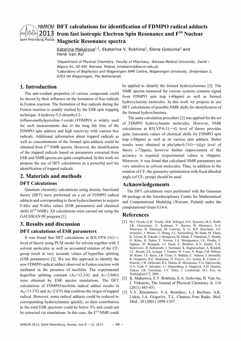
NMRCM 2013, Saint Petersburg, Russia, July 8 – 12, 2013 – 94 –
1. Introduction The anti-oxidant properties of various compounds could
be shown by their influence on the formation of free radicals in Fenton reaction. The formation of free radicals during the Fenton reaction is usually studied by the ESR spin trapping technique. 4-hydroxy-5,5-dimethyl-2- trifluoromethylpyrroline-1-oxide (FDMPO) is widely used for such measurements due to the long life time of the FDMPO spin adducts and high reactivity with various free radicals. Additional information about trapped radicals as well as concentrations of the formed spin adducts could be obtained from F19 NMR spectra. However, the identification of the trapped radicals based on parameters extracted from ESR and NMR spectra are quite complicated. In this work we propose the use of DFT calculations as a powerful tool for identification of trapped radicals.
2. Materials and methods DFT Calculations
Quantum chemistry calculations using density functional theory (DFT) were performed on a set of FDMPO radical adducts and corresponding to them hydorxilamines to acquire F-hfcs and N-hfcs values (ESR parameters) and chemical shifts (F19 NMR). All calculations were carried out using the GAUSSIAN 09 program [1].
3. Results and discussion DFT calculations of ESR parameters
It was found that DFT calculations at B3LYP/6-31G++ level of theory using PCM model for solvent together with 2 solvent molecules as well as accounted rotation of the CF3 group result in very accurate values of hyperfine splitting (ESR parameters) [2]. We use this approach to identify the new FDMPO radical adduct observed in Fenton reaction with methanol in the presence of taxifolin. The experimental hyperfine splitting constant (AN=13.31G and AF=2.94G) were obtained by ESR spectra simulations. The DFT calculations of FDMPO/taxifolin radical adduct results in AN=13.37G and AF=2.87G that confirms the origin of trapped radical. However, some radical adducts could be reduced to corresponding hydroxylamine quickly, so their contribution to the total ESR spectrum could be below 5% and could not be extracted via simulations. In this case, the F19 NMR could
be applied to identify the formed hydroxylamines [3]. The NMR spectra measured for various systems contains signal from FDMPO spin trap (-60ppm) as well as formed hydroxylamine molecules. In this work we propose to use DFT calculations of possible NMR shifts for identification of the formed hydroxilamines.
The same calculation procedure [2] was applied for the set of FDMPO hydroxylamine molecules. However, NMR calculations at B3LYP/6-31++G level of theory provides quite inaccurate values of chemical shifts for FDMPO spin trap (-88ppm) as well as its various spin adducts. Better results were obtained at pbe1pbe/6-31G++(d,p) level of theory (-72ppm), however further improvement of the accuracy is required (experimental values is -66ppm). Moreover, it was found that calculated NMR parameters are very sensitive to solvent molecules. Thus, in addition to the rotation of CF3 the geometry optimization with fixed dihedral angle (of CF3 group) should be used.
Acknowledgements The DFT calculations were performed with the Gaussian
09 package in the Interdisciplinary Centre for Mathematical and Computational Modeling (Warsaw, Poland) under the computational Grant G14-6.
References [1] M.J. Frisch, G.W. Trucks, H.B. Schlegel, G.E. Scuseria, M.A. Robb,
J.R. Cheeseman, G. Scalmani, V. Barone, B. Mennucci, G.A. Petersson, H. Nakatsuji, M. Caricato, X. Li, H.P. Hratchian, A.F. Izmaylov, J. Bloino, G. Zheng, J.L. Sonnenberg, M. Hada, M. Ehara, K. Toyota, R. Fukuda, J. Hasegawa, M. Ishida, T. Nakajima, Y. Honda, O. Kitao, H. Nakai, T. Vreven, J.A. Montgomery, J.E. Peralta, F. Ogliaro, M. Bearpark, J.J. Heyd, E. Brothers, K.N. Kudin, V.N. Staroverov, R. Kobayashi, J. Normand, K. Raghavachari, A. Rendell, J.C. Burant, S.S. Iyengar, J. Tomasi, M. Cossi, N. Rega, J.M. Millam, M. Klene, J.E. Knox, J.B. Cross, V. Bakken, C. Adamo, J. Jaramillo, R. Gomperts, R.E. Stratmann, O. Yazyev, A.J. Austin, R. Cammi, C. Pomelli, J.W. Ochterski, R.L. Martin, K. Morokuma, V.G. Zakrzewski, G.A. Voth, P. Salvador, J.J. Dannenberg, S. Dapprich, A.D. Daniels, Farkas, J.B. Foresman, J.V. Ortiz, J. Cioslowski, D.J. Fox, in, Wallingford CT, 2009.
[2] K. Makarova, E.V. Rokhina, E.A. Golovina, H. Van As, J. Virkutyte, The Journal of Physical Chemistry A, 116 (2011) 443-451.
[3] V.V. Khramtsov, V.A. Reznikov, L.J. Berliner, A.K. Litkin, I.A. Grigor'ev, T.L. Clanton, Free Radic. Biol. Med., 30 (2001) 1099-1107.
DFT calculations for identification of FDMPO radical adducts from fast isotropic Electron Spin Resonance and F19 Nuclear Magnetic Resonance spectra Katerina Makarova1,2, Ekaterina V. Rokhina2, Elena Golovina2 and Henk Van As2
1Department of Physical Chemistry, Faculty of Pharmacy, Warsaw Medical University, Zwirki i
Wigury 61, 02-091 Warsaw, Poland, [email protected]; 2Laboratory of Biophysics and Wageningen NMR Centre, Wageningen University, Dreijenlaan 3,
6703 HA Wageningen, The Netherlands
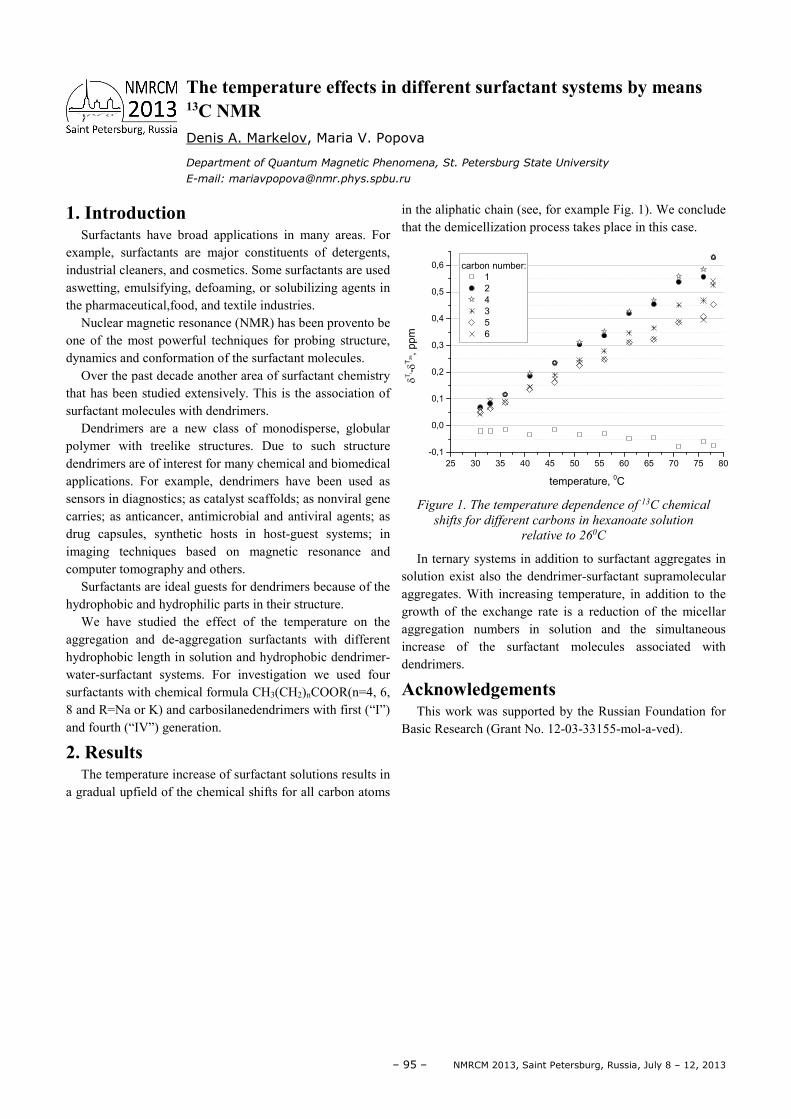
– 95 – NMRCM 2013, Saint Petersburg, Russia, July 8 – 12, 2013
1. Introduction Surfactants have broad applications in many areas. For
example, surfactants are major constituents of detergents, industrial cleaners, and cosmetics. Some surfactants are used aswetting, emulsifying, defoaming, or solubilizing agents in the pharmaceutical,food, and textile industries.
Nuclear magnetic resonance (NMR) has been provento be one of the most powerful techniques for probing structure, dynamics and conformation of the surfactant molecules.
Over the past decade another area of surfactant chemistry that has been studied extensively. This is the association of surfactant molecules with dendrimers.
Dendrimers are a new class of monodisperse, globular polymer with treelike structures. Due to such structure dendrimers are of interest for many chemical and biomedical applications. For example, dendrimers have been used as sensors in diagnostics; as catalyst scaffolds; as nonviral gene carries; as anticancer, antimicrobial and antiviral agents; as drug capsules, synthetic hosts in host-guest systems; in imaging techniques based on magnetic resonance and computer tomography and others.
Surfactants are ideal guests for dendrimers because of the hydrophobic and hydrophilic parts in their structure.
We have studied the effect of the temperature on the aggregation and de-aggregation surfactants with different hydrophobic length in solution and hydrophobic dendrimer-water-surfactant systems. For investigation we used four surfactants with chemical formula CH3(CH2)nCOOR(n=4, 6, 8 and R=Na or K) and carbosilanedendrimers with first (“I”) and fourth (“IV”) generation.
2. Results The temperature increase of surfactant solutions results in
a gradual upfield of the chemical shifts for all carbon atoms
in the aliphatic chain (see, for example Fig. 1). We conclude that the demicellization process takes place in this case.
Figure 1. The temperature dependence of 13C chemical
shifts for different carbons in hexanoate solution
relative to 260C
In ternary systems in addition to surfactant aggregates in solution exist also the dendrimer-surfactant supramolecular aggregates. With increasing temperature, in addition to the growth of the exchange rate is a reduction of the micellar aggregation numbers in solution and the simultaneous increase of the surfactant molecules associated with dendrimers. Acknowledgements
This work was supported by the Russian Foundation for Basic Research (Grant No. 12-03-33155-mol-a-ved).
25 30 35 40 45 50 55 60 65 70 75 80
-0,1
0,0
0,1
0,2
0,3
0,4
0,5
0,6
δΤi -δ
T2
6,
pp
m
carbon number:
1
2
4
3
5
6
temperature, 0C
The temperature effects in different surfactant systems by means 13C NMR
Denis A. Markelov, Maria V. Popova
Department of Quantum Magnetic Phenomena, St. Petersburg State University
E-mail: [email protected]
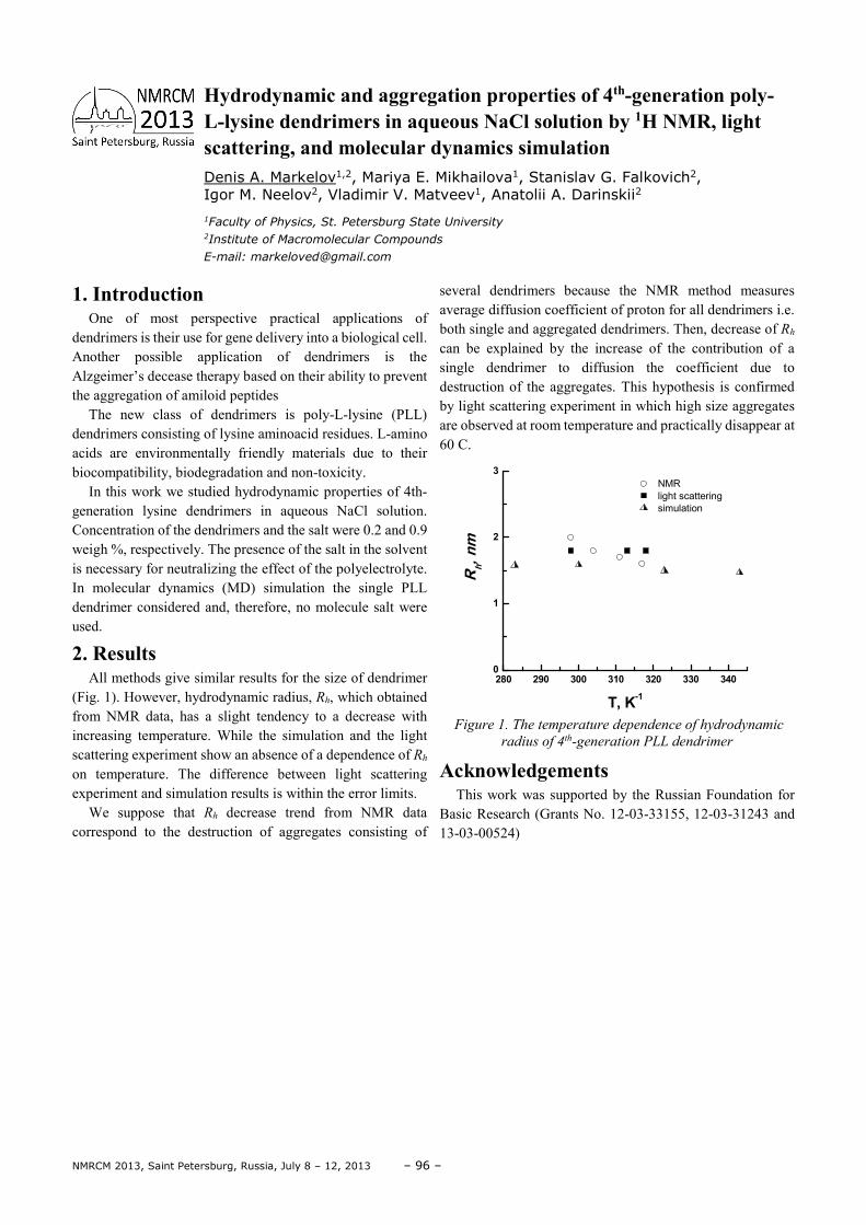
NMRCM 2013, Saint Petersburg, Russia, July 8 – 12, 2013 – 96 –
1. Introduction One of most perspective practical applications of
dendrimers is their use for gene delivery into a biological cell. Another possible application of dendrimers is the Alzgeimer’s decease therapy based on their ability to prevent the aggregation of amiloid peptides
The new class of dendrimers is poly-L-lysine (PLL) dendrimers consisting of lysine aminoacid residues. L-amino acids are environmentally friendly materials due to their biocompatibility, biodegradation and non-toxicity.
In this work we studied hydrodynamic properties of 4th-generation lysine dendrimers in aqueous NaCl solution. Concentration of the dendrimers and the salt were 0.2 and 0.9 weigh %, respectively. The presence of the salt in the solvent is necessary for neutralizing the effect of the polyelectrolyte. In molecular dynamics (MD) simulation the single PLL dendrimer considered and, therefore, no molecule salt were used.
2. Results All methods give similar results for the size of dendrimer
(Fig. 1). However, hydrodynamic radius, Rh, which obtained from NMR data, has a slight tendency to a decrease with increasing temperature. While the simulation and the light scattering experiment show an absence of a dependence of Rh on temperature. The difference between light scattering experiment and simulation results is within the error limits.
We suppose that Rh decrease trend from NMR data correspond to the destruction of aggregates consisting of
several dendrimers because the NMR method measures average diffusion coefficient of proton for all dendrimers i.e. both single and aggregated dendrimers. Then, decrease of Rh can be explained by the increase of the contribution of a single dendrimer to diffusion the coefficient due to destruction of the aggregates. This hypothesis is confirmed by light scattering experiment in which high size aggregates are observed at room temperature and practically disappear at 60 C.
280 290 300 310 320 330 3400
1
2
3
NMR
light scattering
simulation
Rh, n
m
T, K-1
Figure 1. The temperature dependence of hydrodynamic
radius of 4th-generation PLL dendrimer
Acknowledgements This work was supported by the Russian Foundation for
Basic Research (Grants No. 12-03-33155, 12-03-31243 and 13-03-00524)
Hydrodynamic and aggregation properties of 4th-generation poly-L-lysine dendrimers in aqueous NaCl solution by 1H NMR, light scattering, and molecular dynamics simulation Denis A. Markelov1,2, Mariya E. Mikhailova1, Stanislav G. Falkovich2, Igor M. Neelov2, Vladimir V. Matveev1, Anatolii A. Darinskii2
1Faculty of Physics, St. Petersburg State University 2Institute of Macromolecular Compounds
E-mail: [email protected]
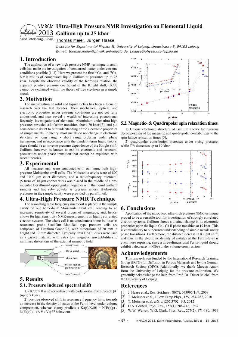
– 97 – NMRCM 2013, Saint Petersburg, Russia, July 8 – 12, 2013
1. Introduction The application of a new high pressure NMR technique in anvil
cells has made the investigation of condensed matter under extreme conditions possible [1, 2]. Here we present the first 69Ga- and 71Ga- NMR results of compressed liquid Gallium at pressures up to 25 kbar. Despite the observed validity of the Korringa relation, the apparent positive pressure coefficient of the Knight shift, ∂K/∂p cannot be explained within the theory of free electrons in a simple metal.
2. Motivation The investigation of solid and liquid metals has been a focus of
research over the last decades. Their mechanical, optical, and electronic properties under extreme conditions are not yet fully understood, and may reveal a wealth of interesting phenomena. Recently, investigations of elemental Aluminium under ultra-high pressures revealed a Lifschitz transition above 70 kbar [3], and put considerable doubt to our understanding of the electronic properties of simple metals. In theory, most metals do not change in electronic structure or long range - short range ordering under phase transisiton, and in accordance with the Landau-Fermi liquid theory, there should be an inverse pressure dependence of the Knight shift. Gallium, however, is known to exhibit electronic and structural pecularities under phase transition that cannot be explained with recent theories.
3. Experimental All measurements were conducted with our home-built high-
pressure Moissanite anvil cells. The Moissanite anvils were of 800 and 1000 µm culet diameters, and a radiofrequency microcoil (5 turns of 18 µm copper wire) was placed in the middle of a pre-indented Beryllium-Copper gasket, together with the liquid Gallium samples and fine ruby powder as pressure senors. Hydrostatic pressures in the sample cavity were provided by paraffin oil.
4. Ultra-High Pressure NMR Technique The resonating radio frequency microcoil is placed in the sample
cavity of our home-built Moissanite anvil cell, leading to an increased sensitivity of several orders of magnitude, and, hence, allows for high sensitivity NMR-measurements on highly correlated electron systems. The whole cell is mounted onto a home-built series resonance probe head.Our Mao-Bell type pressure cells are composed of Titanium Grade 23, with dimensions of 20 mm in height and 17 mm diameter. Typically, thin Be-Cu disks were used as a gasket material, with extra low magnetic susceptibilities to minimise distortions of the external magnetic field.
5. Results 5.1. Pressure induced spectral shift
1) ∂K/∂p > 0 is in accordance with early works from Cornell [4] (up to 5 kbar);
2) positive observed shift in resonance frequency hints towards an increase in the density of states at the Fermi level under volume compression, whereas theory predicts a Ks(p)/Ks(0) ~ N(EF)(p) / N(EF)(0) ~ (∆ V / V0)-3/2 behaviour.
5.2. Magnetic- & Quadrupolar spin relaxation times
1) Unique electronic structure of Gallium allows for rigorous decomposition of the magnetic and quadrupolar contributions to the spin-lattice relaxation times [5];
2) quadrupolar contribution increases under rising pressure, while Tm1 decreases up to 19 kbar.
6. Conclusions
Application of the introduced ultra-high pressure NMR technique proved to be a versatile tool for investigation of strongly correlated electron systems. Gallium shows a distinct change in its electronic properties upon the liquid Ga - Ga II phase transition at 19 kbar. This is contradictory to our current understanding of simple metals under phase transitions. Furthermore, the distinct increase in Knight shift, and thus in the electronic density of s-states at the Fermi-level is even more suprising, since a three-dimensional Fermi-liquid should exhibit a decrease in N(EF) under volume compression.
Acknowledgements This research was funded by the International Research Training
Group (IRTG) for Diffusion in Porous Materials and by the German Research Society (DFG). Additionally, we thank Marcus Anton from the University of Leipzig for the pressure calibration. We gratefully acknowledge the help from Prof. Dr. Dieter Michel from the University of Leipzig.
References
[1] J. Haase et.al., Rev. Sci.Instr., 80(7), 073905/1-/4, 2009 [2] T. Meissner et.al., J.Low.Temp.Phys., 159, 284-287, 2010 [3] T. Meissner et.al, arXiv:1207.3702, 1-5, 2012 [4] D.A. Cornell, Phys. Rev., 153(1), 208-216, 1967 [5] W.W. Warren, W.G. Clark, Phys. Rev., 277(2), 171-180, 1969
Ultra-High Pressure NMR Investigation on Elemental Liquid Gallium up to 25 kbar
Thomas Meier, Jürgen Haase
Institute for Experimental Physics II, University of Leipzig, Linnestrasse 5, 04103 Leipzig
E-mail: [email protected], [email protected]
NMRCM 2013, Saint Petersburg, Russia, July 8 – 12, 2013 – 98 –
1. Introduction Composite pulse basically is a sequence of radiofrequency
pulses with varying duration and phase. Composite pulses has been widely used in NMR as a tool for elimination of dipole-dipole interactions, to increase signal-to-noise ratio and to reduce errors in quantum computing computation. In NQR, composite pulses can increase SNR [1], radiofrequency field homogeneity and effective volume of a detection coil for single-sided acquisition. It can be useful for NQR remote sensing applications. Our approach to composite pulses design based on use of NQR fictious spin operators for I=1 and product operator formalism.
2. Theory In NMR, theory of composite pulses design is often
straightforward. A possible way to create a composite pulce sequence is to use a geometrical vector approach or perturbation theory. However, simple vector model has no
physical sense in NQR. Instead, it is possible to divide NQR density matrix to the set of independent orthogonal subspaces, corresponding to the NQR energy levels. This fictional spin-1/2 operators [2] may be presented as individual vector models for every NQR frequency. In single-frequency experiment it is possible to implement a vector model for single subspace and trace an evolution of magnetization vector, emerging under the radiofrequency field. The goal is to calculate a trajectory of magnetization vector which will lead to minimal variance. It is impossible in NQR to excite every crystallite in powder sample, so the calculations suggest directional averaging.
References [1] Ramamoorthy A., Chandracumar N., Dubey A.K.,
Narasimhan P.T. Analysis or the performance of NQR composite pulses // Journal of Magnetic Resonance, 1993. Series A102, P. 274-286
[2] Vega S., Pines A.: J. Chem. Phys. 66, 5624-5644 (1977)
Broadband composite pulses for Spin=1NQR in powders Ivan Mershiev, Galina Kupriyanova
Faculty of physics and technology, Immanuel Kant Baltic federal university
E-mail: [email protected]
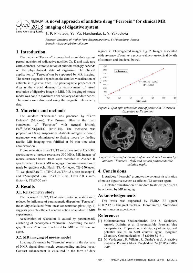
– 99 – NMRCM 2013, Saint Petersburg, Russia, July 8 – 12, 2013
1. Introduction The medicine “Ferrocin” is prescribed as antidote against
peroral nutrition of radioactive nuclides Cs, K and toxic rare earth elements. Antitoxic action of antidote strongly depends on the physiological state of organism. The clinical application of “Ferrocin”can be supported by MR imaging. The robust diagnosis depends on the detailed visualization of antidote in digestive tract. The paramagnetic properties of drug is the crucial demand for enhancement of visual resolution of digestive image in MRI. MR imaging of mouse model was done in dynamics after delivery of antidote per os. The results were discussed using the magnetic relaxometry data.
2. Materials and methods The antidote “Ferrocine” was produced by “Farm
Defence” (Moscow). The Prussian Blue is the main component of “Ferrocine” with general formula Fe4
ΙΙΙ[FeΙΙ(CN)6]3nH2O (n=14-16). The medicine was prepared as 1% aq. suspensions. Antidote intragastric dose 6 mg/mouse was administered to fasting mouse by feeding needle. MR imaging was fulfilled at 30 min time after administration.
Proton relaxation times T1, T2 were measured at CXP-300 spectrometer at proton resonance 300 MHz. MR images of mouse stomach-bowel tract were recorded at Avanch ΙΙ spectrometer (Bruker). MR imagings of mouse stomach were made by gradient echo Flash (TR=5.4 мсек, TE=350 мсек, T1-weighted Rare T1 ( TE=7.5 мs, TR=1.5 s, rare-фактор=4) and Т2-weighted Rare T2 (TE=12 мs. TR=4.200 s, rare-factor=8, TEeff=36 мs).
3. Results 3.1. Relaxometry study
The measured T1, T2, T2 of water proton relaxation were reduced by influence of paramagnetic dispersion “Ferrocin”. Relaxivity calculated from linear concentration plots (Fig. 1) suggests possible efficient contrast action of antidote in MRI experiments.
Acceleration of relaxation is caused by paramagnetic clustering of nanocrystals “Ferrocin”. According the ratio r2/r1 “Ferrocin” is more preferred for MRI as T2 contrast agent.
3.2. MR imaging of mouse model Loading of stomach by “Ferrocin” results in the decrease
of NMR signal from voxels corresponding antidote locus. Contrast enhancement is visualized in the form of dark
regions in T1-weigheted images Fig. 2. Images associated with presence of contrast agent reveal new anatomical details of stomach and duodenal bowel.
Figure 1. Spin-spin relaxation rate of protons in “Ferrocin”
dispersion vs Fe content
Figure 2. T1-weighted images of mouse stomach loaded by
antidote “Ferrocin” (left) and control polysaccharide
solution (right)
4. Conclusions 1. Antidote “Ferrocin” promotes the contrast visualisation
of mouse digestive system as efficient T2 contrast agent. 2. Detailed visualization of antidote treatment per os can
be achieved by MR imaging.
Acknowledgements This work was supported by FMBA RF (grant
40.002.12.0). Our great thanks A. Dobrodumov, I. Voevodina for assistance in experiments.
References [1] Mohammadreza Shokouhimehr, Eric S. Soehnlen,
Anatoly Khitrin et al. Biocompatible Prussian blue nanoparticles: Preparation, stability, cytotoxicity, and potential use as an MRI contrast agent. Inorganic Chemistry Communications 13 (2010) 58–61.
[2] M. Verdaguer , F. Villain , R. Ouahe`s et al. Attractive magnetic Prussian blues. Polyhedron 24 (2005) 2906–2908.
y = 3,5633x + 0,6329
R2 = 0,9996
0,00
5,00
10,00
15,00
20,00
25,00
30,00
35,00
40,00
0,0 2,0 4,0 6,0 8,0 10,0 12,0
C(Fe), мМ/лR
2, 1
/c
Ферроцин
A novel approach of antidote drug “Ferrocin” for clinical MR imaging of digestive system B. P. Nikolaev, Ya. Yu. Marchenko, L. Y. Yakovleva
Reseach Institute of Highly Pure Biopreparations, St.Petersburg, Russia
E-mail: [email protected]
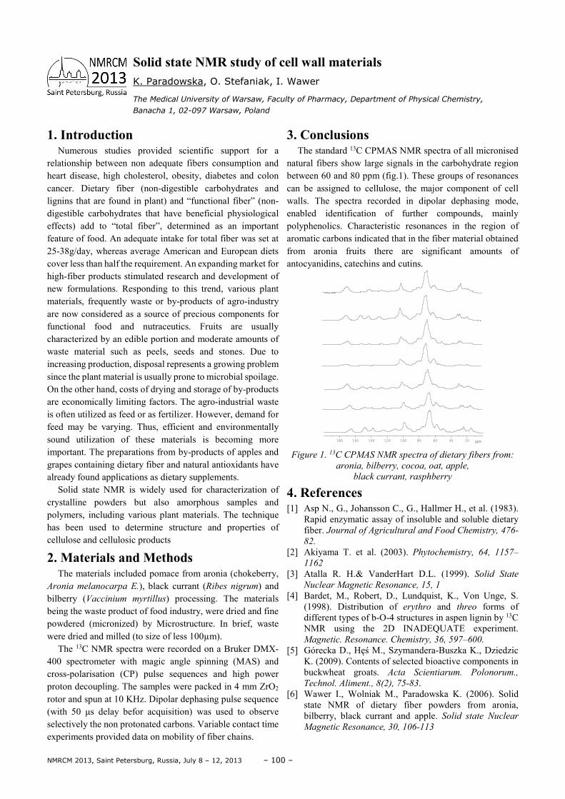
NMRCM 2013, Saint Petersburg, Russia, July 8 – 12, 2013 – 100 –
1. Introduction Numerous studies provided scientific support for a
relationship between non adequate fibers consumption and heart disease, high cholesterol, obesity, diabetes and colon cancer. Dietary fiber (non-digestible carbohydrates and lignins that are found in plant) and “functional fiber” (non-digestible carbohydrates that have beneficial physiological effects) add to “total fiber”, determined as an important feature of food. An adequate intake for total fiber was set at 25-38g/day, whereas average American and European diets cover less than half the requirement. An expanding market for high-fiber products stimulated research and development of new formulations. Responding to this trend, various plant materials, frequently waste or by-products of agro-industry are now considered as a source of precious components for functional food and nutraceutics. Fruits are usually characterized by an edible portion and moderate amounts of waste material such as peels, seeds and stones. Due to increasing production, disposal represents a growing problem since the plant material is usually prone to microbial spoilage. On the other hand, costs of drying and storage of by-products are economically limiting factors. The agro-industrial waste is often utilized as feed or as fertilizer. However, demand for feed may be varying. Thus, efficient and environmentally sound utilization of these materials is becoming more important. The preparations from by-products of apples and grapes containing dietary fiber and natural antioxidants have already found applications as dietary supplements.
Solid state NMR is widely used for characterization of crystalline powders but also amorphous samples and polymers, including various plant materials. The technique has been used to determine structure and properties of cellulose and cellulosic products
2. Materials and Methods The materials included pomace from aronia (chokeberry,
Aronia melanocarpa E.), black currant (Ribes nigrum) and bilberry (Vaccinium myrtillus) processing. The materials being the waste product of food industry, were dried and fine powdered (micronized) by Microstructure. In brief, waste were dried and milled (to size of less 100µm).
The 13C NMR spectra were recorded on a Bruker DMX-400 spectrometer with magic angle spinning (MAS) and cross-polarisation (CP) pulse sequences and high power proton decoupling. The samples were packed in 4 mm ZrO2 rotor and spun at 10 KHz. Dipolar dephasing pulse sequence (with 50 µs delay befor acquisition) was used to observe selectively the non protonated carbons. Variable contact time experiments provided data on mobility of fiber chains.
3. Conclusions The standard 13C CPMAS NMR spectra of all micronised
natural fibers show large signals in the carbohydrate region between 60 and 80 ppm (fig.1). These groups of resonances can be assigned to cellulose, the major component of cell walls. The spectra recorded in dipolar dephasing mode, enabled identification of further compounds, mainly polyphenolics. Characteristic resonances in the region of aromatic carbons indicated that in the fiber material obtained from aronia fruits there are significant amounts of antocyanidins, catechins and cutins.
Figure 1. 13C CPMAS NMR spectra of dietary fibers from:
aronia, bilberry, cocoa, oat, apple,
black currant, rasphberry
4. References [1] Asp N., G., Johansson C., G., Hallmer H., et al. (1983).
Rapid enzymatic assay of insoluble and soluble dietary fiber. Journal of Agricultural and Food Chemistry, 476-
82.
[2] Akiyama T. et al. (2003). Phytochemistry, 64, 1157–
1162
[3] Atalla R. H.& VanderHart D.L. (1999). Solid State
Nuclear Magnetic Resonance, 15, 1
[4] Bardet, M., Robert, D., Lundquist, K., Von Unge, S. (1998). Distribution of erythro and threo forms of different types of b-O-4 structures in aspen lignin by 13C NMR using the 2D INADEQUATE experiment. Magnetic. Resonance. Chemistry, 36, 597–600.
[5] Górecka D., Hęś M., Szymandera-Buszka K., Dziedzic K. (2009). Contents of selected bioactive components in buckwheat groats. Acta Scientiarum. Polonorum.,
Technol. Aliment., 8(2), 75-83. [6] Wawer I., Wolniak M., Paradowska K. (2006). Solid
state NMR of dietary fiber powders from aronia, bilberry, black currant and apple. Solid state Nuclear
Magnetic Resonance, 30, 106-113
180 160 140 120 100 80 60 40 20 ppm
Solid state NMR study of cell wall materials K. Paradowska, O. Stefaniak, I. Wawer
The Medical University of Warsaw, Faculty of Pharmacy, Department of Physical Chemistry,
Banacha 1, 02-097 Warsaw, Poland
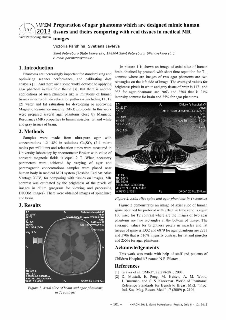
– 101 – NMRCM 2013, Saint Petersburg, Russia, July 8 – 12, 2013
1. Introduction Phantoms are increasingly important for standardizing and
optimizing scanner performance, and calibrating data analysis [1]. And there are a some works devoted to applying agar phantom in this field theme [3]. But there is another applications of such phantoms like a imitations of human tissues in terms of their relaxation pathways, including T1, T2 [2] water and fat saturation for developing or approving Magnetic Resonance imaging (MRI) protocols. In this work were prepared several agar phantoms close by Magnetic Resonance (MR) properties to human muscles, fat and white and gray tissues of brain.
2. Methods Samples were made from ultra-pure agar with
concentrations 1.2-1.8% in solutions Cu2SO4 (2-4 micro moles per milliliter) and relaxation times were measured in University laboratory by spectrometer Bruker with value of constant magnetic fields is equal 2 T. When necessary parameters were achieved by varying of agar and paramagnetic concentrations samples were placed near human body in medical MRI system (Toshiba ExelArt Atlas Vantage XGV) for comparing with tissues on images. MR contrast was estimated by the brightness of the pixels of images in eFilm (program for viewing and processing DICOM images). There were obtained images of spine,knee and brain.
3. Results
Figure 1. Axial slice of brain and agar phantoms
in T1 contrast
In picture 1 is shown an image of axial slice of human brain obtained by protocol with short time repetition for T1 , contrast where are images of two agar phantoms are two rectangles on the left side of image. The averaged values for brightness pixels in white and gray tissue of brain is 1171 and 938 for agar phantoms are 2063 and 2504 that is 21% intensity contrast for brain and 25% for agar phantoms.
Figure 2. Axial slice spine and agar phantoms in T2 contrast
Figure 2 demonstrates an image of axial slice of human spine obtained by protocol with effective time echo is equal 100 msec for T2 contrast where are the images of two agar phantoms are two rectangles at the bottom of image. The averaged values for brightness pixels in muscles and fat tissues of spine is 1332 and 6879 for agar phantoms are 2233 and 5706 that is 516% intensity contrast for fat and muscles and 255% for agar phantoms.
Acknowledgements This work was made with help of staff and patients of
Children Hospital N5 named N.F. Filatov.
References [1] Graves et al. “JMRI”, 28:278-281, 2008. [2] D. Mustafi, E. Peng, M. Heisen, A. M. Wood,
J. Buurman, and G. S. Karczmar. World of Phantoms: Reference Standards for Bench to Breast MRI. “Proc. Intl. Soc. Mag. Reson. Med.” 17 (2009) p. 2104.
Preparation of agar phantoms which are designed mimic human tissues and theirs comparing with real tissues in medical MR images Victoria Parshina, Svetlana Ievleva
Saint Petersburg State University, 198504 Saint Petersburg, Ulianovskaya st. 1
E-mail: [email protected]
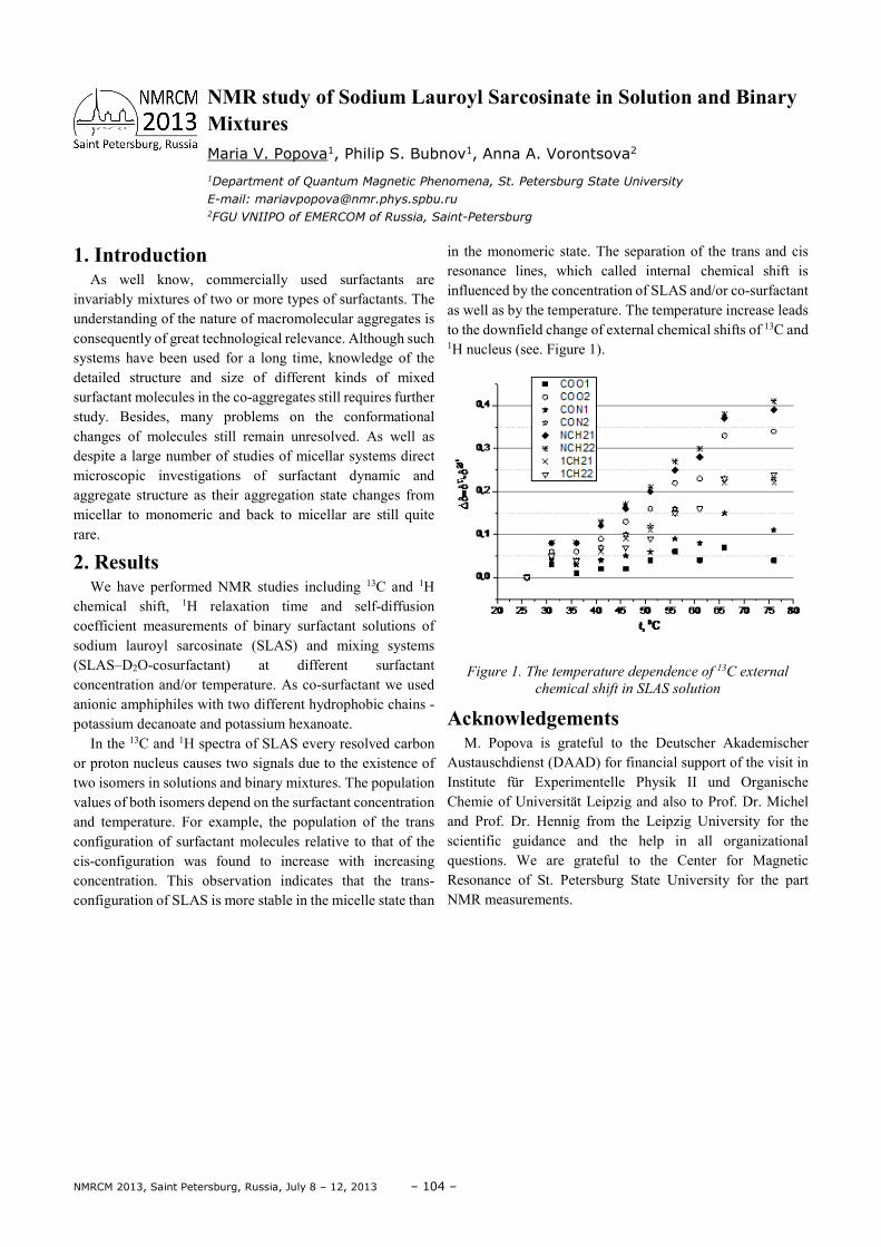
NMRCM 2013, Saint Petersburg, Russia, July 8 – 12, 2013 – 104 –
1. Introduction As well know, commercially used surfactants are
invariably mixtures of two or more types of surfactants. The understanding of the nature of macromolecular aggregates is consequently of great technological relevance. Although such systems have been used for a long time, knowledge of the detailed structure and size of different kinds of mixed surfactant molecules in the co-aggregates still requires further study. Besides, many problems on the conformational changes of molecules still remain unresolved. As well as despite a large number of studies of micellar systems direct microscopic investigations of surfactant dynamic and aggregate structure as their aggregation state changes from micellar to monomeric and back to micellar are still quite rare.
2. Results We have performed NMR studies including 13C and 1H
chemical shift, 1H relaxation time and self-diffusion coefficient measurements of binary surfactant solutions of sodium lauroyl sarcosinate (SLAS) and mixing systems (SLAS–D2O-cosurfactant) at different surfactant concentration and/or temperature. As co-surfactant we used anionic amphiphiles with two different hydrophobic chains - potassium decanoate and potassium hexanoate.
In the 13C and 1H spectra of SLAS every resolved carbon or proton nucleus causes two signals due to the existence of two isomers in solutions and binary mixtures. The population values of both isomers depend on the surfactant concentration and temperature. For example, the population of the trans configuration of surfactant molecules relative to that of the cis-configuration was found to increase with increasing concentration. This observation indicates that the trans-configuration of SLAS is more stable in the micelle state than
in the monomeric state. The separation of the trans and cis resonance lines, which called internal chemical shift is influenced by the concentration of SLAS and/or co-surfactant as well as by the temperature. The temperature increase leads to the downfield change of external chemical shifts of 13C and 1H nucleus (see. Figure 1).
Figure 1. The temperature dependence of 13C external
chemical shift in SLAS solution
Acknowledgements M. Popova is grateful to the Deutscher Akademischer
Austauschdienst (DAAD) for financial support of the visit in Institute für Experimentelle Physik II und Organische Chemie of Universität Leipzig and also to Prof. Dr. Michel and Prof. Dr. Hennig from the Leipzig University for the scientific guidance and the help in all organizational questions. We are grateful to the Center for Magnetic Resonance of St. Petersburg State University for the part NMR measurements.
NMR study of Sodium Lauroyl Sarcosinate in Solution and Binary Mixtures
Maria V. Popova1, Philip S. Bubnov1, Anna A. Vorontsova2
1Department of Quantum Magnetic Phenomena, St. Petersburg State University
E-mail: [email protected] 2FGU VNIIPO of EMERCOM of Russia, Saint-Petersburg
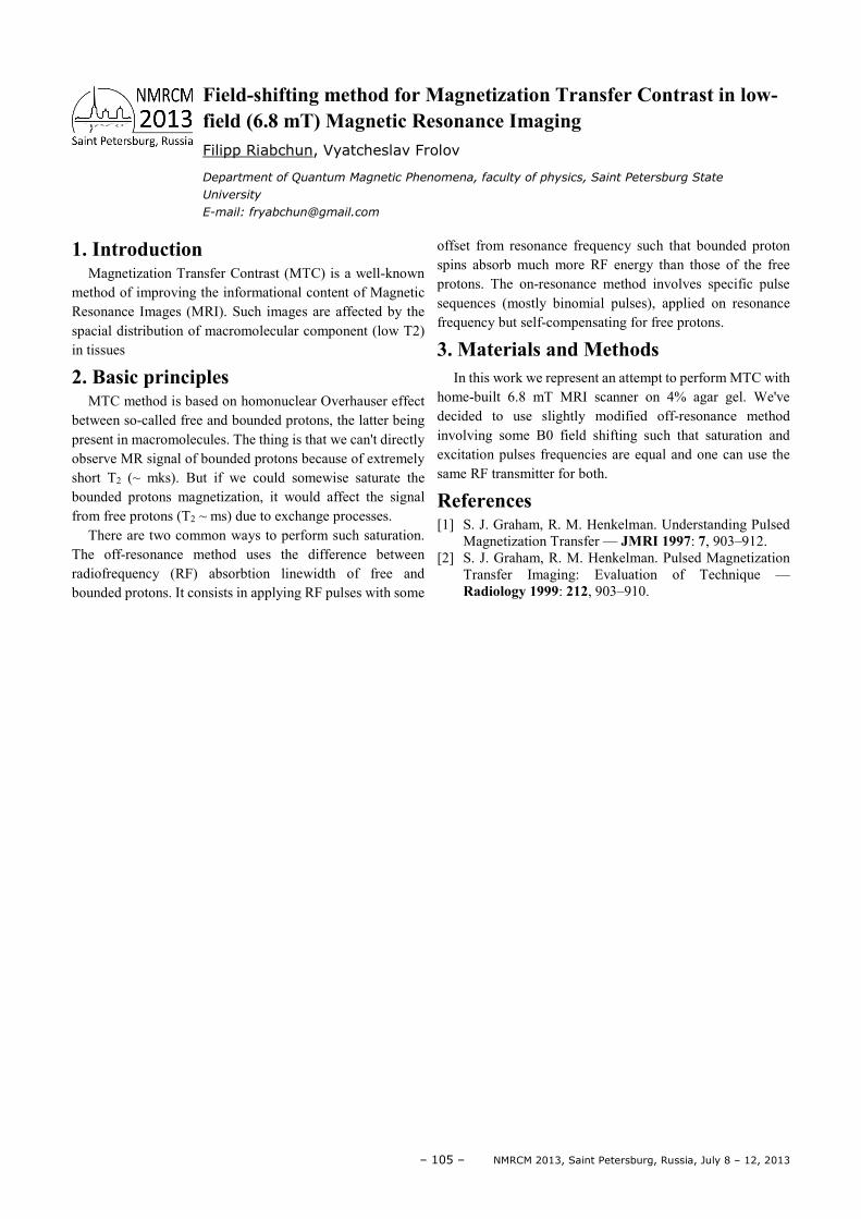
– 105 – NMRCM 2013, Saint Petersburg, Russia, July 8 – 12, 2013
1. Introduction Magnetization Transfer Contrast (MTC) is a well-known
method of improving the informational content of Magnetic Resonance Images (MRI). Such images are affected by the spacial distribution of macromolecular component (low T2) in tissues
2. Basic principles MTC method is based on homonuclear Overhauser effect
between so-called free and bounded protons, the latter being present in macromolecules. The thing is that we can't directly observe MR signal of bounded protons because of extremely short T2 (~ mks). But if we could somewise saturate the bounded protons magnetization, it would affect the signal from free protons (T2 ~ ms) due to exchange processes.
There are two common ways to perform such saturation. The off-resonance method uses the difference between radiofrequency (RF) absorbtion linewidth of free and bounded protons. It consists in applying RF pulses with some
offset from resonance frequency such that bounded proton spins absorb much more RF energy than those of the free protons. The on-resonance method involves specific pulse sequences (mostly binomial pulses), applied on resonance frequency but self-compensating for free protons.
3. Materials and Methods In this work we represent an attempt to perform MTC with
home-built 6.8 mT MRI scanner on 4% agar gel. We've decided to use slightly modified off-resonance method involving some B0 field shifting such that saturation and excitation pulses frequencies are equal and one can use the same RF transmitter for both.
References [1] S. J. Graham, R. M. Henkelman. Understanding Pulsed
Magnetization Transfer — JMRI 1997: 7, 903–912. [2] S. J. Graham, R. M. Henkelman. Pulsed Magnetization
Transfer Imaging: Evaluation of Technique — Radiology 1999: 212, 903–910.
Field-shifting method for Magnetization Transfer Contrast in low-field (6.8 mT) Magnetic Resonance Imaging Filipp Riabchun, Vyatcheslav Frolov
Department of Quantum Magnetic Phenomena, faculty of physics, Saint Petersburg State
University
E-mail: [email protected]
NMRCM 2013, Saint Petersburg, Russia, July 8 – 12, 2013 – 106 –
One of the basic experiment is to obtain a proton NMR spectrum, which allows you to make some assumptions about the chemical composition of the substance.
Modern NMR spectrometry to determine not only the chemical composition, but also the spatial structure of the substance. Such an opportunity came through the use of two-dimensional experiments.
If the chemical structure is known, it is possible to carry out the assignment of the corresponding resonance lines. If the structure is unknown, the structure can be selected from several alternatives. [2] In general, you should answer the question: "Which of the nuclear spins are linked together?" In principle it is possible to get an answer to this question is simple enough for even the structures using one-dimensional methods, such as using isolation or construction of theoretical spectra. However, in general there are more complicated cases when the spectra contain overlapping lines, these methods lead to success only if a large number of labor-intensive and time-consuming experiments. With this method of two-dimensional information can be obtained from a single experiment [3].
The best-known examples of these experiments are COSY and NOESY.
The sample used in this paper the azo-dye, acid-base indikator.CI22120. C.I.Direct red 28 - Congo red.
For identification of the NMR signals of organic dyes we conducted three types of experiments:
1. Removal of the proton spectrum (PROTON); 2. COSY; 3. NOESY. We got the exact values of the chemical shifts of hydrogen
and spent the correlation signal for the dye Congo red by proton spectrum and the experiment COSY.
Also we received correlation maps NOESY, obtained with different mixing time 100, 300 and 500 ms. It was concluded that the growth of the correlation value τm improved card,
namely the intensity of the cross peaks, all the peaks are more soluble and decreases the noise contribution. Were compared with theoretical and practical information for small molecules with large rotational correlation time.
For small values of the mixing interval τm for the intensity of cross-peak between the lines that match the resonance of spins i and j, we have the formula
6~ij ij mrσ τ− , (1)
where σij - intensity cross-peak, and rij - the corresponding distance between the nuclei.
Resulting in a practical way the intensity of the peaks on the mixing time suggests coordination between theory and practice, however, it is worth noting that the construction of further dependence is difficult because of the spin diffusion, which is evident when τm > 500 ms and degrades the testimony of the experiment. Thus, based on the findings of the outcome of the experiment depends on the mixing time, we can choose an optimal value for a sample of Congo red τm = 500 ms. Then, using the formula
060ij
ij
r rσσ
= , (2)
where σ0 and r0 - the calibration values, we have obtained the corresponding relative distance of the observed cross-peaks of the sample congo red, due to the experimental NOESY.
References [1] A. Bax, "Two-Dimensional Nuclear Magnetic
Resonance in fluid", 1988, 160 [2] H. Gunther, "Introduction to the course NMR
Spectroscopy", 1984, 142-207 [3] A. Deroum, "Modern NMR techniques for chemistry
research," 1992. 145-188, 266-348 [4] R. Ernst, G. Bodenhausen, A. Vokaun, "NMR
spectroscopy in one and two dimensions", 1990, 342-353 [5] S. Braun, H.-O. Kalinowski, S. Berger, "150 and more
basic NMR experiments", 1998, 79-82, 92-93
The use of two-dimensional COSY and NOESY methods for the identification of the NMR signals of organic dyes S. A. Ruzheva, A. V. Bogaychuk
Physico-Technical Institute, Department of Radio Physics and Information Security, Immanuel
Kant Baltic Federal Univnrsitet
E-mail: [email protected]
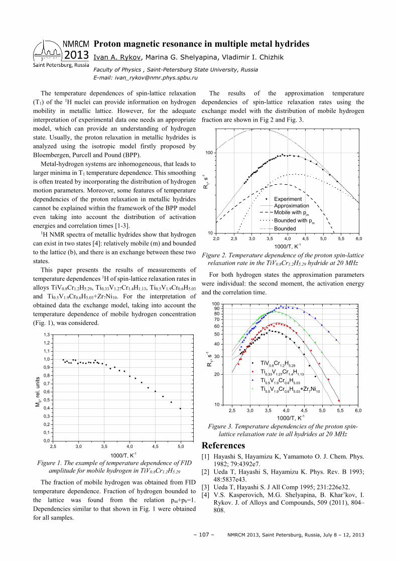
– 107 – NMRCM 2013, Saint Petersburg, Russia, July 8 – 12, 2013
The temperature dependences of spin-lattice relaxation (T1) of the 1H nuclei can provide information on hydrogen mobility in metallic lattice. However, for the adequate interpretation of experimental data one needs an appropriate model, which can provide an understanding of hydrogen state. Usually, the proton relaxation in metallic hydrides is analyzed using the isotropic model firstly proposed by Bloembergen, Purcell and Pound (BPP).
Metal-hydrogen systems are inhomogeneous, that leads to larger minima in T1 temperature dependence. This smoothing is often treated by incorporating the distribution of hydrogen motion parameters. Moreover, some features of temperature dependencies of the proton relaxation in metallic hydrides cannot be explained within the framework of the BPP model even taking into account the distribution of activation energies and correlation times [1-3].
1H NMR spectra of metallic hydrides show that hydrogen can exist in two states [4]: relatively mobile (m) and bounded to the lattice (b), and there is an exchange between these two states.
This paper presents the results of measurements of temperature dependences 1H of spin-lattice relaxation rates in alloys TiV0.8Cr1.2H5.29, Ti0.33V1.27Cr1.4H1.13, Ti0.5V1.9Cr0.6H5.03 and Ti0.5V1.9Cr0.6H5.03+Zr7Ni10. For the interpretation of obtained data the exchange model, taking into account the temperature dependence of mobile hydrogen concentration (Fig. 1), was considered.
2,5 3,0 3,5 4,0 4,5 5,0
0,0
0,1
0,2
0,3
0,4
0,5
0,6
0,7
0,8
0,9
1,0
1,1
1,2
1,3
M0,
rel. u
nits
1000/T, K-1
Figure 1. The example of temperature dependence of FID
amplitude for mobile hydrogen in TiV0.8Cr1.2H5.29
The fraction of mobile hydrogen was obtained from FID temperature dependence. Fraction of hydrogen bounded to the lattice was found from the relation pm+pb=1. Dependencies similar to that shown in Fig. 1 were obtained for all samples.
The results of the approximation temperature dependencies of spin-lattice relaxation rates using the exchange model with the distribution of mobile hydrogen fraction are shown in Fig 2 and Fig. 3.
2,0 2,5 3,0 3,5 4,0 4,5 5,0 5,5 6,0
10
100
Experiment
Approximation
Mobile with pm
Bounded with pm
Bounded
R1, s
-1
1000/T, K-1
Figure 2. Temperature dependence of the proton spin-lattice
relaxation rate in the TiV0.8Cr1.2H5.29 hydride at 20 MHz
For both hydrogen states the approximation parameters were individual: the second moment, the activation energy and the correlation time.
2,5 3,0 3,5 4,0 4,5 5,0 5,5 6,0
10
20
30
40
50
60
70
8090
100
R1,
s-1
1000/T, K-1
TiV0.8
Cr1.2
H5.29
Ti0.33
V1.27
Cr1.4
H1.13
Ti0.5
V1.9
Cr0.6
H5.03
Ti0.5
V1.9
Cr0.6
H5.03
+Zr7Ni
10
Figure 3. Temperature dependencies of the proton spin-
lattice relaxation rate in all hydrides at 20 MHz
References [1] Hayashi S, Hayamizu K, Yamamoto O. J. Chem. Phys.
1982; 79:4392e7. [2] Ueda T, Hayashi S, Hayamizu K. Phys. Rev. B 1993;
48:5837e43. [3] Ueda T, Hayashi S. J All Comp 1995; 231:226e32. [4] V.S. Kasperovich, M.G. Shelyapina, B. Khar’kov, I.
Rykov. J. of Alloys and Compounds, 509 (2011), 804– 808.
Proton magnetic resonance in multiple metal hydrides Ivan A. Rykov, Marina G. Shelyapina, Vladimir I. Chizhik
Faculty of Physics , Saint-Petersburg State University, Russia
E-mail: [email protected]
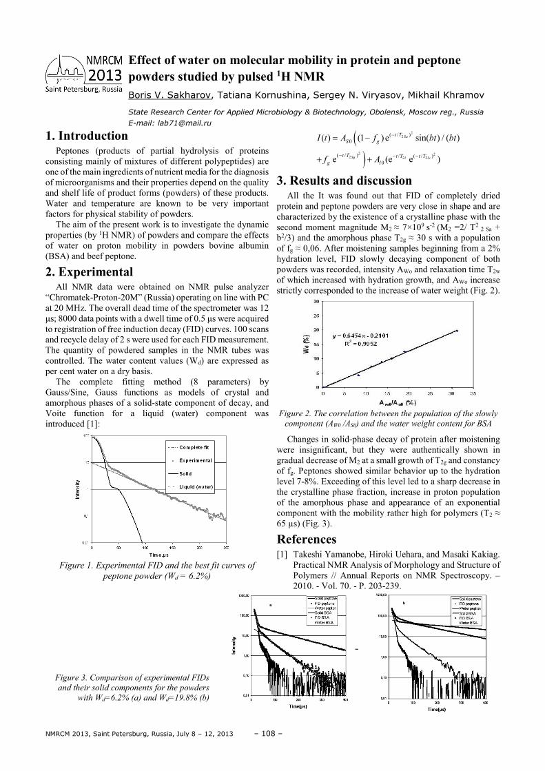
NMRCM 2013, Saint Petersburg, Russia, July 8 – 12, 2013 – 108 –
1. Introduction Peptones (products of partial hydrolysis of proteins
consisting mainly of mixtures of different polypeptides) are one of the main ingredients of nutrient media for the diagnosis of microorganisms and their properties depend on the quality and shelf life of product forms (powders) of these products. Water and temperature are known to be very important factors for physical stability of powders.
The aim of the present work is to investigate the dynamic properties (by 1H NMR) of powders and compare the effects of water on proton mobility in powders bovine albumin (BSA) and beef peptone.
2. Experimental All NMR data were obtained on NMR pulse analyzer
“Chromatek-Proton-20M” (Russia) operating on line with PC at 20 MHz. The overall dead time of the spectrometer was 12 µs; 8000 data points with a dwell time of 0.5 µs were acquired to registration of free induction decay (FID) curves. 100 scans and recycle delay of 2 s were used for each FID measurement. The quantity of powdered samples in the NMR tubes was controlled. The water content values (Wd) are expressed as per cent water on a dry basis.
The complete fitting method (8 parameters) by Gauss/Sine, Gauss functions as models of crystal and amorphous phases of a solid-state component of decay, and Voite function for a liquid (water) component was introduced [1]:
Figure 1. Experimental FID and the best fit curves of
peptone powder (Wd = 6.2%)
()
22
2 22 2 2
( / )0
( / ) / ( / )0
( ) (1 )e sin( ) / ( )
e (e e )
Sa
Sg l lv
t T
S g
t T t T t T
g l
I t A f bt bt
f A
−
− − −
= −
+ +
3. Results and discussion All the It was found out that FID of completely dried
protein and peptone powders are very close in shape and are characterized by the existence of a crystalline phase with the second moment magnitude М2 ≈ 7×10⁹ s-2 (М2 =2/ T2
2 Sa + b2/3) and the amorphous phase T2g ≈ 30 s with a population of fg ≈ 0,06. After moistening samples beginning from a 2% hydration level, FID slowly decaying component of both powders was recorded, intensity AWo and relaxation time T2w of which increased with hydration growth, and AWo increase strictly corresponded to the increase of water weight (Fig. 2).
Figure 2. The correlation between the population of the slowly
component (AW0 /AS0) and the water weight content for BSA
Changes in solid-phase decay of protein after moistening were insignificant, but they were authentically shown in gradual decrease of М2 at a small growth of T2g and constancy of fg. Peptones showed similar behavior up to the hydration level 7-8%. Exceeding of this level led to a sharp decrease in the crystalline phase fraction, increase in proton population of the amorphous phase and appearance of an exponential component with the mobility rather high for polymers (T2 ≈ 65 µs) (Fig. 3).
References [1] Takeshi Yamanobe, Hiroki Uehara, and Masaki Kakiag.
Practical NMR Analysis of Morphology and Structure of Polymers // Annual Reports on NMR Spectroscopy. – 2010. - Vol. 70. - P. 203-239.
Figure 3. Comparison of experimental FIDs
and their solid components for the powders
with Wd=6.2% (a) and Wd=19.8% (b)
Effect of water on molecular mobility in protein and peptone powders studied by pulsed 1H NMR Boris V. Sakharov, Tatiana Kornushina, Sergey N. Viryasov, Mikhail Khramov
State Research Center for Applied Microbiology & Biotechnology, Obolensk, Moscow reg., Russia
E-mail: [email protected]
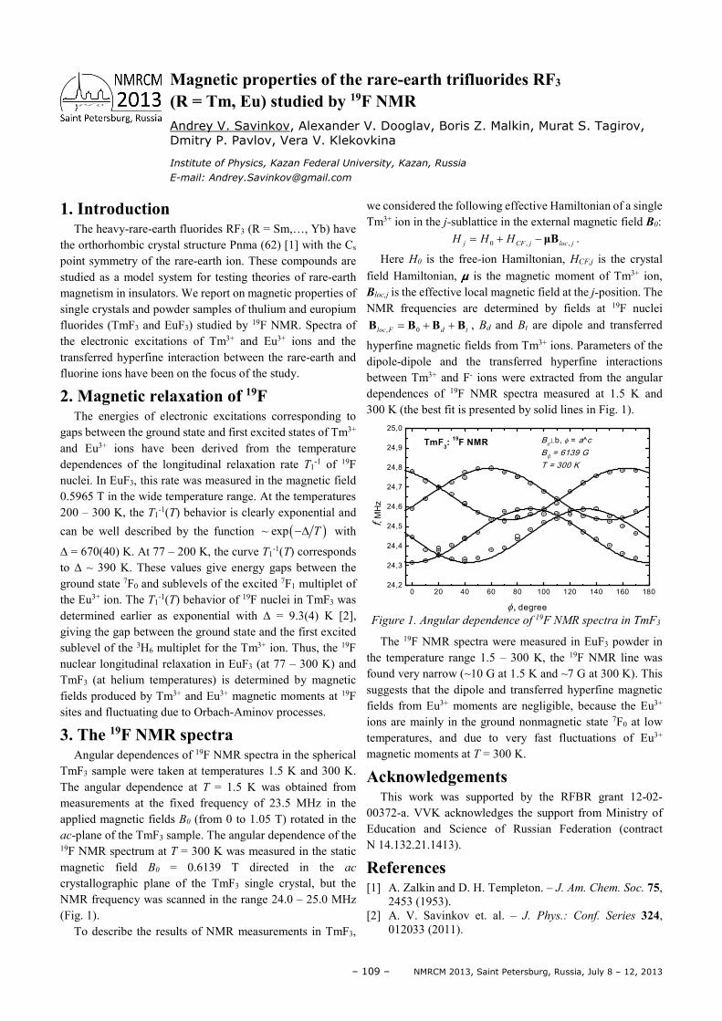
– 109 – NMRCM 2013, Saint Petersburg, Russia, July 8 – 12, 2013
1. Introduction The heavy-rare-earth fluorides RF3 (R = Sm,…, Yb) have
the orthorhombic crystal structure Pnma (62) [1] with the Cs point symmetry of the rare-earth ion. These compounds are studied as a model system for testing theories of rare-earth magnetism in insulators. We report on magnetic properties of single crystals and powder samples of thulium and europium fluorides (TmF3 and EuF3) studied by 19F NMR. Spectra of the electronic excitations of Tm3+ and Eu3+ ions and the transferred hyperfine interaction between the rare-earth and fluorine ions have been on the focus of the study.
2. Magnetic relaxation of 19F The energies of electronic excitations corresponding to
gaps between the ground state and first excited states of Tm3+ and Eu3+ ions have been derived from the temperature dependences of the longitudinal relaxation rate T1
-1 of 19F nuclei. In EuF3, this rate was measured in the magnetic field 0.5965 T in the wide temperature range. At the temperatures 200 – 300 K, the T1
-1(T) behavior is clearly exponential and
can be well described by the function ( )~ exp T−∆ with
∆ = 670(40) K. At 77 – 200 K, the curve T1-1(T) corresponds
to ∆ ~ 390 K. These values give energy gaps between the ground state 7F0 and sublevels of the excited 7F1 multiplet of the Eu3+ ion. The T1
-1(T) behavior of 19F nuclei in TmF3 was determined earlier as exponential with ∆ = 9.3(4) K [2], giving the gap between the ground state and the first excited sublevel of the 3H6 multiplet for the Tm3+ ion. Thus, the 19F nuclear longitudinal relaxation in EuF3 (at 77 – 300 K) and TmF3 (at helium temperatures) is determined by magnetic fields produced by Tm3+ and Eu3+ magnetic moments at 19F sites and fluctuating due to Orbach-Aminov processes.
3. The 19F NMR spectra Angular dependences of 19F NMR spectra in the spherical
TmF3 sample were taken at temperatures 1.5 K and 300 K. The angular dependence at T = 1.5 K was obtained from measurements at the fixed frequency of 23.5 MHz in the applied magnetic fields B0 (from 0 to 1.05 T) rotated in the ac-plane of the TmF3 sample. The angular dependence of the 19F NMR spectrum at T = 300 K was measured in the static magnetic field B0 = 0.6139 T directed in the ac crystallographic plane of the TmF3 single crystal, but the NMR frequency was scanned in the range 24.0 – 25.0 MHz (Fig. 1).
To describe the results of NMR measurements in TmF3,
we considered the following effective Hamiltonian of a single Tm3+ ion in the j-sublattice in the external magnetic field B0:
0 , ,j CF j loc jH H H= + − µB .
Here H0 is the free-ion Hamiltonian, HCF,j is the crystal field Hamiltonian, µµµµ is the magnetic moment of Tm3+ ion, Bloc,j is the effective local magnetic field at the j-position. The NMR frequencies are determined by fields at 19F nuclei
, 0loc F d t= + +B B B B , Bd and Bt are dipole and transferred
hyperfine magnetic fields from Tm3+ ions. Parameters of the dipole-dipole and the transferred hyperfine interactions between Tm3+ and F- ions were extracted from the angular dependences of 19F NMR spectra measured at 1.5 K and 300 K (the best fit is presented by solid lines in Fig. 1).
0 20 40 60 80 100 120 140 160 18024,2
24,3
24,4
24,5
24,6
24,7
24,8
24,9
25,0
f, M
Hz
φ, degree
B0⊥b, φ = a^c
B0 = 6139 G
T = 300 K
TmF3:
19F NMR
Figure 1. Angular dependence of 19F NMR spectra in TmF3
The 19F NMR spectra were measured in EuF3 powder in the temperature range 1.5 – 300 K, the 19F NMR line was found very narrow (~10 G at 1.5 K and ~7 G at 300 K). This suggests that the dipole and transferred hyperfine magnetic fields from Eu3+ moments are negligible, because the Eu3+ ions are mainly in the ground nonmagnetic state 7F0 at low temperatures, and due to very fast fluctuations of Eu3+ magnetic moments at T = 300 K.
Acknowledgements This work was supported by the RFBR grant 12-02-
00372-а. VVK acknowledges the support from Ministry of Education and Science of Russian Federation (contract N 14.132.21.1413).
References [1] A. Zalkin and D. H. Templeton. – J. Am. Chem. Soc. 75,
2453 (1953). [2] A. V. Savinkov et. al. – J. Phys.: Conf. Series 324,
012033 (2011).
Magnetic properties of the rare-earth trifluorides RF3 (R = Tm, Eu) studied by 19F NMR Andrey V. Savinkov, Alexander V. Dooglav, Boris Z. Malkin, Murat S. Tagirov, Dmitry P. Pavlov, Vera V. Klekovkina
Institute of Physics, Kazan Federal University, Kazan, Russia
E-mail: [email protected]
NMRCM 2013, Saint Petersburg, Russia, July 8 – 12, 2013 – 110 –
1. Introduction Nafion® ionomers are solid polymer electrolytes with a
high conductivity for protons when they are hydrated. Therefore they are well suited for the application in fuel cells. Nafion consists of a hydrophobic main chain with hydrophilic side chains having a sulfonic acid group at the ends. Hydration leads to a selforganisation of the hydrophilic and hydrophobic groups building up a nanostructured network of ionic clusters interconnected by channels where water molecules migrate along restricted pathways [1]. The conductivity increases with water content and temperature. A main problem of fuel cells is the temperature limitation of about 80oC due to a rapid loss of water [2].
The objective is the enhancement of the conductivity at low relative humidity as well as the improvement of the water management and hydration stability at high temperatures. This can be achieved by the addition of nanomaterials. Fullerenes are one of the dopants with the advantage of their spherical shape, such that a high density of chemical attachments is possible [3].
During this study we investigated the long range proton transport in dry and wet samples of Nafion doped with two fullerene-based materials C60(OH)n and C60(SO3)n having different additional functional groups up to n=24. Diffusion in pure Nafion is presented for comparison.
2. Experimental We used NMR diffusometry in an ultrahigh static field
gradient (SFG) realized by a special NMR magnet with an anti-Helmholtz arrangement of superconducting field coils. The field B and the gradient g are presented in Fig. 1.
Figure 1. Field profile and gradient strength
along the z-axis
By using a 1H Larmor frequency of 100 MHz four positions correspond to a magnetic field of 2.35 T. There are two positions zHG where a gradient of 178 T/m is reached and two positions zLG with 50.5 T/m. The diffusion measurement was done by stimulated echos at two different gradients to exclude the influence of T1- and T2-relaxation. Several data sets with different constant evolution times τ (tM varied) and different mixing times tM (τ varied) are recorded. The sample temperature has been stabilized by a cryostat.
3. Results For different τ and tM the results of the diffusion
coefficients are consistent with each other. The resulting diffusion coefficients as a function of temperature, shown in Fig. 2, are described by an Arrhenius law. At low temperatures there is a change towards higher activation energy. In contrast to all wet samples where the diffusion is the same at high temperatures the dry samples show the strongest diffusion in the entire temperature range for the C60(SO3)n added Nafion. In comparison to pure Nafion the achieved diffusion in this functionalized fullerene doped sample is increased by a factor of 2. Thus the additive C60(SO3)n might be promising for practical application of Nafion.
Figure 2. Arrhenius plot of the diffusion coefficients
References [1] K. A. Mauritz, R. B. Moore, Chem. Rev., 104, 4535
(2004). [2] A. Wieckowski, J. Nørskov, Fuel Cell Science, Wiley,
388 (2010) [3] K. Tasaki, J. Gasa, H. Wang, R. DeSousa, Polymer 48,
4438 (2007)
1H diffusion in chemically functionalized fullerene doped Nafion proton electrolyte membranes measured by SFG-NMR M. Schäfer1, A. F. Privalov1, F. Fujara, I. V. Murin1, V. N. Postnov2, D. V. Postnov2
1Institut für Festkörperphysik, TU Darmstadt, Hochschulstr. 6, 64289 Darmstadt, Germany
E-mail: [email protected] 2Department of Chemistry St. Petersburg State University,
Universitetskij pr.26, 198504, St. Petersburg, Russia
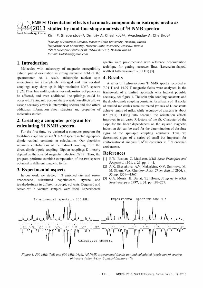
– 111 – NMRCM 2013, Saint Petersburg, Russia, July 8 – 12, 2013
1. Introduction Molecules with anisotropy of magnetic susceptibility
exhibit partial orientation in strong magnetic field of the spectrometer. As a result, anisotropic nuclear spin interactions are incompletely averaged and thus residual couplings may show up in high-resolution NMR spectra [1, 2]. Thus, line widths, intensities and positions of peaks can be affected, and even additional line-splittings could be observed. Taking into account these orientation effects allows escape accuracy errors in interpreting spectra and also offers additional information about structure and properties of molecules studied.
2. Creating a computer program for calculating 1H NMR spectra
For the first time, we designed a computer program for total-line-shape analysis of 1H NMR spectra including dipole-dipole residual constants in calculations. Our algorithm separates contributions of the indirect coupling from the direct dipole-dipole coupling. Dipolar couplings D linearly depend on the squared magnetic induction B0
2 [2]. Thus, the program performs combine computation of the two spectra obtained in different magnetic fields.
3. Experimental aspects In our work we studied 15N enriched cis- and trans-
azobenzene, substituted naphthalenes, styrene and tetrahydrofuran in different isotropic solvents. Degassed and sealed-off in vacuum samples were used. Experimental
spectra were pre-processed with reference deconvolution technique for getting narrower lines (Lorenzian-shaped, width at half-maximum ~ 0.1 Hz) [3].
4. Results A series of high-resolution 1H NMR spectra recorded at
7.04 T and 14.09 T magnetic fields were analyzed in the framework of a unified approach with highest possible accuracy, see figure 1. The spin-spin coupling constants and the dipole-dipole coupling constants for all pairs of 1H nuclei of studied molecules were estimated (values of D constants achieve tenths of mHz, while accuracy of analysis is about 0.5 mHz). Taking into account, the orientation effects improves in all cases R-factors of the fit. Character of the slope for the linear dependences on the squared magnetic induction B0
2 can be used for the determination of absolute signs of the spin-spin coupling constants. Thus we determined signs of a series of small but important for conformational analysis 1H-15N constants in 15N enriched azobenzene.
References [1] E.W. Bastian, C. MacLean, NMR basic Principles and
Progress // 1991, v. 25, pp. 1–44. [2] A.K. Shestakova, A.V. Makarkina, O.V. Smirnova, M.
M. Shtern, V.A. Chertkov, Russ. Chem. Bull., // 2006, v. 55, pp. 1359—1367.
[3] G.A. Morris, H. Barjat, T.J. Home, Progress in NMR
Spectroscopy // 1997, v. 31. pp. 197–257.
Figure 1. 300 MHz (left) and 600 MHz (right) 1H NMR experimental (peaks up) and calculated (peaks down) spectra
of trans-1-(phenyl-D5) -2-phenyldiazido-1-15N
Orientation effects of aromatic compounds in isotropic media as studied by total-line-shape analysis of 1H NMR spectra Kirill F. Sheberstov1,2, Dmitriy A. Cheshkov2,3, Vyacheslav A. Chertkov2
1Faculty of Materials Science, Moscow State University, Moscow, Russia 2Department of Chemistry, Moscow State University, Moscow, Russia 3State Scientific Centre of RF “GNIIChTEOS”, Moscow Russia
E-mail: [email protected]
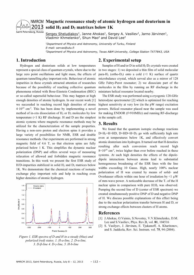
NMRCM 2013, Saint Petersburg, Russia, July 8 – 12, 2013 – 112 –
1. Introduction Hydrogen and deuterium solids at low temperatures
represent a special class of quantum crystals, where due to the large zero point oscillations and light mass, the effects of quantum tunnelling play important role. Behaviour of atomic impurities in these crystals attracted attention of researches because of the possibility of reaching collective quantum phenomena related with Bose-Einstein Condensation (BEC) or so-called supersolid behaviour. This may happen at high enough densities of atomic hydrogen. In our recent work [1] we succeeded in reaching record high densities of atoms 4·1019 cm-3. This has been done by implementing a novel method of in-situ dissociation of H2 or D2 molecules by low temperature (<1 K) RF discharge. H and D are the simplest atomic systems where magnetic resonance methods may be utilized for the characterization of the sample properties. Having a non-zero proton and electron spins it provides a large variety of possibilities for NMR, ESR and double resonance methods. Our experiments are performed in strong magnetic field of 4.6 T, so that electron spins are fully polarized below 1 K. This simplifies the dynamic nuclear polarization (DNP) and offers several ways of measuring relaxation of allowed and forbidden magnetic resonance transitions. In this work we present the first ESR study of H/D-impurities stabilized in solid H2 and D2 matrices below 1K. We demonstrate that the chemical reactions of isotopic exchange play important role and help in reaching even higher densities of atomic hydrogen.
Figure 1. ESR spectra of D and H in a steady (blue) and
polarized (red) states. 1. H-a-line, 2. D-α-line,
3. D-β-line 4. D-γ-line, 5. H-b-line
2. Experimental setup Samples of H and/or D in solid H2/D2 crystals were created
in two stages: 1) we deposited a thin film of solid molecular para-H2 (ortho-D2) onto a cold (<1 K) surface of quartz microbalance crystal, which served also as a mirror of 128 GHz Fabry-Perot resonator; 2) we dissociate part of the molecules in the film by running an RF discharge in the miniature helical resonator located nearby.
The ESR study was performed using a cryogenic 128-GHz heterodyne spectrometer [2] which is optimized for reaching highest sensitivity at very low (in the pW range) excitation powers. Helical resonator placed near the sample was used for making ENDOR (f=910MHz) and running RF-discharge in the sample cell.
3. Results We found that the quantum isotopic exchange reactions
D+H2=H+HD, D+HD=H+D2 go with sufficiently high rate even at temperatures below 1K, and effectively convert atomic deuterium into hydrogen. It turned out that H densities resulting after such conversion reach record high 8×1019 cm-3, twice higher than ever before reached in these systems. At such high densities the effects of the dipole-dipole interactions between atoms lead to substantial homogeneous broadening of the ESR lines with the line widths exceeding 10 Gauss. High, nearly 100% nuclear polarization of H was created by means of solid- and Overhauser effects within one hour of irradiation by <1 µW of mm-wave power. A noticeable decrease of the T1 of the H nuclear spins in comparison with pure H:H2 was observed. Pumping the second line of D (center of ESR spectrum) we created simultaneously positive DNP of D and negative DNP of H. We discuss possible explanations of this effect being due to the nuclear polarization transfer between H and D, or strong exchange effects between clusters of H atoms.
References [1] J.Ahokas, O.Vainio, S.Novotny, V.V.Khmelenko, D.M.
Lee and S.Vasiliev, Phys. Rev.B, vol. 81, 104516 [2] S. Vasilyev, J. Järvinen, E. Tjukanoff, A. Kharitonov,
and S. Jaakkola, Rev. Sci. Instrum. vol. 75, 94 (2004).
Magnetic resonance study of atomic hydrogen and deuterium in solid H2 and D2 matrixes below 1K Sergey Sheludiakov1, Janne Ahokas1, Sergey A. Vasiliev1, Jarno Järvinen1, Vladimir Khmelenko2, Shun Mao2 and David Lee2
1Department of Physics and Astronomy, University of Turku, Finland
E-mail: [email protected] 2Department of Physics and Astronomy, Texas A&M University, College Station TX77843, USA
– 113 – NMRCM 2013, Saint Petersburg, Russia, July 8 – 12, 2013
1. Introduction Stable complexes of azacrownethers and cryptands with
metal ions are known more than two decades. Of special interest are the lanthanide derivatives The application of lanthanides in many fields is closely related to their unusually high co-ordination number and large charge density. Many uses require complexation of the lanthanide with suitable organic ligands, which then allow, for example, introduction of other functions into systems like chemical nucleases. There are not many structural investigations with such systems (see e.g. [1, 2]). Presently we report here the results for a complex between hydrated lanthanum triflate and [221]-cryptand obtained with three independent methods: X-ray diffraction, quantum chemistry calculations and NMR measurements of vicinal spin-spin coupling constants.
2. Results and discussion Complexation leads to dramatic effect on conformation of
the ligand. X-Ray diffraction data revealed the structure of innersphere almost C2v–symmetrical complex with [221]cryptand and hydrated lanthanum triflate. We performed a series of ECP quantum chemical calculations (Lanl2dz approach), which support the structure of complexes under study. Experimental vicinal spin-spin coupling constants between protons of neighboring CH2-groups for complexes of lanthanum salts with a series of model macrocyclic ligands (1,10-diaza-18-crown-6 [2] and [2,2,2]-cryptand [1]) in solutions (CDCl3, CD3OD and CD3CN) are in good agreement with the ab’inito calculated ones (PFT DFT with Ub3lyp and 6G/311(d) basis set). The conformation of the biggest loop of the [221]-cryptand is close to that of 1,10-diaza-18-crown-6 in their lanthanum complexes.
The complex is of particular interest due to a specific co-ordination sphere with all seven binding sites of the cryptand (all five O and both N-atoms, see fig. 1) and extra three water molecules grouping from the same side of the compound (through the biggest loop of the cryptand). The analysis of the crystal packing revealed, that in crystal molecules are
assembled into the two layers by O-H…O bonds (O…O 2.766(6)-2.867(6)Å) formed by the water molecules and СF3SO3 anions.
Figure 1. Scheme of the spatial structure for binding of
La(3+) with [2,2,1]-cryptand extra three co-ordinated
water molecules
ECP quantum chemical calculations (Lanl2dz approach) support the structure. Experimental vicinal spin-spin coupling constants between protons of neighbouring CH2-groups in solutions (CD3OD and CD3CN) are in good agreement with the ab’inito calculated ones (PFT DFT with Ub3lyp and 6G/311(d) basis set, see e.g. [3]). So, the specific conformation of the ligand in solutions of the complex and in the solid state is entirely the same.
References [1] A.K. Shestakova, V.A. Chertkov, H.-J. Schneider, Tetr.
Letters, 41, 6753-6756 (2000). [2] A.K. Shestakova, V.A. Chertkov, H.-J. Schneider, K.A.
Lysenko, Org. Letters, 3, 325-327 (2001). [3] W. Deng, J.R. Cheeseman, M.J. Frisch, J. Chem. Theory
and Comput., 2 1028-37 (2006).
A comparison of solid state and solution structure for a complex between hydrated lanthanum triflate and [221]cryptand Alla K. Shestakova1, Vyacheslav A. Chertkov2
1State Research Institute of Chemistry and Technology of Organoelement Compounds, sh.
Entuiastov 38, 111123 Moscow, Russia
E-mail: [email protected] 2Department of Chemistry, Moscow State University, Leninskie Gory 1/3, 119992 Moscow Russia
E-mail: [email protected]
NMRCM 2013, Saint Petersburg, Russia, July 8 – 12, 2013 – 114 –
1. Introduction In preliminary study we have shown that the high proton
relaxivity of Gd-containing water soluble fullerene provide the enhancement of contrast of liver image in mouse MR imaging [1]. Antiangiogenesis activity of Gd@Ful is supplementary reason for using this paramagnetic agent for theranostic study of cancer [2,3]. The rat glioma is suitable model for study of Gd@Ful fitness for diagnostic application in brain cancer cases.
2. Materials and methods Water soluble gadofullerene was synthesized as
Gd@C82(OH)x x ≥20 as described [4]. Water solutions of Gd@Ful were used for magnetic relaxation measurements in vitro and for MR imaging of rat with implanted glioma. C6 glioma cells were injected into brain. Gd@Ful was delivered to animal intravenously and directly into tumor through intracranical hole after tumor formation at 3d week. Magnetic relaxation times and relaxivity of water protons under action of Gd@Ful were estimated with help CXP-300 spectrometer (Bruker). MR imaging of brain tumor was made by scanning by Avance II NMR spectrometer with microimaging accessory.
3. Results MR scanning of experimental glioma demonstrated the
enhancement of contrast tumor image after intravenous administration of Gd@Ful solution. The T1 weighted images of rat glioma are shown in Figure.1.
Figure 1. T1-weighted transvers images of rat glioma after
iv injection Gd@Ful (right). Left image – control
The intravenous injection caused the shortening of T1 time relaxation in the area of blood vessels. The same effect of resonance was achieved after local injection into tumor Figure 2. The paramagnetic Gd inside Fullerene cage strongly affects the spin state of water protons in homogeneous magnetic field. The reduction of tumor angiogenesis doesn’t inhibit the contrast action of Gd@Ful as contrast agent.
Figure 2. T1-weighted image of rat glioma after injection
Gd@Ful into the tumor body
The scanning in T2-weighted regime represents the decrease of signal intensity of necrotic tissue Figure 3.
Figure 3. T2-weighted image of rat glioma after iv Gd@Ful
injection (right). Control-left
The Gd@Ful administration promotes the improvement of lining tumor border due to accumulation of paramagnetic substance in tumor locus. The traffic of Gd@Ful into glioma is ensured by destruction of blood-brain barrier by cancereous cells. The contrast efficiency of Gd@Ful corresponds to the characteristics of negative contrast agent.
4. Conclusion The Gd@Ful preparation is shown to be effective contrast
enhancer for theranostic study of brain tumors.
Acknowledgements This work was supported by the FMBA RF (grant
40.002.12.0). Authors thank A.Dobrodumov for MRI scanning.
References [1] Ya.Yu. Marchenko, B.P. Nikolaev et al. Magnetic
Relaxation Study of Water–soluble Gadofullerene as potential contrast agent for theranostics. NMRCM 2012, St.Petersburg, Book of abstracts, p.102.
[2] H. Meng et al. Potent Angiogenesis Inhibition by the Particulate Form of Fullerene Derivatives. // ACNano, 4(2010), 5, p. 2773–2783.
[3] Zhiyun Chen et al Applications of Functionalized Fullerenes in Tumor Theranostics.// Theranostics 2 (2012), 3, P.238-250.
[4] Patent RF 2396207.
Magnetic Resonance Imaging of Rat Glioma Model enhanced by using water-soluble Gadolinium Fullerene M. A. Shevzov1, B. P. Nikolaev2, Ya. Yu. Marchenko2, L. Yu. Yakovleva2, V. T. Lebedev3
1Institute of Cytology of RAS, St.Petersburg, Russia 2Reseach Institute of Highly Pure Biopreparation,St.Petersburg, Russia 3Institute of Nuclear Physics, St.Petersburg, Russia
E-mail: [email protected]
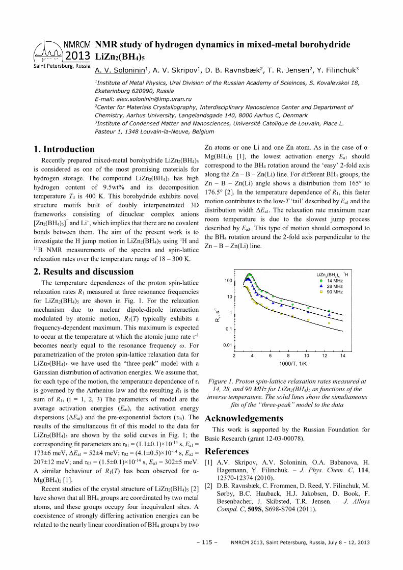
– 115 – NMRCM 2013, Saint Petersburg, Russia, July 8 – 12, 2013
1. Introduction Recently prepared mixed-metal borohydride LiZn2(BH4)5
is considered as one of the most promising materials for hydrogen storage. The compound LiZn2(BH4)5 has high hydrogen content of 9.5wt% and its decomposition temperature Td is 400 K. This borohydride exhibits novel structure motifs built of doubly interpenetrated 3D frameworks consisting of dinuclear complex anions [Zn2(BH4)5]¯ and Li+, which implies that there are no covalent bonds between them. The aim of the present work is to investigate the H jump motion in LiZn2(BH4)5 using 1H and 11B NMR measurements of the spectra and spin-lattice relaxation rates over the temperature range of 18 – 300 K.
2. Results and discussion The temperature dependences of the proton spin-lattice
relaxation rates R1 measured at three resonance frequencies for LiZn2(BH4)5 are shown in Fig. 1. For the relaxation mechanism due to nuclear dipole-dipole interaction modulated by atomic motion, R1(T) typically exhibits a frequency-dependent maximum. This maximum is expected to occur at the temperature at which the atomic jump rate τ-1 becomes nearly equal to the resonance frequency ω. For parametrization of the proton spin-lattice relaxation data for LiZn2(BH4)5 we have used the “three-peak” model with a Gaussian distribution of activation energies. We assume that, for each type of the motion, the temperature dependence of τi is governed by the Arrhenius law and the resulting R1 is the sum of R1i (i = 1, 2, 3) The parameters of model are the average activation energies (Eai), the activation energy dispersions (∆Eai) and the pre-exponential factors (τ0i). The results of the simultaneous fit of this model to the data for LiZn2(BH4)5 are shown by the solid curves in Fig. 1; the corresponding fit parameters are τ01 = (1.1±0.1)×10-14 s, Ea1 = 173±6 meV, ∆Ea1 = 52±4 meV; τ02 = (4.1±0.5)×10-14 s, Ea2 = 207±12 meV; and τ03 = (1.5±0.1)×10-14 s, Ea3 = 302±5 meV. A similar behaviour of R1(T) has been observed for α-Mg(BH4)2 [1].
Recent studies of the crystal structure of LiZn2(BH4)5 [2] have shown that all BH4 groups are coordinated by two metal atoms, and these groups occupy four inequivalent sites. A coexistence of strongly differing activation energies can be related to the nearly linear coordination of BH4 groups by two
Zn atoms or one Li and one Zn atom. As in the case of α-Mg(BH4)2 [1], the lowest activation energy Ea1 should correspond to the BH4 rotation around the ‘easy’ 2-fold axis along the Zn – B – Zn(Li) line. For different BH4 groups, the Zn – B – Zn(Li) angle shows a distribution from 165° to 176.5° [2]. In the temperature dependence of R1, this faster motion contributes to the low-T ‘tail’ described by Ea1 and the distribution width ∆Ea1. The relaxation rate maximum near room temperature is due to the slowest jump process described by Ea3. This type of motion should correspond to the BH4 rotation around the 2-fold axis perpendicular to the Zn – B – Zn(Li) line.
2 4 6 8 10 12 14
0.01
0.1
1
10
100
LiZn2(BH
4)
5
1H
14 MHz
28 MHz
90 MHz
R1, s
-1
1000/T, 1/K
Figure 1. Proton spin-lattice relaxation rates measured at
14, 28, and 90 MHz for LiZn2(BH4)5 as functions of the
inverse temperature. The solid lines show the simultaneous
fits of the “three-peak” model to the data
Acknowledgements This work is supported by the Russian Foundation for
Basic Research (grant 12-03-00078).
References [1] A.V. Skripov, A.V. Soloninin, O.A. Babanova, H.
Hagemann, Y. Filinchuk. – J. Phys. Chem. C, 114, 12370-12374 (2010).
[2] D.B. Ravnsbæk, C. Frommen, D. Reed, Y. Filinchuk, M. Sørby, B.C. Hauback, H.J. Jakobsen, D. Book, F. Besenbacher, J. Skibsted, T.R. Jensen. – J. Alloys
Compd. C, 509S, S698-S704 (2011).
NMR study of hydrogen dynamics in mixed-metal borohydride LiZn2(BH4)5 A. V. Soloninin1, A. V. Skripov1, D. B. Ravnsbæk2, T. R. Jensen2, Y. Filinchuk3
1Institute of Metal Physics, Ural Division of the Russian Academy of Scieinces, S. Kovalevskoi 18,
Ekaterinburg 620990, Russia
E-mail: [email protected] 2Center for Materials Crystallography, Interdisciplinary Nanoscience Center and Department of
Chemistry, Aarhus University, Langelandsgade 140, 8000 Aarhus C, Denmark 3Institute of Condensed Matter and Nanosciences, Université Catolique de Louvain, Place L.
Pasteur 1, 1348 Louvain-la-Neuve, Belgium
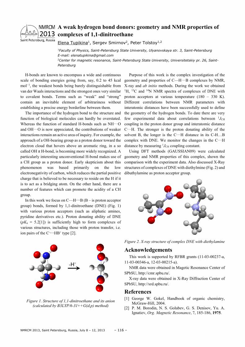
NMRCM 2013, Saint Petersburg, Russia, July 8 – 12, 2013 – 116 –
H-bonds are known to encompass a wide and continuous scale of bonding energies going from, say, 0.2 to 45 kcal mol−1, the weakest bonds being barely distinguishable from van der Waals interactions and the strongest ones very similar to covalent bonds. Terms such as “weak” and “strong” contain an inevitable element of arbitrariness without establishing a precise energy borderline between them.
The importance of the hydrogen bond to the structure and function of biological molecules can hardly be overstated. Whereas the function of standard H-bonds such as NH···O and OH···O is now appreciated, the contributions of weaker interactions remain an active area of inquiry. For example, the approach of a OH-bearing group as a proton donor toward the electron cloud that hovers above an aromatic ring, in a so called OH π H-bond, is becoming more widely recognized. A particularly interesting unconventional H-bond makes use of a CH group as a proton donor. Early skepticism about this phenomenon was based primarily on the low electronegativity of carbon, which reduces the partial positive charge that is believed to be necessary to reside on the H if it is to act as a bridging atom. On the other hand, there are a number of features which can promote the acidity of a CH group.
In this work we focus on C—H···B (B – is proton acceptor group) bonds, formed by 1,1-dinitroethane (DNE) (Fig. 1) with various proton acceptors (such as aliphatic amines, pyridine derivatives etc.). Proton donating ability of DNE (pKa = 5.2[1]) is sufficiently high to form complexes of various structures, including those with proton transfer, i.e. ion pairs of the C-···HB+ type [2].
Figure 1. Structure of 1,1-dinitroethane and its anion
(calculated by B3LYP/6-31++G(d,p) method)
Purpose of this work is the complex investigation of the geometry and properties of C—H···B complexes by NMR, X-ray and ab initio methods. During the work we obtained 1H, 13C and 14N NMR spectra of complexes of DNE with proton acceptors at various temperature (180 – 330 K). Different correlations between NMR parameters with interatomic distances have been successfully used to define the geometry of the hydrogen bonds. To date there are very few experimental data about correlations between 1JCH coupling in the proton donor group and interatomic distance C···H. The stronger is the proton donating ability of the solvent B, the longer is the C···H distance in its C-H…B complex with DNE. We monitor the changes in the C···H distance by measuring 1JCH coupling constant.
Using DFT methods (GAUSSIAN09) were calculated geometry and NMR properties of this complex, shown the comparison with the experiment data. Also discussed X-Ray structures of complexes of DNE with diethylmine (Fig. 2) and dibuthylamine as proton acceptor group.
Figure 2. X-ray structure of complex DNE with diethylamine
Acknowledgements This work is supported by RFBR grants (11-03-00237-a,
11-03-00346-а, 12-03-00215-а). NMR data were obtained in Magetic Resonance Center of
SPbSU, http://cmr.spbu.ru/. X-ray data were obtained in X-Ray Diffraction Center of
SPbSU, http://xrd.spbu.ru/.
References [1] George W. Gokel, Handbook of organic chemistry,
McGraw-Hill, 2004. [2] P. M. Borodin, N. S. Golubev, G. S. Denisov, Yu. A.
Ignatiev, Org. Magnetic Resonance, 7, 185-186, 1975.
A weak hydrogen bond donors: geometry and NMR properties of complexes of 1,1-dinitroethane Elena Tupikina1, Sergev Smirnov2, Peter Tolstoy1,2
1Faculty of Physics, Saint-Petersburg State University, Ulyanovskaya str. 3, Saint-Petersburg
E-mail: [email protected] 2Center for magnetic resonance, Saint-Petersburg State University, Universitetskiy pr. 26, Saint-
Petersburg
-Н+
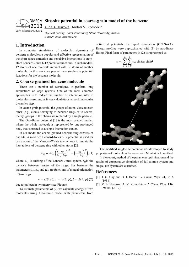
– 117 – NMRCM 2013, Saint Petersburg, Russia, July 8 – 12, 2013
1. Introduction In computer simulations of molecular dynamics of
benzene molecules, a popular and effective representation of the short-range attractive and repulsive interactions is atom-atom Lennard-Jones 6-12 potential functions. In such models, 12 atoms of one molecule interact with 12 atoms of another molecule. In this work we present new single-site potential functions for the benzene molecule.
2. Coarse-grained benzene molecule There are a number of techniques to perform long
simulations of large systems. One of the most common approaches is to reduce the number of interaction sites in molecules, resulting in fewer calculations at each molecular dynamics step.
In coarse-grain potential the groups of atoms close to each other (e.g., atoms belonging to benzene rings or to several methyl groups in the chain) are replaced by a single particle.
The Gay-Berne potential [1] is the most grained model, where the whole molecule is represented by one prolonged body that is treated as a single interaction center.
In our model the coarse-grained benzene ring consists of one site. A modified Lennard-Jones 6-12 potential is used for calculation of the Van-der-Waals interactions to imitate the interactions of benzene ring with other atoms [2]:
#$ = 4&$ '( )*+*∆*, − ( )*
+*∆*,-., (1)
where ∆$ is shifting of the Lennard-Jones sphere, /$is the distance between centers of the rings. For benzene the parameters &$, 0$ and ∆$ are functions of mutual orientation of two rings:
& = & 1, 3, 0 = 0 1, 3, ∆= ∆ 1, 3 2
due to molecular symmetry (see Figure). To estimate parameters of (2) we calculate energy of two
molecules using full-atomic model with parameters from
optimized potentials for liquid simulation (OPLS-AA). Energy profiles were approximated with (1) by non-linear fitting. Final form of parameters in (2) is represented as
& = 4 4 567 sin ;3 sin <1-
7=
-
6=
The modified single-site potential was developed to study
properties of molecule of benzene with Monte-Carlo method. In the report, method of the parameter optimization and the
results of comparative simulation of full-atomic system and single-site system are discussed.
References [1] J. G. Gay and B. J. Berne – J. Chem. Phys. 74, 3316
(1981) [2] V. S. Neverov, A. V. Komolkin – J. Chem. Phys. 136,
094102 (2012)
Site-site potential in coarse-grain model of the benzene Alina A. Uskova, Andrei V. Komolkin
Physical Faculty, Saint-Petersburg State University, Russia
E-mail: [email protected]
1
3
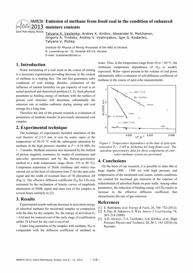
NMRCM 2013, Saint Petersburg, Russia, July 8 – 12, 2013 – 118 –
1. Introduction Water moistening of a coal seam in the course of mining
is a necessary requirement providing decrease in the content of methane in a stoping face. The last fact guarantees safer conditions of coal mining. Besides, estimation of the influence of natural humidity on gas capacity of coal is an actual practical and theoretical problem [1, 2]. Such physical parameter as binding energy of methane with the surface of porous coal structure will determine substantially the emission rate at sudden outbursts during mining and coal storage for a long time.
Therefore the aim of the present research is evaluation of parameters of methane transfer in previously moistened coal samples.
2. Experimental technique The technique of experiments included saturation of the
coal fraction of 2-2.5 mm in size by water vapor at the temperature of 30-35 °C with the subsequent saturation by methane in the high pressure chamber at Р = 8-10 MPs for 1 - 3 months. Methane emission was measured by the method of proton magnetic resonance by means of continuous and spin-echo spectrometers and by the thermo-gravimetric method in a wide temperature range (from -155 to 80 °C). Component separation of fluids (methane and water) was carried out on the base of relaxation time T2 for the spin-echo signal and the width of resonant lines of 1H absorption ∆H (Fig.1). The effective diffusion coefficient Deff for CH4 was estimated by the inclination of kinetic curves of amplitude attenuation of NMR signal and mass loss of the samples in several hours similarly to [3].
3. Results Experimental results indicate decrease in activation energy
of adsorbed methane for moistened samples in comparison with the data for dry samples. So, the energy of activation Ea = 6 kJ/mol for watered coal of the early stage of coalification unlike 18 kJ/mol for dry coal is received.
Under long saturation of the samples with methane, Deff is comparable with the diffusion coefficient of methane in
water. Thus, in the temperature range from-10 to + 80 °C, the Arrhenius temperature dependence of Deff is weakly expressed. Water vapors present in the volume of coal pores substantially affect evaluation of self-diffusion coefficient of methane in the course of spin-echo measurements.
Figure 1. Temperature dependence of the time of spin-spin
relaxation Т2 ~ 1/∆H in Arrhenius for long-flame-coal.. The
autodyne spectrometry data for three components of coal-
water-methane system are presented
4. Conclusions On the basis of our research, it is possible to state that at
large depths (800 - 1500 m) with high pressure and temperatures of the moistened coal seams, certain conditions are created for increased gas emission at the expense of redistribution of adsorbed fluids on pore walls. Among other parameters, the reduction of binding energy of CH4 results in increase in the effective diffusion coefficient that characterizes the rate of gas emission.
References [1] E. Battistutta, et al. Energy & Fuels, 26, 746–752 (2012). [2] S. Day, R. Sakurovs, S. Weir. Intern. J. Coal Geolog, 74,
203–214 (2008). [3] А.D. Аlexeev, Т.А. Vasilenko, А.К. Кirillov, et al., High
Pressure Physics and Technics, 20, 3, 143 (2010) (in Russian).
Emission of methane from fossil coal in the condition of enhanced moisture contents Tatyana А. Vasilenko, Andrey K. Kirillov, Alexander N. Molchanov, Grigoriy A. Troitsky, Andrey V. Vyshnyakov, Igor G. Kostenko, Tatyana V. Pichka
Institute for Physics of Mining Processes of the NАS of Ukraine
R. Luxemburg str. 72, Donetsk 83114, Ukraine
E-mail: [email protected]
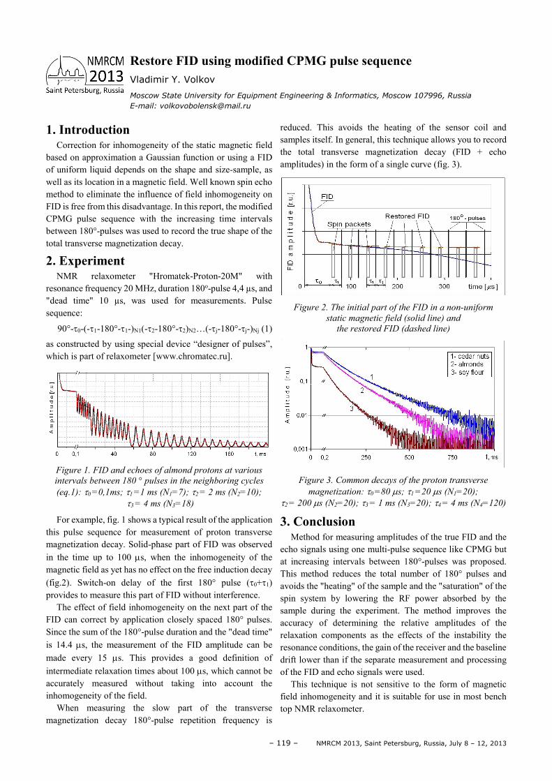
– 119 – NMRCM 2013, Saint Petersburg, Russia, July 8 – 12, 2013
1. Introduction Correction for inhomogeneity of the static magnetic field
based on approximation a Gaussian function or using a FID of uniform liquid depends on the shape and size-sample, as well as its location in a magnetic field. Well known spin echo method to eliminate the influence of field inhomogeneity on FID is free from this disadvantage. In this report, the modified CPMG pulse sequence with the increasing time intervals between 180°-pulses was used to record the true shape of the total transverse magnetization decay.
2. Experiment NMR relaxometer "Hromatek-Proton-20M" with
resonance frequency 20 MHz, duration 180o-pulse 4,4 µs, and "dead time" 10 µs, was used for measurements. Pulse sequence:
90°-τ0-(-τ1-180°-τ1-)N1(-τ2-180°-τ2)N2…(-τj-180°-τj-)Nj (1)
as constructed by using special device “designer of pulses”, which is part of relaxometer [www.chromatec.ru].
Figure 1. FID and echoes of almond protons at various
intervals between 180 ° pulses in the neighboring cycles
(eq.1): τ0 =0,1ms; τ1 =1 ms (N1=7); τ2 = 2 ms (N2=10);
τ3 = 4 ms (N3=18)
For example, fig. 1 shows a typical result of the application this pulse sequence for measurement of proton transverse magnetization decay. Solid-phase part of FID was observed in the time up to 100 µs, when the inhomogeneity of the magnetic field as yet has no effect on the free induction decay (fig.2). Switch-on delay of the first 180° pulse (τ0+τ1)
provides to measure this part of FID without interference. The effect of field inhomogeneity on the next part of the
FID can correct by application closely spaced 180° pulses. Since the sum of the 180°-pulse duration and the "dead time" is 14.4 µs, the measurement of the FID amplitude can be made every 15 µs. This provides a good definition of intermediate relaxation times about 100 µs, which cannot be accurately measured without taking into account the inhomogeneity of the field.
When measuring the slow part of the transverse magnetization decay 180°-pulse repetition frequency is
reduced. This avoids the heating of the sensor coil and samples itself. In general, this technique allows you to record the total transverse magnetization decay (FID + echo amplitudes) in the form of a single curve (fig. 3).
Figure 2. The initial part of the FID in a non-uniform
static magnetic field (solid line) and
the restored FID (dashed line)
Figure 3. Common decays of the proton transverse
magnetization: τ0 =80 µs; τ1 =20 µs (N1=20);
τ2 = 200 µs (N2=20); τ3 = 1 ms (N3=20); τ4 = 4 ms (N4=120)
3. Conclusion Method for measuring amplitudes of the true FID and the
echo signals using one multi-pulse sequence like CPMG but at increasing intervals between 180°-pulses was proposed. This method reduces the total number of 180° pulses and avoids the "heating" of the sample and the "saturation" of the spin system by lowering the RF power absorbed by the sample during the experiment. The method improves the accuracy of determining the relative amplitudes of the relaxation components as the effects of the instability the resonance conditions, the gain of the receiver and the baseline drift lower than if the separate measurement and processing of the FID and echo signals were used.
This technique is not sensitive to the form of magnetic field inhomogeneity and it is suitable for use in most bench top NMR relaxometer.
Restore FID using modified CPMG pulse sequence Vladimir Y. Volkov
Moscow State University for Equipment Engineering & Informatics, Moscow 107996, Russia
E-mail: [email protected]
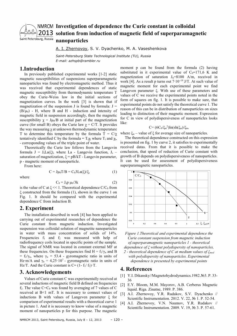
NMRCM 2013, Saint Petersburg, Russia, July 8 – 12, 2013 – 120 –
1.Introduction In previously published experimental works [1-2] static
magnetic susceptibilities of suspensions superparamagnetic nanoparticles was found by electromagnetic method. Thus it was received that experimental dependences of static magnetic susceptibility from thermodynamic temperature T obey the Curie-Weiss law in the initial sections of magnetization curves. In the work [3] is shown that if magnetization of the suspension J is found by formula: J = (B/µ0) - H, where B and H - induction and intensity of magnetic field in suspension accordingly, then the magnetic susceptibility χ = Jµ0/B at initial part of the magnetization curve (for small B) obeys the Curie law χ = C/T. It provides the way measuring χ at unknown thermodynamic temperature T to determine this temperature by the formula T = C/χ tentatively identified C by the formula = Ttχt where Tt and χt, - corresponding values of the triple point of water.
Theoretically the Curie law follows from the Langevin formula J = JsLa(ξ), where La - Langevin function, Js – saturation of magnetization, ξ = pB/kT - Langevin parameter, p - magnetic moment of nanoparticle.
From here: С = Jµ0T/B = C03La(ξ)/ξ, (1)
where С0 = Jsр µ0/3k (2)
is the value of C at ξ << 1. Theoretical dependence C/C0 from ξ constructed from the formula (1), shown in the curve 1 on Fig. 1. It should be compared with the experimental dependence C from induction B.
2. Experiment The installation described in work [4] has been applied to
carrying out of experimental researches of dependence the Curie constant from magnetic induction. Investigated suspension was colloidal solution of magnetite nanoparticles in water with mass concentration of solids of 14%. Frequencies f1 and f2 was measured with help of radiofrequency coils located in specific points of the sample. The signal of NMR was located in constant external MF at these frequencies. On these frequencies find Н = f1/γн and В = f2/γв, where γн = 53.4 - gyromagnetic ratio in units of Hz·m/A and γв = 4,25·107 - gyromagnetic ratio in units of Hz/T. And the Curie constant is С= (1- f1/ f2)·Т.
3. Acknowledgements
Values of Curie constant C was experimentally received at several inductions of magnetic field B defined on frequencies f2. The value C=C0 was found by averaging of 7 values of C received at B<1 mT. It is necessary to connect values of inductions B with values of Langeven parameter ξ for comparison of experimental results with a theoretical curve 1 in picture 1. And it is necessary to know value of a magnetic moment of nanoparticles р for this purpose. The magnetic
moment р can be found from the formula (2) having substituted in it experimental value of С0=171,6 K and magnetization of saturation Jн=8100 A/m, received in work [4]. As a result р turns out 7·10–19 J/T. At such value of magnetic moment for each experimental point we find Langeven parameter ξ. With use of these parameters and values of C we receive the experimental points noted in the form of squares on fig. 1. It is possible to make sure, that experimental points do not satisfy the theoretical curve 1. The reason of this can be in distribution of nanoparticles on sizes leading to distinction of their magnetic moment. Expression for C in view of polydispersiveness of nanoparticles looks like:
С= (6С0/ξм2)ln(sh(ξм)/ξм,
where ξм – value of ξ for average size of nanoparticles. The theoretical dependence constructed on this expression
is presented on fig. 1 by curve 2, it satisfies to experimentally received datas. From that it is possible to make the conclusion, that speed of reduction of Curie constant with growth of B depends on polydispersiveness of nanoparticles. It can be used for assessment of polydispersiveness superparamagnetic nanoparticles.
Figure 1.Theoretical and experimental dependence the
Curie constant suspension from magnetic induction
of.superparamagnetic nanoparticles 1 - theoretical
dependence of ξ without polydispersity of nanoparticles,
2 - theoretical dependence of C at medium values of ξmed
with polydispersity of nanoparticles. Experimental
dependence is presented by experimental points
4. References [1] Y.I. Dikansky//Magnetohydrodynamics.1982.3. Р. 33-
36 [2] E.Y. Bloom, M.M. Mayorov, A.B. Cerberus Magnetic
liquid. Riga. Zinatne, 1989. P. 386. [3] A.I. Zhernovoy, Y.R. Rudakov, S.V. Dyachenko //
Scientific Instrumentation. 2012. V. 22, 1. P. 52-54. [4] A.I. Zhernovoy, V.N. Naumov, Y.R. Rudakov //
Scientific Instrumentation. 2009. V. 19, 3. P. 57-61.
Investigation of dependence the Curie constant in colloidal solution from induction of magnetic field of superparamagnetic nanoparticles A. I. Zhernovoy, S. V. Dyachenko, M. A. Vaseshenkova
Saint-Petersburg State Technological Institute (TU), Russia
E-mail: [email protected]
ξ, ξcp
C/C0
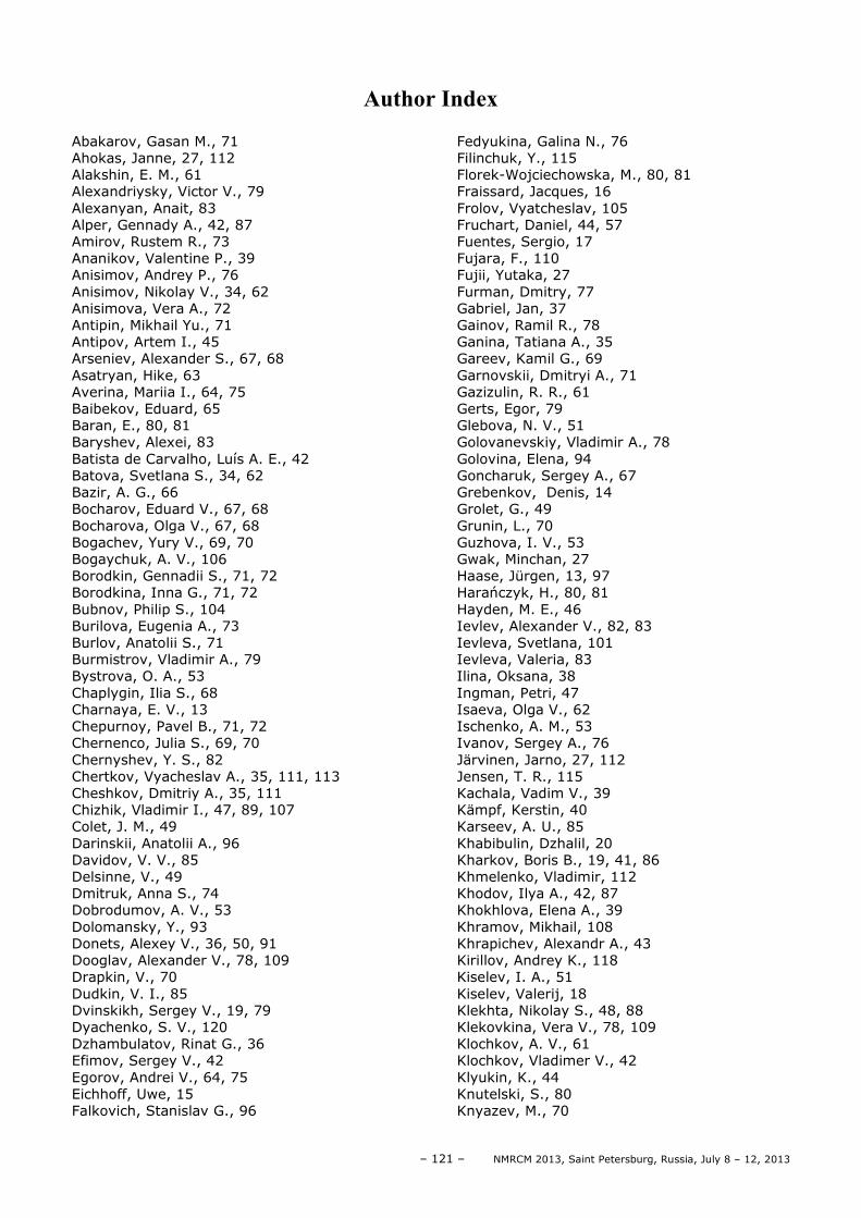
– 121 – NMRCM 2013, Saint Petersburg, Russia, July 8 – 12, 2013
Author Index
Abakarov, Gasan M., 71 Ahokas, Janne, 27, 112 Alakshin, E. M., 61 Alexandriysky, Victor V., 79 Alexanyan, Anait, 83 Alper, Gennady A., 42, 87 Amirov, Rustem R., 73 Ananikov, Valentine P., 39 Anisimov, Andrey P., 76 Anisimov, Nikolay V., 34, 62 Anisimova, Vera A., 72 Antipin, Mikhail Yu., 71 Antipov, Artem I., 45 Arseniev, Alexander S., 67, 68 Asatryan, Hike, 63 Averina, Mariia I., 64, 75 Baibekov, Eduard, 65 Baran, E., 80, 81 Baryshev, Alexei, 83 Batista de Carvalho, Luís A. E., 42 Batova, Svetlana S., 34, 62 Bazir, A. G., 66 Bocharov, Eduard V., 67, 68 Bocharova, Olga V., 67, 68 Bogachev, Yury V., 69, 70 Bogaychuk, A. V., 106 Borodkin, Gennadii S., 71, 72 Borodkina, Inna G., 71, 72 Bubnov, Philip S., 104 Burilova, Eugenia A., 73 Burlov, Anatolii S., 71 Burmistrov, Vladimir A., 79 Bystrova, O. A., 53 Chaplygin, Ilia S., 68 Charnaya, E. V., 13 Chepurnoy, Pavel B., 71, 72 Chernenco, Julia S., 69, 70 Chernyshev, Y. S., 82 Chertkov, Vyacheslav A., 35, 111, 113 Cheshkov, Dmitriy A., 35, 111 Chizhik, Vladimir I., 47, 89, 107 Colet, J. M., 49 Darinskii, Anatolii A., 96 Davidov, V. V., 85 Delsinne, V., 49 Dmitruk, Anna S., 74 Dobrodumov, A. V., 53 Dolomansky, Y., 93 Donets, Alexey V., 36, 50, 91 Dooglav, Alexander V., 78, 109 Drapkin, V., 70 Dudkin, V. I., 85 Dvinskikh, Sergey V., 19, 79 Dyachenko, S. V., 120 Dzhambulatov, Rinat G., 36 Efimov, Sergey V., 42 Egorov, Andrei V., 64, 75 Eichhoff, Uwe, 15 Falkovich, Stanislav G., 96
Fedyukina, Galina N., 76 Filinchuk, Y., 115 Florek-Wojciechowska, M., 80, 81 Fraissard, Jacques, 16 Frolov, Vyatcheslav, 105 Fruchart, Daniel, 44, 57 Fuentes, Sergio, 17 Fujara, F., 110 Fujii, Yutaka, 27 Furman, Dmitry, 77 Gabriel, Jan, 37 Gainov, Ramil R., 78 Ganina, Tatiana A., 35 Gareev, Kamil G., 69 Garnovskii, Dmitryi A., 71 Gazizulin, R. R., 61 Gerts, Egor, 79 Glebova, N. V., 51 Golovanevskiy, Vladimir A., 78 Golovina, Elena, 94 Goncharuk, Sergey A., 67 Grebenkov, Denis, 14 Grolet, G., 49 Grunin, L., 70 Guzhova, I. V., 53 Gwak, Minchan, 27 Haase, Jürgen, 13, 97 Harańczyk, H., 80, 81 Hayden, M. E., 46 Ievlev, Alexander V., 82, 83 Ievleva, Svetlana, 101 Ievleva, Valeria, 83 Ilina, Oksana, 38 Ingman, Petri, 47 Isaeva, Olga V., 62 Ischenko, A. M., 53 Ivanov, Sergey A., 76 Järvinen, Jarno, 27, 112 Jensen, T. R., 115 Kachala, Vadim V., 39 Kämpf, Kerstin, 40 Karseev, A. U., 85 Khabibulin, Dzhalil, 20 Kharkov, Boris B., 19, 41, 86 Khmelenko, Vladimir, 112 Khodov, Ilya A., 42, 87 Khokhlova, Elena A., 39 Khramov, Mikhail, 108 Khrapichev, Alexandr A., 43 Kirillov, Andrey K., 118 Kiselev, I. A., 51 Kiselev, Valerij, 18 Klekhta, Nikolay S., 48, 88 Klekovkina, Vera V., 78, 109 Klochkov, A. V., 61 Klochkov, Vladimer V., 42 Klyukin, K., 44 Knutelski, S., 80 Knyazev, M., 70
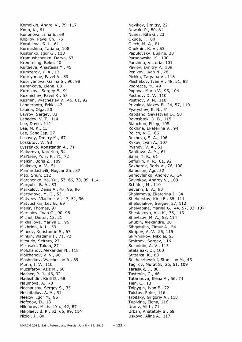
NMRCM 2013, Saint Petersburg, Russia, July 8 – 12, 2013 – 122 –
Komolkin, Andrei V., 79, 117 Kono, K., 61 Kononova, Irina E., 69 Kopilov, Pavel Ch., 76 Korableva, S. L., 61 Kornushina, Tatiana, 108 Kostenko, Igor G., 118 Kramushchenko, Darya, 63 Kremmling, Beke, 40 Kultaeva, Anastasia Y., 45 Kumzerov, Y. A., 13 Kupriyanov, Pavel A., 89 Kupriyanova, Galina S., 90, 98 Kurenkova, Elena, 83 Kurnikov, Sergey E., 91 Kuzmichev, Pavel K., 67 Kuzmin, Vyacheslav V., 46, 61, 92 Lähderanta, Erkki, 47 Lapina, Olga, 20 Lavrov, Sergey, 83 Lebedev, V. T., 114 Lee, David, 112 Lee, M. K., 13 Lee, SangGap, 27 Lesovoy, Dmitry M., 67 Loskutov, V., 93 Lyssenko, Konstantin A., 71 Makarova, Katerina, 94 Mal’tsev, Yuriy F., 71, 72 Malkin, Boris Z., 109 Malkova, A. V., 51 Mamardashvili, Nugzar Zh., 87 Mao, Shun, 112 Marchenko, Ya. Yu., 53, 66, 70, 99, 114 Margulis, B. A., 53 Markelov, Denis A., 47, 95, 96 Martynova, M. G., 53 Matveev, Vladimir V., 47, 51, 96 Matyushkin, Lev B., 69 Meier, Thomas, 97 Mershiev, Ivan G., 90, 98 Michel, Dieter, 13, 21 Mikhailova, Mariya E., 96 Mikhrina, A. L., 53 Mineev, Konstantin S., 67 Minkin, Vladimir I., 71, 72 Mitsudo, Seitaro, 27 Mizusaki, Takao, 27 Molchanov, Alexander N., 118 Molchanov, V. V., 90 Moshnikov, Vyacheslav A., 69 Murin, I. V., 110 Muzafarov, Aziz M., 56 Nacher, P.-J., 46, 92 Nadezhdin, Kirill D., 68 Naumova, A., 70 Nechausov, Sergey S., 35 Nechitailov, A. A., 51 Neelov, Igor M., 96 Nefedov, D., 13 Nikiforov, Mikhail Yu., 42, 87 Nikolaev, B. P., 53, 66, 99, 114 Nizioł, J., 80
Novikov, Dmitry, 22 Nowak, P., 80, 81 Nunes, Rita G., 23 Okuda, T., 80 Olech, M. A., 81 Onokhin, K. V., 53 Papulovsky, Eugine, 20 Paradowska, K., 100 Parshina, Victoria, 101 Pavlov, Dmitry P., 109 Pen’kov, Ivan N., 78 Pichka, Tatyana V., 118 Pleshakov, Ivan V., 48, 51, 88 Podrecca, M., 49 Popova, Maria V., 95, 104 Postnov, D. V., 110 Postnov, V. N., 110 Privalov, Alexey F., 24, 57, 110 Pyatyshev, E. N., 51 Rabdano, Sevastyan O., 50 Ravnsbæk, D. B., 115 Riabchun, Filipp, 105 Rokhina, Ekaterina V., 94 Rolich, V. I., 66 Ruzheva, S. A., 106 Rykov, Ivan A., 107 Ryzhov, V. A., 51 Sabitova, A. M., 61 Safin, T. R., 61 Safiullin, K. R., 61, 92 Sakharov, Boris V., 76, 108 Samoson, Ago, 52 Samoylenko, Andrey A., 34 Savinkov, Andrey V., 109 Schäfer, M., 110 Severin, E. A., 90 Shalamova, Ekaterina I., 34 Sheberstov, Kirill F., 35, 111 Sheludiakov, Sergey, 27, 112 Shelyapina, Marina G., 44, 57, 83, 107 Shestakova, Alla K., 35, 113 Shevtsov, M. A., 53, 114 Shubin, Alexandre, 20 Sibgatullin, Timur A., 54 Skripov, A. V., 25, 115 Skrynnikov, Nikolai, 55 Smirnov, Sergev, 116 Soloninin, A. V., 115 Stefaniak, O., 100 Strzałka, K., 80 Sukharzhevskii, Stanislav M., 45 Tagirov, Murat S., 26, 61, 109 Tarasiuk, J., 80 Tastevin, G., 46 Tatarinova, Elena A., 56, 74 Tien, C., 13 Tolpygin, Ivan E., 72 Tolstoy, Peter, 116 Troitsky, Grigoriy A., 118 Tupikina, Elena, 116 Uraev, Ali I., 71 Urban, Anatatoly S., 68 Uskova, Alina A., 117

– 123 – NMRCM 2013, Saint Petersburg, Russia, July 8 – 12, 2013
Uspenskaya, Yulia, 63 Vagizov, Farit G., 78 Vainio, Otto, 27 Van As, Henk, 54, 94 Vaseshenkova, M. A., 120 Vasil’ev, Sergey G., 56, 74 Vasilchenko, Igor S., 71, 72 Vasilenko, Tatyana А., 118 Vasiliev, Sergey A., 27, 112 Vergeldt, Frank J., 54 Vezo, O. S., 66 Viryasov, Sergey N., 108 Vogel, Michael, 37, 40 Vojtylov, V. V., 66
Volkov, Vitaly I., 28, 56 Volkov, Vladimir Y., 119 Volynski, Pavel E., 67, 68 Vorontsova, Anna A., 104 Vyshnyakov, Andrey V., 118 Vyvodtceva, Anna V., 57 Wawer, I., 100 Weigel, Matthias, 29 Yakovleva, L. Yu., 53, 66, 99, 114 Zhakov, S., 93 Zhernovoy, A. I., 120 Ziyatdinova, Anna B., 73 Zvezdov, Denis, 27

– 125 – NMRCM 2013, Saint Petersburg, Russia, July 8 – 12, 2013
List of Participants Aktas Bekir Gebze Institute of Technology Gebze Turkey [email protected] Aleksashina Nadejda Alekseevna Institute Metal Physics Ekaterinburg Russia [email protected] Alexandriysky Viktor, V. Research Institute of Macroheterocyclic Compounds, Ivanovo State University of Chemistry and Technology Ivanovo Russian Federation [email protected] Aminova Roza Kazan Federal University Kazan Russia [email protected] Amirov Rustem Kazan Federal University Kazan Russia [email protected] Andrey Gurinov Saint Petersburg State University Saint Petersburg Russia [email protected] Anisimov Nikolay Lomonosov Moscow State University Moscow Russia [email protected] Asatryan Hike A.F. Ioffe physical-technical institute St. Petersburg Russia [email protected] Averina Mariia Saint Petersburg State University Saint Petersburg Russia [email protected] Babailov Sergey A.V.Nikolaev’s Institute of Inorganic Chemistry Novosibirsk Russian Federation [email protected] Baibekov Eduard Il’darovich Kazan Federal University Kazan Russia [email protected] Baran Ewelina Institute of Physics, Jagiellonian University Kraków Poland [email protected]
Barzilovich Petr Yurievich Institute of Problems of Chemical Physics Chernogolovka Russia [email protected] Bazir Anna Russian Federation [email protected] Benkevich Vladimir Russia [email protected] Bocharov Eduard Valerievich Laboratory of Biomolecular NMR-Spectroscopy, Department of Structural Biology, Shemyakin-Ovchinnikov Institute of Bioorganic Chemistry RAS Moscow Russia [email protected] Bocharova Olga Vladimirovna Shemyakin–Ovchinnikov Institute of Bioorganic Chemistry RAS Moscow Russia [email protected] Borodkin Gennadii Sergeevich Institute of Physical and Organic Chemistry at Southern Federal University, Rostov-on-Don Russia [email protected] Borodkina Inna Gennadyevna Institute of Physical and Organic Chemistry at Southern Federal University, Rostov-on-Don Russia [email protected] Bubnov Philipp Saint Petersburg State University Saint Petersburg Russia [email protected] Céspedes-Camacho Isaac Fabián Leiden Institute of Chemistry, Universiteit Leiden Leiden The Netherlands [email protected] Charnaya Elena V. Saint Petersburg State University Saint Petersburg Russia [email protected] Chernyak Alexander Institute of Problem of Chemical Physics RAS Chernogolovka Russia [email protected] Chernyshev Yuri Saint Petersburg State University Saint Petersburg Russia [email protected]
Chertkov Vyacheslav Alekseevich Chemistry department M.V. Lomonosov Moscow State University Moscow Russia [email protected] Chizhik Anna Saint Petersburg State Universiry Saint Petersburg Russia [email protected] Chizhik Vladimir Ivanovich Saint Petersburg State University Saint Petersburg Russia [email protected] Davydov Vadim Vladimirovich Saint Petersburg state politechnical university Saint Petersburg Russia [email protected] Dmitruk Anna Sergeevna Moscow State University Moscow Russia [email protected] Dolinenkov Filipp Baltic Federal University of Immanuel Kant Kaliningrad Russian Federation [email protected] Donets Alexey V. St. Petersburg State University Saint-Petersburg Russian Federation [email protected] Dvinskikh Sergey V. Royal Insitute of Technology KTH Stockholm Sweden [email protected] Dyachenko Semen Saint Petersburg State Technological Institute Saint Petersburg Russia [email protected] Dzhambulatov Rinat Saint Petersburg State University Saint Petersburg Russia [email protected] Egorov Andrei Saint Petersburg State University Saint Petersburg Russia [email protected] Eichhoff Uwe Bruker HTS GmbH, MGU im. M.V.Lomonosova Gaggenau Germany [email protected]

NMRCM 2013, Saint Petersburg, Russia, July 8 – 12, 2013 – 126 –
Fedyukina Galina Nikolaevna FBUN State Reseach Centre for Applied Microbiology and Bionechnology Obolensk Russia [email protected] Fraissard Jacques University P. and M. Curie, and ESPCI Paris France [email protected] Frolov Viatcheslav Saint Petersburg State University Saint Petersburg Russia [email protected] Fuentes Sergio CNyN-UNAM Ensenada Mexico [email protected] Furman Dmitry Saint Petersburg State University Saint Petersburg Russia [email protected] Gabriel Jan TU Darmstadt, Institut für Festkörperphysik Darmstadt Germany [email protected] Gareev Kamil Saint-Petersburg Electrotechnical University “LETI” Saint-Petersburg Russia [email protected] Gerts Egor Saint-Petersburg State University Saint-Petersburg Russia [email protected] Golovina Eugeniia Saint Petersburg State University Saint Petersburg Russia [email protected] Grebenkov Denis CNRS Palaiseau France [email protected] Ievlev Alexandr Saint Petersburg State University Saint Petersburg Russia [email protected] Ievleva Valleriya Olegovna Saint Petersburg State University Saint Petersburg Russia [email protected] Ilina Oksana Saint Petersburg State University Saint Petersburg Russia [email protected]
Isaeva Olga Lomonosov Moscow State University Moscow Russian [email protected] Kachala Vadim V. Bruker Ltd/N.D. Zelinskii IOC Moscow Russia [email protected] Kämpf Kerstin Technical University Darmstadt Darmstadt Germany [email protected] Karseev Anton St. Petersburg State Politechnical University Saint-Petersburg Russia [email protected] Kharkov Boris Saint Petersburg State University Saint Petersburg Russia [email protected] Khodov Ilya G.A. Krestov Institute of Solution Chemistry of the Russian Academy of Sciences Ivanovo Russia [email protected] Khrapichev Alexandr Alexandrovich University of Oxford Oxford United Kingdom [email protected] Kirillov Andrey Kuzmich Institute for Physics of mining processes NAS of Ukraine Donetsk Ukraine [email protected] Klehta Nikolay Sergeevich Saint-Petersburg State Polytechnic University Saint-Petersburg Russia [email protected] Klekovkina Vera Vadimovna Kazan Federal University, Institute of Physics Kazan Russia [email protected] Klyukin Konstantin Saint Petersburg State University Saint Petersburg Russia [email protected] Komolkin Andrei V. Saint Petersburg State University Saint Petersburg Russia [email protected] Koriakina Vladilina Institute of Oil and Gas Problems Yakutsk Russia [email protected]
Kultaeva Anastasia Yur’evna Saint Petersburg State University Saint Petersburg Russia [email protected] Kupriyanov Paul Saint Petersburg State University Saint Petersburg Russia [email protected] Kurnikov Sergey Saint Petersburg State University Saint Petersburg Russia [email protected] Kuzmichev Pavel Konstantinovich Shemyakin–Ovchinnikov Institute of Bioorganic Chemistry RAS Moscow Russia [email protected] Kuzmin Viacheslav Laboratoire Kastler Brossel, ENS Paris France [email protected] Lapina Olga Boreskov institute of catalysis Novosibirsk Russia [email protected] Lebedev Vasily Timofeevich Petersburg Nuclear Physics Institute Gatchina Russia [email protected] Likhachev Ilya Vyatcheslavovitch IMPB RAS Tula Russia [email protected] Loskutov Valentin Valentinovich Mari State University Yoshkar-Ola Russia [email protected] Makarova Katerina Medical University of Warsaw Warsaw Poland [email protected] Markelov Denis Anatolivich Saint Petersburg State University Saint Petersburg Russia [email protected] Matveev Vladimir V. Saint Petersburg State University Saint Petersburg Russia [email protected] Mazur Anton, Stanislavovich Donetsk Institute for Physics and Engineering n.a. O.O. Galkin NAS of Ukraine Donetsk Ukraine [email protected]

– 127 – NMRCM 2013, Saint Petersburg, Russia, July 8 – 12, 2013
Mehrotra Gopal Krishna MNNIT Allahabad Allahabad India [email protected] Meier Thomas University of Leipzig, Institute for Experimental Physics II Leipzig Germany [email protected] Mershiev Ivan Immanuel Kant Baltic Federal University Kaliningrad Russia [email protected] Michel Dieter University Leipzig, Faculty of Physics and Earthsciences Leipzig Germany [email protected] Mikhailova Mariya Evgen’evna Department of Polymer Physics Faculty of Physics Saint Petersburg State University Saint Petersburg Russia [email protected] Minasyan Nune Sergo Molecule Structure Research Center of STC OPC of NAS, RA Yerevan Armenia [email protected] Molchanov Vitaliy Immanuel Kant Baltic federal university Kaliningrad Russian Federation [email protected] Mozzhukhin Georgy Gebze Institute of Technology Istanbul Turkey [email protected] Nagarajarao Suryaprakash Indian Institute of Science Bangalore India [email protected] Naumova Anna Saint Petersburg Electrotechnical University “LETI” Saint Petersburg Russia [email protected] Nikolaev Boris Petrovich Institute of highly pure biopreparations Saint-petersburg Russian Federation [email protected] Nunes Rita, Gouveia Univ. de Lisboa Lisbon Portugal [email protected] Oteju Adeleke Qudus Ikorodu, Lagos Nigeria [email protected]
Padlyak Bohdan Volodymyrovych Institute of Physical Optics of the Ministry of Education and Science of Ukraine Lviv Ukraine [email protected] Paluch Piotr Center of Molecular and Macrolomecular Study PAS Łódź Poland [email protected] Paradowska Katarzyna Medical University of Warsaw Warsaw Poland [email protected] Parshina Victoria Saint Petersburg State University Saint Petersburg Russia [email protected] Pawlak Tomasz Centre of Molecular and Macromolecular Studies, Polish Academy of Science Lodz Poland [email protected] Pleshakov Ivan Ioffe Physical-Technical Institute of the Russian Academy of Sciences Saint-Petersburg Russia [email protected] Podrecca Manuel UMONS - Dept of Human Biology and Toxicology Mons Belgium [email protected] Polyakov Peter Institute For Physics of Mining Processes Donetsk Ukraine [email protected] Popova Maria V. Saint Petersburg State University Saint Petersburg Russia [email protected] Privalov Victor Ivanovich Kurnakov institute of general and inorganic chemistry of RAS Moscow Russia [email protected] Privalov Alexei Institute für Festkörperphysik Darmstadt Germany [email protected] Rabdano Sevastyan Saint Petersburg State University Saint Petersburg Russia [email protected] Razumovski Andrean University of Leiden Leiden Netherlands [email protected]
Riabchun Filipp Saint Petersburg State University Saint Petersburg Russia [email protected] Ruzheva Svetlana, Alexandrovna Immanuel Kant Baltic Federal University Kaliningrad Russia [email protected] Rykov Ivan Saint Petersburg State University Saint Petersburg Russia [email protected] Ryzhov Vyacheslav Anatolievich St. Petersburg Nuclear Physics Institute, NRC Kurchatov Institute Gatchina Russia [email protected] Samoson Ago NMR Institute and Tallinn University of Technology Estonia [email protected] Schäfer Max Institut of solid matter, TU-Darmstadt Alsbach-Hähnlein Germany [email protected] Severin Evgeniy Immanuel Kant Baltic Federal University Kaliningrad Russian Federation [email protected] Shalamova Ekaterina Lomonosov Moscow State University Moscow Russia [email protected] Sheberstov Kirill Fedorovich Lomonosov Moscow State University Moscow Russia [email protected] Sheludiakov Sergey University of Turku Turku Finland [email protected] Shelyapina Marina Saint Petersburg State University Saint Petersburg Russia [email protected] Shestakova Alla Konstantinovna State Research Institute of Chemistry and Technology of Organoelement Compounds Moscow Russia [email protected] Shevtsov Maxim Alexeyevich Institute of Cytology of the Russian Academy of Sciences Saint Petersburg Russia [email protected]

NMRCM 2013, Saint Petersburg, Russia, July 8 – 12, 2013 – 128 –
Shishmakova Elena Vyacheslavovna Saint Petersburg State University Saint Petersburg Russia [email protected] Sibgatullin Timur Anvarovich Kazan Institute of Biochemistry and Biophysics Kazan Russia [email protected] Skripov Alexander Vladimirovich Institute of Metal Physics, Ural Branch of the Russian Academy of Sciences Ekaterinburg Russia [email protected] Skrynnikov Nikolai R. Purdue University West Lafayette USA [email protected] Smolyak Svitlana G. Kurdyumov Institute for Metal Physics NAS of Ukraine Kyiv Ukraine [email protected] Soloninin Alexey Viktorovich Institute Metal Physics Ekaterinburg Russia [email protected] Stefaniak Olga Medical University of Warsaw Warsaw Poland [email protected] Sukharzhevskii Stanislav Saint Petersburg State University Saint Petersburg Russia [email protected] Tagirov Murat Kazan Federal University Kazan Russian Federation [email protected] Tupikina Elena Saint Petersburg State University Saint Petersburg Russia [email protected] Tutukin Konstantin Victorovich Saint Petersburg State University Saint Petersburg Russia [email protected] Uskova Alina Saint Petersburg State University Saint Petersburg Russia [email protected] Vasil’ev Sergey Institute of Problems of Chemical Physics Chernogolovka Russia [email protected]
Vasilenko Tatyana Anatolievna Institute for Physics of Mining Processes Donetsk Ukraine [email protected] Vasiliev Sergey Aleksandrovich University of Turku Turku Finland [email protected] Vezo Olga Saint Petersburg State University Saint Petersburg Russian Federation [email protected] Viryasov Sergey Obolensk Russia [email protected] Volkov Vitaly Institute of Problems of Chemical Physics Chernogolovka Russia [email protected] Volkov Vladimir Ya. Moscow State University for Equipment Engineering & Informatics Serpukhov Russia [email protected] Vorontsova Anna Saint Petersburg State University Saint Petersburg Russia [email protected] Vyvodtceva Anna Saint Petersburg State University Saint Petersburg Russia [email protected] Zagorodny Yuriy G. Kurdyumov Institute for Metal Physics NAS of Ukraine Kyiv Ukraine [email protected] Zhernovoy Alexander Saint Petersburg State Technological Institute Saint Petersburg Russia [email protected]

– 129 – NMRCM 2013, Saint Petersburg, Russia, July 8 – 12, 2013

NMRCM 2013, Saint Petersburg, Russia, July 8 – 12, 2013 – 130 –

– 131 – NMRCM 2013, Saint Petersburg, Russia, July 8 – 12, 2013

NMRCM 2013, Saint Petersburg, Russia, July 8 – 12, 2013 – 132 –


