Clinical StudyAccelerated Corneal Collagen Cross-Linking in PediatricPatients: Two-Year Follow-Up Results
Rohit Shetty,1 Harsha Nagaraja,1 Chaitra Jayadev,1 Natasha Kishore Pahuja,1
Mathew Kurian Kummelil,1 and Rudy M. M. A. Nuijts2
1 Narayana Nethralaya Eye Hospital, 121/C Chord Road, 1 “R” Block, Rajajinagar, Bangalore, Karnataka 560 010, India2University Eye Clinic Maastricht, Maastricht University Medical Center, Maastricht, The Netherlands
Correspondence should be addressed to Harsha Nagaraja; [email protected]
Received 29 May 2014; Revised 11 August 2014; Accepted 25 August 2014; Published 11 September 2014
Academic Editor: George Asimellis
Copyright © 2014 Rohit Shetty et al. This is an open access article distributed under the Creative Commons Attribution License,which permits unrestricted use, distribution, and reproduction in any medium, provided the original work is properly cited.
Purpose. To evaluate the effectiveness and safety of accelerated corneal collagen cross-linking (ACXL) in patients below 14 yearsof age with progressive keratoconus. Materials and Methods. Thirty eyes of 18 patients with established progressive keratoconusunderwent preoperative and postoperative visual acuity assessment, topography, and specular microscopy prior to ACXL andwere followed up for 24 months. Results. Mean age of the patients was 12.7 years with ten males and eight females. There wasan improvement in the mean postoperative uncorrected distant visual acuity (from 0.76 ± 0.26 to 0.61 ± 0.25; 𝑃 = 0.005), meancorrected distant visual acuity (from 0.24 ± 0.19 to 0.12 ± 0.12; 𝑃 < 0.001), mean spherical refraction (from −3.04DS ± 3.60 to−2.38DS ± 3.37; 𝑃 = 0.28), mean cylinder (from −3.63DC ± 1.82 to −2.80DC ± 1.48; 𝑃 = 0.008), and spherical equivalent (from−4.70D±3.86 to −3.75D±3.49; 𝑃 = 0.15).Three eyes of two patients with vernal keratoconjunctivitis (VKC) showed progression.There were no intra- or postoperative complications. Conclusion. In pediatric patients ACXL is an effective and safe procedure forthe management of keratoconus. Optimal management of VKC is important to arrest the progression of keratoconus.
1. Introduction
Keratoconus is characterized by progressive corneal pro-trusion and thinning, leading to irregular astigmatism andimpairment in visual function, secondary to changes inthe structure and organization of corneal collagen [1, 2].The ectasia progresses at a variable rate but may be morerapid in pediatric patients afflicted with vernal keratocon-junctivitis (VKC) [3]. Reeves et al. found that keratoconusprogression was more frequent and faster in patients under18 years of age, with a seven-fold higher risk of requiringcorneal transplantation [4]. There have been some studiesthat have used corneal collagen cross-linking (CXL) in themanagement of young patients with progressive keratoconusand found it to be effective [5]. Corneal collagen cross-linking causes photopolymerization of the stromal collagenfibers by using the combined action of a photosensitizingsubstance (riboflavin or vitamin B2) and ultraviolet- (UV-)A irradiation. This results in corneal stiffening due to anincrease in the number of intrafibrillar and interfibrillar
covalent bonds with heightened corneal collagen resistanceagainst enzymatic degradation [6, 7]. A shorter durationof treatment may offer some advantage to the pediatricage group. Hence we undertook a pilot study to analyzethe safety and effectiveness of “accelerated” collagen cross-linking (ACXL) in patients under 14 years of age.
2. Materials and Methods
This was a prospective interventional study of 30 eyes of 18patients. The inclusion criteria for the study were eyes withprogressive keratoconus documented by serial topographyfor at least six months, corneal thickness >400 microns at thethinnest location, and children in the age group of 11–14 years.An increase in the steep K-value bymore than 1.0 to 1.5 D anda corresponding change (form >1.0 to 1.5 D) in the subjectiverefraction in the last six months or a 5% or more decreasein the thinnest pachymetry in the preceding six months wasdefined as “progression.”
Hindawi Publishing CorporationBioMed Research InternationalVolume 2014, Article ID 894095, 5 pageshttp://dx.doi.org/10.1155/2014/894095
2 BioMed Research International
Eyes with corneal thickness <400 microns at the thinnestpoint, concurrent corneal infections, central or paracentralscarring, and those who had a history of herpetic keratitiswere excluded.
Written informed consent was obtained from parents ofall patients undergoing the procedure, and the study protocolwas approved by the hospital’s ethics committee and wasperformed according to the tenets of the Declaration ofHelsinki. All patients underwent a detailed ophthalmic exam-ination including assessment of the uncorrected distant visualacuity (UDVA) and corrected distant visual acuity (CDVA),subjective acceptance, slit lamp, specular microscopy, anddilated fundus examination. Both UDVA and CDVA wererecorded using Snellen’s chart and later converted to logMARvalues. All patients underwent corneal topography using theScheimpflug camera Pentacam (Oculus, Wetzlar, Germany).Keratometric values (K1 and K2) and minimum pachymetryvalues were derived from the Pentacam and the pachymetrywas confirmed with an ultrasound pachymetry. All patientsunderwent the above tests at baseline and at all subsequentvisits.
2.1. Surgical Technique. Corneal collagen cross-linkingwas performed under sterile conditions in the operatingroom. Topical proparacaine hydrochloride 0.5% eye dropswere instilled preoperatively. The central 8mm of thecorneal epithelium was removed using an epithelial scraper.Riboflavin 0.1% solution (10mg riboflavin-5-phosphatein 10mL dextran-T-500 20% solution) was applied as aphotosensitizer every 2 minutes for 30 minutes. Afterconfirming permeation of riboflavin through the corneausing a PSL Portable Slit Lamp (Reichert, Depew, NY), UV-Airradiation of 9mW/cm2 with a wavelength of 365 nm wasinitiated using the Avedro KXL system (Waltham, MA) for10 minutes. During irradiation, drops of riboflavin solutionwere reapplied to the cornea every two minutes to sustainthe necessary concentration and to prevent desiccation ofthe cornea. A 9mm sized beam was used with care takento avoid any damage to the limbus by using a limbal guardmade of polymethyl methacrylate. A silicone hydrogelbandage contact lens (Pure Vision, Bausch and Lomb) wasapplied following the treatment and was removed on the 3rdpostoperative day or once the epithelium healed.
Postoperative treatment included prednisolone acetate1% eye drops in tapering dosage for threeweeks,moxifloxacinhydrochloride 0.5% eye drops for one week, nepafenac 0.1%eye drops three times a day for three days, and topical artificialtears supplements for three months. Patients with associatedVKC were treated with topical antiallergic medication andcyclosporine as prophylaxis to prevent acute exacerbations.
For residual refractive errors three months after ACXL,patients were prescribed contact lenses (rigid gas permeable,Rose-K, or hybrid lenses). Patients who had allergic eyedisease or were intolerant to contact lenses were givenspectacle correction.
2.2. Statistical Analyses. The raw data was entered on excelsheets (Microsoft Corp.) and imported to the Statistical
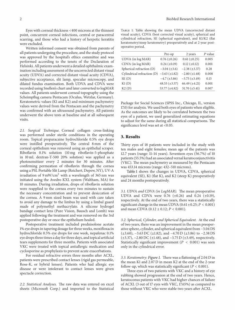
Table 1: Table showing the mean UDVA (uncorrected distantvisual acuity), CDVA (best corrected visual acuity), spherical andcylindrical refraction, SE (spherical equivalent), and K1/K2 (flatkeratometry/steep keratometry) preoperatively and at 2-year post-operative period.
Pre-op 2 years 𝑃 valueUDVA (in logMAR) 0.76 (±0.26) 0.61 (±0.25) 0.005CDVA (in logMAR) 0.24 (±0.19) 0.12 (±0.12) 0.001Spherical refraction (D) −3.04 (±3.6) −2.38 (±3.37) 0.28Cylindrical refraction (D) −3.63 (±1.82) −2.80 (±1.48) 0.008SE (D) −4.7 (±3.86) −3.75 (±3.49) 0.15K1 (D) 48.53 (±3.57) 46.49 (±4.21) 0.001K2 (D) 53.77 (±4.82) 51.70 (±5.41) 0.007
Package for Social Sciences (SPSS Inc., Chicago, IL, version17.0) for analysis. We used both eyes of patients when eligible.As the outcomes are likely to be correlated between the twoeyes of a patient, we used generalized estimating equationsto adjust for the same during all statistical comparisons. Thesignificance level was set at <0.05.
3. Results
Thirty eyes of 18 patients were included in the study withten males and eight females; mean age of the patients was12.7 years (range: 11–14 years). Seventeen eyes (56.7%) of 10patients (55.5%) had an associated vernal keratoconjunctivitis(VKC). The mean pachymetry as measured by the Pentacamwas 453.14 microns (range: 432–510 microns).
Table 1 shows the changes in UDVA, CDVA, sphericalequivalent (SE), K1 (flat K), and K2 (steep K) preoperativelyand 24 months postoperatively.
3.1. UDVA and CDVA (in LogMAR). The mean preoperativeUDVA and CDVA were 0.76 (±0.26) and 0.24 (±0.19),respectively. At the end of two years, there was a statisticallysignificant change in themeanUDVA (0.61±0.25;𝑃 < 0.001)and mean CDVA (0.12 ± 0.12; 𝑃 < 0.001).
3.2. Spherical, Cylinder, and Spherical Equivalent. At the endof two years, there was an improvement in themean preoper-ative sphere, cylinder, and spherical equivalent from−3.04DS(±3.60), −3.63DC (±1.82), and −4.70D (±3.86) to −2.38DS(±3.37), −2.80DC (±1.48), and −3.75D (±3.49), respectively.Statistically significant improvement (𝑃 < 0.001) was seenonly in the cylindrical error.
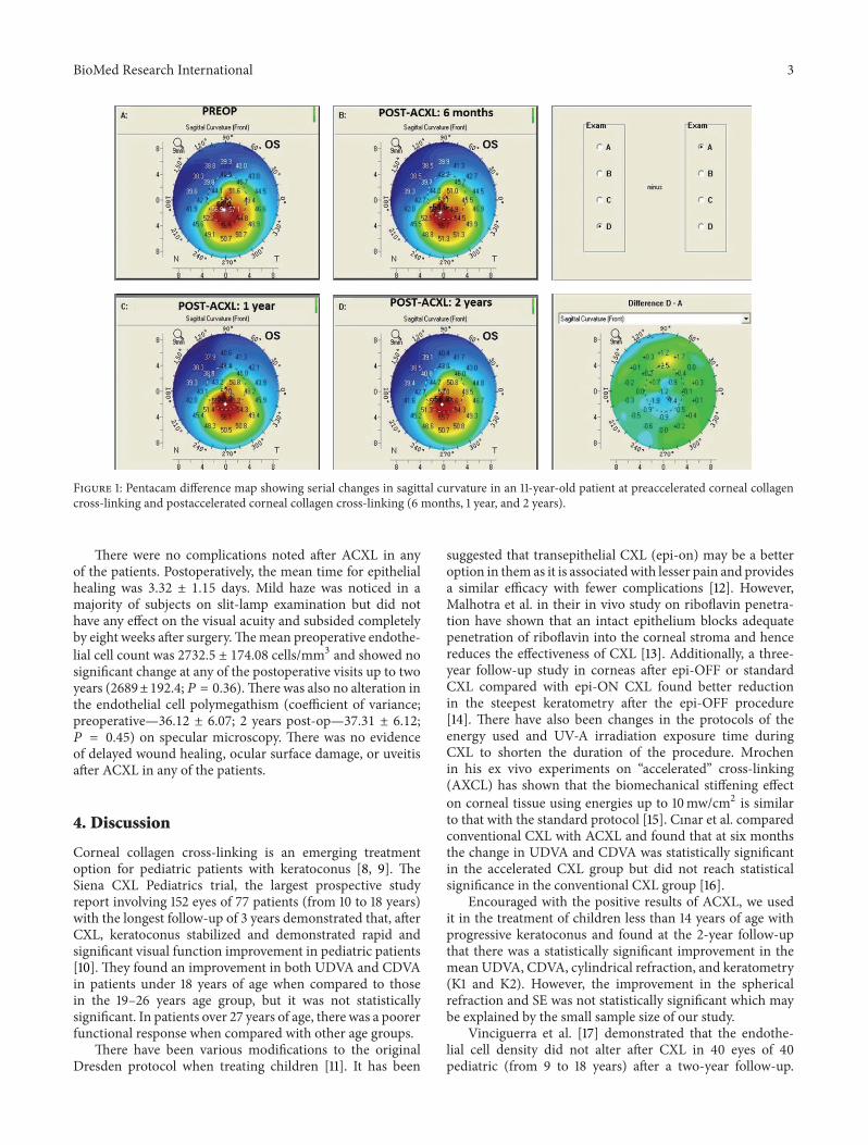
3.3. Keratometry: Figure 1. There was a flattening of 2.04D inthe mean K1 and 2.07D in mean K2 at the end of the 2-yearfollow-up, which was statistically significant (𝑃 < 0.001).
Three eyes of two patients with VKC and a history of eyerubbing showed progression at the end of two years. Hence,keratoconus patients with VKC had higher chances of failureof ACXL (3 out of 17 eyes with VKC, 17.65%) as compared tothose without VKC who were stable two years after ACXL.
BioMed Research International 3
Figure 1: Pentacam difference map showing serial changes in sagittal curvature in an 11-year-old patient at preaccelerated corneal collagencross-linking and postaccelerated corneal collagen cross-linking (6 months, 1 year, and 2 years).
There were no complications noted after ACXL in anyof the patients. Postoperatively, the mean time for epithelialhealing was 3.32 ± 1.15 days. Mild haze was noticed in amajority of subjects on slit-lamp examination but did nothave any effect on the visual acuity and subsided completelyby eight weeks after surgery.Themean preoperative endothe-lial cell count was 2732.5 ± 174.08 cells/mm3 and showed nosignificant change at any of the postoperative visits up to twoyears (2689±192.4; 𝑃 = 0.36).There was also no alteration inthe endothelial cell polymegathism (coefficient of variance;preoperative—36.12 ± 6.07; 2 years post-op—37.31 ± 6.12;𝑃 = 0.45) on specular microscopy. There was no evidenceof delayed wound healing, ocular surface damage, or uveitisafter ACXL in any of the patients.
4. Discussion
Corneal collagen cross-linking is an emerging treatmentoption for pediatric patients with keratoconus [8, 9]. TheSiena CXL Pediatrics trial, the largest prospective studyreport involving 152 eyes of 77 patients (from 10 to 18 years)with the longest follow-up of 3 years demonstrated that, afterCXL, keratoconus stabilized and demonstrated rapid andsignificant visual function improvement in pediatric patients[10]. They found an improvement in both UDVA and CDVAin patients under 18 years of age when compared to thosein the 19–26 years age group, but it was not statisticallysignificant. In patients over 27 years of age, there was a poorerfunctional response when compared with other age groups.
There have been various modifications to the originalDresden protocol when treating children [11]. It has been
suggested that transepithelial CXL (epi-on) may be a betteroption in them as it is associatedwith lesser pain and providesa similar efficacy with fewer complications [12]. However,Malhotra et al. in their in vivo study on riboflavin penetra-tion have shown that an intact epithelium blocks adequatepenetration of riboflavin into the corneal stroma and hencereduces the effectiveness of CXL [13]. Additionally, a three-year follow-up study in corneas after epi-OFF or standardCXL compared with epi-ON CXL found better reductionin the steepest keratometry after the epi-OFF procedure[14]. There have also been changes in the protocols of theenergy used and UV-A irradiation exposure time duringCXL to shorten the duration of the procedure. Mrochenin his ex vivo experiments on “accelerated” cross-linking(AXCL) has shown that the biomechanical stiffening effecton corneal tissue using energies up to 10mw/cm2 is similarto that with the standard protocol [15]. Cınar et al. comparedconventional CXL with ACXL and found that at six monthsthe change in UDVA and CDVA was statistically significantin the accelerated CXL group but did not reach statisticalsignificance in the conventional CXL group [16].
Encouraged with the positive results of ACXL, we usedit in the treatment of children less than 14 years of age withprogressive keratoconus and found at the 2-year follow-upthat there was a statistically significant improvement in themean UDVA, CDVA, cylindrical refraction, and keratometry(K1 and K2). However, the improvement in the sphericalrefraction and SE was not statistically significant which maybe explained by the small sample size of our study.
Vinciguerra et al. [17] demonstrated that the endothe-lial cell density did not alter after CXL in 40 eyes of 40pediatric (from 9 to 18 years) after a two-year follow-up.

4 BioMed Research International
In our study with ACXL, we found similar results of nosignificant reduction in the endothelial cell count at theend of two years. Failure of CXL to arrest progressionof keratoconus is attributed to different genetic patterns,relative biomechanical modifications potentially occurringin the corneal stroma and the negative influences of otherconditions such as allergy and atopy [18–21]. Ghosh et al.studied reviewed various proteomic and genomic expressionsand discoveredmolecules that can serve as biomarkers whichmay have potential role in the management of keratoconus[20]. They also found that a history of allergy, atopy (eczema,asthma, and hayfever), corneal injury, eye rubbing, and rigidcontact lens usage has been shown to be associated with thedevelopment of keratoconus. In our studywe noticed a higherincidence of VKC in children with progressive keratoconus(55.5%).There was also a failure to stabilize keratoconus withACXL in these patients (17.65%). Hence, management of theunderlying cause in such children is of prime importance aspersistent eye rubbingmay “nullify” the effect of CXL. Topicaltreatment with steroids, mast cell stabilizers, and also the useof immunomodulators such as cyclosporine eye drops mayhelp in alleviating allergy.
Other factors that may influence treatment are limbalstem cell (LSC) damage caused by chronic VKC [22] andUV-A damage [23]. The change in ocular flora due to chronicuse of topical steroids in keratoconus patients with VKCcan lead to an increased risk of postoperative keratitis [24].Hence, while using ACXL to treat children, we recommendthe usage of a limbal guard to protect the LSCs from damageand stoppage of the use of topical steroids (once the VKC iscontrolled) for at least two weeks prior to the procedure.
5. Conclusion
Very few studies have been published about the effectivenessof CXL in the younger age group. To the best of ourknowledge this is the first study to evaluate the effectivenessof ACXL in children less than 14 years of age. The higherenergy and shorter duration of treatment (9mW/cm2 for10mins) of ACXLmay prove to be a good option in children.It can potentially prevent amblyopia, improve the fit ofcontact lenses, and deter an early penetrating keratoplasty.For children with keratoconus and chronic VKC undergoingCXL, it would be prudent to treat the allergy aggressivelywith topical steroids during the active phase and with topicalantiallergics and topical cyclosporine in chronic cases toreduce the chances of failure of CXL.
Conflict of Interests
The authors have no proprietary or commercial interests inany concept or product discussed in this paper.
References
[1] J. Vazirani and S. Basu, “Keratoconus: current perspectives,”Clinical Ophthalmology, vol. 7, pp. 2019–2030, 2013.
[2] M. K. Smolek and W. H. Beekhuis, “Collagen fibril orientationin the human corneal stroma and its implications in kerato-conus,” Investigative Ophthalmology & Visual Science, vol. 38,no. 7, pp. 1289–1290, 1997.
[3] U. Rehany and S. Rumelt, “Corneal hydrops associated withvernal conjunctivitis as a presenting sign of keratoconus inchildren,” Ophthalmology, vol. 102, no. 12, pp. 2046–2049, 1995.
[4] S. W. Reeves, S. Stinnett, R. A. Adelman, and N. A. Afshari,“Risk factors for progression to penetrating keratoplasty inpatients with keratoconus,”American Journal of Ophthalmology,vol. 140, no. 4, pp. 607.e1–607.e6, 2005.
[5] N. Chatzis and F. Hafezi, “Progression of keratoconus andefficacy of corneal collagen cross-linking in children and ado-lescents,” Journal of Refractive Surgery, vol. 28, no. 11, pp. 753–758, 2012.
[6] E. Spoerl, G. Wollensak, and T. Seiler, “Increased resistance ofcrosslinked cornea against enzymatic digestion,” Current EyeResearch, vol. 29, no. 1, pp. 35–40, 2004.
[7] E. Sporl, F. Raiskup-Wolf, and L. E. Pillunat, “Biophysicalprinciples of collagen cross-linking,”KlinischeMonatsblatter furAugenheilkunde, vol. 225, no. 2, pp. 131–137, 2008.
[8] R. Arora, D. Gupta, J. L. Goyal, and P. Jain, “Results ofcorneal collagen cross-linking in pediatric patients,” Journal ofRefractive Surgery, vol. 28, no. 11, pp. 759–762, 2012.
[9] P. G. Zotta, K. A. Moschou, V. F. Diakonis et al., “Cornealcollagen cross-linking for progressive keratoconus in pediatricpatients: a feasibility study,” Journal of Refractive Surgery, vol.28, no. 11, pp. 793–796, 2012.
[10] A. Caporossi, C.Mazzotta, S. Baiocchi, T. Caporossi, R. Denaro,and A. Balestrazzi, “Riboflavin-UVA-induced corneal collagencross-linking in pediatric patients,” Cornea, vol. 31, no. 3, pp.227–231, 2012.
[11] V. P. Kankariya, G. D. Kymionis, V. F. Diakonis et al., “Manage-ment of pediatric keratoconus—evolving role of corneal colla-gen cross-linking: an update,” Indian Journal of Ophthalmology,vol. 61, no. 8, pp. 435–440, 2013.
[12] A. Magli, R. Forte, A. Tortori, L. Capasso, G. Marsico, and E.Piozzi, “Epithelium-off corneal collagen cross-linking versustransepithelial cross-linking for pediatric keratoconus,”Cornea,vol. 32, no. 5, pp. 597–601, 2013.
[13] C. Malhotra, R. Shetty, R. S. Kumar, H. Veluri, H. Nagaraj, andK. B. Shetty, “In vivo imaging of riboflavin penetration duringcollagen cross-linking with hand-held spectral domain opticalcoherence tomography,” Journal of Refractive Surgery, vol. 28,no. 11, pp. 776–780, 2012.
[14] L. Gualdi, “Epion crosslinking: 3 years results and suggestionsfor the selection of the patient,” in Proceedings of the Interna-tional Congress of Corneal Cross Linking, Geneva, Switzerland,December 2012.
[15] M. Mrochen, “Current status of accelerated corneal cross-linking,” Indian Journal of Ophthalmology, vol. 61, no. 8, pp.428–429, 2013.
[16] Y. Cınar, A. K. Cingu, F. M. Turkcu et al., “Comparison ofaccelerated and conventional corneal collagen cross-linking forprogressive keratoconus,”Cutaneous andOcular Toxicology, vol.33, no. 3, pp. 218–222, 2014.
[17] P. Vinciguerra, E. Albe, B. E. Frueh, S. Trazza, and D. Epstein,“Two-year corneal cross-linking results in patients youngerthan 18 years with documented progressive keratoconus,”Amer-ican Journal of Ophthalmology, vol. 154, no. 3, pp. 520–526, 2012.
BioMed Research International 5
[18] T. Koller, M. Mrochen, and T. Seiler, “Complication andfailure rates after corneal crosslinking,” Journal of Cataract andRefractive Surgery, vol. 35, no. 8, pp. 1358–1362, 2009.
[19] T. Georgiou, C. L. Funnell, A. Cassels-Brown, and R. O’Conor,“Influence of ethnic origin on the incidence of keratoconus andassociated atopic disease in Asians and white patients,” Eye, vol.18, no. 4, pp. 379–383, 2004.
[20] A.Ghosh, L. Zhou, A. Ghosh et al., “Proteomic and gene expres-sion patterns of keratoconus,” Indian Journal of Ophthalmology,vol. 61, no. 8, pp. 389–391, 2013.
[21] A. S. Roy, R. Shetty, and M. K. Kummelil, “Keratoconus: abiomechanical perspective on loss of corneal stiffness,” IndianJournal of Ophthalmology, vol. 61, no. 8, pp. 392–393, 2013.
[22] V. S. Sangwan, V. Jain, G. K. Vemuganti, and S. I. Murthy,“Vernal keratoconjunctivitis with limbal stem cell deficiency,”Cornea, vol. 30, no. 5, pp. 491–496, 2011.
[23] H. Matalia, R. Shetty, K. Dhamodaran, M. Subramani, V.Arokiaraj, and D. Das, “Potential apoptotic effect of ultraviolet-A irradiation during cross-linking: a study on ex vivo cultivatedlimbal epithelial cells,”British Journal of Ophthalmology, vol. 96,no. 10, pp. 1339–1345, 2012.
[24] S. S. Ermis, O. C. Aktepe, U. U. Inan, F. Ozturk, andM. Altindis,“Effect of topical dexamethasone and ciprofloxacin on bacterialflora of healthy conjunctiva,” Eye, vol. 18, no. 3, pp. 249–252,2004.

Submit your manuscripts athttp://www.hindawi.com
Stem CellsInternational
Hindawi Publishing Corporationhttp://www.hindawi.com Volume 2014
Hindawi Publishing Corporationhttp://www.hindawi.com Volume 2014
MEDIATORSINFLAMMATION
of
Hindawi Publishing Corporationhttp://www.hindawi.com Volume 2014
Behavioural Neurology
EndocrinologyInternational Journal of
Hindawi Publishing Corporationhttp://www.hindawi.com Volume 2014
Hindawi Publishing Corporationhttp://www.hindawi.com Volume 2014
Disease Markers
Hindawi Publishing Corporationhttp://www.hindawi.com Volume 2014
BioMed Research International
OncologyJournal of
Hindawi Publishing Corporationhttp://www.hindawi.com Volume 2014
Hindawi Publishing Corporationhttp://www.hindawi.com Volume 2014
Oxidative Medicine and Cellular Longevity
Hindawi Publishing Corporationhttp://www.hindawi.com Volume 2014
PPAR Research
The Scientific World JournalHindawi Publishing Corporation http://www.hindawi.com Volume 2014
Immunology ResearchHindawi Publishing Corporationhttp://www.hindawi.com Volume 2014
Journal of
ObesityJournal of
Hindawi Publishing Corporationhttp://www.hindawi.com Volume 2014
Hindawi Publishing Corporationhttp://www.hindawi.com Volume 2014
Computational and Mathematical Methods in Medicine
OphthalmologyJournal of
Hindawi Publishing Corporationhttp://www.hindawi.com Volume 2014
Diabetes ResearchJournal of
Hindawi Publishing Corporationhttp://www.hindawi.com Volume 2014
Hindawi Publishing Corporationhttp://www.hindawi.com Volume 2014
Research and TreatmentAIDS
Hindawi Publishing Corporationhttp://www.hindawi.com Volume 2014
Gastroenterology Research and Practice
Hindawi Publishing Corporationhttp://www.hindawi.com Volume 2014
Parkinson’s Disease
Evidence-Based Complementary and Alternative Medicine
Volume 2014Hindawi Publishing Corporationhttp://www.hindawi.com


