Int. J. Pharm. Sci. Rev. Res., 45(2), July - August 2017; Article No. 08, Pages: 36-47 ISSN 0976 – 044X
International Journal of Pharmaceutical Sciences Review and Research International Journal of Pharmaceutical Sciences Review and Research Available online at www.globalresearchonline.net
© Copyright protected. Unauthorised republication, reproduction, distribution, dissemination and copying of this document in whole or in part is strictly prohibited. Available online at www.globalresearchonline.net
36
1Gore S. A., 1Satpute V. V., 1Gholve S. B.*, 1Bhusnure O. G., 2Bhosale P. H., 3Phutke P. B 1Channabasweshwar Pharmacy College (Degree), India.
2 Channabasweshwar Pharmacy Polytechnic, India. Kava Road, BasweshwarChowk, Latur, Maharashtra, India.
3 WarpudkarInstitute of Pharmacy, Pedgaon, Dist. Parbhani, (MS), India. *Corresponding author’s E-mail: [email protected]
Received: 03-05-2017; Revised: 12-07-2017; Accepted: 28-07-2017.
ABSTRACT
Innovations in transdermal delivery systems (TDS) have made important contributions to medical practice by providing advances in the delivery of treatment with existing and novel drugs. Today about 74% of drugs are taken orally and are found not to be as effective as desired. To improve such characters transdermal drug delivery system was emerged. Drug delivery through the skin to achieve a systemic effect of a drug is commonly known as transdermal drug delivery and differs from traditional topical drug delivery. Transdermal drug delivery system can improve the therapeutic efficacy and safety of the drugs because drug delivered through the skin at a predetermined and controlled rate. Skin is the important site of drug application for both the local and systemic effects. Transdermal drug delivery system (TDDS) has several advantages over conventional system; TDDS offers sustained drug release, avoidance of first pass effect, patient compliance, ease of application and removal in case of toxicity as well as decrease in the side effects as compared with conventional therapy. The stratum corneum acts as a barrier that limits the penetration of substances through the skin and this limitation can be overcome by permeation enhancing techniques. The main disadvantage to transdermal delivery systems stems from the fact that the skin is a very effective barrier; as a result, only medications whose molecules are small enough to penetrate the skin can be delivered in this method. A wide variety of pharmaceuticals are now available in transdermal patch form. Characterization of transdermal patch is use to check it’s quality, size, time of onset & duration, adhesive property, thickness, weight of patch, moisture of content, uniformity & cutaneous toxicological studies.
Keywords: Transdermal Patches, Transdermal Drug Delivery System, Skin, Novel approaches in transdermal drug delivery system.
INTRODUCTION
irst transdermal patch approved in 1979 by FDA was of Scopolamine for motion sickness. Second patch approved in 1981 was of Nitroglycerine. Now a
day’s several patches are available in market for transdermal use. Some of them are: Clonidine, Testosterone, Fentanyl, Nicotine, Hormones etc. these patches usually applied from 1to7 days depending uponvarious conditions. Oral route is most commonly used route for drug delivery, but due to some major shortcomings such as poor B.A., first pass effect and the ability to create fluctuation of drug level in blood.
1
Topical or Transdermal delivery of anti-analgesic drugs has gained prominence in recent years, owing to its ability to provide concentrated and highly localized pain relief directly to a specific area of the body, unlike oral drug delivery which often causes side-effects as it winds its way through the gastrointestinal tract. However, in spite of its benefits like targeted and concentrated drug delivery, the transdermal application of hydrophobic drugs is significantly limited by the outermost layer of human skin (stratum corneum). 2
Transdermal delivery not only provides controlled, constant administration of the drug, but also allows
continuous input of drugs with short biological half-lives and eliminates pulsed entry into systemic circulation, which often causes undesirable side effects. Thus various forms of Novel drug delivery system such as Transdermal drug delivery systems, Controlled release systems, Transmucosal delivery systems etc.
Several important advantages of transdermal drug delivery are limitation of hepatic first pass metabolism, enhancement of therapeutic efficiency and maintenance of steady plasma level of the drug. The first Transdermal system, Transderm-SCOP was approved by FDA in 1979 for the prevention of nausea and vomiting associated with travel, particularly by sea. The evidence of percutaneous drug absorption may be found through measurable blood levels of the drug, detectable excretion of the drug and its metabolites in the urine and through the clinical response of the patient to the administered drug therapy. 3
Selection of drug candidate for transdermal delivery The transdermal route of administration cannot be employed for a large number of drugs. Judicious choice of the drug substance is the most important decision in the successful development of a
A Systematic Review on Transdermal Patches
F
Review Article
Int. J. Pharm. Sci. Rev. Res., 45(2), July - August 2017; Article No. 08, Pages: 36-47 ISSN 0976 – 044X
International Journal of Pharmaceutical Sciences Review and Research International Journal of Pharmaceutical Sciences Review and Research Available online at www.globalresearchonline.net
© Copyright protected. Unauthorised republication, reproduction, distribution, dissemination and copying of this document in whole or in part is strictly prohibited. Available online at www.globalresearchonline.net
37
transdermal system. The drug candidate should have following ideas characteristics:
Adequate skin permeability
Drugs with low molecular weight
Drugs with low melting point
Drugs with moderate oil and water solubility
Adequate skin acceptability
Non-irritating drugs
Non-metabolizing drugs
Adequate clinical need
Need to prolong administration
Need to reduce side effects on target tissues
Need to increase patient compliance.4
Advantages of Transdermal drug delivery over the conventional dosage forms
1. The Transdermal drug delivery system (TDDS) can be defined as a delivery device, which upon application on a suitable skin surface will be able to deliver the drug into the systemic circulation at sufficient concentration to ensure therapeutic efficacy, an additional limitation to oral drug delivery, can be avoided with transdermal administration.
2. Steady permeation of drug across the skin, allowing consistent serum drug level, often a Goal of therapy.
3. Similar to intravenous infusion, it also achieves consistent plasma levels, but noninvasive in nature.
4. In addition, if toxicity develops from a drug administered transdermally, the effects could be moderated by removing the patch.
5. Transdermal drug delivery can be used as an alternative delivery system for patients who cannot tolerate oral dosage forms.
6. It is of great advantage in patients who are nauseated or unconscious.
7. Drugs that cause gastrointestinal upsets can be good candidates for transdermal delivery because this method avoids direct effects on stomach and intestine.5,6,7
8. Topical patches are a painless, non-invasive way to deliver substances directly into the body.
9. Topical patches over a controlled, steady delivery of medication over long periods of time.
10. Topical patches have fewer side effects than oral medications or supplements.
11. Topical patches over an alternative to people who cannot, or prefer not to take Medications or supplements orally.
12. Topical patches are cost-effective. 13. People prefer topical patches.
8
Disadvantages of TDDS
1. Some drugs penetrate into the skin slowly which may affect the efficacy of the treatment.
2. Example: hydrophilic drugs. 3. The components in the TDDS formulation
may produce skin irritation, local edema& erythema.
4. Damage to a transdermal patch may cause the poor control on the release rate.
5. The barrier function of the skin changes from one site to another on the same person from person to person and with age.
6. Adhere of patches on the skin is very difficult. 7. It is not suitable for drugs with higher doses. 8. It is not suitable for the substance having higher
molecular weight. 9. Drugs are metabolized by the skin and
undergoes protein binding in skin are not suitable in
10. TDDS formulation.9,10,11
Components of Transdermal Pathches
Polymer Matrix
The polymer controls the release of the drug fromthe device. The following criteria should be satisfied for a polymer to be used in transdermal patches.
(a) Molecular weight, chemical functionality of the polymer should be such that the specific drug diffuses properly and gets released through it.
(b) The polymer should be stable.
(c) The polymer should be nontoxic
(d) The polymer should be easily of manufactured
(e) The polymer should be inexpensive
(f) The polymer and its degradation product must be nontoxic or non-antagonistic to the host.
(g) Large amounts of the active agent are incorporated into it.
Types of polymer
Natural polymers
Cellulose derivative, Gelatin, Waxes, Proteins, Gum, Shellac, Natural rubber, starch.
Synthetic Elastomers
Hydrin rubber, silicone rubber, Nitrile, Acrylonitrile, Neoprene.
Synthetic polymers
Polyvinyl alcohol, polyvinyl chloride, polyethylene, polypropylene, polyamiode, polyurea, epoxy.12
Int. J. Pharm. Sci. Rev. Res., 45(2), July - August 2017; Article No. 08, Pages: 36-47 ISSN 0976 – 044X
International Journal of Pharmaceutical Sciences Review and Research International Journal of Pharmaceutical Sciences Review and Research Available online at www.globalresearchonline.net
© Copyright protected. Unauthorised republication, reproduction, distribution, dissemination and copying of this document in whole or in part is strictly prohibited. Available online at www.globalresearchonline.net
38
(I) Drug
For successfully developing a Transdermal drug delivery system, the drug should be chosen with great care. The following are some of the desirable properties of a drug for Transdermal delivery.
Physicochemical Properties
The drug should have a molecular weight less than approximately 1000 Daltons.
The drug should have affinity for both- lipophilic and hydrophilic phases.
Extreme partitioning characteristics are not conducive to successful drug delivery via the skin.
The drug should have a low melting point.
Biological Properties
The drug should be potent with a daily dose of the order of a few mg/day.
The half-life (t1/2) of the drug should be short.
The drug must not induce a cutaneous irritation or allergic response.
Drugs, which degrade in the GI tract or are inactivated by hepatic first-pass effect, are suitable candidates for Transdermal delivery.
Tolerance to the drug must not develop under the near zero-order release profile of Transdermal delivery.
Drugs, which have to be administered for a long period of time or which cause adverse effects to non-target tissues can also, be formulated for Transdermal delivery.
Permeation Enhancers
Permeation enhancers or promoters are agents that have no therapeutic properties of their own but can transport the sorption of drugs from drug delivery systems onto the skin.
The flux, of drugs across the skin can be written as:
J = D Xdc/dx
Where D is the diffusion coefficient and is a function of size, shape and flexibility of the diffusing molecule as well as the membrane resistance; C is the concentration of the diffusing species; x is the spatial coordinate.
Although the solution for J with various boundary conditions and membrane heterogeneities can be very complex, the basic concepts regarding flux enhancement can be found in above equation. The concentration gradient is thermodynamic in origin, and the diffusion coefficient is related to the size and shape of penetrate and the energy required to make a hole for diffusion.
Thus enhancement of flux across membranes reduces to considerations of
Thermodynamics (lattice energies, distribution coefficients).
Molecular size and shape.
Reducing the energy required to make a molecular hole in the membrane.
Permeation enhancers are hypothesized to affect one or more of the layers to achieve skin penetration enhancement. A large number of compounds have been investigated for their ability to enhance stratum corneum permeability. These conveniently classified
Under the following main headings:
Solvents
These compounds increase penetration possibly by
1) Swelling the polar pathways in the skin.
2) Fluidization of lipids.
Examples include water alcohols-methanol and ethanol; alkyl methyl sulfoxides-dimethyl sulfoxide, alkyl homologs of methyl sulphoxide, dimethyl acetamide and dimethyl formamide; pyrrolidones-2-pyrrolidone; laurocapram (Azone), miscellaneous solvents-propylene glycol, glycerol, silicone fluids, isopropyl palmitate.
Surfactants
These compounds are proposed to enhance polar pathway transport, especially of hydrophilic drugs. The ability of a surfactant to alter penetration is a function of the head group and the hydrocarbon chain length. These compounds are skin irritants, therefore, a balance between penetration enhancement and irritation have to be considered. Anionic surfactant can penetrate and interact strongly with the skin. Once these surfactants have penetrated the skin, they can induce large alterations. Cationic surfactants are reportedly more irritant than the anionic surfactants and they have not been widely studied as skin permeation enhancers. Of the three major classes of surfactants, the nonionic have long been recognized as those with the least potential for irritation and have been widely studied.
Examples of commonly used surfactants are:
Anionic Surfactants
Dioctylsulphosuccinate, Sodium lauryl sulphate, Decodecylmethylsulphoxide etc.
Nonionic Surfactants
Pluronic F127, Pluronic F68, etc. Bile Salts: Sodiumtaurocholate, Sodium deoxycholate, Sodium tauroglycocholate.
Int. J. Pharm. Sci. Rev. Res., 45(2), July - August 2017; Article No. 08, Pages: 36-47 ISSN 0976 – 044X
International Journal of Pharmaceutical Sciences Review and Research International Journal of Pharmaceutical Sciences Review and Research Available online at www.globalresearchonline.net
© Copyright protected. Unauthorised republication, reproduction, distribution, dissemination and copying of this document in whole or in part is strictly prohibited. Available online at www.globalresearchonline.net
39
Miscellaneous Chemicals
These include urea, a hydrating and keratolytic agent; N, N-dimethyl-m-toluamide; Calcium thioglycolate; Anticholinergic agents. Some potential permeation enhancers have recently been described but the available data on their effectiveness are sparse. These include eucalyptol, di-o-methyl-beta-cyclodextrin and soyabean casein.
12, 13, 14
Lipid action
The enhancer interacts with the organized intracellular lipid structure of the stratum corneum so as to disrupt it and make it more permeable to drug molecules. Very many enhancers operate in this way. Some solvents act by extracting the lipid Components and thus make the horny layer more permeable.
Protein modification
Ionic surface active molecules in particular tend to interact well with the keratin in the corneocytes, to open up the dense keratin structure and make it more permeable. The intracellular route is not usually prominent in drug permeation, although drastic reductions to this routes resistance could open up an alternative path for drug penetration.
Partitioning promotion
Many solvents can enter the stratum corneum, change its solvent properties and thus increase the partitioning of a second molecule into the horny layer. This molecule may be a drug, a coenhancer or a cosolvent.
For example ethanol has been used to increase the penetration of the drug molecules
Adhesive
Serves to adhere the patch to the skin for systemic delivery of drug.
Ex: Silicones, Polyisobutylene.
Backing Layer
Backing layer protects patch from outer environment.
Ex: Cellulose derivatives, Polypropylene silicon rubber.15-23
Adhesives sensitive to pressure 24-26
It maintains a close contact between the skin surface & the transdermal system. It adheres with skin surface by applying finger pressure. Mostlyused adhesives in the preparation oftransdermal patches are:
1. Poly isobutylene
2. Polyacrylates
3. Silicone-based adhesives.
They can be placed on the front or back of the patch. Adhesives should possess following
Characteristics
Non irritant
Ease of removal
Firm contact with skin
Biocompatible
Do not affect drug permeation
Remove without leaving residue behind.
Backing laminates
Chemical resistance, excipient compatibility & flexibility along with better tensile strength must be considered in designing the backing layers. It must allow oxygen& water vapor transmission. At Present, backing layer itself act as a drug reservoir as compared to conventional dosage form. Generally following materials are used for the preparation of backing laminates:
Polyesters
Polyolefin
Low density polyethylene
Aluminum plastic laminate
Resin. 25,27,28
Skin
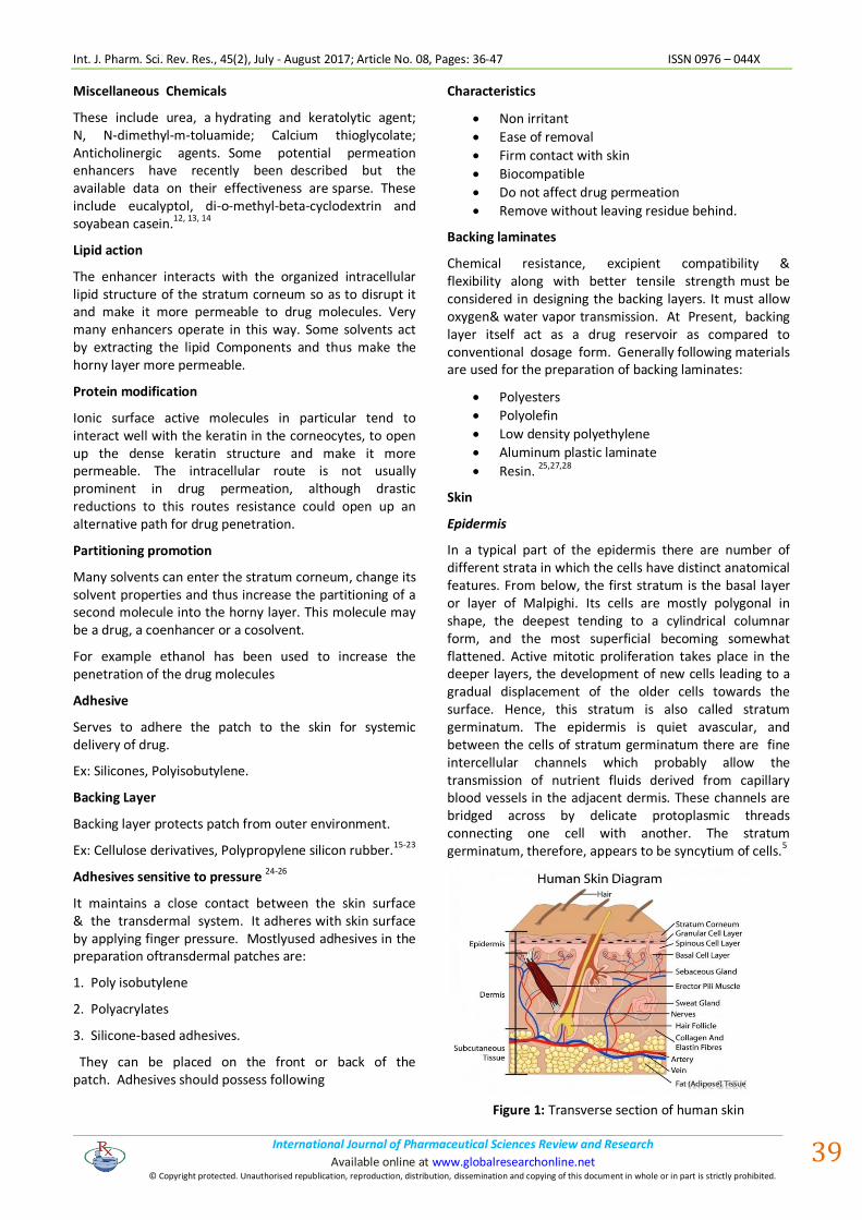
Epidermis
In a typical part of the epidermis there are number of different strata in which the cells have distinct anatomical features. From below, the first stratum is the basal layer or layer of Malpighi. Its cells are mostly polygonal in shape, the deepest tending to a cylindrical columnar form, and the most superficial becoming somewhat flattened. Active mitotic proliferation takes place in the deeper layers, the development of new cells leading to a gradual displacement of the older cells towards the surface. Hence, this stratum is also called stratum germinatum. The epidermis is quiet avascular, and between the cells of stratum germinatum there are fine intercellular channels which probably allow the transmission of nutrient fluids derived from capillary blood vessels in the adjacent dermis. These channels are bridged across by delicate protoplasmic threads connecting one cell with another. The stratum germinatum, therefore, appears to be syncytium of cells.5
Figure 1: Transverse section of human skin
Int. J. Pharm. Sci. Rev. Res., 45(2), July - August 2017; Article No. 08, Pages: 36-47 ISSN 0976 – 044X
International Journal of Pharmaceutical Sciences Review and Research International Journal of Pharmaceutical Sciences Review and Research Available online at www.globalresearchonline.net
© Copyright protected. Unauthorised republication, reproduction, distribution, dissemination and copying of this document in whole or in part is strictly prohibited. Available online at www.globalresearchonline.net
40
Above stratum granulosamis stratumlucidam - it called so, because it has a clear translucent appearance in stained sections. At this level of their growth towards the surface, the cells have lost their clear-cut outline, the nuclei are becoming indistinct, and the granules of the subjacent stratum have become converted in to larger masses of an achromatic substance. The surface stratum forms the greater part of’ the whole thickness of the epidermis in many parts of the skin. Because of’ horny character of the cellular elements that composes this layer; henceforth it is called the stratum corneum. In stratum corneum the cell structure has become completely obscured and nuclei are no longer evident.
30
Dermis
The dermis or corium consists of a dense felt work of connective tissue in which bundles of collagenous fibers predominate, mingled with a certain proportion of elastic tissue in superficial levels. Dermis contains fine plexuses of blood vessels, lymphaticsand nerves, hair follicles, sweat glands and sebaceous glands.31, 32 the thicker the epidermis; therefore, the more prominent are the papillae.33
Factors Affecting Transdermal Bioavailability
(Physicochemical factors)
Skin hydration
In contact with water the permeability of skin increases significantly. Hydration is most important factor increasing the permeation of skin. So use of humectants is done in transdermal delivery.
Temperature and pH
The permeation of drug increase ten folds with temperature variation. The diffusion coefficient decreases as temperature falls. Weak acids and weak bases dissociate depending on the pH and pKa or pKb values. The proportion of unionized drug determines the drugconcentration in skin. Thus, temperature and pH are important factors affecting drug penetration.
Diffusion Coefficient
Penetration of drug depends on diffusion coefficient of drug. At a constant temperature, the diffusion coefficient of drug depends on properties of drug, diffusion medium and interaction between them3
Drug Concentration
The flux is proportional to the concentration gradient across the barrier and concentration gradient will be higher if the concentration of drug will be more across the barrier.
Partition Coefficient
The optimal partition coefficient (K) is required for good action. Drugs with high K are not ready to leave the lipid portion of skin. Also, drugs with low K will not be permeated. Molecular Size and Shape Drug absorption is
inversely related to molecular weight, small molecules penetrate faster than large ones.
Biological Factors
Skin Condition
Chloroform, methanols damage the skin cells and promotes penetration. Diseased state of patient alters the skin conditions. The intact skin is better barrier but the above mentioned conditions affect penetration.
Skin age
The young skin is more permeable than older. Children are more sensitive for skin absorption of toxins. Thus, skin age is one of the factors affecting penetration of drug in TDDS.
Blood flow
Changes in peripheral circulation can affect Transdermal absorption.
Regional Skin Sites
Thickness of skin, nature of stratum corneum and density of appendages vary site to site. These factors affect significantly penetration.
Skin metabolism
Skin metabolizes steroids, hormones, chemical carcinogens and some drugs. So skin metabolism determines efficacy of drug permeated through the skin.
Species Differences
The skin thickness, density of appendages and keratinization of skin vary species to species, so affects the penetration.34
Pathways of Drug Absorption through the Skin
The drug can be absorbed by various pathways through the skin depending on the physicochemical properties of the drug. Both lipophilic and hydrophilic drugs are absorbed from different routes. The upper stratum corneum of the skin opposes the absorption of drug but presence of various absorption routes facilitates the entry of drug and transport of drug to the systemic circulation. Various drug absorption routes (figure 1) are as follows:
Transfollicular route
Transfollicular route is the shortest pathway that drug has to follow to reach the systemic circulation that provides a large area for diffusion of drugs. Skin has various sweat glands, oil glands, hair follicles and pores opening to the outer surface of the skin via their ducts. These ducts offer a continuous channel across the stratum corneum for drug transport but various factors like secretion from glands, content and amount of secretion etc., affect the transport of drugs through this route. However Trans appendage route occupies

Int. J. Pharm. Sci. Rev. Res., 45(2), July - August 2017; Article No. 08, Pages: 36-47 ISSN 0976 – 044X
International Journal of Pharmaceutical Sciences Review and Research International Journal of Pharmaceutical Sciences Review and Research Available online at www.globalresearchonline.net
© Copyright protected. Unauthorised republication, reproduction, distribution, dissemination and copying of this document in whole or in part is strictly prohibited. Available online at www.globalresearchonline.net
41
only 0.1% of total skinsurface and therefore contributes a little.1
Transcellular route
Drug delivering through this route passes from corneocytes which has highly hydrated keratin creating hydrophilic pathway. Corneocytesare surrounded bylipids connecting these cells. So a drug requires a number of partitioning and diffusion steps. It is the most widelyused route by various types of drugs. In transcellularroute drug passes through the matrix (cytoplasm) of thecells. This route is suitable for hydrophilic drugs. The drug passes through the corneocytes of stratum corneum. The highly hydrated keratin provide aqueous pathway to thehydrophilic drugs. A number of partitioning and diffusion steps are needed to pass the drug through the cell matrix12
Intercellular route
As name indicates in intercellular pathway the drug diffuses through the continuous lipid matrix present between the cells. The barrier property of this route is due tortuous structure formed by corneocytes and the drug has to pass through the alternating lipid and aqueous domain by partitioning into the lipid bilayer and diffusing to the inner side. It has been found that water has to travel 50 times more by this route so; it is suitable mainly for uncharged lipophilic drugs.35
Approaches to Development Transdermal Therapeutic Systems
Various technologies have been successfully developed to provide a rate control over the release and the transdermal permeation of a drugs. These technologies can be classified into two major categories as follows.
Rate-programmed transdermal DDS
1. Membrane permeation-controlled systems 2. Adhesive dispersion-type systems. 3. Matrix diffusion-controlled systems. 4. Micro reservoir type or micro sealed dissolution
controlled systems.
Physical stimuli-activated transdermal DDS
1. Structure based
1. Microneedles 2. Macroflux 3. MDTS
2. Electrically based
1. Iontophoresis 2. Ultrasound 3. Photochemical waves 4. Electroporation 5. Electroosmosis
3. Velocity based
1. Powder jet
2. Needle free injection
4. Others
1. Transferosomes 2. Medicated tattoos 3. Skin abrasion 4. Heat 5. Laser radiation 6. Magnetophoresis.
36
Membrane permeation – controlled system
These system can be multilaminate processe.g. Transdermal Nitro. These products consist of three substrates held together by two layers of drug containing adhesive. First the drug is processed into the physical / chemical form required for incorporation into the product. Then the drug adhesive components and excipient are mixed with a solvent to achieve uniform solution. These adhesive composition are deposited as a thin film on moving substances rate which are subsequently dried to remove solvent. Then lamination of the dried adhesive film and other layer to form the five layer product consisting of release linear contact adhesive control membrane, drug reservoir and backing substrate. The lamination then printed and die cut into final dosage form. The production isthen packed in individual foil pouches. After inspection the products are automatically inserted into a continuously moving web of pouch stock which is sealed around the dosage form.12
Adhesive Dispersion-Type Systems
Figure 2: Adhesive Dispersion-Type Systems
This is a simplified form of membrane permeation controlled system. The drug reservoir is formulated by directly dispersing drug in an adhesive polymer e.g. poly (acrylate) or Poly(isobutylene) adhesive and then spreading the medicated adhesive, by hot melt or solvent casting, on to a flat sheet of drug impermeable metallic plastic backing to form a thin drug reservoir layer. On the top of drug reservoir layer, thin layers of non-medicated, rate-controlling adhesive polymer of a specific permeability & constant thickness are applied to manufacture an adhesive diffusion-controlled delivery system.
Int. J. Pharm. Sci. Rev. Res., 45(2), July - August 2017; Article No. 08, Pages: 36-47 ISSN 0976 – 044X
International Journal of Pharmaceutical Sciences Review and Research International Journal of Pharmaceutical Sciences Review and Research Available online at www.globalresearchonline.net
© Copyright protected. Unauthorised republication, reproduction, distribution, dissemination and copying of this document in whole or in part is strictly prohibited. Available online at www.globalresearchonline.net
42
Releases of Isosorbide dinitrate for once-a-day medication of angina pectoris This system is characterized by the inclusion of a liquid compartment containing a drug suspension or solution separated from release liner by a semi-permeable membrane and adhesive. The adhesive component of product responsible for skin adhesion can either incorporated as a continuous layer between the membrane and release liner or in concentric configuration around membrane. The rate of drug release from this system is given by,
dQ/dt = Ka/r. Da A ( ha ) ∕ha ( t )
In the above equation, the thickness of adhesive layer for drug molecules to diffuse through enhances with time ha (t). To compensate this time dependent increase in diffusional path due to the depletion of drug dose by release, drug loading level is also enhanced with the thickness of diffusion path A (ha). In the above equation, thickness of adhesive layer to diffuse through enhances with time ha (t).
Matrix diffusion controlled systems
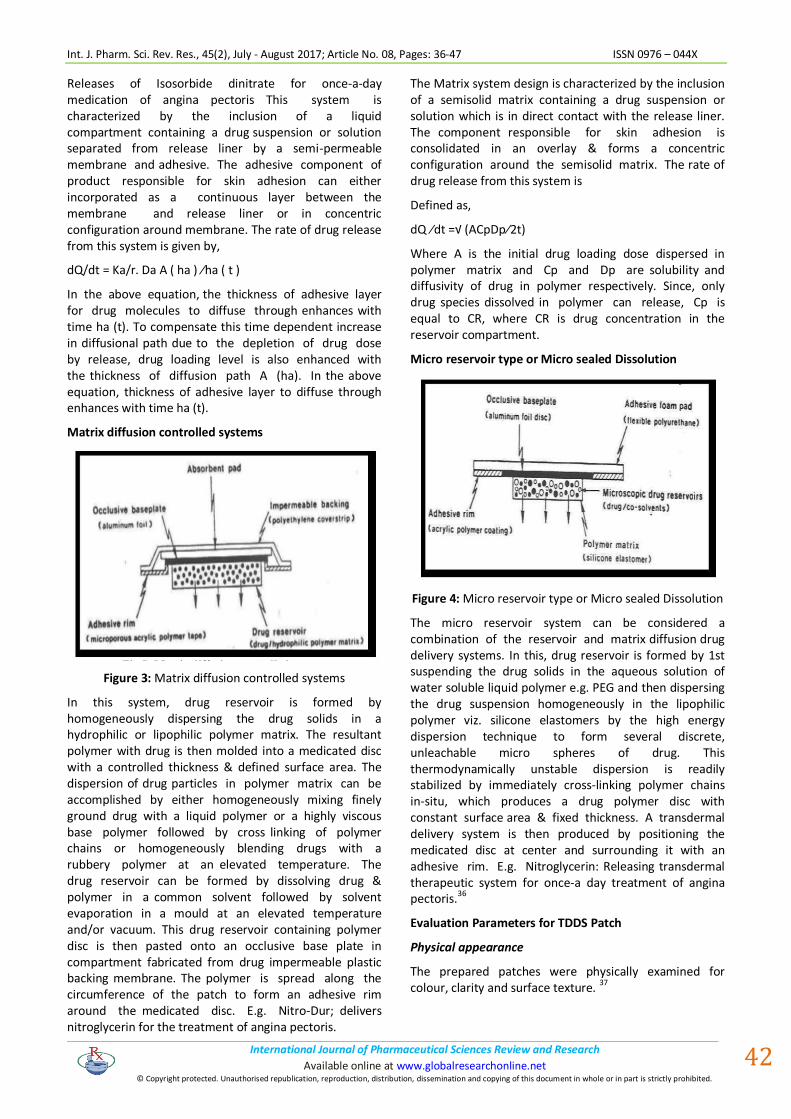
Figure 3: Matrix diffusion controlled systems
In this system, drug reservoir is formed by homogeneously dispersing the drug solids in a hydrophilic or lipophilic polymer matrix. The resultant polymer with drug is then molded into a medicated disc with a controlled thickness & defined surface area. The dispersion of drug particles in polymer matrix can be accomplished by either homogeneously mixing finely ground drug with a liquid polymer or a highly viscous base polymer followed by cross linking of polymer chains or homogeneously blending drugs with a rubbery polymer at an elevated temperature. The drug reservoir can be formed by dissolving drug & polymer in a common solvent followed by solvent evaporation in a mould at an elevated temperature and/or vacuum. This drug reservoir containing polymer disc is then pasted onto an occlusive base plate in compartment fabricated from drug impermeable plastic backing membrane. The polymer is spread along the circumference of the patch to form an adhesive rim around the medicated disc. E.g. Nitro-Dur; delivers nitroglycerin for the treatment of angina pectoris.
The Matrix system design is characterized by the inclusion of a semisolid matrix containing a drug suspension or solution which is in direct contact with the release liner. The component responsible for skin adhesion is consolidated in an overlay & forms a concentric configuration around the semisolid matrix. The rate of drug release from this system is
Defined as,
dQ ∕dt =√ (ACpDp∕2t)
Where A is the initial drug loading dose dispersed in polymer matrix and Cp and Dp are solubility and diffusivity of drug in polymer respectively. Since, only drug species dissolved in polymer can release, Cp is equal to CR, where CR is drug concentration in the reservoir compartment.
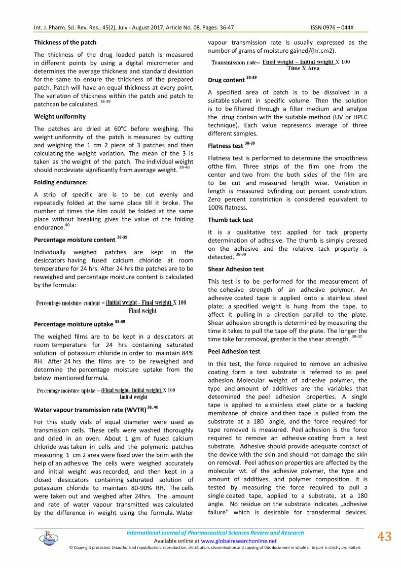
Micro reservoir type or Micro sealed Dissolution
Figure 4: Micro reservoir type or Micro sealed Dissolution
The micro reservoir system can be considered a combination of the reservoir and matrix diffusion drug delivery systems. In this, drug reservoir is formed by 1st suspending the drug solids in the aqueous solution of water soluble liquid polymer e.g. PEG and then dispersing the drug suspension homogeneously in the lipophilic polymer viz. silicone elastomers by the high energy dispersion technique to form several discrete, unleachable micro spheres of drug. This thermodynamically unstable dispersion is readily stabilized by immediately cross-linking polymer chains in-situ, which produces a drug polymer disc with constant surface area & fixed thickness. A transdermal delivery system is then produced by positioning the medicated disc at center and surrounding it with an adhesive rim. E.g. Nitroglycerin: Releasing transdermal therapeutic system for once-a day treatment of angina pectoris.
36
Evaluation Parameters for TDDS Patch
Physical appearance
The prepared patches were physically examined for colour, clarity and surface texture.
37
Int. J. Pharm. Sci. Rev. Res., 45(2), July - August 2017; Article No. 08, Pages: 36-47 ISSN 0976 – 044X
International Journal of Pharmaceutical Sciences Review and Research International Journal of Pharmaceutical Sciences Review and Research Available online at www.globalresearchonline.net
© Copyright protected. Unauthorised republication, reproduction, distribution, dissemination and copying of this document in whole or in part is strictly prohibited. Available online at www.globalresearchonline.net
43
Thickness of the patch
The thickness of the drug loaded patch is measured in different points by using a digital micrometer and determines the average thickness and standard deviation for the same to ensure the thickness of the prepared patch. Patch will have an equal thickness at every point. The variation of thickness within the patch and patch to patchcan be calculated.
38-39
Weight uniformity
The patches are dried at 60°C before weighing. The weight uniformity of the patch is measured by cutting and weighing the 1 cm 2 piece of 3 patches and then calculating the weight variation. The mean of the 3 is taken as the weight of the patch. The individual weight should notdeviate significantly from average weight. 39-40
Folding endurance:
A strip of specific are is to be cut evenly and repeatedly folded at the same place till it broke. The number of times the film could be folded at the same place without breaking gives the value of the folding endurance.40
Percentage moisture content 38-39
Individually weighed patches are kept in the desiccators having fused calcium chloride at room temperature for 24 hrs. After 24 hrs the patches are to be reweighed and percentage moisture content is calculated by the formula:
Percentage moisture uptake 38-39
The weighed films are to be kept in a desiccators at room temperature for 24 hrs containing saturated solution of potassium chloride in order to maintain 84% RH. After 24 hrs the films are to be reweighed and determine the percentage moisture uptake from the below mentioned formula.
Water vapour transmission rate (WVTR) 38, 40
For this study vials of equal diameter were used as transmission cells. These cells were washed thoroughly and dried in an oven. About 1 gm of fused calcium chloride was taken in cells and the polymeric patches measuring 1 cm 2 area were fixed over the brim with the help of an adhesive. The cells were weighed accurately and initial weight was recorded, and then kept in a closed desiccators containing saturated solution of potassium chloride to maintain 80-90% RH. The cells were taken out and weighed after 24hrs. The amount and rate of water vapour transmitted was calculated by the difference in weight using the formula. Water
vapour transmission rate is usually expressed as the number of grams of moisture gained/(hr.cm2).
Drug content 38-39
A specified area of patch is to be dissolved in a suitable solvent in specific volume. Then the solution is to be filtered through a filter medium and analyze the drug contain with the suitable method (UV or HPLC technique). Each value represents average of three different samples.
Flatness test 38-39
Flatness test is performed to determine the smoothness ofthe film. Three strips of the film one from the center and two from the both sides of the film are to be cut and measured length wise. Variation in length is measured byfinding out percent constriction. Zero percent constriction is considered equivalent to 100% flatness.
Thumb tack test
It is a qualitative test applied for tack property determination of adhesive. The thumb is simply pressed on the adhesive and the relative tack property is detected. 38-39
Shear Adhesion test
This test is to be performed for the measurement of the cohesive strength of an adhesive polymer. An adhesive coated tape is applied onto a stainless steel plate; a specified weight is hung from the tape, to affect it pulling in a direction parallel to the plate. Shear adhesion strength is determined by measuring the time it takes to pull the tape off the plate. The longer the time take for removal, greater is the shear strength. 39-42
Peel Adhesion test
In this test, the force required to remove an adhesive coating form a test substrate is referred to as peel adhesion. Molecular weight of adhesive polymer, the type and amount of additives are the variables that determined the peel adhesion properties. A single tape is applied to a stainless steel plate or a backing membrane of choice and then tape is pulled from the substrate at a 180 angle, and the force required for tape removed is measured. Peel adhesion is the force required to remove an adhesive coating from a test substrate. Adhesive should provide adequate contact of the device with the skin and should not damage the skin on removal. Peel adhesion properties are affected by the molecular wt. of the adhesive polymer, the type and amount of additives, and polymer composition. It is tested by measuring the force required to pull a single coated tape, applied to a substrate, at a 180 angle. No residue on the substrate indicates „adhesive failure‟ which is desirable for transdermal devices.
Int. J. Pharm. Sci. Rev. Res., 45(2), July - August 2017; Article No. 08, Pages: 36-47 ISSN 0976 – 044X
International Journal of Pharmaceutical Sciences Review and Research International Journal of Pharmaceutical Sciences Review and Research Available online at www.globalresearchonline.net
© Copyright protected. Unauthorised republication, reproduction, distribution, dissemination and copying of this document in whole or in part is strictly prohibited. Available online at www.globalresearchonline.net
44
Remnants on thesubstrate indicate „cohesive failure‟ signifying a deficit of cohesive strength in the coating. 38-41
Rolling ball tack test
In this test a steel ball of 7/16 inch in diameter is rolled down an inclined having horizontally placed patch facing adhesive surface upward. The ball rolls down and runs horizontal distance on the patch. The distance run by theball gives the tack property of the adhesive patch.
38
Quick Stick (peel-tack) test
In this test, the tape is pulled away from the substrate at 90 C at a speed of 12 inches/min. The peel force required breaking the bond between adhesive and substrate is measured and recorded as tack value, which is expressed in ounces or grams per inch width.42-43
Uniformity of dosage unit test
An accurately weighed portion of the patch is to be cut into small pieces and transferred to a specific volume volumetric flask, dissolved in a suitable solvent and sonicate for complete extraction of drug from the patch and made up to the mark with same. The resulting solution was allowed to settle for about an hour, and the supernatant was suitably diluted to give the desired concentration with suitable solvent. The solution was filtered using 0.2m membrane filter and analyzed by suitable analytical technique (UV or HPLC) and the drug content per piece will be calculated. 42-43
Polariscope examination
This test is performed to examine the drug crystals from patch by polariscope. A specific surface area of the piece is kept on the object slide and observed for the drugs crystals to distinguish whether the drug is present as crystalline form or amorphous form in the patch.
Skin Irritation study
Skin irritation and sensitization testing can be performed on healthy rabbits (average weight 1.2 to 1.5 kg). The dorsal surface (50cm2) of the rabbit is to be cleaned and remove the hair from the clean dorsal surface by shaving and clean the surface by using rectified spirit and the representative formulations can be applied over the skin. The patch is to be removed after 24 hr and the skin is to be observed and classified into 5 grades on the basis of the severity of skin injury.
41-42
In vitro drug release studies
The paddles over disc method (USP apparatus V) can be employed for assessment of the release of the drug from the prepared patches. Dry films of known thickness is to be cut into definite shape, weighed, and fixed over a glass plate with an adhesive. The glass plate was then placed in a 500-mL of the dissolution medium or phosphate buffer (pH 7.4), and the apparatus was equilibrated to 32± 0.5°C. The paddle was then set at a
distance of 2.5 cm from the glass plate and operated at a speed of 50 rpm. Samples (5- mL aliquots) can be withdrawn at appropriate time intervals up to 24 h and analyzed by UV spectrophotometer or HPLC. The experiment is to be performed in triplicate and the mean value can be calculated.
42-43
Stability studies
Stability studies are to be conducted according to the ICH guidelines by storing the TDDS samples at 40±0.5°c and 75±5% RH for 6 months. The samples were withdrawn at 0, 30, 60, 90 and 180 days and analyze suitably for the drug content.
42
In vivo study: The In vivo study involves:
a) Animal model
b) Human model
Most preferably In vivo study is conducted on animal models as compared to human models because of easy availability of animals, ease of experiment and toxicity and safety parameters associated with the experiment. Various species of mouse, rat, dogs, monkey, pig, cat, rabbit and squirrel are used for animal study. Mainly hairless animalare preferred over hairy animals for transdermalformulation evaluation. In final stage of formulation development human volunteers are studied to determine the pharmacokinetic and pharmacodynamic profile of the drug including safety and efficacy of the formulation. Clinical trials are conducted in IV phases. Phase I trials are conducted on small group of volunteers to determine the safety and toxicity profile. Phase II trials are conducted on a small group of patients for safety and toxicity for short term. Phase III study is conducted on a large group of patients and phase IV is the post marketing survey. 42-43
General clinical considerations in the use of TDDS 44
The patient should be advised of the following general guidelines. The patient should be advised of the importance of using the recommended site and rotating locations within the site.
1. TDDS should be applied to clean, dry skin relatively free of hair and not oily, inflamed, irritated, broken.
2. Use of skin lotion should be avoided at the application site, because lotions affect the hydration of skin and can alter partition coefficient of drug.
3. Patient should not physically alter TDDS, since this destroys integrity of the system.
4. The protecting backing should be removed with care not to touch fingertips. The TDDS should be pressed firmly against skin site with the heel of hand for about 10 seconds
5. A TDDS should be placed at a site that will not subject it to being rubbed off by clothing or movement. TDDS should be left on when showering, bathing or swimming.
Int. J. Pharm. Sci. Rev. Res., 45(2), July - August 2017; Article No. 08, Pages: 36-47 ISSN 0976 – 044X
International Journal of Pharmaceutical Sciences Review and Research International Journal of Pharmaceutical Sciences Review and Research Available online at www.globalresearchonline.net
© Copyright protected. Unauthorised republication, reproduction, distribution, dissemination and copying of this document in whole or in part is strictly prohibited. Available online at www.globalresearchonline.net
45
6. A TDDS should be worn for full period as stated in the product’s instructions followed by removal and replacement with fresh system.
7. The patient or caregiver should clean the hands after applying a TDDS. Patient should not rub eye or touch the mouth during handling of the system.
8. If the patient exhibits sensitivity or intolerance to a TDDS or if undue skin irritation results, the patient should seek reevaluation.
9. Upon removal, a used TDDS should be folded in its half with the adhesive layer together so that it cannot be reused. The used patch discarded in a manner safe to children and pets. It is important to use a different application site everyday to avoid skin irritation. Suggested rotation is: Day 1 – Upper right arm; Day 2 – upper right chest; Day 3 – Upper left chest; Day 4 – Upper left arm. [Then repeat from Day 1]
A Novel Approach in Transdermal Drug Delivery: Micro fabricated Micro needles
The development of more sophisticated drugs has demanded the need for more sophisticated methods to deliver those drugs. Conventional drug delivery techniques using pills and injections are often not suitable for Transdermal protein based, DNA-based, and other therapeutic compounds produced by modern biotechnology. An attractive alternative method of delivery involves drug administration across the skin. This approach avoids degradation in the gastrointestinal tract and first-pass effects of the liver associated with oral delivery as well as the pain and inconvenience of intravenous injection.
Despite its many potential advantages, transdermal drug delivery is severely limited by the poor permeability of human skin; most drugs do not cross skin at therapeutically relevant rates. A number of methods have been developed to increase rates of transdermal transport with varied levels of success. Chemical enhancers can increase permeability of skin to small molecules but also trigger skin irritation or other safety concerns which limit their use. Iontophoresis employs an electric field to drive ionized molecules across skin by electrophoresis and nonionized molecules by electroosmosis. Despite concerns about skin irritation, Iontophoresis may be useful to deliver some peptides and small proteins. Recently, physical methods to transiently increase skin permeability using electroporation and ultrasound have shown promise for delivery of both small drugs and macromolecules.
In this study, we present a novel approach to transdermal drug delivery which dramatically enhances transport of molecules across skin. We have used standard micro fabrication techniques to etch arrays of micron-size needles into silicon. When these microneedle arrays are inserted into the skin, they create conduits for transport across the stratum corneum, the outer layer of skin which forms the primary barrier to transport. Once a
compound crosses the stratum corneum it can diffuse rapidly through deeper tissue and be taken up by the underlying capillaries for systemic administration. The design of microneedles which painlessly permeabilize skin is based on an understanding of skin anatomy. Human skin is made of three layers: stratum corneum, viable epidermis, and dermis. The outer 10-15 ím of skin, called stratum corneum, is a dead tissue that forms the primary barrier to drug transport. Below lies the viable epidermis (50-100 ím),a tissue containing living cells and nerves, but no blood vessels. Deeper still, the dermis forms the bulk of skin volume and contains living cells, nerves, and blood vessels. Therefore, microneedles which penetrate the skin just a little more than 10-15 ím should provide transport pathways across the stratum corneum, but do so painlessly since the microneedles do not reach nerves found in deeper tissue.
Microneedles were made using micro fabrication technology, which is the same technology used to make integrated circuits. An advantage of this approach is that micro fabrication readily makes structures of micron dimensions in a way that is easily scaled up for cheap and reproducible mass production. To adapt this technology for transdermal drug delivery, we created three-dimensional arrays of sharp-tipped microneedles of approximately 150 ím in length.
A deep reactive ion etching process was used to micro fabricate the needles for this study. In this process, a chromium masking material is deposited onto silicon wafers and patterned into dots which have a diameter approximately equal to the base of the desired microneedles. The wafers are then loaded into a reactive ion etcher and subjected to carefully controlled plasma based on fluorine/oxygen chemistries to etch very deep, high aspect ratio valleys into the silicon. Those regions protected by the metal mask remain and form the microneedles.45
CONCLUSION
This article provide an valuable information regarding the transdermal drug delivery systems and its evaluation process details as a ready reference for the research scientist who are involved in TDDS. The foregoing shows that TDDS have great potentials, being able to use for both hydrophobic and hydrophilic active substance into promising deliverable drugs. To optimize this drug delivery system, greater understanding of the different mechanisms of biological interactions, and polymer are required. TDDS a realistic practical application as the next generation of drug delivery system.
REFERENCES
1. Vyas S.P. and R.K Khar, Controlled drug delivery concepts and advances. VallabhPrakashan, 1, 2002 411-447.
2. Prausnitz, M. R., Mitragotri, S. & Langer, R. Current Status and Future Potential of Transdermal Drug Delivery. Nature Reviews 3(2), 2004, 115-124.
Int. J. Pharm. Sci. Rev. Res., 45(2), July - August 2017; Article No. 08, Pages: 36-47 ISSN 0976 – 044X
International Journal of Pharmaceutical Sciences Review and Research International Journal of Pharmaceutical Sciences Review and Research Available online at www.globalresearchonline.net
© Copyright protected. Unauthorised republication, reproduction, distribution, dissemination and copying of this document in whole or in part is strictly prohibited. Available online at www.globalresearchonline.net
46
3. Loyd V. Allen Jr, Nicholas G. Popovich, Howard C. Ansel. Pharmaceutical dosage forms and drug delivery systems, 8th Edition., Wolter Kluwer Publishers, New Delhi, 2005 pp. 298-299.
4. R.Sowjanya, Salman Khan, D.Bhowmik, Harish.G, S.Duraivel TRANSDERMAL DRUG DELIVERY SYSTEMS Indian Journal of Research in Pharmacy and Biotechnology Volume 1(4), July-August 2013, Page 489,490.
5. Chien YW, Novel drug delivery systems, drugs and the Pharmaceutical sciences, Vol.50, Marcel Dekkar, New York, NY, 1992, 797.
6. Guy RH. Current status and future prospects of transdermal drug delivery, Pharm Res 13, 1996, 1765-1769.
7. Guy RH, Hadgraft J, Bucks DA. Transdermal drug delivery and cutaneous metabolism, Xonobiotica 7, 1987, 325-343.
8. Dipen Patel, Sunita A. Chaudhary, BhaveshParmar, NikunjBhura Transdermal Drug Delivery System: A Review 1Vol. 1 No. 4 2012 www.thepharmajournal.com page no 67.
9. jain A, Mishra A, Nayak S and Soni V. Transdermal delivery of antihypertensive agents: A tabular update. International Journal of Drug Delivery. 3, 2011, 1-13.
10. Patel RP and Baria AH. Formulation and evaluation considerations of transdermal drug delivery system. International Journal of Pharmaceutical Research. 3, 2011, 1-9.
11. Bhargava T, Ramchandani U, Shrivastava SK and Dubey PK. Current trends in NDDS withspecial reference to NSAIDs. International Journal of Pharmacy and Bio Sciences. 2, 2011, 92-114.
12. Jain NK.Controlled and Novel drug delivery, Published by CBS Publishers & distributors, New Delhi-110002, 1st Edn, 1997, 100-129.
13. SwarnlataSonl and Vinod K, Dixit, Transdermal Penetration Enhancers, Categorization, Indian Drugs, 29(11), 1992, 465-471.
14. TanuBhargava, Current trends in NDDS with special reference to NSAIDS, International Journal of Pharma and Bio Sciences, 2, 2011, 92-114.
15. Bernar B and John V.A. Pharmacokinetic characterization of Transdermal delivery systems. Jour. Clinical pharmacokinetics 26(2), 1994, 1`21-34.
16. Singh J, Tripathi K.T and Sakia T.R. Effect of penetration enhancers on the in vitro transport of ephedrine through rate skin and human epidermis from matrix based Transdermal formulations. Drug Dev. Ind Pharm. 19, 1993, 1623-1628.
17. Wade A, Wellar P.J. Handbook of pharmaceutical Excipients. Washington, DC; American pharmaceutical publishing Association; 1994, 362-366.
18. Raghuramreddy k, Muttalik s and Reddy S. Once-daily sustained release matrix tablets of nicorandil: formulation and in vitro evaluations. AAPS Pharm Sci.Tech.2003, 4:4.
19. Costa P, Ferrica DC, Morgado R, Soussa Lobo JM. Design and evaluation of a lorazepam transdermal delivery system, Drug DevIndPharm 23, 1997, 939-944.
20. Shaila L., Pandey s and Udupa N. Design and evaluation of matrix controlled Transdermal drug delivery system of nicitin suitable for use in smoking cessation. Indian Journ. Pharm. Sci. 68, 2006, 179-184.
21. Bagyalakshmi J, Vamsikrishna RP, Manavalan R, Ravi TK, Manna PK. Formulation development and in vitro and in vivo evaluation of membrane moderated transdermal systems of ampicilline sodium in ethanol: pH 4.7 buffer solvent systems AAPS PharmSciTec.2007, 8, Article7.
22. Ubaidulla U, Reddy MV, Ruckmani K, Ahmed FJ, Khar RK. Transdermal the rapeutic system of Carvedilol: effect of hydrophilic and hydrophobic matrix on invitro and invivo characteristics, AAPS PharmSciTech 2007, 8(1), Article 2.
23. Wade A andwellar P.J. Handbook of pharmaceutical Excipients. Washington, DC: American Pharmaceutical publishing Association 1994, 362-366.
24. Arunachalam A, et al., Transdermal drug delivery system: A review. Current PharmaResearch, 1(1), 2010, 70-81.
25. Kandavilli S, V. Nair and R. Panchagnula, Polymers in transdermal drug delivery systems. Pharmaceutical Technology, 2002, 26(5), 62-81.
26. Sheth N.S. And R.B. Mistry, Formulation and evaluation of transdermal patches and to study permeation enhancement effect of eugenol. 2011.
27. Pfister, W.R. and D. Hsieh, Permeation enhancers compatible with transdermal drugdelivery systems. Part I: selection and formulation considerations. Medical device technology, 1989, 1(5), 48-55.
28. Godbey, K., Improving patient comfort with nonocclusive transdermal backings. American Association of Pharmaceutical Scientists, 1996, 1-2.
29. Chen K., Schmidt C F., The action of ephedrine, the active principle of Chinese drug Ma Huang, J. Pharmacology. Exp. Ther., 24, 1925, 339-357.
30. Nelson H.S., Beta adrenergic bronchodilator, Eng. J. Med., 333, 1995, 499-506.
31. Gros L., Clark W.E., “The structure of skin” in “The tissue of the body” Le Gros&Clark W. E. (editors) edition VI, ELBS and Oxford University Press, London, 1980, 29-313.
32. Chien Y.W., ”Systemic delivery of pharmacologically active molecules across skin” in “Targeted drug delivery” Radolph L., Juliano(Editor) Springer- Verlag, Berlin, 1991, 182-230.
33. The skin & the sensory organs,chapter-12, “Cunningham’s text book of anatomy”, Romans G.J., (Editor), Edition XXII, Oxford University press, NY, US, 1981, 829-34.
34. Wilkosz M F. Transdermal Drug Delivery: Part I.U.S. Pharmacist, Jobson publication, 28, 2003, 04.
35. Morow D.I.J., Carron P.A. Mc, Woolfson A.D., Donnelly R.F., Innovative Strategies for Enhancing Topical and Transdermal Drug Delivery, The Open Drug Delivery Journal, 1, 2007, 36-59.
36. AnujaDhas, Ganesh Deshmukh, ShripadPansare and Ravish QureshiA REVIEW ON TRANSDERMAL PATCHES World Journal of Pharmaceutical and Life Sciences WJPLS www.wjpls.org wjpls, Vol. 2, 2016, 2454-2229 Issue 3, 381-399ISSN
Int. J. Pharm. Sci. Rev. Res., 45(2), July - August 2017; Article No. 08, Pages: 36-47 ISSN 0976 – 044X
International Journal of Pharmaceutical Sciences Review and Research International Journal of Pharmaceutical Sciences Review and Research Available online at www.globalresearchonline.net
© Copyright protected. Unauthorised republication, reproduction, distribution, dissemination and copying of this document in whole or in part is strictly prohibited. Available online at www.globalresearchonline.net
47
37. Prochazka AV, New developments in smoking cessation, Chest. 2000, 117 (4), 169-175.
38. Crawford R.R and Esmerian O.K. Effect of plasticizers on some physical properties of cellulose acetate phthalate films. J. Pharm. Sci. 1997, 60, 312- 314.
39. Shaila L, Pandey S and Udupa N, Design and evaluation of matrix type membrane controlled Transdermal drug delivery system of nicotine suitable for use in smoking cessation, Indian Journ. Pharm. Sci, 2006, 68, 179-18.
40. N Aarti, Louk A.R.M.P, Russsel.O.P and Richard H.G. Mechanism of oleic acid induced skin permeation enhancement in vivo in humans, Jour. Control, Release 1995, 37, 299-306.
41. Wade A and Weller P.J. Handbook of pharmaceutical Excipients. Washington, DC: American Pharmaceutical Publishing Association 1994, 362-366.
42. Lec S.T, Yac S.H, Kim S.W and Berner B, One way membrane for Transdermal drug delivery systems / system optimization, Int. J Pharm., 1991, 77, 231 - 237.
43. Singh J, Tripathi K.T and Sakia T.R. Effect of penetration enhancers on the invitro transport of ephedrine through rate skin and human epidermis from matrix based Transdermal formulations, Drug Dev.Ind. Pharm., 1993, 19, 1623-1628.
44. Sharma N, Agarwal G, Rana AC, Bhat Z, Kumar D. A Review: Transdermal Drug Delivery System: A Tool for Novel drug delivery system. IJDDR 2011, 3(3), 70-84
45. P. M. Patil, Dr. P. D. Chaudhari, Jalpa K Patel, K. A. Kedar, P. P. Katolkar “Recent trends in challenges and opportunities of Transdermal drug delivery system”, Int. J. Drug Dev. & Res., Jan-March 2012, 4(1), 39-50.
Source of Support: Nil, Conflict of Interest: None.


