dorsolateral prefrontal cortex activity predicts responsiveness to cognitive–behavioral therapy in...
TRANSCRIPT

DRiVA
Bew
Matc
Rs(0
Ce
Ks
Appettp
as(fpdsddab
F
A
R
0d
orsolateral Prefrontal Cortex Activity Predictsesponsiveness to Cognitive–Behavioral Therapy
n Schizophreniaeena Kumari, Emmanuelle R. Peters, Dominic Fannon, Elena Antonova, Preethi Premkumar,nantha P. Anilkumar, Steven C.R. Williams, and Elizabeth Kuipers
ackground: Given the variable response to cognitive–behavioral therapy (CBT) when added to antipsychotic medication in psychosis and thevidence for a role of pretherapy level of frontal lobe–based cognitive function in responsiveness to CBT in other disorders, this study examinedhether pretherapy brain activity associated with working memory neural network predicts clinical responsiveness to CBT in schizophrenia.
ethods: Fifty-two outpatients stable on medication with at least one distressing symptom of schizophrenia and willing to receive CBT inddition to their usual treatment and 20 healthy participants underwent functional magnetic resonance imaging during a parametric n-backask. Subsequently, 26 patients received CBT for psychosis (CBT�treatment-as-usual [TAU], 19 completers) for 6 – 8 months, and 26ontinued with TAU alone (17 completers). Symptoms in both patient groups were assessed (blindly) at entry and follow-up.
esults: The CBT�TAU and TAU-alone groups did not differ clinically or in performance at baseline. The CBT�TAU group showedignificant improvement in relation to the TAU-alone group, which showed no change, at follow-up. Stronger dorsolateral prefrontal cortexDLPFC) activity (within the normal range) and DLPFC– cerebellum connectivity during the highest memory load condition (2-back �-back) were associated with post-CBT clinical improvement.
onclusions: DLPFC activity and its connectivity with the cerebellum predict responsiveness to CBT for psychosis in schizophrenia. These
ffects may be mediated by PFC– cerebellum contributions to executive processing.ey Words: CBT, cerebellum, connectivity, DLPFC, fMRI, psychosis,chizophrenia
number of randomized controlled trials (RCTs) havedemonstrated that persistent positive symptoms, particu-larly delusions, and secondary disturbances such as de-
ression, are improved by cognitive–behavioral therapy forsychosis (CBT-P) in patients with schizophrenia (1–3). Theseffects, however, are seen with modest effect sizes and present,o a noticeable degree, in only about 50% of such patients. Givenhe variable response to CBT-P, it is important to elucidate theredictors of CBT efficacy for psychosis.
Cognitive functions may play a role in CBT responsivenesscross most disorders but perhaps more so in disorders, such aschizophrenia, which are characterized by cognitive impairment4). Although skilled therapy adaptations attempt to compensateor such impairment, it may still hinder effectiveness of CBT,erhaps by impeding patients’ ability to remember informationiscussed in sessions; to acquire new, more flexible thinkingtyles or coping strategies; or to generalize specific issuesiscussed in therapy to other situations in life. For example, inepression better executive functioning and problem-solvingbility predict a more favorable clinical outcome following CBT,ut not following treatment with antidepressant or placebo or
rom the Department of Psychology (VK, ERP, EA, PP, EK), Division of Psycho-logical Medicine (DF, APA), and Centre for Neuroimaging Sciences(SCRW), Institute of Psychiatry, King’s College London; National Institutefor Health Research (NIHR) Biomedical Research Centre for MentalHealth (VK, ERP, EK), South London and Maudsley National Health Ser-vice Foundation Trust, London, United Kingdom.
ddress correspondence to Veena Kumari, Ph.D., PO78, Institute of Psychi-atry, King’s College London, De Crespigny Park, London SE5 8AF, UnitedKingdom. E-mail: [email protected].
eceived Oct 22, 2008; revised Mar 24, 2009; accepted Apr 21, 2009.
006-3223/09/$36.00oi:10.1016/j.biopsych.2009.04.036
waiting for CBT (5,6). Intact executive functioning also predicts afavorable response to CBT for generalized anxiety disorder (7).Similarly, initial cognitive flexibility predicted a better outcome inan RCT of CBT-P (8). In a recent study (9), neurocognitivedeficits predicted poorer function overall in older psychoticpatients but did not moderate CBT effects. The therapy in thisstudy, however, was targeted specifically at social functioningrather than at distress and symptom reduction. In general, clinicalresponsiveness to CBT across disorders may relate to executiveprocesses (5–8) modulated by the frontal lobes, especially thedorsolateral prefrontal cortex (DLPFC) (10–12).
This study tested the hypothesis, for the first time to ourknowledge, that pretherapy DLPFC activity, elicited with aparametric n-back working memory (WM) task (13,14) anddetected with functional magnetic resonance imaging (fMRI),would predict clinical responsiveness to CBT-P in schizophrenia.Given known associations between impaired cognition anddisrupted DLPFC connectivity in schizophrenia (15), we alsoexamined DLPFC connectivity with other brain regions as apredictor of responsiveness to CBT-P.
Methods and Materials
Participants and DesignThis investigation involved three groups. Group 1 consisted
of 26 outpatients with schizophrenia (16) who received CBT-Pfor 6–8 months in addition to their treatment-as-usual (TAU;CBT�TAU), Group 2 of 26 outpatients with schizophrenia whoreceived TAU during the course of this investigation (TAUalone), and Group 3 of 20 healthy participants (HC) who did nothave a clinical diagnosis (17).
All participants were right-handed and had no history ofneurological conditions or head injury. All patients 1) had beenon stable doses of antipsychotics for �2 years and on theirpresent antipsychotic for �3 months, 2) received a rating of �60
on the Positive and Negative Symptoms scale (PANSS) (18) andBIOL PSYCHIATRY 2009;66:594–602© 2009 Society of Biological Psychiatry
rwtThCtIS(opamCma(sT
T
DAEPPA
R
C
ADb
b
b
b
A
A
(t‘
V. Kumari et al. BIOL PSYCHIATRY 2009;66:594–602 595
eported at least one positive “distressing” symptom, and 3)ished/agreed to receive 6–8 months of CBT-P in addition to
heir usual drug treatment. Patients for both (CBT�TAU,AU-alone) groups 1) were from the same geographic area, 2)ad been identified by their psychiatrists as suitable forBT-P, and 3) wished to receive CBT in addition to their usual
reatment. Those accepted for CBT-P by the Psychologicalnterventions Clinic for Outpatients with Psychosis (PICuP),outh London and Maudsley (SLAM) National Health ServiceNHS) Foundation Trust went into the CBT�TAU group;thers, matched to those in the CBT�TAU group as much asossible, went into the TAU-alone group. With the resourcesvailable to the SLAM NHS Foundation Trust, only approxi-ately 10% of eligible patients are offered CBT-P. The finalBT�TAU group had 19 patients (consent withdrawal, n � 4;edication noncompliance before follow-up, n � 1; nonus-
ble data, n � 2), and the TAU-alone group had 17 patientsconsent withdrawal, n � 5; acutely unwell/admitted to aecure hospital before follow-up, n � 2; nonusable data, n � 2).able 1 presents participants’ characteristics. All participants pro-
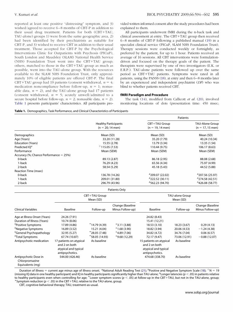
able 1. Demographics, Task Performance, and Clinical Characteristics of P
Healthy Partic(n � 20; 14
emographics Mean (SDge (Years) 33.20 (1ducation (Years) 15.55 (2redicted IQa �115.05 (7erformance Mean (SEccuracy (%; Chance Performance � 25%)0-back 89.13 (21-back 76.29 (42-back 58.54 (5
eaction Time (msec)0-back 136.78 (11-back 209.01 (32-back 296.79 (4
Patie
CBT�TAU GroupMean (SD)
linical Variables Baseline Follow-upCM
ge at Illness Onset (Years) 24.26 (7.91)uration of Illness (Years) 10.74 (8.06)
Positive Symptoms 17.89 (4.86) ’14.79 (4.39)Negative Symptoms 16.89 (3.52) 15.21 (4.04)General Psychopathology 32.95 (5.27) ’28.05 (7.48)Total Symptoms 67.74 (10.87) ’58.05 (14.93)ntipsychotic medication 17 patients on atypical
and 2 on bothatypical and typicalantipsychotics.
As baseline
ntipsychotic Dose inChlorpromazineEquivalents (mg)
544.00 (426.46) As baseline
Duration of illness � current age minus age of illness onset; aNationamissing IQ data in one healthy participant) and IQ in healthy participants sigo healthy participants even when controlling for age; ’Lower symptom sSymptom reduction (p � .05) in the CBT�TAU, relative to the TAU-alone,
CBT, cognitive-behavioral therapy; TAU, treatment-as-usual.
vided written informed consent after the study procedures had beenexplained to them.
All participants underwent fMRI during the n-back task andclinical assessment at entry. The CBT�TAU group then received6–8 months of CBT-P following a published manual (19) in aspecialist clinical service (PICuP, SLAM NHS Foundation Trust).Therapy sessions were conducted weekly or fortnightly, aspreferred by the patient, for up to 1 hour. Patients received anaverage of 16 sessions. All CBT interventions were formulation-driven and focused on the therapy goals of the patient. Thetherapists were supervised by one of two investigators (E.K. orE.R.P.). TAU-alone patients were followed up over the sameperiod as CBT�TAU patients. Symptoms were rated in allpatients, using the PANSS (18), at entry and then 6–8 months laterby an experienced and independent psychiatrist (DF) who wasblind to whether patients received CBT.
fMRI Paradigm and ProcedureThe task (14), modified from Callicott et al. (20), involved
monitoring locations of dots (presentation time: 450 msec;
pants
s
Patients
CBT�TAU Group(n � 19, 14 men)
TAU-Alone Group(n � 17, 15 men)
Mean (SD) Mean (SD)35.20 (7.79) 40.24 (10.34)13.79 (3.34) 13.35 (1.54)
110.44 (9.75) 106.17 (8.63)Mean (SEM) Mean (SEM)
86.18 (2.95) 88.08 (2.68)65.56 (4.34) 75.97 (4.99)45.18 (5.43) 44.52 (5.88)
�209.07 (22.02) �207.56 (25.97)�222.52 (30.11) �274.58 (42.51)�562.23 (94.70) �426.08 (58.77)
nly
TAU-alone GroupMean (SD)
e (BaselineFollow-up) Baseline Follow-up
Change (BaselineMinus Follow-up)
24.82 (8.43)15.41 (12.21)
1 (3.88) 18.53 (3.10) 18.23 (3.67) 0.29 (4.13)8 (3.90) 18.82 (3.94) 20.06 (4.53) �1.24 (4.38)9 (7.06) 34.82 (4.72) 34.76 (7.04) 0.06 (6.57)8 (12.29) 72.17 (9.47) 73.06 (12.91) �0.88 (12.07)
15 patients on atypicaland 2 on bothatypical and typicalantipsychotics.
As baseline
474.68 (338.70) As baseline
t Reading Test (21); bPositive and Negative Symptom Scale (18); �N � 19antly higher than TAU-alone; �Longer latencies (p � .05) in patients relative(p � .05) at follow-up in the CBT�TAU, but not in the TAU-alone, group;.
artici
ipantmen)
)1.28).78).53)M)
.87)
.23)
.29)
4.26)1.80)3.96)
nts O
hanginus
‘3.1‘1.6‘4.8‘9.6
l Adulnific
coresgroup
www.sobp.org/journal

io(apew7iPtbcppt
I
Somoisitra11
DM
oaab1rGs
F
596 BIOL PSYCHIATRY 2009;66:594–602 V. Kumari et al.
w
nterstimulus-interval: 1500 msec) within a diamond-shaped boxn the screen at a given delay from the original occurrence0-back, 1-back, or 2-back; Figure 1). There were three 30-secctive conditions (0-back, 1-back, 2-back) presented to partici-ants five times in pseudo-random order, controlling for orderffect. Each active block had 15 stimulus presentations, startedith a 15-sec rest block (“Rest” on the screen), and began with a50-msec text delay allowing the participants to notice a changen task demand/condition. The experiment lasted 11.25 min.articipants viewed the paradigm projected onto a screenhrough a prismatic mirror. They were required to press theutton on every trial, using the right thumb, corresponding to theorrect location of the 0-back, 1-back, or 2-back stimulus (chanceerformance � 25%; location of dots purely random). Partici-ants abstained from alcohol for at least 24 hours and underwentask familiarization before scanning.
mage AcquisitionEchoplanar MR brain images were acquired using a 1.5-T GE
igna system (General Electric, Milwaukee, Wisconsin). In eachf 16 near-axial noncontiguous planes parallel to the intercom-issural plane, 225 T2*-weighted MR images depicting bloodxygen level–dependent contrast were acquired over the exper-ment with echo time (TE) � 40 msec, repetition time (TR) � 3ec, in-plane resolution � 3.1 mm, slice thickness � 7.0 mm, andnterslice gap � .7 mm. In the same session, a high-resolutionhree-dimensional inversion recovery prepared spoiled gradientecalled acquisition in a steady state volume data set wascquired with TE � 5.3 msec, inversion time � 300 msec, TR �2.2 msec, in-plane resolution � .94 mm, and slice thickness �.5 mm.
ata Analysis: Demographic, Clinical, and BehavioraleasuresThe HC, CBT�TAU, and TAU-alone groups were compared
n age, education, and predicted IQ (21) using a one-waynalysis of variance (ANOVA), followed by mean comparisons asppropriate. Group differences in performance were examinedy a Group (HC, CBT�TAU, TAU-alone) � Load (0-back,-back, 2-back) ANOVA (separately for accuracy [% correctesponses] and latency [in msec] of correct responses) withroup as a between-subjects factor and Load as the within-
igure 1. Illustration of 1-back and 2-back trials.
ubjects factor, followed by analysis of lower order effects as
ww.sobp.org/journal
appropriate. A significant Group effect in latency (Results), giventhe potential effect of age in this measure, was reevaluated usinganalyses of covariance (ANCOVA), covarying for age.
The CBT�TAU and TAU-alone groups were compared onclinical variables using independent-sample t tests. The changein symptoms from baseline to follow-up was investigated using aGroup (CBT�TAU, TAU-alone) � Time (baseline, follow-up)ANOVA with Group as a between-subjects factor and Time as awithin-subjects factor. A significant Group � Time effect wasfollowed up by paired t tests on total and subscale PANSS scoresseparately in the CBT�TAU and TAU-alone groups. Followingthe observation of significant symptom reduction in theCBT�TAU group, but not in the TAU-alone group, we examinedpotential associations between baseline symptom severity andsymptom change (baseline minus follow-up) in the CBT�TAUgroup using Pearson’s correlations and confirmed the effects ofCBT-P using ANCOVAs on symptom change scores covarying forbaseline symptoms. We also computed the degree of change insymptoms independent of initial severity as residual change insymptoms by regressing the initial PANSS (total and subscales)scores on follow-up scores as a further outcome measure of CBTresponsiveness for fMRI analysis following the method used bySiegle and colleagues (22). The association between perfor-mance variables and responsiveness to CBT was examined usingPearson’s correlations.
All analyses were performed in SPSS windows (version 15).Before running the described analyses, each variable was eval-uated for the normality of the distribution to ensure it met thecriteria of parametric statistics. Alpha level for testing significanceof effects was maintained at p � .05.
Functional MRI: Image Pre-ProcessingFor each participant, the 225-volume functional time series
were motion corrected, transformed into stereotactic space(Montreal Neurological Institute), smoothed with a 10 mmfull-width-at-half-maximum Gaussian filter, and band-pass fil-tered using statistical parametric mapping software (SPM2;http://www.fil.ion.ucl.ac.uk/spm).
Models and Statistical InferencesData were analyzed using a random-effect procedure (23).
Subject-specific activations were identified with a factorial modelconsisting of three active conditions and rest as an implicitbaseline. Generic task-related activity changes were identifiedusing one-sample t tests (height threshold, p � .001; cluster-corrected p � .05) separately in CBT�TAU and HC groups.
To examine the relationship of CBT response with pretherapybrain activity in patients, we regressed residual symptom changescores on task-related activations (0-back vs. rest; 1- and 2-backvs. 0-back) across the entire brain (height threshold p � .05,cluster-corrected p � 05). For the positive associations of a priorihypothesized regions in the frontal lobe with CBT responsive-ness, the following significance criteria to maxima voxels ofclusters that did not survive whole-brain correction for multiplecomparisons were applied: 1) T value of �3.80 (correspondingto uncorrected voxel p � .001) and �100 contiguous voxels, and2) survival of small volume correction (SVC) within a locallydefined volume (15-mm radius sphere) with family-wise errorcorrected p � .05. (No cluster in any other regions met this SVCcriterion for positive associations with CBT response.) We ex-plored negative associations between CBT responsiveness andpretherapy activations using a more conservative criteria (height
threshold, p � .005; whole-brain cluster-corrected p � .05)
bi
trCurR
resDpP0twwg(rrsp
R
D
dHe2y3Taf
sPFgCP3pdsttF.tnisce(
t��p
V. Kumari et al. BIOL PSYCHIATRY 2009;66:594–602 597
ecause we did not have a specific hypothesis or a region ofnterest (ROI).
To examine whether a positive association between pre-herapy brain activity and CBT responsiveness reflected a hyper-esponse or a stronger response within the normal range inBT�TAU patients, we compared CBT�TAU patients and HCsing two-sample t tests on 2-back � 0 back contrasts (theseevealed the strongest association with CBT responsiveness; seeesults).
Further, we examined functional connectivity of the left andight frontal regions (that associated with CBT responsiveness inarlier analyses) with other regions as predictors of CBT respon-iveness. For this purpose, the activity time series from the leftLPFC (seed �48[x], 34[y], 30[z]; this had the most consistentositive association with CBT response) and right inferior-middleFC (seed 52[x], 24[y], and 22[z]) were extracted (2-back �-back) and used as a regressor (separately for left and right PFC)o investigate their connectivity with other regions. The regionsith significantly covarying increases or decreases in activityith the two ROIs were identified for each participant, and theroup connectivity maps constructed using one-sample t testsheight threshold p � .005; cluster-corrected p � .05). Theelationship of CBT response with connectivity of the left andight PFC with other regions was identified by regressing residualymptom change scores on connectivity SPM maps in CBT�TAUatients (height threshold p � .005; cluster-corrected p � .05).
esults
emographic, Clinical, and Behavioral MeasuresThere was a trend for the effect of Group in age (F � 2.44,
f � 2,56, p � .10); TAU-alone patients were slightly older thanC (t � 1.96, df � 35, p � .06). There were Group effects inducation (F � 3.53, df � 2,56, p � .04) and IQ (F � 4.70, df �,55, p � .01); TAU� alone patients, relative to HC, had fewerears in education (t � 1.79, df � 35, p � .01) and lower IQ (t �.30, df � 34, p � .002). CBT�TAU patients did not differ fromAU-alone patients in age, education, IQ, baseline symptoms,ge at illness onset, illness duration, and antipsychotic dose orrom HC in demographic characteristics (Table 1).
CBT�TAU, but not TAU-alone, patients showed changes inymptoms from baseline to follow-up (Group � Time: totalANSS scores, F � 6.74, df � 1,34, p � .014; positive symptoms,� 4.42, p � .043; negative symptoms, F � 4.47, p � .042;
eneral psychopathology: F � 4.49, p � .041; Table 1). Only theBT�TAU group showed reduced symptoms at follow-up (totalANSS scores: t � 3.43, df � 18, p � .003; positive symptoms: t �.48, p � .003; negative symptoms: t � 1.88, p � .07; generalsychopathology: t � 3.02, p � .007). Baseline symptom severityid not correlate with CBT responsiveness (change in totalymptoms, r � .17, p � .48). Covarying for baseline symptoms,here was significant symptom improvement (change scores) inhe CBT�TAU, relative to TAU-alone, group (total PANSS scores:� 8.16, df � 1,36, p � .007; positive symptoms: F � 7.01, p �
012; negative symptoms: F � 8.27, p � .007; general psychopa-hology: F � 5.98, p � .02). As can be expected given earlieroted independence between baseline symptoms and symptommprovement in CBT�TAU patients, residual symptom changecore correlated highly positively with the absolute symptomhange scores (total PANSS; r � .962, p � .001). Illness duration,ducation, and IQ were not associated with CBT responsiveness
p values � .40).For performance accuracy, there was a main effect of Load(F � 91.85, df � 4,106, p � .001; lower accuracy with increasingload); Group (F � 1.86, df � 2,53, p � .17) and Group � Load(F � 1.66, df � 2,53, p � .20) effects were nonsignificant. Forlatency, there were significant Group (F � 4.24, df � 2,53, p �.02; covarying for age, F � 4.02, df � 2,52, p � .024) and Group �Load (F � 4.24, df � 2,53, p � .02; covarying for age, F � 4.02,df � 2,52, p � .024) effects indicating longer latencies in bothpatient groups compared to HC, especially at 1-back and 2-back(p values � .05); there was no difference between the CBT�TAUand TAU-alone groups (p values � .40). The relationshipsbetween CBT responsiveness and performance (accuracy:1-back, r � .33, 2-back, r � .21; latency: r � �.08, r � �.25),although in the expected direction (better performance withpositive CBT response), failed to reach significance.
Generic fMRI PatternsThe generic WM network (Table 1 in Supplement 1) identified in
both the CBT�TAU and HC groups included bilateral activations inthe inferior-middle-superior frontal gyrus and the parietal lobe. Thedeactivated regions included the posterior cingulate, medial pre-frontal and middle temporal gyri, insula, and precuneus.
Pretherapy Brain Activity and CBT ResponsivenessTask-Related Activity Changes. Expected associations emer-
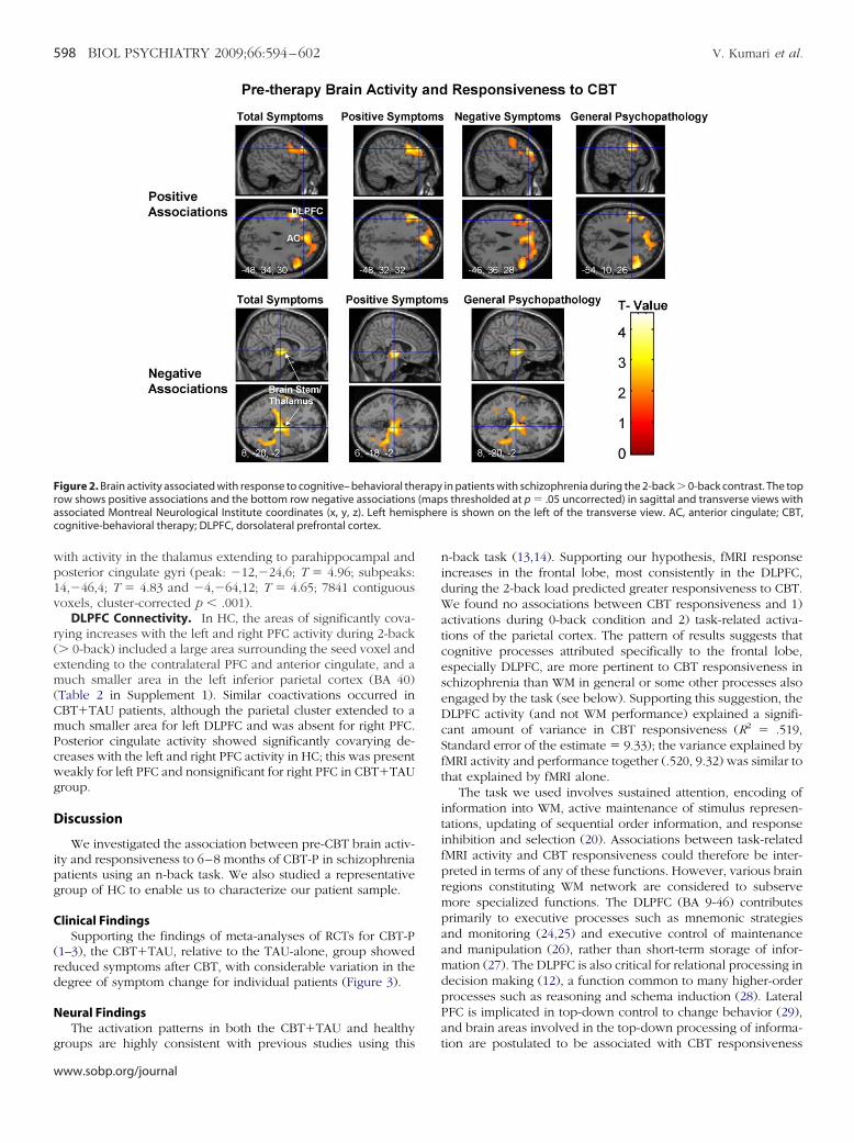
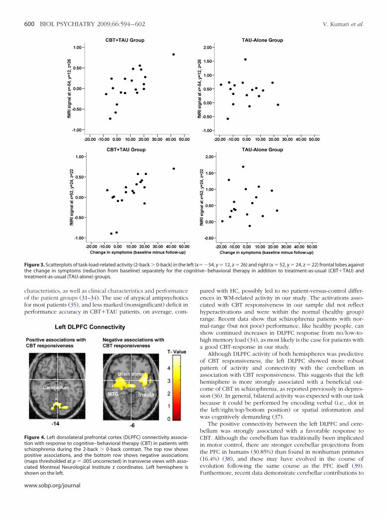
ged between pretherapy task-related activity at 2-back (� 0-back) and CBT responsiveness (Figure 2, Table 2); theserelationships were absent in the TAU-alone group (Figure 3).Specifically, a reduction in total PANSS scores was associatedwith greater activity bilaterally in the inferior-middle frontalgyrus, mainly the DLPFC (BA 46). A medial PFC–anterior cingu-late cluster (uncorrected p � .012) failed to survive SVC. Areduction in positive symptoms was associated with greater leftinferior-middle frontal gyrus, most consistently Brodmann’s area[BA] 9-46, activity. A reduction in negative symptoms wasassociated with greater pretherapy activity in a large left-sidedcluster including the caudate, dorsomedial PFC, and DLPFC (BA9-46). A reduction in general psychopathology was associatedwith greater activity bilaterally in the inferior-middle frontalgyrus, primarily BA 46.
No other activation was positively associated with a change intotal or subscale symptom scores at any task load. Activity inseveral regions, mainly those found to be deactivated duringmemory load relative to no memory load (0-back/rest) condi-tions (Supplement 1), was associated negatively with CBT re-sponsiveness during 1-back and 2-back (� 0-back) conditions(Figure 2, Table 2). These relationships are due to relativelystronger deactivations during memory load conditions in patientswith the strongest CBT response.
The CBT�TAU and HC groups did not differ in activity of theregions positively or negatively associated with CBT responsive-ness (p values � .20).
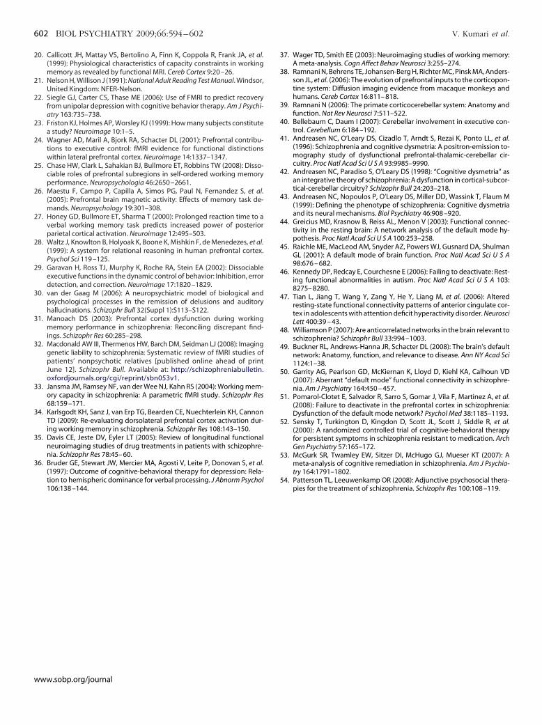
Within the left DLPFC connectivity maps (2-back � 0-back),CBT responsiveness associated positively with covarying in-creases in activity in a lingual gyrus-cerebellum cluster (peak:�4[x], �70[y], �4[z], T � 5.20; subpeaks: 8,�70, 0, T � 4.84 and�2,�70,�14; T � 4.30; 874 contiguous voxels; cluster-correctedp � .04), and negatively with activity in the insula extending tohalamus/brainstem and middle/superior temporal gyrus (peak:38,6,�6; T � 5.04; subpeaks: �4,�14,�4; T � 4.74 and40,�12,0, T � 4.67; 4187 contiguous voxels, cluster-corrected� .001) (Figure 4). Within the right DLPFC connectivity maps
(2-back � 0-back), CBT responsiveness associated negatively
www.sobp.org/journal
wp1v
r(em(CmPcwg
D
ipg
C
(rd
N
g
Frac
598 BIOL PSYCHIATRY 2009;66:594–602 V. Kumari et al.
w
ith activity in the thalamus extending to parahippocampal andosterior cingulate gyri (peak: �12,�24,6; T � 4.96; subpeaks:4,�46,4; T � 4.83 and �4,�64,12; T � 4.65; 7841 contiguousoxels, cluster-corrected p � .001).
DLPFC Connectivity. In HC, the areas of significantly cova-ying increases with the left and right PFC activity during 2-back� 0-back) included a large area surrounding the seed voxel andxtending to the contralateral PFC and anterior cingulate, and auch smaller area in the left inferior parietal cortex (BA 40)
Table 2 in Supplement 1). Similar coactivations occurred inBT�TAU patients, although the parietal cluster extended to auch smaller area for left DLPFC and was absent for right PFC.osterior cingulate activity showed significantly covarying de-reases with the left and right PFC activity in HC; this was presenteakly for left PFC and nonsignificant for right PFC in CBT�TAUroup.
iscussion
We investigated the association between pre-CBT brain activ-ty and responsiveness to 6–8 months of CBT-P in schizophreniaatients using an n-back task. We also studied a representativeroup of HC to enable us to characterize our patient sample.
linical FindingsSupporting the findings of meta-analyses of RCTs for CBT-P
1–3), the CBT�TAU, relative to the TAU-alone, group showededuced symptoms after CBT, with considerable variation in theegree of symptom change for individual patients (Figure 3).
eural FindingsThe activation patterns in both the CBT�TAU and healthy
igure 2. Brain activity associated with response to cognitive– behavioral thow shows positive associations and the bottom row negative associationsssociated Montreal Neurological Institute coordinates (x, y, z). Left hemisognitive-behavioral therapy; DLPFC, dorsolateral prefrontal cortex.
roups are highly consistent with previous studies using this
ww.sobp.org/journal
n-back task (13,14). Supporting our hypothesis, fMRI responseincreases in the frontal lobe, most consistently in the DLPFC,during the 2-back load predicted greater responsiveness to CBT.We found no associations between CBT responsiveness and 1)activations during 0-back condition and 2) task-related activa-tions of the parietal cortex. The pattern of results suggests thatcognitive processes attributed specifically to the frontal lobe,especially DLPFC, are more pertinent to CBT responsiveness inschizophrenia than WM in general or some other processes alsoengaged by the task (see below). Supporting this suggestion, theDLPFC activity (and not WM performance) explained a signifi-cant amount of variance in CBT responsiveness (R2 � .519,Standard error of the estimate � 9.33); the variance explained byfMRI activity and performance together (.520, 9.32) was similar tothat explained by fMRI alone.
The task we used involves sustained attention, encoding ofinformation into WM, active maintenance of stimulus represen-tations, updating of sequential order information, and responseinhibition and selection (20). Associations between task-relatedfMRI activity and CBT responsiveness could therefore be inter-preted in terms of any of these functions. However, various brainregions constituting WM network are considered to subservemore specialized functions. The DLPFC (BA 9-46) contributesprimarily to executive processes such as mnemonic strategiesand monitoring (24,25) and executive control of maintenanceand manipulation (26), rather than short-term storage of infor-mation (27). The DLPFC is also critical for relational processing indecision making (12), a function common to many higher-orderprocesses such as reasoning and schema induction (28). LateralPFC is implicated in top-down control to change behavior (29),and brain areas involved in the top-down processing of informa-
in patients with schizophrenia during the 2-back � 0-back contrast. The tops thresholded at p � .05 uncorrected) in sagittal and transverse views withe is shown on the left of the transverse view. AC, anterior cingulate; CBT,
erapy(mappher
tion are postulated to be associated with CBT responsiveness

(aoDc
T
P
T
P
N
G
N
T
P
N
G
e
V. Kumari et al. BIOL PSYCHIATRY 2009;66:594–602 599
30). The positive association we report here between DLPFCctivity and CBT responsiveness may be mediated by facilitationf effective CBT by executive processes modulated by theLPFC. Patients with greater DLPFC response may be more
able 2. Brain Areas Showing Positive and Negative Associations with CBT
ositive Associations BA Cluster Size
otal Symptoms2-back � 0-back
Inferior-middle frontal gyrus 44 10446
Inferior-middle frontal gyrus 45/46 11346
ositive Symptoms2-back � 0-back
Inferior-middle frontal gyrus 9–46 12644
9egative Symptoms2-back � 0-back
Caudate nucleus n/a 1280Anterior cingulate/medial prefrontal cortex 32/8Middle frontal gyrus 9–46
eneral Psychopathology2-back � 0-back
Inferior-middle frontal gyrus 44 8746
Middle frontal gyrus 46 72
egative Associations
otal Symptoms1-back � 0-back
Insula n/a 1574Middle temporal gyrus 20/21Parahippocampal gyrus 36
2-back � 0-backBrain stem n/a 772Middle temporal gyrus 21Inferior parietal lobe 40
ositive Symptoms1-back � 0-back
Transverse temporal gyrus 41 1170Posterior cingulate 23
23/312-back � 0-back
Brain stem n/a 659n/a
Middle temporal gyrus 39egative SymptomsNone
eneral Psychopathology1-back � 0 back
Middle temporal gyrus 21/20 1624Parahippocampal gyrus 35/36Fusiform gyrus 37
2-back � 0-backBrain stem n/a 1743Posterior cingulate 31Inferior parietal lobe 5
BA, Brodmann area; MNI, Montreal Neurological Institute. In italics: SVC crntire brain.
apable of schema induction (facilitating transfer of learning
from one situation to other, similar, situations), reasoning, andrelational processing (pooling together and comparing decision-relevant information) and gain most from CBT.
Previous studies have reported hyper-, hypo-, or normal-
onsiveness in Patients
ls) Side
MNI Coordinates
Voxel T Value Corrected pX Y Z
L �54 12 26 4.85 .015L �48 34 30 4.43R 52 24 22 3.99 .033R 42 24 26 2.95
L �48 32 32 4.61 .021L �52 12 28 4.03L �54 22 32 3.75
L �14 2 14 5.60 �.001L �6 22 52 4.57L �46 36 28 4.55
L �54 10 26 4.51 .025L �48 34 30 3.83R 52 26 24 4.48 .026
L �30 �28 6 5.50 �.001L �36 �34 �14 5.32L �20 �40 �14 4.65
L �8 �20 �2 3.83 .006L �60 0 �10 3.65L �50 �40 48 3.54
L �30 �30 10 4.94 �.001R 10 �36 28 4.91R 14 �56 20 4.82
R 6 �18 �2 4.24 .016R 8 �12 �8 3.92R 36 �62 18 3.60
L �38 �32 �14 5.31 �.001L �18 �38 �14 5.14L �32 �40 �16 5.02
R 8 �20 �2 4.37 �.001L �24 �60 18 4.09L �24 �44 54 3.94
n applied. All others: cluster p corrected for multiple comparisons across the
Resp
(voxe
8
2
3
2
4
3
6
9
4
3
5
5
iterio
range frontal activations in schizophrenia depending on task
www.sobp.org/journal
cofp
Ftt
Ftsp(cs
600 BIOL PSYCHIATRY 2009;66:594–602 V. Kumari et al.
w
haracteristics, as well as clinical characteristics and performancef the patient groups (31–34). The use of atypical antipsychoticsor most patients (35), and less marked (nonsignificant) deficit inerformance accuracy in CBT�TAU patients, on average, com-
igure 3. Scatterplots of task-load-related activity (2-back � 0-back) in the lehe change in symptoms (reduction from baseline) separately for the coreatment-as-usual (TAU-alone) groups.
igure 4. Left dorsolateral prefrontal cortex (DLPFC) connectivity associa-ion with response to cognitive– behavioral therapy (CBT) in patients withchizophrenia during the 2-back � 0-back contrast. The top row showsositive associations, and the bottom row shows negative associations
maps thresholded at p � .005 uncorrected) in transverse views with asso-iated Montreal Neurological Institute z coordinates. Left hemisphere is
hown on the left.ww.sobp.org/journal
pared with HC, possibly led to no patient-versus-control differ-ences in WM-related activity in our study. The activations asso-ciated with CBT responsiveness in our sample did not reflecthyperactivations and were within the normal (healthy group)range. Recent data show that schizophrenia patients with nor-mal-range (but not poor) performance, like healthy people, canshow continued increases in DLPFC response from no/low-to-high memory load (34), as most likely is the case for patients witha good CBT-response in our study.
Although DLPFC activity of both hemispheres was predictiveof CBT responsiveness, the left DLPFC showed more robustpattern of activity and connectivity with the cerebellum inassociation with CBT responsiveness. This suggests that the lefthemisphere is more strongly associated with a beneficial out-come of CBT in schizophrenia, as reported previously in depres-sion (36). In general, bilateral activity was expected with our taskbecause it could be performed by encoding verbal (i.e., dot inthe left/right/top/bottom position) or spatial information andwas cognitively demanding (37).
The positive connectivity between the left DLPFC and cere-bellum was strongly associated with a favorable response toCBT. Although the cerebellum has traditionally been implicatedin motor control, there are stronger cerebellar projections fromthe PFC in humans (30.85%) than found in nonhuman primates(16.4%) (38), and these may have evolved in the course ofevolution following the same course as the PFC itself (39).
�54, y � 12, z � 26) and right (x � 52, y � 24, z � 22) frontal lobes againste– behavioral therapy in addition to treatment-as-usual (CBT�TAU) and
ft (x�gnitiv
Furthermore, recent data demonstrate cerebellar contributions to

hmTacnAttsh
nrtiimlnasb
L
rggwtpfobdmtsFt(crc(shFftvC
C
tnPemDi
V. Kumari et al. BIOL PSYCHIATRY 2009;66:594–602 601
igher-order cognitive functions, especially the task manage-ent and multitasking components of executive processing (40).he DLPFC-cerebellum connectivity and CBT responsivenessssociation may thus be explained by the PFC–cerebellumontributions to executive control, facilitating CBT responsive-ess in the same way as the DLPFC activity itself. According tondreasen et al. (41–43), disruption in the corticocerebellar-
halamo-cortical circuitry results in deficient processing, priori-izing, retrieval, coordination, and responding to information inchizophrenia. Our findings suggest that this circuitry may alsoave a role in responsiveness to CBT in schizophrenia.
Finally, we observed strong associations between a low, oro, response to CBT in patients and reduced deactivation of theegions that were deactivated during the rest/0-back, relative tohe memory load, conditions in HC. These have generally beenmplicated in “mind-wandering” default states (44,45). Our find-ng may indicate an association between a reduced ability toaintain focus on, or switch to, a goal (task in this case) and a
ess favorable response to CBT. Clinically, disruption of defaultetwork activity has been reported in several disorders includingutism (46), attention-deficit/hyperactivity disorder (47), andchizophrenia (48–51). Our findings suggest that default mode ofrain action has a role in CBT efficacy in schizophrenia.
imitationsFirst, this study used a parallel-group, rather than a purely
andom, design for allocation to CBTp�TAU and TAU-aloneroups. Although we cannot prove that the patients in the TAUroup would also improve if they received CBT, the patientsere randomly distributed across both groups in their desire for
his intervention. Second, it could be argued that CBT�TAUatients showed clinical improvement simply because of bene-iting from therapist contact, independent of the specific effectsf the CBT methods applied to them. It is, however, unlikelyecause the standard care provided to patients before, anduring, the study consisted of management offered by a caseanagement team with a dedicated care coordinator who saw
he patient regularly, in addition to psychiatrists and otherpecialists, such as a benefits adviser and occupational therapist.urthermore, CBT for psychosis has specific effects on symp-oms, distinct from interventions such as social skills traininghelp acquire social skills) or cognitive remediation (improveognitive functioning) (1,3), and has been found superior ineducing the symptoms to a nonspecific befriending interventionontrolling for the amount of contact with treating professionals52). Third, the CBT�TAU and TAU-alone groups differedlightly in IQ, education, and illness duration; none of these,owever, had a noticeable influence in CBT responsiveness.ourth, the use of a block design limited the interpretation ofMRI findings in terms of the component processes involved inask performance. Finally, we employed a spatial MW task;erbal WM may be more pertinent to skills needed to engage inBT.
onclusionsWithin the WM network, the DLPFC activity and its connec-
ivity with the cerebellum are associated with CBT responsive-ess in schizophrenia. These effects may be mediated by theFC–cerebellum contributions to executive processes facilitatingffective CBT within a psychotherapeutic context. Our resultsay imply that addressing cognitive deficits associated withLPFC in schizophrenia would maximize benefit from CBT. This
s in line with recent data showing better outcomes with a
combination of cognitive training and psychiatric rehabilitationin schizophrenia (53,54).
This research was supported by the Wellcome Trust (Grant No.067427/z/02/z). We thank Dr. Angus MacDonald for his com-ments on an earlier version of this article and Ms. Ingrid Aasenand Dr. Michael A Cooke for their help with data collection.
Competing Interests: The authors report no biomedical finan-cial interests or potential conflicts of interest.
Supplementary material cited in this article is availableonline.
1. Pfammatter M, Junghan UM, Brenner HD (2006): Efficacy of psycholog-ical therapy in schizophrenia: Conclusions from meta-analyses. Schizo-phr Bull 32(suppl 1):S64 –S80.
2. Wykes T, Steel C, Everitt B, Tarrier N (2008): Cognitive behavior therapyfor schizophrenia: Effect sizes, clinical models, and methodologicalrigor. Schizophr Bull 34:523–537.
3. Pilling S, Bebbington P, Kuipers E, Garety P, Geddes J, Orbach G, et al.(2002): Psychological treatments in schizophrenia. I. Meta-analysis offamily intervention and cognitive behaviour therapy. Psychol Med 32:763–782.
4. Reichenberg A, Harvey PD (2007): Neuropsychological impairments inschizophrenia: Integration of performance-based and brain imagingfindings. Psychol Bull 133:833– 858.
5. Moorey S, Holting C, Hughes P, Knynenberg P, Michael A (2001): Doesproblem solving ability predict therapy outcome in a clinical setting?Behav Cognit Psychother 29:485– 495.
6. Julian LJ, Mohr DC (2006): Cognitive predictors of response to treatmentfor depression in multiple sclerosis. J Neuropsychiatr Clin Neurosci 18:356 –363.
7. Mohlman J, Gorman JM (2005): The role of executive functioning in CBT:A pilot study with anxious older adults. Behav Res Ther 43:447– 465.
8. Garety P, Fowler D, Kuipers E, Freeman D, Dunn G, Bebbington P, et al.(1997): London-East Anglia randomised controlled trial of cognitive-behavioural therapy for psychosis. II: predictors of outcome. Br J Psychi-atry 171:420 – 426.
9. Granholm E, McQuaid JR, Link PC, Fish S, Patterson T, Jeste DV (2008):Neuropsychological predictors of functional outcome in cognitive be-havioral social skills training for older people with schizophrenia. Schizo-phr Res 100:133–143.
10. Cabeza R, Nyberg L (2000): Imaging cognition II: An empirical review of275 PET and fMRI studies. J Cogn Neurosci 12:1– 47.
11. Duncan J, Owen AM (2000): Common regions of the human frontal loberecruited by diverse cognitive demands. Trends Neurosci 23:475– 483.
12. Krawczyk DC (2002): Contributions of the prefrontal cortex to the neuralbasis of human decision making. Neurosci Biobehav Rev 26:631– 664.
13. Kumari V, Aasen I, Taylor P, Ffytche DH, Das M, Barkataki I, et al. (2006):Neural dysfunction and violence in schizophrenia: An fMRI investiga-tion. Schizophr Res 84:144 –164.
14. Kumari V, Aasen I, Ffytche D, Williams SC, Sharma T (2006): Neuralcorrelates of adjunctive rivastigmine treatment to antipsychotics inschizophrenia: A randomized, placebo-controlled, double-blind fMRIstudy. Neuroimage 29:545–556.
15. Yoon JH, Minzenberg MJ, Ursu S, Walters R, Wendelken C, Ragland JD, etal. (2008): Association of dorsolateral prefrontal cortex dysfunction withdisrupted coordinated brain activity in schizophrenia: Relationship withimpaired cognition, behavioral disorganization, and global function.Am J Psychiatry 165:1006 –1014.
16. First MB, Spitzer RL, Gibbon M, Williams JBW (1995) : Structured ClinicalInterview for DSM-IV Axis I Disorders, Patient Edition (SCID-P), Version 2:New York: New York State Psychiatric Institute, Biometrics Research.
17. First MB, Spitzer RL, Gibbon M, Williams JBW (2002): Structured ClinicalInterview for DSM-IV-TR Axis I Disorders, Research Version, Non-PatientEdition. SCID-I/NP. New York: New York State Psychiatric Institute, Bio-metrics Research.
18. Kay SR, Fiszbein A, Opler LA (1987): The positive and negative syndromescale (PANSS) for schizophrenia. Schizophr Bull 13:261–276.
19. Fowler D, Garety PA, Kuipers E (1995): Cognitive Behaviour Therapy for
Psychosis: Theory and Practice. Chichester, UK: Wiley.www.sobp.org/journal
2
2
2
2
2
2
2
2
2
2
3
3
3
3
3
3
3
602 BIOL PSYCHIATRY 2009;66:594–602 V. Kumari et al.
w
0. Callicott JH, Mattay VS, Bertolino A, Finn K, Coppola R, Frank JA, et al.(1999): Physiological characteristics of capacity constraints in workingmemory as revealed by functional MRI. Cereb Cortex 9:20 –26.
1. Nelson H, Willison J (1991): National Adult Reading Test Manual. Windsor,United Kingdom: NFER-Nelson.
2. Siegle GJ, Carter CS, Thase ME (2006): Use of FMRI to predict recoveryfrom unipolar depression with cognitive behavior therapy. Am J Psychi-atry 163:735–738.
3. Friston KJ, Holmes AP, Worsley KJ (1999): How many subjects constitutea study? Neuroimage 10:1–5.
4. Wagner AD, Maril A, Bjork RA, Schacter DL (2001): Prefrontal contribu-tions to executive control: fMRI evidence for functional distinctionswithin lateral prefrontal cortex. Neuroimage 14:1337–1347.
5. Chase HW, Clark L, Sahakian BJ, Bullmore ET, Robbins TW (2008): Disso-ciable roles of prefrontal subregions in self-ordered working memoryperformance. Neuropsychologia 46:2650 –2661.
6. Maestu F, Campo P, Capilla A, Simos PG, Paul N, Fernandez S, et al.(2005): Prefrontal brain magnetic activity: Effects of memory task de-mands. Neuropsychology 19:301–308.
7. Honey GD, Bullmore ET, Sharma T (2000): Prolonged reaction time to averbal working memory task predicts increased power of posteriorparietal cortical activation. Neuroimage 12:495–503.
8. Waltz J, Knowlton B, Holyoak K, Boone K, Mishkin F, de Menedezes, et al.(1999): A system for relational reasoning in human prefrontal cortex.Psychol Sci 119 –125.
9. Garavan H, Ross TJ, Murphy K, Roche RA, Stein EA (2002): Dissociableexecutive functions in the dynamic control of behavior: Inhibition, errordetection, and correction. Neuroimage 17:1820 –1829.
0. van der Gaag M (2006): A neuropsychiatric model of biological andpsychological processes in the remission of delusions and auditoryhallucinations. Schizophr Bull 32(Suppl 1):S113–S122.
1. Manoach DS (2003): Prefrontal cortex dysfunction during workingmemory performance in schizophrenia: Reconciling discrepant find-ings. Schizophr Res 60:285–298.
2. Macdonald AW III, Thermenos HW, Barch DM, Seidman LJ (2008): Imaginggenetic liability to schizophrenia: Systematic review of fMRI studies ofpatients’ nonpsychotic relatives [published online ahead of printJune 12]. Schizophr Bull. Available at: http://schizophreniabulletin.oxfordjournals.org/cgi/reprint/sbn053v1.
3. Jansma JM, Ramsey NF, van der Wee NJ, Kahn RS (2004): Working mem-ory capacity in schizophrenia: A parametric fMRI study. Schizophr Res68:159 –171.
4. Karlsgodt KH, Sanz J, van Erp TG, Bearden CE, Nuechterlein KH, CannonTD (2009): Re-evaluating dorsolateral prefrontal cortex activation dur-ing working memory in schizophrenia. Schizophr Res 108:143–150.
5. Davis CE, Jeste DV, Eyler LT (2005): Review of longitudinal functionalneuroimaging studies of drug treatments in patients with schizophre-nia. Schizophr Res 78:45– 60.
6. Bruder GE, Stewart JW, Mercier MA, Agosti V, Leite P, Donovan S, et al.(1997): Outcome of cognitive-behavioral therapy for depression: Rela-tion to hemispheric dominance for verbal processing. J Abnorm Psychol
106:138 –144.ww.sobp.org/journal
37. Wager TD, Smith EE (2003): Neuroimaging studies of working memory:A meta-analysis. Cogn Affect Behav Neurosci 3:255–274.
38. Ramnani N, Behrens TE, Johansen-Berg H, Richter MC, Pinsk MA, Anders-son JL, et al. (2006): The evolution of prefrontal inputs to the corticopon-tine system: Diffusion imaging evidence from macaque monkeys andhumans. Cereb Cortex 16:811– 818.
39. Ramnani N (2006): The primate corticocerebellar system: Anatomy andfunction. Nat Rev Neurosci 7:511–522.
40. Bellebaum C, Daum I (2007): Cerebellar involvement in executive con-trol. Cerebellum 6:184 –192.
41. Andreasen NC, O’Leary DS, Cizadlo T, Arndt S, Rezai K, Ponto LL, et al.(1996): Schizophrenia and cognitive dysmetria: A positron-emission to-mography study of dysfunctional prefrontal-thalamic-cerebellar cir-cuitry. Proc Natl Acad Sci U S A 93:9985–9990.
42. Andreasen NC, Paradiso S, O’Leary DS (1998): “Cognitive dysmetria” asan integrative theory of schizophrenia: A dysfunction in cortical-subcor-tical-cerebellar circuitry? Schizophr Bull 24:203–218.
43. Andreasen NC, Nopoulos P, O’Leary DS, Miller DD, Wassink T, Flaum M(1999): Defining the phenotype of schizophrenia: Cognitive dysmetriaand its neural mechanisms. Biol Psychiatry 46:908 –920.
44. Greicius MD, Krasnow B, Reiss AL, Menon V (2003): Functional connec-tivity in the resting brain: A network analysis of the default mode hy-pothesis. Proc Natl Acad Sci U S A 100:253–258.
45. Raichle ME, MacLeod AM, Snyder AZ, Powers WJ, Gusnard DA, ShulmanGL (2001): A default mode of brain function. Proc Natl Acad Sci U S A98:676 – 682.
46. Kennedy DP, Redcay E, Courchesne E (2006): Failing to deactivate: Rest-ing functional abnormalities in autism. Proc Natl Acad Sci U S A 103:8275– 8280.
47. Tian L, Jiang T, Wang Y, Zang Y, He Y, Liang M, et al. (2006): Alteredresting-state functional connectivity patterns of anterior cingulate cor-tex in adolescents with attention deficit hyperactivity disorder. NeurosciLett 400:39 – 43.
48. Williamson P (2007): Are anticorrelated networks in the brain relevant toschizophrenia? Schizophr Bull 33:994 –1003.
49. Buckner RL, Andrews-Hanna JR, Schacter DL (2008): The brain’s defaultnetwork: Anatomy, function, and relevance to disease. Ann NY Acad Sci1124:1–38.
50. Garrity AG, Pearlson GD, McKiernan K, Lloyd D, Kiehl KA, Calhoun VD(2007): Aberrant “default mode” functional connectivity in schizophre-nia. Am J Psychiatry 164:450 – 457.
51. Pomarol-Clotet E, Salvador R, Sarro S, Gomar J, Vila F, Martinez A, et al.(2008): Failure to deactivate in the prefrontal cortex in schizophrenia:Dysfunction of the default mode network? Psychol Med 38:1185–1193.
52. Sensky T, Turkington D, Kingdon D, Scott JL, Scott J, Siddle R, et al.(2000): A randomized controlled trial of cognitive-behavioral therapyfor persistent symptoms in schizophrenia resistant to medication. ArchGen Psychiatry 57:165–172.
53. McGurk SR, Twamley EW, Sitzer DI, McHugo GJ, Mueser KT (2007): Ameta-analysis of cognitive remediation in schizophrenia. Am J Psychia-try 164:1791–1802.
54. Patterson TL, Leeuwenkamp OR (2008): Adjunctive psychosocial thera-pies for the treatment of schizophrenia. Schizophr Res 100:108 –119.