p40 elite - sonoscape
TRANSCRIPT
U-P
40E2
0200
715© 2020 SonoScape Medical Corp.
All rights are reserved.
SonoScape Medical reserves the right to change the above information and discontinue any product at any time without prior noti�cation and will not be liable for any consequences resulting from the use of this publication.
SonoScape Medical Corp.2F, 12th Building, Shenzhen Software Park Phase II, Keji Middle 2nd Road, Nanshan District, Shenzhen 518057, Guangdong, ChinaTel: +86-755-26722890 Fax: +86-755-26722850Email: [email protected] www.sonoscape.com
P40 EliteElite Experience Meets Value
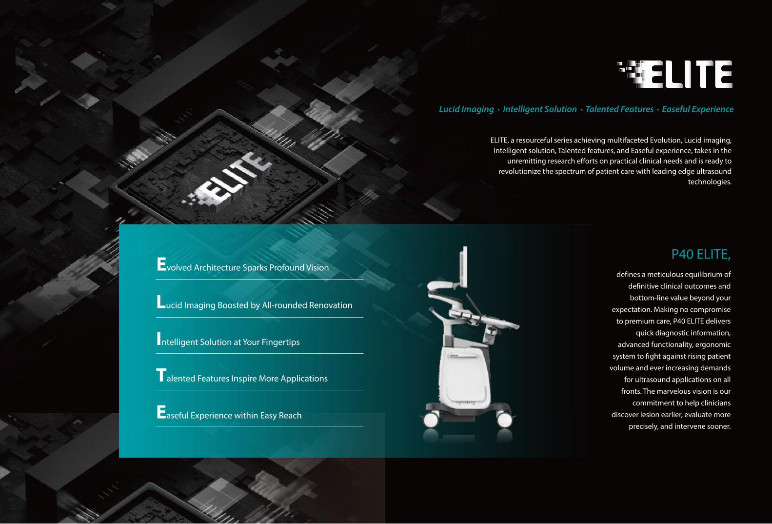
P40 ELITE, defines a meticulous equilibrium of
definitive clinical outcomes and bottom-line value beyond your
expectation. Making no compromise to premium care, P40 ELITE delivers
quick diagnostic information, advanced functionality, ergonomic
system to fight against rising patient volume and ever increasing demands
for ultrasound applications on all fronts. The marvelous vision is our
commitment to help clinicians discover lesion earlier, evaluate more
precisely, and intervene sooner.
Evolved Architecture Sparks Profound Vision
Lucid Imaging Boosted by All-rounded Renovation
Intelligent Solution at Your Fingertips
Talented Features Inspire More Applications
Easeful Experience within Easy Reach
Lucid Imaging Intelligent Solution Talented Features Easeful Experience
ELITE, a resourceful series achieving multifaceted Evolution, Lucid imaging, Intelligent solution, Talented features, and Easeful experience, takes in the
unremitting research efforts on practical clinical needs and is ready to revolutionize the spectrum of patient care with leading edge ultrasound
technologies.
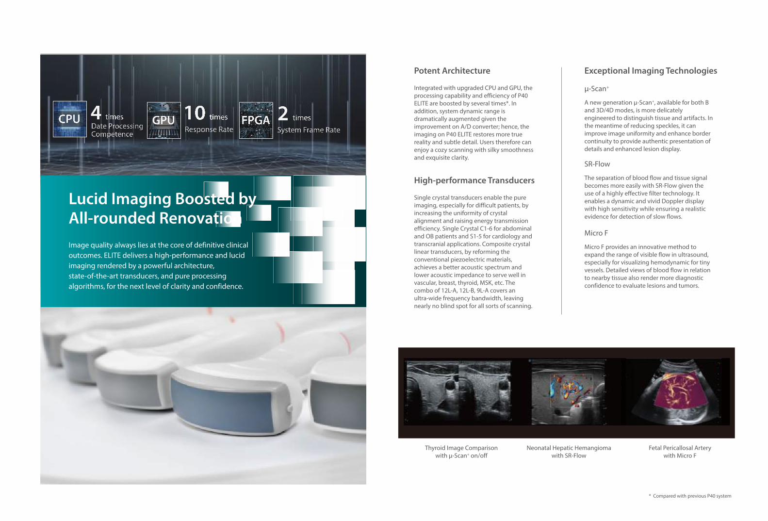
GPU FPGACPUDate ProcessingCompetence
4 times
Response Rate
10 times
System Frame Rate
2 times
Potent Architecture
Integrated with upgraded CPU and GPU, the processing capability and efficiency of P40 ELITE are boosted by several times*. In addition, system dynamic range is dramatically augmented given the improvement on A/D converter; hence, the imaging on P40 ELITE restores more true reality and subtle detail. Users therefore can enjoy a cozy scanning with silky smoothness and exquisite clarity.
High-performance Transducers
Single crystal transducers enable the pure imaging, especially for difficult patients, by increasing the uniformity of crystal alignment and raising energy transmission efficiency. Single Crystal C1-6 for abdominal and OB patients and S1-5 for cardiology and transcranial applications. Composite crystal linear transducers, by reforming the conventional piezoelectric materials, achieves a better acoustic spectrum and lower acoustic impedance to serve well in vascular, breast, thyroid, MSK, etc. The combo of 12L-A, 12L-B, 9L-A covers an ultra-wide frequency bandwidth, leaving nearly no blind spot for all sorts of scanning.
Exceptional Imaging Technologies
A new generation μ-Scan+, available for both B and 3D/4D modes, is more delicately engineered to distinguish tissue and artifacts. In the meantime of reducing speckles, it can improve image uniformity and enhance border continuity to provide authentic presentation of details and enhanced lesion display.
μ-Scan+
The separation of blood flow and tissue signal becomes more easily with SR-Flow given the use of a highly effective filter technology. It enables a dynamic and vivid Doppler display with high sensitivity while ensuring a realistic evidence for detection of slow flows.
SR-Flow
Micro F provides an innovative method to expand the range of visible flow in ultrasound, especially for visualizing hemodynamic for tiny vessels. Detailed views of blood flow in relation to nearby tissue also render more diagnostic confidence to evaluate lesions and tumors.
Micro F
Image quality always lies at the core of definitive clinical outcomes. ELITE delivers a high-performance and lucid imaging rendered by a powerful architecture, state-of-the-art transducers, and pure processing algorithms, for the next level of clarity and confidence.
Lucid Imaging Boosted by All-rounded Renovation
Thyroid Image Comparisonwith μ-Scan+ on/off
* Compared with previous P40 system
Neonatal Hepatic Hemangiomawith SR-Flow
Fetal Pericallosal Arterywith Micro F
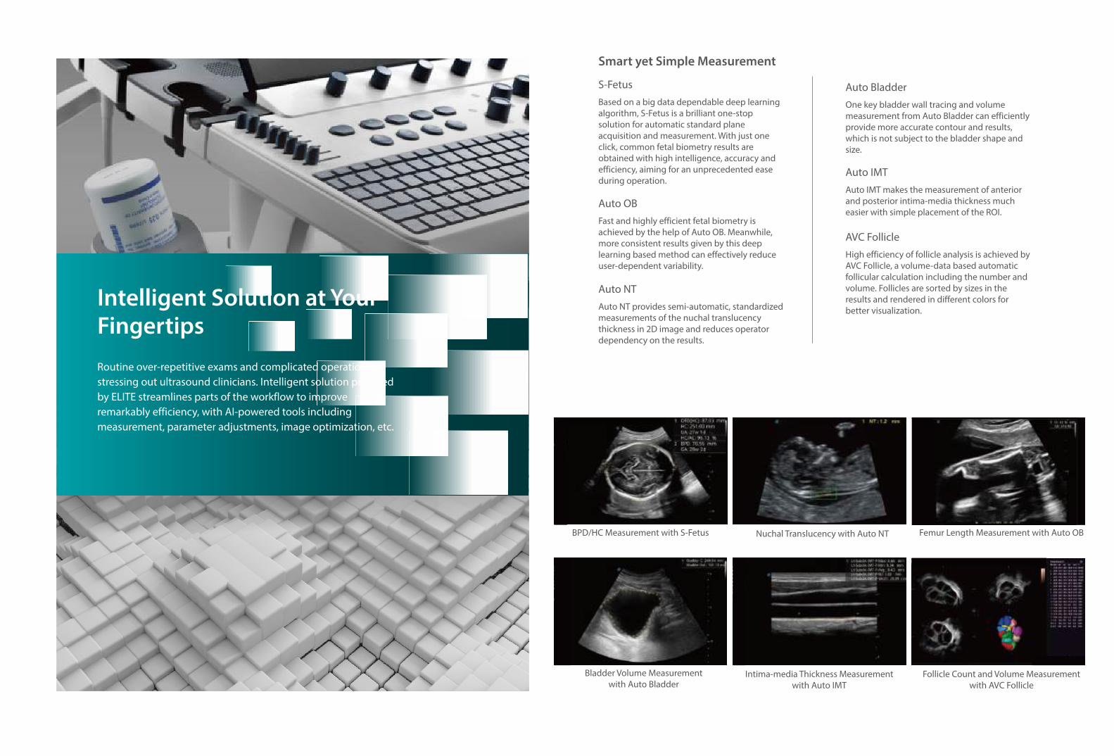
Smart yet Simple Measurement
Based on a big data dependable deep learning algorithm, S-Fetus is a brilliant one-stop solution for automatic standard plane acquisition and measurement. With just one click, common fetal biometry results are obtained with high intelligence, accuracy and efficiency, aiming for an unprecedented ease during operation.
S-Fetus
Fast and highly efficient fetal biometry is achieved by the help of Auto OB. Meanwhile, more consistent results given by this deep learning based method can effectively reduce user-dependent variability.
Auto OB
Auto NT provides semi-automatic, standardized measurements of the nuchal translucency thickness in 2D image and reduces operator dependency on the results.
Auto NT
BPD/HC Measurement with S-Fetus Femur Length Measurement with Auto OB
Follicle Count and Volume Measurementwith AVC Follicle
One key bladder wall tracing and volume measurement from Auto Bladder can efficiently provide more accurate contour and results, which is not subject to the bladder shape and size.
Auto Bladder
Auto IMT makes the measurement of anterior and posterior intima-media thickness much easier with simple placement of the ROI.
Auto IMT
High efficiency of follicle analysis is achieved by AVC Follicle, a volume-data based automatic follicular calculation including the number and volume. Follicles are sorted by sizes in the results and rendered in different colors for better visualization.
AVC Follicle
Routine over-repetitive exams and complicated operation are stressing out ultrasound clinicians. Intelligent solution provided by ELITE streamlines parts of the workflow to improve remarkably efficiency, with AI-powered tools including measurement, parameter adjustments, image optimization, etc.
Intelligent Solution at Your Fingertips
Intima-media Thickness Measurementwith Auto IMT
Bladder Volume Measurementwith Auto Bladder
Nuchal Translucency with Auto NT
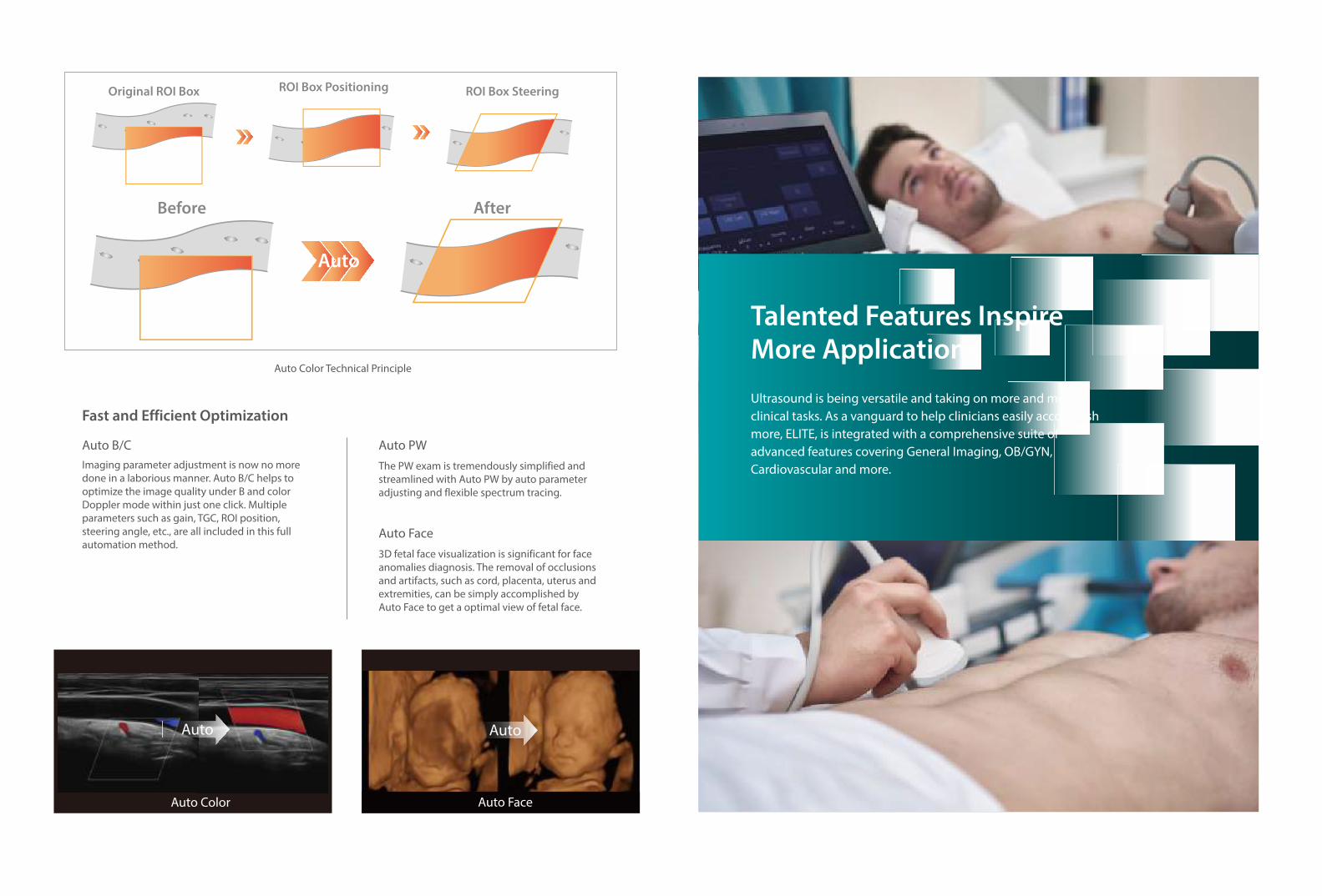
Fast and Efficient Optimization
Auto Color Technical Principle
Imaging parameter adjustment is now no more done in a laborious manner. Auto B/C helps to optimize the image quality under B and color Doppler mode within just one click. Multiple parameters such as gain, TGC, ROI position, steering angle, etc., are all included in this full automation method.
Auto B/CThe PW exam is tremendously simplified and streamlined with Auto PW by auto parameter adjusting and flexible spectrum tracing.
Auto PW
3D fetal face visualization is significant for face anomalies diagnosis. The removal of occlusions and artifacts, such as cord, placenta, uterus and extremities, can be simply accomplished by Auto Face to get a optimal view of fetal face.
Auto Face
Ultrasound is being versatile and taking on more and more clinical tasks. As a vanguard to help clinicians easily accomplish more, ELITE, is integrated with a comprehensive suite of advanced features covering General Imaging, OB/GYN, Cardiovascular and more.
Talented Features Inspire More Applications
AutoAuto
Auto
Before
Original ROI Box ROI Box Positioning ROI Box Steering
After
Auto Color Auto Face
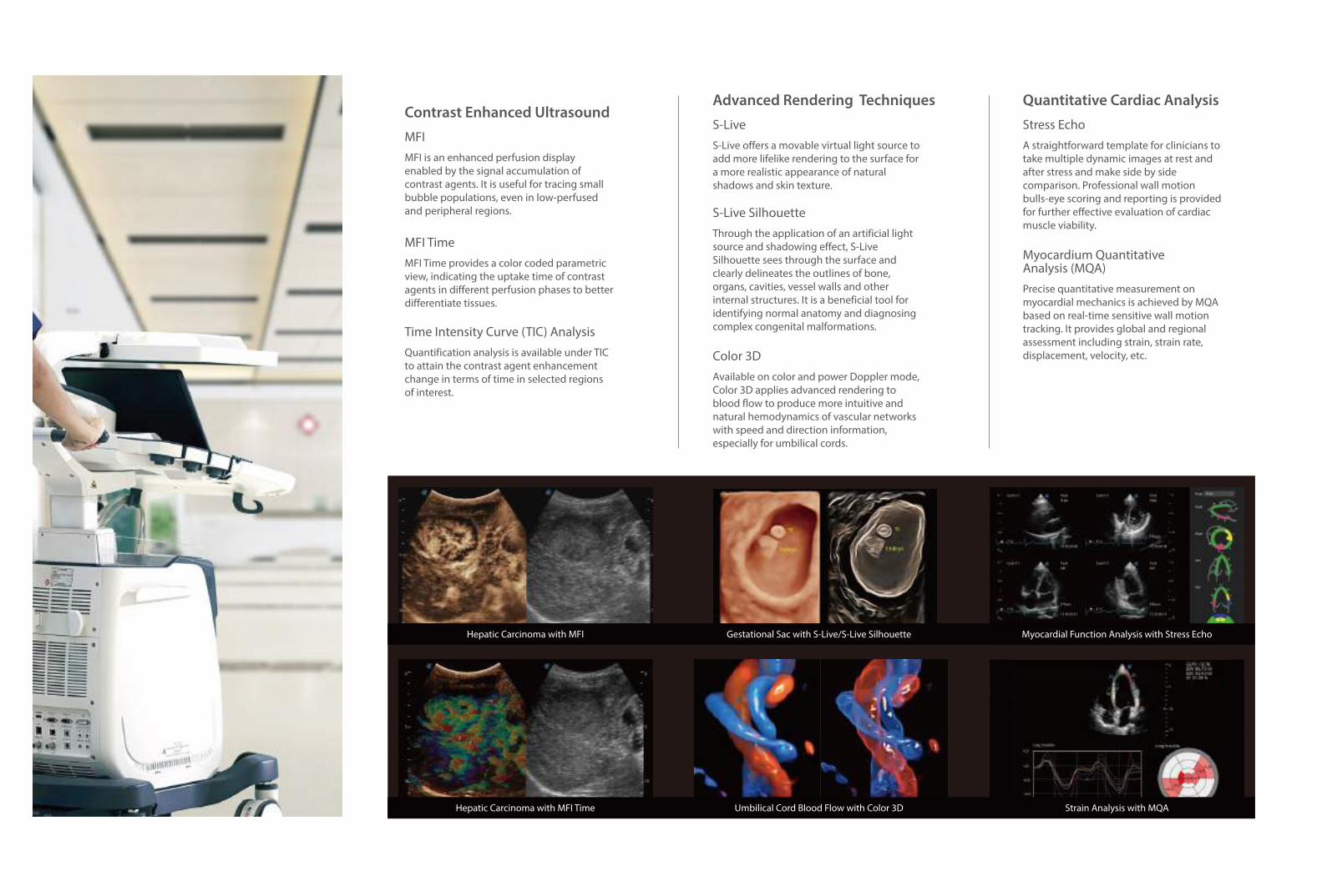
Contrast Enhanced Ultrasound
MFI is an enhanced perfusion display enabled by the signal accumulation of contrast agents. It is useful for tracing small bubble populations, even in low-perfused and peripheral regions.
MFI
Quantification analysis is available under TIC to attain the contrast agent enhancement change in terms of time in selected regions of interest.
Time Intensity Curve (TIC) Analysis
MFI Time provides a color coded parametric view, indicating the uptake time of contrast agents in different perfusion phases to better differentiate tissues.
MFI Time
Advanced Rendering Techniques
S-Live offers a movable virtual light source to add more lifelike rendering to the surface for a more realistic appearance of natural shadows and skin texture.
S-Live
Through the application of an artificial light source and shadowing effect, S-Live Silhouette sees through the surface and clearly delineates the outlines of bone, organs, cavities, vessel walls and other internal structures. It is a beneficial tool for identifying normal anatomy and diagnosing complex congenital malformations.
S-Live Silhouette
Available on color and power Doppler mode, Color 3D applies advanced rendering to blood flow to produce more intuitive and natural hemodynamics of vascular networks with speed and direction information, especially for umbilical cords.
Color 3D
Quantitative Cardiac Analysis
A straightforward template for clinicians to take multiple dynamic images at rest and after stress and make side by side comparison. Professional wall motion bulls-eye scoring and reporting is provided for further effective evaluation of cardiac muscle viability.
Stress Echo
Precise quantitative measurement on myocardial mechanics is achieved by MQA based on real-time sensitive wall motion tracking. It provides global and regional assessment including strain, strain rate, displacement, velocity, etc.
Myocardium QuantitativeAnalysis (MQA)
Hepatic Carcinoma with MFI Gestational Sac with S-Live/S-Live Silhouette Myocardial Function Analysis with Stress Echo
Hepatic Carcinoma with MFI Time Umbilical Cord Blood Flow with Color 3D Strain Analysis with MQA
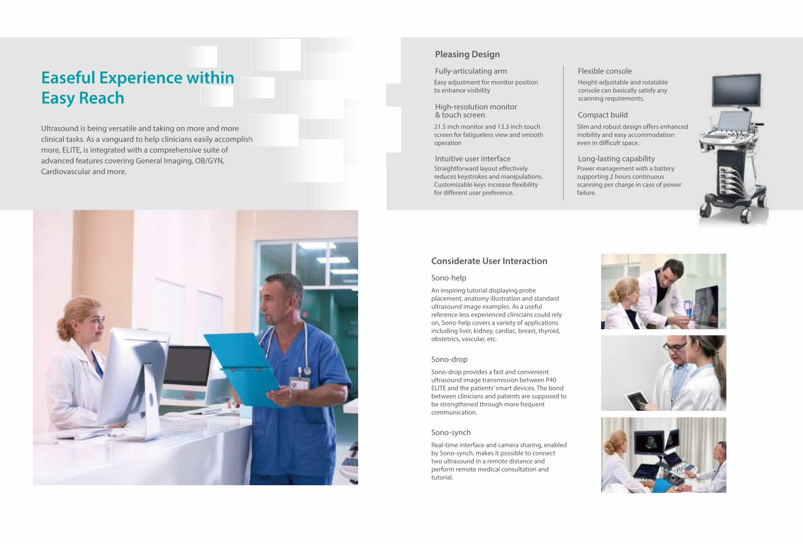
Considerate User Interaction
An inspiring tutorial displaying probe placement, anatomy illustration and standard ultrasound image examples. As a useful reference less experienced clinicians could rely on, Sono-help covers a variety of applications including liver, kidney, cardiac, breast, thyroid, obstetrics, vascular, etc.
Sono-help
Sono-drop provides a fast and convenient ultrasound image transmission between P40 ELITE and the patients’ smart devices. The bond between clinicians and patients are supposed to be strengthened through more frequent communication.
Sono-drop
Real-time interface and camera sharing, enabled by Sono-synch, makes it possible to connect two ultrasound in a remote distance and perform remote medical consultation and tutorial.
Sono-synch
Pleasing Design
Fully-articulating armEasy adjustment for monitor position to enhance visibility
High-resolution monitor& touch screen21.5 inch monitor and 13.3 inch touch screen for fatigueless view and smooth operation
Intuitive user interfaceStraightforward layout effectively reduces keystrokes and manipulations. Customizable keys increase flexibility for different user preference.
Flexible consoleHeight-adjustable and rotatable console can basically satisfy any scanning requirements.
Compact buildSlim and robust design offers enhanced mobility and easy accommodation even in difficult space.
Long-lasting capabilityPower management with a battery supporting 2 hours continuous scanning per charge in case of power failure.
Ultrasound is being versatile and taking on more and more clinical tasks. As a vanguard to help clinicians easily accomplish more, ELITE, is integrated with a comprehensive suite of advanced features covering General Imaging, OB/GYN, Cardiovascular and more.
Easeful Experience within Easy Reach
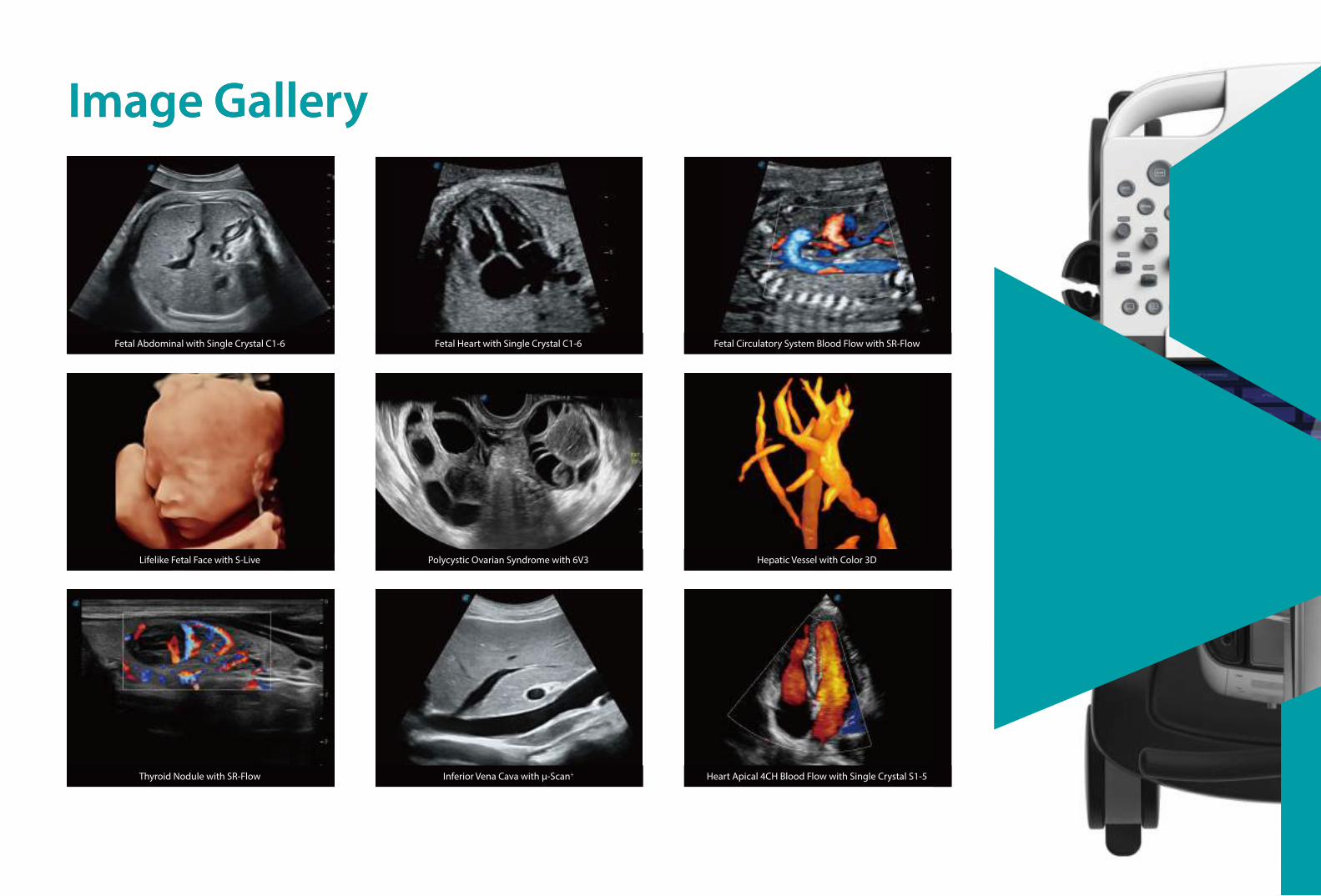
Image GalleryImage Gallery
Fetal Abdominal with Single Crystal C1-6 Fetal Heart with Single Crystal C1-6 Fetal Circulatory System Blood Flow with SR-Flow
Lifelike Fetal Face with S-Live Polycystic Ovarian Syndrome with 6V3 Hepatic Vessel with Color 3D
Thyroid Nodule with SR-Flow Inferior Vena Cava with μ-Scan+ Heart Apical 4CH Blood Flow with Single Crystal S1-5