dna the genetic material · semiconservative replication meselson & stahl label “parent”...
TRANSCRIPT
AP Biology 2006-2007
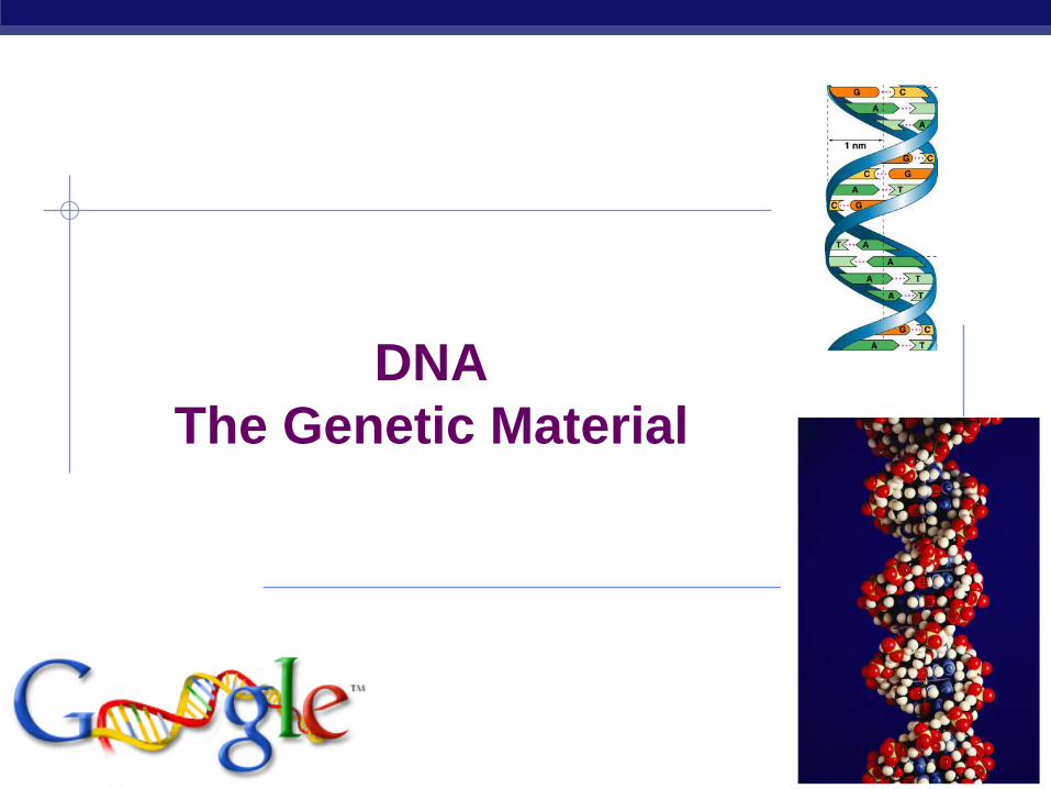
DNA
The Genetic Material

AP Biology
Chromosomes related to phenotype
T.H. Morgan
working with Drosophila
fruit flies
associated phenotype with
specific chromosome
white-eyed male had specific
X chromosome
1908 | 1933
AP Biology
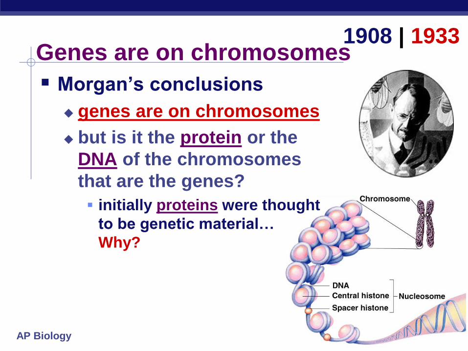
Genes are on chromosomes
Morgan’s conclusions
genes are on chromosomes
but is it the protein or the
DNA of the chromosomes
that are the genes?
initially proteins were thought
to be genetic material…
Why?
1908 | 1933

AP Biology
The “Transforming Principle” 1928
Frederick Griffith
Streptococcus pneumonia bacteria was working to find cure for pneumonia
harmless live bacteria (“rough”)
mixed with heat-killed pathogenic
bacteria (“smooth”) causes fatal
disease in mice
a substance passed from dead
bacteria to live bacteria to change
their phenotype
“Transforming Principle”

AP Biology
The “Transforming Principle”
Transformation = change in phenotype
something in heat-killed bacteria could still transmit
disease-causing properties
live pathogenicstrain of bacteria
live non-pathogenicstrain of bacteria
mice die mice live
heat-killed pathogenic bacteria
mix heat-killed pathogenic & non-pathogenicbacteria
mice live mice die
A. B. C. D.

AP Biology
DNA is the “Transforming Principle”
Avery, McCarty & MacLeod
purified both DNA & proteins separately from
Streptococcus pneumonia bacteria
which will transform non-pathogenic bacteria?
injected protein into bacteria
no effect
injected DNA into bacteria
transformed harmless bacteria into
virulent bacteria
1944
mice die

AP BiologyOswald Avery Maclyn McCarty Colin MacLeod
Avery, McCarty & MacLeod Conclusion
first experimental evidence that DNA was the genetic material
1944 | ??!!
AP Biology
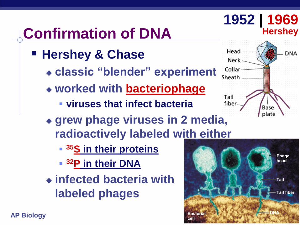
Confirmation of DNA
Hershey & Chase
classic “blender” experiment
worked with bacteriophage
viruses that infect bacteria
grew phage viruses in 2 media,
radioactively labeled with either
35S in their proteins
32P in their DNA
infected bacteria with
labeled phages
1952 | 1969Hershey
AP Biology
Protein coat labeledwith 35S
DNA labeled with 32P
bacteriophages infectbacterial cells
T2 bacteriophagesare labeled with
radioactive isotopesS vs. P
bacterial cells are agitatedto remove viral protein coats
35S radioactivityfound in the medium
32P radioactivity foundin the bacterial cells
Which radioactive marker is found inside the cell?
Which molecule carries viral genetic info?
Hershey
& Chase

AP Biology
Blender experiment
Radioactive phage & bacteria in blender
35S phage
radioactive proteins stayed in supernatant
therefore viral protein did NOT enter bacteria
32P phage
radioactive DNA stayed in pellet
therefore viral DNA did enter bacteria
Confirmed DNA is “transforming factor”

AP Biology
Hershey & Chase
Alfred HersheyMartha Chase
1952 | 1969Hershey

AP Biology
Chargaff
DNA composition: “Chargaff’s rules”
varies from species to species
all 4 bases not in equal quantity
bases present in characteristic ratio
humans:
A = 30.9%
T = 29.4%
G = 19.9%
C = 19.8%
1947
RulesA = TC = G

AP Biology
Rosalind Franklin (1920-1958)
AP Biology
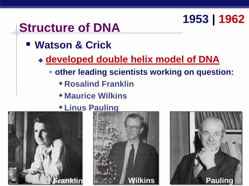
Structure of DNA
Watson & Crick
developed double helix model of DNA
other leading scientists working on question:
Rosalind Franklin
Maurice Wilkins
Linus Pauling
1953 | 1962
Franklin Wilkins Pauling

AP Biology
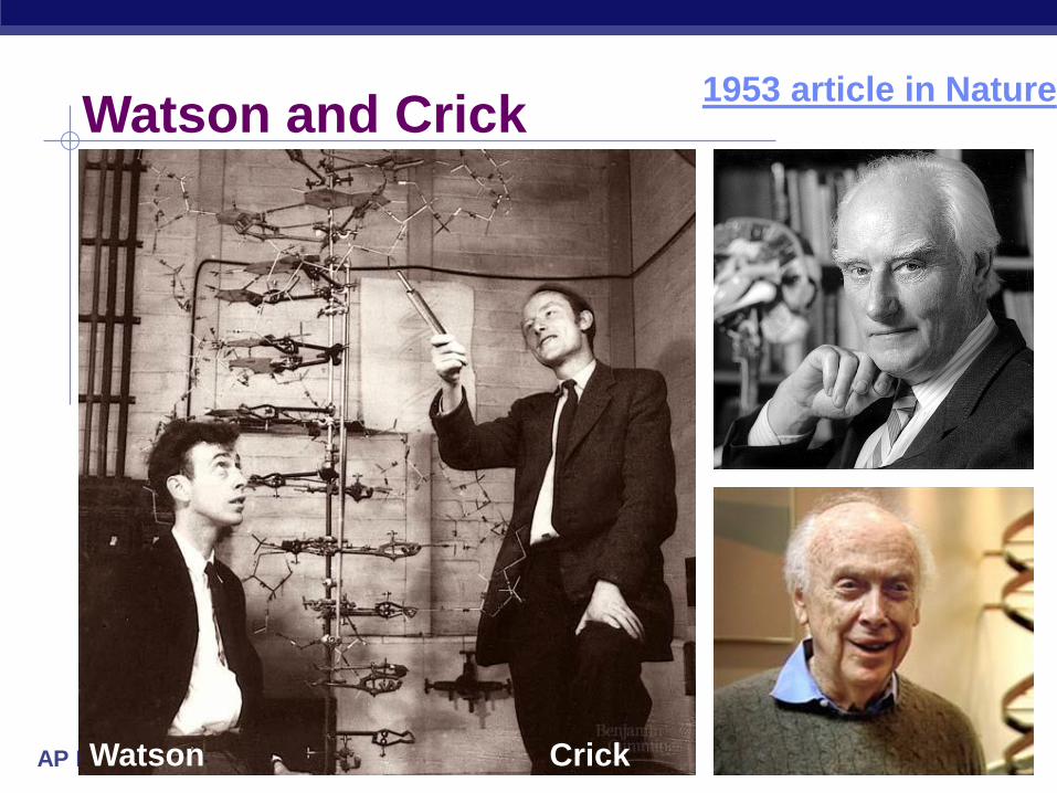
Double helix structure of DNA
“It has not escaped our notice that the specific pairing we have postulated
immediately suggests a possible copying mechanism for the genetic
material.” Watson & Crick
AP Biology
Watson and Crick1953 article in Nature
CrickWatson

AP Biology
But how is DNA copied?
Replication of DNA
base pairing suggests
that it will allow each
side to serve as a
template for a new
strand
“It has not escaped our notice that the specific pairing we have postulated
immediately suggests a possible copying mechanism for the genetic
material.” — Watson & Crick
AP Biology
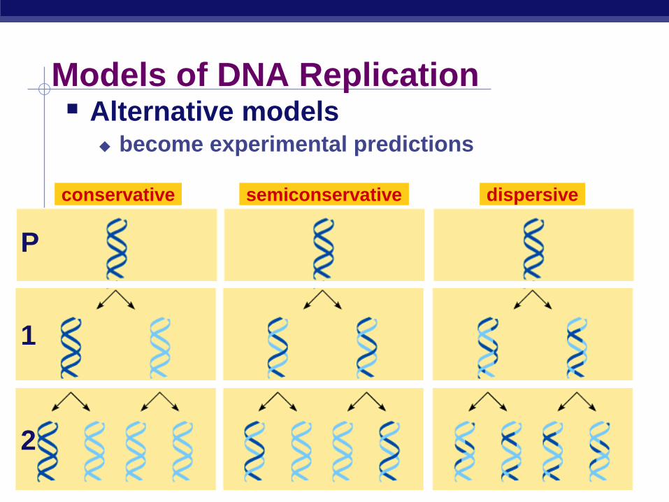
Models of DNA Replication Alternative models
become experimental predictions
conservative semiconservative dispersive
1
2
P

AP Biology
Semiconservative replication Meselson & Stahl
label “parent” nucleotides in DNA strands with heavy nitrogen = 15N
label new nucleotides with lighter isotope = 14N
“The Most Beautiful Experiment in Biology”
1958
parent replication
15N parent
strands
15N/15N
AP Biology
Predictions
1st round of
replication
conservative
15N/15N
14N/14N
semi-
conservative
15N/14N
dispersive
15N/14N
conservative
15N/15N
14N/14N
semi-
conservative
15N/14N
dispersive
15N/14N
2nd round of
replication
14N/14N
15N parent
strands
15N/15N
1
2
P

AP Biology
Franklin Stahl
Matthew Meselson
Matthew Meselson Franklin Stahl
Meselson & Stahl

AP Biology
Scientific History March to understanding that DNA is the genetic material
T.H. Morgan (1908)
genes are on chromosomes
Frederick Griffith (1928)
a transforming factor can change phenotype
Avery, McCarty & MacLeod (1944)
transforming factor is DNA
Erwin Chargaff (1947)
Chargaff rules: A = T, C = G
Hershey & Chase (1952)
confirmation that DNA is genetic material
Watson & Crick (1953)
determined double helix structure of DNA
Meselson & Stahl (1958)
semi-conservative replication
AP Biology 2007-2008
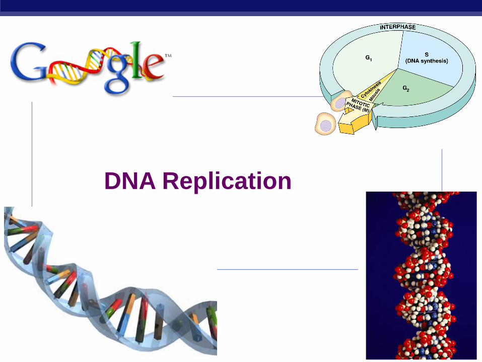
DNA Replication
AP Biology
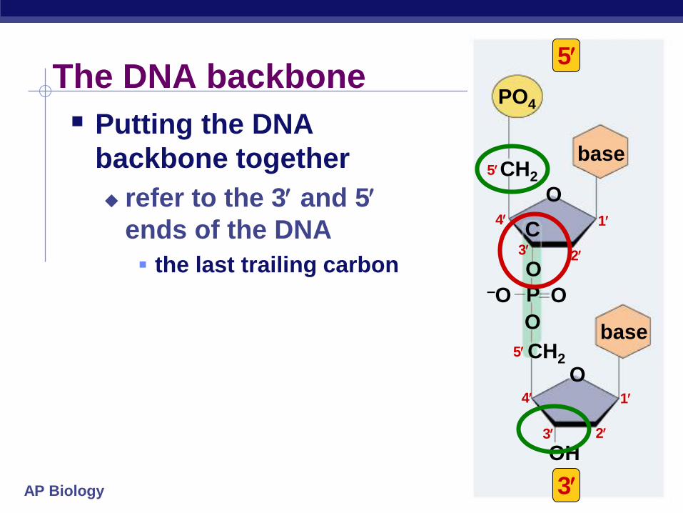
The DNA backbone
Putting the DNA
backbone together
refer to the 3 and 5
ends of the DNA
the last trailing carbon
OH
O
3
PO4
base
CH2
O
base
O
P
O
C
O–O
CH2
1
2
4
5
1
2
3
3
4
5
5

AP Biology
Anti-parallel strands
Nucleotides in DNA
backbone are bonded from
phosphate to sugar
between 3 & 5 carbons
DNA molecule has
“direction”
complementary strand runs
in opposite direction
3
5
5
3

AP Biology
Bonding in DNA
….strong or weak bonds?
How do the bonds fit the mechanism for copying DNA?
3
5 3
5
covalent
bonds
hydrogen
bonds
AP Biology
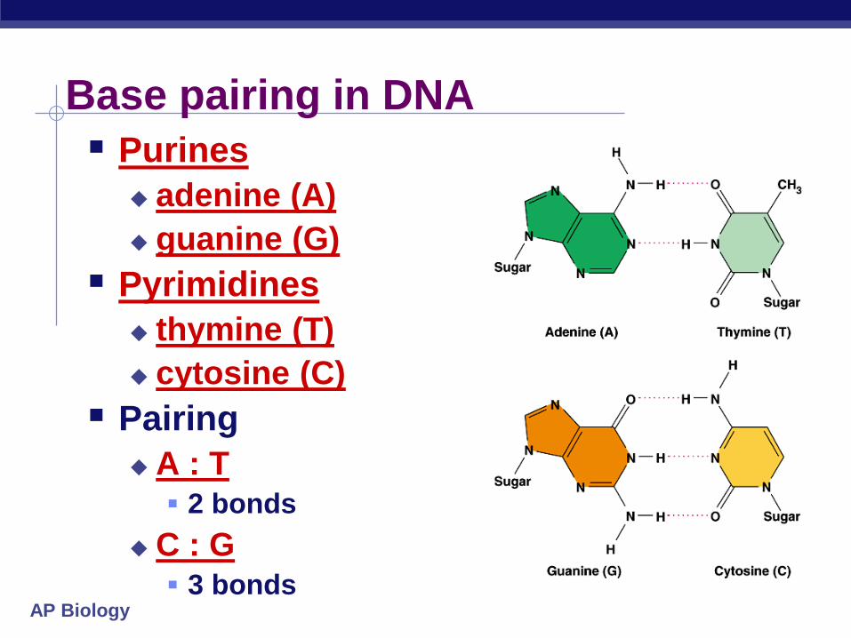
Base pairing in DNA
Purines
adenine (A)
guanine (G)
Pyrimidines
thymine (T)
cytosine (C)
Pairing
A : T 2 bonds
C : G 3 bonds

AP Biology
Copying DNA
Replication of DNA
base pairing allows each strand to serve as a template for a new strand
new strand is 1/2 parent template & 1/2 new DNA semi-conservative
copy process

AP Biology
DNA Replication Large team of enzymes coordinates replication
AP Biology
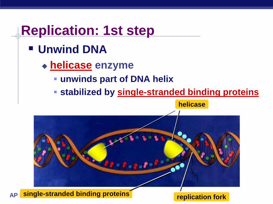
Replication: 1st step
Unwind DNA
helicase enzyme
unwinds part of DNA helix
stabilized by single-stranded binding proteins
single-stranded binding proteinsreplication fork
helicase
AP Biology
DNA
Polymerase III
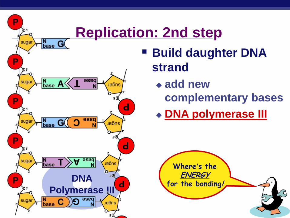
Replication: 2nd step
But…
We’re missing something!
What?
Where’s theENERGY
for the bonding!
Build daughter DNA
strand
add new
complementary bases
DNA polymerase III

AP Biology
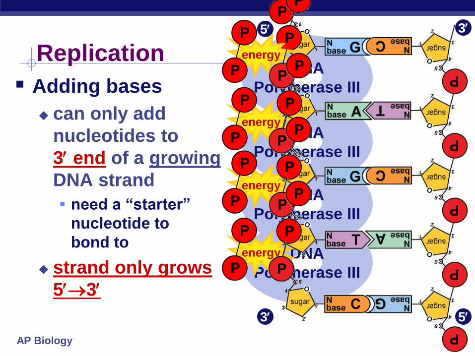
Energy of Replication The nucleotides arrive as nucleosides
DNA bases with P–P–P P-P-P = energy for bonding
DNA bases arrive with their own energy source for bonding
bonded by enzyme: DNA polymerase III
ATP GTP TTP CTP
AP Biology
Adding bases
can only add
nucleotides to
3 end of a growing
DNA strand
need a “starter”
nucleotide to
bond to
strand only grows
53
DNA
Polymerase III
DNA
Polymerase III
DNA
Polymerase III
DNA
Polymerase III
energy
energy
energy
Replication energy
3
3
5
5

AP Biology
Limits of DNA polymerase III
can only build onto 3 end of
an existing DNA strand
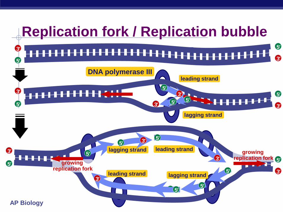
Leading & Lagging strands
5
5
5
5
3
3
3
5
35
3 3
Leading strand
Lagging strandligase
Okazaki
Leading strand
continuous synthesis
Lagging strand
Okazaki fragments
joined by ligase
“spot welder” enzyme
DNA polymerase III
3
5
growing replication fork
AP Biology
DNA polymerase III
Replication fork / Replication bubble
5
35
3
leading strand
lagging strand
leading strand
lagging strandleading strand
5
3
3
5
5
3
5
3
5
3 5
3
growing replication fork
growing replication fork
5
5
5
5
5
3
3
5
5lagging strand
5 3

AP Biology
DNA polymerase III
RNA primer
built by primase
serves as starter sequence for DNA polymerase III
Limits of DNA polymerase III
can only build onto 3 end of
an existing DNA strand
Starting DNA synthesis: RNA primers
5
5
5
3
3
3
5
35
3 5 3
growing replication fork
primase
RNA
AP Biology
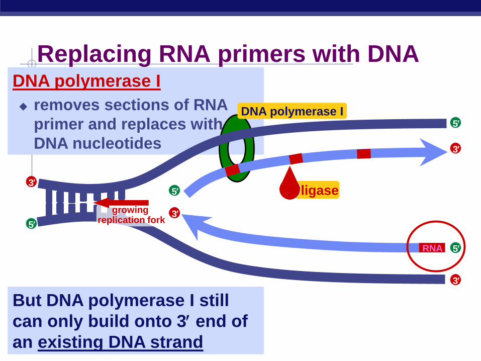
DNA polymerase I
removes sections of RNA
primer and replaces with
DNA nucleotides
But DNA polymerase I still
can only build onto 3 end of
an existing DNA strand
Replacing RNA primers with DNA
5
5
5
5
3
3
3
3
growing replication fork
DNA polymerase I
RNA
ligase

AP Biology
Replication fork
3’
5’
3’
5’
5’
3’
3’ 5’
helicase
direction of replication
SSB = single-stranded binding proteins
primase
DNA polymerase III
DNA polymerase III
DNA polymerase I
ligase
Okazaki fragments
leading strand
lagging strand
SSB
AP Biology
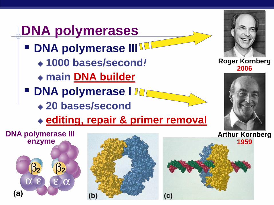
DNA polymerases
DNA polymerase III
1000 bases/second!
main DNA builder
DNA polymerase I
20 bases/second
editing, repair & primer removal
DNA polymerase III enzyme
Arthur Kornberg1959
Roger Kornberg2006
AP Biology
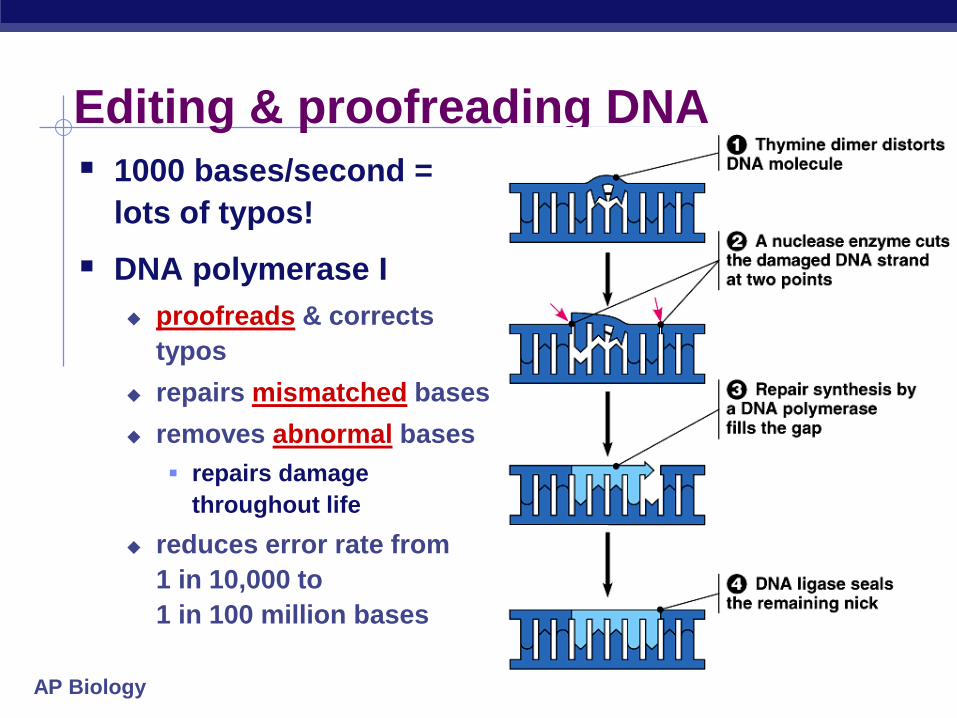
Editing & proofreading DNA
1000 bases/second =
lots of typos!
DNA polymerase I
proofreads & corrects
typos
repairs mismatched bases
removes abnormal bases
repairs damage
throughout life
reduces error rate from
1 in 10,000 to
1 in 100 million bases
AP Biology
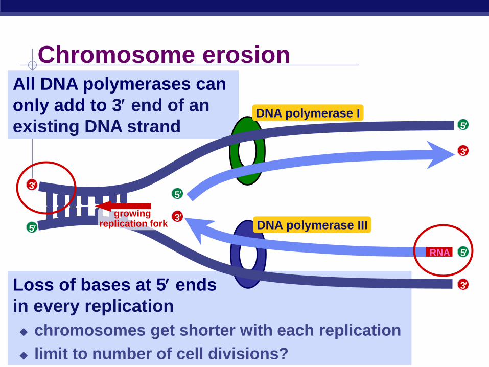
Loss of bases at 5 ends
in every replication
chromosomes get shorter with each replication
limit to number of cell divisions?
DNA polymerase III
All DNA polymerases can
only add to 3 end of an
existing DNA strand
Chromosome erosion
5
5
5
5
3
3
3
3
growing replication fork
DNA polymerase I
RNA

AP Biology
Repeating, non-coding sequences at the end
of chromosomes = protective cap
limit to ~50 cell divisions
Telomerase
enzyme extends telomeres
can add DNA bases at 5 end
different level of activity in different cells
high in stem cells & cancers -- Why?
telomerase
Telomeres
5
5
5
5
3
3
3
3
growing replication fork
TTAAGGGTTAAGGG