diversification of spatiotemporal expression and copy ... · research article diversification of...
TRANSCRIPT

RESEARCH ARTICLE
Diversification of spatiotemporal expression
and copy number variation of the echinoid
hbox12/pmar1/micro1 multigene family
Vincenzo Cavalieri1,2*, Fabiana Geraci1, Giovanni Spinelli1*
1 Department of Biological, Chemical and Pharmaceutical Sciences and Technologies (STEBICEF),
University of Palermo, Viale delle Scienze Edificio 16, Palermo, Italy, 2 Advanced Technologies Network
Center (ATeN), University of Palermo, Viale delle Scienze Edificio 18, Palermo, Italy
* [email protected] (VC); [email protected] (GS)
Abstract
Changes occurring during evolution in the cis-regulatory landscapes of individual members of
multigene families might impart diversification in their spatiotemporal expression and function.
The archetypal member of the echinoid hbox12/pmar1/micro1 family is hbox12-a, a homeo-
box-containing gene expressed exclusively by dorsal blastomeres, where it governs the dorsal/
ventral gene regulatory network during embryogenesis of the sea urchin Paracentrotus lividus.
Here we describe the inventory of the hbox12/pmar1/micro1 genes in P. lividus, highlighting
that gene copy number variation occurs across individual sea urchins of the same species. We
show that the various hbox12/pmar1/micro1 genes group into three subfamilies according to
their spatiotemporal expression, which ranges from broad transcription throughout develop-
ment to transient expression in either the animal hemisphere or micromeres of the early em-
bryo. Interestingly, the promoter regions of those genes showing comparable expression
patterns are highly similar, while differing from those of the other subfamilies. Strikingly, phylo-
genetic analysis suggests that the hbox12/pmar1/micro1 genes are species-specific, exhibiting
extensive divergence in their noncoding, but not in their coding, sequences across three distinct
sea urchin species. In spite of this, two micromere-specific genes of P. lividus possess a TCF/
LEF-binding motif in a similar position, and their transcription relies on Wnt/β-catenin signaling,
similar to the pmar1 and micro1 genes, which in other sea urchin species are involved in micro-
mere specification. Altogether, our findings suggest that the hbox12/pmar1/micro1 gene family
evolved rather rapidly, generating paralogs whose cis-regulatory sequences diverged following
multiple rounds of duplication from a common ancestor.
Introduction
The last two decades of research in the field of molecular embryology have provided a detailed
mechanistic explanation of how fates of different cell types are encoded in the genome and
sculpted through the sequential progression of transcriptional states of defined regulatory
genes [1–4]. By contrast, much less is known about the driving forces underlying dynamic
rewiring of gene regulatory network circuitries during evolution [5–9]. In this regard, it is
PLOS ONE | https://doi.org/10.1371/journal.pone.0174404 March 28, 2017 1 / 21
a1111111111
a1111111111
a1111111111
a1111111111
a1111111111
OPENACCESS
Citation: Cavalieri V, Geraci F, Spinelli G (2017)
Diversification of spatiotemporal expression and
copy number variation of the echinoid hbox12/
pmar1/micro1 multigene family. PLoS ONE 12(3):
e0174404. https://doi.org/10.1371/journal.
pone.0174404
Editor: Michael Schubert, Laboratoire de Biologie
du Developpement de Villefranche-sur-Mer,
FRANCE
Received: October 8, 2016
Accepted: March 8, 2017
Published: March 28, 2017
Copyright: © 2017 Cavalieri et al. This is an open
access article distributed under the terms of the
Creative Commons Attribution License, which
permits unrestricted use, distribution, and
reproduction in any medium, provided the original
author and source are credited.
Data Availability Statement: All relevant data are
within the paper and its Supporting Information
files.
Funding: The authors received no specific funding
for this work.
Competing interests: The authors have declared
that no competing interests exist.

commonly accepted that gene duplication provides a major source of both evolutionary nov-
elty and species diversification. In fact, in all three domains of life a substantial fraction of
genes underwent a series of duplications that originated multicopy gene families [10,11], and
among these is the echinoid hbox12/pmar1/micro1 family.Hbox12 was originally identified
in the Mediterranean Paracentrotus lividus species as a cDNA coding for a paired-like class
homeodomain-containing factor [12]. Whole mount in situ hybridization and cis-regulatory
analysis highlighted that the archetypal hbox12 gene, namely hbox12-a, is expressed transiently
during the very early cleavage stages exclusively by presumptive dorsal blastomeres [12–15].
We also showed that hbox12-a encodes a key transcription repressor functioning at the top of
the symmetry-breaking sequence of events within the dorsal-ventral gene regulatory network.
In particular, by transient inactivation of p38-MAP kinase activity during very early cleavage,
Hbox12-a defines the future dorsal side of the embryo, allowing the expression of the TGF-βsuperfamily member Nodal on the opposite side [14–16]. Afterwards, Nodal-dependent sig-
naling imposes the dorsal-ventral polarity in the developing embryo [17–20].
To date, proteins that show high sequence similarity to the Hbox12-a regulator are encoded
by the pmar1/micro1 genes identified in Strongylocentrotus purpuratus [21], Hemicentrituspulcherrimus [22], Anthocidaris crassispina [23], and Lytechinus variegatus [24]. Similarly to
hbox12-a, multiple copies of pmar1/micro1 genes are clustered in the respective sea urchin
genomes [13,23,25]. However, the Pmar1 factor is produced solely by micromeres, where it
drives their specification by inhibiting transcription of the ubiquitous repressor HesC, which
otherwise negatively regulates the repertoire of micromere specification genes [26–28]. The
fact that the Hbox12-a and Pmar1/Micro1 regulators display high sequence similarity across
species but serve different functions poses the question of whether diversification of their cis-regulatory sequences has arisen during evolution by duplication of a common ancestor.
In the current paper, we address this question by describing the inventory of the hbox12/pmar1/micro1 genes present in P. lividus, and highlighting that gene copy number variation
occurs across the genome of distinct individual sea urchins of the same species. We also show
that members of this gene family exhibit extensive divergence in their noncoding, but not in
their coding, sequences among three different urchin species, as well as substantial differences
of spatiotemporal expression during embryogenesis of P. lividus. While some members coher-
ently recapitulate the animal-restricted expression pattern formerly described for the hbox12-agene [12,13], others are either transcribed broadly throughout development or transiently
expressed only in micromeres of the fourth cleavage embryo, mimicking the expression pat-
tern reported for pmar1/micro1 in other species. Furthermore, a predicted TCF/LEF-binding
motif exists in a similar position, compared to pmar1 genes of S. purpuratus, near the tran-
scription start site of the micromere-specific genes of P. lividus, and the expression of these
genes specifically relies on the nuclerization of β-catenin in vegetal blastomeres. We propose
that the echinoid hbox12/pmar1/micro1 family includes distinct paralogs that most likely
evolved independently following multiple rounds of duplication from a common ancestor.
Materials and methods
Quantitative PCR analysis
For gene copy number estimation, genomic DNA was purified from sperm of seven individual
urchins, and then used as template in SYBR Green-based qPCR reactions for hbox12/pmar1/micro1 genes (see below). As known single copy controls we used the otp and cmpl genes
[29,30]. The number of hbox12, otp and cmpl amplicons was determined using standard curves
with different dosages of plasmid DNA containing the mentioned amplicon sequences, and
gene ratios were eventually calculated. The primers for the assay are listed in S1 Table.
The echinoid hbox12/pmar1/micro1 multigene family
PLOS ONE | https://doi.org/10.1371/journal.pone.0174404 March 28, 2017 2 / 21
For gene expression analysis, reverse-transcription and qPCR were performed essentially as
described [15,31,32]. Briefly, total RNA from batches of dissected or injected embryos grown
at the desired stage was extracted using the Power SYBR Green Cells-to-CT kit (Ambion) and
reverse transcribed following the manufacturer’s recommendations. The resulting cDNA sam-
ple was further diluted and the equivalent amount corresponding to one embryo was used as
template for qPCR analysis, with the oligonucleotide primers indicated in S1 Table. qPCR
experiments were performed from at least three distinct batches and all reactions were run in
triplicate on a StepOnePlus Real-Time PCR System (Thermo Fisher Scientific) using SYBR
Green detection chemistry. ROX was used as a measure of background fluorescence and, at
the end of the amplification reactions, a melting-curve analysis was run to confirm the homo-
geneity of all amplicons. Calculations from qPCR raw data were performed using the StepOne
software version 2.3 (Thermo Fisher Scientific), with the comparative Ct method. Primer
efficiencies were found to exceed 1.85. In each experiment, a no-template control was included
for each primer set. A cytochrome oxidase or thembf1mRNA, which are known to be ex-
pressed at a constant level during development, was used to normalize all data, in order to
account for fluctuations among different preparations. The number of transcripts per embryo
at the very early blastula stage was estimated assuming a reference standard number of 1000
copies/embryo of the z12mRNA [33,34].
Embryo dissection and microinjection of synthetic mRNA
Embryo dissection was carried out as described [14,35]. Briefly, unperturbed P. lividusembryos nearing the end of the fourth cleavage were transferred into a modified Kiehart
chamber in Ca2+-free sea water and manipulated with fine glass needles under a Leica
M165FC stereomicroscope equipped with micromanipulators (Narishige). Groups of three
embryos at one time were manipulated by removing micromeres immediately following their
formation from the overlying macromeres. After surgery, the isolated micromeres and their
complementary micromere-less embryoids, as well as control intact 16-cell stage embryos,
were promptly processed for qPCR analysis.
Microinjection was conducted as described [36,37]. ΔLvG-Cad [38], dnTCF [39], and
strim out-of-frame [35] constructs were linearized and used as template to synthesize capped
mRNAs using the mMessage mMachine kit (Ambion). Approximately 1–2 pl of the purified
RNAs were injected in 30% glycerol at the following concentrations: ΔLvG-cad 0.1 pg/pl,
dnTCF 0.4 pg/pl, and strim out-of-frame 0.5 pg/pl.
For each dissection or injection experiment, roughly one hundred embryos were processed
for qPCR analysis, and each experiment was repeated twice with different batches of eggs.
Results
Inventory of the hbox12/pmar1/micro1 homeobox genes residing in the
P. lividus genome
As a first step toward identification of the complete set of hbox12/pmar1/micro1 genes of the
Mediterranean sea urchin P. lividus, an in-silico analysis was carried out using the most recent
assembly of the genome (v4.0, 01/2016; http://octopus.obs-vlfr.fr/blast/oursin/blast_oursin.
php). A BLASTn search retrieved a total of thirty hits distributed in six distinct genomic scaf-
folds of different size (Fig 1). Neither hbox12-a nor -b, the two originally identified genes [13],
were found among these hits, probably because in some cases the gene anatomy was partially
obscured by the incomplete nature of the available genome draft. Nevertheless, Genscan analy-
sis revealed that each of the thirty genes share an identical structure, which consists of two
The echinoid hbox12/pmar1/micro1 multigene family
PLOS ONE | https://doi.org/10.1371/journal.pone.0174404 March 28, 2017 3 / 21

exons split by a single intron (Fig 1), mirroring the genomic organization formerly described
for the prototypical hbox12-a and -b genes [13]. The only exception is the hbox12-29 gene, for
which the first exon has not been identified (Fig 1). The newly identified gene copies were
often oriented divergently from one another, being transcribed outwardly in the opposite
direction irrespective of their genomic location (Fig 1). Interestingly, some of the mentioned
copies were arranged in contiguous bigenic clusters with a head-to-tail orientation (Fig 1), a
peculiar feature that we observed previously in the genomic λ-clone containing the hbox12-aand -b genes [13]. The fact that all of the hbox12/pmar1/micro1 family members share the
same exon-intron structure, and that chimeric copies were not detected, indicates that gene
units have presumably duplicated as intact copies. Importantly, the amino acid sequences pre-
dicted for the thirty genes showed extensive similarity to each other and to the Hbox12-a regu-
lator, especially in the homeodomain (S1 Fig), with minor differences likely due to allelic
polymorphisms.
The various hbox12/pmar1/micro1 copies were flanked by unrelated genes that were appar-
ently distinguished among scaffolds, indicating that the mentioned scaffolds were not overlap-
ping, and that therefore the gene copy number obtained by the in silico analysis was not
overestimated. Nevertheless, this possibility cannot be excluded because in the provisional
genome draft technical complications hindered the separation of the two haplotypes mixed
during preparation of the sequencing library (indeed, the current assembly size of 1.4 Gb is
well above the expected 0.8 Gb for the haploid genome).
In a recent paper [40], the partial or complete protein sequences of five hbox12/pmar1/micro1 family members were inferred from one contig of P. lividus genomic sequence. We
found that two of these proteins, termed by the authors Hbox12-9 and -2, were 100% identical
to those we deduced for the hbox12-01 and -20 genes, respectively (S2 Fig). However, in
Fig 1. Genomic organization of the hbox12/pmar1/micro1 family members of the sea urchin P. lividus. The exon-
intron structure, orientation, and location for each gene are shown. Scaffold number identifiers and coordinates are based on
v4.0 of the P. lividus genome assembly (http://octopus.obs-vlfr.fr/blast/oursin/blast_oursin.php). The hbox12-27 gene
displays a normal genomic organization, but the 3’ portion of the exon 2 is missing due to lack of genomic sequence.
Unrelated genes are omitted for simplicity.
https://doi.org/10.1371/journal.pone.0174404.g001
The echinoid hbox12/pmar1/micro1 multigene family
PLOS ONE | https://doi.org/10.1371/journal.pone.0174404 March 28, 2017 4 / 21

contrast to Hbox12-9 and -2, these genes map into distinct scaffolds (Fig 1), suggesting that
they would lie in different genomic regions. Analogously, pairwise alignments showed that the
proteins predicted for the hbox12-02, -04 and -27 genes displayed the highest similarity to the
three remaining sequences, although various differences were detected (S2 Fig). Although
these discrepancies could be at least in part due to allelic polymorphisms, in the absence of
genomic sequence for the contig described in [40], we cannot rule out the possibility that we
were looking at further distinct copies of the same gene family, which probably escaped from
the sequencing and/or assembly of the P. lividus genomic reads.
Hence, to measure the hbox12/pmar1/micro1 gene dosage in P. lividusmore precisely, we
performed qPCR using a universal primer pair that completely matched the sequence of all the
known family members (S1 Table), and specific primers for the otp and cmpl genes [29,30],
used as single copy controls. As a template for this assay, we utilized the genomes derived from
sperm of seven randomly selected individuals and, much to our surprise, we found that the
number of hbox12/pmar1/micro1 genes varied noticeably among individuals, ranging from 6
to 22 copies (Fig 2).
Taken together, our findings not only confirm that several copies of these genes exist in the
genome of P. lividus, but also suggest that the hbox12/pmar1/micro1 family expanded via mul-
tiple episodes of segmental duplication.
Heterogeneity of spatiotemporal expression profiles within the hbox12/
pmar1/micro1 gene family
The transcription level of about half of the hbox12/pmar1/micro1 family members was assessed
by qPCR analysis from unfertilized eggs and embryos at distinct developmental time points,
using gene-specific primer pairs (S1 Table). The results of this survey revealed that transcripts
derived from the examined genes are not maternally stored in the unfertilized egg (Fig 3 and
S3 Fig), and that three zygotic expression groups are distinguishable on the basis of the abun-
dance and temporal accumulation of transcripts during embryogenesis (Fig 3 and S3 Fig). The
hbox12-04, -10, -24, and -29 genes, altogether falling in a first group, were found to be tran-
scribed at significantly higher levels throughout development (Figs 3A and S3). In the case of
hbox12-04, however, the result could reflect the sum of separate transcriptional activities, as
we noticed that the potential cDNAs derived from hbox12-21 and -25 genes are co-amplified
using the hbox12-04 primer pair. The hbox12-09 and -28 genes were both expressed at reduced
levels when compared to the former group, and their transcripts were not detected at the late
gastrula stage (Fig 3B and S3 Fig). A third group included hbox12-a, -06, -12, -16, -17, and
-19, the genes displaying the lowest detectable expression levels among those investigated (Fig
3C and S3 Fig). It is also interesting to note that the mRNA abundance of most of the genes
belonging to the third group varied significantly across the different cDNA batches used, and
there was no apparent reciprocal correlation in the transcriptional status of these genes within
a given cDNA sample (Fig 3D). Moreover, transcripts of the hbox12-07, -08, and -14 genes
were not detected at all in the cDNA batches we examined (not shown), suggesting that they
were either absent or accumulated at an extent that cannot be determined under our experi-
mental conditions. Altogether, these results indicate remarkable heterogeneity in gene ex-
pression occurring among both the hbox12/pmar1/micro1 family members and the distinct
population of embryos from which cDNA templates were derived.
As mentioned, the expression profile of the P. lividus hbox12-a gene differs from those of
the pmar1 andmicro1 genes described in S. purpuratus andH. pulcherrimus, respectively
[12,13,21,22]. To distinguish the hbox12-a-like from the pmar1/micro1-like genes in P. lividus,we assessed the spatial distribution in the developing embryo of the transcripts derived from
The echinoid hbox12/pmar1/micro1 multigene family
PLOS ONE | https://doi.org/10.1371/journal.pone.0174404 March 28, 2017 5 / 21

the various hbox12/pmar1/micro1 family members. Unfortunately, collecting of probes that
unambiguously recognize the mRNA of each copy in whole mount in situ hybridization assays
was hampered by the very extensive identity of both coding sequences and flanking UTRs
within the hbox12/pmar1/micro1 family. To overcome this drawback, we combined a dissec-
tion approach to qPCR analysis, focusing on the 16-cell stage embryo, when the discordance
in the spatial expression patterns of hbox12-a and pmar1/micro1 genes is apparent [12,21,22].
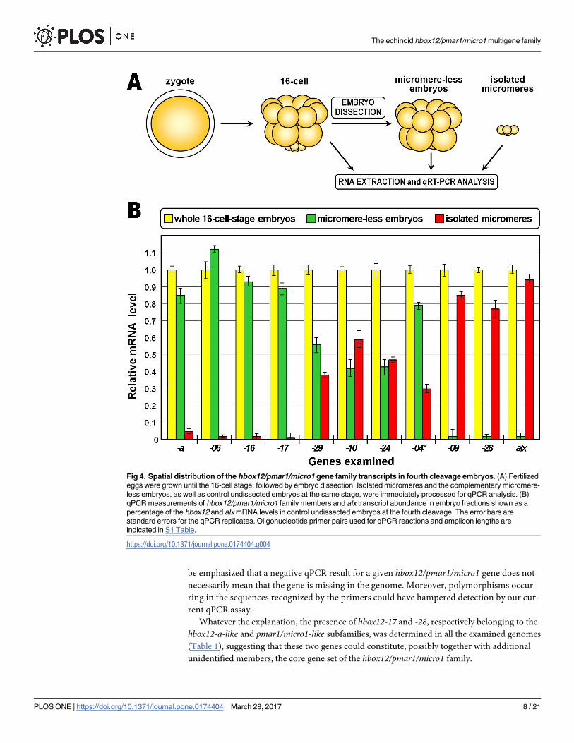
Unperturbed P. lividus embryos were grown until the fourth cleavage, followed by micromere
removal. Isolated micromeres and the complementary micromere-less embryos, as well as
control undissected embryos at the same stage, were immediately processed for qPCR analysis
(Fig 4A). As expected, the micromere-specific marker alx1 [41], used as a control, was ex-
pressed in isolated micromeres at a level nearly equal to that of the whole embryos, and there
was no evidence of alx1 expression in micromere-less embryos (Fig 4B). By contrast, at the
time of micromere removal the hbox12-a gene was selectively transcribed in micromere-less
embryos, in perfect accordance with previous observations [13–15]. Other members of the
family, such as hbox12-06, -16, and -17, grouped together coherently reiterating the spatial
expression profile established for hbox12-a (Fig 4B). The remaining hbox12/pmar1/micro1
Fig 2. Hbox12/pmar1/micro1 gene copy number determination in the genome of seven distinct P.
lividus individuals. The histogram derived from qPCR data represent the number of hbox12 copies
normalized to the single copy gene controls otp and cmpl. Error bars are standard errors for the qPCR
replicates.
https://doi.org/10.1371/journal.pone.0174404.g002
The echinoid hbox12/pmar1/micro1 multigene family
PLOS ONE | https://doi.org/10.1371/journal.pone.0174404 March 28, 2017 6 / 21

copies were subdivided into additional groups according to their divergent spatial expression.
In particular, mRNAs produced by hbox12-10, -24, and -29 genes were conjointly detected in
isolated micromeres and the micromere-less counterpart, in both cases at a similar reduced
level compared to the sibling control intact embryos (Fig 4B), suggesting that probably the men-
tioned genes were either ubiquitously expressed or broadly transcribed along the animal-vegetal
axis of unperturbed 16-cell stage embryos. Alternately, the transcript population derived from
the hbox12-04 gene was found in both the dissected embryonic fractions, although with prefer-
ential accumulation in micromere-less embryos (Fig 4B). In the absence of a specific primer
pair (hbox12-04, -21 and -25were co-amplified, as mentioned), this result should be considered
as the sum of potentially separate transcriptional profiles. Finally, hbox12-09 and -28 transcripts
were found to be specifically restricted to isolated micromeres (Fig 4B).
Collectively, these results highlight the complexity of spatiotemporal expression profiles
achieved by the hbox12/pmar1/micro1 family in the developing P. lividus embryo, leading
us to further focus on those members showing the hbox12-a-like and pmar1/micro1-likeexpression patterns. In particular, we performed qPCR with specific primer pairs to de-
termine the presence/absence of each of these subfamily members in the genome of the
seven sea urchin individuals described in the previous section. We found distinct composi-
tion of the mentioned subfamilies in the genome of the examined individuals (Table 1), as
expected from the high copy number variation of the hbox12/pmar1/micro1 family. In light
of this, it could be reasoned that genes that did not generate amplicons in the expression
analyses were probably missing, rather than silenced, in those batches. However, it should
Fig 3. Temporal expression of the hbox12/pmar1/micro1 gene family members in the P. lividus embryo. (A-C) Individual representation of
each expression profile presented as the mean relative to global hbox12/pmar1/micro1 transcript abundance measured with the universal primer pair
at the indicated developmental stages. Expression profiles with standard errors of the mean between replicates are shown individually in S3 Fig. The
developmental stages are as follows: Egg, unfertilized egg; 16-cell, fourth cleavage embryo; VEB, very early blastula; MB, mesenchyme blastula; LG, late
gastrula. The asterisk indicates that the hbox12-04 amplicon was obtained together with the hbox12-21 and -25 amplicons by co-amplification with the
same primer pair. (D) The absolute number of transcripts per embryo given at the very early blastula stage derives from independent qPCR experiments
using distinct cDNA batches. Further detail for the qPCR procedure is given in Materials and Methods. The error bars are standard errors for qPCR
replicates. The oligonucleotide primer pairs used for the qPCR reactions and amplicon lengths are indicated in S1 Table.
https://doi.org/10.1371/journal.pone.0174404.g003
The echinoid hbox12/pmar1/micro1 multigene family
PLOS ONE | https://doi.org/10.1371/journal.pone.0174404 March 28, 2017 7 / 21
be emphasized that a negative qPCR result for a given hbox12/pmar1/micro1 gene does not
necessarily mean that the gene is missing in the genome. Moreover, polymorphisms occur-
ring in the sequences recognized by the primers could have hampered detection by our cur-
rent qPCR assay.
Whatever the explanation, the presence of hbox12-17 and -28, respectively belonging to the
hbox12-a-like and pmar1/micro1-like subfamilies, was determined in all the examined genomes
(Table 1), suggesting that these two genes could constitute, possibly together with additional
unidentified members, the core gene set of the hbox12/pmar1/micro1 family.
Fig 4. Spatial distribution of the hbox12/pmar1/micro1 gene family transcripts in fourth cleavage embryos. (A) Fertilized
eggs were grown until the 16-cell stage, followed by embryo dissection. Isolated micromeres and the complementary micromere-
less embryos, as well as control undissected embryos at the same stage, were immediately processed for qPCR analysis. (B)
qPCR measurements of hbox12/pmar1/micro1 family members and alx transcript abundance in embryo fractions shown as a
percentage of the hbox12 and alx mRNA levels in control undissected embryos at the fourth cleavage. The error bars are
standard errors for the qPCR replicates. Oligonucleotide primer pairs used for qPCR reactions and amplicon lengths are
indicated in S1 Table.
https://doi.org/10.1371/journal.pone.0174404.g004
The echinoid hbox12/pmar1/micro1 multigene family
PLOS ONE | https://doi.org/10.1371/journal.pone.0174404 March 28, 2017 8 / 21

Relationships between hbox12/pmar1/micro1 genes across sea urchin
species
To explore how the hbox12/pmar1/micro1 genes evolved, we first used the Phylogeny.fr plat-
form [42] to build a phylogenetic tree with a set of complete gene sequences (5’-flanking
+exons+intron) available for the P. lividus, S. purpuratus, and L. variegatus species. Notably, in
this analysis the various genes formed three distinct species-specific clades supported by reli-
able bootstrap values (Fig 5), suggesting that these genes diverged after the split among the
mentioned sea urchin species.
Consistently with this finding, a phylogenetic footprinting analysis using the mVISTA soft-
ware package to compare the genomic sequences of the hbox12/pmar1/micro1 family members
of P. lividus with those of S. purpuratus and L. variegatus, failed to detect significant evolution-
ary conservation in their cis-regulatory apparatuses over the entire gene units, at any setting of
the algorithm (Fig 6 and S4 Fig). As additional reassuring evidence that we were looking at
unrelated loci, no syntenic association was detected between the genes surrounding the
hbox12/pmar1/micro1 genomic regions of the three sea urchin species (not shown).
Furthermore, from this analysis we appraised that the hbox12/pmar1/micro1 family mem-
bers of P. lividus could be subdivided into distinct categories on the basis of conservation of
their promoter sequences (Fig 6), that generally correlated with the diversification of their
expression profiles. For instance, the hbox12-a, -16, -17, and -19 genes yielded fairly equivalent
spatiotemporal expression and consistently exhibited pervasive conservation of their promoter
sequences, differing in terms of both expression profile and promoter composition from the
group formed by hbox12-09 and -28 genes (Figs 3, 4 and 6). The hbox12-12 gene, which also
belongs to the hbox12-a-like expression group, is an exception to this trend, displaying poor
sequence conservation limited to the 5’-most portion of the promoter (Fig 6).
Similarly, the regulatory regions of hbox12-09 and -28 apparently diverged from those of
the pmar1/micro1 genes of other sea urchin species (S4 Fig), but maintained the capability to
drive a similar spatiotemporal expression profile restricted to micromeres at the fourth cleav-
age stage. As a fundamental input triggering the expression of the genes required for the speci-
fication of the micromere-lineage is represented by the nuclearization of maternal β-catenin in
vegetal blastomeres [23,26,39,41,43], we looked at the promoter sequences of both the pmar1/micro1 and hbox12-09/-28 genes for TCF/LEF-binding consensus sites. Interestingly, multiple
canonical sequence motifs (TCAAAG) were predicted (S5 Fig), and one of them occupied a
rather similar position with respect to the transcription start site of the mentioned hbox12/pmar1/micro1 family members from S. purpuratus and P. lividus (S5 Fig), suggesting that these
genes could be similarly regulated by the Wnt/β-catenin pathway. To assess this possibility, we
Table 1. Composition of the hbox12-a-like and pmar1/micro1-like gene subfamilies in seven P. lividus individuals.
Gene sub-family Gene name Individual genomic DNA
#1 #2 #3 #4 #5 #6 #7
hbox12-a-like hbox12-a - + - - - - +
hbox12-06 - - + - - - +
hbox12-12 - - + - - - +
hbox12-16 + + - + + + -
hbox12-17 + + + + + + +
hbox12-19 + - - + - + -
pmar1/micro1-like hbox12-09 - + - + - - +
hbox12-28 + + + + + + +
https://doi.org/10.1371/journal.pone.0174404.t001
The echinoid hbox12/pmar1/micro1 multigene family
PLOS ONE | https://doi.org/10.1371/journal.pone.0174404 March 28, 2017 9 / 21

used two different perturbation approaches. First, we disrupted this signal by overexpression
of ΔLvG-cad, a synthetic mRNA encoding the transmembrane and intracellular domains of
the cell adhesion molecule LvG-cadherin [38]. ΔLvG-cad binds and traps β-catenin in the cyto-
plasm, leading to depletion of the signalling pool of β-catenin [44,45]. Strim out-of-frame-injected controls [35] and ΔLvG-cad-injected embryos at the 60/120-cell stage were collected
Fig 5. Phylogenetic relationship among the hbox12/pmar1/micro1 genes of P. lividus, S. purpuratus
and L. variegatus. After alignment of sequences with MUSCLE v3.8.31, ambiguous regions (i.e. containing
gaps and/or poorly aligned) were removed with Gblocks v0.91b. A rooted Maximum Likelihood phylogenetic
tree was reconstructed using the PhyML v3.1/3.0 aLRT, and graphically represented using TreeDyn v198.3.
The ant gene (AY060407.1) of Drosophila melanogaster was identified by a BLAST search with hbox12-a
against the NCBI databases as one of the homeobox-containing genes with higher sequence similarity within
the homeobox, and it was therefore used as an outgroup. Numbers above nodes record percent bootstrap
values, and branches with support value smaller than 50% are collapsed. pmar1 genes from L. variegatus and
S. purpuratus were retrieved by Genscan analysis of the following scaffold items: AC131562.1 (Lvpmar1a-j),
AC168388.2 (Sppmar1a-b), AC179748.1 (Sppmar1c), and AC149920.2 (Sppmar1d).
https://doi.org/10.1371/journal.pone.0174404.g005
The echinoid hbox12/pmar1/micro1 multigene family
PLOS ONE | https://doi.org/10.1371/journal.pone.0174404 March 28, 2017 10 / 21

and processed for qPCR. An extra aliquot of ΔLvG-cad-injected embryos set aside and
observed later in development, showed a typically animalized phenotype, whereas unperturbed
embryos normally gastrulated (Fig 7A). As a control for this assay we used alx1, a known
micromere-specific target of β-catenin [41], and as expected the mRNA abundance of alx1decreased abruptly following overexpression of ΔLvG-cad (Fig 7B). Although to a lower extent
compared to alx1, a reduction in the transcriptional activity was also detected for the micro-
mere-specific genes hbox12-09, -28, and for the broadly expressed gene hbox12-29 (Fig 7B),
suggesting that their maximal expression in the unperturbed embryo normally relies either
directly or indirectly on the positive input given by nuclear β-catenin. This conclusion was
also supported by equivalent results obtained following overexpression of dnTCF (Fig 7), a
dominant negative form of the sea urchin TCF lacking the β-catenin binding domain [39].
Fig 6. Comparison of hbox12/pmar1/micro1 loci across sea urchin species. Structural annotation of the hbox12/pmar1/micro1 locus is
shown in the drawing on top. The genomic sequences used in Fig 5 were compared using the mVISTA software package [46,47] to
determine evolutionary conserved regions among the three sea urchin species indicated, using hbox12-a as the reference sequence. Each
graph show a pairwise alignment with the extent of sequence identity plotted on the Y-axis against the indicated sequence. The grey arrow
below each graph shows the extent of the sequence used, while the filled portions indicate conservation (>70% over 100 bp) of either exons
(labeled in blue) or noncoding sequences (pink). Note that significant sequence similarity across all species is found exclusively in the protein
coding regions.
https://doi.org/10.1371/journal.pone.0174404.g006
The echinoid hbox12/pmar1/micro1 multigene family
PLOS ONE | https://doi.org/10.1371/journal.pone.0174404 March 28, 2017 11 / 21

Importantly, in both the perturbation assays we did not notice any significant reduction in the
transcript amounts of the vast majority of the P. lividus hbox12/pmar1/micro1 family members
(Fig 7B), confirming that their expression occurs independently of the β-catenin nuclear inter-
nalization at the vegetal pole.
Taken together, these results would suggest that the P. lividus hbox12-09/-28 and the pmar1/micro1 genes of other species are regulated in a similar way by β-catenin dependent signalling.
Discussion
Evolutionary expansion and expression of the hbox12/pmar1/micro1
family members
Comparative genomic studies revealed that duplication of a gene encoding a transcription fac-
tor represents an effective strategy for a transcription factor to explore new functions during
Fig 7. Inhibition of the wnt/β-catenin signalling pathway and effect of hbox12/pmar1/micro1 family gene expression. (A)
Representative examples of control gastrulae and embryos at the same stage injected with either ΔLvG-cad or dnTCF synthetic
transcripts. (B) Changes in gene expression level of hbox12/pmar1/micro1 family members assessed by qPCR in ΔLvG-cad- and
dnTCF-injected embryos. Data are indicated as normalized ΔCt (ΔΔCt, left ordinate), and as the corresponding fold difference in
transcript abundance (right ordinate), with respect to control embryos, at the same stage of development, derived from zygotes
injected with the strim1 out-of-frame transcript. The gray region represents ΔΔCt values corresponding to non-significant variation
(less than 3-fold difference). Error bars are standard errors for the qPCR replicates. Oligonucleotide primer pairs used for qPCR
reactions and amplicon lengths are indicated in S1 Table.
https://doi.org/10.1371/journal.pone.0174404.g007
The echinoid hbox12/pmar1/micro1 multigene family
PLOS ONE | https://doi.org/10.1371/journal.pone.0174404 March 28, 2017 12 / 21

evolution, without significantly decreasing the fitness of an organism [48,49]. Although one of
the duplicates could be loss by pseudogenization [50], different models have been proposed to
describe the functional outcomes of duplicate genes [50–54]. In the subfunctionalization model
of divergence, the biological functions of the ancestor could become partitioned between the
two paralogs. Alternatively, in the presence of positive selection pressure one copy maintains
the ancestral expression pattern, while the other one acquires a new expression pattern, and per-
haps a new function. Possibly, the chance to evolve paralogs with new functions is even higher
when multiple rounds of duplication of the ancestral gene succeed over evolutionary time.
Accordingly, regulatory divergence in the form of expression pattern changes appears very fre-
quently when comparing genes derived following a duplication event, both within and between
metazoan genomes [51–58]. For instance, members of multigene families expanded rapidly as
tandem duplicates displaying a multitude of highly diverse tissue-specific expression during
embryogenesis of drosophilids [58–60]. In striking parallel to these observations, the hbox12/pmar1/micro1 family of P. lividus combines members showing assorted spatiotemporal expres-
sion profiles, encompassing broad embryonic territories or only micromeres. The latter case
pertains to hbox12-09 and -28, whose transcription depends upon β-catenin signalling, similar
to pmar1/micro1 in other sea urchin species. Nevertheless, the mentioned genes cover incongru-
ent genomic locations among sea urchin species, and there is total lack of conservation in their
cis-regulatory elements, excluding a potential TCF/LEF-binding motif. From these findings, at
least two interdependent conclusions can be inferred. First, the expression patterns among dif-
ferent sea urchin species is probably due, at least in part, to convergent evolution. Second, dis-
tinct selection pressures may have acted independently on each species to establish the current
hbox12/pmar1/micro1 gene copies. An explanation for the fact that these genes have different
chromosomal positions among sea urchin species is that they underwent genomic rearrange-
ment or, alternatively, they may have arisen by independent duplications. Whatever process
changed the chromosomal disposition of these genes, it must have taken place rather rapidly on
an evolutionary scale, since L. variegatus, S. purpuratus, and P. lividus diverged from a common
ancestor roughly 30–50 million years ago, while the last two species diverged less than 20 mil-
lion years ago [61–63].
In the case of P. lividus, it seems reasonable to suggest that the evolution process could also
have involved transposition of single gene units to new physical locations in the genome,
where subsequent waves of duplication would have led to expansion of the hbox12/pmar1/micro1 family. A similar pattern of duplication and transposition events has been postulated to
explain the evolution of various multigene families, such as those of the histone, TTY2, and
vertebrate immune system genes [64–66]. Moreover, the occurrence of transposition and
inversion episodes is supported by the different gene orientations in the DNA strands, and by
the presence of several transposon relic sequences in the genomic regions neighbouring the
hbox12/pmar1/micro1 genes (not shown).
It is difficult to judge at this stage how many hbox12/pmar1/micro1 family members repre-
sent functional genes. Indeed, although the presence of transcripts has been demonstrated in
the developing embryo by qRT-PCR (Fig 3), we cannot rule out the possibility that some of the
copies are pseudogenes. The transition between functional gene and pseudogene is usually
gradual, and at the initial stages, the gene may continue to be transcribed. We noted that the
predicted open reading frames of the four genes broadly transcribed in time and embryo terri-
tories are disrupted by premature translation termination codons (not shown). This observa-
tion may simply reflect artefactual mutations introduced during the sequencing procedure.
Alternatively, it could be interpreted as decay of the functional sequence, and perhaps it sup-
ports the idea that these copies are indeed pseudogenes. Intriguingly, the lack of a regular ORF
may also imply that these members of the hbox12/pmar1/micro1 gene family function at the
The echinoid hbox12/pmar1/micro1 multigene family
PLOS ONE | https://doi.org/10.1371/journal.pone.0174404 March 28, 2017 13 / 21

RNA level rather than encoding a protein product. In this regard, current literature of biosci-
ences is dominated by examples of transcriptional units that, although lacking protein-coding
capacity, are able to produce long functional RNAs [67].
Copy number variation of the hbox12/pmar1/micro1 family
We show that hbox12/pmar1/micro1 exists in the P. lividus genome as an extensive gene family
exhibiting multiallelic copy number variation (CNV). CNV has been commonly associated
with animal genomic portions containing gene families evolved by segmental duplication,
which specifically refers to duplication of DNA fragments of at least 1 Kb [68,69]. The best
characterized examples pertain to mammalian genes, such as human genes for olfactory recep-
tors [70], the major histocompatibility complex class III and β-defensin antimicrobial gene
clusters [71,72], genes at the amylase locus [73], and the paired-like homebox RHOXF2 genes
[74]. The hbox12/pmar1/micro1 example adds to this list, representing the first report of CNV
of a regulatory gene family of an invertebrate ambulacrarian organism.
Evidence in plants and case reports involving human patients have suggested that in-
creasing the copy number of regulatory genes could modify phenotypes affecting, either posi-
tively or negatively, the expression level of target genes [75–78]. Conversely, a recent study
highlighted substantial changes in the mRNA abundance of pmar1 among embryo populations
derived from mating of six male and female S. purpuratus individuals in every combination,
but these incongruences had no detectable impact on the expression of known downstream
genes involved in the specification of the micromere lineage [79]. The potential relationship
between CNV of the hbox12/pmar1/micro1 family and phenotypic variation deserves to be
evaluated in similar large-scale studies in P. lividus, first requiring the identification of the
direct target genes controlled by the various member of the family.
Another important consequence of CNV is that it could provide a mechanism for acquiring
novel functions, and therefore phenotypes, through the occurrence of subsequent mutations
of the different copies. For example, evolution of polychromatic color vision in primates
resulted from sequence divergence within the opsin multicopy gene family [80,81], and struc-
tural variation of this visual pigment gene cluster affects red-green color discrimination capac-
ity [82,83]. Similarly, it could be hypothesized that CNV at the hbox12/pmar1/micro1 family
may have provided a genetic substrate for the functional diversification of the hbox12-a-likeand pmar1/micro1-like genes (see below).
The hbox12-a-like and pmar1/micro1-like subfamilies
Our findings clearly indicate that in P. lividus the expression of the genes belonging to the
hbox12-a-like and pmar1/micro1-like subfamilies is respectively restricted to animal blasto-
meres and micromeres of the early embryo. Equivalent, albeit indirect, findings were reported
from studies in other sea urchin species. For example, L. variegatus micromere-less embryos
assessed by qPCR were found to express pmar1 at a reduced, although comparable, level with
respect to intact embryos, indirectly revealing the expression of one or more members of
the hbox12-a-like subfamily [84]. Indeed, given the remarkable conservation in the coding
sequence within the hbox12/pmar1/micro1 family, it is reasonable to presume that the oligonu-
cleotide primers used in this study did not meet the required specificity to distinguish tran-
scripts from the two subfamilies.
Another pertinent example comes from studies inH. pulcherrimus embryos, whereas a com-
bination of whole mount in situ hybridization (WMISH), Northern blot hybridization using
RNA extracted from blastomeres fractionated from 16-/32-cell stage embryos, and RT-PCR
assays revealed two distinct and reproducible spatial expression domains ofmicro1 expression,
The echinoid hbox12/pmar1/micro1 multigene family
PLOS ONE | https://doi.org/10.1371/journal.pone.0174404 March 28, 2017 14 / 21

involving both the micromere and part of the mesomere lineages of 16-/32-cell stage embryos
[22]. Similarly, and in accordance with unpublished evidence obtained during the optimization of
our WMISH protocol, WMISH in P. lividus embryos at the early blastula stage clearly highlighted
the apparent accumulation of hbox12/pmar1/micro1 transcripts in broad spatial embryonic sec-
tors, likely derived from both micromeres and animal blastomeres of earlier developmental stages
[40]. In light of the findings described in the current study, we assume that cross-hybridization
of the probe with transcripts from both the hbox12-a-like and pmar1/micro1-like subfamilies
occurred during WMISH experiments in both the sea urchin species mentioned.
Systematic identification, as well as expression and cis-regulatory analysis of all the hbox12/pmar1/micro1 genes from various sea urchin species will definitively clarify whether their tran-
scripts are ubiquitous, circumscribed to certain embryonic sectors, or expressed at specific
developmental stages.
In spite of the heterogeneity of their spatial expression profiles, the known hbox12/pmar1/micro1 genes exhibit overt similarity in the coding sequences across distinct sea urchin species.
This is not surprising, because the majority of duplicated genes with conserved coding
sequences tend to rapidly acquire divergent expression pattern in organisms as diverse as
yeast, humans, and plants [85–87]. A change in the spatial expression of duplicated genes cod-
ing for transcription factors could result in functional outcomes because most, if not all, tran-
scription regulators do not work alone, but engage in complex interactions with other partners
that may be differentially distributed among distinct cell types. Consistently with this, and
assuming the obvious difference in the nuclear environment of animal blastomeres and micro-
meres of the early sea urchin embryo, it is possible that the different spatial transcription pro-
files of the genes belonging to the hbox12-a-like and pmar1/micro1 subfamilies could account
for divergent functions. In particular, the hbox12-a-like genes would be conjointly involved in
the dorsal/ventral gene regulatory network, while the pmar1/micro1-like genes would be
required to program the specification of the micromere lineage.
The reported difference in the transcriptional level of the two subfamilies could provide an
additional layer of functional diversification, as change in the absolute amount of the corre-
sponding transcription factors may affect selection of subset of target sequences with differential
binding affinity. In this regard, it was clear to us from many foregoing experiments that the
severity of the morphological effects inflicted by overexpression of hbox12-a correlated with the
proportional increase of the injected hbox12-amRNA. In particular, when ectopically expressed
at sub-physiological dosages, hbox12-a partially affects nodal expression and exerts a reproduc-
ible phenotype in a fraction (up to 50%) of the injected embryos [14]. By contrast, when higher
amounts of hbox12-amRNA are injected, nearly all embryos (n>1000) observed from the mes-
enchyme blastula stage onwards undergo massive epithelial-mesenchymal transition (not
shown). It could be argued that, over a threshold of concentration, the ectopic expression of
hbox12-amight have unspecifically phenocopied the developmental aberrations described in S.
purpuratus andH. pulcherrimus embryos misexpressing pmar1 andmicro1, respectively [21,23].
Significantly, a comparable effect was also observed in P. lividus embryos following injection
of a synthetic mRNA corresponding to a not well-defined member of the hbox12/pmar1/micro1family, at doses above or equal to 5 μg/ml [40]. By contrast, the same transcript did not produce
detectable effects on dorsal-ventral polarization when injected in the developing zygote at con-
centration below 5 μg/ml [40]. Given the partial penetrance of the phenotype inflicted by ubiq-
uitous hbox12-a overexpression [14], in the absence of statistics and analysis of nodal and/or
nodal-dependent gene transcription at early stages, these results could likely reflect a limited
effect on dorsal-ventral polarization, followed by recovery during embryogenesis.
In conclusion, our study provides the first comprehensive analysis of the hbox12/pmar1/micro1 multigene family. Members of this family appear to have evolved separately in different
The echinoid hbox12/pmar1/micro1 multigene family
PLOS ONE | https://doi.org/10.1371/journal.pone.0174404 March 28, 2017 15 / 21

indirectly developing sea urchin species, where they fulfil distinct functions. Future work in
this direction will benefit from investigating this gene family in further echinoid species.
Supporting information
S1 Table. List of gene-specific oligonucleotides used in the quantitative PCR and RT-PCR.
(DOC)
S1 Fig. Multiple ClustalW alignment of the homeodomain sequences deduced for the
hbox12/pmar1/micro1 genes of P. lividus. Identical residues in all of the aminoacid sequences
are marked by asterisks. Dashes represent the gaps inserted for maximal alignment, while
stretches of dashes located either at the COOH- or NH2-terminal end of hbox12-19, -21, and
-27 indicate lack of protein sequence.
(TIF)
S2 Fig. Pairwise comparisons of the amino acid sequences deduced from the indicated
hbox12/pmar1/micro1 family members. The names of the sequences identified in this study
are indicated in black, while those of the proteins previously described [40] are shown in
blue. For each alignment, identical residues are indicated by asterisks, while differences are
highlighted in red. The sequences of Hbox12-5 and -27 are incomplete at the COOH termi-
nus.
(TIF)
S3 Fig. Temporal expression of P. lividus hbox12/pmar1/micro1 genes investigated by
qPCR surveys. The expression profile of every gene is displayed individually, together with
standard errors of the mean between replicates for each developmental stage assayed. The
developmental stages are as follows: Egg, unfertilized egg; 16-cell, fourth cleavage embryo;
VEB, very early blastula; MB, mesenchyme blastula; LG, late gastrula.
(TIF)
S4 Fig. Comparison of hbox12/pmar1/micro1 loci across sea urchin species. The mVISTA
software package was used to determine evolutionary conserved regions among the two sea
urchin species indicated, using pmar1b from S. purpuratus as the reference sequence. Each
graph show a pairwise alignment with the extent of sequence identity plotted on the Y-axis
against the indicated sequence. The grey arrow below each graph shows the extent of sequence
used, while filled portions indicate conservation (>70% over 100 bp) of either exons (labeled
in blue) or noncoding sequences (pink). Note that significant sequence similarity is found
exclusively in the protein coding regions across the two species.
(TIF)
S5 Fig. Diagrammatic representation of the hbox12/pmar1/micro1 genes from P. lividus(hbox12-09 and -28) and S. purpuratus (pmar1a-d), indicating the relative location of the
predicted TCF/LEF consensus binding sites. The motifs mapping on the sense and antisense
DNA strand are represented respectively above and below the diagram. Pink shading indicates
conservation in the relative position of a TCF/LEF motif in 5 out of 6 genes.
(TIF)
Acknowledgments
We warmly thanks Dave McClay and Christian Gache for generously providing the ΔLvG-Cadherin and dnTCF constructs, respectively. This research did not receive any specific grant
from funding agencies in the public, commercial, or not-for-profit sectors.
The echinoid hbox12/pmar1/micro1 multigene family
PLOS ONE | https://doi.org/10.1371/journal.pone.0174404 March 28, 2017 16 / 21

Author Contributions
Conceptualization: VC GS.
Data curation: VC FG GS.
Formal analysis: VC.
Investigation: VC.
Methodology: VC.
Project administration: VC.
Resources: VC.
Supervision: VC.
Validation: VC.
Visualization: VC.
Writing – original draft: VC.
Writing – review & editing: VC GS.
References1. Ettensohn CA. Encoding anatomy: developmental gene regulatory networks and morphogenesis. Gen-
esis 2013; 51:383–409. https://doi.org/10.1002/dvg.22380 PMID: 23436627
2. Martik ML, Lyons DC, McClay DR. Developmental gene regulatory networks in sea urchins and what
we can learn from them. F1000Res 2016; 5.
3. Parfitt DE, Shen MM. From blastocyst to gastrula: gene regulatory networks of embryonic stem cells
and early mouse embryogenesis. Philos Trans R Soc Lond B Biol Sci 2014; 369.
4. Satou Y, Imai KS. Gene regulatory systems that control gene expression in the Ciona embryo. Proc Jpn
Acad Ser B Phys Biol Sci 2015; 91:33–51. https://doi.org/10.2183/pjab.91.33 PMID: 25748582
5. Hinman VF, Nguyen AT, Cameron RA, Davidson EH. Developmental gene regulatory network architec-
ture across 500 million years of echinoderm evolution. Proc Natl Acad Sci USA 2003; 100:13356–61.
https://doi.org/10.1073/pnas.2235868100 PMID: 14595011
6. McCauley BS, Weideman EP, Hinman VF. A conserved gene regulatory network subcircuit drives dif-
ferent developmental fates in the vegetal pole of highly divergent echinoderm embryos. Dev Biol 2012;
340:200–8.
7. Peter IS, Davidson EH. Evolution of gene regulatory networks controlling body plan development. Cell
2011; 144:970–85. https://doi.org/10.1016/j.cell.2011.02.017 PMID: 21414487
8. Prud’homme B, Gompel N, Rokas A, Kassner VA, Williams TM, Yeh SD, et al. Repeated morphological
evolution through cis-regulatory changes in a pleiotropic gene. Nature 2006; 440:1050–3. https://doi.
org/10.1038/nature04597 PMID: 16625197
9. Royo JL, Maeso I, Irimia M, Gao F, Peter IS, Lopes CS, et al. Transphyletic conservation of develop-
mental regulatory state in animal evolution. Proc Natl Acad Sci USA 2011; 108:14186–91. https://doi.
org/10.1073/pnas.1109037108 PMID: 21844364
10. Zhang JZ. Evolution by gene duplication: an update. Trends Ecol Evol 2003; 18:292–8.
11. Lynch M. The origins of genome architecture. Sunderland: Sinauer Associates; 2007.
12. Di Bernardo M, Russo R, Oliveri P, Melfi R, and Spinelli G. Homeobox-containing gene transiently
expressed in a spatially restricted pattern in the early sea urchin embryo. Proc Natl Acad Sci USA 1995;
92:8180–4. PMID: 7667265
13. Cavalieri V, Di Bernardo M, Anello L, and Spinelli G. cis-Regulatory sequences driving the expression
of the Hbox12 homeobox-containing gene in the presumptive aboral ectoderm territory of the Paracen-
trotus lividus sea urchin embryo. Dev Biol 2008; 321:455–69. https://doi.org/10.1016/j.ydbio.2008.06.
006 PMID: 18585371
14. Cavalieri V, Spinelli G. Early asymmetric cues triggering the dorsal/ventral gene regulatory network of
the sea urchin embryo. Elife 2014; 3:e04664. https://doi.org/10.7554/eLife.04664 PMID: 25457050
The echinoid hbox12/pmar1/micro1 multigene family
PLOS ONE | https://doi.org/10.1371/journal.pone.0174404 March 28, 2017 17 / 21

15. Cavalieri V, Spinelli G. Ectopic hbox12 Expression Evoked by Histone Deacetylase Inhibition Disrupts
Axial Specification of the Sea Urchin Embryo. PLoS One 2015; 10:e0143860. https://doi.org/10.1371/
journal.pone.0143860 PMID: 26618749
16. Cavalieri V, Spinelli G. Symmetry Breaking and Establishment of Dorsal/Ventral Polarity in the Early
Sea Urchin Embryo. Symmetry 2015; 7(4): 1721–33.
17. Duboc V, Rottinger E, Besnardeau L, and Lepage T. Nodal and BMP2/4 signaling organizes the oral-
aboral axis of the sea urchin embryo. Dev Cell 2004; 6:397–410. PMID: 15030762
18. Duboc V, Lapraz F, Saudemont A, Bessodes N, Mekpoh F, Haillot E, et al. Nodal and BMP2/4 pattern
the mesoderm and endoderm during development of the sea urchin embryo. Development 2010;
137:223–35. https://doi.org/10.1242/dev.042531 PMID: 20040489
19. Flowers VL, Courteau GR, Poustka AJ, Weng W, Venuti JM. Nodal/activin signaling establishes oral-
aboral polarity in the early sea urchin embryo. Dev Dyn 2004; 231:727–40. https://doi.org/10.1002/
dvdy.20194 PMID: 15517584
20. Materna SC, Ransick A, Li E, Davidson EH. Diversification of oral and aboral mesodermal regulatory
states in pregastrular sea urchin embryos. Dev Biol 2013; 375:92–104. https://doi.org/10.1016/j.ydbio.
2012.11.033 PMID: 23261933
21. Oliveri P, Carrick DM, Davidson EH. A regulatory gene network that directs micromere specification in
the sea urchin embryo. Dev Biol 2002; 246:209–28. https://doi.org/10.1006/dbio.2002.0627 PMID:
12027443
22. Kitamura K, Nishimura Y, Kubotera N, Higuchi Y, Yamaguchi M. Transient activation of the micro1
homeobox gene family in the sea urchin (Hemicentrotus pulcherrimus) micromere. Dev Genes Evol
2002; 212:1–10. https://doi.org/10.1007/s00427-001-0202-3 PMID: 11875651
23. Nishimura Y, Sato T, Morita Y, Yamazaki A, Akasaka K, Yamaguchi M. Structure, regulation, and func-
tion of micro1 in the sea urchin Hemicentrotus pulcherrimus. Dev Genes Evol 2004; 214:525–36.
https://doi.org/10.1007/s00427-004-0442-0 PMID: 15480758
24. Wu SY, McClay DR. The Snail repressor is required for PMC ingression in the sea urchin embryo.
Development 2007; 134:1061–70. https://doi.org/10.1242/dev.02805 PMID: 17287249
25. Ettensohn CA, Kitazawa C, Cheers MS, Leonard JD, Sharma T. Gene regulatory networks and devel-
opmental plasticity in the early sea urchin embryo: alternative deployment of the skeletogenic gene reg-
ulatory network. Development 2007; 134:3077–87. https://doi.org/10.1242/dev.009092 PMID:
17670786
26. Oliveri P, Davidson EH, McClay DR. Activation of pmar1 controls specification of micromeres in the sea
urchin embryo. Dev Biol 2003; 258:32–43. PMID: 12781680
27. Yamazaki A, Kawabata R, Shiomi K, Amemiya S, Sawaguchi M, Mitsunaga-Nakatsubo K, et al. The
micro1 gene is necessary and sufficient for micromere differentiation and mid/hindgut-inducing activity
in the sea urchin embryo. Dev Genes Evol 2005; 215:450–9. https://doi.org/10.1007/s00427-005-
0006-y PMID: 16078091
28. Revilla-i-Domingo R, Oliveri P, Davidson EH. A missing link in the sea urchin embryo gene regulatory
network: hesC and the double-negative specification of micromeres. Proc Natl Acad Sci USA 2007;
104:12383–8. https://doi.org/10.1073/pnas.0705324104 PMID: 17636127
29. Di Bernardo M, Castagnetti S, Bellomonte D, Oliveri P, Melfi R, Palla F, et al. Spatially restricted expres-
sion of PlOtp, a Paracentrotus lividus orthopedia-related homeobox gene, is correlated with oral ecto-
dermal patterning and skeletal morphogenesis in late-cleavage sea urchin embryos. Development
1999; 126:2171–9. PMID: 10207142
30. Cavalieri V, Melfi R, and Spinelli G. The Compass-like locus, exclusive to the ambulacrarians, encodes
a chromatin insulator binding protein in the sea urchin embryo. PLoS Genet 2013; 9:e1003847. https://
doi.org/10.1371/journal.pgen.1003847 PMID: 24086165
31. Cavalieri V, Melfi R, and Spinelli G. Promoter activity of the sea urchin (Paracentrotus lividus) nucleoso-
mal H3 and H2A and linker H1 α-histone genes is modulated by enhancer and chromatin insulator.
Nucleic Acids Res 2009; 37:7407–15. https://doi.org/10.1093/nar/gkp859 PMID: 19843609
32. Baiamonte E, Spinelli G, Maggio A, Acuto S, Cavalieri V. The Sea Urchin sns5 Chromatin Insulator
Shapes the Chromatin Architecture of a Lentivirus Vector Integrated in the Mammalian Genome.
Nucleic Acid Ther 2016; 26:318–26. https://doi.org/10.1089/nat.2016.0614 PMID: 27248156
33. Wang DGW, Britten RJ, Davidson EH. Maternal and embryonic provenance of a sea urchin embryo
transcription factor, SpZ12-1. Mol Marine Biol Biotechnol 1995; 4:148–53. PMID: 7773332
34. Materna SC, Nam J, Davidson EH. High accuracy, high-resolution prevalence measurement for the
majority of locally expressed regulatory genes in early sea urchin development. Gene Expr Patterns
2010; 10:177–84. https://doi.org/10.1016/j.gep.2010.04.002 PMID: 20398801
The echinoid hbox12/pmar1/micro1 multigene family
PLOS ONE | https://doi.org/10.1371/journal.pone.0174404 March 28, 2017 18 / 21

35. Cavalieri V, Guarcello R, and Spinelli G. Specific expression of a TRIM-containing factor in ectoderm
cells affects the skeletal morphogenetic program of the sea urchin embryo, Development 2011;
138:4279–90. https://doi.org/10.1242/dev.066480 PMID: 21896632
36. Cavalieri V, Di Bernardo M, Spinelli G. Regulatory sequences driving expression of the sea urchin Otp
homeobox gene in oral ectoderm cells. Gene Expr Patterns 2007; 7:124–30. https://doi.org/10.1016/j.
modgep.2006.06.001 PMID: 16843737
37. Cavalieri V, Di Bernardo M, and Spinelli G. Functional studies of regulatory genes in the sea urchin
embryo. Methods Mol Biol 2009; 518:175–88. https://doi.org/10.1007/978-1-59745-202-1_13 PMID:
19085138
38. Logan CY, Miller JR, Ferkowicz MJ, McClay DR. Nuclear beta-catenin is required to specify vegetal cell
fates in the sea urchin embryo. Development 1999; 126:345–57. PMID: 9847248
39. Rottinger E, Besnardeau L, Lepage T. A Raf/MEK/ERK signaling pathway is required for development
of the sea urchin embryo micromere lineage through phosphorylation of the transcription factor Ets.
Development 2004; 131:1075–87. https://doi.org/10.1242/dev.01000 PMID: 14973284
40. Haillot E, Molina MD, Lapraz F, Lepage T. The Maternal Maverick/GDF15-like TGF-β Ligand Panda
Directs Dorsal-Ventral Axis Formation by Restricting Nodal Expression in the Sea Urchin Embryo.
PLoS Biol 2015; 13(9):e1002247. https://doi.org/10.1371/journal.pbio.1002247 PMID: 26352141
41. Ettensohn CA, Illies MR, Oliveri P, De Jong DL. Alx1, a member of the Cart1/Alx3/Alx4 subfamily of
Paired-class homeodomain proteins, is an essential component of the gene network controlling skeleto-
genic fate specification in the sea urchin embryo. Development 2003; 130:2917–28. PMID: 12756175
42. Dereeper A, Guignon V, Blanc G, Audic S, Buffet S, Chevenet F, et al. Phylogeny.fr: robust phyloge-
netic analysis for the non-specialist. Nucleic Acids Res 2008; 36:W465–9. https://doi.org/10.1093/nar/
gkn180 PMID: 18424797
43. Wikramanayake AH, Peterson R, Chen J, Huang L, Bince JM, McClay DR, et al. Nuclear beta-catenin-
dependent Wnt8 signaling in vegetal cells of the early sea urchin embryo regulates gastrulation and dif-
ferentiation of endoderm and mesodermal cell lineages. Genesis 2004; 39(3):194–205. https://doi.org/
10.1002/gene.20045 PMID: 15282746
44. Fagotto F, Funayama N, Gluck U, Gumbiner BM. Binding to cadherins antagonizes the signaling activity
of beta-catenin during axis formation in Xenopus. J Cell Biol 1996; 132:1105–14. PMID: 8601588
45. Sanson B, White P, Vincent JP. Uncoupling cadherin-based adhesion from wingless signalling in Dro-
sophila. Nature 1996; 383:627–30. https://doi.org/10.1038/383627a0 PMID: 8857539
46. Brudno M, Do CB, Cooper GM, Kim MF, Davydov E, NISC Comparative Sequencing Program, et al.
LAGAN and Multi-LAGAN: efficient tools for large-scale multiple alignment of genomic DNA. Genome
Res 2003; 13:721–31. https://doi.org/10.1101/gr.926603 PMID: 12654723
47. Mayor C, Brudno M, Schwartz JR, Poliakov A, Rubin EM, Frazer KA, et al. VISTA: visualizing global
DNA sequence alignments of arbitrary length. Bioinformatics 2000; 16:1046–7. PMID: 11159318
48. Hoekstra HE, Coyne JA. The locus of evolution: evo devo and the genetics of adaptation. Evolution
2007; 61:995–1016. https://doi.org/10.1111/j.1558-5646.2007.00105.x PMID: 17492956
49. Teichmann SA, Babu MM. Gene regulatory network growth by duplication. Nat Genet 2004; 36:492–6.
https://doi.org/10.1038/ng1340 PMID: 15107850
50. Lynch M, Conery JS. The evolutionary fate and consequences of duplicate genes. Science 2000;
290:1151–5. PMID: 11073452
51. Assis R, Bachtrog D. Neofunctionalization of young duplicate genes in Drosophila. Proc Natl Acad Sci
USA 2013; 110:17409–14. https://doi.org/10.1073/pnas.1313759110 PMID: 24101476
52. Castillo-Davis CI, Hartl DL, Achaz G. cis-Regulatory and protein evolution in orthologous and duplicate
genes. Genome Res 2004; 14:1530–6. https://doi.org/10.1101/gr.2662504 PMID: 15256508
53. Force A, Lynch M, Pickett FB, Amores A, Yan YL, Postlethwait J. Preservation of duplicate genes by
complementary, degenerative mutations. Genetics 1999; 151:1531–45. PMID: 10101175
54. Ohno S. Evolution by gene duplication. Berlin: Springer-Verlag; 1970.
55. Gu Z, Rifkin SA, White KP, Li WH. Duplicate genes increase gene expression diversity within and
between species. Nat Genet 2004; 36:577–9. https://doi.org/10.1038/ng1355 PMID: 15122255
56. Ha M, Kim ED, Chen ZJ. Duplicate genes increase expression diversity in closely related species and
allopolyploids. Proc Natl Acad Sci USA 2009; 106:2295–300. https://doi.org/10.1073/pnas.
0807350106 PMID: 19168631
57. Katju V. To the beat of a different drum: determinants implicated in the asymmetric sequence diver-
gence of Caenorhabditis elegans paralogs. BMC Evol Biol 2013; 13:73. https://doi.org/10.1186/1471-
2148-13-73 PMID: 23530733
The echinoid hbox12/pmar1/micro1 multigene family
PLOS ONE | https://doi.org/10.1371/journal.pone.0174404 March 28, 2017 19 / 21

58. Tanaka K, Diekmann Y, Hazbun A, Hijazi A, Vreede B, Roch F, et al. Multispecies Analysis of Expres-
sion Pattern Diversification in the Recently Expanded Insect Ly6 Gene Family. Mol Biol Evol 2015;
32:1730–47. https://doi.org/10.1093/molbev/msv052 PMID: 25743545
59. Fradkin LG, Kamphorst JT, DiAntonio A, Goodman CS, Noordermeer JN. Genome-wide analysis of the
Drosophila tetraspanins reveals a subset with similar function in the formation of the embryonic syn-
apse. Proc Natl Acad Sci USA 2002; 99:13663–8. https://doi.org/10.1073/pnas.212511099 PMID:
12370414
60. Patel MV, Hallal DA, Jones JW, Bronner DN, Zein R, Caravas J, et al. Dramatic expansion and develop-
mental expression diversification of the methuselah gene family during recent Drosophila evolution. J
Exp Zool Part B Mol Dev Evol 2012; 318: 368–87.
61. Cameron RA, Samanta M, Yuan A, He D, Davidson E. SpBase: the sea urchin genome database and
web site. Nucleic Acids Res 2009; 37:D75075–4.
62. Littlewood DT., Smith AB. A combined morphological and molecular phylogeny for sea urchins (Echinoi-
dea: Echinodermata). Philos Trans R Soc Lond B Biol Sci 1995, 347:213–34. https://doi.org/10.1098/
rstb.1995.0023 PMID: 7746863
63. Smith AB, Pisani D, Mackenzie-Dodds JA, Stockley B, Webster BL, Littlewood DT. Testing the molecu-
lar clock: molecular and paleontological estimates of divergence times in the Echinoidea (Echinoder-
mata). Mol Biol Evol 2006; 23:1832–51. https://doi.org/10.1093/molbev/msl039 PMID: 16777927
64. Eirın-Lopez JM, Gonzalez-Tizon AM, Martınez A, Mendez J. Birth-and-death evolution with strong puri-
fying selection in the histone H1 multigene family and the origin of orphon H1 genes. Mol Biol Evol
2004; 21:1992–2003. https://doi.org/10.1093/molbev/msh213 PMID: 15254261
65. Makrinou E, Fox M, Lovett M, Haworth K, Cameron JM, Taylor K, et al. TTY2: a multicopy Y-linked
gene family. Genome Res 2001; 11:935–45. https://doi.org/10.1101/gr.175901 PMID: 11381023
66. Sitnikova T, Nei M. Evolution of immunoglobulin kappa chain variable region genes in vertebrates. Mol
Biol Evol 1998; 15:50–60. PMID: 9491604
67. Quinn JJ, Chang HY. Unique features of long non-coding RNA biogenesis and function. Nat Rev Genet
2016; 17:47–62. https://doi.org/10.1038/nrg.2015.10 PMID: 26666209
68. Feuk L, Carson AR, and Scherer SW. Structural variation in the human genome. Nat Rev Genet 2006;
7:85–97. https://doi.org/10.1038/nrg1767 PMID: 16418744
69. Freeman JL, Perry GH, Feuk L, Redon R, McCarroll SA, Altshuler DM, et al. Copy number variation:
new insights in genome diversity. Genome Res 2006; 16:949–61. https://doi.org/10.1101/gr.3677206
PMID: 16809666
70. Trask BJ, Friedman C, Martin-Gallardo A, Rowen L, Akinbami C, Blankenship J, et al. Members of the
olfactory receptor gene family are contained in large blocks of DNA duplicated polymorphically near the
ends of human chromosomes. Hum Mol Genet 1998; 7:13–26. PMID: 9384599
71. Ghanem N, Uring-Lambert B, Abbai M, Hauptmann G, Lefranc MP, and Lefranc G. Polymorphism of
MHC class III genes: Definition of restriction fragment linkage groups and evidence for frequent dele-
tions and duplications. Hum Genet 1988; 79:209–18. PMID: 2900212
72. Hollox EJ, Armour JAL, and Barber JCK. Extensive normal copy number variation of a β-Defensin anti-
microbial-gene cluster. Am J Hum Genet 2003; 73:591–600. https://doi.org/10.1086/378157 PMID:
12916016
73. Groot PC, Mager WH, and Frants RF. Interpretation of polymorphic DNA patterns in the human α-amy-
lase multigene family. Genomics 1991; 10:779–85. PMID: 1679752
74. Niu AL, Wang YQ, Zhang H, Liao CH, Wang JK, Zhang R, et al. Rapid evolution and copy number varia-
tion of primate RHOXF2, an X-linked homeobox gene involved in male reproduction and possibly brain
function. BMC Evol Biol 2011; 11:298. https://doi.org/10.1186/1471-2148-11-298 PMID: 21988730
75. Cook DE, Lee TG, Guo X, Melito S, Wang K, Bayless AM, et al. Copy number variation of multiple
genes at Rhg1 mediates nematode resistance in soybean. Science 2012; 338:1206–9. https://doi.org/
10.1126/science.1228746 PMID: 23065905
76. Somerville MJ, Mervis CB, Young EJ, Seo EJ, del Campo M, Bamforth S, et al. Severe Expressive-Lan-
guage Delay Related to Duplication of the Williams–Beuren Locus. N Engl J Med 2005; 353:1694–701.
https://doi.org/10.1056/NEJMoa051962 PMID: 16236740
77. Lee JA, Madrid RE, Sperle K, Ritterson CM, Hobson GM, Garbern J, et al. Spastic paraplegia type 2
associated with axonal neuropathy and apparent PLP1 position effect. Ann Neurol 2006; 59:398–403.
https://doi.org/10.1002/ana.20732 PMID: 16374829
78. Linzmeier RM, Ganz T. Human defensin gene copy number polymorphisms: comprehensive analysis
of independent variation in alpha- and beta-defensin regions at 8p22-p23. Genomics 2005; 86(4):423–
30. https://doi.org/10.1016/j.ygeno.2005.06.003 PMID: 16039093
The echinoid hbox12/pmar1/micro1 multigene family
PLOS ONE | https://doi.org/10.1371/journal.pone.0174404 March 28, 2017 20 / 21

79. Garfield DA, Runcie DE, Babbitt CC, Haygood R, Nielsen WJ, Wray GA. The impact of gene expression
variation on the robustness and evolvability of a developmental gene regulatory network. PLoS Biol
2013; 11(10):e1001696. https://doi.org/10.1371/journal.pbio.1001696 PMID: 24204211
80. Nei M, Zhang J, Yokoyama S. Color vision of ancestral organisms of higher primates. Mol Biol Evol
1997; 14: 611–8. PMID: 9190062
81. Yokoyama S, Radlwimmer FB. The molecular genetics of red and green color vision in mammals.
Genetics 1999; 153:919–32. PMID: 10511567
82. Jagla WM, Jagle H, Hayashi T, Sharpe LT, Deeb SS. The molecular basis of dichromatic color vision in
males with multiple red and green visual pigment genes. Hum Mol Genet 2002; 11:23–32. PMID:
11772996
83. Neitz J, Neitz M, Kainz PM. Visual pigment gene structure and the severity of color vision defects. Sci-
ence 1996; 274:801–4. PMID: 8864125
84. Cheng X, Lyons DC, Socolar JE, McClay DR. Delayed transition to new cell fates during cellular repro-
gramming. Dev Biol 2014; 391:147–57. https://doi.org/10.1016/j.ydbio.2014.04.015 PMID: 24780626
85. Gu Z, Cavalcanti A, Chen FC, Bouman P, Li WH. Extent of gene duplication in the genomes of Drosoph-
ila, nematode, and yeast. Mol Biol Evol 2002; 19:256–62. PMID: 11861885
86. Makova KD, and Li WH. Divergence in the spatial pattern of gene expression between human duplicate
genes. Genome Res 2003; 13:1638–45. https://doi.org/10.1101/gr.1133803 PMID: 12840042
87. Blanc G, and Wolfe KH. Functional Divergence of Duplicated Genes Formed by Polyploidy during Ara-
bidopsis Evolution. Plant Cell 2004; 16:1679–91. https://doi.org/10.1105/tpc.021410 PMID: 15208398
The echinoid hbox12/pmar1/micro1 multigene family
PLOS ONE | https://doi.org/10.1371/journal.pone.0174404 March 28, 2017 21 / 21