dissecting signaling and functions of adhesion g protein
TRANSCRIPT
Ann. N.Y. Acad. Sci. ISSN 0077-8923
ANNALS OF THE NEW YORK ACADEMY OF SCIENCESIssue: Annals Meeting Reports
Dissecting signaling and functions of adhesionG protein–coupled receptors
Demet Arac,1 Gabriela Aust,2 Davide Calebiro,3 Felix B. Engel,4 Caroline Formstone,5
Andre Goffinet,6 Jorg Hamann,7 Robert J. Kittel,8 Ines Liebscher,9 Hsi-Hsien Lin,10
Kelly R. Monk,11 Alexander Petrenko,12 Xianhua Piao,13 Simone Promel,9 Helgi B. Schioth,14
Thue W. Schwartz,15 Martin Stacey,16 Yuri A. Ushkaryov,17 Manja Wobus,18 Uwe Wolfrum,19
Lei Xu,20 and Tobias Langenhan8
1Stanford University, Stanford, California. 2Department of Surgery, Research Laboratories, University of Leipzig, Leipzig,Germany. 3Institute of Pharmacology and Rudolf Virchow Center, DFG-Research Center for Experimental Biomedicine,University of Wurzburg, Wurzburg, Germany. 4Department of Cardiac Development and Remodelling, Max-Planck-Institute forHeart and Lung Research, Bad Nauheim, Germany, and Laboratory of Experimental Renal and Cardiovascular Research,Department of Nephropathology, Institute of Pathology, University of Erlangen-Nurnberg, Erlangen, Germany. 5MRC Centre forDevelopmental Neurobiology, King’s College London, New Hunts House, London, United Kingdom. 6Universite Catholique deLouvain, Institute of Neuroscience, Developmental Neurobiology, Brussels, Belgium. 7Department of ExperimentalImmunology, Academic Medical Center, University of Amsterdam, Amsterdam, The Netherlands. 8Institute of Physiology,Department of Neurophysiology, University of Wurzburg, Wurzburg, Germany. 9Institute of Biochemistry, MolecularBiochemistry, Medical Faculty, University of Leipzig, Leipzig, Germany. 10Department of Microbiology and Immunology,College of Medicine, Chang Gung University, Tao-Yuan, Taiwan. 11Department of Developmental Biology, WashingtonUniversity School of Medicine, St. Louis, Missouri. 12Shemyakin-Ovchinnikov Institute of Bioorganic Chemistry, Moscow,Russia. 13Division of Newborn Medicine, Department of Medicine, Boston Children’s Hospital and Harvard Medical School,Boston, Massachusetts. 14Department of Neuroscience, Functional Pharmacology, Uppsala University, Uppsala, Sweden.15Laboratory for Molecular Pharmacology, Department of Neuroscience and Pharmacology, and the Novo Nordisk FoundationCenter for Basic Metabolic Research, University of Copenhagen, Copenhagen, Denmark. 16Faculty of Biological Sciences,University of Leeds, Leeds, United Kingdom. 17Division of Cell and Molecular Biology, Imperial College London, London,United Kingdom, and Medway School of Pharmacy, University of Kent, Chatham, United Kingdom. 18Medical Clinic andPoliclinic I, University Hospital Carl Gustav Carus, Dresden, Germany. 19Cell and Matrix Biology, Institute of Zoology,Johannes Gutenberg University of Mainz, Mainz, Germany. 20University of Rochester Medical Center, Rochester, New York
Address for correspondence: Tobias Langenhan, Institute of Physiology, Department of Neurophysiology, University ofWurzburg, Rontgenring 9, 97070 Wurzburg, Germany. [email protected]
G protein–coupled receptors (GPCRs) comprise an expanded superfamily of receptors in the human genome.Adhesion class G protein–coupled receptors (adhesion-GPCRs) form the second largest class of GPCRs. Despitethe abundance, size, molecular structure, and functions in facilitating cell and matrix contacts in a variety oforgan systems, adhesion-GPCRs are by far the most poorly understood GPCR class. Adhesion-GPCRs possess aunique molecular structure, with extended N-termini containing various adhesion domains. In addition, manyadhesion-GPCRs are autoproteolytically cleaved into an N-terminal fragment (NTF, NT, �-subunit) and C-terminalfragment (CTF, CT, �-subunit) at a conserved GPCR autoproteolysis–inducing (GAIN) domain that contains aGPCR proteolysis site (GPS). These two features distinguish adhesion-GPCRs from other GPCR classes. Thoughactive research on adhesion-GPCRs in diverse areas, such as immunity, neuroscience, and development and tumorbiology has been intensified in the recent years, the general biological and pharmacological properties of adhesion-GPCRs are not well known, and they have not yet been used for biomedical purposes. The “6th InternationalAdhesion-GPCR Workshop,” held at the Institute of Physiology of the University of Wurzburg on September 6–8,2012, assembled a majority of the investigators currently actively pursuing research on adhesion-GPCRs, includingscientists from laboratories in Europe, the United States, and Asia. The meeting featured the nascent mechanisticunderstanding of the molecular events driving the signal transduction of adhesion-GPCRs, novel models to evaluatetheir functions, and evidence for their involvement in human disease.
Keywords: G protein–coupled receptors; GPS motif; autoproteolysis; molecular and genetic analysis
doi: 10.1111/j.1749-6632.2012.06820.xAnn. N.Y. Acad. Sci. 1276 (2012) 1–25 c© 2012 New York Academy of Sciences. 1
Adhesion G protein–coupled receptors Arac et al.
Introduction
The biennial adhesion-GPCR workshops evolvedfrom a European grassroots initiative that be-gan in 2002 and have been fostering informalcommunication on topics concerning adhesion-GPCR research. The workshops slowly evolved fromgatherings of a group of immunologists workingon the founding adhesion-GPCR class members,F4/80 and CD97, to the only international meet-ing solely dedicated to adhesion-GPCR research.This year the tenth anniversary workshop gavemore than 20 laboratories a platform on whichto exchange their latest insights into the biologyof this poorly understood receptor class. At thesame occasion, the Adhesion-GPCR Consortium(AGC) was formed by the constituting memberassembly (http://www.adhesiongpcr.org). The AGCwill represent and disseminate adhesion-GPCR re-search and coordinate concerted funding initiativeson the topic. The workshop program was dividedinto (1) evolutionary aspects of adhesion-GPCRs,(2) signaling of adhesion-GPCRs, (3) adhesion-GPCRs in development, (4) adhesion-GPCRsin neurobiology, and (5) adhesion-GPCRs indisease.
The origin of the adhesion-GPCR family
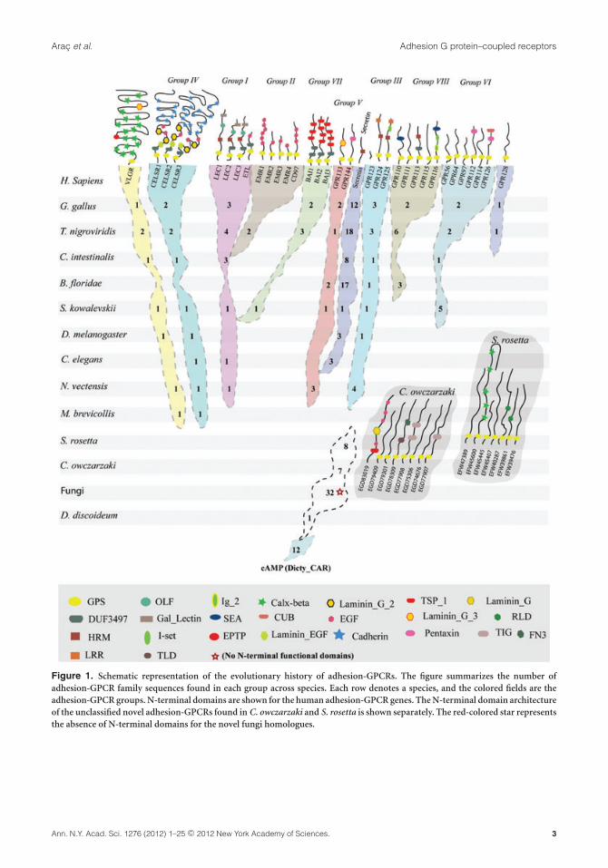
Helgi Schioth (Uppsala University) introduced hisstudies on the origin of the adhesion-GPCR class ofseven-span transmembrane (7TM) receptors. Theadhesion G protein–coupled receptors (GPCRs) arethe second largest family of GPCRs, with genes con-taining multiple exons that are presumed to havearisen through several mechanisms.1,2 Adhesion-GPCRs are of ancient origin and are found in severaleukaryotes that include most of the vertebrates, theclosest relatives to the vertebrates (Ciona intestinalisand Branchiostoma floridae), and the most primi-tive animals (Nematostella vectensis and Trichoplaxadhaerens; Fig. 1).3 Intriguingly, gene mining inamphioxus B. floridae has revealed several noveladhesion-GPCR domains such as somatomedin B,kringle, lectin C-type, SRCR, LDLa, immunoglobu-lin I-set, CUB, and TNFR, typically not found in themammalian receptors.4 Further, unique adhesion-GPCRs have been identified in urochordates (C. in-testinalis) and in the Strongylocentrotus purpuratus(sea urchin) genome, and some of these are species
specific. There are at least 21 adhesion-GPCRs inC. intestinalis that possess just the GPCR proteoly-sis site (GPS) proteolytic domain in the N-termini,while in the sea urchin there are 40 adhesion-GPCRscontaining multiple leucine-rich repeats (LRRs) butlacking GPS sites.5,6 Furthermore, comprehensiveanalysis of the entire set of adhesion and related se-cretin, and Methuselah groups of GPCRs providedthe first evolutionary hierarchy among the five mainclasses of vertebrate GPCRs. Schioth’s group pro-vided convincing evidence that the secretin GPCRsdescended from the family of adhesion-GPCRs,probably from group V of the adhesion-GPCRs.7
Moreover, they clarified the origin of the adhesion-GPCRs by providing the first evidence for the pres-ence of adhesion-GPCR homologues in fungi.8 Thisstudy estimated that the adhesion-GPCRs evolvedfrom Dictyostelium cAMP receptors before the splitof unikonts from a common ancestor of all ma-jor eukaryotic lineages.8 In addition, they minedthe close unicellular relatives of the metazoan lin-eage Salpingoeca rosetta and Capsaspora owczarzaki.These species have a rich group of the adhesion-GPCRs that provided additional insight into the firstemergence of the N-terminal domains of the ad-hesion family.8 Prime examples are the emergenceof the characteristic adhesion-family domains, GPSand the Calx-� domain in C. owczarzaki, and theEGF-CA domain in S. rosetta.8 Further, Schiothanalyzed the hemichordate Saccoglossus kowalevskii(acorn worm), which serves as an important modelorganism for developmental biologists to under-stand the evolution of the central nervous system(CNS). Unlike vertebrates that have a centralizednervous system, the acorn worm has a diffuse nervenet. Despite this, the acorn worm contains well-conserved orthologues for several of the adhesionfamily members with a similar N-terminal domainarchitecture. This is particularly apparent for thosegenes responsible for CNS development and regula-tion in vertebrates (Krishnan, A., et al. unpublisheddata). Overall, adhesion-GPCRs have a remarkablylong and complex evolutionary history that can betraced down to the common ancestors of metazoaand fungi (Fig. 1). Knowledge of the origin andevolution of the unique N-terminal domain archi-tectures of these genes may offer opportunities tobetter understand their functional roles and to aiddeorphanization.
2 Ann. N.Y. Acad. Sci. 1276 (2012) 1–25 c© 2012 New York Academy of Sciences.
Arac et al. Adhesion G protein–coupled receptors
Figure 1. Schematic representation of the evolutionary history of adhesion-GPCRs. The figure summarizes the number ofadhesion-GPCR family sequences found in each group across species. Each row denotes a species, and the colored fields are theadhesion-GPCR groups. N-terminal domains are shown for the human adhesion-GPCR genes. The N-terminal domain architectureof the unclassified novel adhesion-GPCRs found in C. owczarzaki and S. rosetta is shown separately. The red-colored star representsthe absence of N-terminal domains for the novel fungi homologues.
Ann. N.Y. Acad. Sci. 1276 (2012) 1–25 c© 2012 New York Academy of Sciences. 3
Adhesion G protein–coupled receptors Arac et al.
Signaling of adhesion-GPCRs
Intense focus during the 6th Adhesion-GPCR Work-shop was directed toward the mechanistic under-standing of adhesion-GPCR signal transduction inrelation to the structural components of adhesion-GPCRs. A group of researchers presented their find-ings on details of the molecular signaling mech-anism of different adhesion-GPCRs, providing acomprehensive overview of methods, models, andreceptors currently used to understand adhesion-GPCR signal transduction.
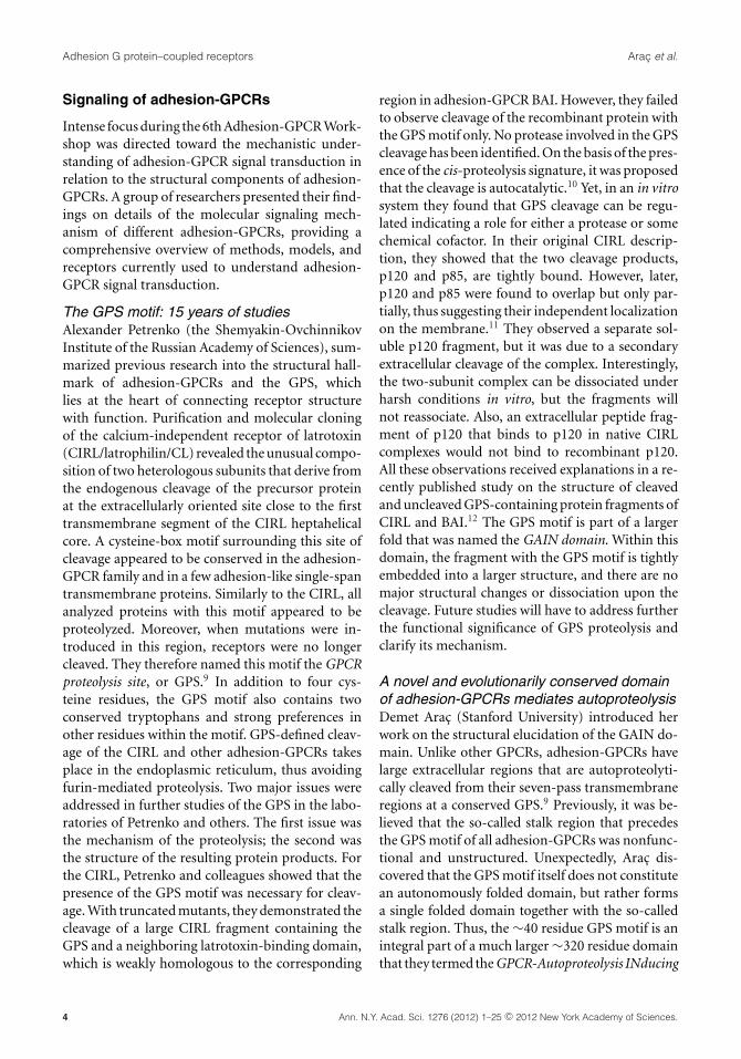
The GPS motif: 15 years of studiesAlexander Petrenko (the Shemyakin-OvchinnikovInstitute of the Russian Academy of Sciences), sum-marized previous research into the structural hall-mark of adhesion-GPCRs and the GPS, whichlies at the heart of connecting receptor structurewith function. Purification and molecular cloningof the calcium-independent receptor of latrotoxin(CIRL/latrophilin/CL) revealed the unusual compo-sition of two heterologous subunits that derive fromthe endogenous cleavage of the precursor proteinat the extracellularly oriented site close to the firsttransmembrane segment of the CIRL heptahelicalcore. A cysteine-box motif surrounding this site ofcleavage appeared to be conserved in the adhesion-GPCR family and in a few adhesion-like single-spantransmembrane proteins. Similarly to the CIRL, allanalyzed proteins with this motif appeared to beproteolyzed. Moreover, when mutations were in-troduced in this region, receptors were no longercleaved. They therefore named this motif the GPCRproteolysis site, or GPS.9 In addition to four cys-teine residues, the GPS motif also contains twoconserved tryptophans and strong preferences inother residues within the motif. GPS-defined cleav-age of the CIRL and other adhesion-GPCRs takesplace in the endoplasmic reticulum, thus avoidingfurin-mediated proteolysis. Two major issues wereaddressed in further studies of the GPS in the labo-ratories of Petrenko and others. The first issue wasthe mechanism of the proteolysis; the second wasthe structure of the resulting protein products. Forthe CIRL, Petrenko and colleagues showed that thepresence of the GPS motif was necessary for cleav-age. With truncated mutants, they demonstrated thecleavage of a large CIRL fragment containing theGPS and a neighboring latrotoxin-binding domain,which is weakly homologous to the corresponding
region in adhesion-GPCR BAI. However, they failedto observe cleavage of the recombinant protein withthe GPS motif only. No protease involved in the GPScleavage has been identified. On the basis of the pres-ence of the cis-proteolysis signature, it was proposedthat the cleavage is autocatalytic.10 Yet, in an in vitrosystem they found that GPS cleavage can be regu-lated indicating a role for either a protease or somechemical cofactor. In their original CIRL descrip-tion, they showed that the two cleavage products,p120 and p85, are tightly bound. However, later,p120 and p85 were found to overlap but only par-tially, thus suggesting their independent localizationon the membrane.11 They observed a separate sol-uble p120 fragment, but it was due to a secondaryextracellular cleavage of the complex. Interestingly,the two-subunit complex can be dissociated underharsh conditions in vitro, but the fragments willnot reassociate. Also, an extracellular peptide frag-ment of p120 that binds to p120 in native CIRLcomplexes would not bind to recombinant p120.All these observations received explanations in a re-cently published study on the structure of cleavedand uncleaved GPS-containing protein fragments ofCIRL and BAI.12 The GPS motif is part of a largerfold that was named the GAIN domain. Within thisdomain, the fragment with the GPS motif is tightlyembedded into a larger structure, and there are nomajor structural changes or dissociation upon thecleavage. Future studies will have to address furtherthe functional significance of GPS proteolysis andclarify its mechanism.
A novel and evolutionarily conserved domainof adhesion-GPCRs mediates autoproteolysisDemet Arac (Stanford University) introduced herwork on the structural elucidation of the GAIN do-main. Unlike other GPCRs, adhesion-GPCRs havelarge extracellular regions that are autoproteolyti-cally cleaved from their seven-pass transmembraneregions at a conserved GPS.9 Previously, it was be-lieved that the so-called stalk region that precedesthe GPS motif of all adhesion-GPCRs was nonfunc-tional and unstructured. Unexpectedly, Arac dis-covered that the GPS motif itself does not constitutean autonomously folded domain, but rather formsa single folded domain together with the so-calledstalk region. Thus, the ∼40 residue GPS motif is anintegral part of a much larger ∼320 residue domainthat they termed the GPCR-Autoproteolysis INducing
4 Ann. N.Y. Acad. Sci. 1276 (2012) 1–25 c© 2012 New York Academy of Sciences.
Arac et al. Adhesion G protein–coupled receptors
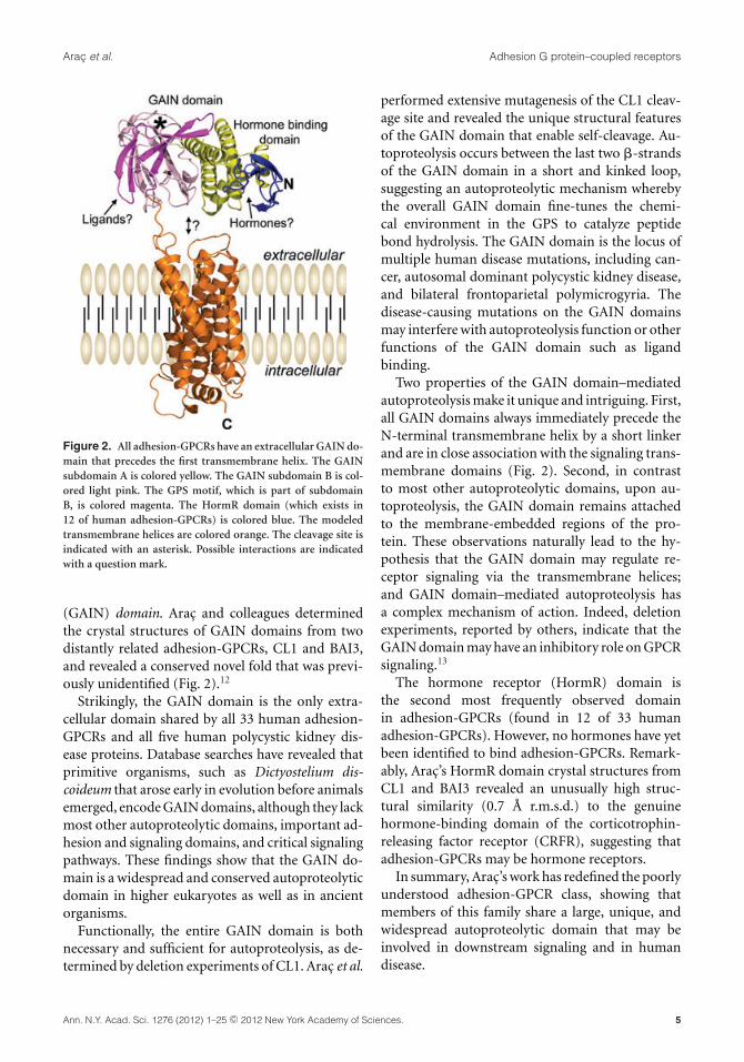
Figure 2. All adhesion-GPCRs have an extracellular GAIN do-main that precedes the first transmembrane helix. The GAINsubdomain A is colored yellow. The GAIN subdomain B is col-ored light pink. The GPS motif, which is part of subdomainB, is colored magenta. The HormR domain (which exists in12 of human adhesion-GPCRs) is colored blue. The modeledtransmembrane helices are colored orange. The cleavage site isindicated with an asterisk. Possible interactions are indicatedwith a question mark.
(GAIN) domain. Arac and colleagues determinedthe crystal structures of GAIN domains from twodistantly related adhesion-GPCRs, CL1 and BAI3,and revealed a conserved novel fold that was previ-ously unidentified (Fig. 2).12
Strikingly, the GAIN domain is the only extra-cellular domain shared by all 33 human adhesion-GPCRs and all five human polycystic kidney dis-ease proteins. Database searches have revealed thatprimitive organisms, such as Dictyostelium dis-coideum that arose early in evolution before animalsemerged, encode GAIN domains, although they lackmost other autoproteolytic domains, important ad-hesion and signaling domains, and critical signalingpathways. These findings show that the GAIN do-main is a widespread and conserved autoproteolyticdomain in higher eukaryotes as well as in ancientorganisms.
Functionally, the entire GAIN domain is bothnecessary and sufficient for autoproteolysis, as de-termined by deletion experiments of CL1. Arac et al.
performed extensive mutagenesis of the CL1 cleav-age site and revealed the unique structural featuresof the GAIN domain that enable self-cleavage. Au-toproteolysis occurs between the last two �-strandsof the GAIN domain in a short and kinked loop,suggesting an autoproteolytic mechanism wherebythe overall GAIN domain fine-tunes the chemi-cal environment in the GPS to catalyze peptidebond hydrolysis. The GAIN domain is the locus ofmultiple human disease mutations, including can-cer, autosomal dominant polycystic kidney disease,and bilateral frontoparietal polymicrogyria. Thedisease-causing mutations on the GAIN domainsmay interfere with autoproteolysis function or otherfunctions of the GAIN domain such as ligandbinding.
Two properties of the GAIN domain–mediatedautoproteolysis make it unique and intriguing. First,all GAIN domains always immediately precede theN-terminal transmembrane helix by a short linkerand are in close association with the signaling trans-membrane domains (Fig. 2). Second, in contrastto most other autoproteolytic domains, upon au-toproteolysis, the GAIN domain remains attachedto the membrane-embedded regions of the pro-tein. These observations naturally lead to the hy-pothesis that the GAIN domain may regulate re-ceptor signaling via the transmembrane helices;and GAIN domain–mediated autoproteolysis hasa complex mechanism of action. Indeed, deletionexperiments, reported by others, indicate that theGAIN domain may have an inhibitory role on GPCRsignaling.13
The hormone receptor (HormR) domain isthe second most frequently observed domainin adhesion-GPCRs (found in 12 of 33 humanadhesion-GPCRs). However, no hormones have yetbeen identified to bind adhesion-GPCRs. Remark-ably, Arac’s HormR domain crystal structures fromCL1 and BAI3 revealed an unusually high struc-tural similarity (0.7 A r.m.s.d.) to the genuinehormone-binding domain of the corticotrophin-releasing factor receptor (CRFR), suggesting thatadhesion-GPCRs may be hormone receptors.
In summary, Arac’s work has redefined the poorlyunderstood adhesion-GPCR class, showing thatmembers of this family share a large, unique, andwidespread autoproteolytic domain that may beinvolved in downstream signaling and in humandisease.
Ann. N.Y. Acad. Sci. 1276 (2012) 1–25 c© 2012 New York Academy of Sciences. 5
Adhesion G protein–coupled receptors Arac et al.
Activation of adhesion-GPCRs by anendogenous tethered ligandSimone Promel (University of Leipzig) introducedher studies on the GPS motif of latrophilins, whichhave shed light on the molecular interactions thatthe GAIN/GPS element of adhesion-GPCRs par-ticipates in. Latrophilins (LPHN/CL/CIRL) havebeen characterized to be one of the receptors for�-latrotoxin, a component of the black widow spi-der toxin.14 Binding of �-latrotoxin to LPHN1 leadsto calcium-independent release of neurotransmit-ters in neurons.15 Besides CELSR/Stan/Flamingo,latrophilins are the only members of the adhesion-GPCR family that are present in vertebrates andinvertebrates. They can be found throughout sev-eral phyla and species, making them prototypic forthe adhesion-GPCR class and suggesting that theyhave essential roles that are conserved in all bi-laterians. Three latrophilin homologs exist in themammalian genome (lphn1–3), whereas the nema-tode Caenorhabditis elegans contains two homologs,lat-1 and lat-2. A lat-1 null mutant causes vari-able morphogenic defects during embryonic andlarval development, leading to embryonic and lar-val lethality. Previous studies indicate that LAT-1participates in the control tissue polarity duringembryogenesis.16
The molecular mechanisms of LAT-1 signalingremain elusive. Very few in vivo approaches to ad-dress such questions have been developed thus far.However, Promel and colleagues employed an invivo assay in C. elegans to assess molecular functionof the latrophilin homolog LAT-1. The assay is basedon transgenic rescue of lat-1 mutant phenotypes bya wild-type or a modified lat-1 copy, which allowsanalysis of structure–function relationships in thebiological context of the receptor without detailedknowledge of input or output signals.
Using this assay, Promel performed a comprehen-sive receptor analysis indicating that LAT-1 signalsvia two different types of interactions.17 The firstmode requires the presence and structural preser-vation of the GPS—which is an integral part ofthe GAIN domain12—and 7TM domains. By con-trast, the other interaction is independent from the7TM/C terminus of the receptor. Importantly, forboth modes of receptor activity the GPS is essen-tial. On a more mechanistic level, Promel and col-leagues uncovered that the GAIN/GPS domain me-diates receptor activity by interacting with the 7TM
of the receptor, consistent with its function as anendogenous tethered ligand of the 7TM domain(Fig. 3). This finding suggests that the GAIN/GPSstructure might exert a similar function amongseveral, if not all, adhesion-GPCRs by modulatingthe signaling of the 7TM domain.17 Further, theassay system allowed intermolecular complemen-tation experiments with pairs of LAT-1 receptorvariants in vivo. These experiments revealed thatthe GPS cross-interacts with the 7TM domain ofa homologous partner receptor, likely in a dimericcomplex, for the 7TM-dependent function (Fig. 3).Promel also tested the requirement of GPS prote-olysis, which cleaves latrophilins and most otheradhesion-GPCRs autoproteolytically.10 Thus far thiscleavage event has been assumed to be essentialfor receptor function.18 In contrast to that assump-tion, Promel and colleagues showed that GPS cleav-age is not essential for receptor function.17 Finally,the GAIN/GPS structure is required for a secondfunction in C. elegans fertility that operates indepen-dently of the 7TM domain, indicating the molecu-lar versatility of the GAIN/GPS region in adhesion-GPCRs (Fig. 3).17
The work by Promel describes novel insights intoadhesion-GPCR function on a molecular level andanalyses under in vivo conditions. These insights,based on a complete receptor analysis, are the firststeps toward a better understanding of the entireclass of adhesion-GPCRs and a general mechanismfor their mode of signaling. In future studies bothmodes of receptor activity, 7TM-dependent and-independent, and their impact on latrophilin func-tion, will be analyzed in more detail. Additionally,current research by Promel addresses the questionof how GPS and the seven-transmembrane domainmight interact to mediate function and which otherinteraction partners play a role.
G protein–mediated signal transductionof adhesion-GPCRsInes Liebscher (University of Leipzig) demon-strated a high-throughput approach to determineG protein–mediated signal transduction ofadhesion-GPCRs. Over the last several years at-tempts have been made to unravel the issue of signaltransduction of adhesion-GPCRs. Although severalligands as interacting partners with these receptorshad been identified, activation of specific intracel-lular signal cascades remained obscure. There were
6 Ann. N.Y. Acad. Sci. 1276 (2012) 1–25 c© 2012 New York Academy of Sciences.
Arac et al. Adhesion G protein–coupled receptors
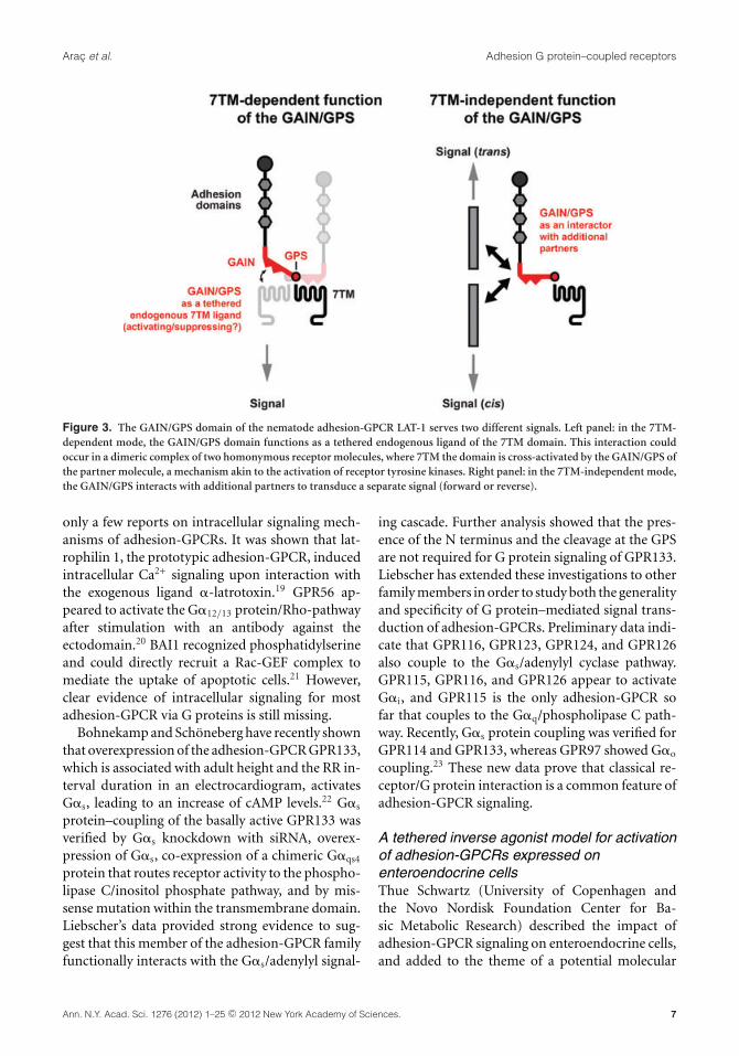
Figure 3. The GAIN/GPS domain of the nematode adhesion-GPCR LAT-1 serves two different signals. Left panel: in the 7TM-dependent mode, the GAIN/GPS domain functions as a tethered endogenous ligand of the 7TM domain. This interaction couldoccur in a dimeric complex of two homonymous receptor molecules, where 7TM the domain is cross-activated by the GAIN/GPS ofthe partner molecule, a mechanism akin to the activation of receptor tyrosine kinases. Right panel: in the 7TM-independent mode,the GAIN/GPS interacts with additional partners to transduce a separate signal (forward or reverse).
only a few reports on intracellular signaling mech-anisms of adhesion-GPCRs. It was shown that lat-rophilin 1, the prototypic adhesion-GPCR, inducedintracellular Ca2+ signaling upon interaction withthe exogenous ligand �-latrotoxin.19 GPR56 ap-peared to activate the G�12/13 protein/Rho-pathwayafter stimulation with an antibody against theectodomain.20 BAI1 recognized phosphatidylserineand could directly recruit a Rac-GEF complex tomediate the uptake of apoptotic cells.21 However,clear evidence of intracellular signaling for mostadhesion-GPCR via G proteins is still missing.
Bohnekamp and Schoneberg have recently shownthat overexpression of the adhesion-GPCR GPR133,which is associated with adult height and the RR in-terval duration in an electrocardiogram, activatesG�s, leading to an increase of cAMP levels.22 G�s
protein–coupling of the basally active GPR133 wasverified by G�s knockdown with siRNA, overex-pression of G�s, co-expression of a chimeric G�qs4
protein that routes receptor activity to the phospho-lipase C/inositol phosphate pathway, and by mis-sense mutation within the transmembrane domain.Liebscher’s data provided strong evidence to sug-gest that this member of the adhesion-GPCR familyfunctionally interacts with the G�s/adenylyl signal-
ing cascade. Further analysis showed that the pres-ence of the N terminus and the cleavage at the GPSare not required for G protein signaling of GPR133.Liebscher has extended these investigations to otherfamily members in order to study both the generalityand specificity of G protein–mediated signal trans-duction of adhesion-GPCRs. Preliminary data indi-cate that GPR116, GPR123, GPR124, and GPR126also couple to the G�s/adenylyl cyclase pathway.GPR115, GPR116, and GPR126 appear to activateG�i, and GPR115 is the only adhesion-GPCR sofar that couples to the G�q/phospholipase C path-way. Recently, G�s protein coupling was verified forGPR114 and GPR133, whereas GPR97 showed G�o
coupling.23 These new data prove that classical re-ceptor/G protein interaction is a common feature ofadhesion-GPCR signaling.
A tethered inverse agonist model for activationof adhesion-GPCRs expressed onenteroendocrine cellsThue Schwartz (University of Copenhagen andthe Novo Nordisk Foundation Center for Ba-sic Metabolic Research) described the impact ofadhesion-GPCR signaling on enteroendocrine cells,and added to the theme of a potential molecular
Ann. N.Y. Acad. Sci. 1276 (2012) 1–25 c© 2012 New York Academy of Sciences. 7
Adhesion G protein–coupled receptors Arac et al.
signaling mechanism of adhesion-GPCRs. En-teroendocrine cells function as specialized sensorsof food components and nutrient metabolites. Thecells are flask shaped with dense core secretorygranules located at the base, from which pep-tide hormones are released, and with an apicalmicrovillus–decorated sensory extension reachingthe gut lumen.24 Much attention has recently fo-cused on the expression and function of 7TM recep-tors as chemosensors for metabolites, for example,of triglycerides—long chain fatty acids and 2-OG—and of complex carbohydrates generated by the gutmicrobiota—short chain fatty acids.24,25 The en-teroendocrine cells are, along with their neighbor-ing enterocytes, renewed every week from pluripo-tent stem cells located at the bottom of the mucosalcrypts.
Individual enteroendocrine cells are isolated andFACS purified after genetic labeling with GFP orRFP expressed under the control of promoters forgut hormones such as CCK.26 Through quanti-tative polymerase chain reaction (qPCR) analy-sis, classical nutrient receptors were identified asbeing both highly expressed and highly enrichedin the cells. Surprisingly, a number of adhesion-GPCRs were identified as being expressed at sim-ilar high levels in enteroendocrine cells as werespecific nutrient metabolite receptors, for example,Celrs1, GPR128, Celsr3, CD97, and Lphn1. How-ever, the majority of the adhesion-GPCRs were alsohighly expressed in the neighboring enterocytes,though a few were found to be both highly ex-pressed and highly enriched in the enteroendocrinecells.
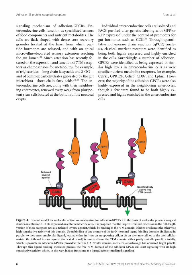
Figure 4. General model for molecular activation mechanism for adhesion-GPCRs. On the basis of molecular pharmacologicalstudies on adhesion-GPCRs expressed on enteroendocrine cells, it is proposed that the large N-terminal extension in the full-lengthversion of these receptors acts as a tethered inverse agonist, which, by binding to the 7TM domain, inhibits or silences the otherwisehigh constitutive activity of this domain. Upon binding of one or more of the far N-terminal ligand-binding domains (indicated inpurple) to their macromolecular ligand, located either in trans on an opposing cell, in cis on the same cell, or in the intercellularmatrix, the tethered inverse agonist (indicated in red) is removed from the 7TM domain, either partly (middle panel) or totally,which is possible in adhesion-GPCRs, provided that the GAIN/GPS domain–mediated autocleavage has occurred (right panel).Through this ligand binding–mediated process the free 7TM domain of the adhesion-GPCR will start signaling with its highconstitutive activity, which, in this way, in fact, functions as a ligand/agonist-mediated signaling.
8 Ann. N.Y. Acad. Sci. 1276 (2012) 1–25 c© 2012 New York Academy of Sciences.
Arac et al. Adhesion G protein–coupled receptors
It has been rather unclear to what degreeadhesion-GPCRs couple through classical G pro-tein pathways. A number of the adhesion-GPCRsexpressed in enteroendocrine cells were cloned andexpressed heterologously in HEK293 cells usingan optimized signal peptide construct to improveexpression levels, though generally they displayedonly minimal signaling. However, strong signalingthrough both Gq and Gi and, in particular, the SREtranscriptional activation pathway—that is, pre-sumably through G12/13—was observed when thereceptors were expressed in an N-terminally trun-cated form in which only the small N-terminal seg-ment from the autocleavage site to TM-I was in-tact. Thus, a general activation model for adhesion-GPCRs was proposed (see Fig. 4). In which the 7TMdomain of the adhesion-GPCRs is highly constitu-tively active and the large N-terminal segment ofthe receptors functions as a tethered inverse ago-nist. That is, in the intact receptor, the N-terminalextension, or presumably the 3D-conserved GAINdomain, which after the autocleavage is noncova-lently bound to the 7TM domain, will silence theconstitutive signaling of this domain. It is proposedthat binding of one or more of the far N-terminallylocated binding domains (which differ among thedifferent adhesion-GPCRs) to a ligand attached onthe neighboring cell or located on the same cell orin the intercellular matrix will lead to dissociationof the N-terminally tethered inverse agonist, whichresults in high constitutive signaling of the unbound7TM domain left at the cell surface (Fig. 4).
This model is in agreement with a similar modelsuggested by the Hall and Xu groups on the basisof classical biochemical structure–function studiesof GPR56, and by a model proposed by Langenhanand coworkers on the basis of in vivo structure–function studies performed in C. elegans with LAT-1/latrophilin.13,17,27
Shear stress–dependent downregulation ofCD97 on circulating leukocytes by CD55Jorg Hamann (University of Amsterdam) pre-sented work on the ligand interactions of CD97,a prototypic adhesion-GPCR broadly expressed byhematopoietic and nonhematopoietic cells. CD97interacts, through different regions in its extracel-lular subunit, with at least four other molecules:CD55, chondroitin sulfate B, �5�1 integrin, andCD90/Thy-1. The ability of CD97 to engage with
seemingly unrelated binding partners has triggeredstudies that aim to address the importance of in-dividual ligands in vivo using the interaction withCD55 (Ref. 28) as a paradigm. These studies re-vealed that mice lacking either CD97 or CD55 hadhigher granulopoietic activity, resulting in increasednumbers of circulating granulocytes.29 Moreover,the absence of CD97 or CD55 reduced disease ac-tivity in two experimental models of arthritis.30 Inboth cases, CD97 and CD55 knockout mice devel-oped a highly similar phenotype; yet a causativerelationship between the molecules could not beestablished.
Hamann described that circulating leukocytesfrom CD55-deficient mice express significantly in-creased levels of CD97. After adoptive transfer intoof CD55-deficient leukocytes wild-type mice, CD97expression on CD55-deficient leukocytes droppedto normal levels due to contact with CD55 expressedon wild-type leukocytes and stromal cells. Down-regulation of CD97 occurred within minutes afterfirst contact with CD55, involved both the extra-cellular and transmembrane subunit of the recep-tor, and correlated with an increase in plasma levelsof soluble CD97. In vitro, downregulation of CD97on CD55-deficient leukocytes cocultured with wild-type blood cells was strictly dependent on the shearstress from rigorous agitation of the cell cultures. Invivo, CD55-mediated downregulation of CD97 re-quired intact circulation, as shown in experimentswith wild-type recipient mice that were pretreatedwith heparin to prevent blood coagulation and thensacrificed immediately after adoptive transfer, fol-lowed by blood collection at later time points; trans-ferred CD55-deficient leukocytes did not downreg-ulate CD97 under these conditions. To test whetherligation by CD55 triggers CD97 signaling, CD55-deficient leukocytes were cocultured with wild-typeblood cells. Notably, de novo ligation did not activatesignaling molecules that recently were shown to beconstitutively engaged by CD97 in cancer cells, suchas ERK, PKB/Akt, and RhoA.31
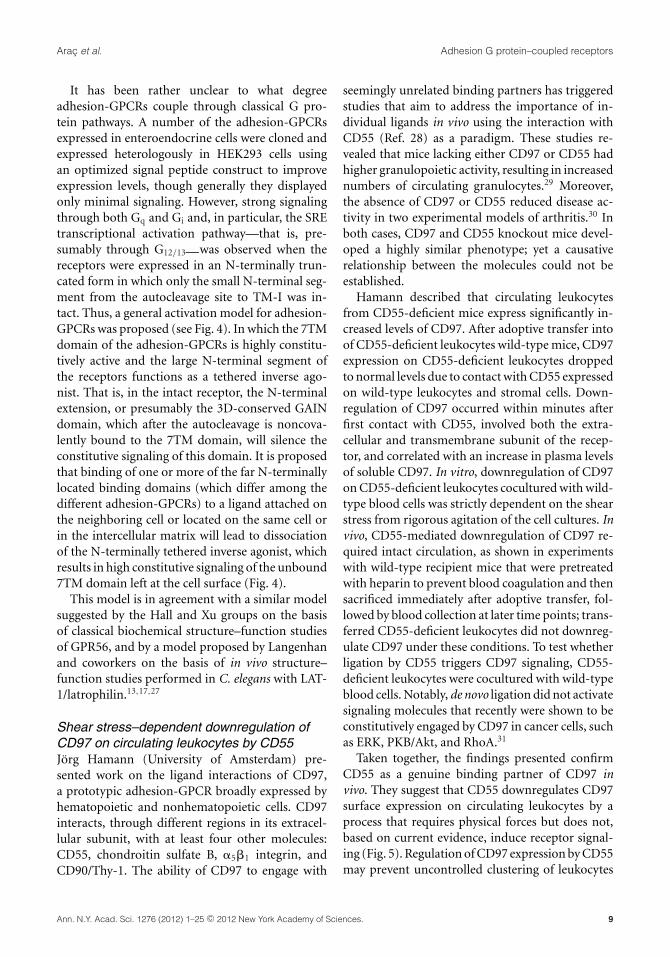
Taken together, the findings presented confirmCD55 as a genuine binding partner of CD97 invivo. They suggest that CD55 downregulates CD97surface expression on circulating leukocytes by aprocess that requires physical forces but does not,based on current evidence, induce receptor signal-ing (Fig. 5). Regulation of CD97 expression by CD55may prevent uncontrolled clustering of leukocytes
Ann. N.Y. Acad. Sci. 1276 (2012) 1–25 c© 2012 New York Academy of Sciences. 9
Adhesion G protein–coupled receptors Arac et al.
Figure 5. Consequences of the CD97–CD55 interaction invivo. (A) In tissue, contacts between the adhesion-GPCR CD97and its ligand CD55 likely facilitate cell adhesion. (B) In thecirculation, CD97 expression is constantly downregulated bycontact with CD55 on blood and stromal cells. This regulationprocess may prevent uncontrolled clustering of leukocytes inthe blood stream, thereby restricting CD97–CD55 interaction-mediated adhesion to tissue sites.
due to homo- or heterotypic cellular contacts inthe blood stream, thereby restricting CD97–CD55interaction-mediated adhesion to tissue sites. Thedata support the hypothesis that adhesion-GPCRsare two-part entities with distinct roles for the ex-tracellular and the seven-transmembrane subunitsin cell adhesion and signaling, respectively.
Activation of the EMR2 receptor vialigation-induced translocation, and interactionof receptor subunits in lipid rafts activatesmacrophagesHsi-Hsien Lin (Chang Gung University) analyzedthe signaling mechanism of the adhesion-GPCREMR2 in macrophages. Directed migration ofphagocytes to infected sites is a critical step ininnate immunity for pathogen elimination. Acti-vated phagocytes clear invading pathogens by multi-ple mechanisms, including phagocytosis and releaseof proteases, antimicrobial peptides, and cytokine/chemokines. As a myeloid cell–restricted memberof the adhesion-GPCR family, the EMR2 receptorhas been shown previously to play a role in thecellular functions of innate immune cells.32,33 In-deed, ligation of the EMR2 receptor not only canincrease neutrophil adhesion and migration, but itcan also augment the production of antimicrobialmediators.32,33
As with the majority of adhesion-GPCRs, EMR2is posttranslationally modified by GPS autoprote-olysis in the endoplasmic reticulum and cleavedinto a large extracellular domain (�-subunit) and
a seven-transmembrane domain (�-subunit).10,34
To investigate the role of GPS autoproteolysisin mediating the cellular functions of adhesion-GPCRs and the mechanistic relevance of the re-ceptor subunit interaction, Lin and coworkers firstdemonstrated that GPS proteolysis is necessary forEMR2-mediated cell migration. Next, the structuralorganization of EMR2 receptor subunits was ex-amined. Surprisingly, two distinct receptor com-plexes were identified: one is a noncovalent �-�heterodimer, while the other consists of two inde-pendent receptor subunits with differential distribu-tion in lipid raft microdomains. More specifically,the EMR2 �-subunit was shown to locate mostly inthe nonraft regions, while the �-subunit was foundin both the raft and nonraft regions. These datasuggest that the two EMR2 receptor subunits do notalways interact on the cell surface but behave, inpart, as two independent molecules.35
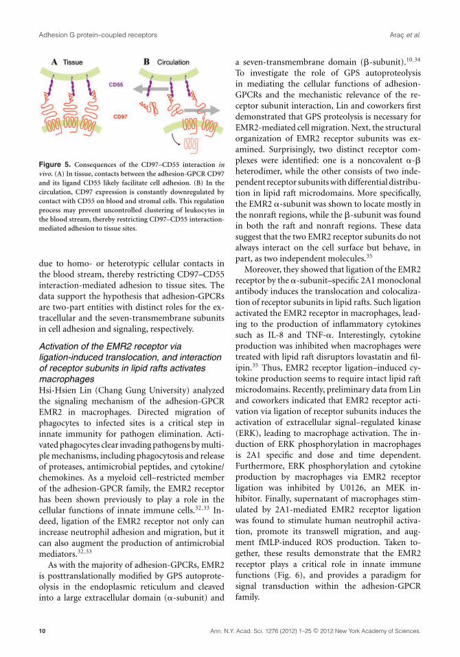
Moreover, they showed that ligation of the EMR2receptor by the �-subunit–specific 2A1 monoclonalantibody induces the translocation and colocaliza-tion of receptor subunits in lipid rafts. Such ligationactivated the EMR2 receptor in macrophages, lead-ing to the production of inflammatory cytokinessuch as IL-8 and TNF-�. Interestingly, cytokineproduction was inhibited when macrophages weretreated with lipid raft disruptors lovastatin and fil-ipin.35 Thus, EMR2 receptor ligation–induced cy-tokine production seems to require intact lipid raftmicrodomains. Recently, preliminary data from Linand coworkers indicated that EMR2 receptor acti-vation via ligation of receptor subunits induces theactivation of extracellular signal–regulated kinase(ERK), leading to macrophage activation. The in-duction of ERK phosphorylation in macrophagesis 2A1 specific and dose and time dependent.Furthermore, ERK phosphorylation and cytokineproduction by macrophages via EMR2 receptorligation was inhibited by U0126, an MEK in-hibitor. Finally, supernatant of macrophages stim-ulated by 2A1-mediated EMR2 receptor ligationwas found to stimulate human neutrophil activa-tion, promote its transwell migration, and aug-ment fMLP-induced ROS production. Taken to-gether, these results demonstrate that the EMR2receptor plays a critical role in innate immunefunctions (Fig. 6), and provides a paradigm forsignal transduction within the adhesion-GPCRfamily.
10 Ann. N.Y. Acad. Sci. 1276 (2012) 1–25 c© 2012 New York Academy of Sciences.
Arac et al. Adhesion G protein–coupled receptors
Figure 6. Activation of the EMR2 receptor in macrophages is mediated through the translocation and interaction of its twoindependent subunits in the lipid raft. The independent EMR2 �- and �-subunits are localized in the nonraft and lipid raftregions, respectively. Following receptor ligation by the � subunit–specific 2A1 mAb, the independent �-subunit translocates andreassociates with the �-subunit in the lipid raft region. Such interaction induces intracellular signaling via the MAPK pathway(mainly ERK1/2 phosphorylation), leading eventually to proinflammatory cytokine (IL-8, TNF-�) secretion.
Real-time monitoring of GPCR signaling inliving cellsDavide Calebiro (University of Wurzburg) con-cluded the workshop section on signaling ofadhesion-GPCRs with an overview of in vivo imag-ing for GPCR signaling, which might be extended toadhesion-GPCRs one day. The approximately 1000different GPCRs present on the surface of cells pro-vide fundamental links between the extracellularenvironment and the intracellular milieu, allowingcells to respond and adapt to a wide variety of stim-uli, such as hormones, neurotransmitters, light, andodors, as well as cell and matrix contacts. Whereasthe basic molecular mechanisms of GPCR signal-ing have been elucidated, how such diverse stim-uli are integrated via a few common signaling cas-cades while achieving highly specific responses is stillpoorly understood. In the last few years, Calebiroand colleagues developed a series of optical meth-ods using fluorescence resonance energy transfer
(FRET), which enables imaging GPCR activationand signaling directly in living cells.36
More recently, in order to analyze GPCR signalingunder highly physiological conditions, Calebiro andcolleagues have generated a transgenic mouse37 withubiquitous expression of a FRET sensor for cAMP38
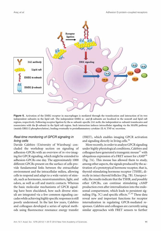
(Fig. 7A). This mouse has allowed them to study,among other aspects, the signals produced by the ac-tivation of a prototypical hormone receptor, that is,thyroid-stimulating hormone receptor (TSHR), di-rectly in intact thyroid follicles (Fig. 7B). Unexpect-edly, the results indicate that the TSHR, and possiblyother GPCRs, can continue stimulating cAMPproduction even after internalization into the endo-somal compartment, which leads to persistent sig-naling (Fig. 7C) and specific effects.37,39 These datareveal new and important functions for receptorinternalization in regulating GPCR-mediated re-sponses. Calebiro and colleagues are currently usingsimilar approaches with FRET sensors to further
Ann. N.Y. Acad. Sci. 1276 (2012) 1–25 c© 2012 New York Academy of Sciences. 11
Adhesion G protein–coupled receptors Arac et al.
Figure 7. Real-time monitoring of GPCR signaling in livingcells. (A) Transgenic mouse with ubiquitous expression of aFRET sensor for cAMP (Epac1-camps). (B) Confocal image ofa thyroid follicle isolated from the cAMP reporter mouse. (C)Representative FRET trace obtained in a thyroid follicle, show-ing persistent cAMP elevations after transient TSH stimulation.(D) Single GPCRs on the surface of living cells visualized byTIRF microscopy.
explore this and other novel aspects of GPCR-cAMPsignaling in fundamental physiological processessuch as thyroid hormone production and femalereproduction. Whereas these approaches allowa precise characterization of GPCR and secondmessenger signaling with high spatiotemporal res-olution, a full characterization of GPCR signalingcascades will likely require observing the signalsproduced by the activation of a single receptor.
To achieve this goal, Calebiro and colleaguesare developing new methods using labeling withsmall organic fluorophores and total internal re-flection fluorescence (TIRF) microscopy, whichallow visualizing signaling proteins at the sur-face of living cells with single-molecule sensi-tivity (Fig 7D). They are using these methodsto monitor individual protein–protein interac-tions, such as those involved in ligand bind-ing, receptor di-/oligomerization, or coupling toG proteins with high spatiotemporal resolution. Ini-tial data suggest that GPCRs are targeted to differ-ent microdomains of the cell surface, where theyare present in a dynamic equilibrium, with con-stant formation and dissociation of new receptorcomplexes. Taken together, these data provide novel
insights into the complex dynamic events at the ba-sis of the spatiotemporal compartmentalization ofGPCR signaling cascades.
Adhesion-GPCRs in development
Accumulating evidence demonstrates thatadhesion-GPCRs fulfill plentiful functions duringthe genesis of various organ systems. For someadhesion-GPCRs, these functions are well definedon a cell biological level, whereas other receptorsremain to be placed in a physiological context.
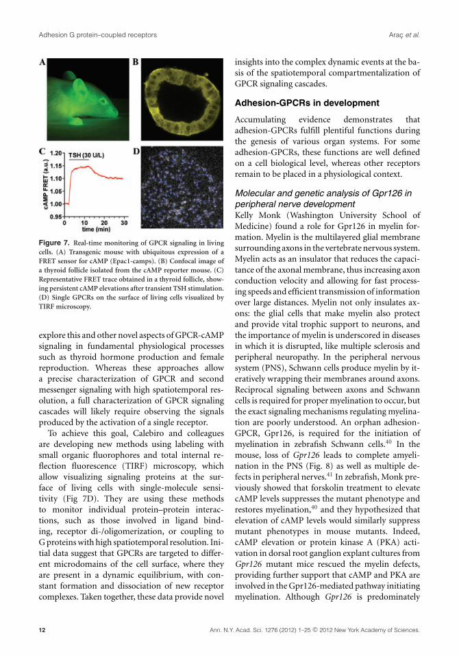
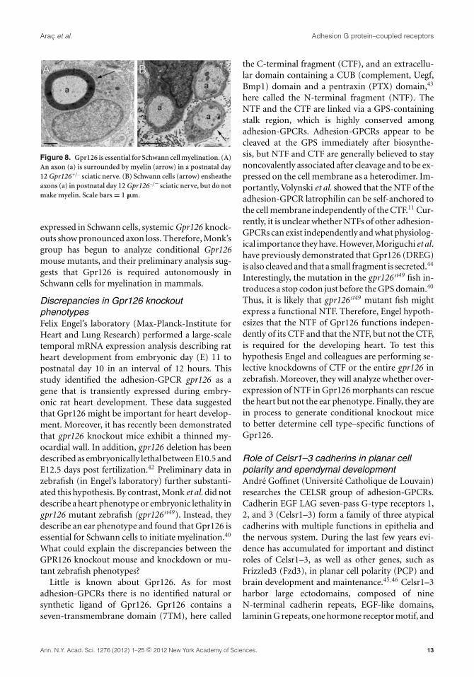
Molecular and genetic analysis of Gpr126 inperipheral nerve developmentKelly Monk (Washington University School ofMedicine) found a role for Gpr126 in myelin for-mation. Myelin is the multilayered glial membranesurrounding axons in the vertebrate nervous system.Myelin acts as an insulator that reduces the capaci-tance of the axonal membrane, thus increasing axonconduction velocity and allowing for fast process-ing speeds and efficient transmission of informationover large distances. Myelin not only insulates ax-ons: the glial cells that make myelin also protectand provide vital trophic support to neurons, andthe importance of myelin is underscored in diseasesin which it is disrupted, like multiple sclerosis andperipheral neuropathy. In the peripheral nervoussystem (PNS), Schwann cells produce myelin by it-eratively wrapping their membranes around axons.Reciprocal signaling between axons and Schwanncells is required for proper myelination to occur, butthe exact signaling mechanisms regulating myelina-tion are poorly understood. An orphan adhesion-GPCR, Gpr126, is required for the initiation ofmyelination in zebrafish Schwann cells.40 In themouse, loss of Gpr126 leads to complete amyeli-nation in the PNS (Fig. 8) as well as multiple de-fects in peripheral nerves.41 In zebrafish, Monk pre-viously showed that forskolin treatment to elevatecAMP levels suppresses the mutant phenotype andrestores myelination,40 and they hypothesized thatelevation of cAMP levels would similarly suppressmutant phenotypes in mouse mutants. Indeed,cAMP elevation or protein kinase A (PKA) acti-vation in dorsal root ganglion explant cultures fromGpr126 mutant mice rescued the myelin defects,providing further support that cAMP and PKA areinvolved in the Gpr126-mediated pathway initiatingmyelination. Although Gpr126 is predominately
12 Ann. N.Y. Acad. Sci. 1276 (2012) 1–25 c© 2012 New York Academy of Sciences.
Arac et al. Adhesion G protein–coupled receptors
Figure 8. Gpr126 is essential for Schwann cell myelination. (A)An axon (a) is surrounded by myelin (arrow) in a postnatal day12 Gpr126+/– sciatic nerve. (B) Schwann cells (arrow) ensheatheaxons (a) in postnatal day 12 Gpr126–/– sciatic nerve, but do notmake myelin. Scale bars = 1 �m.
expressed in Schwann cells, systemic Gpr126 knock-outs show pronounced axon loss. Therefore, Monk’sgroup has begun to analyze conditional Gpr126mouse mutants, and their preliminary analysis sug-gests that Gpr126 is required autonomously inSchwann cells for myelination in mammals.
Discrepancies in Gpr126 knockoutphenotypesFelix Engel’s laboratory (Max-Planck-Institute forHeart and Lung Research) performed a large-scaletemporal mRNA expression analysis describing ratheart development from embryonic day (E) 11 topostnatal day 10 in an interval of 12 hours. Thisstudy identified the adhesion-GPCR gpr126 as agene that is transiently expressed during embry-onic rat heart development. These data suggestedthat Gpr126 might be important for heart develop-ment. Moreover, it has recently been demonstratedthat gpr126 knockout mice exhibit a thinned my-ocardial wall. In addition, gpr126 deletion has beendescribed as embryonically lethal between E10.5 andE12.5 days post fertilization.42 Preliminary data inzebrafish (in Engel’s laboratory) further substanti-ated this hypothesis. By contrast, Monk et al. did notdescribe a heart phenotype or embryonic lethality ingpr126 mutant zebrafish (gpr126st49). Instead, theydescribe an ear phenotype and found that Gpr126 isessential for Schwann cells to initiate myelination.40
What could explain the discrepancies between theGPR126 knockout mouse and knockdown or mu-tant zebrafish phenotypes?
Little is known about Gpr126. As for mostadhesion-GPCRs there is no identified natural orsynthetic ligand of Gpr126. Gpr126 contains aseven-transmembrane domain (7TM), here called
the C-terminal fragment (CTF), and an extracellu-lar domain containing a CUB (complement, Uegf,Bmp1) domain and a pentraxin (PTX) domain,43
here called the N-terminal fragment (NTF). TheNTF and the CTF are linked via a GPS-containingstalk region, which is highly conserved amongadhesion-GPCRs. Adhesion-GPCRs appear to becleaved at the GPS immediately after biosynthe-sis, but NTF and CTF are generally believed to staynoncovalently associated after cleavage and to be ex-pressed on the cell membrane as a heterodimer. Im-portantly, Volynski et al. showed that the NTF of theadhesion-GPCR latrophilin can be self-anchored tothe cell membrane independently of the CTF.11 Cur-rently, it is unclear whether NTFs of other adhesion-GPCRs can exist independently and what physiolog-ical importance they have. However, Moriguchi et al.have previously demonstrated that Gpr126 (DREG)is also cleaved and that a small fragment is secreted.44
Interestingly, the mutation in the gpr126st49 fish in-troduces a stop codon just before the GPS domain.40
Thus, it is likely that gpr126st49 mutant fish mightexpress a functional NTF. Therefore, Engel hypoth-esizes that the NTF of Gpr126 functions indepen-dently of its CTF and that the NTF, but not the CTF,is required for the developing heart. To test thishypothesis Engel and colleagues are performing se-lective knockdowns of CTF or the entire gpr126 inzebrafish. Moreover, they will analyze whether over-expression of NTF in Gpr126 morphants can rescuethe heart but not the ear phenotype. Finally, they arein process to generate conditional knockout miceto better determine cell type–specific functions ofGpr126.
Role of Celsr1–3 cadherins in planar cellpolarity and ependymal developmentAndre Goffinet (Universite Catholique de Louvain)researches the CELSR group of adhesion-GPCRs.Cadherin EGF LAG seven-pass G-type receptors 1,2, and 3 (Celsr1–3) form a family of three atypicalcadherins with multiple functions in epithelia andthe nervous system. During the last few years evi-dence has accumulated for important and distinctroles of Celsr1–3, as well as other genes, such asFrizzled3 (Fzd3), in planar cell polarity (PCP) andbrain development and maintenance.45,46 Celsr1–3harbor large ectodomains, composed of nineN-terminal cadherin repeats, EGF-like domains,laminin G repeats, one hormone receptor motif, and
Ann. N.Y. Acad. Sci. 1276 (2012) 1–25 c© 2012 New York Academy of Sciences. 13
Adhesion G protein–coupled receptors Arac et al.
a potential GPS. This is followed by seven trans-membrane domains and a cytoplasmic tail. Liketheir fruit fly ortholog Flamingo (Fmi), Celsr1–3are thought to work in interaction with other corePCP proteins such as Frizzled (particularly Fzd3 andFzd6 in mammals), Van Gogh (Vangl1–2), Dishev-elled (Dvl1–3), and Prickle (Pk1–4).
Observations of constitutive and conditionalCelsr2 and 3 mutant mice uncovered importantfunctions of these proteins during ependymal de-velopment. In double mutant animals, a severe hy-drocephalus develops rapidly during the early post-natal period (P), leading to death around P8–P10.Studies with scanning and transmission microscopyand immunohistochemistry found prominent ab-normalities of ciliogenesis in mutants. Basal bod-ies remained frequently embedded in the subapicalcytoplasm and failed to become aligned normally(rotational polarity defect). As a result, manyependymal cells are completely devoid of cilia andthe circulation of the CSF is severely impaired, lead-ing to lethal hydrocephalus.46
More recently, Goffinet has studied the role ofCelsr1 during ependymal development using con-ditional and floxed Celsr147 and Vangl248 mutantalleles, showing that they are distinct from that ofCelsr2 and 3. Whereas cilia appear at the normaltime and are of normal length, in Celsr1 mutantmice there is a clear defect in translational polar-ity, in that cilia tufts are not consistently displacedrostrally, as in normal animals; a translational po-larity defect was seen even at P0, when ependymalcells had one nonmotile cilium, in Celsr1 mutantsand in Fzd3 and Vangl2 mutants. Rotational po-larization of basal bodies (BB) was studied usingdouble labeling with gamma-catenin for BB andphosphocatenin, which labels an area adjacent toBB, opposite the basal foot. Rotational polarizationof BB was found to be similarly defective in Celsr1and Vangl2 mutants (Fzd3 mutants die at P0 andcannot be studied). In addition, the mean vectorsof translational and rotational polarization are notaligned in mutant ependymal cells, unlike normalependymal cells.
Goffinet’s data show that Celsr1 regulates trans-lational and rotational polarity of ependymal cellsfrom the time that they are generated to the adultstage. Celsr1 works together with Vangl2 and Fzd3 inthis process. Such PCP regulation in the ependymalepithelium is complementary to the role of Celsr2
and 3, which regulate ependymal differentiation andciliogenesis.
Adhesion-GPCR Celsr1 in the complexmorphogenesis of mammalian organprimordiaCaroline Formstone (King’s College London) re-ported about additional functions of Celsr-likeadhesion-GPCRs. Mice with disrupted core PCPcomponent function die at birth owing to catas-trophic developmental defects in neural tubeclosure49 and lung branching.50 Defects in the de-velopment of other organ systems are also appar-ent including the epidermis.51 The adhesion-GPCRFlamingo plays a central role in the local transmis-sion of PCP information among neighboring cells.Of three Flamingo homologues in mammals, Celsr1is predominantly found in epithelial precursorswithin organ primordia. Several studies strongly in-dicate a major role for Celsr1 in the coordination ofPCP during mammalian organ development,47,51
but how it functions to coordinate epithelial mor-phogenesis is unclear. Formstone’s recent data sug-gest that Celsr1 protein exhibits a differential distri-bution along the apicobasal axis of some epithelia.In particular, Celsr1 exhibits a novel enrichment tothe basal membrane of neuroepithelial precursorsand lung tubules.52,53 Studies on how differentialCelsr1 protein distribution links to its function intissue morphogenesis and whether Celsr1 protein atthe epithelial basal membrane elicits PCP signalingwill provide insight into the complex roles of Celsr1in mammalian organ development.
CD97 overexpression induces amegaintestineInterestingly, CD97 is also implicated in intestinaldevelopment, as presented by Gabriela Aust (Uni-verstiy of Leipzig). Adhesion-GPCRs are involved inadhesion, guidance, and positioning of cells. CD97,in contrast to the other EGF-TM7 adhesion-GPCRsubfamily members restricted to immune cells, ispresent in normal and malignant epithelial cells. Innormal human intestine, CD97 is located in ente-rocytic cell–cell contacts54 and, in the cytoplasm,shows an expression gradient along the crypt–villusaxis.
To understand the role of CD97 in intestinal phys-iology, Aust et al. generated transgenic Tg(villin-CD97) mice.54 Unexpectedly, overexpressionof CD97 resulted in upper megaintestine, depending
14 Ann. N.Y. Acad. Sci. 1276 (2012) 1–25 c© 2012 New York Academy of Sciences.

Arac et al. Adhesion G protein–coupled receptors
on the CD97 cDNA copy number integrated. In-testinal enlargement involved an increase in length,diameter, and weight. Remarkably, the megaintes-tine phenotype develops with normal microscopicmorphology, and thereby clearly differs fromexisting megaintestine models in which intestinalenlargement is often accompanied by dramaticmorphological changes.
The megaintestine phenotype is acquired afterbirth before weaning, which makes this a uniquemodel for investigating the mechanisms underlyingpostnatal expansion of the mammalian small intes-tine by way of two consecutive growth patterns: (1)cylindrical growth in length and diameter withoutalteration of microscopic morphology (as seen inthe Tg(villin-CD97) mice), and (2) luminal growthwith amplification of the internal surface area.55
Notably and in accordance with a cylindricalgrowth pattern, suckling but not adult Tg(villin-CD97) mice showed more crypt fission comparedwith wild-type mice. Consistently, acquisition of themegaintestine was independent of altered cell lin-eage determination, Wnt signaling, and an increaseof intestinal stem cell markers. Suckling Tg(villin-CD97) pups developed the phenotype independentof the genotype of the feeding dam, thus excludingregulation of a milk growth factor by CD97. Mostlikely, CD97 regulates the binding or signaling of anintestinal receptor for a milk constituent.
The Tg(villin-CD97) mice provide new evidencesupporting the conclusion that adhesion-GPCRshave distinct functions that may depend on thecellular context in which a given receptor is ex-pressed. Tg(villin-CD97) mice not expressing CD55that binds to the extracellular EGF-like domains ofCD97 also developed a megaintestine, suggestingthat the adhesive extracellular part is not necessaryfor phenotype induction. By contrast, mice overex-pressing a truncated CD97 with only the first twotransmembrane helices did not develop a megain-testine, which implies signaling through CD97 inphenotype induction.
Overall, these transgenic mice provide suitablemodels to uncover and understand functions of ad-hesion GPCRs in normal epithelial cells.
Neurobiological roles of adhesion-GPCRs
The connection between adhesion-GPCRs and neu-ral function was postulated early on. Some groupsreported about their ongoing efforts to pinpoint the
exact relationship between these receptors and theproperties of this highly specialized cell type.
Drosophila synapses as an in vivo model tostudy structure–function relationships oflatrophilinTobias Langenhan and Robert Kittel (Universityof Wurzburg) introduced the fruit fly Drosophilamelanogaster as a new in vivo model for researchon evolutionarily conserved adhesion-GPCRs ofthe latrophilin group, which is extendable to theFlamingo/CELSR group. Latrophilins have beenimplicated in the control of synaptic transmis-sion as well as planar cell polarity, raising thequestions of whether and how these phenomenaare interlinked.15,16,56 Thus far, models in whichcell polarity and neuronal exocytosis could betested at the same time and in the same celltype have been lacking in the adhesion-GPCRfield. The versatile model system of Drosophilaincludes high-throughput transgenesis with singlecopy integration, homologous recombination forknockout/knockin studies of selected target genes,cell- and time-specific transgene expression throughbinary expression systems, and a vast arsenal of al-lelic variants covering the entire genome for geneticinteraction studies. In particular, the fruit fly larvapossesses a well-defined synaptic contact, the neuro-muscular junction (NMJ), ideally suited for inves-tigating adhesion-GPCR expression and functionwith biochemical, imaging, and electrophysiologicalmethods. Langenhan and Kittel presented prelimi-nary data indicating that latrophilin/dCIRL is resi-dent at the NMJ, and that its removal by mutation orRNA interference causes changes in the molecular,structural, and functional properties of this synapsetype. After full characterization of the phenotypicprofile due to dCirl deficiency, Langenhan and Kit-tel will use the NMJ as an in vivo platform to testmodified dCirl variants and correlate molecular le-sions in the receptor with functional consequenceson a cell biological level.
Transsynaptic interaction between presynapticlatrophilin and postsynaptic LassoYuri Ushkaryov (University of Kent) is interestedin the function of mammalian latrophilins andpresented his latest data. Latrophilin 1 (LPH1),57
a neuronal adhesion G-protein–coupled receptorthat binds �-latrotoxin, is implicated in controlof presynaptic Ca2+ and in the modulation of
Ann. N.Y. Acad. Sci. 1276 (2012) 1–25 c© 2012 New York Academy of Sciences. 15
Adhesion G protein–coupled receptors Arac et al.
neurotransmitter release.58,59 To understand themolecular mechanisms of these physiological func-tions, Ushkaryov’s group isolated the endogenousligand of LPH1, Lasso.60 This protein is a splicevariant of teneurin-2. Teneurins are brain-specific,orphan, cell surface receptors with functionsin neuronal pathfinding and synaptogenesis.Ushkaryov’s data indicate that LPH1, located onpresynaptic terminals, forms strong and specifictranssynaptic complexes with Lasso, which is foundon postsynaptic spines. This interaction is not onlystructural but also functional: soluble fragmentsof Lasso induce intracellular Ca2+ signals uponbinding to LPH1 in presynaptic boutons of culturedhippocampal neurons and in nonneuronal cellsexpressing exogenous LPH1. Furthermore, theLPH1–Lasso complexes play an important role insynaptic development and activity. Thus, Lassofragments acting via LPH1 strongly increase the rateof spontaneous exocytosis in mouse neuromuscularjunctions.61 LPH1 expressed on nonneuronal cellsinduces postsynaptic differentiation in coculturedhippocampal neurons.60 On the other hand, whilesynapses in which the interaction between LPH1and Lasso is inhibited, appear morphologicallynormal, they remain physiologically silent.61 Takentogether, the data from the Ushkaryov groupindicate that while the transsynaptic interactionof LPH1 and Lasso is not necessary for the initialestablishment of central synapses, it participates inpresynaptic Ca2+ control and is required for func-tional maturation of presynaptic nerve terminals.
The very large G PCR Vlgr1b/GPR98 – a keycomponent of the Usher syndrome proteinnetworksUwe Wolfrum (University of Mainz) described therole of the very large G protein–coupled receptor-1(VLGR1) in the inner ear and in retinal biology.VLGR1, also known as MASS1 or GPR98, has amolecular weight of up to ∼700 kDa and is by farthe largest GPCR and the largest cell surface pro-tein known to date.62 The large ectodomain of thelargest splice variant VLGR1b contains several re-peated motifs, including calcium binding, Calx-�repeats, and seven copies of an epitempin repeat.It is linked to the 7TM moiety via a proteolyticsite (GPS) containing a region typical for adhesion-GPCRs. The short intracellular C-terminus containsa consensus PDZ binding motif, suggesting interac-
tions with cellular scaffold proteins. In the absenceof any known ligand VLGR1/GPR98 is one of thefew adhesion-GPCRs in which mutations are dis-ease relevant. VLGR1/GPR98 defects are thoughtto be associated with epilepsy. Mouse vlgr1 mu-tants are characterized by the susceptibility to au-diogenic seizures and to the development of sen-soneuronal defects, namely hearing impairment andvisual dysfunction.63,64 Mutations in the humanVLGR1/GPR98 gene cause Usher syndrome (USH)type 2C.65
Human USH is the most common form ofcombined hereditary deaf-blindness. Three clini-cal subtypes (USH1–3) are differentiated on thebasis of severity, age of onset, and progression ofthe symptom.65 Wolfrum and others have identi-fied VLGR1/GPR98 as a component of USH pro-tein networks in inner ear hair cells and retinalphotoreceptor cells. In hair cells VLGR1/GPR98 ispart of the ankle link complex essential for theformation of the ankle links between the mem-branes of neighboring stereocilia and thereby for thecorrect development of the mechanosensitive hair
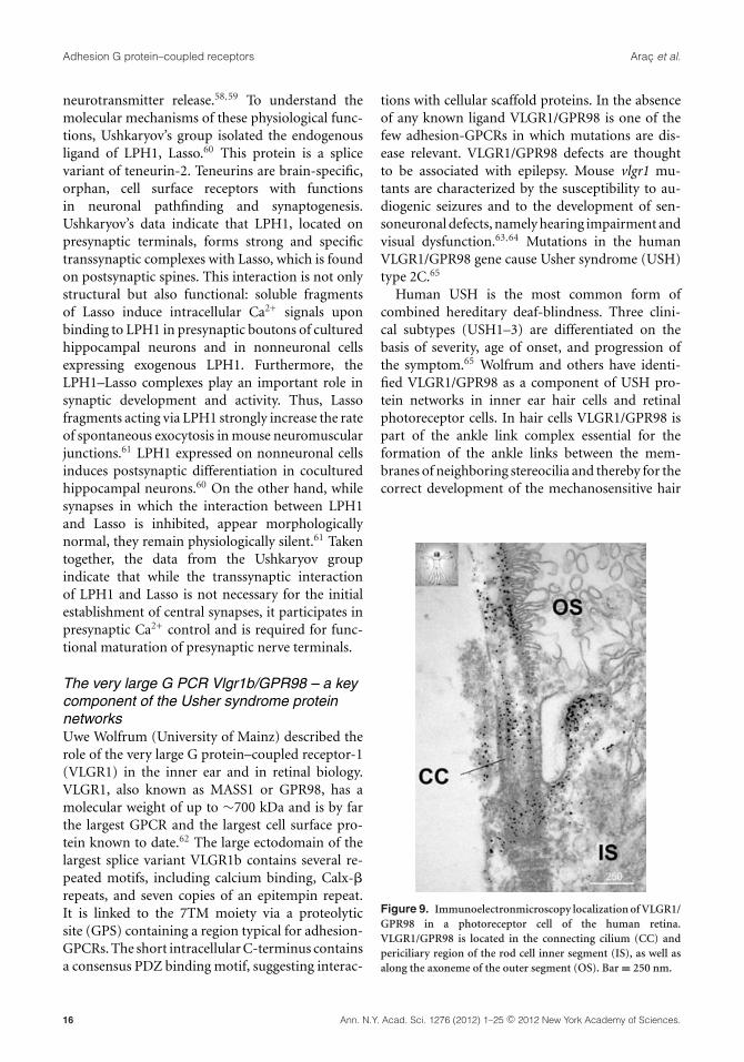
Figure 9. Immunoelectronmicroscopy localization of VLGR1/GPR98 in a photoreceptor cell of the human retina.VLGR1/GPR98 is located in the connecting cilium (CC) andpericiliary region of the rod cell inner segment (IS), as well asalong the axoneme of the outer segment (OS). Bar = 250 nm.
16 Ann. N.Y. Acad. Sci. 1276 (2012) 1–25 c© 2012 New York Academy of Sciences.
Arac et al. Adhesion G protein–coupled receptors
bundles.63 In photoreceptor cells VLGR1/GPR98 isa component of the periciliary USH protein net-work, which is crucial for cargo transport to the pho-toreceptor cilium (Fig. 9).64 In this periciliary net-work, VLGR1/GPR98 is required for the assemblyof fibrous links communicating between the mem-branes of the inner segment and the connecting cil-ium of photoreceptor cells. In both sensory systems,VLGR1/GPR98 is additionally found at synapses,where it is specifically localized in postsynapses ofthe dendritic tips of retinal bipolar cells and in spiralganglion neuritis, respectively.66
The identification of further components of theseprotein networks, the decipherment of the down-stream cellular signaling pathway, and knowledgeabout ligands of VLGR1/GPR98 will lead not onlyto a better understanding of protein function, butwill also enlighten the pathomechanisms underlyingthe USH disease, which is a necessary prerequisitefor the development of future therapy concepts.
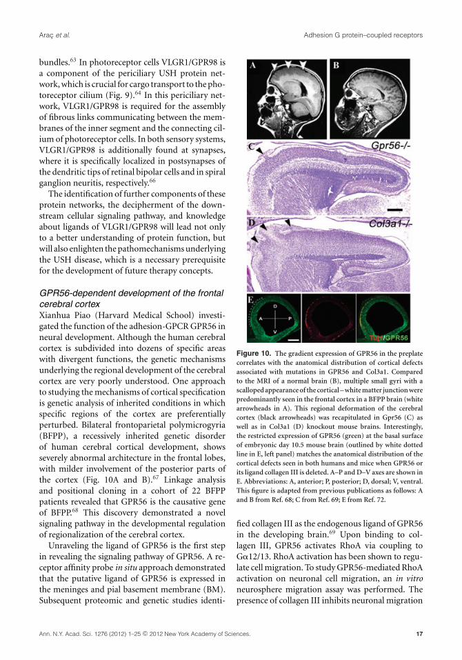
GPR56-dependent development of the frontalcerebral cortexXianhua Piao (Harvard Medical School) investi-gated the function of the adhesion-GPCR GPR56 inneural development. Although the human cerebralcortex is subdivided into dozens of specific areaswith divergent functions, the genetic mechanismsunderlying the regional development of the cerebralcortex are very poorly understood. One approachto studying the mechanisms of cortical specificationis genetic analysis of inherited conditions in whichspecific regions of the cortex are preferentiallyperturbed. Bilateral frontoparietal polymicrogyria(BFPP), a recessively inherited genetic disorderof human cerebral cortical development, showsseverely abnormal architecture in the frontal lobes,with milder involvement of the posterior parts ofthe cortex (Fig. 10A and B).67 Linkage analysisand positional cloning in a cohort of 22 BFPPpatients revealed that GPR56 is the causative geneof BFPP.68 This discovery demonstrated a novelsignaling pathway in the developmental regulationof regionalization of the cerebral cortex.
Unraveling the ligand of GPR56 is the first stepin revealing the signaling pathway of GPR56. A re-ceptor affinity probe in situ approach demonstratedthat the putative ligand of GPR56 is expressed inthe meninges and pial basement membrane (BM).Subsequent proteomic and genetic studies identi-
Figure 10. The gradient expression of GPR56 in the preplatecorrelates with the anatomical distribution of cortical defectsassociated with mutations in GPR56 and Col3a1. Comparedto the MRI of a normal brain (B), multiple small gyri with ascalloped appearance of the cortical – white matter junction werepredominantly seen in the frontal cortex in a BFPP brain (whitearrowheads in A). This regional deformation of the cerebralcortex (black arrowheads) was recapitulated in Gpr56 (C) aswell as in Col3a1 (D) knockout mouse brains. Interestingly,the restricted expression of GPR56 (green) at the basal surfaceof embryonic day 10.5 mouse brain (outlined by white dottedline in E, left panel) matches the anatomical distribution of thecortical defects seen in both humans and mice when GPR56 orits ligand collagen III is deleted. A–P and D–V axes are shown inE. Abbreviations: A, anterior; P, posterior; D, dorsal; V, ventral.This figure is adapted from previous publications as follows: Aand B from Ref. 68; C from Ref. 69; E from Ref. 72.
fied collagen III as the endogenous ligand of GPR56in the developing brain.69 Upon binding to col-lagen III, GPR56 activates RhoA via coupling toG�12/13. RhoA activation has been shown to regu-late cell migration. To study GPR56-mediated RhoAactivation on neuronal cell migration, an in vitroneurosphere migration assay was performed. Thepresence of collagen III inhibits neuronal migration
Ann. N.Y. Acad. Sci. 1276 (2012) 1–25 c© 2012 New York Academy of Sciences. 17
Adhesion G protein–coupled receptors Arac et al.
in a GPR56-dependent fashion. This observationwas further confirmed in Gpr56 and Col3a1 knock-out mouse brains (Fig. 10C and D).70,71 Taken to-gether, Piao’s data indicate that the interaction ofGPR56 and its ligand collagen III inhibits migratingneurons from breaking through the pial BM, thusconveying a positional cue during cortical develop-ment.
Because the regulation of rostral cortical develop-ment by GPR56 signaling could be accomplished byregional expression of either GPR56 or its ligand col-lagen III, Piao and coworkers studied the expressionprofile of both proteins in the developing cortex.Immunohistochemistry of collagen III on sagittalsections of mouse embryonic brains ranging in agefrom E10.5 to E11.5 did not reveal an expressiongradient of collagen III during these developmentalstages.72 In contrast, an anterior-to-posterior gra-dient of GPR56 protein expression was found onthe basal surface of the neocortex in both E10.5 andE11.5 brains (Fig. 10E), but dissipated by E12.5. Thisfinding is particularly interesting, as the change inthe expression pattern occurs in the region wherepreplate neurons reside.
During cerebral cortical development first-bornneurons form the preplate directly beneath the pialBM and function as a framework for further de-velopment of the cortex. However, the molecularmechanism underlying the function of the preplateneurons remains largely unknown. The fact that agradient expression of GPR56 in preplate neuronsmatches the regional cortical defects associated withloss of GPR56, or its ligand collagen III, (Fig. 10)suggests that a novel receptor–ligand pair is respon-sible for mediating the interaction between preplateneurons and the pial BM, thus defining the bound-ary between the neocortex and the meninges, whileproviding a framework for the developing cortex.Further testing of this hypothesis will undoubtedlyadvance our understanding of the molecular mech-anisms underlying how preplate neurons regulatecortical development.
Emerging roles of adhesion-GPCRsin disease
It becomes increasingly clear that adhesion-GPCRdysfunction is involved in several human condi-tions. Research into these pathological states notonly helps elucidate molecular breaking points ofadhesion-GPCR signals, but also assists in devel-
oping remedies and direct pharmacological effortsto counteract adhesion-GPCR–dependent diseases.At the workshop, the roles of adhesion-GPCRs intumorigenesis were discussed.
GPR56 and cancerLei Xu (University of Rochester Medical Center)presented studies from her lab on the roles ofthe adhesion-GPCR GPR56 in cancer progression.She and colleagues previously showed that GPR56is downregulated in highly metastatic melanomacells and that its re-expression led to inhibitionof metastasis and melanoma growth.73 Insights onhow this might occur came from the identifica-tion of a putative ligand of GPR56, TG2.73 TG2,also called tissue transglutaminase, is a crosslink-ing enzyme in the extracellular matrix (ECM) thatmodulates ECM biophysical properties.74 TG2 alsopossesses crosslinking-independent functions andinteracts with integrins and ECM proteins to reg-ulate cell adhesion.75 The signaling mechanisms ofGPR56, as well as whether and how TG2–GPR56 in-teraction regulates melanoma progression, are out-standing questions for which progress was then dis-cussed.
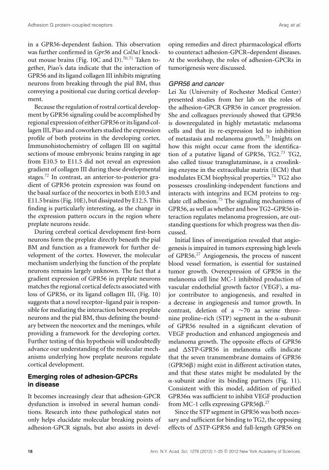
Initial lines of investigation revealed that angio-genesis is impaired in tumors expressing high levelsof GPR56.27 Angiogenesis, the process of nascentblood vessel formation, is essential for sustainedtumor growth. Overexpression of GPR56 in themelanoma cell line MC-1 inhibited production ofvascular endothelial growth factor (VEGF), a ma-jor contributor to angiogenesis, and resulted ina decrease in angiogenesis and tumor growth. Incontrast, deletion of a ∼70 aa serine threo-nine proline–rich (STP) segment in the �-subunitof GPR56 resulted in a significant elevation ofVEGF production and enhanced angiogenesis andmelanoma growth. The opposite effects of GPR56and �STP-GPR56 in melanoma cells indicatethat the seven transmembrane domains of GPR56(GPR56�) might exist in different activation states,and that these states might be modulated by the�-subunit and/or its binding partners (Fig. 11).Consistent with this model, addition of purifiedGPR56� was sufficient to inhibit VEGF productionfrom MC-1 cells expressing GPR56�.27
Since the STP segment in GPR56 was both neces-sary and sufficient for binding to TG2, the opposingeffects of �STP-GPR56 and full-length GPR56 on
18 Ann. N.Y. Acad. Sci. 1276 (2012) 1–25 c© 2012 New York Academy of Sciences.
Arac et al. Adhesion G protein–coupled receptors
Figure 11. Different activation states of GPR56.
VEGF production implied that the GPR56–TG2interaction was required for regulation of VEGFproduction by GPR56. Nevertheless, in contrast tothe effect of �STP-GPR56, knockdown of TG2 byshRNAs did not result in elevated VEGF produc-tion, indicating that the roles of the TG2–GPR56interaction in melanomas might be more complex.To investigate this further, researchers analyzedgrowth of melanoma cells expressing GPR56cDNA or shRNAs in immunodeficient Tg2–/–
mice. Preliminary data suggested an unexpectedantagonistic relationship between GPR56 and TG2in melanomas: while GPR56 inhibited melanomagrowth, TG2 promoted it (work in progress). Fur-thermore, the absence of TG2 abolished the effectsof GPR56 on melanoma growth, indicating thatTG2 might act downstream of GPR56, i.e., GPR56might inhibit the tumor-promoting role of TG2.The mechanisms of this antagonism were revealedthrough a series of immunohistochemical andbiochemical analyses. GPR56 expression was foundto induce changes in the distribution pattern of TG2in melanomas, probably due to a downregulation ofTG2 in the ECM of GPR56-expressing melanomacells. This downregulation was confirmed by in vitrostudies that showed that the extracellular TG2 wasinternalized by GPR56 and subsequently degradedintracellularly in a lysosome-dependent manner.
Aberrant adhesion-GPCR expression inbreast cancer—a potential role in metastasis?Martin Stacey (University of Leeds) reported on theconnection between adhesion-GPCRs and metas-tasis development. EGF-TM7 adhesion-GPCRs are
predominantly expressed on leukocytes, includ-ing macrophages, dendritic cells, and neutrophils.Through the use of a stimulating antibody (2A1),Stacey and colleagues have shown that ligation of thehuman-restricted EGF-TM7 receptor EMR2 resultsin the enhanced activation of human neutrophils.Data demonstrate an increase in reactive oxygenspecies generation, degranulation (myeloperoxi-dase), and surface marker expression (CD11b andl-selectin shedding) upon ligation by 2A1. Further-more, EMR2 transfectants displayed an increase inin vitro cell migration and invasion.33 Truncations ofthe transmembrane domains and mutants prevent-ing cleavage at the GPS site demonstrated the re-quirement of an intact transmembrane domain andreceptor processing to elicit cell signaling,33 showingthat signaling is indeed required for EMR2 function.Overall, the data suggest an important role in theactivation and migration of human leukocytes. In-terestingly despite its leukocyte-restricted profile ofEMR2, Stacey and colleagues show that mRNA andprotein are aberrantly present in epithelial cells ofbreast cancer tissue.76 Moreover receptor isoformexpression is similar to that seen in neutrophilsand macrophages, suggesting a potential hijackingof the normal function of EMR2 for tumor acti-vation, migration, and progression. qPCR and flowcytometric analysis of EMR2-transfected breast can-cer cell lines demonstrated increased expression ofthe epithelial-to-mesenchymal transcription factorssnail and twist and decreased expression of the ep-ithelial marker E-cadherin. Further, potential rolesof EMR2 in tumor progression are to be investi-gated. Tools for targeting of EGF-TM7 receptorshave been generated through the use of recombinantantibody fragments; for example, scFv and diabod-ies of antibodies against EMR2 and F4/80 have beencloned and fused to either toxins or model peptides.These reagents will be used in future depletion stud-ies and in the receptor-specific targeting of antigensto leukocyte subsets.
The potential role of CD97 in the biology ofacute myeloid leukemiaManja Wobus (University Hospital, Dresden) foundthat the adhesion-GPCR CD97 is involved in acutemyeloid leukemia (AML). AML cells home to aspecified region of the bone marrow (BM), wherethey interact with stromal components, includingextracellular matrix proteins, glycosaminoglycans,
Ann. N.Y. Acad. Sci. 1276 (2012) 1–25 c© 2012 New York Academy of Sciences. 19
Adhesion G protein–coupled receptors Arac et al.
and stromal cells, by which they derive proliferativeand growth inhibitory signals. Different receptors,for example, VLA- (very late antigen-) 4, CXCR4,and CD44, described to play a critical role in normalstem cell homing, also appear to be paramount tothe homing of AML cells to, or retention within, thebone marrow.77
CD97 is differentially expressed in murinehematopoietic stem and progenitor cells (HSPCs),78
but nothing is known about its expression in hu-man hematopoietic progenitor cells. Wobus hypoth-esizes that CD97 is involved in AML progressionand manifestation, potentially by interaction withits recently described ligand CD90/Thy-1, whichis expressed by nonhematopoietic cells in the BMmicroenvironment.79 They therefore initiated acomprehensive investigation of de novo AML sam-ples and correlated the CD97 expression to clinicallyimportant parameters, such as NPM1 and FLT3 mu-
tations.80 The AML cell lines MV4–11 and EOL-1,as well as CD34+ HSPCs, were used to study CD97expression and regulation in vitro.
Compared to BM blasts of healthy donors,they detected significantly higher CD97 expres-sion (mean fluorescence intensity, MFI) in 42%of AML samples. Patients with CD97 expressionabove the mean on leukemic blasts also showedincreased expression of the molecule within theresidual granulo- and monopoiesis. Of note, higherCD97 expression was accompanied by a signif-icantly higher BM blast count (75% vs. 53%,P < 0.001). Interestingly, elevated CD97 expres-sion was associated with mutations in NPM1 (46%vs. 18%, P = 0.003) and FLT3 genes (39% vs. 7%,P < 0.001), as well as lower CD34 expression (46%vs. 81%, P <0.001). Furthermore, no AML1/ETOor CBFb/MYH11 fusion genes were detectable inCD97+ AML versus 6% in CD97-AML.
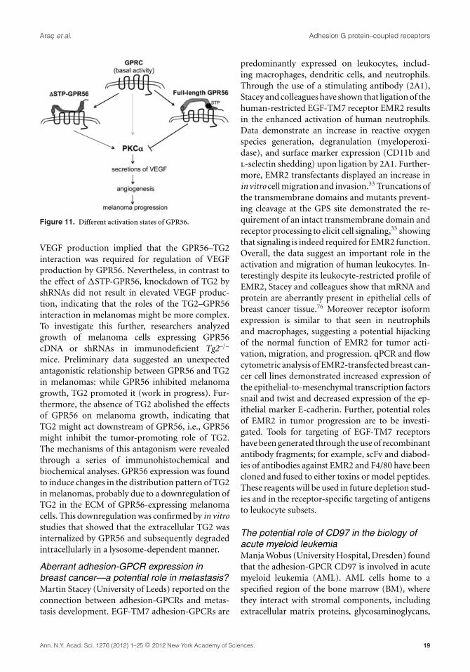
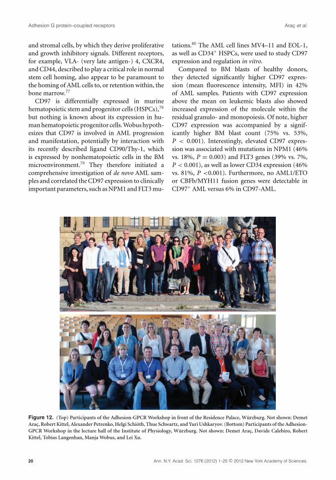
Figure 12. (Top) Participants of the Adhesion-GPCR Workshop in front of the Residence Palace, Wurzburg. Not shown: DemetArac, Robert Kittel, Alexander Petrenko, Helgi Schioth, Thue Schwartz, and Yuri Ushkaryov. (Bottom) Participants of the Adhesion-GPCR Workshop in the lecture hall of the Institute of Physiology, Wurzburg. Not shown: Demet Arac, Davide Calebiro, RobertKittel, Tobias Langenhan, Manja Wobus, and Lei Xu.
20 Ann. N.Y. Acad. Sci. 1276 (2012) 1–25 c© 2012 New York Academy of Sciences.
Arac et al. Adhesion G protein–coupled receptors
In vitro, Wobus detected lower CD97 expres-sion levels in primary CD34+ HSPC compared tothe AML cell lines. Of note, in FLT3-ITD mutatedMV4–11 cells, CD97 was expressed significantlyhigher. Treatment of this cell line with different tyro-sine kinase inhibitors resulted in a decreased CD97expression. The lower CD97 expression levels corre-lated with inhibition of the spontaneous migratorycapacity. By using a dicer-substrate 27-mer duplextargeting CD97 in MV4–11 cells, Wobus knockeddown expression to about 45%, which correlatedwith decreased transwell migration.
In summary, Wobus provides the first evidenceof higher CD97 expression in AML cells comparedto normal CD34+ hematopoietic cells in vivo and invitro, which correlates with FLT3-ITD mutation. Inongoing studies the underlying regulatory mecha-nisms will be investigated. The possible impact ofCD97 as well as other molecules of that receptorfamily on AML biology and clinical outcome will beevaluated in a larger patient cohort.
Conclusions
The 6th International Adhesion-GPCR Workshop(photographs of workshop participants shown inFig. 12) has yielded intricate details regarding themolecular faculty of these peculiar membrane pro-teins. The collective efforts of all the labs now allowus for the first time to speculate on the events fromstimulus reception by an adhesion-GPCR via trans-duction to signal generation inside the cell, whichis a remarkable milestone in the history of researchon adhesion-GPCRs. The biological setting, the un-usual important signaling route, became definedmore clearly, allowing novel glimpses of adhesion-GPCR function and dysfunction. It is clear that in-terdisciplinarity and open scientific exchange is animportant prerequisite and driving force to achiev-ing this current level of understanding.
Acknowledgments
All participants of the 6th Adhesion-GPCR Work-shop are grateful to Nicole Hartmann and JenniferGehring for their excellent organizational skills, andto the Interdisciplinary Center for Clinical Research(IZKF) of the University of Wurzburg for its gen-erous contribution toward the accommodation ofworkshop guests. We also acknowledge all colleaguesthat presented their data on adhesion-GPCRs dur-ing the poster session.
Conflicts of interest
The authors declare no conflicts of interest.
References
1. Yona, S., H.-H. Lin, W.O. Siu, et al. 2008. Adhesion-GPCRs:emerging roles for novel receptors. Trends Biochem. Sci. 33:491–500.
2. Kwakkenbos, M.J., E.N. Kop, M. Stacey, et al. 2004. TheEGF-TM7 family: a postgenomic view. Immunogenetics 55:655–666.
3. Nordstrom, K.J.V., M. Sallman Almen, M.M. Edstam, et al.2011. Independent HHsearch, Needleman–Wunsch-based,and motif analyses reveal the overall hierarchy for most ofthe G protein-coupled receptor families. Mol. Biol. Evol. 28:2471–2480.
4. Nordstrom, K.J.V., R. Fredriksson & H.B. Schioth. 2008.The amphioxus (Branchiostoma floridae) genome containsa highly diversified set of G protein-coupled receptors. BMCEvol. Biol. 8: 9.
5. Kamesh, N., G.K. Aradhyam & N. Manoj. 2008. The reper-toire of G protein-coupled receptors in the sea squirt Cionaintestinalis. BMC Evol. Biol. 8: 129.
6. Whittaker, C.A., K.-F. Bergeron, J. Whittle, et al. 2006. Theechinoderm adhesome. Dev. Biol. 300: 252–266.
7. Nordstrom, K.J.V., M.C. Lagerstrom, L.M.J. Waller, et al.2009. The secretin GPCRs descended from the family of ad-hesion GPCRs. Mol. Biol. Evol. 26: 71–84.
8. Krishnan, A., M.S. Almen, R. Fredriksson & H.B. Schioth.2012. The origin of GPCRs: identification of mammalianlike rhodopsin, adhesion, glutamate and frizzled GPCRs infungi. PLoS ONE 7: e29817.
9. Krasnoperov, V., M.A. Bittner, R.W. Holz, et al. 1999.Structural requirements for alpha-latrotoxin binding andalpha-latrotoxin-stimulated secretion. A study with calcium-independent receptor of alpha-latrotoxin (CIRL) deletionmutants. J. Biol. Chem. 274: 3590–3596.
10. Lin, H.-H., G.-W. Chang, J.Q. Davies, et al. 2004. Autocat-alytic cleavage of the EMR2 receptor occurs at a conserved Gprotein-coupled receptor proteolytic site motif. J. Biol. Chem.279: 31823–31832.
11. Volynski, K.E., J.-P. Silva, V.G. Lelianova, et al. 2004. Lat-rophilin fragments behave as independent proteins that as-sociate and signal on binding of LTX(N4C). EMBO J. 23:4423–4433.
12. Arac, D., A.A. Boucard, M.F. Bolliger, et al. 2012. A novel evo-lutionarily conserved domain of cell-adhesion GPCRs medi-ates autoproteolysis. EMBO J. 31: 1364–1378.
13. Paavola, K.J., J.R. Stephenson, S.L. Ritter, et al. 2011. The Nterminus of the adhesion G protein-coupled receptor GPR56controls receptor signaling activity. J. Biol. Chem. 286: 28914–28921.
14. Krasnoperov, V.G., R. Beavis, O.G. Chepurny, et al. 1996.The calcium-independent receptor of alpha-latrotoxin is nota neurexin. Biochem. Biophys. Res. Commun. 227: 868–875.
15. Krasnoperov, V.G., M.A. Bittner, R. Beavis, et al. 1997.alpha-Latrotoxin stimulates exocytosis by the interactionwith a neuronal G-protein-coupled receptor. Neuron 18: 925–937.
Ann. N.Y. Acad. Sci. 1276 (2012) 1–25 c© 2012 New York Academy of Sciences. 21
Adhesion G protein–coupled receptors Arac et al.
16. Langenhan, T., S. Promel, L. Mestek, et al. 2009. Latrophilinsignaling links anterior-posterior tissue polarity and orientedcell divisions in the C. elegans embryo. Dev. Cell 17: 494–504.
17. Promel, S., M. Frickenhaus, S. Hughes, et al. 2012. The GPSmotif is a molecular switch for bimodal activities of adhe-sion class G protein-coupled receptors. Cell Reports 2: 321–331.
18. Okajima, D., G. Kudo & H. Yokota. 2010. Brain-specific an-giogenesis inhibitor 2 (BAI2) may be activated by prote-olytic processing. J. Recept. Signal Transduct. Res. 30: 143–153.
19. Silva, J.-P., V. Lelianova, C. Hopkins, et al. 2009. Functionalcross-interaction of the fragments produced by the cleav-age of distinct adhesion G-protein-coupled receptors. J. Biol.Chem. 284: 6495–6506.
20. Iguchi, T., K. Sakata, K. Yoshizaki, et al. 2008. Orphan Gprotein-coupled receptor GPR56 regulates neural progenitorcell migration via a G alpha 12/13 and Rho pathway. J. Biol.Chem. 283: 14469–14478.
21. Park, D., A.-C. Tosello-Trampont, M.R. Elliott, et al. 2007.BAI1 is an engulfment receptor for apoptotic cells upstreamof the ELMO/Dock180/Rac module. Nature 450: 430–434.
22. Bohnekamp, J. & T. Schoneberg. 2011. Cell adhesion receptorGPR133 couples to Gs protein. J. Biol. Chem. 286: 41912–41916.
23. Gupte, J., G. Swaminath, J. Danao, et al. 2012. Signaling prop-erty study of adhesion G-protein-coupled receptors. FEBSLett. 586: 1214–1219.
24. Engelstoft, M.S., K.L. Egerod, B. Holst & T.W. Schwartz. 2008.A gut feeling for obesity: 7TM sensors on enteroendocrinecells. Cell Metab. 8: 447–449.
25. Hansen, H.S., M.M. Rosenkilde, J.J. Holst & T.W. Schwartz.2012. GPR119 as a fat sensor. Trends Pharmacol. Sci. 33: 374–381.
26. Egerod, K.L., M.S. Engelstoft, K.V. Grunddal, et al. A ma-jor lineage of enteroendocrine cells co-express CCK, GLP-1,GIP, PYY, neurotensin, and secretin but not somatostatin.Endocrinology. In press.
27. Yang, L., G. Chen, S. Mohanty, et al. 2011. GPR56 regulatesVEGF production and angiogenesis during melanoma pro-gression. Cancer Res. 71: 5558–5568.
28. Hamann, J., B. Vogel, G.M. van Schijndel & R.A. van Lier.1996. The seven-span transmembrane receptor CD97 hasa cellular ligand (CD55, DAF). J. Exp. Med. 184: 1185–1189.
29. Veninga, H., R.M. Hoek, A.F. de Vos, et al. 2011. A novel rolefor CD55 in granulocyte homeostasis and anti-bacterial hostdefense. PLoS ONE 6: e24431.
30. Hoek, R.M., D. de Launay, E.N. Kop, et al. 2010. Deletion ofeither CD55 or CD97 ameliorates arthritis in mouse models.Arthritis. Rheum. 62: 1036–1042.
31. Ward, Y., R. Lake, J.J. Yin, et al. 2011. LPA receptor het-erodimerizes with CD97 to amplify LPA-initiated RHO-dependent signaling and invasion in prostate cancer cells.Cancer Res. 71: 7301–7311.
32. Chen, T.-Y., T.-L. Hwang, C.-Y. Lin, et al. 2011. EMR2 recep-tor ligation modulates cytokine secretion profiles and cell sur-vival of lipopolysaccharide-treated neutrophils. Chang GungMed. J. 34: 468–477.
33. Yona, S., H.-H. Lin, P. Dri, et al. 2008. Ligation of theadhesion-GPCR EMR2 regulates human neutrophil func-tion. FASEB J. 22: 741–751.
34. Lin, H.-H., M. Stacey, S. Yona & G.-W. Chang. 2010. GPSproteolytic cleavage of adhesion-GPCRs. Adv. Exp. Med. Biol.706: 49–58.
35. Huang, Y.-S., N.-Y. Chiang, C.-H. Hu, et al. Lin. 2012. Activa-tion of myeloid cell-specific adhesion class G protein-coupledreceptor EMR2 via ligation-induced translocation and inter-action of receptor subunits in lipid raft microdomains. Mol.Cell Biol. 32: 1408–1420.
36. Lohse, M.J., V.O. Nikolaev, P. Hein, et al. 2008. Optical tech-niques to analyze real-time activation and signaling of G-protein-coupled receptors. Trends Pharmacol. Sci. 29: 159–165.
37. Calebiro, D., V.O. Nikolaev, M.C. Gagliani, et al. 2009. Per-sistent cAMP-signals triggered by internalized G-protein-coupled receptors. PLoS Biol. 7: e1000172.
38. Nikolaev, V.O., M. Bunemann, L. Hein, et al. 2004. Novelsingle chain cAMP sensors for receptor-induced signal prop-agation. J. Biol. Chem. 279: 37215–37218.
39. Calebiro, D., V.O. Nikolaev, L. Persani & M.J. Lohse.2010. Signaling by internalized G-protein-coupled receptors.Trends Pharmacol. Sci. 31: 221–228.
40. Monk, K.R., S.G. Naylor, T.D. Glenn, et al. 2009. A G protein-coupled receptor is essential for Schwann cells to initiatemyelination. Science 325: 1402–1405.
41. Monk, K.R., K. Oshima, S. Jors, et al. 2011. Gpr126 is es-sential for peripheral nerve development and myelination inmammals. Development 138: 2673–2680.
42. Waller-Evans, H., S. Promel, T. Langenhan, et al. 2010. Theorphan adhesion-GPCR GPR126 is required for embryonicdevelopment in the mouse. PLoS ONE 5: e14047.
43. Stehlik, C., R. Kroismayr, A. Dorfleutner, et al. 2004. VIGR—a novel inducible adhesion family G-protein coupled receptorin endothelial cells. FEBS Lett. 569: 149–155.
44. Moriguchi, T., K. Haraguchi, N. Ueda, et al. 2004. DREG, adevelopmentally regulated G protein-coupled receptor con-taining two conserved proteolytic cleavage sites. Genes Cells9: 549–560.
45. Tissir, F., I. Bar, Y. Jossin, et al. 2005. Protocadherin Celsr3is crucial in axonal tract development. Nat. Neurosci. 8: 451–457.
46. Tissir, F., Y. Qu, M. Montcouquiol, et al. 2010. Lack of cad-herins Celsr2 and Celsr3 impairs ependymal ciliogenesis,leading to fatal hydrocephalus. Nat. Neurosci. 13: 700–707.
47. Qu, Y., D.M. Glasco, L. Zhou, et al. 2010. Atypical cad-herins Celsr1–3 differentially regulate migration of facialbranchiomotor neurons in mice. J. Neurosci. 30: 9392–9401.
48. Song, H., J. Hu, W. Chen, et al. 2010. Planar cell polaritybreaks bilateral symmetry by controlling ciliary positioning.Nature 466: 378–382
49. Curtin, J.A., E. Quint, V. Tsipouri, et al. 2003. Mutation ofCelsr1 disrupts planar polarity of inner ear hair cells andcauses severe neural tube defects in the mouse. Curr. Biol. 13:1129–1133.
50. Yates, L.L., C. Schnatwinkel, J.N. Murdoch, et al. 2010.The PCP genes Celsr1 and Vangl2 are required for normallung branching morphogenesis. Hum Mol Genet 19: 2251–2267.
22 Ann. N.Y. Acad. Sci. 1276 (2012) 1–25 c© 2012 New York Academy of Sciences.
Arac et al. Adhesion G protein–coupled receptors
51. Devenport, D., D. Oristian, E. Heller & E. Fuchs. 2011. Mitoticinternalization of planar cell polarity proteins preserves tissuepolarity. Nat. Cell Biol. 13: 893–902.
52. Formstone, C.J., C. Moxon, J. Murdoch, et al. 2010. Basalenrichment within neuroepithelia suggests novel function(s)for Celsr1 protein. Mol. Cell. Neurosci. 44: 210–222.
53. Yates, L.L., C. Schnatwinkel, L. Hazelwood, et al. Scribble isrequired for normal epithelial cell-cell contacts and lumenmorphogenesis in the mammalian lung. Dev Biol. In press.
54. Becker, S., E. Wandel, M. Wobus, et al. 2010. Overexpressionof CD97 in intestinal epithelial cells of transgenic mice atten-uates colitis by strengthening adherens junctions. PLoS ONE5: e8507.
55. Cummins, A.G. & F.M. Thompson. 2002. Effect of breastmilk and weaning on epithelial growth of the small intestinein humans. Gut 51: 748–754.
56. Tobaben, S., T.C. Sudhof & B. Stahl. 2002. Genetic analysisof alpha-latrotoxin receptors reveals functional interdepen-dence of CIRL/latrophilin 1 and neurexin 1 alpha. J. Biol.Chem. 277: 6359–6365.
57. Davletov, B.A., O.G. Shamotienko, V.G. Lelianova, et al.1996. Isolation and biochemical characterization of a Ca2+-independent alpha-latrotoxin-binding protein. J. Biol. Chem.271: 23239–23245.
58. Davletov, B.A., F.A. Meunier, A.C. Ashton, et al. 1998. Vesicleexocytosis stimulated by alpha-latrotoxin is mediated by la-trophilin and requires both external and stored Ca2. EMBOJ. 17: 3909–3920.
59. Silva, J.-P. & Y.A. Ushkaryov. 2010. The latrophilins, “split-personality” receptors. Adv. Exp. Med. Biol. 706: 59–75.
60. Silva, J.-P., V.G. Lelianova, Y.S. Ermolyuk, et al. 2011. Lat-rophilin 1 and its endogenous ligand Lasso/teneurin-2 forma high-affinity transsynaptic receptor pair with signaling ca-pabilities. Proc. Natl. Acad. Sci. USA 108: 12113–12118.
61. Silva, J.P., N. Vysokov, G.V. Lelianova, et al. Interaction be-tween latrophilin-1 and Lasso is required for functional mat-uration of synapses. Manuscript in preparation.
62. McMillan, D.R. & P.C. White. 2010. Studies on the very largeG protein-coupled receptor: from initial discovery to deter-mining its role in sensorineural deafness in higher animals.Adv. Exp. Med. Biol. 706: 76–86.
63. McGee, J., R.J. Goodyear, D.R. McMillan, et al. 2006. The verylarge G-protein-coupled receptor VLGR1: a component ofthe ankle link complex required for the normal developmentof auditory hair bundles. J. Neurosci. 26: 6543–6553.
64. Maerker, T., E. van Wijk, N. Overlack, et al. 2008. A novelUsher protein network at the periciliary reloading point be-tween molecular transport machineries in vertebrate pho-toreceptor cells. Hum. Mol. Genet. 17: 71–86.
65. Wolfrum, U. 2011. Protein networks related to the Ushersyndrome gain insights in the molecular basis of the disease.In Usher Syndrome: Pathogenesis, Diagnosis and Therapy. S.A.Ed.: 51–73. Nova Science Publishers, Inc. Hauppauge, NY.
66. Reiners, J., E. van Wijk, T. Marker, et al. 2005. Scaffold proteinharmonin (USH1C) provides molecular links between Ushersyndrome type 1 and type 2. Hum. Mol. Genet. 14: 3933–3943.
67. Piao, X., B.S. Chang, A. Bodell, et al. 2005. Genotype-phenotype analysis of human frontoparietal polymicrogyriasyndromes. Ann. Neurol. 58: 680–687.
68. Piao, X., R.S. Hill, A. Bodell, et al. 2004. G protein-coupledreceptor-dependent development of human frontal cortex.Science 303: 2033–2036.
69. Luo, R., S.-J. Jeong, Z. Jin, et al. 2011. G protein-coupledreceptor 56 and collagen III, a receptor-ligand pair, regulatescortical development and lamination. Proc. Natl. Acad. Sci.USA 108: 12925–12930.
70. Li, S., Z. Jin, S. Koirala, et al. 2008. GPR56 regulates pialbasement membrane integrity and cortical lamination. J.Neurosci. 28: 5817–5826.
71. Jeong, S.-J., S. Li, R. Luo, et al. 2012. Loss of Col3a1, the genefor Ehlers-Danlos syndrome type IV, results in neocorticaldyslamination. PLoS ONE 7: e29767.
72. Jeong, S.-J., R. Luo, S. Li, et al. 2012. Characterization of Gprotein-coupled receptor 56 protein expression in the mousedeveloping neocortex. J. Comp. Neurol. 520: 2930–2940.
73. Xu, L., S. Begum, J.D. Hearn & R.O. Hynes. 2006. GPR56,an atypical G protein-coupled receptor, binds tissue transg-lutaminase, TG2, and inhibits melanoma tumor growth andmetastasis. Proc. Natl. Acad. Sci. USA 103: 9023–9028.
74. Lorand, L. & R.M. Graham. 2003. Transglutaminases:crosslinking enzymes with pleiotropic functions. Nat. Rev.Mol. Cell Biol. 4: 140–156.
75. Belkin, A.M. 2011. Extracellular TG2: emerging functionsand regulation. FEBS J. 278: 4704–4716.
76. Davies, J.Q., H.-H. Lin, M. Stacey, et al. 2011. Leukocyteadhesion-GPCR EMR2 is aberrantly expressed in humanbreast carcinomas and is associated with patient survival.Oncol. Rep. 25: 619–627.
77. Becker, P.S. 2012. Dependence of acute myeloid leukemia onadhesion within the bone marrow microenvironment. TheScientific World Journal 2012: 856467.
78. van Pel, M., H. Hagoort, J. Hamann & W.E. Fibbe. 2008.CD97 is differentially expressed on murine hematopoieticstem-and progenitor-cells. Haematologica 93: 1137–1144.
79. Wandel, E., A. Saalbach, D. Sittig, et al. 2012. Thy-1 (CD90)is an interacting partner for CD97 on activated endothelialcells. J. Immunol. 188: 1442–1450.
80. Kiyoi, H. & T. Naoe. 2006. Biology, clinical relevance, andmolecularly targeted therapy in acute leukemia with FLT3mutation. Int. J. Hematol. 83: 301–308.
Appendix
6th International Adhesion-GPCRWorkshop
6–8 September 2012 – Wurzburg – Germany
Scientific program
Thursday, Sep 6 2012
9:00 – 9:10Opening remarksTobias Langenhan, University of Wurzburg
Ann. N.Y. Acad. Sci. 1276 (2012) 1–25 c© 2012 New York Academy of Sciences. 23
Adhesion G protein–coupled receptors Arac et al.
Session A – Structural hallmarks ofAdhesion-GPCR
9:10 – 9:35The GPS motif: 15 years of studiesAlexander Petrenko, Russian Academy of Sciences,Moscow
9:35 – 10:00A Novel Evolutionarily Conserved Domain ofCell-Adhesion GPCRs Mediates AutoproteolysisDemet Arac-Ozkan, Stanford University
10:00 – 10:25Structural insights into the adhesion-GPCRCD97Martin Stacey, University of Leeds
10:25 – 10:45Coffee break
Session B – Neurobiological roles ofAdhesion-GPCR
10:45 – 11:10Latrophilin receptors regulate presynaptic trans-mitter releaseTobias Langenhan, University of Wurzburg
11:10 – 11:35High-affinity functional trans-synaptic receptorpairs between presynaptic latrophilin and post-synaptic Lasso (teneurin-2)Yuri Ushkaryov, University of Kent
11:35 – 12:00GPR56-dependent development of the frontalcerebral cortexXianhua Piao, Harvard Medical School
12:00 – 14:00Lunch break
Session C – Neurobiological roles ofAdhesion-GPCR (continued)
14:00 – 14:25GPR56, together with �3�1 Integrin, RegulatesCortical LaminationKathleen Singer, Harvard Medical School
14:25 – 14:50Role of Celsr1–3 cadherins in planar cell polarityand brain developmentAndre Goffinet, University of Louvain
14:50 – 15:15The very large G protein coupled receptorVlgr1b/GPR98 as a key component of the
Usher syndrome protein networks in the innerear and the retinaUwe Wolfrum, University of Mainz
15:15 – 15:40Molecular and genetic analysis of Gpr126 in pe-ripheral nerve developmentKelly Monk, Washington University School ofMedicine, St. Louis
15:40 – 16:30Coffee break
19:00Evening program
Friday, Sep 7 2012
Session D – Adhesion-GPCR indevelopment
9:00 – 9:25Basal enrichment of Celsr1 protein within epithe-lia: novel function or apico-basal dependent pla-nar cell polarity (PCP) signalling?Caroline Formstone, King’s College London
9:25 – 9:50Knockdown of the orphan G protein-coupled receptor 126 influences ventricularmorphogenesis and heart function in zebrafishand miceFelix Engel, Max-Planck-Institute Bad Nauheim
9:50 – 10:15Mice constitutively overexpressing CD97 in ente-rocytes develop a megaintestine without alter-ations in histology and cell fate decisionGabriela Aust, University of Leipzig
10:15 – 10:45Coffee break
Session E – Adhesion-GPCR in tumorbiology
10:45 – 11:10Roles of GPR56 and TG2 during MelanomaProgressionLei Xu, Rochester School of Medicine
11:10 – 11:35Molecular characterization of the interaction ofGPR56 and a novel ligandHsi-Hsien Lin, Chang Gung University
11:35 – 12:00The expression of the EGF-TM7 receptor CD97is higher in CD34-negative and NPM1/FLT3-ITD mutated AMLManja Wobus, University of Dresden
24 Ann. N.Y. Acad. Sci. 1276 (2012) 1–25 c© 2012 New York Academy of Sciences.
Arac et al. Adhesion G protein–coupled receptors
12:00 – 14:00Lunch break
Session F – Mechanisms of signaltransduction of Adhesion-GPCR
14:00 – 14:25Shear stress-dependent downregulation of theadhesion-GPCR CD97 on circulating leukocytes
upon contact with its ligand CD55Jorg Hamann, University of Amsterdam
14:25 – 14:50Insights into the molecular function of lat-rophilins – logic of adhesion-GPCR signallingSimone Promel, University of Oxford & Universityof Leipzig
14:50 – 15:15G protein-mediated signal transduction of adhe-sion GPCRInes Liebscher, University of Leipzig
15:15 – 15:40Real-time monitoring of GPCR signaling in livingcells: from intracellular signaling microdomainsto single moleculesDavide Calebiro, University of Wurzburg
15:40 – 16:30Coffee break
16:30 – 17:30General Meeting of the Adhesion-GPCR Consor-tium
19:00Evening program
Saturday, Sep 8 2012
Session G – Adhesion-GPCR in endocrine,cardiovascular, and immune functions
9:00 – 9:25The origin of the Adhesion-GPCR familyHelgi Schioth, University of Uppsala
9:25 – 9:50The ADHD-susceptibility gene lphn3.1 modu-lates dopaminergic neuron formation andlocomotor activity during zebrafish developmentKlaus-Peter Lesch, University of Wurzburg
9:50 – 10:15Adhesion 7TM receptors – major players in theendocrine and enteroendocrine systemThue Schwartz, University of Copenhagen
10:15 – 10:45Coffee break
10:45 – 11:45Future initiatives of the Adhesion-GPCR com-munity
11:45 – 12:00Concluding remarksJorg Hamann, University of Amsterdam & TobiasLangenhan, University of Wurzburg
Ann. N.Y. Acad. Sci. 1276 (2012) 1–25 c© 2012 New York Academy of Sciences. 25