dissecting functions of katanin and wrinkled1 in cotton fiber development by virus-induced ·...
TRANSCRIPT
Dissecting Functions of KATANIN and WRINKLED1in Cotton Fiber Development by Virus-InducedGene Silencing1[C][W][OA]
Jing Qu2, Jian Ye2, Yun-Feng Geng, Yan-Wei Sun, Shi-Qiang Gao,Bi-Pei Zhang, Wen Chen, and Nam-Hai Chua*
Temasek Life Sciences Laboratory, National University of Singapore, 117604 Singapore (J.Q., J.Y., Y.-F.G.,Y.-W.S., S.-Q.G., B.-P.Z., W.C.); and Laboratory of Plant Molecular Biology, Rockefeller University,New York, New York 10021 (N.-H.C.)
Most of the world’s natural fiber comes from cotton (Gossypium spp.), which is an important crop worldwide. Characterizinggenes that regulate cotton yield and fiber quality is expected to benefit the sustainable production of natural fiber. Although ahuge number of expressed sequence tag sequences are now available in the public database, large-scale gene function analysishas been hampered by the low-efficiency process of generating transgenic cotton plants. Tobacco rattle virus (TRV) has recentlybeen reported to trigger virus-induced gene silencing (VIGS) in cotton leaves. Here, we extended the utility of this method byshowing that TRV-VIGS can operate in reproductive organs as well. We used this method to investigate the function ofKATANIN and WRINKLED1 in cotton plant development. Cotton plants with suppressed KATANIN expression producedshorter fibers and elevated weight ratio of seed oil to endosperm. By contrast, silencing of WRINKLED1 expression resultedin increased fiber length but reduced oil seed content, suggesting the possibility to increase fiber length by repartitioning carbonflow. Our results provide evidence that the TRV-VIGS system can be used for rapid functional analysis of genes involved incotton fiber development.
Cotton (Gossypium spp.) is the most important fiber-producing plant in the world and also a significant oil-seed crop. This crop is grown in more than 80 countrieswith a worldwide production of 123 million bales (480pounds per bale) during the 2011/2012 growing season(United States Department of Agriculture, 2012).
As the most important agronomic traits of cotton arefiber quality and yield, it is important to improve ourunderstanding of genes that directly or indirectly im-pact such traits. To this end, a public effort was initi-ated in 2007 to determine the complete cotton genomicsequence. While this effort is underway, there is anever-expanding set of cotton EST sequences (about400,000 now) being deposited in the public database.Notwithstanding the availability of such a huge amountof cotton gene sequences so far, only a relatively smallnumber of genes have been shown to directly affect cot-ton fiber development. Most of these genes are involved
either in cytoskeletal dynamics or in carbohydrate bi-osynthesis. In the case of cytoskeletal genes, the cottonactin gene has been shown to be important for fiberelongation but not fiber initiation (Li et al., 2005).Overexpression of a fiber-specific profilin (GhPFN2),an actin-binding protein, blocked cell elongation pre-maturely (Wang et al., 2010), whereas down-regulationof the actin-depolymerizing factor gene increased fiberlength and fiber strength (Wang et al., 2009). Sincecellulose is the major constituent of cotton fiber, it is notsurprising that several carbohydrate biosynthetic geneshave been shown to modulate fiber development.Transgenic overexpression of a SUCROSE SYNTHASEgene, a SUCROSE PHOSPHATE SYNTHASE gene, andcellulose synthesis genes improved cotton fiber lengthand strength (Li et al., 2004; Haigler et al., 2007; Jiang et al.,2012). Similarly, higher xyloglucan endotransglycosylase/hydrolase (XTH) activity can promote fiber cell elon-gation, and transgenic cotton overexpressing the xthgene has been shown to increase mature fiber length(Lee et al., 2010).
The major impediment to analyzing cotton genefunction on a large scale is the laborious and time-consuming process of generating transgenic cotton.Moreover, many cotton cultivars and Gossypium speciesthat contain important genes for cotton improvementare recalcitrant to genetic transformation. Therefore,there is an urgent need to develop a rapid method forfunctional analysis of cotton genes on a genomic scale.Virus-induced gene silencing (VIGS) offers such a pos-sibility because it allows the investigation of gene func-tions without plant transformation (Ruiz et al., 1998;
1 This work was supported by the Temasek Life Sciences Labora-tory and the Singapore Millennium Foundation (to N.-H.C.).
2 These authors contributed equally to the article.* Corresponding author; e-mail [email protected] author responsible for distribution of materials integral to the
findings presented in this article in accordance with the policy de-scribed in the Instructions for Authors (www.plantphysiol.org) is:Nam-Hai Chua ([email protected]).
[C] Some figures in this article are displayed in color online but inblack and white in the print edition.
[W] The online version of this article contains Web-only data.[OA] Open Access articles can be viewed online without a subscrip-
tion.www.plantphysiol.org/cgi/doi/10.1104/pp.112.198564
738 Plant Physiology�, October 2012, Vol. 160, pp. 738–748, www.plantphysiol.org � 2012 American Society of Plant Biologists. All Rights Reserved. www.plantphysiol.orgon June 3, 2018 - Published by Downloaded from
Copyright © 2012 American Society of Plant Biologists. All rights reserved.
Burch-Smith et al., 2004). A partial fragment of a candi-date gene is inserted into a virus vector to generate arecombinant virus. Inoculation with the recombinantvirus leads to the production of virus-related small in-terfering RNAs (Baulcombe, 2004) in infected plants. Thesmall interfering RNAs generated can mediate thedegradation of related endogenous gene transcriptsresulting in the silencing of candidate gene expression(Brigneti et al., 2004; Burch-Smith et al., 2004). The si-lencing effect on endogenous gene expression can usu-ally be assayed 1 to 2 weeks after virus inoculation.VIGS has become one of the most widely used and in-deed important reverse genetics tools, especially fornonmodel plants, such as tomato (Solanum lycopersicum;Liu et al., 2002), barley (Hordeum vulgare; Holzberg et al.,2002), soybean (Glycine max; Zhang and Ghabrial, 2006),Medicago truncatula (Grønlund et al., 2008), poppy (Pa-paver somniferum; Hileman et al., 2005), and the shrubtree jatropha (Jatropha curcas; Ye et al., 2009a). Usingdifferent infection and treatment protocols, VIGS hasbeen shown to be effective in leaves (Liu et al., 2002),roots (Valentine et al., 2004), flowers (Liu et al., 2004),and even fruits (Fu et al., 2005).Recently, two different VIGS systems have been shown
to be effective in cotton. Using the geminivirus Cotton leafcrumple virus (CLCrV) as a vector, Tuttle et al. (2008)silenced two visible marker genes encoding magnesiumchelatase subunit I and phytoene desaturase with anefficiency of 70%. Gao et al. (2011a, 2011b), on the otherhand, used the Tobacco rattle virus (TRV) as a vector tosilence another marker gene, chloroplastos alterados1, withan efficiency of 100%. The TRV-VIGS system appears tobe better than the CLCrV-VIGS system because the for-mer has a higher silencing efficiency and a more homog-enous silencing phenotype with a milder virus symptom.Although both VIGS systems have been shown towork in cotton leaves, it was not known whetherthese methods can be used to assess gene function inreproductive tissues and particularly in developingcotton fibers. Nevertheless, using a GFP-expressingCLCrV, Tuttle et al. (2008) observed occasional GFPfluorescence in outer boll walls, the central column ofthe ovary, and ovule integuments.Here, we used two anthocyanin biosynthetic genes,
ANTHOCYANIDIN SYNTHASE (ANS) and ANTHO-CYANIDIN REDUCTASE (ANR), as reporters to opti-mize the TRV-mediated VIGS system in cotton plants,and we showed that it can be used to silence gene ex-pression in reproductive tissues and developing fibers.We used this method to characterize the functions of twogenes, KATANIN (KTN) and WRINKLED1 (WRI1), incotton fiber development.
RESULTS
Synthetic TRV-Induced Silencing of ProanthocyanidinBiosynthetic Genes ANS and ANR
We first chemically synthesized two TRV genomes,TRV1 and TRV2 (Ye et al., 2009b, 2010) to construct a
synthetic TRV (sTRV) VIGS system. Our method foragroinfection of cotton plants was different from that ofGao et al. (2011a, 2011b), who used a syringe to infiltrateAgrobacterium tumefaciens culture into two fully expandedcotyledons. By contrast, we used cotton plants with twoto four true leaves, vacuum infiltrated the leaves with A.tumefaciens culture, and subsequently drenched the rootsystem using the same culture.
We used two proanthocyanidin (PA) biosyntheticgenes as reporter genes to test whether the sTRV-induced silencing was effective throughout the wholecotton plant, especially in cotton fibers. PAs are a majorclass of flavonoids, one of the largest groups of plantsecondary metabolites. Most genetic studies on the PAbiosynthesis pathway were performed in Arabidopsis(Arabidopsis thaliana) and M. truncatula (Xie et al., 2003;Pang et al., 2007). Two enzymes, ANS and ANR,function at branches between anthocyanin and PA bio-synthesis. ANS converts the substrate flavan-3,4-diol(leucoanthocyanidin) to anthocyanidin, which serves asa substrate for ANR to produce a major PA unit, 2,3-cis-flavan-3-ol (epicatechin), in Arabidopsis and M. trunca-tula (Xie et al., 2003; Pang et al., 2007). As far as weknow, cotton genes involved in the PA biosynthesispathway have not been well characterized. Here, wetook advantage of the sTRV-VIGS system in cotton toconfirm the function of two genes in the PA biosyn-thetic pathway.
Two cotton gene sequences with homology to Arab-idopsis genes encoding ANS and ANR were used forfunctional analysis. The partial fragments of putativecotton ANS and ANR were cloned into the psTRVvector to give psTRV:ANS and psTRV:ANR, respec-tively. Plants infiltrated with psTRV empty vector(psTRV1+psTRV2) were used as control plants in thisstudy. All plants infiltrated with empty vector (hence-forth referred to as vector control plants) grew normallyand did not display any abnormal phenotype on veg-etative and reproductive organs (data not shown). Nophenotypic differences were observed between unin-fected plants and vector control plants. This is one of theimportant advantages of sTRV-mediated gene silenc-ing. At 7 to 10 d post inoculation, leaf veins and leafmargins around new systemic leaves began to appearbrownish in all of the psTRV:ANR-treated plants. Withtime, the brownish phenotype became visible on leavesand leaf petioles (Fig. 1A) and stems (Fig. 1B). Thebrownish phenotype was also seen in more than 90%of floral buds in psTRV:ANR-treated plants (Fig. 1D).We surmised that this brownish phenotype was dueto the anthocyanidin accumulation resulting from theblockage of ANR function. By contrast, all of the or-gans of psTRV:ANS plants (Fig. 1, A–E) showed noobvious visible differences from vector control plants.Nevertheless, the difference between psTRV:ANS andcontrol plants can be uncovered by the use of thearomatic aldehyde agent p-dimethylaminocinna-maldehyde (DMACA), which stains PA (Fig. 1, A–E).After reaction with catechins, the major flavan-3-olsassociated with cotton PAs, DMACA turned deep blue.
Plant Physiol. Vol. 160, 2012 739
Virus-Induced Gene Silencing in Cotton Fiber
www.plantphysiol.orgon June 3, 2018 - Published by Downloaded from Copyright © 2012 American Society of Plant Biologists. All rights reserved.
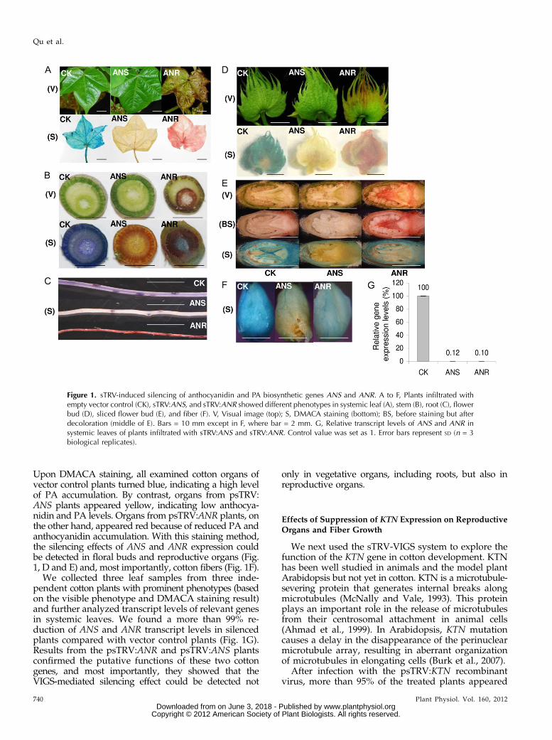
Upon DMACA staining, all examined cotton organs ofvector control plants turned blue, indicating a high levelof PA accumulation. By contrast, organs from psTRV:ANS plants appeared yellow, indicating low anthocya-nidin and PA levels. Organs from psTRV:ANR plants, onthe other hand, appeared red because of reduced PA andanthocyanidin accumulation. With this staining method,the silencing effects of ANS and ANR expression couldbe detected in floral buds and reproductive organs (Fig.1, D and E) and, most importantly, cotton fibers (Fig. 1F).
We collected three leaf samples from three inde-pendent cotton plants with prominent phenotypes (basedon the visible phenotype and DMACA staining result)and further analyzed transcript levels of relevant genesin systemic leaves. We found a more than 99% re-duction of ANS and ANR transcript levels in silencedplants compared with vector control plants (Fig. 1G).Results from the psTRV:ANR and psTRV:ANS plantsconfirmed the putative functions of these two cottongenes, and most importantly, they showed that theVIGS-mediated silencing effect could be detected not
only in vegetative organs, including roots, but also inreproductive organs.
Effects of Suppression of KTN Expression on ReproductiveOrgans and Fiber Growth
We next used the sTRV-VIGS system to explore thefunction of the KTN gene in cotton development. KTNhas been well studied in animals and the model plantArabidopsis but not yet in cotton. KTN is a microtubule-severing protein that generates internal breaks alongmicrotubules (McNally and Vale, 1993). This proteinplays an important role in the release of microtubulesfrom their centrosomal attachment in animal cells(Ahmad et al., 1999). In Arabidopsis, KTN mutationcauses a delay in the disappearance of the perinuclearmicrotubule array, resulting in aberrant organizationof microtubules in elongating cells (Burk et al., 2007).
After infection with the psTRV:KTN recombinantvirus, more than 95% of the treated plants appeared
Figure 1. sTRV-induced silencing of anthocyanidin and PA biosynthetic genes ANS and ANR. A to F, Plants infiltrated withempty vector control (CK), sTRV:ANS, and sTRV:ANR showed different phenotypes in systemic leaf (A), stem (B), root (C), flowerbud (D), sliced flower bud (E), and fiber (F). V, Visual image (top); S, DMACA staining (bottom); BS, before staining but afterdecoloration (middle of E). Bars = 10 mm except in F, where bar = 2 mm. G, Relative transcript levels of ANS and ANR insystemic leaves of plants infiltrated with sTRV:ANS and sTRV:ANR. Control value was set as 1. Error bars represent SD (n = 3biological replicates).
740 Plant Physiol. Vol. 160, 2012
Qu et al.
www.plantphysiol.orgon June 3, 2018 - Published by Downloaded from Copyright © 2012 American Society of Plant Biologists. All rights reserved.
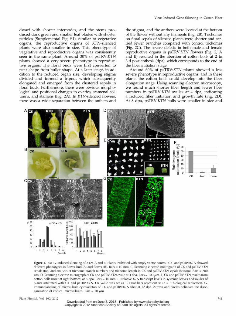
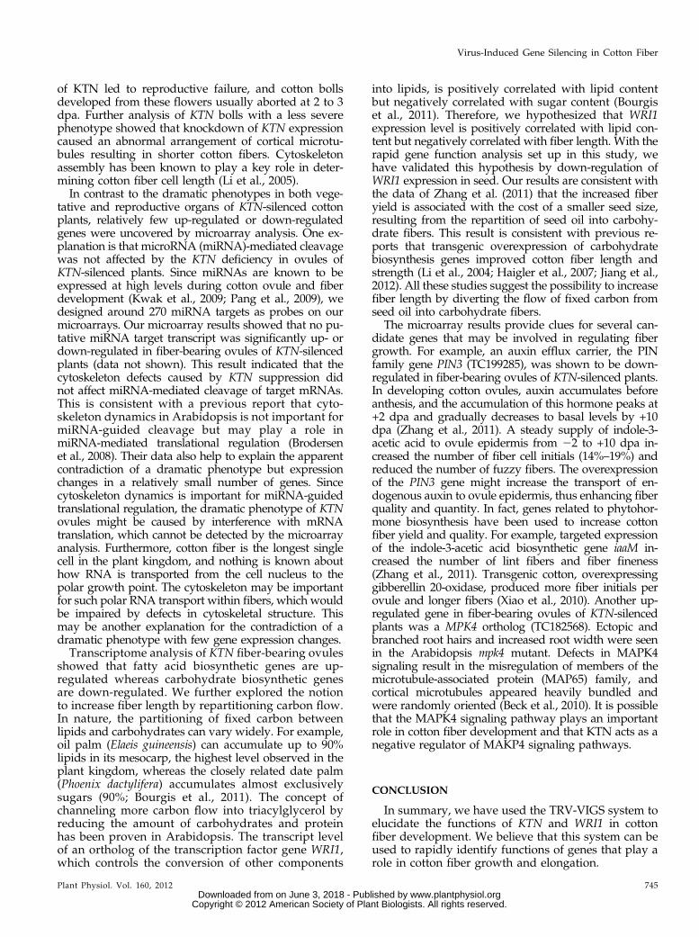
dwarf with shorter internodes, and the stems pro-duced dark green and smaller leaf blades with shorterpetioles (Supplemental Fig. S1). Similar to vegetativeorgans, the reproductive organs of KTN-silencedplants were also smaller in size. This phenotype ofvegetative and reproductive organs was consistentlyseen in the same plant. Around 30% of psTRV:KTNplants showed a very severe phenotype in reproduc-tive organs. The floral buds were first converted topear shape from bullet shape. At a later stage, in ad-dition to the reduced organ size, developing stigmadivided and formed a tripod, which subsequentlyelongated and emerged from the clustered sepals infloral buds. Furthermore, there were obvious morpho-logical and positional changes in ovaries, stamenal col-umns, and stamens (Fig. 2A). In KTN-silenced flowers,there was a wide separation between the anthers and
the stigma, and the anthers were located at the bottomof the flower without any filaments (Fig. 2B). Trichomeson floral sepals of silenced plants were shorter and car-ried fewer branches compared with control trichomes(Fig. 2C). The severe defects in both male and femalereproductive organs in psTRV:KTN flowers (Fig. 2, Aand B) resulted in the abortion of cotton bolls at 2 to3 d post anthesis (dpa), which corresponds to the end ofthe fiber initiation stage.
Around 60% of psTRV:KTN plants showed a lesssevere phenotype in reproductive organs, and in theseplants the cotton bolls could develop into the fiberelongation stage. Using scanning electron microscopy,we found much shorter fiber length and fewer fibernumbers in psTRV:KTN ovules at 4 dpa, indicatinga reduced fiber initiation and growth rate (Fig. 2D).At 8 dpa, psTRV:KTN bolls were smaller in size and
Figure 2. psTRV-induced silencing of KTN. A and B, Plants infiltrated with empty vector control (CK) and psTRV:KTN showeddifferent phenotypes in flower bud (A) and flower (B). Bars = 10 mm. C, Scanning electron micrograph of CK and psTRV:KTNsepals (top) and analysis of trichome branch numbers and trichome length in CK and psTRV:KTN sepals (bottom). Bars = 200mm. D, Scanning electron micrograph of CK and psTRV:KTN ovule at 4 dpa. Bars = 100 mm. E, CK and psTRV:KTN ovules fromcotton bolls (inset at right bottom) at 8 dpa. Bars = 10 mm. F, Relative KTN transcript levels in systemic leaves and ovules ofplants infiltrated with CK and psTRV:KTN. CK value was set as 1. Error bars represent SD (n = 3 biological replicates). G,Immunolabeling of microtubule cytoskeleton of CK and psTRV:KTN fiber at 12 dpa. Arrows and circles delineate the disor-ganization of cortical microtubules. Bars = 10 mm.
Plant Physiol. Vol. 160, 2012 741
Virus-Induced Gene Silencing in Cotton Fiber
www.plantphysiol.orgon June 3, 2018 - Published by Downloaded from Copyright © 2012 American Society of Plant Biologists. All rights reserved.
produced shorter fibers compared with vector controlbolls (Fig. 2E). Compared with the vector control,KTN transcript levels in psTRV:KTN plants were re-duced to 3.3% in systemic leaves and 22.1% in ovulesat 8 dpa (Fig. 2F). In contrast to the parallel array ofcortical microtubules (CMTs) seen in control fibers,immunolabeling of microtubules showed randomCMT orientation (arrows) and microtubule aggrega-tion points (circles) in psTRV:KTN fibers at 12 dpa(Fig. 2G). These defects in the organization of CMTs inthe KTN-silenced cotton fibers indicated that KTNplays important roles in fiber elongation by affectingthe microtubule structure. CMTs are essential fornormal plant morphogenesis because they affect theaxes of cell elongation. The abnormal arrangement ofCMTs, especially the multiple sites of microtubuleconvergence in fibers, led to defects in fiber elongation
and impacted the normal growth and development ofcotton fibers.
To further understand the molecular basis of thedevelopmental defects induced by KTN deficiency,we designed a custom array to investigate possiblechanges of gene expression in fiber-bearing ovules ofKTN-silenced plants at 8 dpa. We found that in fiber-bearing ovules of KTN-silenced plants (one-wayANOVA, P , 0.05), the expression of 37 probe setswas down-regulated by 1.5-fold or more and that of 31probe sets was up-regulated by 1.5-fold or more. Theexpression of some of these probes was confirmedwith real-time PCR (Table I).
Among the 37 probe sets that were down-regulatedin fiber-bearing ovules of psTRV:KTN plants, twodifferent probe sets (TC193948 and TC179681) wereannotated to be the KTN gene. This result provided
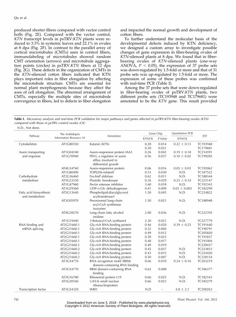
Table I. Microarray analysis and real-time PCR validation for major pathways and genes affected in psTRV:KTN fiber-bearing ovules (KTN)compared with those in psTRV control ovules (CK)
N.D., Not done.
PathwayThe Arabidopsis
Information Resource 10Annotation
Gene Chip Quantitative PCREST
KTN/CK P Value KTN/CK
Cytoskeleton AT1G80350 Katanin (KTN) 0.28 0.014 0.22 6 0.13 TC1939480.30 0.021 TC179681
Auxin transportingand response
AT1G04240 Auxin-responsive protein IAA3 0.26 0.042 0.19 6 0.18 TC214591AT1G70940 PIN3, a regulator of auxin
efflux involved indifferential growth
0.56 0.037 0.10 6 0.02 TC199285
AT4G34760 Auxin-responsive protein 0.06 0.016 0.03 6 0.03 TC192062AT1G80490 TOPLESS-related1 0.53 0.030 N.D. TC187522
Carbohydratemetabolism
AT2G36460 Fru-bisP aldolase 0.62 0.011 N.D. TC188344AT5G23530 Plastidic hexokinase 0.36 0.029 0.25 6 0.14 TC225147AT1G47960 Pectin esterase inhibitor 3.60 0.018 N.D. TC192343AT3G29360 UDP-D-Glc dehydrogenase 0.41 0.009 0.03 6 0.003 TC182299
Fatty acid biosynthesisand metabolism
AT5G13640 Phospholipid:diacylglycerolacyltransferase1
1.50 0.045 N.D. TC224822
AT3G05970 Peroxisomal long-chainacyl-CoA synthetaseisozymes
1.50 0.021 N.D. TC188948
AT4G28570 Long-chain fatty alcoholoxidase
2.00 0.036 N.D. TC222705
AT1G19440 3-Ketoacyl-CoA synthase4 2.30 0.021 N.D. TC227779RNA binding and
mRNA splicingAT2G21660.1 Gly-rich RNA-binding protein 0.46 0.020 0.39 6 0.25 TC191640AT2G21660.1 Gly-rich RNA-binding protein 0.52 0.004 TC190791AT2G21660.1 Gly-rich RNA-binding protein 0.49 0.012 TC205820AT2G21660.1 Gly-rich RNA-binding protein 0.50 0.025 TC191837AT2G21660.1 Gly-rich RNA-binding protein 0.48 0.017 TC191004AT2G21660.1 Gly-rich RNA-binding protein 0.49 0.019 TC228437AT2G21660.2 Gly-rich RNA-binding protein 0.45 0.037 N.D. TC223833AT2G21660.2 Gly-rich RNA-binding protein 0.43 0.015 N.D. TC224420AT2G21660.2 Gly-rich RNA-binding protein 0.50 0.007 N.D. TC228154AT3G54770 RNA recognition motif (RRM)
domain-containing RNA binding0.66 0.010 0.24 6 0.16 TC203219
AT3G54770 RRM domain-containing RNAbinding
0.65 0.008 TC186377
AT3G16780 Ribosomal protein L19 0.66 0.025 N.D. TC182163AT5G20160 U4/U6 small nuclear
ribonucleoprotein0.66 0.023 N.D. TC182279
Transcription factor AT3G54320 WRI1 N.D – 4.8 6 2.1 TC200263
742 Plant Physiol. Vol. 160, 2012
Qu et al.
www.plantphysiol.orgon June 3, 2018 - Published by Downloaded from Copyright © 2012 American Society of Plant Biologists. All rights reserved.
independent confirmation of the silencing of KTN ex-pression in the examined ovules. The remaining 35probe sets included genes of three major categories. Thefirst category encoded proteins related to RNA bindingand mRNA splicing (13 of 35). Most of the RNA-binding proteins were Gly-rich RNA-binding protein.One of the main Gly-rich RNA-binding protein genes,GhGRP1, is the homolog of AtGRP7, which plays a keyrole in alternative mRNA exporting/splicing in Arabi-dopsis (Schöning et al., 2008). This result suggestedpossible expression changes associated with alternativesplicing. The second category of down-regulated geneswas related to auxin transport and response (four of 35),such as genes for the auxin efflux carrier PIN3 (TC199285)and the auxin-responsive protein IAA3 (TC214591). Thethird category of down-regulated genes was related tocarbohydrate metabolism (three of 35), including thoseencoding a plastidic hexokinase (TC225147), a Fru-bisPaldolase (TC188344), and a positive regulator of pecticprecursor synthesis (TC182299; Table I).Among the major up-regulated probe sets were
genes related to fatty acid biosynthesis and metabolism(four of 31), such as a positive regulator of triacyl-glycerol and unusual fatty acid accumulation level(TC224822). Another up-regulated gene was an inhib-itor of pectic precursor synthesis (TC192343), whichwas related to carbohydrate metabolism (Table I). Panget al. (2010) have shown that the biosynthesis of pecticprecursors is important for cotton fiber elongation.The up-regulated fatty acid biosynthetic genes,
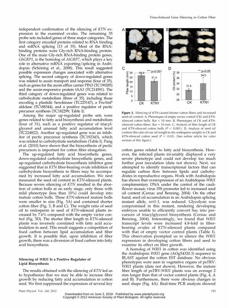
down-regulated carbohydrate biosynthetic genes, andup-regulated carbohydrate biosynthesis inhibitor genesuggested that in KTN fiber-bearing ovule inhibition ofcarbohydrate biosynthesis in fibers may be accompa-nied by increased fatty acid accumulation. We nextmeasured the seed oil content in KTN-silenced plants.Because severe silencing of KTN resulted in the abor-tion of cotton bolls at an early stage, only those withmild phenotype (less than 10%) could develop intomature cotton bolls. These KTN-silenced mature bollswere smaller in size (Fig. 3A) and contained shortercotton fiber (Fig. 3, B and C). The weight ratio of seedoil to endosperm in seed of KTN-silenced plants in-creased by 7.6% compared with the empty vector con-trol (Fig. 3D). The shorter fiber length in KTN-silencedplants was inversely correlated with fatty acid accu-mulation in seed. This result suggests a competition offixed carbon between lipid accumulation and fibergrowth. It is possible that, upon inhibition of fibergrowth, there was a diversion of fixed carbon into fattyacid biosynthesis.
Silencing of WRI1 Is a Positive Regulator ofLipid Biosynthesis
The results obtained with the silencing of KTN led usto hypothesize that we may be able to increase fibergrowth by reducing fatty acid accumulation in cottonseed. We first suppressed the expression of several key
cotton genes related to fatty acid biosynthesis. How-ever, the infected plants invariably displayed a verysevere phenotype and could not develop too muchfurther post inoculation (data not shown). Next, weattempted to identify transcriptional factors that canregulate carbon flow between lipids and carbohy-drates in reproductive organs. Work with Arabidopsishas shown that overexpression of an Arabidopsis WRI1complementary DNA under the control of the cauli-flower mosaic virus 35S promoter led to increased seedoil content (Cernac and Benning, 2004). On the otherhand, seed oil accumulation in an Arabidopsis splicingmutant allele, wri1-1, was reduced. Glycolysis wascompromised in this mutant, rendering developingembryos unable to efficiently convert Suc into pre-cursors of triacylglycerol biosynthesis (Cernac andBenning, 2004). Interestingly, we found that WRI1transcript levels were increased 4.8-fold in fiber-bearing ovules of KTN-silenced plants comparedwith that of empty vector control plants (Table I).This observation prompted us to silence the WRI1expression in developing cotton fibers and seed toexamine its effect on fiber growth.
A homolog of WRI1 in cotton was identified usingthe Arabidopsis WRI1 gene (At3g54320.3) sequence toBLAST against the cotton EST database. No obviousphenotypes were seen in vegetative organs of psTRV:WRI1 plants (data not shown). However, the maturefiber length of psTRV:WRI1 plants was on average 2mm longer than that of vector control plants (Fig. 4, Aand B). Furthermore, there were obvious changes inseed shape (Fig. 4A). Real-time PCR analysis showed
Figure 3. Silencing of KTN caused shorter cotton fibers and increasedseed oil content. A, Phenotypes of empty vector control (CK) and KTN-silenced cotton bolls. Bar = 10 mm. B, Phenotypes of CK and KTN-silenced cotton fibers. Bar = 10 mm. C, Analysis of fiber length of CKand KTN-silenced cotton bolls (P , 0.001). D, Analysis of seed oilcontent (the ratio of raw oil weight to dry endosperm weight) in CK andKTN-silenced cotton seed (P , 0.05). [See online article for colorversion of this figure.]
Plant Physiol. Vol. 160, 2012 743
Virus-Induced Gene Silencing in Cotton Fiber
www.plantphysiol.orgon June 3, 2018 - Published by Downloaded from Copyright © 2012 American Society of Plant Biologists. All rights reserved.
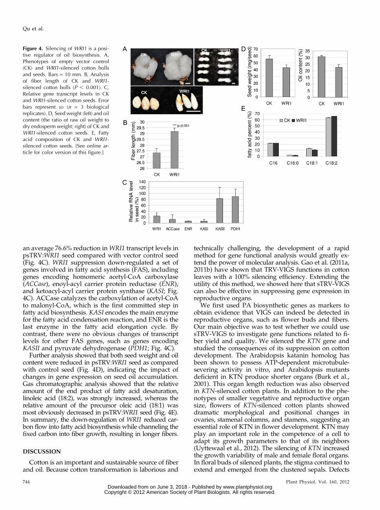
an average 76.6% reduction inWRI1 transcript levels inpsTRV:WRI1 seed compared with vector control seed(Fig. 4C). WRI1 suppression down-regulated a set ofgenes involved in fatty acid synthesis (FAS), includinggenes encoding homomeric acetyl-CoA carboxylase(ACCase), enoyl-acyl carrier protein reductase (ENR),and ketoacyl-acyl carrier protein synthase (KASI; Fig.4C). ACCase catalyzes the carboxylation of acetyl-CoAto malonyl-CoA, which is the first committed step infatty acid biosynthesis. KASI encodes the main enzymefor the fatty acid condensation reaction, and ENR is thelast enzyme in the fatty acid elongation cycle. Bycontrast, there were no obvious changes of transcriptlevels for other FAS genes, such as genes encodingKASII and pyruvate dehydrogenase (PDH1; Fig. 4C).
Further analysis showed that both seed weight and oilcontent were reduced in psTRV:WRI1 seed as comparedwith control seed (Fig. 4D), indicating the impact ofchanges in gene expression on seed oil accumulation.Gas chromatographic analysis showed that the relativeamount of the end product of fatty acid desaturation,linoleic acid (18:2), was strongly increased, whereas therelative amount of the precursor oleic acid (18:1) wasmost obviously decreased in psTRV:WRI1 seed (Fig. 4E).In summary, the down-regulation of WRI1 reduced car-bon flow into fatty acid biosynthesis while channeling thefixed carbon into fiber growth, resulting in longer fibers.
DISCUSSION
Cotton is an important and sustainable source of fiberand oil. Because cotton transformation is laborious and
technically challenging, the development of a rapidmethod for gene functional analysis would greatly ex-tend the power of molecular analysis. Gao et al. (2011a,2011b) have shown that TRV-VIGS functions in cottonleaves with a 100% silencing efficiency. Extending theutility of this method, we showed here that sTRV-VIGScan also be effective in suppressing gene expression inreproductive organs.
We first used PA biosynthetic genes as markers toobtain evidence that VIGS can indeed be detected inreproductive organs, such as flower buds and fibers.Our main objective was to test whether we could usesTRV-VIGS to investigate gene functions related to fi-ber yield and quality. We silenced the KTN gene andstudied the consequences of its suppression on cottondevelopment. The Arabidopsis katanin homolog hasbeen shown to possess ATP-dependent microtubule-severing activity in vitro, and Arabidopsis mutantsdeficient in KTN produce shorter organs (Burk et al.,2001). This organ length reduction was also observedin KTN-silenced cotton plants. In addition to the phe-notypes of smaller vegetative and reproductive organsize, flowers of KTN-silenced cotton plants showeddramatic morphological and positional changes inovaries, stamenal columns, and stamens, suggesting anessential role of KTN in flower development. KTN mayplay an important role in the competence of a cell toadapt its growth parameters to that of its neighbors(Uyttewaal et al., 2012). The silencing of KTN increasedthe growth variability of male and female floral organs.In floral buds of silenced plants, the stigma continued toextend and emerged from the clustered sepals. Defects
Figure 4. Silencing of WRI1 is a posi-tive regulator of oil biosynthesis. A,Phenotypes of empty vector control(CK) and WRI1-silenced cotton bollsand seeds. Bars = 10 mm. B, Analysisof fiber length of CK and WRI1-silenced cotton bolls (P , 0.001). C,Relative gene transcript levels in CKand WRI1-silenced cotton seeds. Errorbars represent SD (n = 3 biologicalreplicates). D, Seed weight (left) and oilcontent (the ratio of raw oil weight todry endosperm weight; right) of CK andWRI1-silenced cotton seeds. E, Fattyacid composition of CK and WRI1-silenced cotton seeds. [See online ar-ticle for color version of this figure.]
744 Plant Physiol. Vol. 160, 2012
Qu et al.
www.plantphysiol.orgon June 3, 2018 - Published by Downloaded from Copyright © 2012 American Society of Plant Biologists. All rights reserved.
of KTN led to reproductive failure, and cotton bollsdeveloped from these flowers usually aborted at 2 to 3dpa. Further analysis of KTN bolls with a less severephenotype showed that knockdown of KTN expressioncaused an abnormal arrangement of cortical microtu-bules resulting in shorter cotton fibers. Cytoskeletonassembly has been known to play a key role in deter-mining cotton fiber cell length (Li et al., 2005).In contrast to the dramatic phenotypes in both vege-
tative and reproductive organs of KTN-silenced cottonplants, relatively few up-regulated or down-regulatedgenes were uncovered by microarray analysis. One ex-planation is that microRNA (miRNA)-mediated cleavagewas not affected by the KTN deficiency in ovules ofKTN-silenced plants. Since miRNAs are known to beexpressed at high levels during cotton ovule and fiberdevelopment (Kwak et al., 2009; Pang et al., 2009), wedesigned around 270 miRNA targets as probes on ourmicroarrays. Our microarray results showed that no pu-tative miRNA target transcript was significantly up- ordown-regulated in fiber-bearing ovules of KTN-silencedplants (data not shown). This result indicated that thecytoskeleton defects caused by KTN suppression didnot affect miRNA-mediated cleavage of target mRNAs.This is consistent with a previous report that cyto-skeleton dynamics in Arabidopsis is not important formiRNA-guided cleavage but may play a role inmiRNA-mediated translational regulation (Brodersenet al., 2008). Their data also help to explain the apparentcontradiction of a dramatic phenotype but expressionchanges in a relatively small number of genes. Sincecytoskeleton dynamics is important for miRNA-guidedtranslational regulation, the dramatic phenotype of KTNovules might be caused by interference with mRNAtranslation, which cannot be detected by the microarrayanalysis. Furthermore, cotton fiber is the longest singlecell in the plant kingdom, and nothing is known abouthow RNA is transported from the cell nucleus to thepolar growth point. The cytoskeleton may be importantfor such polar RNA transport within fibers, which wouldbe impaired by defects in cytoskeletal structure. Thismay be another explanation for the contradiction of adramatic phenotype with few gene expression changes.Transcriptome analysis of KTN fiber-bearing ovules
showed that fatty acid biosynthetic genes are up-regulated whereas carbohydrate biosynthetic genesare down-regulated. We further explored the notionto increase fiber length by repartitioning carbon flow.In nature, the partitioning of fixed carbon betweenlipids and carbohydrates can vary widely. For example,oil palm (Elaeis guineensis) can accumulate up to 90%lipids in its mesocarp, the highest level observed in theplant kingdom, whereas the closely related date palm(Phoenix dactylifera) accumulates almost exclusivelysugars (90%; Bourgis et al., 2011). The concept ofchanneling more carbon flow into triacylglycerol byreducing the amount of carbohydrates and proteinhas been proven in Arabidopsis. The transcript levelof an ortholog of the transcription factor gene WRI1,which controls the conversion of other components
into lipids, is positively correlated with lipid contentbut negatively correlated with sugar content (Bourgiset al., 2011). Therefore, we hypothesized that WRI1expression level is positively correlated with lipid con-tent but negatively correlated with fiber length. With therapid gene function analysis set up in this study, wehave validated this hypothesis by down-regulation ofWRI1 expression in seed. Our results are consistent withthe data of Zhang et al. (2011) that the increased fiberyield is associated with the cost of a smaller seed size,resulting from the repartition of seed oil into carbohy-drate fibers. This result is consistent with previous re-ports that transgenic overexpression of carbohydratebiosynthesis genes improved cotton fiber length andstrength (Li et al., 2004; Haigler et al., 2007; Jiang et al.,2012). All these studies suggest the possibility to increasefiber length by diverting the flow of fixed carbon fromseed oil into carbohydrate fibers.
The microarray results provide clues for several can-didate genes that may be involved in regulating fibergrowth. For example, an auxin efflux carrier, the PINfamily gene PIN3 (TC199285), was shown to be down-regulated in fiber-bearing ovules of KTN-silenced plants.In developing cotton ovules, auxin accumulates beforeanthesis, and the accumulation of this hormone peaks at+2 dpa and gradually decreases to basal levels by +10dpa (Zhang et al., 2011). A steady supply of indole-3-acetic acid to ovule epidermis from 22 to +10 dpa in-creased the number of fiber cell initials (14%–19%) andreduced the number of fuzzy fibers. The overexpressionof the PIN3 gene might increase the transport of en-dogenous auxin to ovule epidermis, thus enhancing fiberquality and quantity. In fact, genes related to phytohor-mone biosynthesis have been used to increase cottonfiber yield and quality. For example, targeted expressionof the indole-3-acetic acid biosynthetic gene iaaM in-creased the number of lint fibers and fiber fineness(Zhang et al., 2011). Transgenic cotton, overexpressinggibberellin 20-oxidase, produced more fiber initials perovule and longer fibers (Xiao et al., 2010). Another up-regulated gene in fiber-bearing ovules of KTN-silencedplants was a MPK4 ortholog (TC182568). Ectopic andbranched root hairs and increased root width were seenin the Arabidopsis mpk4 mutant. Defects in MAPK4signaling result in the misregulation of members of themicrotubule-associated protein (MAP65) family, andcortical microtubules appeared heavily bundled andwere randomly oriented (Beck et al., 2010). It is possiblethat the MAPK4 signaling pathway plays an importantrole in cotton fiber development and that KTN acts as anegative regulator of MAKP4 signaling pathways.
CONCLUSION
In summary, we have used the TRV-VIGS system toelucidate the functions of KTN and WRI1 in cottonfiber development. We believe that this system can beused to rapidly identify functions of genes that play arole in cotton fiber growth and elongation.
Plant Physiol. Vol. 160, 2012 745
Virus-Induced Gene Silencing in Cotton Fiber
www.plantphysiol.orgon June 3, 2018 - Published by Downloaded from Copyright © 2012 American Society of Plant Biologists. All rights reserved.
MATERIALS AND METHODS
Plant Materials
Seeds of upland cotton (Gossypium hirsutum ‘Coker 312’) were germinatedin a greenhouse, and seedlings with two to four true leaves were used forVIGS assays. Infiltrated plants were grown in a growth chamber at 25°C witha 16-h-light/8-h-dark photoperiod cycle. Cotton bolls on treated plants weretagged on the day of anthesis (0 dpa).
Gene Cloning and Generation of RecombinantVIGS Vectors
psTRV1 and psTRV2 (Ye et al., 2009b, 2010) were used for all VIGS assays.Cotton gene sequences were obtained from the EST database of GenBank.Candidate gene fragments were cloned by PCR and inserted into psTRV2 togenerate psTRV2 derivatives. The gene fragment sequences are listed inSupplemental File S1.
Agrobacterium Infiltration
psTRV1 and psTRV2 or psTRV2 derivatives were introduced into Agro-bacterium tumefaciens strain AGL1 by electroporation. Agrobacterial cells weregrown, collected, and resuspended in 10 mM MES, 10 mM MgCl2, and 200 mM
acetylsyringone solution to a final optical density at 600 nm of 1.5 and then leftat room temperature for 3 to 4 h without shaking. Before infiltration, A.tumefaciens cultures containing psTRV1 and psTRV2 or its derivatives weremixed in a 1:1 ratio. Cotton plants were agroinoculated by vacuum infiltration.Whole plants were submerged in agrobacterial inoculum and subjected to 80to 100 kPa vacuum for 1 min, and the vacuum was quickly released to allowthe inoculum to rapidly enter plant tissues. After infiltration, the excessagroinoculum was used to drench the root system of infiltrated plants. Notethat TRV is transmitted by a nematode vector, mechanical inoculation, andgrafting but not by contact between plants (http://www.agls.uidaho.edu/ebi/vdie/descr808.htm). We used separate pots of plants for different TRVtreatments to ensure that the treated plants were not contaminated by a dif-ferent TRV vector. After completion of the experiment, the plants and soilwere autoclaved before disposal to prevent release of the virus into the en-vironment.
RNA Extraction and Quantitative Real-Time PCR Analysis
Leaf, ovule, fiber, or seeds (fresh weight, less than 100 mg) were ground inliquid N2 and extracted with the plant RNA purification kit following themanufacturer’s protocol (Invitrogen). RNAs for microarray experiments wereprepared by a similar method. Harvested bolls (7–8 dpa) of empty vectorcontrol, KTN-silenced, and WRI1-silenced cotton plants were stored on ice,and carefully dissected ovules were further frozen in liquid nitrogen andstored at 280°C until RNA extraction. RNA concentration was measured byNanodrop (Thermo). Moloney murine leukemia virus reverse transcriptase(Promega) was used for reverse transcription reactions. Real-time PCR wasperformed with Power SYBR Green PCR Master (Applied Biosystems) andrun in ABI7900HT. For each construct, we collected three samples from threeplants with significant phenotypes (one sample from each plant). All sampleswere run in triplicate, and data were analyzed with RQ manager at a presetcycle threshold value (Applied Biosystems). The internal control was rbcLmRNA for leaf samples and UBQ mRNA for ovule samples. Cycle thresholdvalues included in the analyses were based on three independent biologicalsamples with three technical replicates for each biological sample. SD wascalculated based on three biological replicates. For real-time PCR primer se-quences, see Supplemental File S2.
Histochemical Assay
The DMACA-HCl protocol for PA staining was adapted from Li et al.(1999). Fresh organs were immediately soaked in ethanol:glacial acetic acidsolution (3:1, v/v) for decoloration. The decolorized organs were stained forabout 20 min with 0.3% (w/v) DMACA in a cold mixture of methanol and 6 M
HCl (1:1, v/v), rinsed with several changes of 70% (v/v) ethanol, and theorgans were observed with a dissecting microscope. Proanthocyanin-containingcells stained blue.
Scanning Electron Microscopy
Organs harvested from treated cotton plants were dissected and fixed withtape inside a sample chamber. Samples were frozen in liquid N2 and visualizedwith a scanning electron microscope (JSM-6360LV; JEOL).
Immunolabeling of Microtubules
Immunolabeling of microtubules was performed according to Zhao et al.(2010). Briefly, ovules were fixed in PEM buffer (50 mM PIPES, 5 mM EGTA,and 1 mM MgSO4, pH 6.9) containing 4% (w/v) paraformaldehyde, 0.1%Triton X-100, and 0.3 M mannitol for 1 h at room temperature. Fixed ovuleswere washed three times with PEM buffer. Fibers were cut off from the ovulesand attached to slides coated with poly-L-Lys (Sigma-Aldrich). The fibers wereincubated with 1% (w/v) Cellulase R-10 and 0.1% (w/v) Macerozyme in PEMbuffer containing 0.3 M mannitol and 1 mM phenylmethylsulfonyl fluoride for10 to 20 min at room temperature and then washed in phosphate-bufferedsaline (PBS) three times. After a preincubation in PBS buffer containing 1%(w/v) bovine serum albumin, the slides were incubated with mouse mono-clonal anti-tubulin antibody (1:1,000) at 4°C overnight. The fibers were thenwashed three times in PBS buffer and incubated for 1 h with fluoresceinisothiocyanate-labeled goat anti-mouse secondary antibody (1:200). The fiberswere washed with PBS buffer and observed using a Zeiss LSM 5 EXCITERupright confocal microscope.
Custom Microarray and Data Analysis
We used Agilent GE 8x60K arrays provided by the full Custom MicroarrayService of Genomax. Forty-four thousand Tentative Consensus sequences fromCotton Gene Index 10.1 cotton (http://compbio.dfci.harvard.edu/cgi-bin/tgi/gimain.pl?gudb=cotton; March 4, 2010) were used for Custom eARRAY probedesign. RNA labeling, hybridization to custom arrays, washing, and scanningwere all performed by the Custom Microarray. Feature extraction and appli-cation support were according to Agilent protocols. Briefly, to perform thecomparative analysis of various samples, we applied total RNA samples fromthe cotton fiber-bearing ovules onto the custom microarrays. Total RNA fromthe fibers was labeled and applied separately. Briefly, 0.1 mg of total RNAfrom each sample was used to synthesize fluorescence-labeled complementaryRNA using Cyanine 3-CTP according to the manufacturer’s protocol (AgilentQuick Amp Labeling Kit, one-color). Labeled RNA was hybridized for 16 h at65°C on the custom array. After hybridization, the microarray slides werewashed according to the standard protocol (Agilent Technologies) and scan-ned on an Agilent microarray scanner. Raw data were filtered, normalized,and further analyzed using the Agilent Feature Extraction Software (version10.7). Statistics analysis was performed with one-way ANOVA, and probeswith P , 0.05 between empty vector control and treated samples were usedfor ontology and pathway analysis. All Tentative Consensus numbers of theprobes generated after significance analysis were retrieved from TentativeConsensus annotator (http://compbio.dfci.harvard.edu/cgi-bin/tgi/gimain.pl?gudb=cotton). BLAST similarity search using BLASTX (E-value , 10210)against The Arabidopsis Information Resource 10 and RefSeq (National Centerfor Biotechnology Information) databases was performed to assign an anno-tation to the transcripts.
Cotton Fiber Length Measurement
Dry and mature fiber length was manually measured against a ruler. Themean fiber length of three measurements from one seed, 10 seeds from one boll,and five typical size bolls from each treated cotton group were recorded andused for Student’s t test analysis. The effect of silencing of KTN1 and WRI1 oncotton fiber length was repeated in two independent experiments.
Fatty Acid Analysis
Oil content and fatty acid profile analysis were performed according to Quet al. (2012). Briefly, the outer seed coat was removed from dried cotton seeds.The dry endosperm part was ground to a fine powder, and the lipids wereextracted with hexane three times. The combined supernatant was transferredto a glass vial, and the hexane was evaporated with a flow of dry nitrogen gasat 50°C. The weight of the raw oil was determined, and the oil content wasrecorded as the ratio of raw oil weight to dry endosperm weight.
746 Plant Physiol. Vol. 160, 2012
Qu et al.
www.plantphysiol.orgon June 3, 2018 - Published by Downloaded from Copyright © 2012 American Society of Plant Biologists. All rights reserved.
Total lipid, extracted from cotton seed, was transmethylated with 3 N
methanolic-HCl (Sigma) plus 400 mL of 2,2-dimethoxypropane (Sigma). Thefatty acid methyl ester was analyzed using GC Agilent 6890 employing heliumas the carrier gas and DB-23 columns for component separation. The fatty acidcomposition value included in the analyses was calculated based on peakareas in three biological replicates and presented as mean 6 SD.
Sequence information for the gene fragment used for VIGS is listed inSupplemental File S1.
Supplemental Data
The following materials are available in the online version of this article.
Supplemental Figure S1. psTRV:KTN plants with severe vegetative phe-notypes.
Supplemental File S1. Sequence information of the gene fragment used forVIGS.
Supplemental File S2. Real-time PCR primer sequences.
ACKNOWLEDGMENTS
We thank Mr. Khar Meng Ng for his help in taking care of cotton plants.We thank Mr. Xue-Zhi Ouyang, Dr. Meredith Calvert, and Ms. Fiona Chia fortheir help in scanning electron microscopy and confocal observation.
Received April 13, 2012; accepted July 23, 2012; published July 26, 2012.
LITERATURE CITED
Ahmad FJ, Yu W, McNally FJ, Baas PW (1999) An essential role for kataninin severing microtubules in the neuron. J Cell Biol 145: 305–315
Baulcombe D (2004) RNA silencing in plants. Nature 431: 356–363Beck M, Komis G, Müller J, Menzel D, Samaj J (2010) Arabidopsis ho-
mologs of nucleus- and phragmoplast-localized kinase 2 and 3 andmitogen-activated protein kinase 4 are essential for microtubule orga-nization. Plant Cell 22: 755–771
Bourgis F, Kilaru A, Cao X, Ngando-Ebongue GF, Drira N, Ohlrogge JB,Arondel V (2011) Comparative transcriptome and metabolite analysis ofoil palm and date palm mesocarp that differ dramatically in carbonpartitioning. Proc Natl Acad Sci USA 108: 12527–12532
Brigneti G, Martín-Hernández AM, Jin H, Chen J, Baulcombe DC, BakerB, Jones JD (2004) Virus-induced gene silencing in Solanum species.Plant J 39: 264–272
Brodersen P, Sakvarelidze-Achard L, Bruun-Rasmussen M, Dunoyer P,Yamamoto YY, Sieburth L, Voinnet O (2008) Widespread translationalinhibition by plant miRNAs and siRNAs. Science 320: 1185–1190
Burch-Smith TM, Anderson JC, Martin GB, Dinesh-Kumar SP (2004)Applications and advantages of virus-induced gene silencing for genefunction studies in plants. Plant J 39: 734–746
Burk DH, Liu B, Zhong R, Morrison WH, Ye ZH (2001) A katanin-likeprotein regulates normal cell wall biosynthesis and cell elongation. PlantCell 13: 807–827
Burk DH, Zhong R, Ye Z-H (2007) The katanin microtubule severingprotein in plants. J Integr Plant Biol 49: 1174–1182
Cernac A, Benning C (2004) WRINKLED1 encodes an AP2/EREB domainprotein involved in the control of storage compound biosynthesis inArabidopsis. Plant J 40: 575–585
Fu DQ, Zhu BZ, Zhu HL, Jiang WB, Luo YB (2005) Virus-induced genesilencing in tomato fruit. Plant J 43: 299–308
Gao X, Britt RC Jr, Shan L, He P (2011a) Agrobacterium-mediated virus-induced gene silencing assay in cotton. J Vis Exp 54: e2938
Gao X, Wheeler T, Li Z, Kenerley CM, He P, Shan L (2011b) SilencingGhNDR1 and GhMKK2 compromises cotton resistance to Verticilliumwilt. Plant J 66: 293–305
Grønlund M, Constantin G, Piednoir E, Kovacev J, Johansen IE, Lund OS(2008) Virus-induced gene silencing in Medicago truncatula and Lathyrusodorata. Virus Res 135: 345–349
Haigler CH, Singh B, Zhang D, Hwang S, Wu C, Cai WX, Hozain M, KangW, Kiedaisch B, Strauss RE, et al (2007) Transgenic cotton over-
producing spinach sucrose phosphate synthase showed enhanced leafsucrose synthesis and improved fiber quality under controlled envi-ronmental conditions. Plant Mol Biol 63: 815–832
Hileman LC, Drea S, Martino G, Litt A, Irish VF (2005) Virus-induced genesilencing is an effective tool for assaying gene function in the basal eudicotspecies Papaver somniferum (opium poppy). Plant J 44: 334–341
Holzberg S, Brosio P, Gross C, Pogue GP (2002) Barley stripe mosaic virus-induced gene silencing in a monocot plant. Plant J 30: 315–327
Jiang Y, Guo W, Zhu H, Ruan YL, Zhang T (2012) Overexpression ofGhSusA1 increases plant biomass and improves cotton fiber yield andquality. Plant Biotechnol J 10: 301–312
Kwak PB, Wang QQ, Chen XS, Qiu CX, Yang ZM (2009) Enrichment of a setof microRNAs during the cotton fiber development. BMC Genomics 10: 457
Lee J, Burns TH, Light G, Sun Y, Fokar M, Kasukabe Y, Fujisawa K,Maekawa Y, Allen RD (2010) Xyloglucan endotransglycosylase/hydrolase genes in cotton and their role in fiber elongation. Planta232: 1191–1205
Li X, Wang XD, Zhao X, Dutt Y (2004) Improvement of cotton fiber qualityby transforming the acsA and acsB genes into Gossypium hirsutum L.by means of vacuum infiltration. Plant Cell Rep 22: 691–697
Li XB, Fan XP, Wang XL, Cai L, Yang WC (2005) The cotton ACTIN1 gene isfunctionally expressed in fibers and participates in fiber elongation.Plant Cell 17: 859–875
Li YG, Tanner G, Larkin P (1999) The DMACA-HCl protocol and thethreshold proanthocyanidin content for bloat safety in forage legumes. JSci Food Agric 70: 89–101
Liu Y, Nakayama N, Schiff M, Litt A, Irish VF, Dinesh-Kumar SP (2004)Virus induced gene silencing of a DEFICIENS ortholog in Nicotianabenthamiana. Plant Mol Biol 54: 701–711
Liu Y, Schiff M, Dinesh-Kumar SP (2002) Virus-induced gene silencing intomato. Plant J 31: 777–786
McNally FJ, Vale RD (1993) Identification of katanin, an ATPase thatsevers and disassembles stable microtubules. Cell 75: 419–429
Pang CY, Wang H, Pang Y, Xu C, Jiao Y, Qin YM, Western TL, Yu SX,Zhu YX (2010) Comparative proteomics indicates that biosynthesis ofpectic precursors is important for cotton fiber and Arabidopsis root hairelongation. Mol Cell Proteomics 9: 2019–2033
Pang M, Woodward AW, Agarwal V, Guan X, Ha M, Ramachandran V,Chen X, Triplett BA, Stelly DM, Chen ZJ (2009) Genome-wide analysisreveals rapid and dynamic changes in miRNA and siRNA sequence andexpression during ovule and fiber development in allotetraploid cotton(Gossypium hirsutum L.). Genome Biol 10: R122
Pang Y, Peel GJ, Wright E, Wang Z, Dixon RA (2007) Early steps inproanthocyanidin biosynthesis in the model legume Medicago truncatula.Plant Physiol 145: 601–615
Qu J, Mao HZ, Chen W, Gao SQ, Bai YN, Sun YW, Geng YF, Ye J (2012)Development of marker-free transgenic Jatropha plants with increasedlevels of seed oleic acid. Biotechnol Biofuels 5: 10
Ruiz MT, Voinnet O, Baulcombe DC (1998) Initiation and maintenance ofvirus-induced gene silencing. Plant Cell 10: 937–946
Schöning JC, Streitner C, Meyer IM, Gao Y, Staiger D (2008) Reciprocalregulation of glycine-rich RNA-binding proteins via an interlockedfeedback loop coupling alternative splicing to nonsense-mediated decayin Arabidopsis. Nucleic Acids Res 36: 6977–6987
Tuttle JR, Idris AM, Brown JK, Haigler CH, Robertson D (2008)Geminivirus-mediated gene silencing from Cotton leaf crumple virus isenhanced by low temperature in cotton. Plant Physiol 148: 41–50
United States Department of Agriculture (2012) Cotton: World Marketsand Trade. http://www.fas.usda.gov/cotton/Current/ (June 8, 2012)
Uyttewaal M, Burian A, Alim K, Landrein B, Borowska-Wykręt D,Dedieu A, Peaucelle A, Ludynia M, Traas J, Boudaoud A, et al (2012)Mechanical stress acts via katanin to amplify differences in growth ratebetween adjacent cells in Arabidopsis. Cell 149: 439–451
Valentine T, Shaw J, Blok VC, Phillips MS, Oparka KJ, Lacomme C(2004) Efficient virus-induced gene silencing in roots using a modifiedtobacco rattle virus vector. Plant Physiol 136: 3999–4009
Wang HY, Wang J, Gao P, Jiao GL, Zhao PM, Li Y, Wang GL, Xia GX(2009) Down-regulation of GhADF1 gene expression affects cotton fibreproperties. Plant Biotechnol J 7: 13–23
Wang J, Wang HY, Zhao PM, Han LB, Jiao GL, Zheng YY, Huang SJ, XiaGX (2010) Overexpression of a profilin (GhPFN2) promotes the pro-gression of developmental phases in cotton fibers. Plant Cell Physiol 51:1276–1290
Plant Physiol. Vol. 160, 2012 747
Virus-Induced Gene Silencing in Cotton Fiber
www.plantphysiol.orgon June 3, 2018 - Published by Downloaded from Copyright © 2012 American Society of Plant Biologists. All rights reserved.
Xiao YH, Li DM, Yin MH, Li XB, Zhang M, Wang YJ, Dong J, Zhao J, LuoM, Luo XY, et al (2010) Gibberellin 20-oxidase promotes initiation andelongation of cotton fibers by regulating gibberellin synthesis. J PlantPhysiol 167: 829–837
Xie DY, Sharma SB, Paiva NL, Ferreira D, Dixon RA (2003) Role of an-thocyanidin reductase, encoded by BANYULS in plant flavonoid bio-synthesis. Science 299: 396–399
Ye J, Chua N-H, Qu J, Geng Y-F, Bu Y-P, inventors. June 10, 2010. Virusinduced gene silencing (VIGS) for functional analysis of genes in cotton.United States Patent Application No. 13/376,902
Ye J, Qu J, Bui HT, Chua N-H (2009a) Rapid analysis of Jatropha curcas genefunctions by virus-induced gene silencing. Plant Biotechnol J 7: 964–976
Ye J, Qu J, Chua N-H, inventors. December 16, 2009b. Functional analysisof Jatropha curcas genes. United States Patent Application no. 13/141,752
Zhang C, Ghabrial SA (2006) Development of Bean pod mottle virus-basedvectors for stable protein expression and sequence-specific virus-induced gene silencing in soybean. Virology 344: 401–411
Zhang M, Zheng X, Song S, Zeng Q, Hou L, Li D, Zhao J, Wei Y, Li X, Luo M,et al (2011) Spatiotemporal manipulation of auxin biosynthesis in cotton ovuleepidermal cells enhances fiber yield and quality. Nat Biotechnol 29: 453–458
Zhao PM, Wang LL, Han LB, Wang J, Yao Y, Wang HY, Du XM, Luo YM,Xia GX (2010) Proteomic identification of differentially expressed pro-teins in the Ligon lintless mutant of upland cotton (Gossypium hirsutumL.). J Proteome Res 9: 1076–1087
748 Plant Physiol. Vol. 160, 2012
Qu et al.
www.plantphysiol.orgon June 3, 2018 - Published by Downloaded from Copyright © 2012 American Society of Plant Biologists. All rights reserved.