disposable miniature pressure sensor for cardiologist...
TRANSCRIPT
Disposable Miniature Pressure Sensor for Cardiologist Use
Xingwei Wang
Department of Electrical and Computer Engineering University of Massachusetts Lowell
[email protected] (978) 934-1981
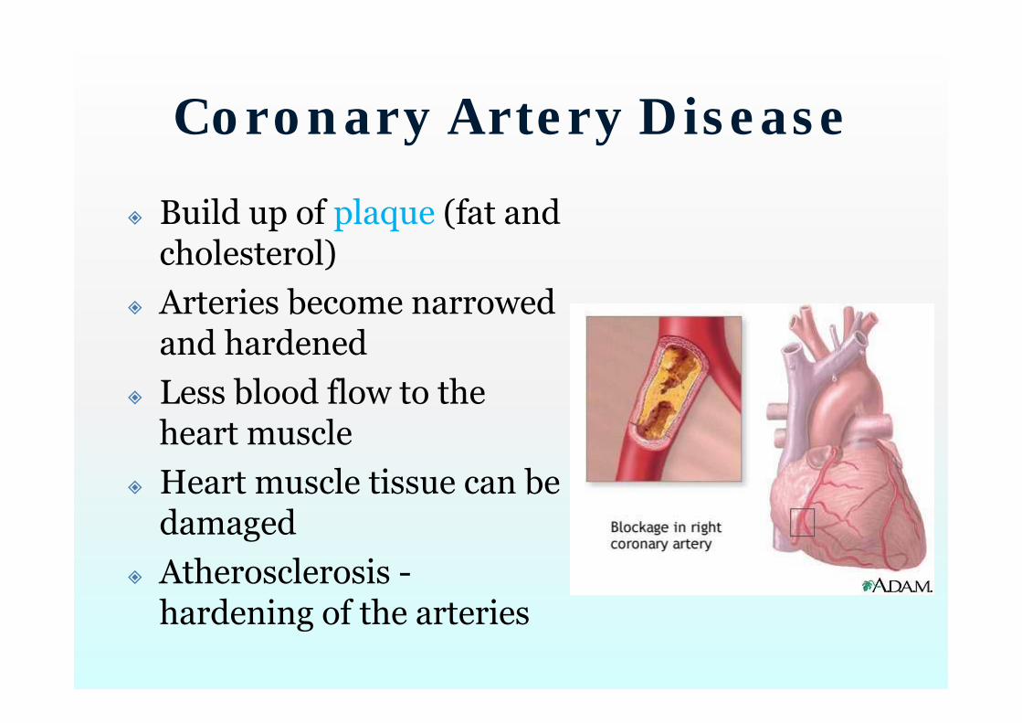
Coronary Artery Disease
Build up of plaque (fat and cholesterol)
Arteries become narrowed and hardened
Less blood flow to the heart muscle
Heart muscle tissue can be damaged
Atherosclerosis -hardening of the arteries
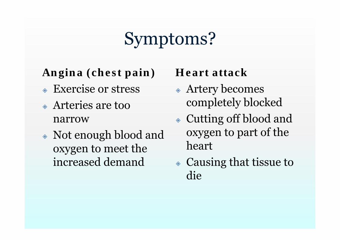
Symptoms?
Angina (chest pain) Exercise or stress Arteries are too
narrow Not enough blood and
oxygen to meet the increased demand
Heart attack Artery becomes
completely blocked Cutting off blood and
oxygen to part of the heart
Causing that tissue to die

Background Coronary artery disease (CAD)
Kills ~ 871,000 people/year Leading cause of death in US 14 million US patients /year [1]
Coronary angioplasty ~650,000 U.S. patients /year More than 2 million /year worldwide Nearly 3 million /year by 2010 Annual medical cost > 112 Billion

The 1st Balloon Angioplasty
Dr. Andreas Gruentzig
The first successful balloon angioplasty in the heart
in (which year?)
in (which country?)
Launched a new medical subspecialty -interventional cardiology
Using catheters with a variety of devices on the tip, to treat heart problems without surgery.
Switzerland
1977
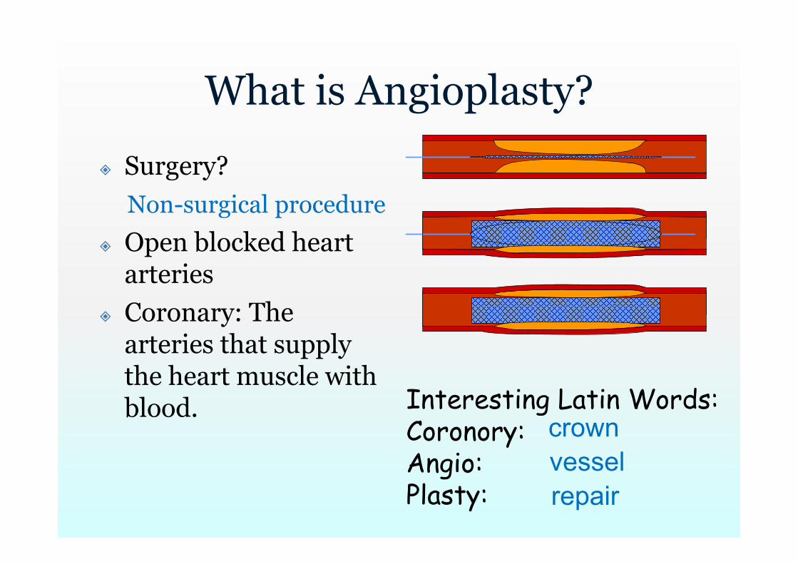
What is Angioplasty?
Surgery?
Open blocked heart arteries
Coronary: The arteries that supply the heart muscle with blood.
Non-surgical procedure
Interesting Latin Words:Coronory:Angio: Plasty:
crownvesselrepair
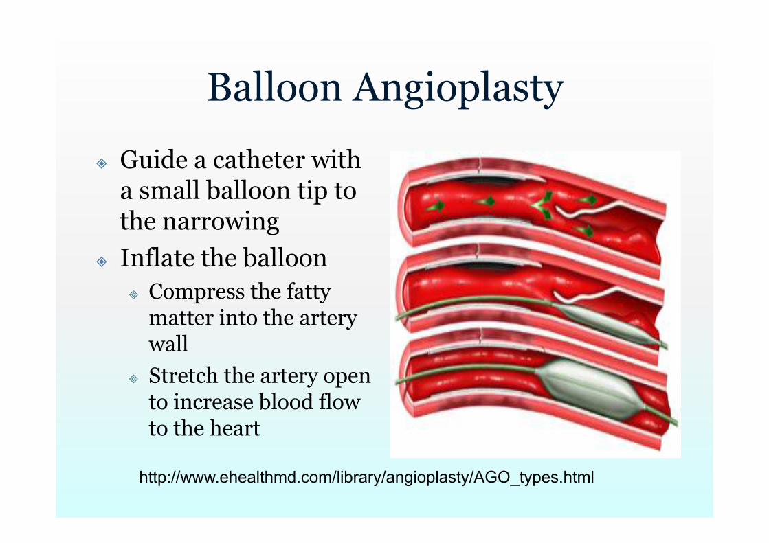
Balloon Angioplasty
Guide a catheter with a small balloon tip to the narrowing
Inflate the balloon Compress the fatty
matter into the artery wall
Stretch the artery open to increase blood flow to the heart
http://www.ehealthmd.com/library/angioplasty/AGO_types.html
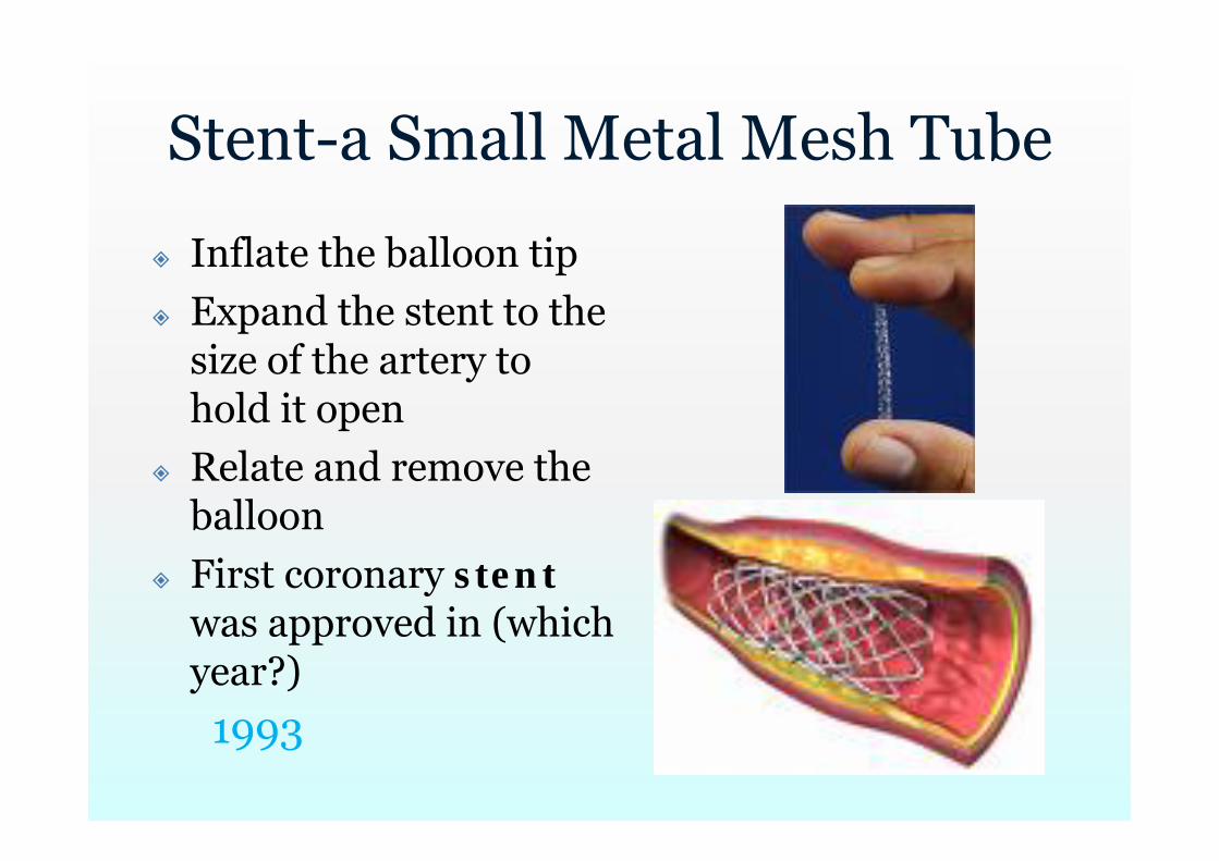
Stent-a Small Metal Mesh Tube
Inflate the balloon tip Expand the stent to the
size of the artery to hold it open
Relate and remove the balloon
First coronary stentwas approved in (which year?)
1993
Rotoblation
Guide a special catheter with an acorn-shaped, diamond-coated tip
The tip spins around at a high speed and grinds away the plaque
The microscopic particles are washed safely away in your blood stream and filtered out by your liver and spleen
Hard, calcified plaque
Atherectomy
Catheter has a hollow cylinder on the tip with an open window on one side and a balloon on the other
Inflate the balloon Pushing the window against the fatty matter A blade (cutter) within the cylinder rotates
and shaves off any fat that protruded into the window. (up to 1,200 revolutions per minute)
The shavings are caught in a chamber within the catheter and removed.

Cutting Balloon
Catheter has a special balloon tip with small blades
Inflate the balloon Small blades score the plaque Balloon compresses the fatty matter into
the artery wall. Laser angioplasty uses laser energy to
destroy plaque
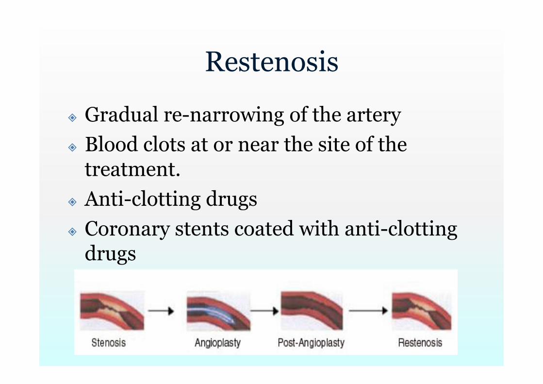
Restenosis
Gradual re-narrowing of the artery Blood clots at or near the site of the
treatment. Anti-clotting drugs Coronary stents coated with anti-clotting
drugs
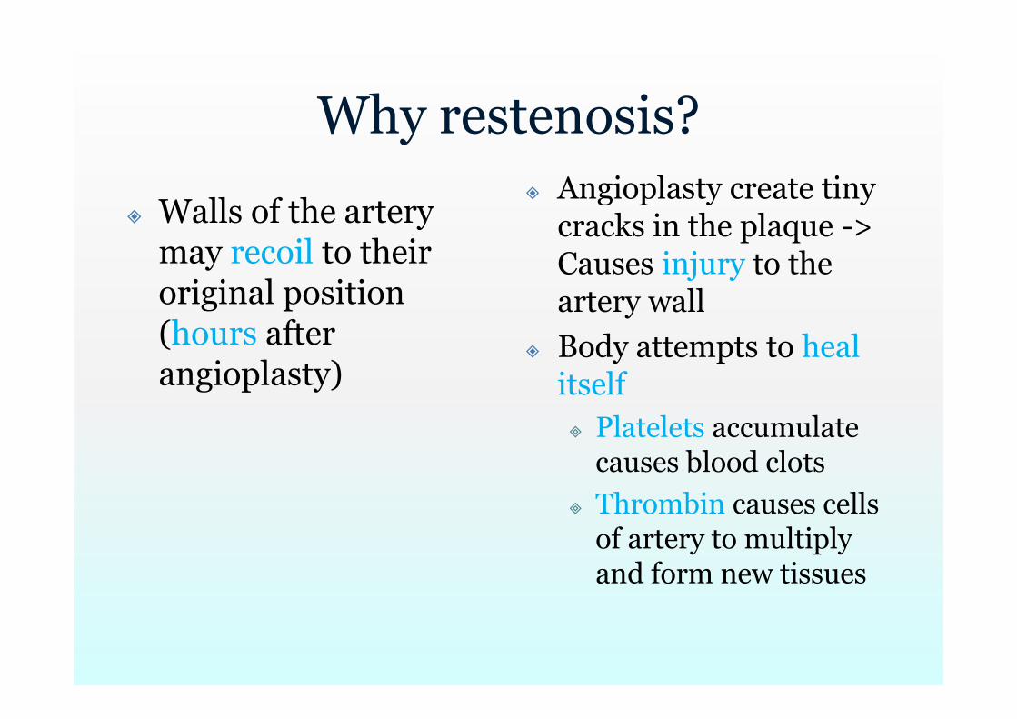
Why restenosis? Angioplasty create tiny
cracks in the plaque -> Causes injury to the artery wall
Body attempts to heal itself Platelets accumulate
causes blood clots Thrombin causes cells
of artery to multiply and form new tissues
Walls of the artery may recoil to their original position (hours after angioplasty)
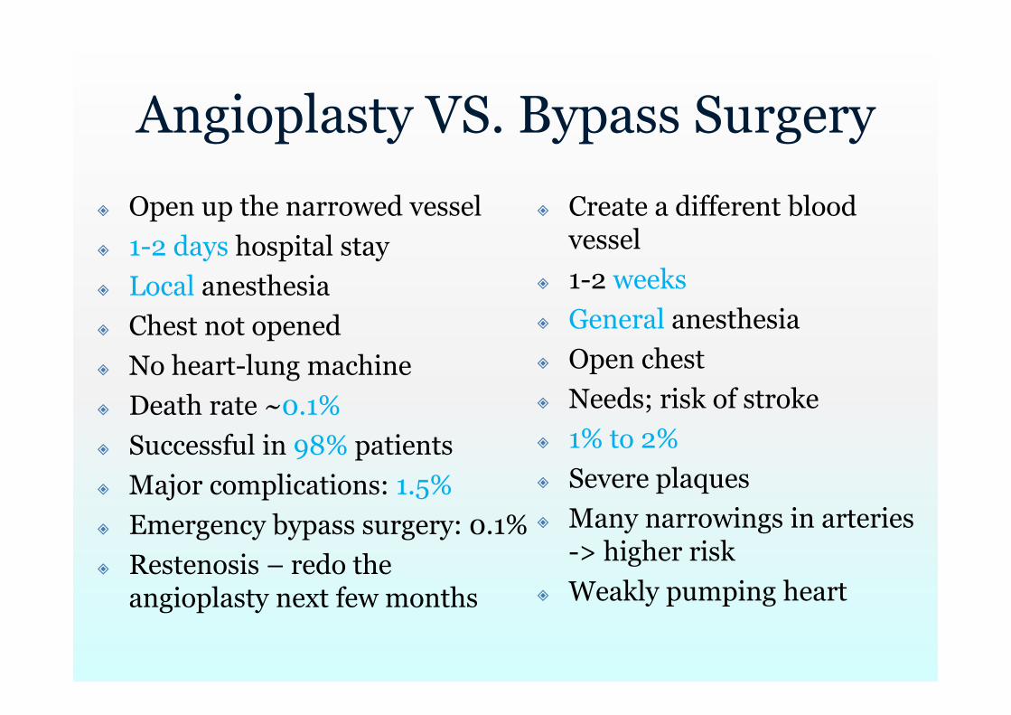
Angioplasty VS. Bypass Surgery
Open up the narrowed vessel 1-2 days hospital stay Local anesthesia Chest not opened No heart-lung machine Death rate ~0.1% Successful in 98% patients Major complications: 1.5% Emergency bypass surgery: 0.1% Restenosis – redo the
angioplasty next few months
Create a different blood vessel
1-2 weeks General anesthesia Open chest Needs; risk of stroke 1% to 2% Severe plaques Many narrowings in arteries
-> higher risk Weakly pumping heart
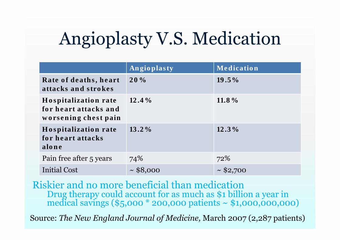
Angioplasty V.S. MedicationAngioplasty Medication
Rate of deaths, heart attacks and strokes
20% 19.5%
Hospitalization rate for heart attacks and worsening chest pain
12.4% 11.8%
Hospitalization rate for heart attacks alone
13.2% 12.3%
Pain free after 5 years 74% 72%
Initial Cost ~ $8,000 ~ $2,700
Source: The New England Journal of Medicine, March 2007 (2,287 patients)
Riskier and no more beneficial than medicationDrug therapy could account for as much as $1 billion a year in medical savings ($5,000 * 200,000 patients ~ $1,000,000,000)
Comments from Doctors
“In low-risk patients with stable coronary arterydisease, aggressive lipid-lowering therapy is at least as effective as angioplasty and usual care in reducing the incidence of ischemic events.” [4]
“In this small pilot study, intensive medicaltherapy and PTCA were comparable at suppressing ischemia in stable patients after AMI. ..Corroboration of these preliminary findings in a larger cardiac-event trial is warranted.” [5]
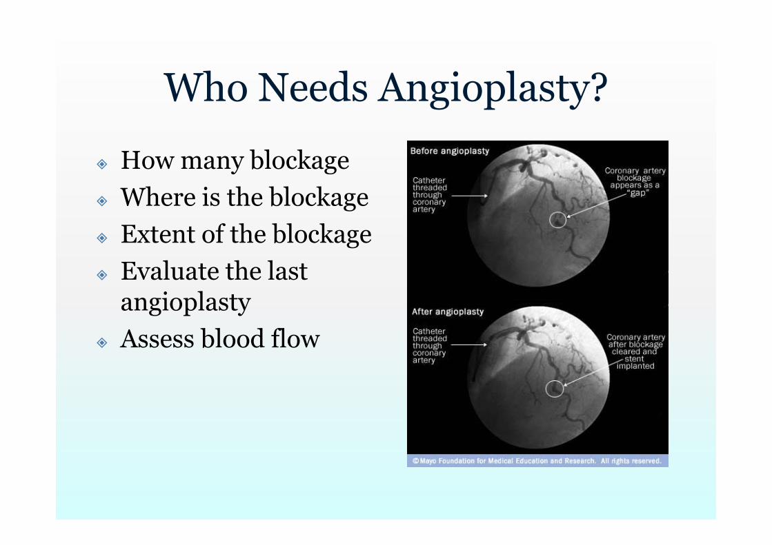
Who Needs Angioplasty?
How many blockage Where is the blockage Extent of the blockage Evaluate the last
angioplasty Assess blood flow
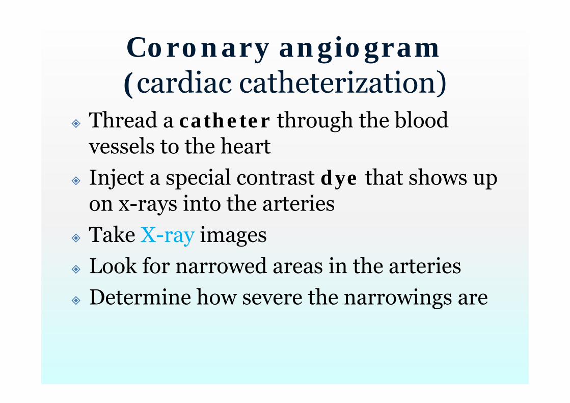
Coronary angiogram (cardiac catheterization)
Thread a catheter through the blood vessels to the heart
Inject a special contrast dye that shows up on x-rays into the arteries
Take X-ray images Look for narrowed areas in the arteries Determine how severe the narrowings are
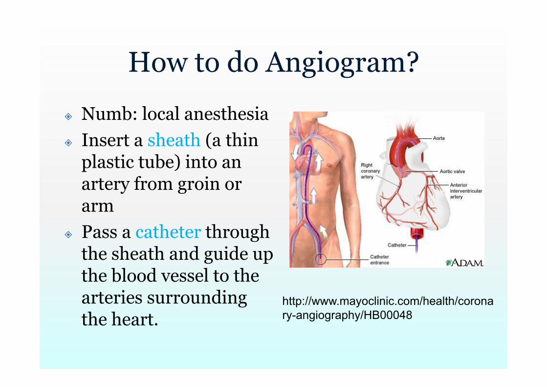
How to do Angiogram?
Numb: local anesthesia Insert a sheath (a thin
plastic tube) into an artery from groin or arm
Pass a catheter through the sheath and guide up the blood vessel to the arteries surrounding the heart.
http://www.mayoclinic.com/health/coronary-angiography/HB00048
How to do Angiogram (2)?
Inject a small amount of contrast material through the catheter
Photograph as it moves through the heart's chambers, valves, and major vessels
Tell whether the coronary arteries are narrowed and/or whether the heart valves are working correctly
Problems of Angiogram
Potential risks -> stroke; heart attacks Allergic to the iodine dyes Kidney damage Infection Trauma to the catheterized arteries Radiation exposure from the X-rays

X-Ray
Body is made up of various substances with differing densities
Denser substances (e.g. calcium rich bones) absorb X-ray photons -> film unexposed ->translucent blue
Lower-density tissues (e.g. fat, skin, organs) -> black part of the film
Reveal the internal structure of the body on film
Computerized Tomography (CT)
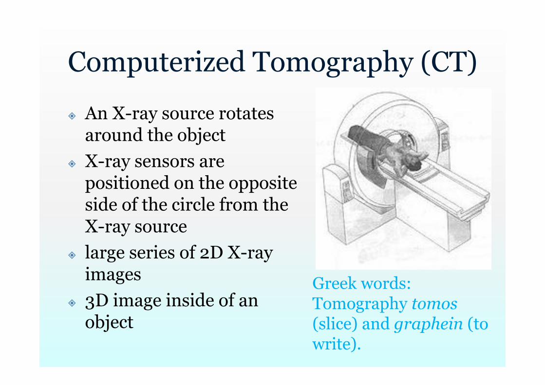
An X-ray source rotates around the object
X-ray sensors are positioned on the opposite side of the circle from the X-ray source
large series of 2D X-ray images
3D image inside of an object
Greek words:Tomography tomos(slice) and graphein (to write).
Problems of Cardiac CT Angiography
Heart is effectively imaged more than once ->a relatively high radiation exposure around 12 mSv
A chest X-ray: ~0.02 to 0.2 mSv Natural background radiation exposure:
~0.01 mSv/day 100-600 chest X-rays or over 3 years
worth of natural background radiation
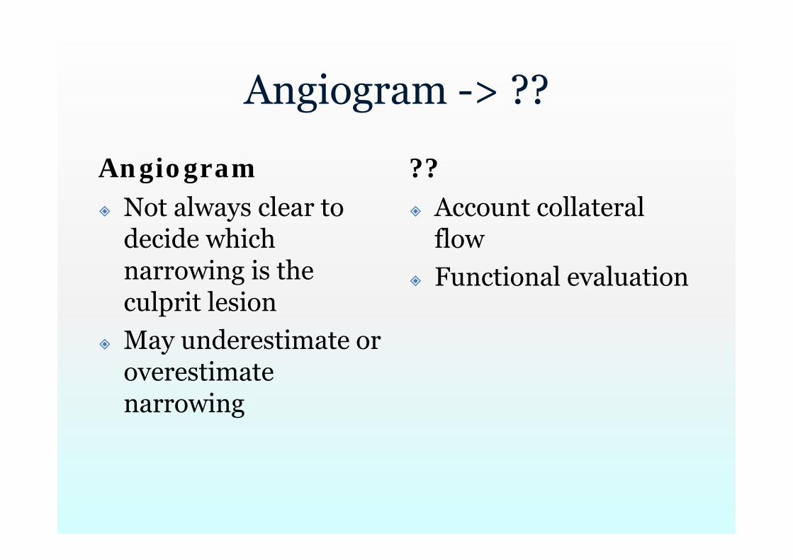
Angiogram -> ??
Angiogram Not always clear to
decide which narrowing is the culprit lesion
May underestimate or overestimate narrowing
?? Account collateral
flow Functional evaluation
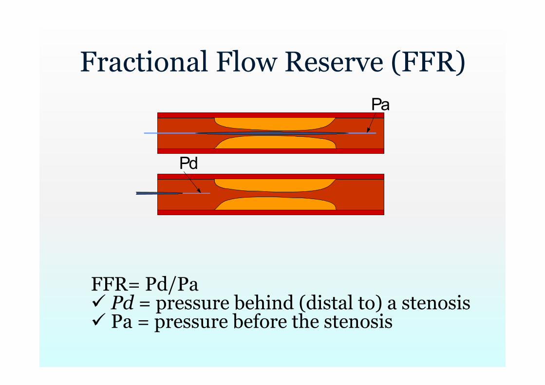
Fractional Flow Reserve (FFR) Pa
Pd
FFR= Pd/Pa Pd = pressure behind (distal to) a stenosis Pa = pressure before the stenosis
FFR
http://www.youtube.com/watch?v=xTaz-OkJPoo
Abnormal?
Maximal flow down a vessel in the presence of a stenosis compared to the maximal flow in the hypothetical absence of the stenosis
No absolute cut-off point Cut-off point: 0.75-0.80
St. Jude’s hydrophilic PressureWire® Certus
http://www.youtube.com/watch?v=DM-L5m9d_2o (sensor) http://www.youtube.com/watch?v=fi37C9rq0nw (pressurewire 8) http://www.youtube.com/watch?v=SgCkKJpVSd8 (insertion of the
sensor) http://www.youtube.com/watch?v=OaTOj8Ct3Pk (pullback) http://www.youtube.com/watch?v=Luq62Mt8rH8 (the stent
placement)

Coronary Artery Disease (CAD) 14 million patients $100 Billion annually
Percutaneous Coronary Intervention (PCI) Angioplasty with or
without stent (90%) Over 1 million/year $8 billion/year
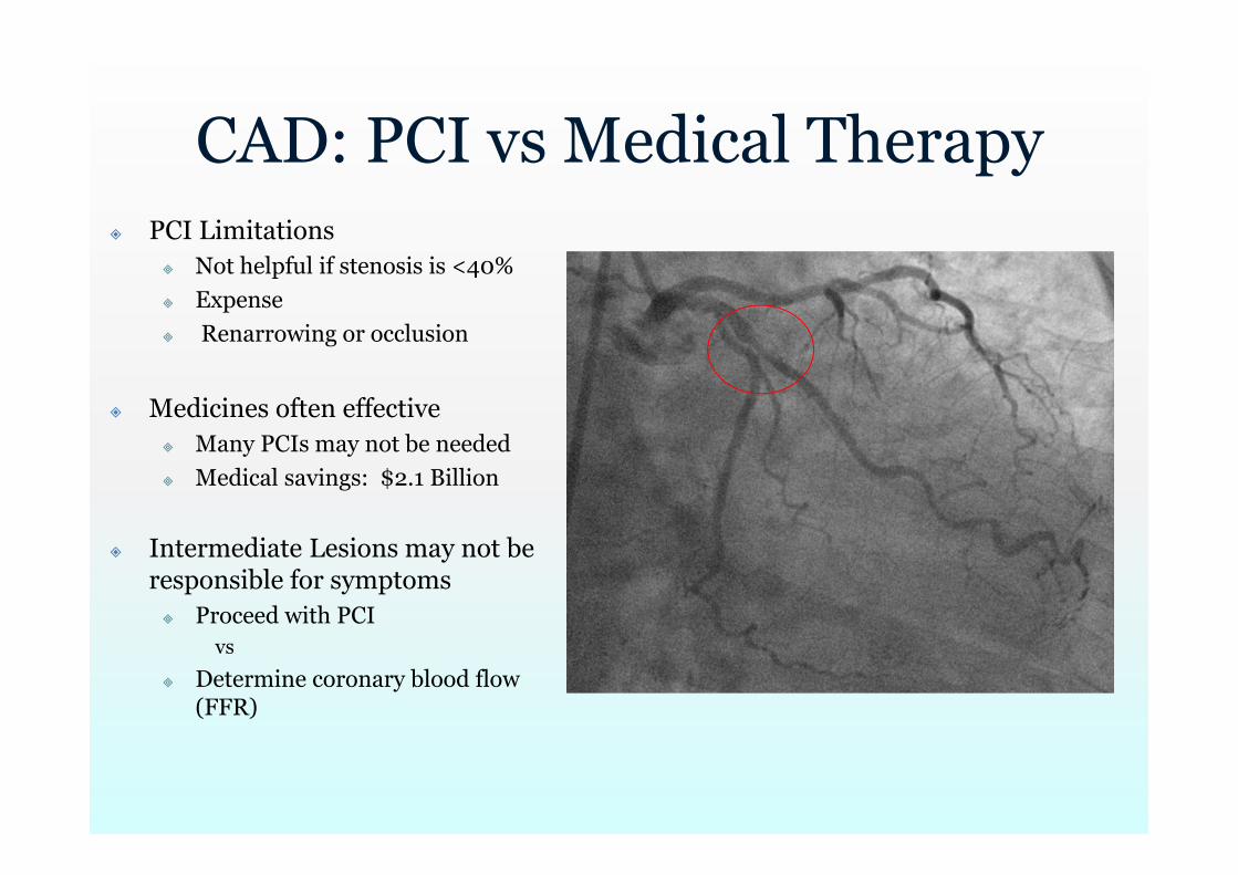
CAD: PCI vs Medical Therapy PCI Limitations
Not helpful if stenosis is <40% Expense Renarrowing or occlusion
Medicines often effective Many PCIs may not be needed Medical savings: $2.1 Billion
Intermediate Lesions may not be responsible for symptoms Proceed with PCI
vs
Determine coronary blood flow (FFR)
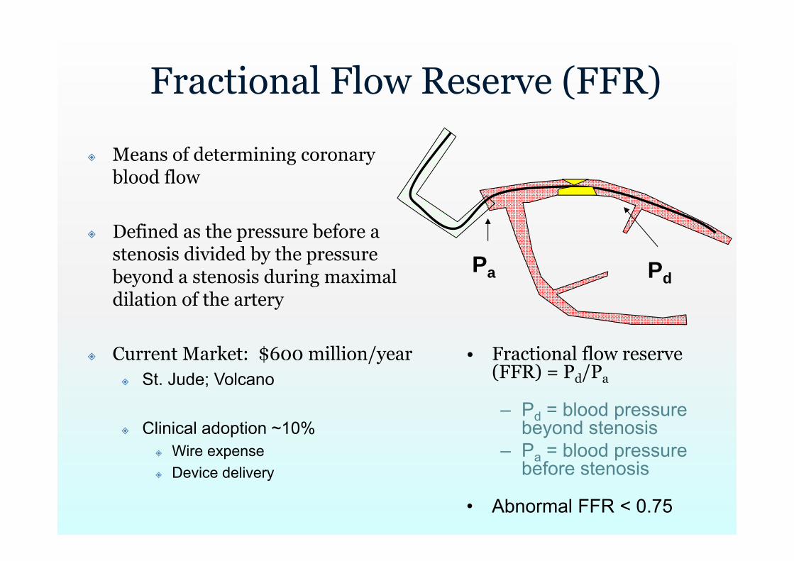
Fractional Flow Reserve (FFR)
• Fractional flow reserve (FFR) = Pd/Pa
– Pd = blood pressure beyond stenosis
– Pa = blood pressure before stenosis
• Abnormal FFR < 0.75
PdPa
Means of determining coronary blood flow
Defined as the pressure before a stenosis divided by the pressure beyond a stenosis during maximal dilation of the artery
Current Market: $600 million/year St. Jude; Volcano
Clinical adoption ~10% Wire expense Device delivery
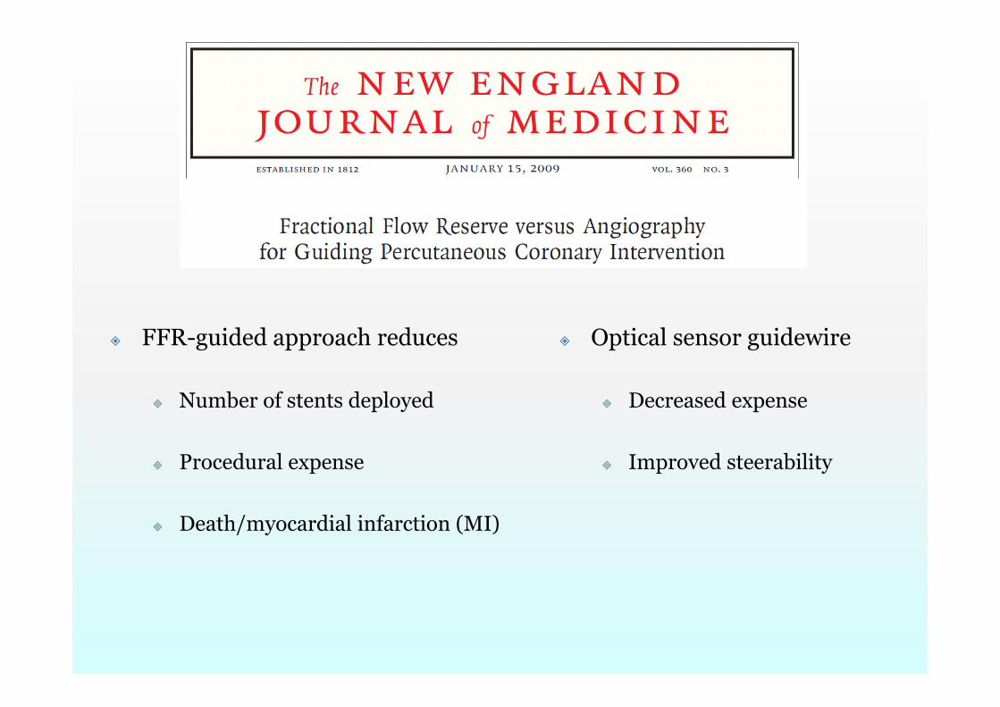
FFR-guided approach reduces
Number of stents deployed
Procedural expense
Death/myocardial infarction (MI)
Optical sensor guidewire
Decreased expense
Improved steerability
FAME Trials
http://www.youtube.com/watch?v=yLfW5k7v2yk
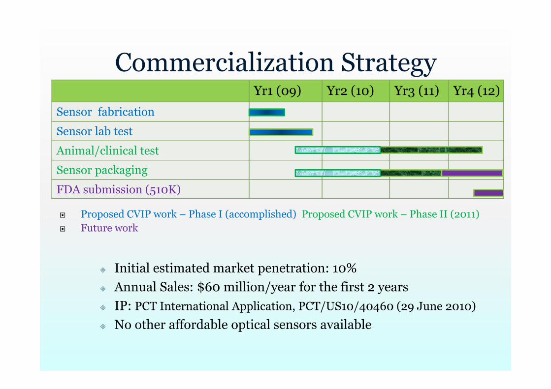
Proposed CVIP work – Phase I (accomplished) Proposed CVIP work – Phase II (2011) Future work
Commercialization StrategyYr1 (09) Yr2 (10) Yr3 (11) Yr4 (12)
Sensor fabrication
Sensor lab test
Animal/clinical test
Sensor packaging
FDA submission (510K)
Initial estimated market penetration: 10% Annual Sales: $60 million/year for the first 2 years IP: PCT International Application, PCT/US10/40460 (29 June 2010)
No other affordable optical sensors available
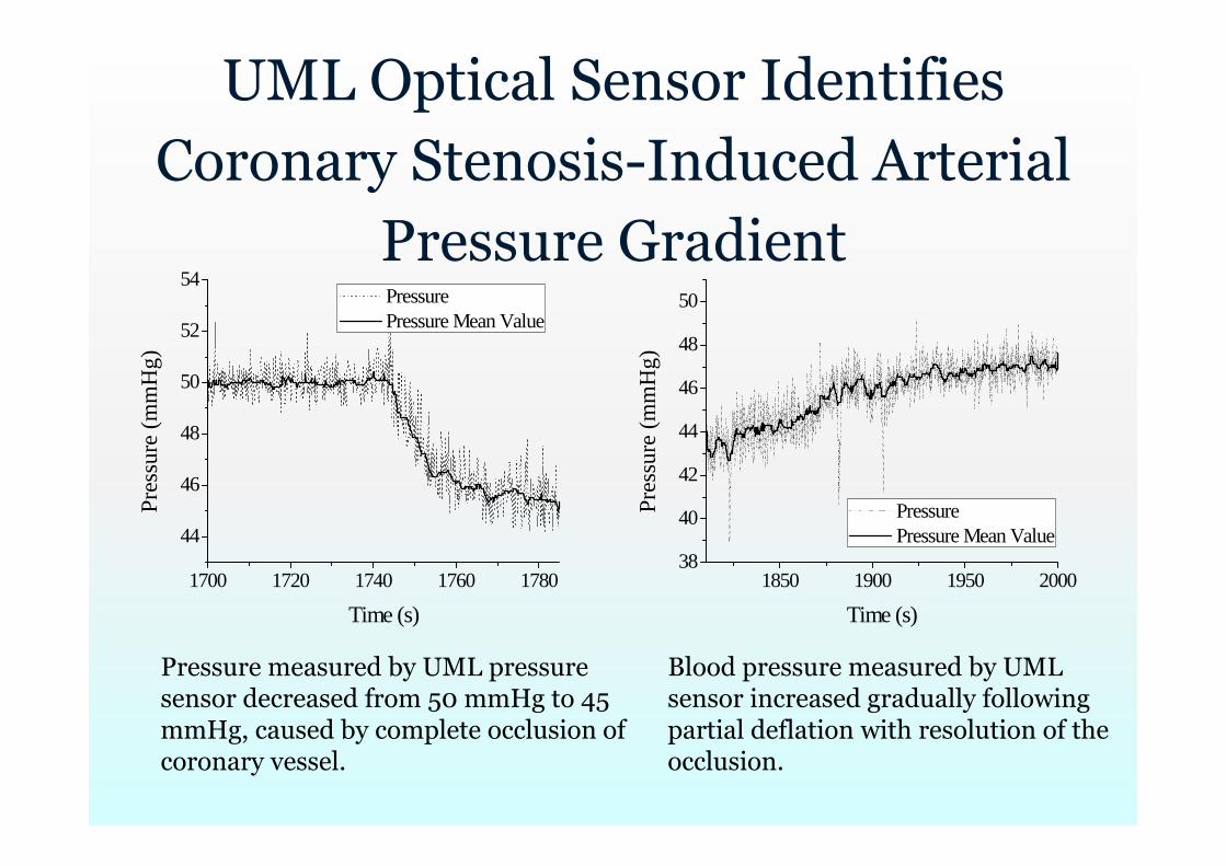
UML Optical Sensor Identifies Coronary Stenosis-Induced Arterial
Pressure Gradient
1700 1720 1740 1760 1780
44
46
48
50
52
54
Pres
sure
(mm
Hg)
Time (s)
Pressure Pressure Mean Value
1850 1900 1950 200038
40
42
44
46
48
50
Pres
sure
(mm
Hg)
Time (s)
Pressure Pressure Mean Value
Pressure measured by UML pressure sensor decreased from 50 mmHg to 45 mmHg, caused by complete occlusion of coronary vessel.
Blood pressure measured by UML sensor increased gradually following partial deflation with resolution of the occlusion.
31 2 54
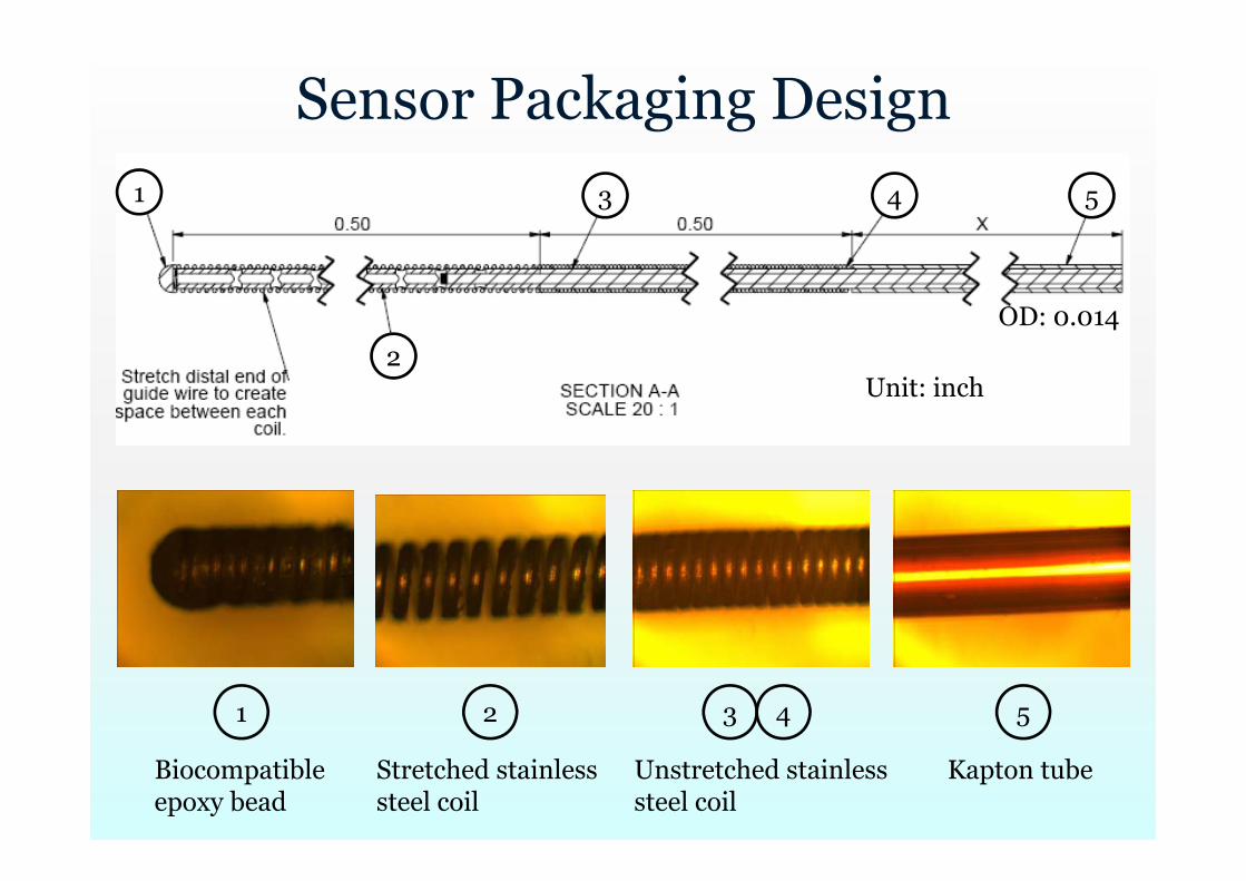
Biocompatible epoxy bead
Stretched stainless steel coil
Unstretched stainless steel coil
Kapton tube
1
2
3 4 5
Unit: inch
OD: 0.014
Sensor Packaging Design
Summary UML optical sensor will advance care of patients with
coronary artery disease in a cost-conscious manner
Wire and system refinements are planned but preclinical studies require additional support
Results from preclinical studies will set the stage for clinical studies and FDA approval
Expansion into other clinical environments and take advantage of the optical sensor’s superior accuracy and resistance to electromagnetic interference
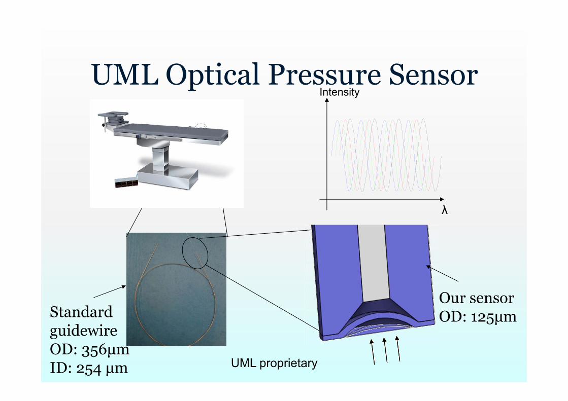
λ
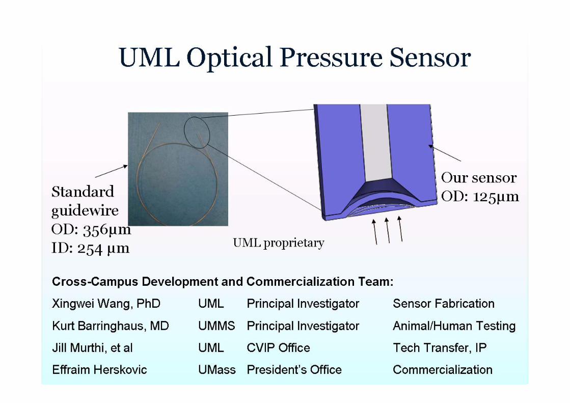
IntensityUML Optical Pressure Sensor
Standard guidewireOD: 356μmID: 254 μm
Our sensorOD: 125μm
UML proprietary
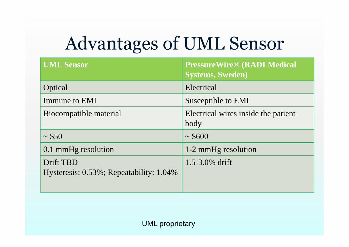
Advantages of UML SensorUML Sensor PressureWire® (RADI Medical
Systems, Sweden)Optical Electrical Immune to EMI Susceptible to EMIBiocompatible material Electrical wires inside the patient
body~ $50 ~ $6000.1 mmHg resolution 1-2 mmHg resolutionDrift TBDHysteresis: 0.53%; Repeatability: 1.04%
1.5-3.0% drift
UML proprietary
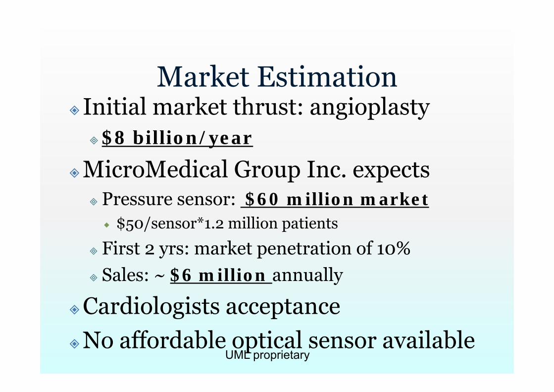
Market EstimationInitial market thrust: angioplasty
$8 billion/year
MicroMedical Group Inc. expects Pressure sensor: $60 million market
$50/sensor*1.2 million patients
First 2 yrs: market penetration of 10% Sales: ~ $6 million annually
Cardiologists acceptanceNo affordable optical sensor available
UML proprietary
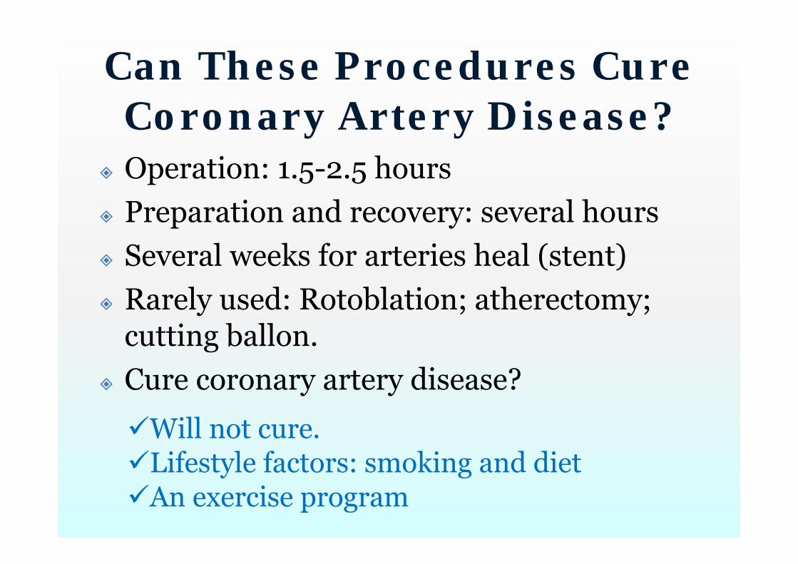
Can These Procedures Cure Coronary Artery Disease?
Operation: 1.5-2.5 hours Preparation and recovery: several hours Several weeks for arteries heal (stent) Rarely used: Rotoblation; atherectomy;
cutting ballon. Cure coronary artery disease?
Will not cure.Lifestyle factors: smoking and dietAn exercise program
46
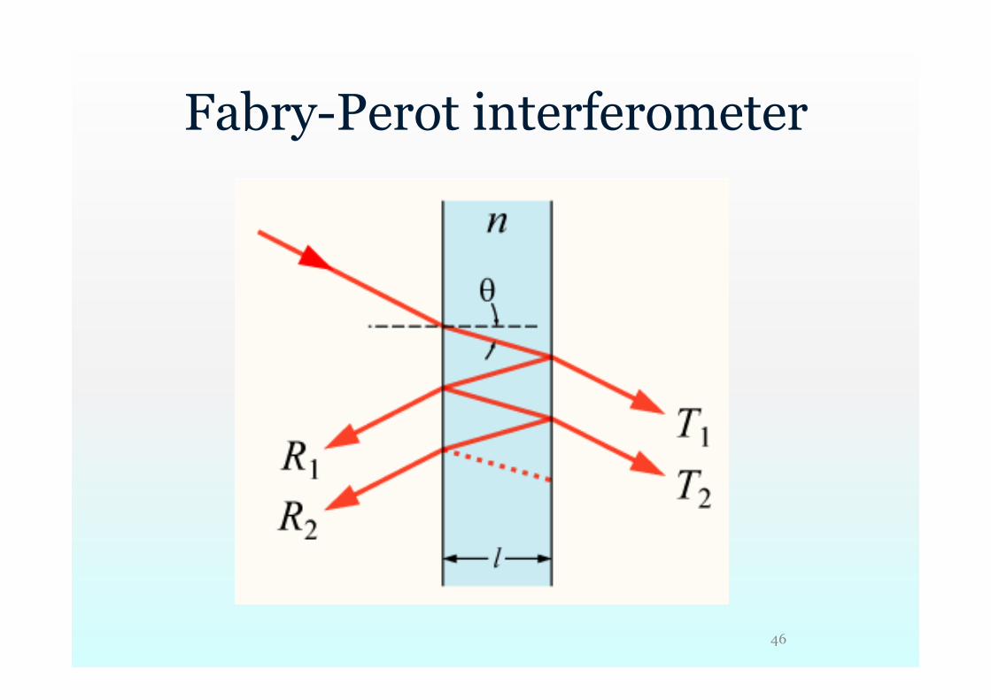
Fabry-Perot interferometer
47
EFPI
The required sensing length may be reduced by having the light double back through multiple reflections along its propagation path.
The use of a single-cavity extrinsic Fabry–Pérot interferometer (EFPI) as a guided-wave/bulk-biomaterial interaction biosensor.
J. L. Elster, M. E. Jones, M. K. Evans, S. M. Lenahan, C. A. Boyce, W. Velander, and R. VanTassell, “Optical fiber extrinsic Fabry–Pérot interferometric (EFPI)-based biosensors,” SPIE, vol. 3911, pp. 105–112,2000.
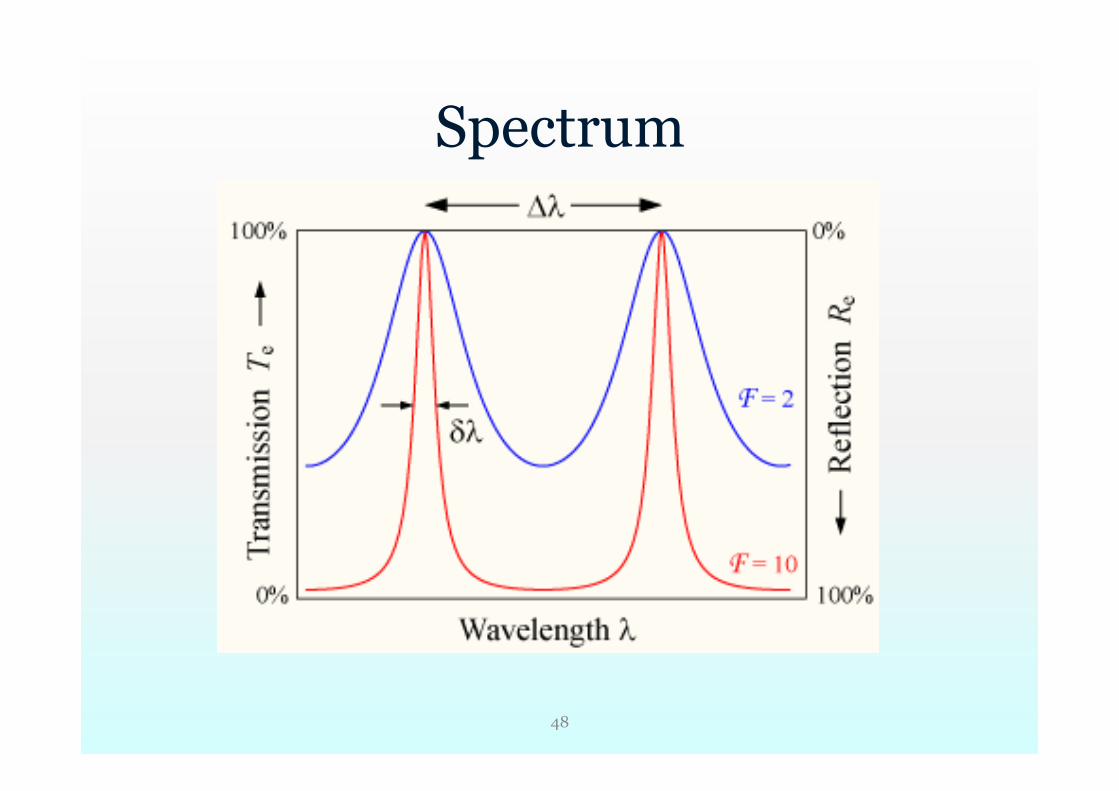
Spectrum
48
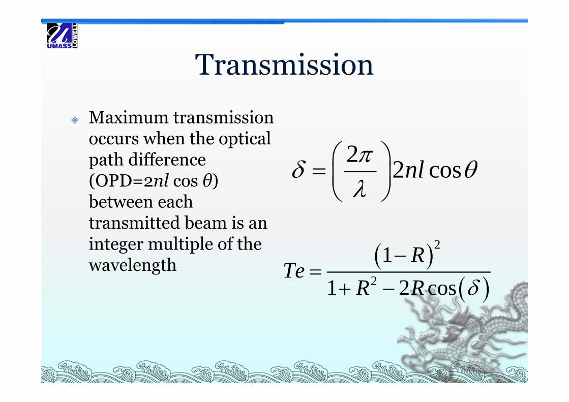
Transmission
Maximum transmission occurs when the optical path difference (OPD=2nl cos θ) between each transmitted beam is an integer multiple of the wavelength
2 2 cosnl
2
2
11 2 cos
RTe
R R
49
FP biosensor
Biomolecule attachment OPD change Spectrum demodulation
50
Reference
X. Wang, K. Cooper, A. Wang, J. Xu, Z. Wang, Y. Zhang, and Z. Tu, "Label-free DNA detection on the surface of silica optical fiber tip". Appl. Phys. Lett. Vol. 89, 163901 (October 2006).
51
References [1] http://www.redwoodeditor.com/content/SCAI/scai/ [2] Tonino PA, De Bruyne B, Pijls NH, et al (January 2009).
"Fractional flow reserve versus angiography for guiding percutaneous coronary intervention". N. Engl. J. Med. 360 (3): 213–24. doi:10.1056/NEJMoa0807611. PMID 19144937.
[3] Cohen D. J., Carrozza J. P., Baim D. S., Ricciardi M. J., Davidson C. J., Bloom J. M., Pitt B., Waters D., Brown W. V., (Dec 9, 1999), “Aggressive Lipid-Lowering Therapy Compared with Angioplasty in Stable Coronary Artery Disease”N Engl J Med 1999; 341:1853-1855.
References [4] Habib A. Dakik, MD; Neal S. Kleiman, MD; John A.
Farmer, MD; Zuo-Xiang He, MD; Juliet A. Wendt, MD; Craig M. Pratt, MD; Mario S. Verani, MD; John J. Mahmarian, MD, (Circulation. 1998;98:2017-2023.) © 1998 American Heart Association, Inc. “Intensive Medical Therapy Versus Coronary Angioplasty for Suppression of Myocardial Ischemia in Survivors of Acute Myocardial Infarction - A Prospective, Randomized Pilot Study”, Presented in part at the 46th Scientific Sessions of the American College of Cardiology, Anaheim, Calif, March 17, 1997 and published in abstract form (J Am Coll Cardiol. 1997;29(suppl A):53A.