disorders of coagulation and … of coagulation and thrombosis immune thrombocytopenia autoimmune...
TRANSCRIPT
DISORDERS OF COAGULATION
AND THROMBOSIS
Zhongshan Hospital,Fudan University
Yunfeng Cheng
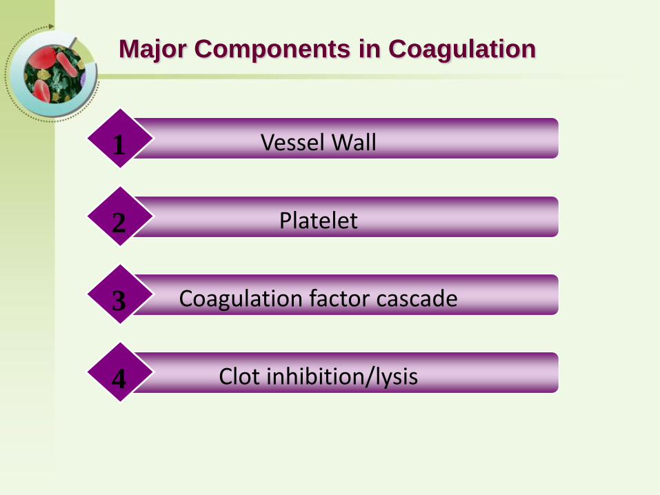
Major Components in Coagulation
Vessel Wall1
Platelet2
Coagulation factor cascade3
Clot inhibition/lysis4
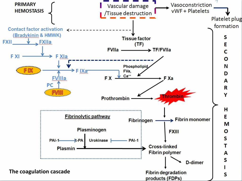
coagulation cascade
The coagulation cascade
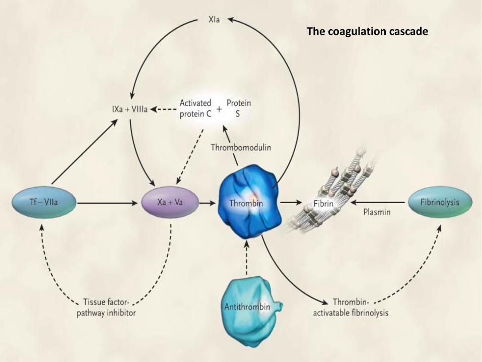
The coagulation cascade
The coagulation cascade
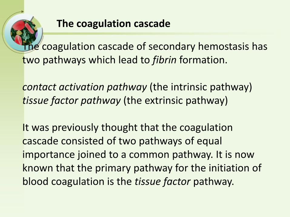
The coagulation cascade of secondary hemostasis has two pathways which lead to fibrin formation.
contact activation pathway (the intrinsic pathway)tissue factor pathway (the extrinsic pathway)
It was previously thought that the coagulation cascade consisted of two pathways of equal importance joined to a common pathway. It is now known that the primary pathway for the initiation of blood coagulation is the tissue factor pathway.
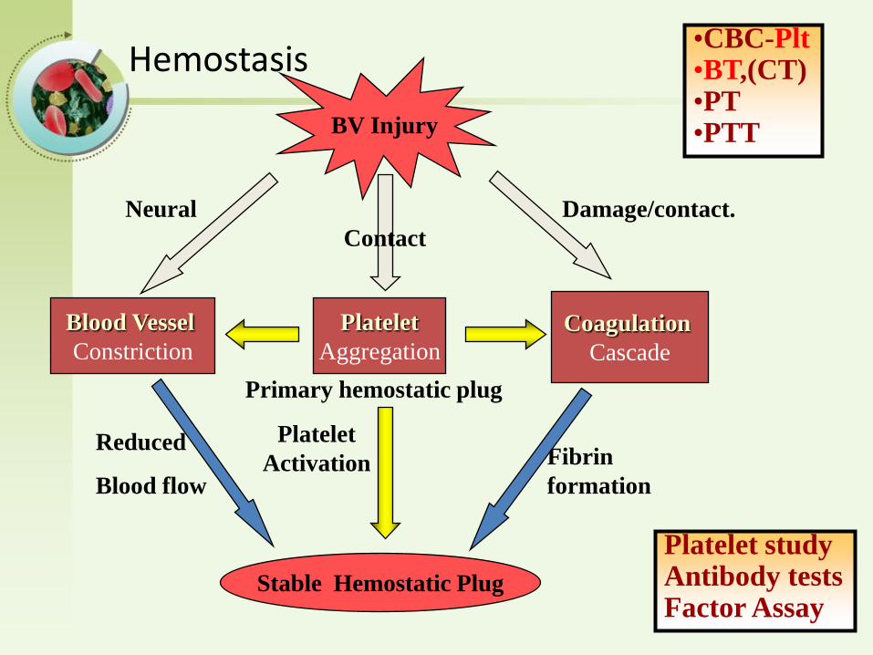
Hemostasis
BV Injury
Platelet
Aggregation
Platelet
Activation
Blood Vessel
Constriction
Coagulation
Cascade
Stable Hemostatic Plug
Fibrin
formation
Reduced
Blood flow
Damage/contact.
Primary hemostatic plug
Neural
•CBC-Plt•BT,(CT)•PT•PTT
Platelet studyAntibody testsFactor Assay
Contact
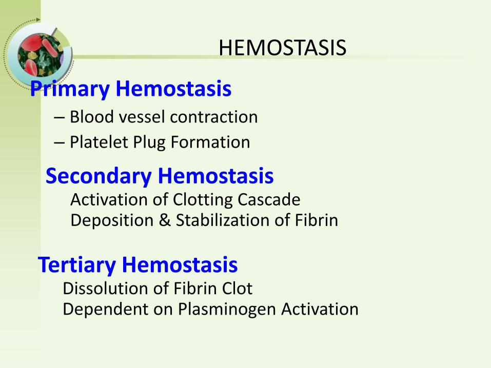
HEMOSTASIS
Primary Hemostasis– Blood vessel contraction
– Platelet Plug Formation
Secondary HemostasisActivation of Clotting Cascade Deposition & Stabilization of Fibrin
Tertiary HemostasisDissolution of Fibrin ClotDependent on Plasminogen Activation
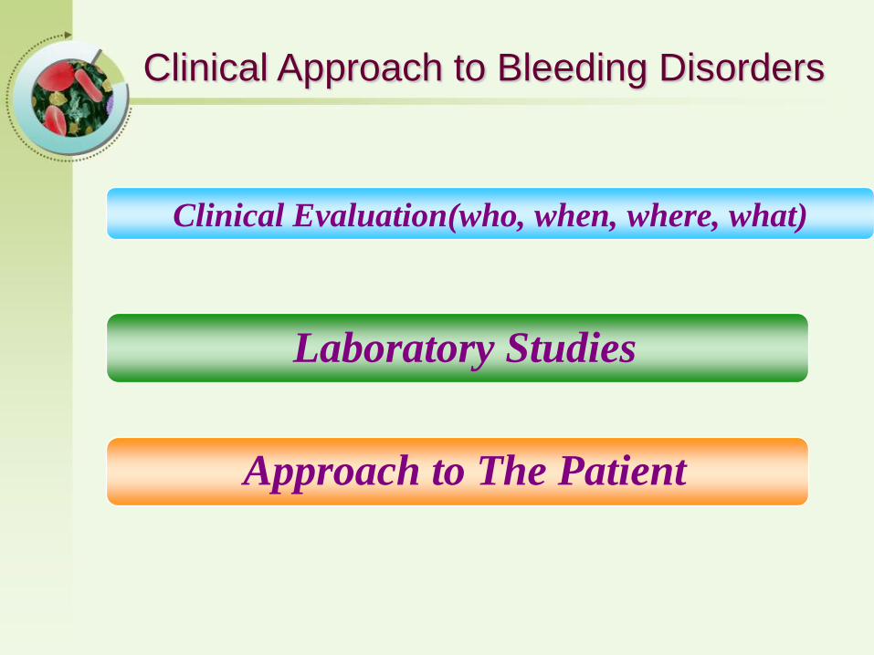
Clinical Approach to Bleeding Disorders
Clinical Evaluation(who, when, where, what)
2 Laboratory Studies
Approach to The Patient
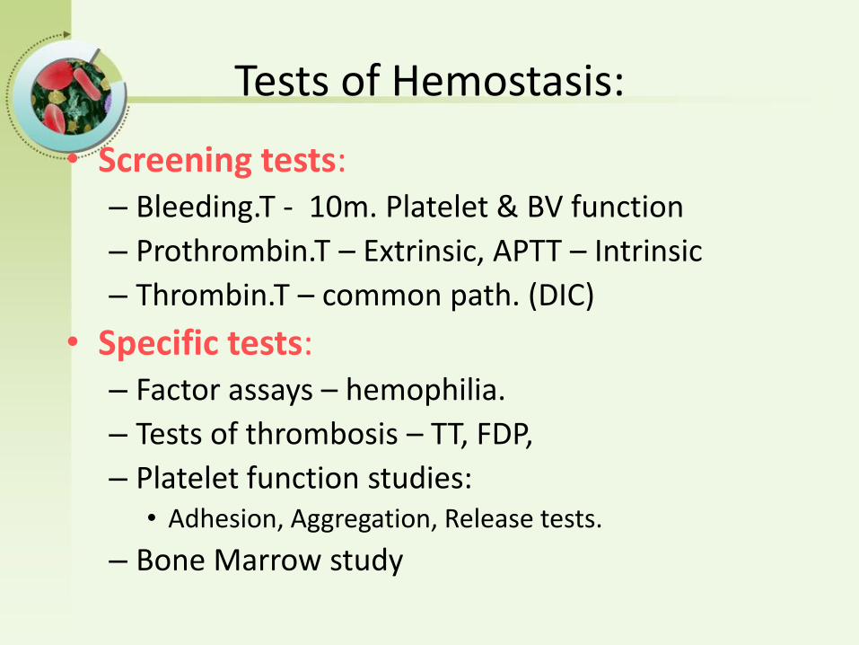
Tests of Hemostasis:
• Screening tests:– Bleeding.T - 10m. Platelet & BV function
– Prothrombin.T – Extrinsic, APTT – Intrinsic
– Thrombin.T – common path. (DIC)
• Specific tests:– Factor assays – hemophilia.
– Tests of thrombosis – TT, FDP,
– Platelet function studies: • Adhesion, Aggregation, Release tests.
– Bone Marrow study
Bleeding: Clinical Features
• Local - Vs - General, spontaneous . .
• Hematoma / Joint Bleeds- Coag• Skin / Mucosal Bleeds – PLT
• wound / surgical bleeding –– Immediate - PLT – Delayed - Coagulation
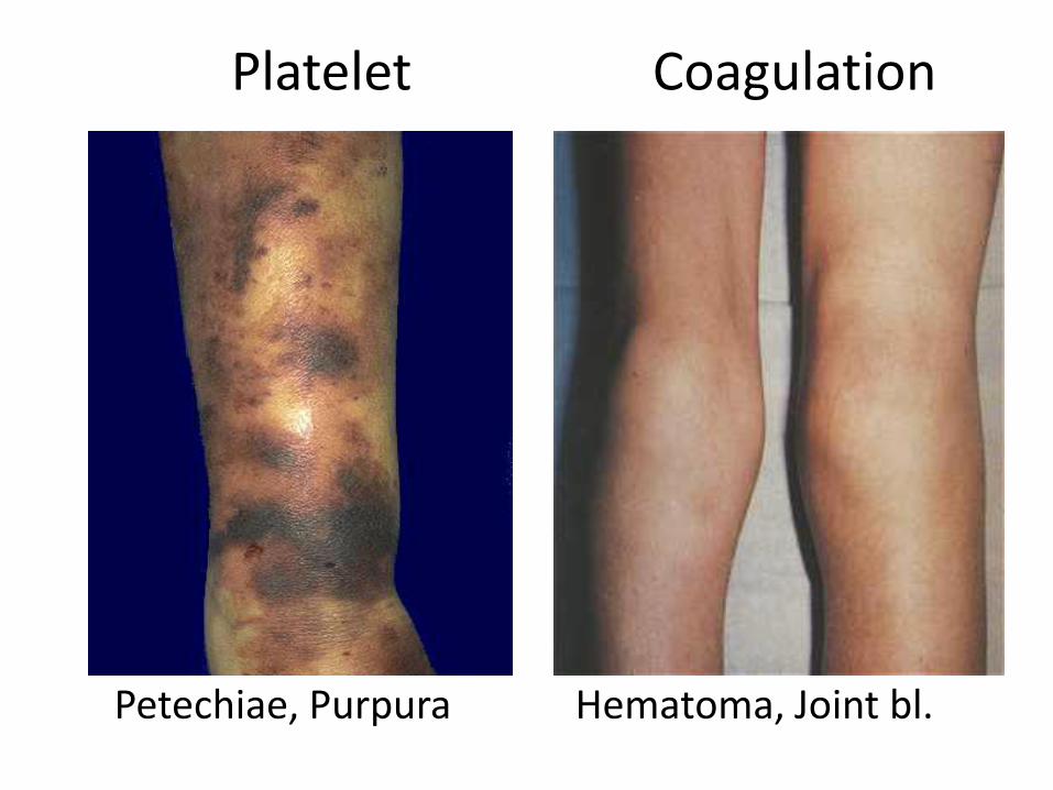
Platelet Coagulation
Petechiae, Purpura Hematoma, Joint bl.
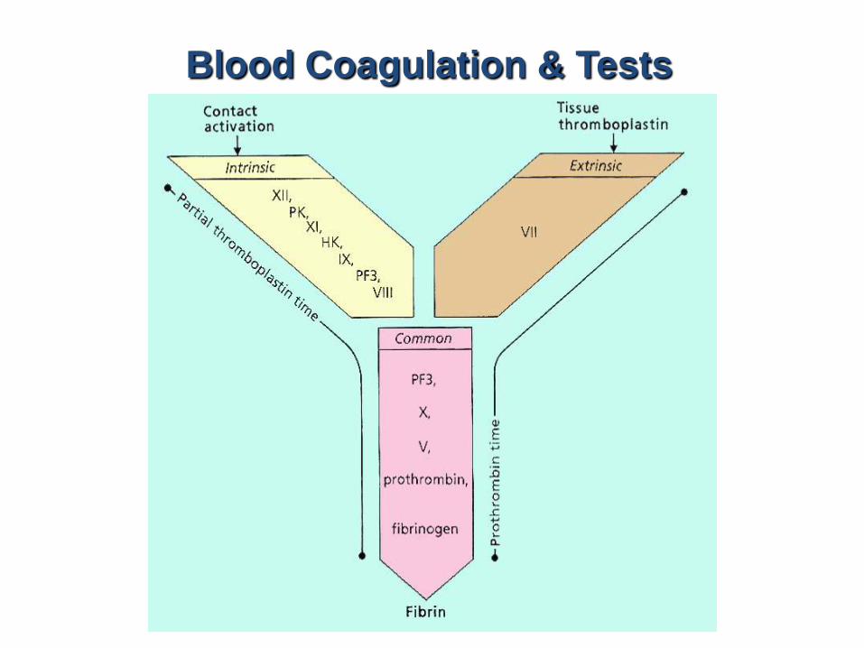
Blood Coagulation & Tests
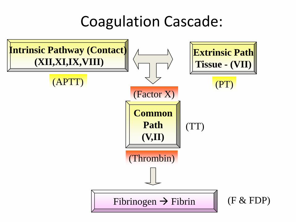
Coagulation Cascade:
Intrinsic Pathway (Contact)
(XII,XI,IX,VIII)Extrinsic Path
Tissue - (VII)
Fibrinogen Fibrin
Common
Path
(V,II)
(PT)(APTT)
(TT)
(F & FDP)
(Factor X)
(Thrombin)
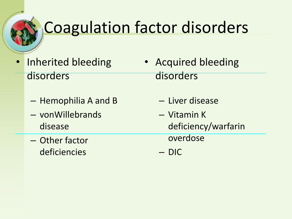
Coagulation factor disorders
• Inherited bleeding disorders
– Hemophilia A and B
– vonWillebrands disease
– Other factor deficiencies
• Acquired bleeding disorders
– Liver disease
– Vitamin K deficiency/warfarin overdose
– DIC
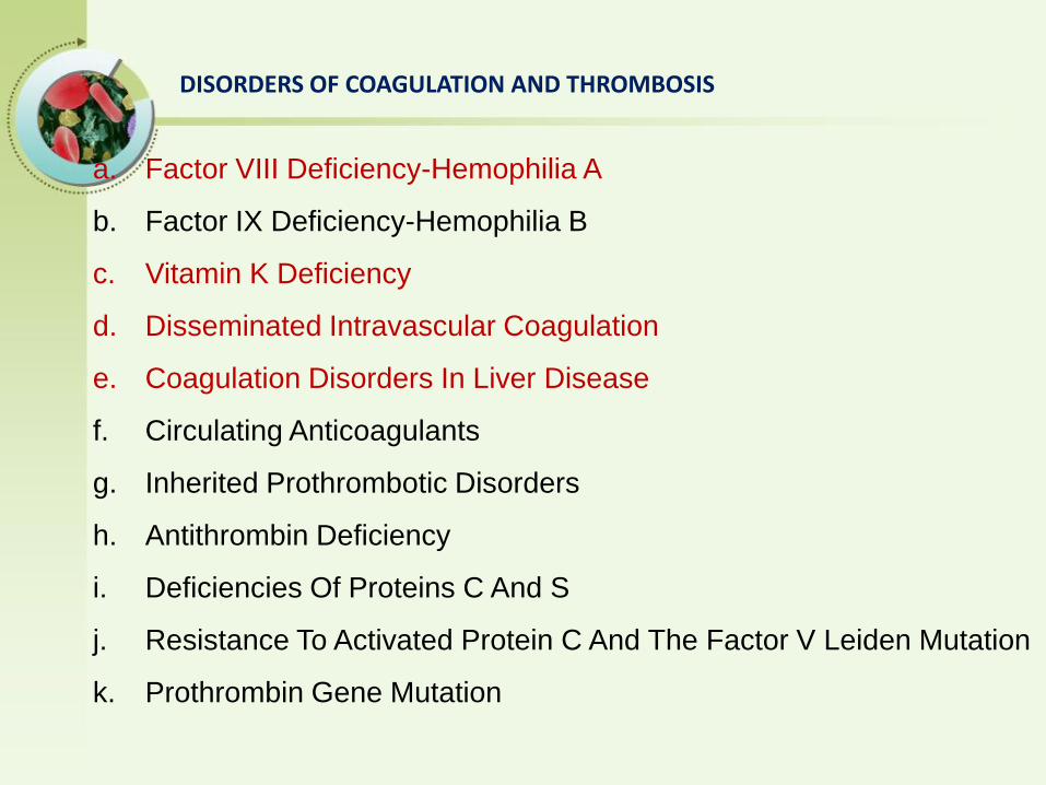
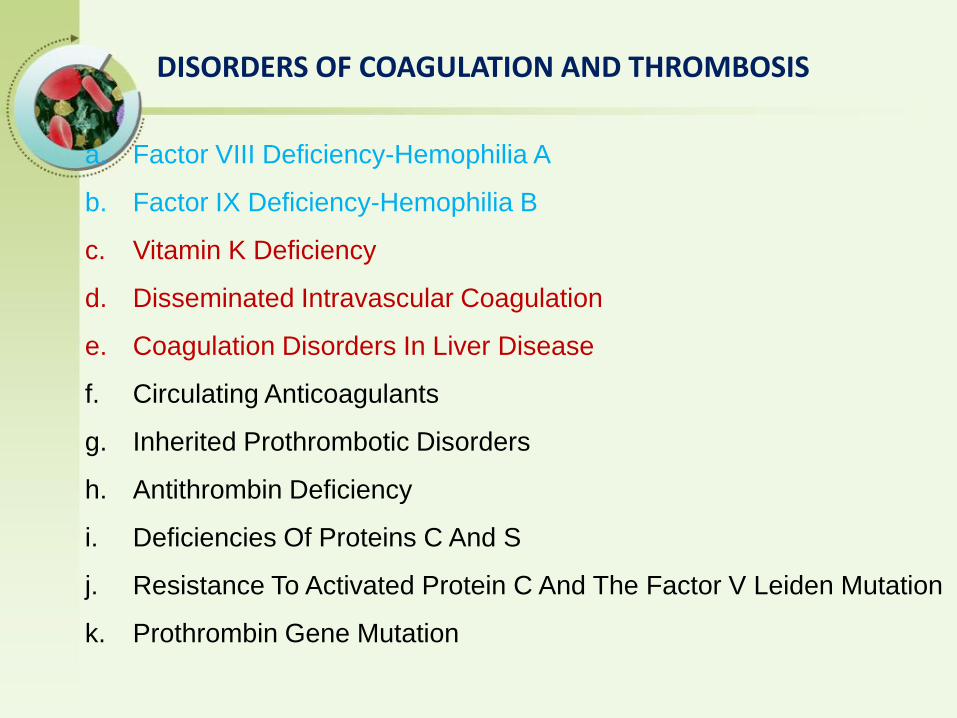
a. Factor VIII Deficiency-Hemophilia A
b. Factor IX Deficiency-Hemophilia B
c. Vitamin K Deficiency
d. Disseminated Intravascular Coagulation
e. Coagulation Disorders In Liver Disease
f. Circulating Anticoagulants
g. Inherited Prothrombotic Disorders
h. Antithrombin Deficiency
i. Deficiencies Of Proteins C And S
j. Resistance To Activated Protein C And The Factor V Leiden Mutation
k. Prothrombin Gene Mutation
DISORDERS OF COAGULATION AND THROMBOSIS
Immune Thrombocytopenia
Autoimmune Disorder
Thrombocytopenia
Heterogeneous
Should know:
What is…
Causes
Signs & Symptoms
Diagnosis
Treatments
Living with
Key points
Links
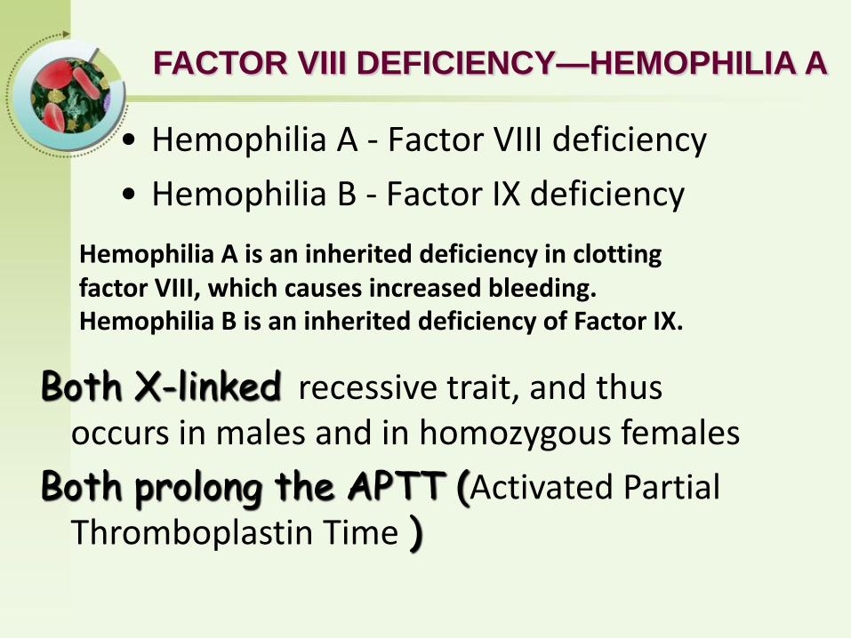
FACTOR VIII DEFICIENCY—HEMOPHILIA A
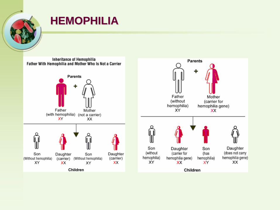
Both X-linked recessive trait, and thus occurs in males and in homozygous females
Both prolong the APTT (Activated Partial Thromboplastin Time )
• Hemophilia A - Factor VIII deficiency
• Hemophilia B - Factor IX deficiency
Hemophilia A is an inherited deficiency in clotting factor VIII, which causes increased bleeding.Hemophilia B is an inherited deficiency of Factor IX.
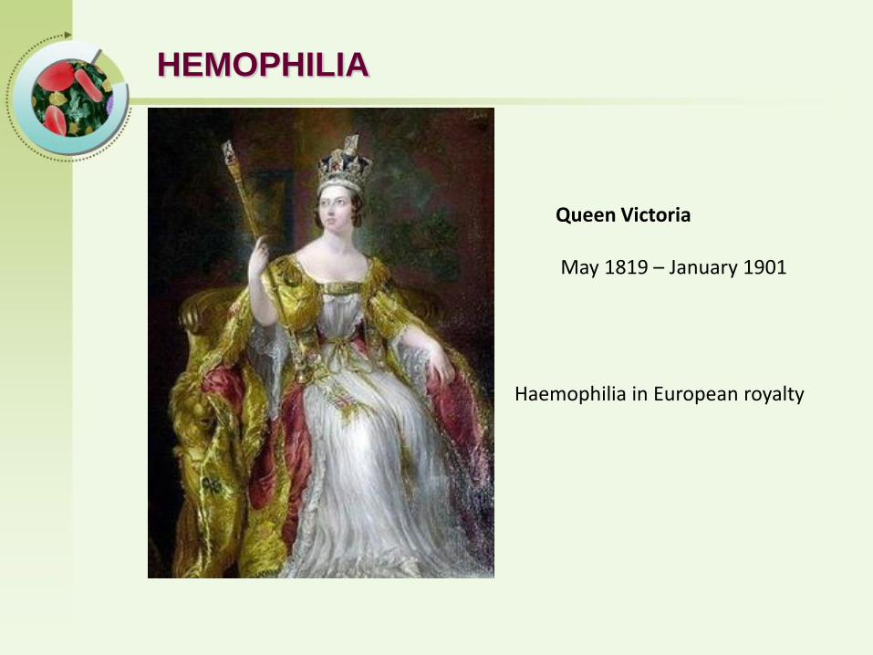
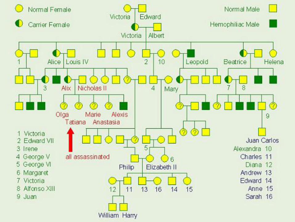
HEMOPHILIA
HEMOPHILIA
Queen Victoria
May 1819 – January 1901
Haemophilia in European royalty
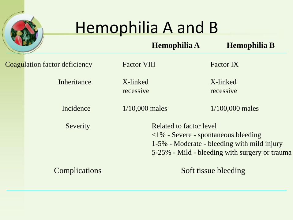
Hemophilia A and BHemophilia A Hemophilia B
Coagulation factor deficiency Factor VIII Factor IX
Inheritance X-linked X-linked
recessive recessive
Incidence 1/10,000 males 1/100,000 males
Severity Related to factor level
<1% - Severe - spontaneous bleeding
1-5% - Moderate - bleeding with mild injury
5-25% - Mild - bleeding with surgery or trauma
Complications Soft tissue bleeding
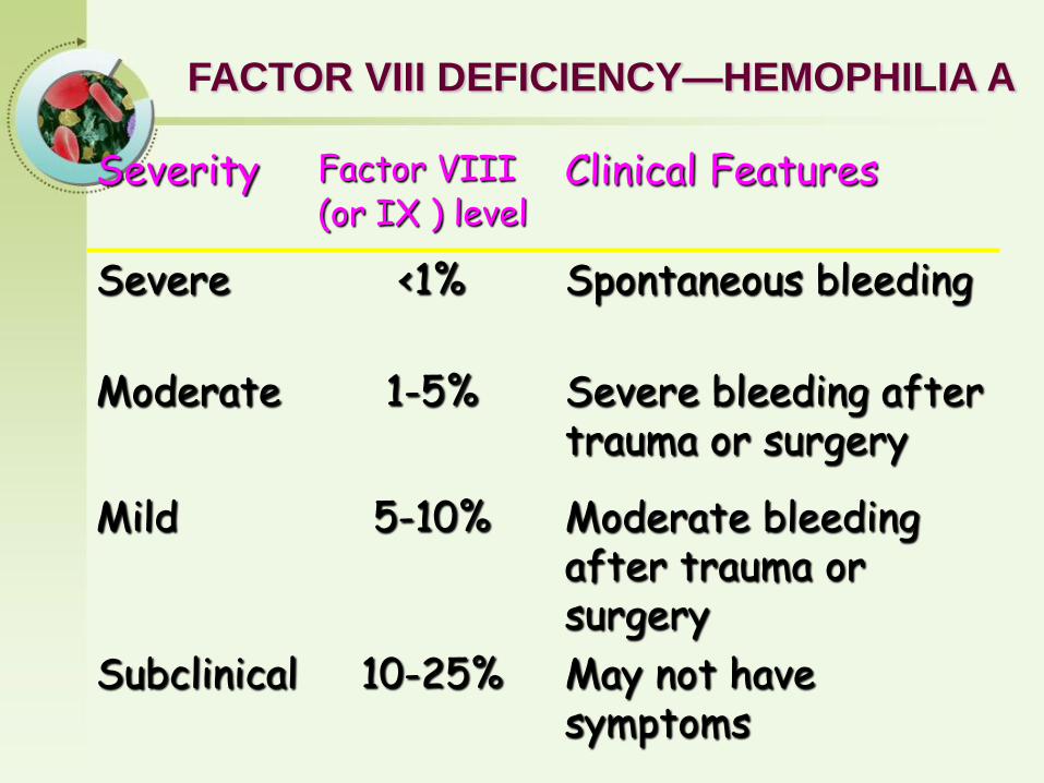
FACTOR VIII DEFICIENCY—HEMOPHILIA A
Severity Factor VIII Clinical Features(or IX ) level
Severe <1% Spontaneous bleeding
Moderate 1-5% Severe bleeding after trauma or surgery
Mild 5-10% Moderate bleeding after trauma or surgery
Subclinical 10-25% May not have symptoms
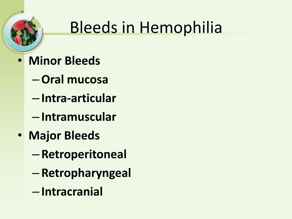
Bleeds in Hemophilia
• Minor Bleeds
– Oral mucosa
– Intra-articular
– Intramuscular
• Major Bleeds
– Retroperitoneal
– Retropharyngeal
– Intracranial
Acutely, bleeding in hemophilics can
• Compromise circulation.
• Close off airway
• Lead to compartment syndrome
• Cause neuropathy
• Cause pain
• Set up inflammatory synovitis, which can trigger more bleeding
Chronically, bleeding in hemophilicscan
• Compromise joint function and lead to accelerated DJD (hemophilic arthropathy)
• Lead to pseudotumor formation
• Weaken muscles
• Lead to permanent neurologic compromise.
Other complications of hemophilia
• Inhibitor formation
• Viral (and other) Infections
• Job bias
Inhibitors in Hemophilia
• Are alloantibodies formed against Factor VIII (and less commonly FIX) in patients with congenital hemophilia
• Inactivate exogenously administered clotting factor
• Must be treated with bypassing agents which less efficacious in hemostasis
• Increase morbidity and mortality
Complications - Orthopedic
• Hemophilic arthropathy
• Surgeries
– synovectomy - arthroscopic or isotopic
– joint fusion - arthrodesis
– joint replacement - arthroplasty
Complications - Viral infection
• HIV and hepatitis B and C
• Blood donation steps to improve viral safety of plasma-derived products
1. blood donors screened
2. donated blood tested for antibodies to hepatitis and HIV
3. all factor concentrate products are virally inactivated and purified

Hemophilia
Clinical manifestations (hemophilia A & B indistinguishable)
Hemarthrosis (most common)Fixed joints
Soft tissue hematomas (e.g., muscle)Muscle atrophyShortened tendons
Other sites of bleedingUrinary tractCNS, neck (may be life-threatening)
Prolonged bleeding after surgery or dental extractions
Treatment of hemophilia A
• Intermediate purity plasma products– Virucidally treated– May contain von Willebrand factor
• High purity (monoclonal) plasma products– Virucidally treated– No functional von Willebrand factor
• Recombinant factor VIII– Virus free/No apparent risk– No functional von Willebrand factor
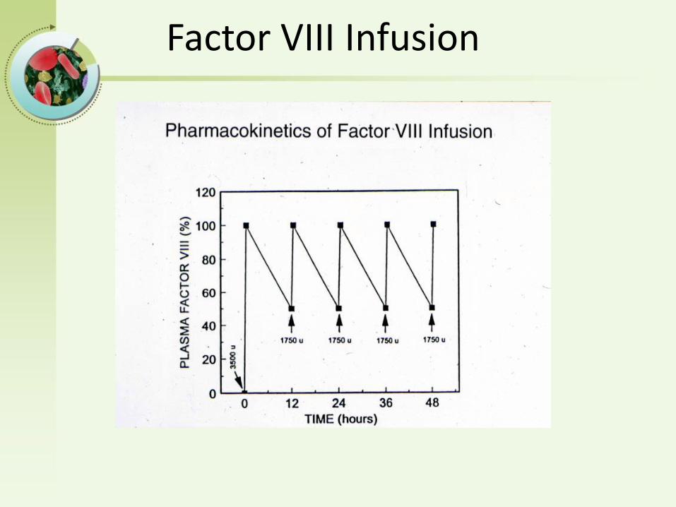
Factor VIII Infusion
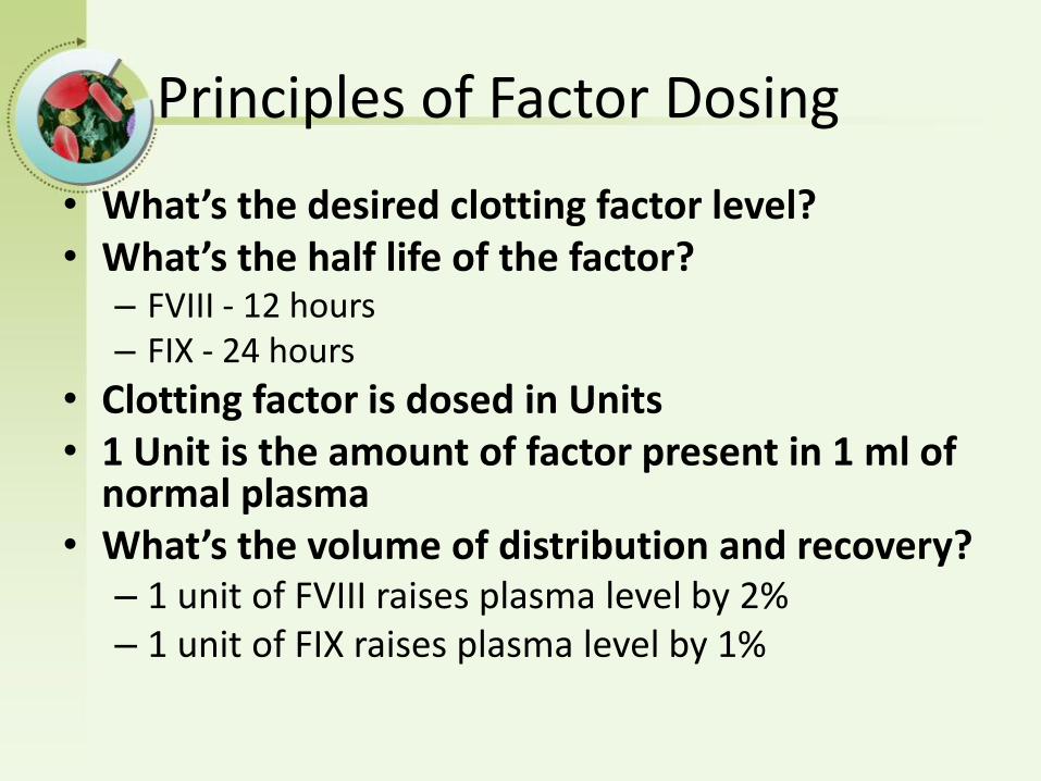
Principles of Factor Dosing
• What’s the desired clotting factor level?• What’s the half life of the factor?
– FVIII - 12 hours– FIX - 24 hours
• Clotting factor is dosed in Units• 1 Unit is the amount of factor present in 1 ml of
normal plasma• What’s the volume of distribution and recovery?
– 1 unit of FVIII raises plasma level by 2%– 1 unit of FIX raises plasma level by 1%
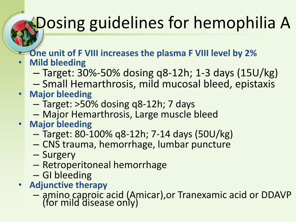
Dosing guidelines for hemophilia A
• One unit of F VIII increases the plasma F VIII level by 2%• Mild bleeding
– Target: 30%-50% dosing q8-12h; 1-3 days (15U/kg)– Small Hemarthrosis, mild mucosal bleed, epistaxis
• Major bleeding – Target: >50% dosing q8-12h; 7 days – Major Hemarthrosis, Large muscle bleed
• Major bleeding– Target: 80-100% q8-12h; 7-14 days (50U/kg)– CNS trauma, hemorrhage, lumbar puncture– Surgery– Retroperitoneal hemorrhage– GI bleeding
• Adjunctive therapy– amino caproic acid (Amicar),or Tranexamic acid or DDAVP
(for mild disease only)
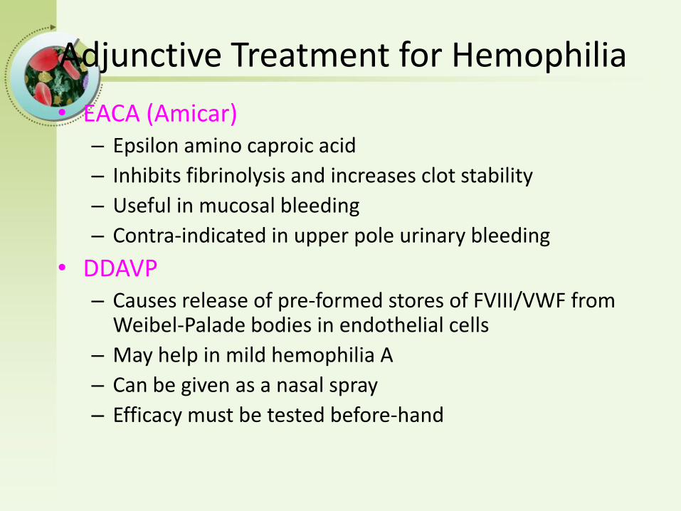
Adjunctive Treatment for Hemophilia
• EACA (Amicar)– Epsilon amino caproic acid
– Inhibits fibrinolysis and increases clot stability
– Useful in mucosal bleeding
– Contra-indicated in upper pole urinary bleeding
• DDAVP– Causes release of pre-formed stores of FVIII/VWF from
Weibel-Palade bodies in endothelial cells
– May help in mild hemophilia A
– Can be given as a nasal spray
– Efficacy must be tested before-hand
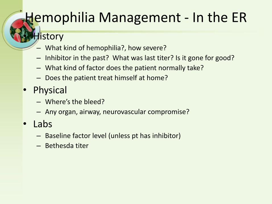
Hemophilia Management - In the ER• History
– What kind of hemophilia?, how severe?
– Inhibitor in the past? What was last titer? Is it gone for good?
– What kind of factor does the patient normally take?
– Does the patient treat himself at home?
• Physical– Where’s the bleed?
– Any organ, airway, neurovascular compromise?
• Labs– Baseline factor level (unless pt has inhibitor)
– Bethesda titer
Hemophilia Management - In the ER
• Give clotting factor and pain meds
• Decide if admission is necessary
Complications of therapy
• Formation of inhibitors (antibodies)– 10-15% of severe hemophilia A patients
– 1-2% of severe hemophilia B patients
• Viral infections– Hepatitis B Human parvovirus
– Hepatitis C Hepatitis A
– HIV Other
Treatment of hemophilia B• Agent
– High purity factor IX
– Recombinant human factor IX
• Dose
– Initial dose: 100U/kg
– Subsequent: 50 U/kg every 24 hours
fresh-frozen plasma or a plasma fraction enriched in the prothrombin complex proteins
Future of Hemophilia
• Primary prophylaxis
• Gene therapy
• Inhibitor prediction/treatment
• Prevention of
– hemophilic arthropathy
– viral contamination of factor products
Zhongshan Hospital,Fudan University

DISORDERS OF COAGULATION
AND THROMBOSIS- part II
Zhongshan Hospital,Fudan University
Yunfeng Cheng
a. Factor VIII Deficiency-Hemophilia A
b. Factor IX Deficiency-Hemophilia B
c. Vitamin K Deficiency
d. Disseminated Intravascular Coagulation
e. Coagulation Disorders In Liver Disease
f. Circulating Anticoagulants
g. Inherited Prothrombotic Disorders
h. Antithrombin Deficiency
i. Deficiencies Of Proteins C And S
j. Resistance To Activated Protein C And The Factor V Leiden Mutation
k. Prothrombin Gene Mutation
DISORDERS OF COAGULATION AND THROMBOSIS
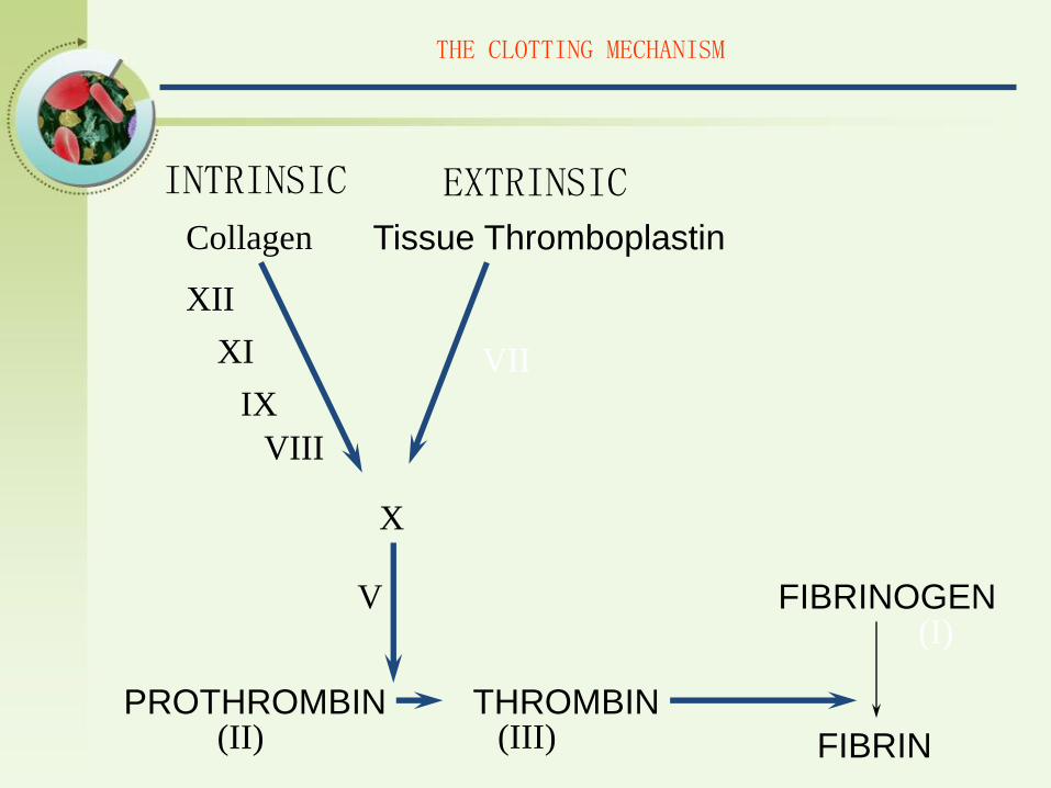
THE CLOTTING MECHANISM
INTRINSIC EXTRINSIC
PROTHROMBIN THROMBIN
FIBRINOGEN
FIBRIN(II) (III)
(I)V
X
Tissue ThromboplastinCollagen
VII
XII
XI
IX
VIII
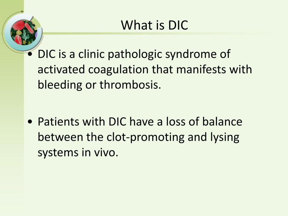
What is DIC
• DIC is a clinic pathologic syndrome of activated coagulation that manifests with bleeding or thrombosis.
• Patients with DIC have a loss of balance between the clot-promoting and lysing systems in vivo.
What is DIC
• This syndrome can have a clinical spectrum ranging from bleeding to a prothrombotic state.
• DIC is not a specific diagnosis, and its presence always indicates another underlying disease.
• Bleeding associated with DIC usually results from excess fibrinolysis; thrombosis associated with DIC results from excess thrombin formation.
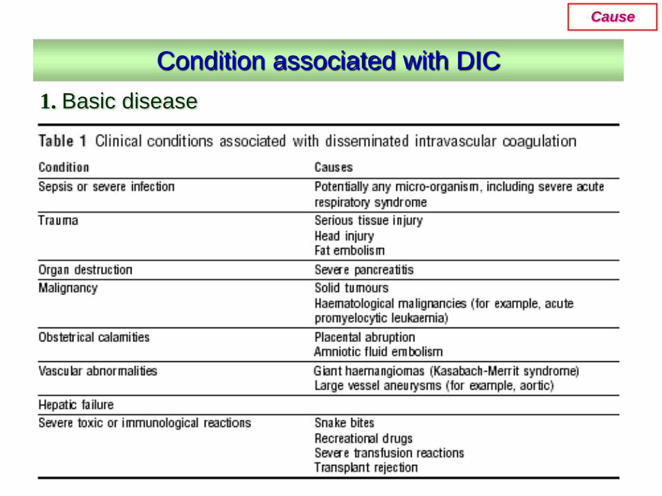
1. Basic disease
Condition associated with DIC
Cause
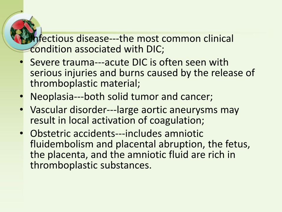
• Infectious disease---the most common clinical condition associated with DIC;
• Severe trauma---acute DIC is often seen with serious injuries and burns caused by the release of thromboplastic material;
• Neoplasia---both solid tumor and cancer;• Vascular disorder---large aortic aneurysms may
result in local activation of coagulation; • Obstetric accidents---includes amniotic
fluidembolism and placental abruption, the fetus, the placenta, and the amniotic fluid are rich in thromboplastic substances.
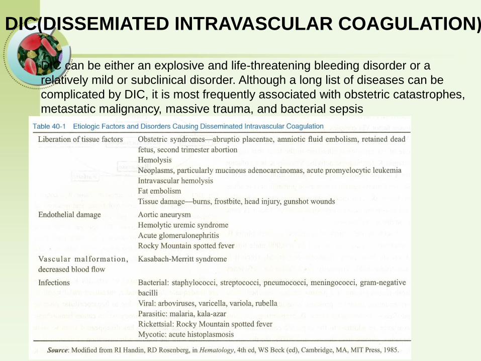
DIC(DISSEMIATED INTRAVASCULAR COAGULATION)
DIC can be either an explosive and life-threatening bleeding disorder or a
relatively mild or subclinical disorder. Although a long list of diseases can be
complicated by DIC, it is most frequently associated with obstetric catastrophes,
metastatic malignancy, massive trauma, and bacterial sepsis
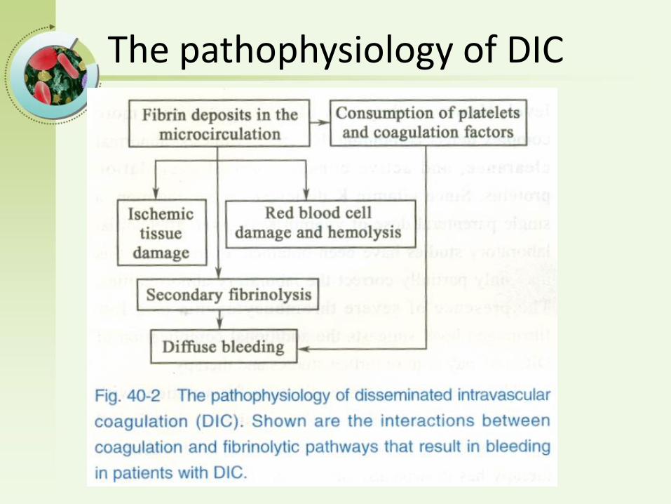
The pathophysiology of DIC
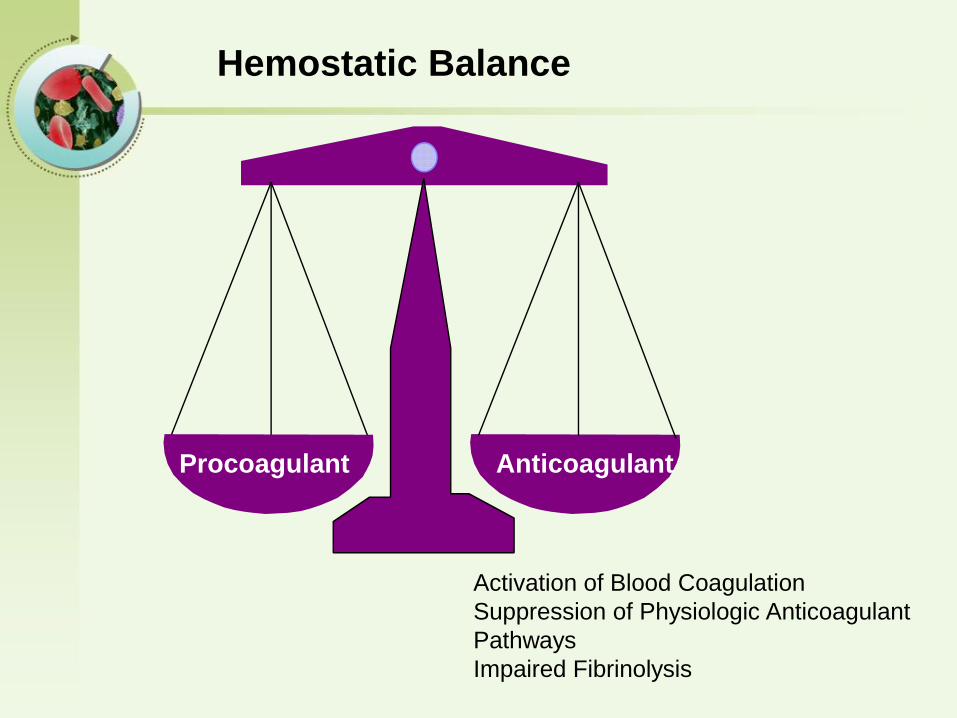
Hemostatic Balance
Procoagulant Anticoagulant
Activation of Blood Coagulation
Suppression of Physiologic Anticoagulant
Pathways
Impaired Fibrinolysis
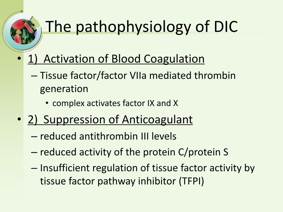
The pathophysiology of DIC
• 1) Activation of Blood Coagulation
– Tissue factor/factor VIIa mediated thrombin generation
• complex activates factor IX and X
• 2) Suppression of Anticoagulant
– reduced antithrombin III levels
– reduced activity of the protein C/protein S
– Insufficient regulation of tissue factor activity by tissue factor pathway inhibitor (TFPI)
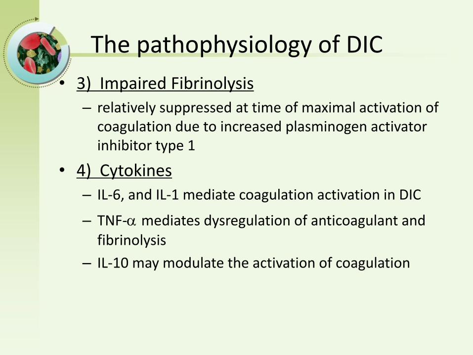
The pathophysiology of DIC
• 3) Impaired Fibrinolysis
– relatively suppressed at time of maximal activation of coagulation due to increased plasminogen activator inhibitor type 1
• 4) Cytokines
– IL-6, and IL-1 mediate coagulation activation in DIC
– TNF- mediates dysregulation of anticoagulant and fibrinolysis
– IL-10 may modulate the activation of coagulation
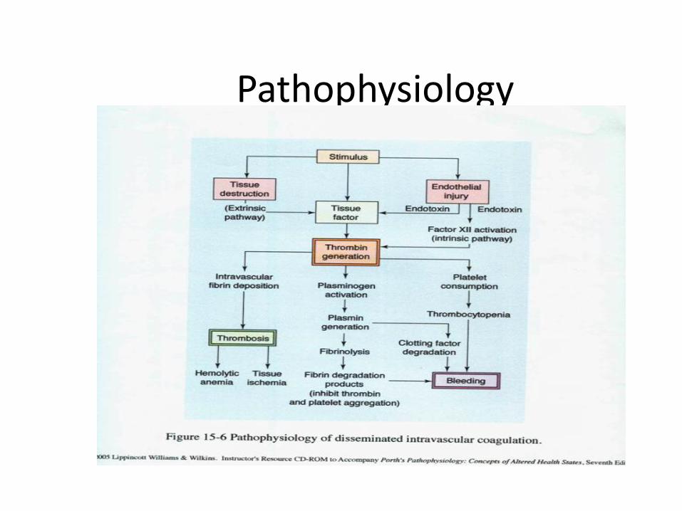
Pathophysiology
In DIC, a systemic activation of the coagulation system simultaneously leads to thrombus formation (compromising blood supply to various organs) and exhaustion of platelets and coagulation factors (results in hemorrhage). This is a disruption of body homeostasis.
(Porth, 2001)
Pathophysiology
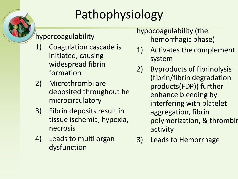
hypercoagulability
1) Coagulation cascade is initiated, causing widespread fibrin formation
2) Microthrombi are deposited throughout he microcirculatory
3) Fibrin deposits result in tissue ischemia, hypoxia, necrosis
4) Leads to multi organ dysfunction
hypocoagulability (the hemorrhagic phase)
1) Activates the complement system
2) Byproducts of fibrinolysis (fibrin/fibrin degradation products(FDP)) further enhance bleeding by interfering with platelet aggregation, fibrin polymerization, & thrombin activity
3) Leads to Hemorrhage
Pathophysiology
Consumption of clotting factors and platelets
Activation of secondary fibrinolytic system
Production of fibrin degradation products
Bleeding mechanisms
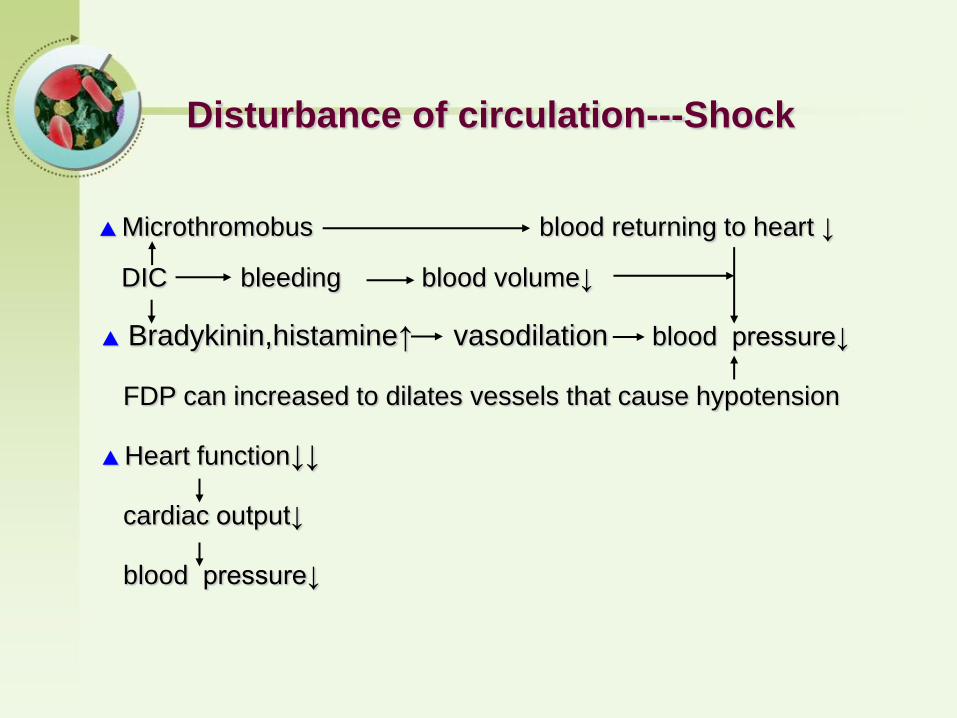
▲ Microthromobus blood returning to heart ↓
DIC bleeding blood volume↓
▲ Bradykinin,histamine↑ vasodilation blood pressure↓
FDP can increased to dilates vessels that cause hypotension
▲ Heart function↓↓
cardiac output↓
blood pressure↓
Disturbance of circulation---Shock
• Renal insufficiency
• Acute adrenal failure
• Adult respiratory distress syndrome
• Infarcted skin syndrome of purpura
Ischemic tissue damage-dysfunction of multiple organs
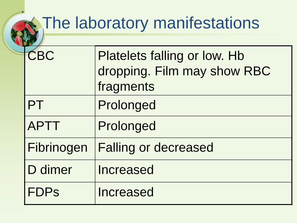
The laboratory manifestations
CBC Platelets falling or low. Hb
dropping. Film may show RBC
fragments
PT Prolonged
APTT Prolonged
Fibrinogen Falling or decreased
D dimer Increased
FDPs Increased
Diagnosis of DIC
• Presence of disease associated with DIC
• Appropriate clinical setting
– Clinical evidence of thrombosis, hemorrhage or both.
• Laboratory studies
– no single test is accurate
– serial test are more helpful than single test
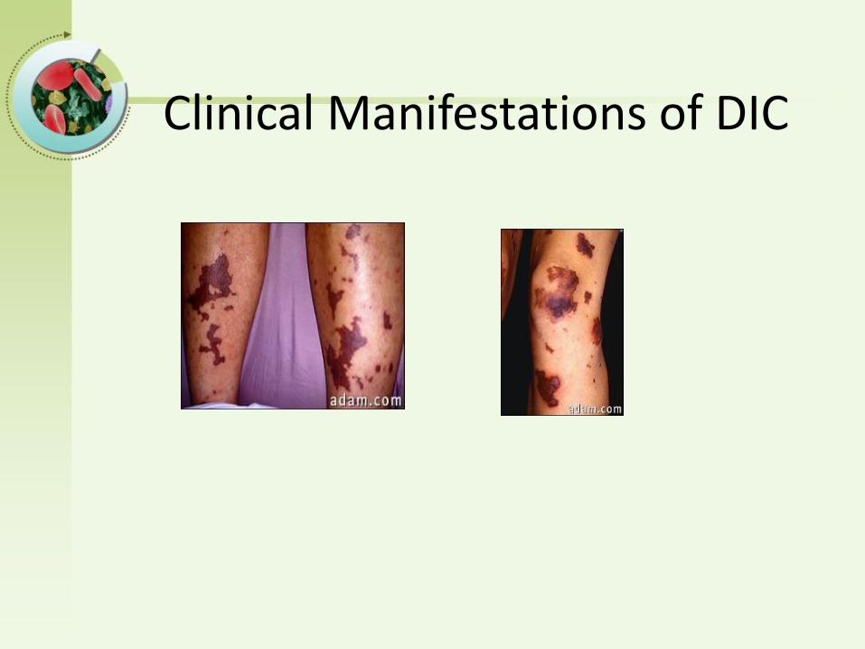
Clinical Manifestations of DIC
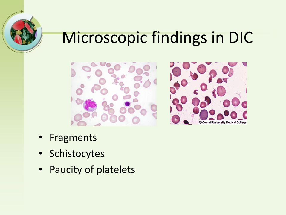
Microscopic findings in DIC
• Fragments
• Schistocytes
• Paucity of platelets
Bachelor of Chinese Medicine
Disseminated intravascular coagulation
• Laboratory results:
– Prolonged PT, APTT and TT
– Reduced fibrinogen level
– Increased D-Dimers
– Thrombocytopenia: <100,000 or rapidly declining
– Microangiopathic changes in blood film
Low levels of coagulation inhibitors: AT III, protein C
Bachelor of Chinese Medicine
Diagnosis of DIC
• Clinical setting
• Laboratory tests
• Criteria– Underlying disease
– Platelet count < 100 X 109/L, or rapid decline
– PT and APTT Prolonged
– Presence of fibrin degradation products
– Low levels of coagulation inhibitors
– Low fibrinogen level in severe cases

Differential Diagnosis
• Severe liver failure
• Vitamin K deficiency
• Liver disease
• Thrombotic thrombocytopenic purpura
• Congenital abnormalities of fibrinogen
• Hypercoagulable stage
• Hypocoagulable stage
• Secondary fibrinolytic stage
Stages of DIC
• Almost always secondary
• Consumptive coagulopathy
• Decreases in both coagulants & anticoagulants
• Severity may relate to levels of anticoagulants
Acute DIC
• Symptoms appear within days to weeks, clotting substances are consumed but not reduced
• Bleeding is rarely seen but the evidence of DIC can be detected by laboratory examinations
• Thrombotic manifestation is commonly seen in malignant patient
Subacute DIC
• DIC develops slowly and the illness goes through within months
• The clinical and laboratory features of chronic DIC present over periods of month and the patient is usually not critically ill
• Usually symptoms are concealed
Chronic DIC
Bachelor of Chinese Medicine
Treatment of DIC
• Treatment of underlying disorder: Stop the triggering process
• Supportive therapy
• Other treatments
– Plasma and platelet substitution therapy
– Anticoagulants
– Physiologic coagulation inhibitors
Bachelor of Chinese Medicine
Treatment of DIC
• Platelets and Plasma
– to treat bleeding tendency
– to cover an invasive procedure for patients with a high risk of bleeding
• Clotting factor concentrates
– overcomes large volumes of plasma
– but not advocated because: 1) contains small amount of activated factors, and 2) DIC results in deficiency of multiple factors
Plasma therapy
• Indications
– Active bleeding
– Patient requiring invasive procedures
– Patient at high risk for bleeding complications
• Fresh frozen plasma(FFP):
– provides clotting factors, fibrinogen, inhibitors, and platelets in balanced amounts.
– Usual dose is 10-15 ml/kg
Bachelor of Chinese Medicine
Treatment of DIC
• Anticoagulants
– low dose heparin
– low molecular weight heparin
– new thrombin inhibitors (ATIII independent)
– useful for clinically overt thromboembolism or extensive deposition of fibrin
Bachelor of Chinese Medicine
Concentrates of coagulation inhibitors
• Antithrombin concentrate
– reduces sepsis related mortality
– improvement of DIC and organ function
• Supportive therapeutic option in severe DIC
Coagulation Inhibitor Therapy
• Antithrombin
• Protein C concentrate
• Tissue Factor Pathway Inhibitor (TFPI)
• Heparin
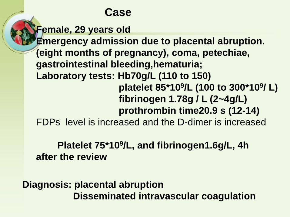
Female, 29 years old
Emergency admission due to placental abruption.
(eight months of pregnancy), coma, petechiae,
gastrointestinal bleeding,hematuria;
Laboratory tests: Hb70g/L (110 to 150)
platelet 85*109/L (100 to 300*109/ L)
fibrinogen 1.78g / L (2~4g/L)
prothrombin time20.9 s (12-14)
FDPs level is increased and the D-dimer is increased
Platelet 75*109/L, and fibrinogen1.6g/L, 4h
after the review
Diagnosis: placental abruption
Disseminated intravascular coagulation
Case
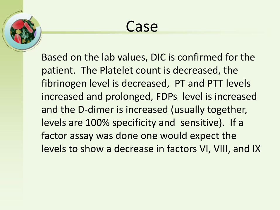
Case
Based on the lab values, DIC is confirmed for the patient. The Platelet count is decreased, the fibrinogen level is decreased, PT and PTT levels increased and prolonged, FDPs level is increased and the D-dimer is increased (usually together, levels are 100% specificity and sensitive). If a factor assay was done one would expect the levels to show a decrease in factors VI, VIII, and IX
Answer for Case Study
Lab abnormalities along with clinical presentation is used to confirm a diagnosis of DIC
If abnormal results are obtained for PT, PTT, platelets, and fibrinogen, then the D-dimer and FDPs levels are used to confirm DIC...FDPs abnormal in 75% and D-dimer in 95%.
Objectives
• Understand the pathophysiology of Disseminated Intravascular Coagulation (DIC)
• Identify risk factors and etiology of DIC
• Describe the signs and symptoms of DIC
• Identify treatment modalities for DIC
• Define, identify and understand Acute vs Chronic DIC
• Develop understanding of diagnosing DIC (lab interpretations)
Glossary
Activated PTT- aPTT tests the intrinsic and common pathways
D-dimer- an antigen formed as a result of plasmin lysis of cross-linked fibrin clots, documents the presence of thrombin
Fibrin degradation product(FDP)- degradation products increase as plasmin biodegrades fibrinogen and fibrin,levels are elevated in 85-100% of patients with DIC
Prothrombin Time(PT)- tests the extrinsic and common pathways
(Porth, 2004) & (Otto, 2001)
Zhongshan Hospital,Fudan University
DISORDERS OF COAGULATION
AND THROMBOSIS- part III
Zhongshan Hospital,Fudan University
Yunfeng Cheng
VITAMIN K DEFICIECY
Vitamin K is a fat-soluble vitamin that plays a
critical role in hemostasis.
Vitamin K is converted to an active epoxide in liver
microsomes and serves as a cofactor in the
enzymatic carboxylation of glutamic acid residues
on prothrombin complex proteins.
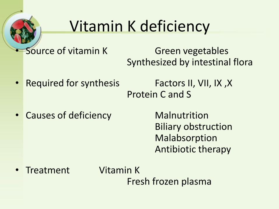
Vitamin K deficiency
• Source of vitamin K Green vegetablesSynthesized by intestinal flora
• Required for synthesis Factors II, VII, IX ,XProtein C and S
• Causes of deficiency MalnutritionBiliary obstructionMalabsorptionAntibiotic therapy
• Treatment Vitamin KFresh frozen plasma
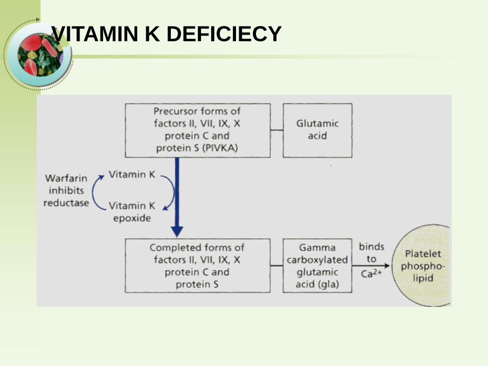
VITAMIN K DEFICIECY
VITAMIN K DEFICIECY
The three major causes of vitamin K deficiency:
• inadequate dietary intake
• intestinal malabsorption
• loss of storage sites due to hepatocellular
disease.
VITAMIN K DEFICIECY
With vitamin K deficiency:
• Plasma levels of all the prothrombin complex
proteins (factors II, VII, IX, X; proteins C and S)
decrease.
• Those with the shortest half-lives, factor VII and
protein C, decrease first. Because of the rapid fall
in factor VII, patients with mild vitamin K deficiency
may have a prolonged PT and a normal APTT.
• Later, as the levels of the other factors fall, the
APTT will also become prolonged.
VITAMIN K DEFICIECY
• Parenteral administration of 10 mg vitamin K
permits normal production of prothrombin
complex proteins within 8 to 10 h.
• Severe hemorrhage can be treated with fresh-
frozen plasma.
• Purified prothrombin complex concentrates
should be avoided because they contain trace
quantities of activated forms of the prothrombin
complex proteins and can cause thrombosis in
patients with liver disease.
Bachelor of Chinese Medicine
Vitamin K antagonism
• Oral anticoagulants (warfarin)
• Both PT and APTT Prolonged
• PT system chosen for monitoring
– due to shortest half life of factor VII
• INR system
– to standardize monitoring of oral anticoagulant therapy
• Important: INR should not be used in other clinical context

COAGULATION DISORDERS IN LIVER
DISEASE
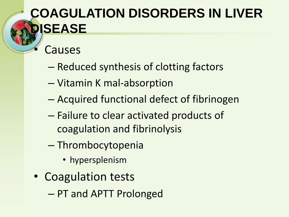
COAGULATION DISORDERS IN LIVER
DISEASE
• Causes
– Reduced synthesis of clotting factors
– Vitamin K mal-absorption
– Acquired functional defect of fibrinogen
– Failure to clear activated products of coagulation and fibrinolysis
– Thrombocytopenia
• hypersplenism
• Coagulation tests
– PT and APTT Prolonged
COAGULATION DISORDERS IN LIVER
DISEASE
– Treatment of Liver disease
– Vitamin K
– Fresh –frozen plasma
– Anticoagulation with heparin has been
advocated to control DIC
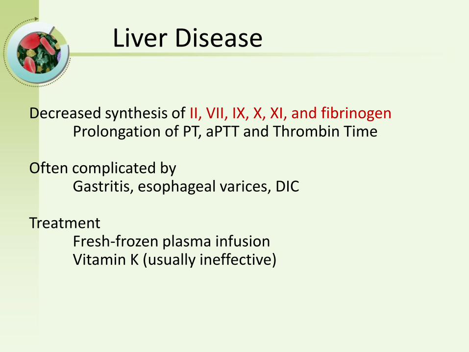
Liver Disease
Decreased synthesis of II, VII, IX, X, XI, and fibrinogenProlongation of PT, aPTT and Thrombin Time
Often complicated byGastritis, esophageal varices, DIC
TreatmentFresh-frozen plasma infusion Vitamin K (usually ineffective)
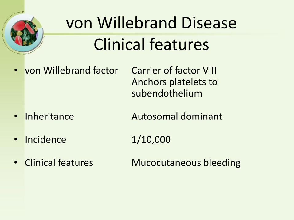
von Willebrand DiseaseClinical features
• von Willebrand factor Carrier of factor VIIIAnchors platelets to subendothelium
• Inheritance Autosomal dominant
• Incidence 1/10,000
• Clinical features Mucocutaneous bleeding
Laboratory evaluation of von Willebrand disease
Classification
– Type 1 Partial quantitative deficiency
– Type 2 Qualitative deficiency
– Type 3 Total quantitative deficiency
• Diagnostic tests: vWF antigen, vWF activity
Treatment of von Willebrand diseaseVaries by Classification
• Cryoprecipitate– Source of fibrinogen, factor VIII and VWF
– Only plasma fraction that consistently contains VWF multimers
– Correction of bleeding time is variable
• DDAVP (Deamino-8-arginine vasopressin)– Increases plasma VWF levels by stimulating secretion from
endothelium
– Duration of response is variable
– Used for type 1 disease
– Dosage 0.3 µg/kg q 12 hr IV
• Factor VIII concentrate– Virally inactivated product
– Used for type 2 and 3
CIRCULATING ANTICOAGULANTS
Circulating anticoagulants, or inhibitors, are usually
IgG antibodies that interfere with coagulation
reactions.
They arise in 15 to 20% of patients with factor VIII
or factor IX deficiency who have received plasma
infusions.
Nonspecific (lupus-like) inhibitors prolong
coagulation tests by binding to phospholipids.
lupus anticoagulant (LA), anticardiolipin antibody
(ACLA)
CIRCULATING ANTICOAGULANTS
PT ,APTT ,
massive plasma or concentrate infusion
activated prothrombin complex concentrates
plasmapheresis or exchange transfusion to lower antibody titer
Women who have had more than one midtrimester abortion,
especially those with SLE, should have a trial of anticoagulant
therapy. Patients with a single thrombotic episode
(stroke or pulmonary embolus)
and no other risk factor except LA or ACLA activity
should be treated.
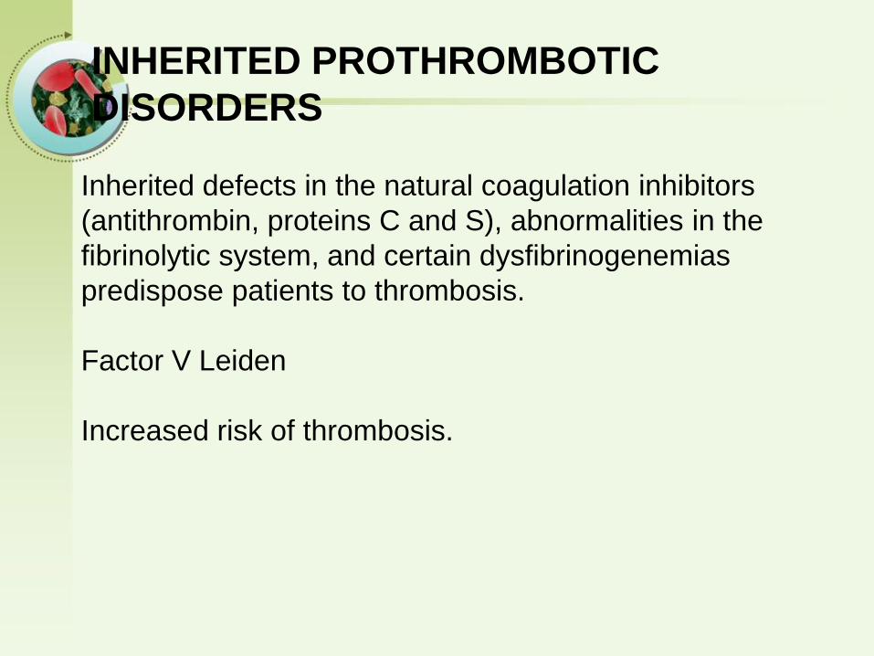
INHERITED PROTHROMBOTIC
DISORDERS
Inherited defects in the natural coagulation inhibitors
(antithrombin, proteins C and S), abnormalities in the
fibrinolytic system, and certain dysfibrinogenemias
predispose patients to thrombosis.
Factor V Leiden
Increased risk of thrombosis.
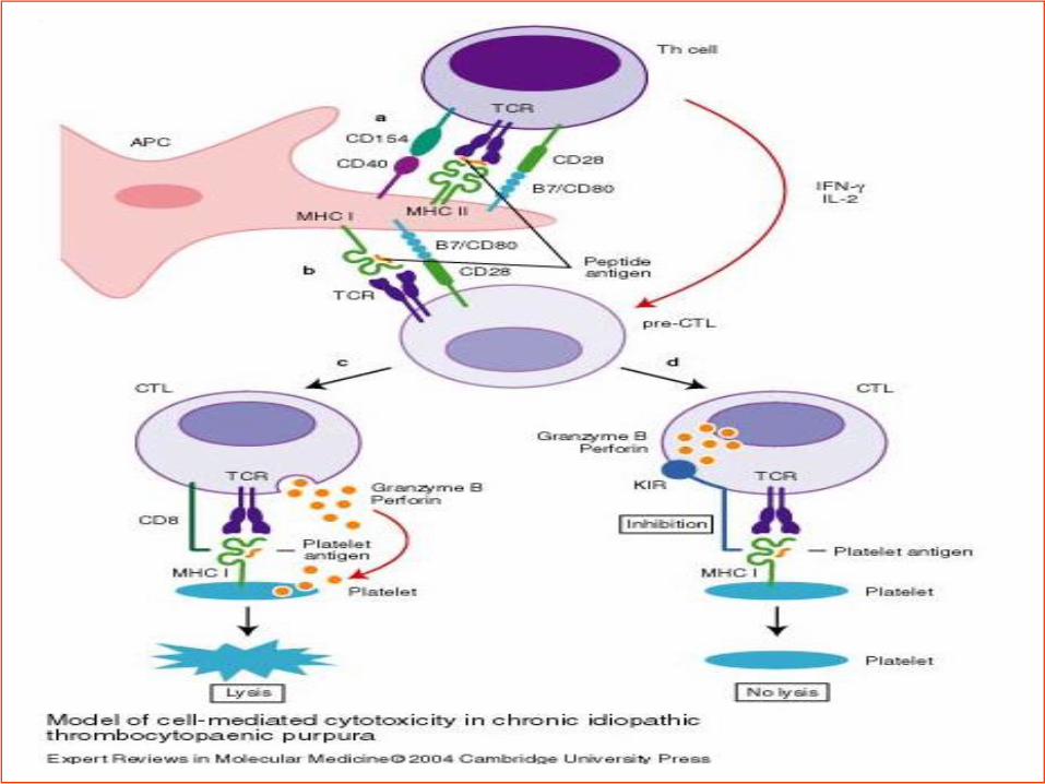
Immune Thrombocytopenia

Immune Thrombocytopenia(ITP)Autoimmune Disorder
Thrombocytopenia(Isolated thrombocytopenia)
(<100×109/L)
Heterogeneous
acquired
Absence of any obvious initiation or underlying
cause
Introduction
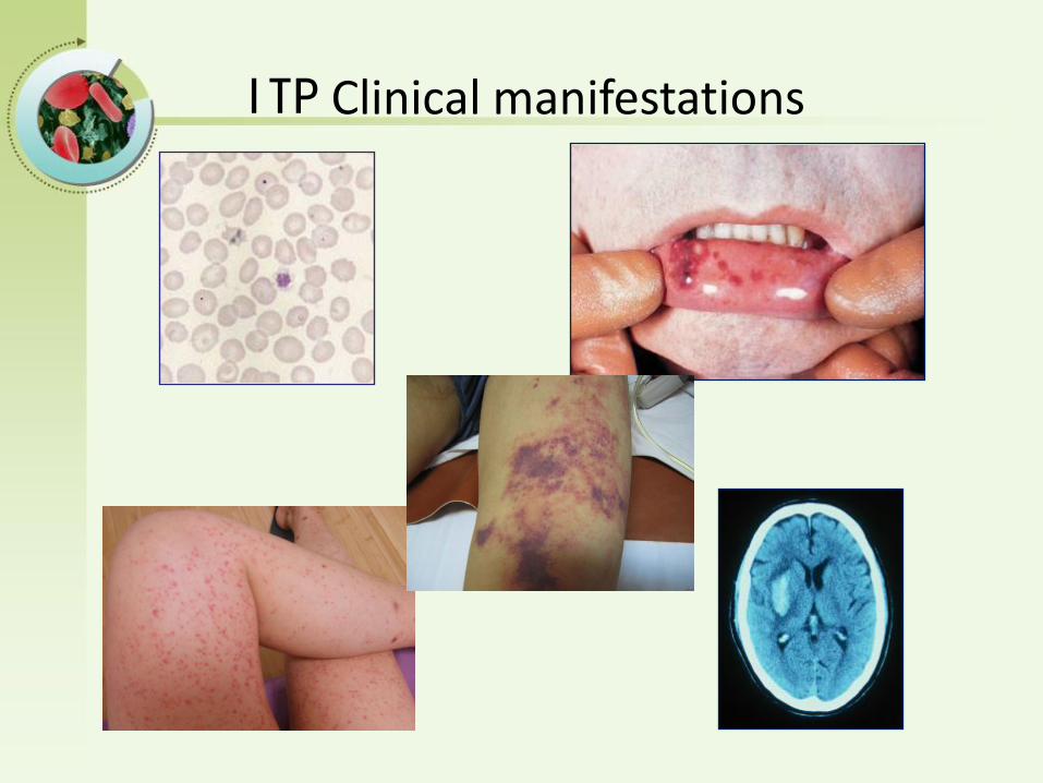
ITP Clinical manifestations
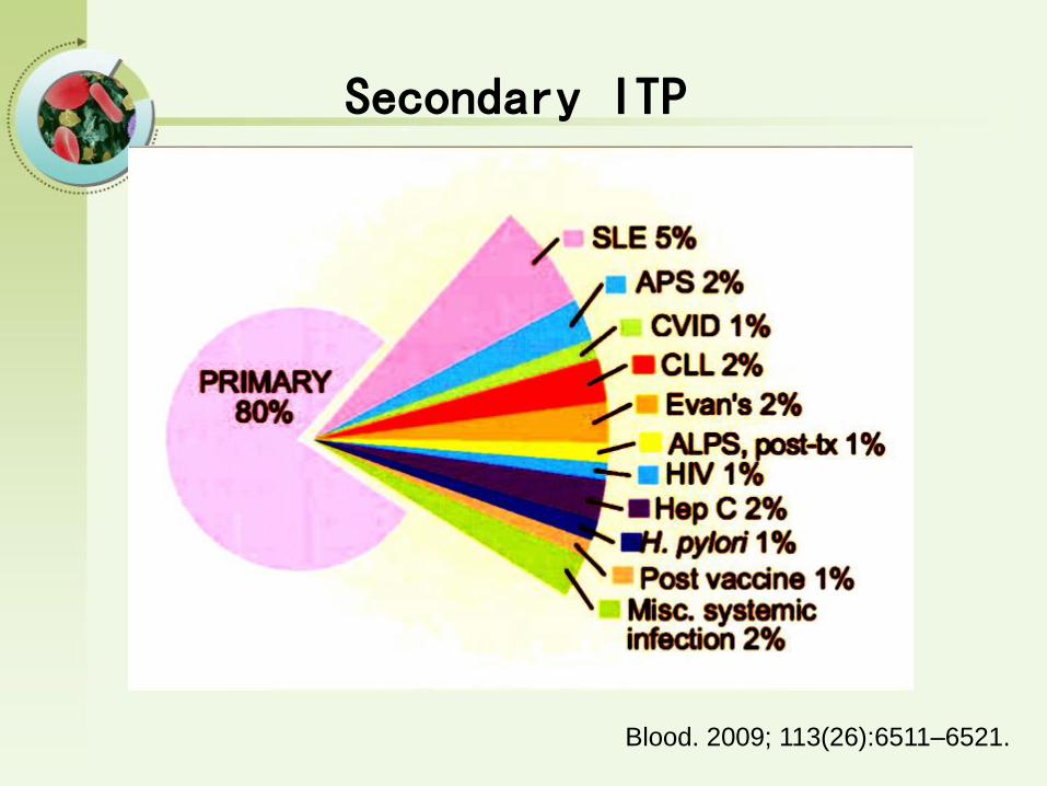
Blood. 2009; 113(26):6511–6521.
Secondary ITP
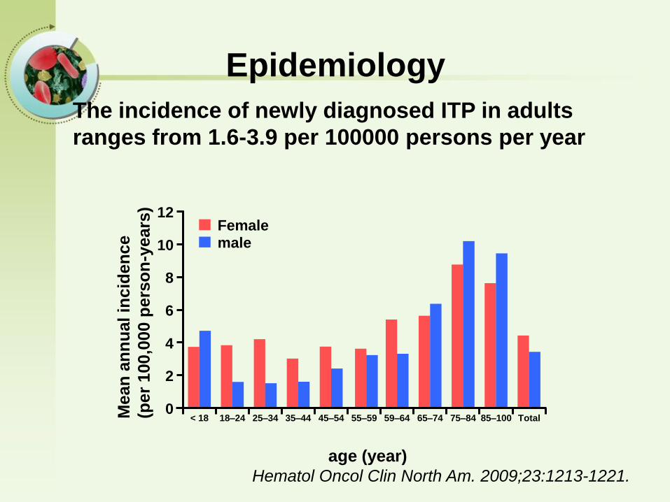
12
10
8
6
4
2
0< 18 18–24 25–34 35–44 45–54 55–59 59–64 65–74 75–84 85–100 Total
age (year)
Mean
an
nu
al in
cid
en
ce
(per
100,0
00 p
ers
on
-years
)
Femalemale
Hematol Oncol Clin North Am. 2009;23:1213-1221.
The incidence of newly diagnosed ITP in adults
ranges from 1.6-3.9 per 100000 persons per year
Epidemiology
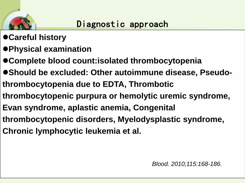
Careful history
Physical examination
Complete blood count:isolated thrombocytopenia
Should be excluded: Other autoimmune disease, Pseudo-
thrombocytopenia due to EDTA, Thrombotic
thrombocytopenic purpura or hemolytic uremic syndrome,
Evan syndrome, aplastic anemia, Congenital
thrombocytopenic disorders, Myelodysplastic syndrome,
Chronic lymphocytic leukemia et al.
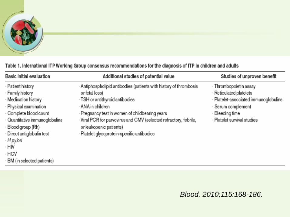
Diagnostic approach
Blood. 2010;115:168-186.
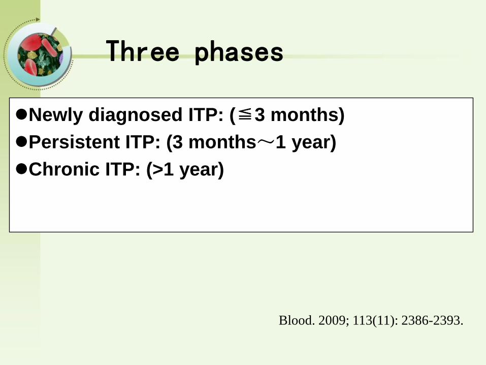
Newly diagnosed ITP: (≦3 months)
Persistent ITP: (3 months~1 year)
Chronic ITP: (>1 year)
Blood. 2009; 113(11): 2386-2393.
Three phases
Blood. 2010;115:168-186.
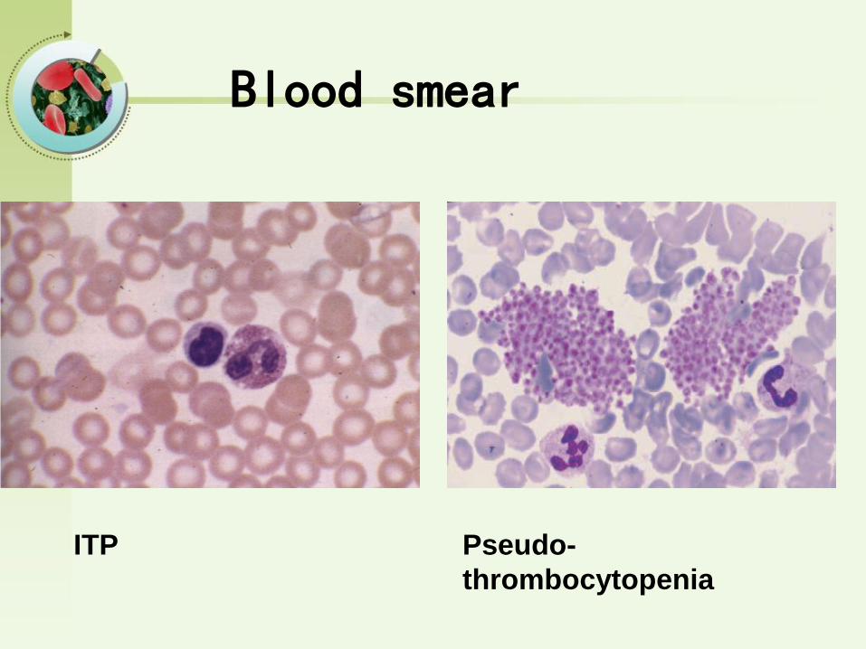
ITP Pseudo-
thrombocytopenia
Blood smear
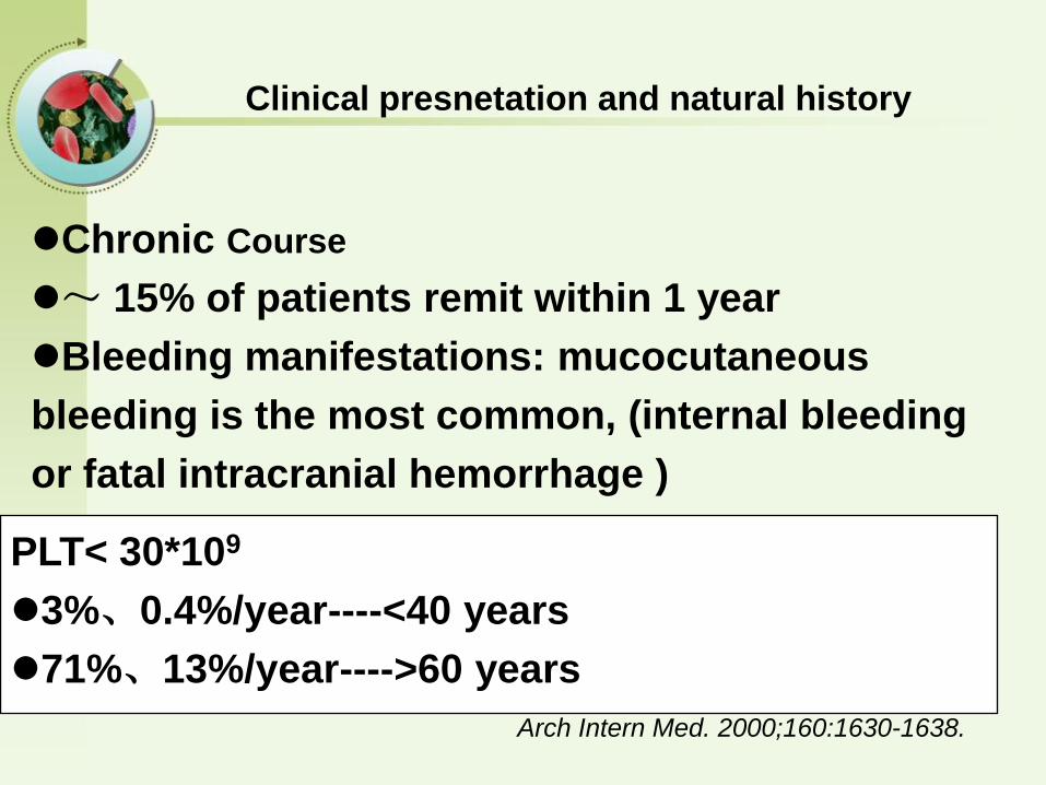
PLT< 30*109
3%、0.4%/year----<40 years
71%、13%/year---->60 years
Arch Intern Med. 2000;160:1630-1638.
Chronic Course
~ 15% of patients remit within 1 year
Bleeding manifestations: mucocutaneous
bleeding is the most common, (internal bleeding
or fatal intracranial hemorrhage )
Clinical presnetation and natural history
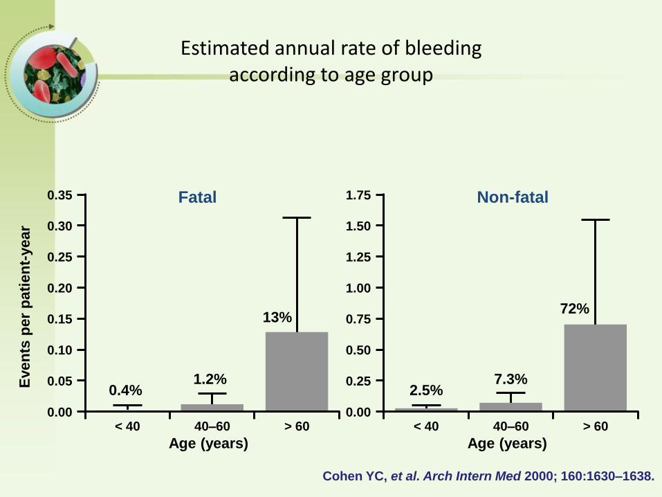
Even
ts p
er
pati
en
t-year
Fatal
0.4%1.2%
13%
0.35
0.30
0.25
0.20
0.15
0.10
0.05
0.00< 40
Age (years)
40–60 > 60
Non-fatal
2.5%7.3%
1.75
1.50
1.25
1.00
0.75
0.50
0.25
0.00< 40
Age (years)
40–60 > 60
72%
Cohen YC, et al. Arch Intern Med 2000; 160:1630–1638.
Estimated annual rate of bleeding according to age group
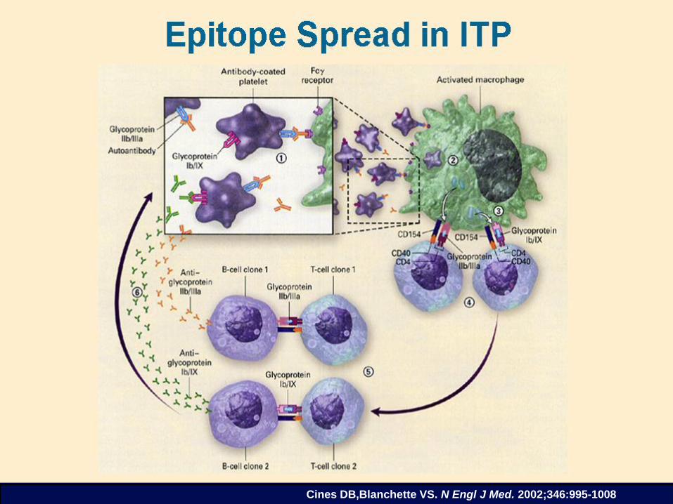
Cines DB,Blanchette VS. N Engl J Med. 2002;346:995-1008
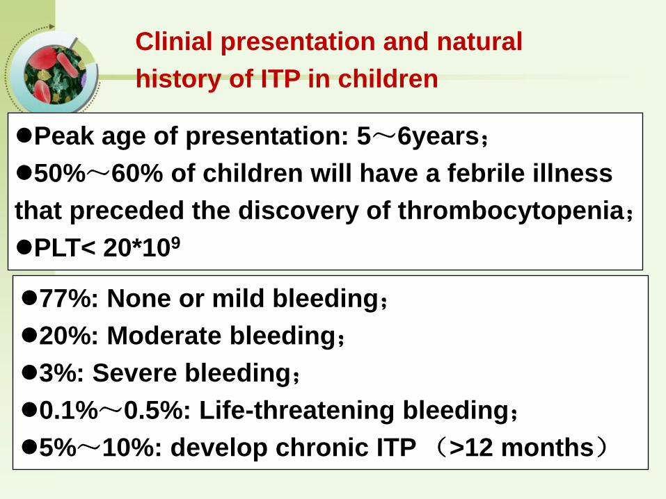
Peak age of presentation: 5~6years;
50%~60% of children will have a febrile illness
that preceded the discovery of thrombocytopenia;
PLT< 20*109
77%: None or mild bleeding;
20%: Moderate bleeding;
3%: Severe bleeding;
0.1%~0.5%: Life-threatening bleeding;
5%~10%: develop chronic ITP (>12 months)
Clinial presentation and natural
history of ITP in children
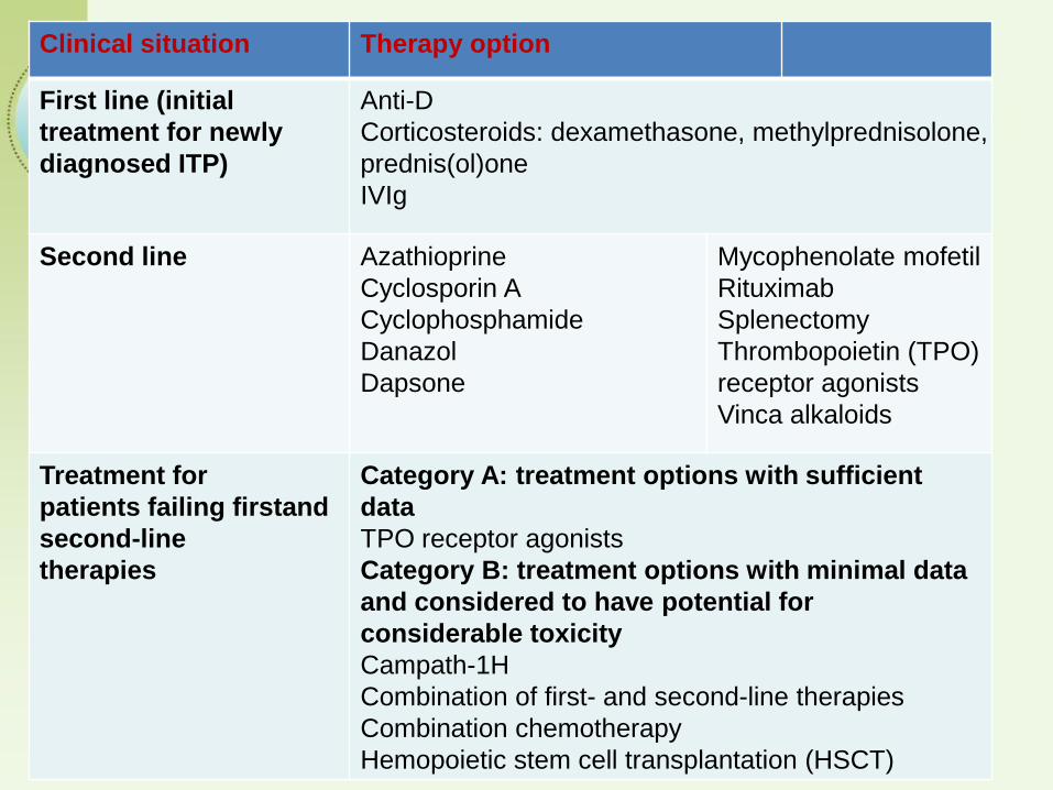
Clinical situation Therapy option
First line (initial
treatment for newly
diagnosed ITP)
Anti-D
Corticosteroids: dexamethasone, methylprednisolone,
prednis(ol)one
IVIg
Second line Azathioprine
Cyclosporin A
Cyclophosphamide
Danazol
Dapsone
Mycophenolate mofetil
Rituximab
Splenectomy
Thrombopoietin (TPO)
receptor agonists
Vinca alkaloids
Treatment for
patients failing firstand
second-line
therapies
Category A: treatment options with sufficient
data
TPO receptor agonists
Category B: treatment options with minimal data
and considered to have potential for
considerable toxicity
Campath-1H
Combination of first- and second-line therapies
Combination chemotherapy
Hemopoietic stem cell transplantation (HSCT)
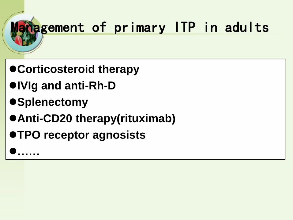
Corticosteroid therapy
IVIg and anti-Rh-D
Splenectomy
Anti-CD20 therapy(rituximab)
TPO receptor agnosists
……
Management of primary ITP in adults

Adjunctive therapy for
bleeding disorders
Adjunctive drug therapy for bleeding
Fresh frozen plasma
Cryoprecipitate
Epsilon-amino-caproic acid (Amicar)
DDAVP
Recombinant human factor VIIa (Novoseven)
Fresh frozen plasma
• Content - plasma (decreased factor V and VIII)
• Indications– Multiple coagulation deficiencies (liver disease, trauma)
– DIC
– Warfarin reversal
– Coagulation deficiency (factor XI or VII)
• Dose (225 ml/unit)– 10-15 ml/kg
• Note– Viral screened product
– ABO compatible
Cryoprecipitate
• Prepared from FFP
• Content– Factor VIII, von Willebrand factor, fibrinogen
• Indications– Fibrinogen deficiency
– Uremia
– von Willebrand disease
• Dose (1 unit = 1 bag)– 1-2 units/10 kg body weight
Aminocaproic acid (Amicar)• Mechanism
– Prevent activation plaminogen -> plasmin
• Dose– 100mg/kg po or IV q 6 hr
• Uses– Primary menorrhagia
– Oral bleeding
– Bleeding in patients with thrombocytopenia
– Blood loss during cardiac surgery
• Side effects– GI toxicity
– Thrombi formation
Desmopressin (DDAVP)• Mechanism
– Increased release of VWF from endothelium
• Dose– 0.3µg/kg IV q12 hrs
– 150mg intranasal q12hrs
• Uses– Most patients with von Willebrand disease
– Mild hemophilia A
• Side effects– Facial flushing and headache
– Water retention and hyponatremia
Recombinant human factor VIIa
• Mechanism
– Activates coagulation system through extrinsic pathway
• Approved Use
– Factor VIII inhibitors in hemophiliacs
• Dose: (1.2 mg/vial)
– 90 µg/kg q 2 hr
– “Adjust as clinically indicated”

Summary Hemostatic Disorders
BT Plt PT APTT
Vascular Dis - - - -
PLT Disorder - - - -
Factor VIII/IX*Congenital - - -
Vit K / Liver*Acquired - - -
Combined (DIC) -
Thrombophilia
Thrombophilia
• Now considered a multicausal disease, with an interplay of acquired and genetic thrombotic risk factors
• Approximately half of venous thromboembolic episodes in patients with inherited thrombophilias occur in relation to events that are generally recognized as a predisposing states, such surgery, pregnancy, and immobilization
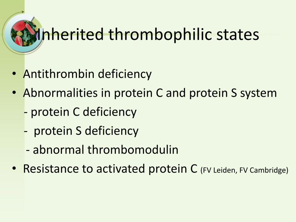
Inherited thrombophilic states
• Antithrombin deficiency
• Abnormalities in protein C and protein S system
- protein C deficiency
- protein S deficiency
- abnormal thrombomodulin
• Resistance to activated protein C (FV Leiden, FV Cambridge)
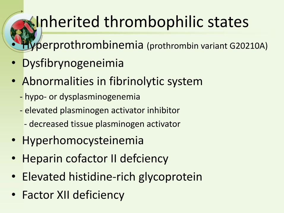
Inherited thrombophilic states• Hyperprothrombinemia (prothrombin variant G20210A)
• Dysfibrynogeneimia
• Abnormalities in fibrinolytic system- hypo- or dysplasminogenemia
- elevated plasminogen activator inhibitor
- decreased tissue plasminogen activator
• Hyperhomocysteinemia
• Heparin cofactor II defciency
• Elevated histidine-rich glycoprotein
• Factor XII deficiency
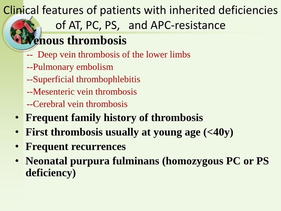
Clinical features of patients with inherited deficiencies of AT, PC, PS, and APC-resistance
• Venous thrombosis -- Deep vein thrombosis of the lower limbs
--Pulmonary embolism
--Superficial thrombophlebitis
--Mesenteric vein thrombosis
--Cerebral vein thrombosis
• Frequent family history of thrombosis
• First thrombosis usually at young age (<40y)
• Frequent recurrences
• Neonatal purpura fulminans (homozygous PC or PS deficiency)
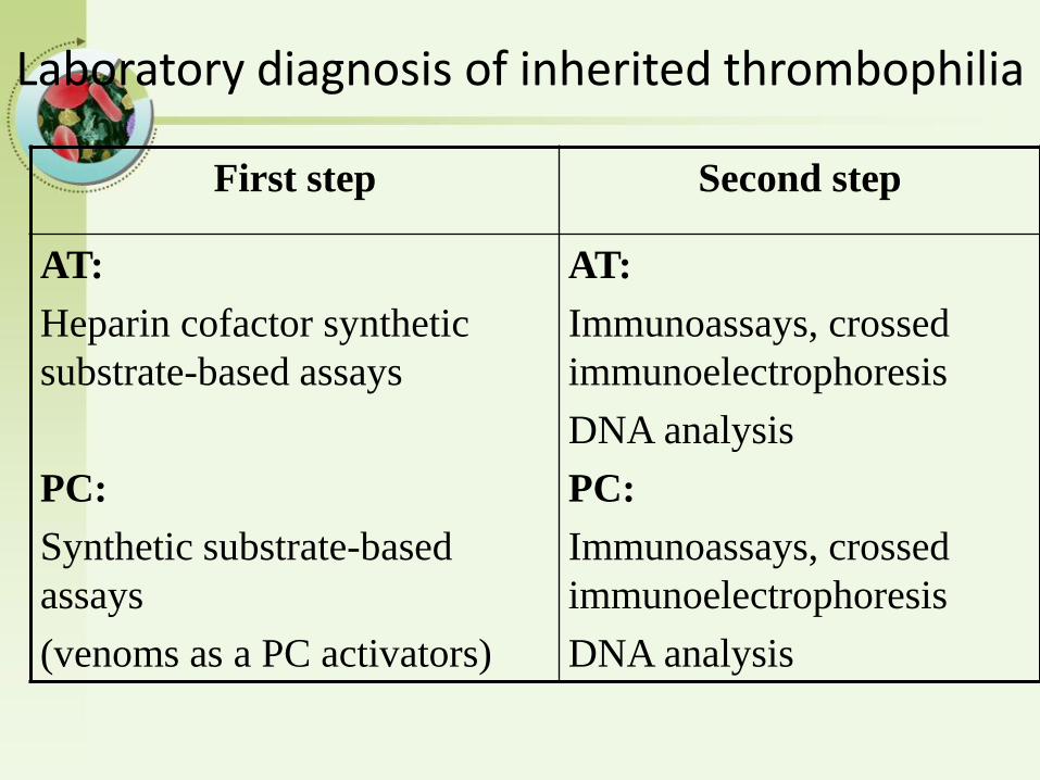
Laboratory diagnosis of inherited thrombophilia
First step Second step
AT:
Heparin cofactor synthetic
substrate-based assays
PC:
Synthetic substrate-based
assays
(venoms as a PC activators)
AT:
Immunoassays, crossed
immunoelectrophoresis
DNA analysis
PC:
Immunoassays, crossed
immunoelectrophoresis
DNA analysis
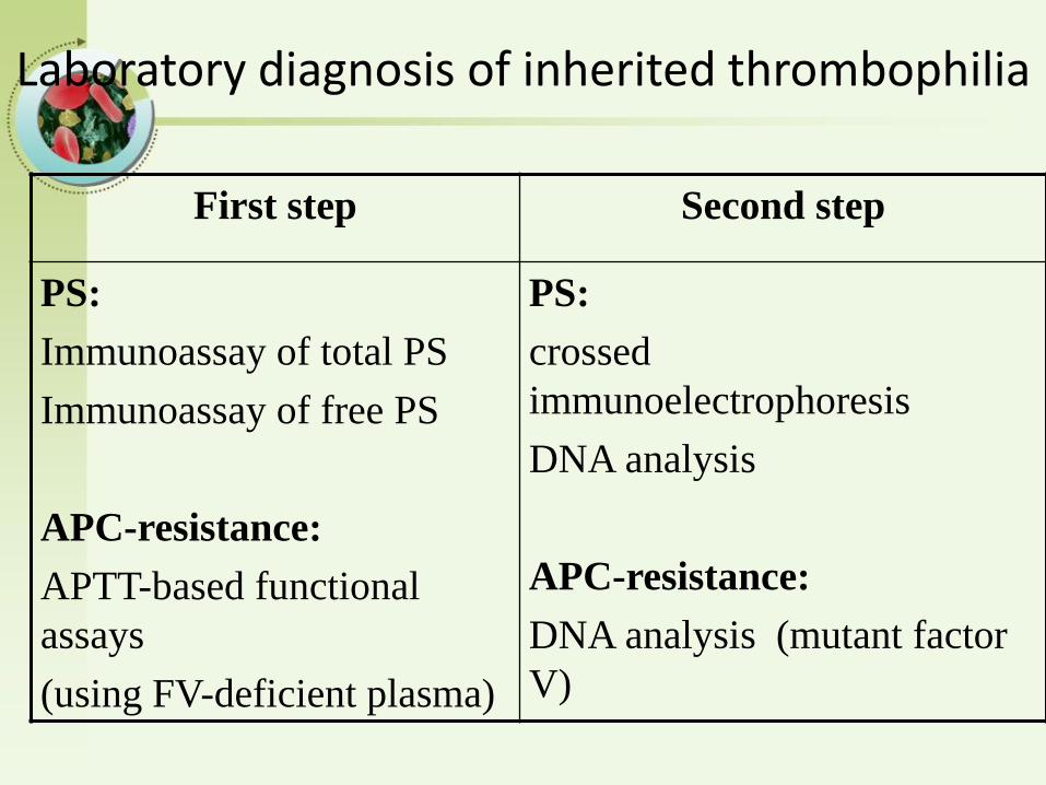
Laboratory diagnosis of inherited thrombophilia
First step Second step
PS:
Immunoassay of total PS
Immunoassay of free PS
APC-resistance:
APTT-based functional
assays
(using FV-deficient plasma)
PS:
crossed
immunoelectrophoresis
DNA analysis
APC-resistance:
DNA analysis (mutant factor
V)
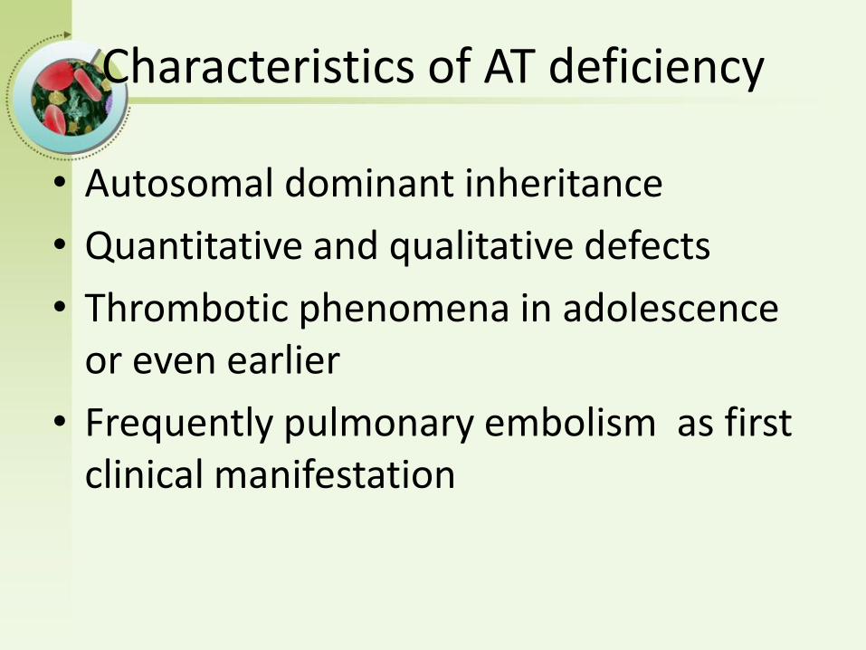
Characteristics of AT deficiency
• Autosomal dominant inheritance
• Quantitative and qualitative defects
• Thrombotic phenomena in adolescence or even earlier
• Frequently pulmonary embolism as first clinical manifestation
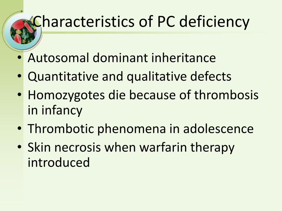
Characteristics of PC deficiency
• Autosomal dominant inheritance
• Quantitative and qualitative defects
• Homozygotes die because of thrombosis in infancy
• Thrombotic phenomena in adolescence
• Skin necrosis when warfarin therapy introduced
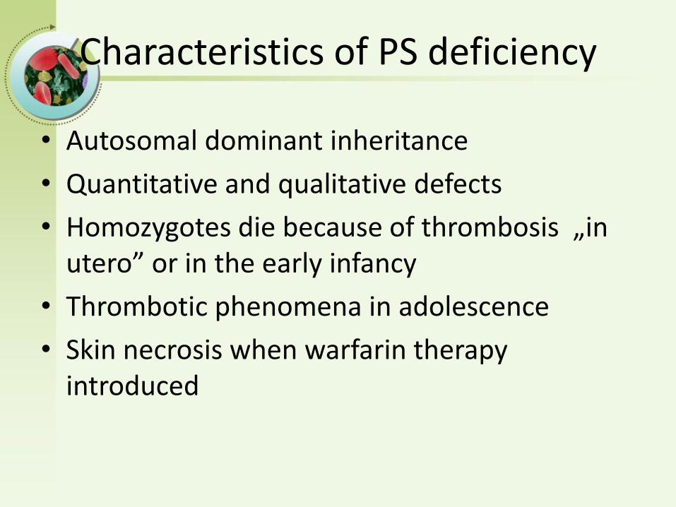
Characteristics of PS deficiency
• Autosomal dominant inheritance
• Quantitative and qualitative defects
• Homozygotes die because of thrombosis „in utero” or in the early infancy
• Thrombotic phenomena in adolescence
• Skin necrosis when warfarin therapy introduced
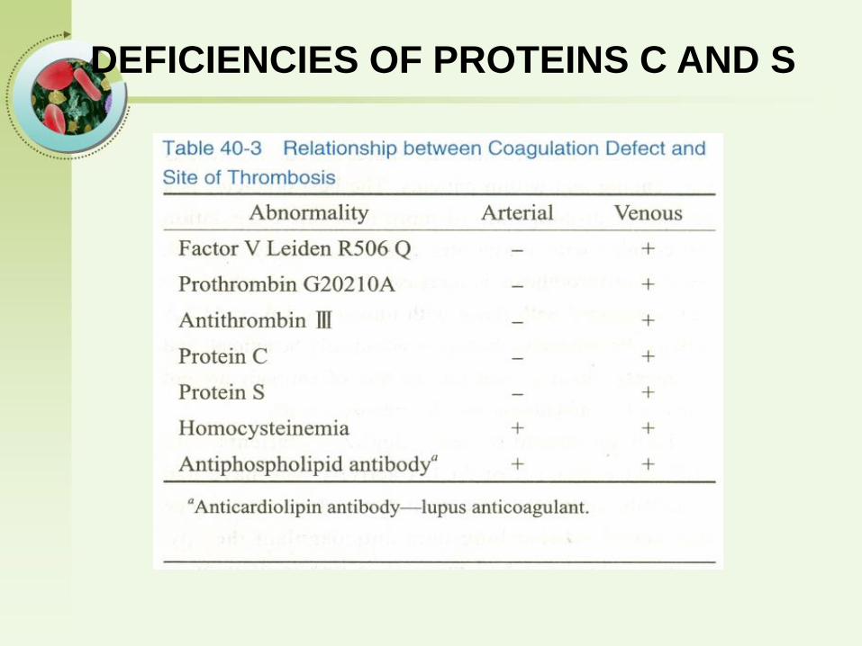
DEFICIENCIES OF PROTEINS C AND S
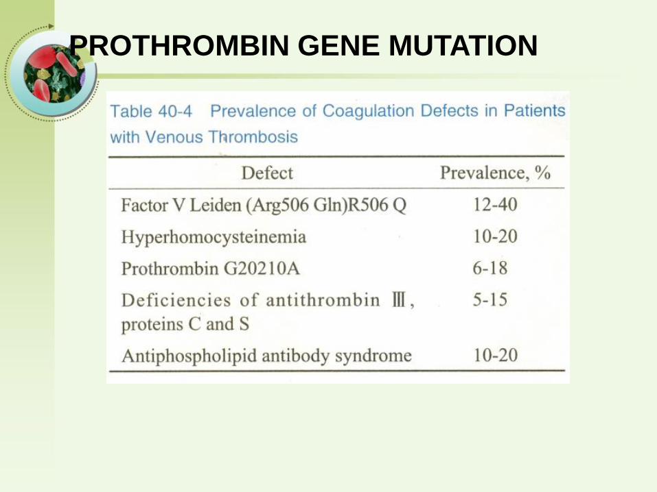
PROTHROMBIN GENE MUTATION
Zhongshan Hospital,Fudan University
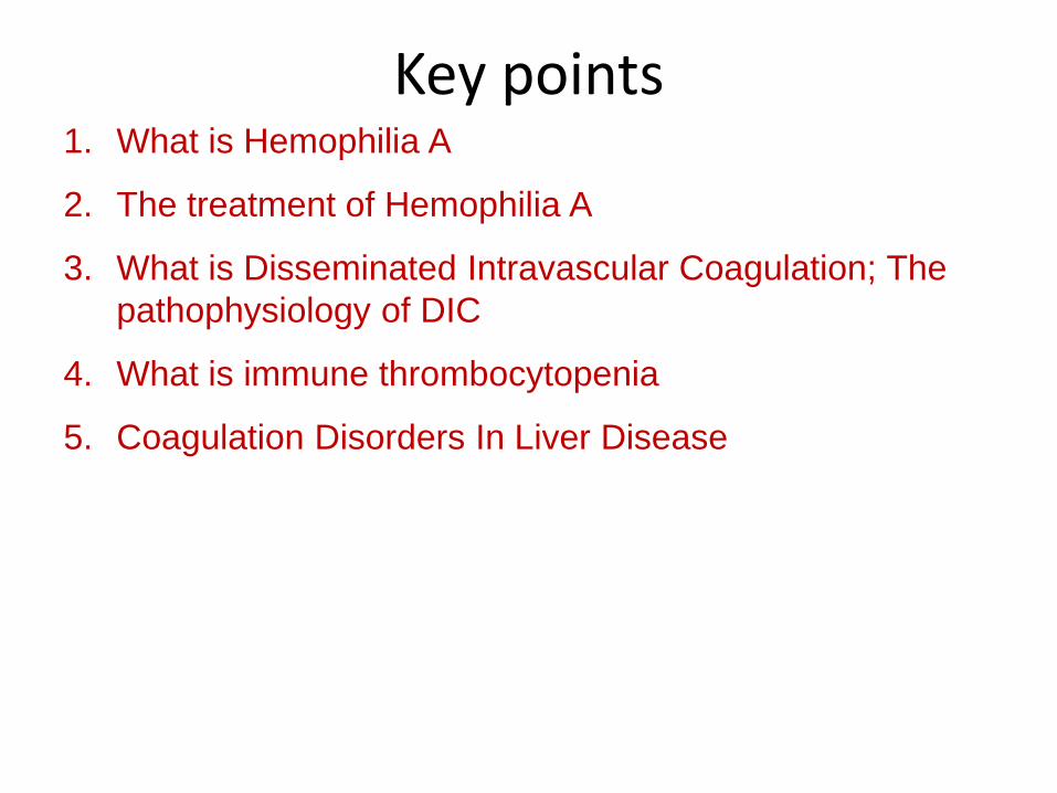
Key points1. What is Hemophilia A
2. The treatment of Hemophilia A
3. What is Disseminated Intravascular Coagulation; The
pathophysiology of DIC
4. What is immune thrombocytopenia
5. Coagulation Disorders In Liver Disease