disorders of blood 23 pressure...
TRANSCRIPT
➤ Blood pressure is probably one of the most variable but best-regulated functions of the body. The purpose of the control ofblood pressure is to keep blood flow constant to vital organs suchas the heart, brain, and kidneys. Without constant blood flow tothese organs, death ensues within seconds, minutes, or days.Although a decrease in flow produces an immediate threat tolife, the continuous elevation of blood pressure that occurs withhypertension is a contributor to premature death and disabilitybecause of its effects on the heart, blood vessels, and kidneys.
The discussion in this chapter focuses on determinants ofblood pressure and conditions of altered arterial pressure—hypertension and orthostatic hypotension.
THE ARTERIAL BLOOD PRESSURE
THE ARTERIAL BLOOD PRESSUREMechanisms of Blood Pressure Regulation
Short-Term RegulationLong-Term Regulation
Blood Pressure MeasurementHYPERTENSION
Essential HypertensionConstitutional Risk FactorsLifestyle Risk FactorsTarget-Organ DamageDiagnosisTreatmentSystolic Hypertension
Secondary HypertensionRenal HypertensionDisorders of Adrenocortical HormonesPheochromocytomaCoarctation of the AortaOral Contraceptive Drugs
Malignant HypertensionHigh Blood Pressure in Pregnancy
ClassificationDiagnosis and Treatment
High Blood Pressure in Children and AdolescentsDiagnosis and Treatment
High Blood Pressure in the ElderlyDiagnosis and Treatment
ORTHOSTATIC HYPOTENSIONPathophysiology and Causative Factors
CausesDiagnosis and Treatment
Disorders of BloodPressure RegulationC A R O L M . P O R T H
Chapter 23
1
After completing this section of the chapter, you shouldbe able to meet the following objectives:
■ Define the terms systolic blood pressure, diastolicblood pressure, pulse pressure, and mean arterialblood pressure.
■ Explain how cardiac output and peripheral vascularresistance interact in determining systolic and dia-stolic blood pressure.
■ Describe the mechanisms for short-term and long-term regulation of blood pressure.
■ Describe the requirements for accurate and reliableblood pressure measurement in terms of cuff size, deter-mining the maximum inflation pressure, and deflationrate.
The arterial blood pressure reflects the rhythmic ejection ofblood from the left ventricle into the aorta.1–3 It rises during sys-tole as the left ventricle contracts and falls as the heart relaxesduring diastole. The contour of the arterial pressure tracingshown in Figure 23-1 is typical of the pressure changes thatoccur in the large arteries of the systemic circulation. There isa rapid rise in the pulse contour during left ventricular contrac-tion, followed by a slower rise to peak pressure. Approximately70% of the blood that leaves the left ventricle is ejected duringthe first one third of systole, accounting for the rapid rise in thepressure contour. The end of systole is marked by a brief down-

2 Unit VI Disorders of Cardiovascular Function
ward deflection and formation of the dicrotic notch, whichoccurs when ventricular pressure falls below that in the aorta.The sudden closure of the aortic valve is associated with a smallrise in pressure caused by continued contraction of the aorta andother large vessels against the closed valve. As the ventriclesrelax and blood flows into the peripheral vessels during dias-tole, the arterial pressure falls rapidly at first and then declinesslowly as the driving force decreases.
In healthy adults, the pressure at the height of the pressurepulse, called the systolic pressure, ideally is less than 120 mm Hg,and the lowest pressure, called the diastolic pressure, is less than80 mm Hg (Fig. 23-2). The difference between the systolic anddiastolic pressure (approximately 40 mm Hg) is called the pulsepressure. The magnitude of the pulse pressure reflects the volumeof blood ejected from the left ventricle during a single beat (strokevolume) and the total distensibility of the atrial tree. The meanarterial pressure (approximately 90 to 100 mm Hg), representsthe average pressure in the arterial system during ventricular con-traction and relaxation and is a good indicator of tissue perfusion.Notice that the mean arterial pressure is not a simple mathemat-ical average of systolic and diastolic pressures. This is because agreater fraction of each cardiac cycle is spent in diastole ratherthan in systole.
The mean arterial blood pressure is determined mainly bythe cardiac output (stroke volume × heart rate) and the peripheralvascular resistance, and can be expressed as the product of thetwo (mean arterial blood pressure = cardiac output × peripheralvascular resistance). The peripheral vascular resistance reflectschanges in the radius of the arterioles as well as the viscosity orthickness of the blood (see Chapter 21 for a discussion of cardiacoutput and peripheral vascular resistance). The arterioles often
are referred to as the resistance vessels because they can selec-tively constrict or relax to control the resistance to outflow ofblood into the capillaries. The body maintains its blood pressureby adjusting the cardiac output to compensate for changes inperipheral vascular resistance and changes the peripheral vascu-lar resistance to compensate for changes in cardiac output.
In hypertension and disease conditions that affect bloodpressure, changes in blood pressure usually are described interms of the systolic and diastolic pressures, pulse pressure, andmean arterial pressure. These pressures are influenced by thestroke volume, the rapidity with which blood is ejected from theheart, the elastic properties of the aorta and large arteries andtheir ability to accept various amounts of blood as it is ejectedfrom the heart, and the properties of the resistance blood ves-sels that control the runoff of blood into the smaller vessels andcapillaries that connect the arterial and venous circulations.
Mechanisms of Blood Pressure RegulationAlthough different tissues in the body are able to regulate theirown blood flow, it is necessary for the arterial pressure to remainrelatively constant as blood shifts from one area of the body toanother. The mechanisms used to regulate the arterial pressuredepend on whether short-term or long-term adaptation is needed2
(Fig. 23-2).
Short-Term Regulation
The mechanisms for short-term regulation of blood pressure,those acting over minutes or hours, are intended to correct tem-porary imbalances in blood pressure, such as occur duringphysical exercise and changes in body position. These mecha-nisms also are responsible for maintenance of blood pressureat survival levels during life-threatening situations such as dur-ing an acute hemorrhagic incident. The short-term regulationof blood pressure relies mainly on neural and humoral mecha-nisms, the most rapid of which are the neural mechanisms.
Neural Mechanisms. The neural control centers for the regulation of blood pressure are located in the reticular forma-tion of the medulla and lower third of the pons, where integra-tion and modulation of autonomic nervous system (ANS)responses occur.2 This area of the brain contains the vasomo-tor and cardiac control centers and is often collectively referredto as the cardiovascular center. The cardiovascular centertransmits parasympathetic impulses to the heart through thevagus nerve and sympathetic impulses to the heart and bloodvessels through the spinal cord and peripheral sympatheticnerves. Vagal stimulation of the heart produces a slowing of heart rate, whereas sympathetic stimulation produces anincrease in heart rate and cardiac contractility. Blood vesselsare selectively innervated by the sympathetic nervous system.Increased sympathetic activity produces constriction of thesmall arteries and arterioles with a resultant increase in periph-eral vascular resistance.
Systolic pressure (peak)
(mm sec)
Meanarterial
pressure
Dicrotic notchPulse
pressure
Diastolicpressure
(minimum)
120
0
40
80
FIGURE 23-1 • Intra-arterial pressure tracing made from the bra-chial artery. Pulse pressure is the difference between systolic anddiastolic pressures. The darker area represents the mean arterial pres-sure, which can be calculated by using the formula of mean arterialpressure = diastolic pressure + pulse pressure/3.
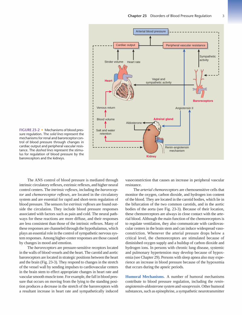
The ANS control of blood pressure is mediated throughintrinsic circulatory reflexes, extrinsic reflexes, and higher neuralcontrol centers. The intrinsic reflexes, including the barorecep-tor and chemoreceptor reflexes, are located in the circulatorysystem and are essential for rapid and short-term regulation ofblood pressure. The sensors for extrinsic reflexes are found out-side the circulation. They include blood pressure responsesassociated with factors such as pain and cold. The neural path-ways for these reactions are more diffuse, and their responsesare less consistent than those of the intrinsic reflexes. Many ofthese responses are channeled through the hypothalamus, whichplays an essential role in the control of sympathetic nervous sys-tem responses. Among higher-center responses are those causedby changes in mood and emotion.
The baroreceptors are pressure-sensitive receptors locatedin the walls of blood vessels and the heart. The carotid and aorticbaroreceptors are located in strategic positions between the heartand the brain (Fig. 23-3). They respond to changes in the stretchof the vessel wall by sending impulses to cardiovascular centersin the brain stem to effect appropriate changes in heart rate andvascular smooth muscle tone. For example, the fall in blood pres-sure that occurs on moving from the lying to the standing posi-tion produces a decrease in the stretch of the baroreceptors witha resultant increase in heart rate and sympathetically induced
vasoconstriction that causes an increase in peripheral vascularresistance.
The arterial chemoreceptors are chemosensitive cells thatmonitor the oxygen, carbon dioxide, and hydrogen ion contentof the blood. They are located in the carotid bodies, which lie inthe bifurcation of the two common carotids, and in the aorticbodies of the aorta (see Fig. 23-3). Because of their location,these chemoreceptors are always in close contact with the arte-rial blood. Although the main function of the chemoreceptors isto regulate ventilation, they also communicate with cardiovas-cular centers in the brain stem and can induce widespread vaso-constriction. Whenever the arterial pressure drops below acritical level, the chemoreceptors are stimulated because ofdiminished oxygen supply and a buildup of carbon dioxide andhydrogen ions. In persons with chronic lung disease, systemicand pulmonary hypertension may develop because of hypox-emia (see Chapter 29). Persons with sleep apnea also may expe-rience an increase in blood pressure because of the hypoxemiathat occurs during the apneic periods.
Humoral Mechanisms. A number of humoral mechanismscontribute to blood pressure regulation, including the renin-angiotensin-aldosterone system and vasopressin. Other humoralsubstances, such as epinephrine, a sympathetic neurotransmitter
Chapter 23 Disorders of Blood Pressure Regulation 3
Sympatheticactivity
Baroreceptors
Angiotensin II
Vagal andsympathetic activity
Heart rateStroke volume
Heart
Venous return
Blood volume
Salt and waterretention
Aldosterone
Kidney
Renin-angiotensinmechanism
Adrenal gland
Cardiac output
Arterial blood pressure
Peripheral vascular resistance
FIGURE 23-2 • Mechanisms of blood pres-sure regulation. The solid lines represent themechanisms for renal and baroreceptorcon-trol of blood pressure through changes incardiac output and peripheral vascular resis-tance. The dashed lines represent the stimu-lus for regulation of blood pressure by thebaroreceptors and the kidneys.

4 Unit VIII Wording of Unit Title4 Unit VIII Wording of Unit Title
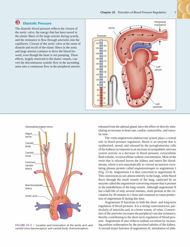
❶ Arterial Blood PressureThe arterial blood pressure represents the force thatdistributes blood to the capillaries throughout thebody. The highest arterial pressure is the systolicpressure and the lowest is the diastolic pressure. Theaorta and its major branches constitute a system ofconduits between the heart and the arterioles. Thearterioles, which are the terminal components of thearterial system, serve as resistance vessels that regu-late the blood pressure at the distribution of blood tothe capillary beds. Because the normal arteries areso compliant and the arterioles present such highresistance to flow, the arterial system acts as a filterthat converts the intermittent flow generated by theheart into a virtually steady flow through the capil-laries. The low-pressure venous system collectsblood from the capillaries and returns it to the heartas a means of maintaining the cardiac output neededto sustain arterial pressure.
❷ Systolic PressureThe systolic blood pressure reflects the amount ofblood (stroke volume) that is ejected from the heartwith each beat, the rate and force with which it isejected, and the elasticity or compliance of theaorta and large arteries. The blood that is ejectedfrom the heart during systole does not movedirectly through the circulation. Instead, a substan-tial fraction of the stroke volume is stored in largearteries. Because the walls of these vessels areelastic, they can be stretched to accommodate alarge volume of blood without an appreciablechange in pressure. The systolic pressure oftenincreases with aging as the aorta and large arterieslose their elasticity and become more rigid.
Understanding • Determinants of Blood PressureThe arterial blood pressure, which is the force that moves blood through the arterial system, reflects theintermittent contraction and relaxation of the left ventricle. It is determined by (1) the properties of the arterial system and the factors that maintain (2) the systolic and (3) the diastolic components of the bloodpressure. These factors include the blood volume, elastic properties of the blood vessels, cardiac output, andperipheral vascular resistance.
Art
eria
l pre
ssur
e (m
m H
g) Systolic
Diastolic
120
80
40
0
Aor
ta
Art
erie
s
Art
erio
les
Cap
illar
ies
Ven
ules
Vei
ns
Ven
a ca
va
Systolic
Peripheralresistance
240
220
200
180
160
140
120
100
80
60
40
20
250
230
210
170
150
130
110
90
70
50
30
10
Aorta
Leftatrium
Leftventricle
4
Chapter 23 Disorders of Blood Pressure Regulation 5
❸ Diastolic PressureThe diastolic blood pressure reflects the closure ofthe aortic valve, the energy that has been stored inthe elastic fibers of the large arteries during systole,and the resistance to flow through arterioles into thecapillaries. Closure of the aortic valve at the onset ofdiastole and recoil of the elastic fibers in the aortaand large arteries continue to drive the blood for-ward, even though the heart is not pumping. Theseeffects, largely restricted to the elastic vessels, con-vert the discontinuous systolic flow in the ascendingaorta into a continuous flow in the peripheral arteries.
Peripheralresistance
Diastolic
240
220
200
180
160
140
120
100
80
60
40
20
250
230
210
170
150
130
110
90
70
50
30
10
Aorta
Leftatrium
Leftventricle
released from the adrenal gland, have the effect of directly stim-ulating an increase in heart rate, cardiac contractility, and vascu-lar tone.
The renin-angiotensin-aldosterone system plays a centralrole in blood pressure regulation. Renin is an enzyme that issynthesized, stored, and released by the juxtaglomerular cellsof the kidneys in response to an increase in sympathetic nervoussystem activity or a decrease in blood pressure, extracellularfluid volume, or extracellular sodium concentration. Most of therenin that is released leaves the kidney and enters the blood-stream, where it acts enzymatically to convert an inactive circu-lating plasma protein called angiotensinogen to angiotensin I(Fig. 23-4). Angiotensin I is then converted to angiotensin II.This conversion occurs almost entirely in the lungs, while bloodflows through the small vessels of the lung, catalyzed by anenzyme called the angiotensin-converting enzyme that is presentin the endothelium of the lung vessels. Although angiotensin IIhas a half-life of only several minutes, renin persists in the cir-culation for 30 minutes to 1 hour and continues to cause produc-tion of angiotensin II during this time.
Angiotensin II functions in both the short- and long-termregulation of blood pressure. It is a strong vasoconstrictor, par-ticularly of arterioles and, to a lesser extent, of veins. Constric-tion of the arterioles increases the peripheral vascular resistance,thereby contributing to the short-term regulation of blood pres-sure. Angiotensin II also reduces sodium excretion by increas-ing sodium reabsorption by the proximal tubules of the kidney.A second major function of angiotensin II, stimulation of aldo-
Glossopharyngeal nerve
V agu s nerve
Carotid body
Carotid sinus
Common carotid artery
Brachiocephalic artery
Aortic arch
FIGURE 23-3 • Location and innervation of the aortic arch andcarotid sinus baroreceptors and carotid body chemoreceptors.

6 Unit VI Disorders of Cardiovascular Function
sterone secretion from the adrenal gland, contributes to thelong-term regulation of blood pressure by increasing salt andwater retention by the kidney.
Vasopressin, also known as antidiuretic hormone (ADH),is released from the posterior pituitary gland in response todecreases in blood volume and blood pressure, an increase in theosmolality of body fluids, and other stimuli. The antidiureticactions of vasopressin are discussed in Chapter 31. Vasopressin
has a direct vasoconstrictor effect, particularly on the vessels ofthe splanchnic circulation that supplies the abdominal viscera.However, long-term increases in vasopressin cannot maintain anincrease in blood pressure, and vasopressin does not enhancehypertension produced by sodium-retaining hormones or othervasoconstricting substances. It has been suggested that vaso-pressin plays a permissive role in hypertension through its water-retaining properties or as a neurotransmitter that serves to mod-ify ANS function.
Long-Term RegulationLong-term mechanisms control the daily, weekly, and monthlyregulation of blood pressure. Although the neural and hormonalmechanisms involved in the short-term regulation of blood pres-sure act rapidly, they are unable to maintain their effectivenessover time. Instead, the long-term regulation of blood pressure islargely vested in the kidneys and their role in the regulation ofthe extracellular fluid volume.2
According to the late Arthur Guyton, a noted physiologist,the extracellular fluid volume and arterial blood pressure are reg-ulated around an equilibrium point, which represents the normalpressure for a given individual2 (Fig. 23-5). When the body con-tains excess extracellular fluids because of increased water andsalt intake, the arterial pressure rises, and the rate at which water(i.e., pressure diuresis) and salt (i.e., pressure natriuresis) areexcreted by the kidney is increased. Accordingly, there are twoways that arterial pressure can be increased using this model:one is by shifting the elimination of salt and water to a higherpressure level (see Fig. 23-5A), and the second is by changingthe extracellular fluid level at which diuresis and natriuresisoccur (see Fig. 23-5B). The function of the kidneys in the long-term regulation of blood pressure can be influenced by a num-ber of factors. For example, excess sympathetic nerve activity orthe release of vasoconstrictor substances can alter the transmis-sion of arterial pressure to the kidney. Similarly, changes inneural and humoral control of kidney function can shift thediuresis–natriuresis process to a higher fluid or pressure level,thereby initiating an increase in arterial pressure.
There are two general mechanisms by which an increase influid volume can elevate blood pressure. One is through a directeffect on cardiac output and the other is indirect, resulting fromthe autoregulation of blood flow and its effect on peripheral vas-cular resistance. Autoregulatory mechanisms function in distrib-uting blood flow to the various tissues of the body according totheir metabolic needs (see Chapter 21). When the blood flow toa specific tissue bed is excessive, local blood vessels constrict,and when the flow is deficient, the local vessels dilate. In situa-tions of increased extracellular fluid volume and a resultantincrease in cardiac output, all of the tissues of the body areexposed to the same increase in flow. This results in a general-ized constriction of arterioles and an increase in the peripheralvascular resistance (and blood pressure).
The role that the kidneys play in blood pressure regulationis emphasized by the fact that many antihypertensive medica-tions produce their blood pressure–lowering effects by increas-ing sodium and water elimination.
Vasoconstriction ofsystemic arterioles
Aldosterone
Sodiumreabsorption
by kidney
Vascular volume andArterial blood pressure
Arterial blood pressure
Juxtaglomerularcells of the kidney
Lungs
Convertingenzyme
Angiotensinogen
Angiotensin I
Angiotensin II
Adrenalcortex
Renin
Arterioles
Extracellular fluidArterial blood pressure
FIGURE 23-4 • Control of blood pressure by the renin-angiotensin-aldosterone system. Renin enzymatically converts the plasma pro-tein angiotensinogen to angiotensin I; angiotensin-convertingenzyme in the lung converts angiotensin I to angiotensin II; andangiotensin II produces vasoconstriction and increases salt andwater retention through direct action on the kidney and throughincreased aldosterone secretion by the adrenal cortex.

Blood Pressure MeasurementArterial blood pressure measurements usually are obtained bythe indirect auscultatory method, which uses a stethoscope anda well-calibrated sphygmomanometer. In the measurement ofblood pressure, a cuff that contains an inflatable rubber blad-der is placed around the upper arm. The bladder of the cuff isinflated to a point at which its pressure exceeds that of theartery, occluding the blood flow. This should be done by pal-pation before the actual pressure is measured to get the pal-pated systolic pressure. By inflating the pressure in the cuff toa level of 30 mm Hg above the palpated pressure, the observercan be certain that the cuff pressure is high enough to avoidmissing the auscultatory gap. This pressure (palpated pressure+ 30 mm Hg) is called the maximum inflation level.4 The cuffis then slowly deflated at 2 mm Hg per second. At the pointwhere the pressure in the vessel again exceeds the pressure inthe cuff, a small amount of blood squirts through the partiallyobstructed artery. The sounds generated by the turbulent floware called the Korotkoff (K) sounds. These low-pitched soundsare best heard with the bell of the stethoscope. Blood pressureis recorded in terms of systolic and diastolic pressures (e.g.,120/70 mm Hg) unless sounds are heard to zero, in which case
three readings are required (122/64/0 or K1/K4/K5). Systolicpressure is defined as the first of two or more Korotkoff soundsheard (K1). Diastolic pressure is recorded as the last soundheard (K5) unless sounds are heard to zero, in which case themuffling sound of K4 is used.
It is important that the bladder of the cuff be appropriatefor the arm size. The width of the bladder should be at least40% of arm circumference and the length at least 80% of armcircumference.4 Undercuffing (using a cuff with a bladder thatis too small) can cause blood pressure to be overestimated.5
This is because a cuff that is too small results in an uneven dis-tribution of pressure across the arm, such that a greater cuffpressure is needed to occlude blood flow. Likewise, overcuff-ing (using a cuff with a bladder that is too large) can cause anunderestimation of blood pressure.
Automated or semiautomated methods of blood pres-sure measurement use a microphone, arterial pressure pulsesensor (oscillometric method), or Doppler equipment fordetecting the equivalent of the Korotkoff sounds. Oscillomet-ric measurement, the most commonly used method, dependson the detection of the pulsatile oscillations of the brachialartery in the blood pressure cuff.6 In contrast to the ausculta-tory method, this method determines the mean arterial pres-sure based on the amplitude of the arterial pulsations andthen uses an algorithm to calculate the systolic and diastolicpressures. Blood pressures obtained by automated devicesare usually less accurate than those obtained by trainedobservers using the auscultatory method, and it is recom-mended that their use be limited to situations in which fre-quent and less accurate measures of blood pressure trendsare needed. They should not be used for the diagnosis andmanagement of hypertension.4
Automated devices are useful for the self-monitoring ofblood pressure and for 24-hour ambulatory monitoring of bloodpressure.7 Ambulatory blood monitors are fully automatic andcan record blood pressure for 24 hours or longer while personsgo about their normal activities. The monitors are typicallyprogrammed to take readings every 15 to 30 minutes through-out the day and night. The readings are stored and downloadedinto a computer for analysis. Automated equipment for self-monitoring of blood pressure is sold in pharmacies and medicalsupply stores throughout the country and is available in manystyles and price ranges. It is important that the equipment be cer-tified as accurate and reliable. The equipment should be a vali-dated aneroid or electronic monitor, should use an appropriate-size cuff, and be checked at least once a year for accuracy. Theaccuracy of an electronic device can be checked by compar-ing its readings with simultaneously obtained auscultatorymeasurements.
Intra-arterial methods provide for direct measurement ofblood pressure. Intra-arterial measurement requires the insertionof a catheter into a peripheral artery. The arterial catheter is con-nected to a pressure transducer, which converts pressure into adigital signal that can be measured, displayed, and recorded.1
The use of this type of blood pressure monitoring usually isrestricted to intensive care units.
Chapter 23 Disorders of Blood Pressure Regulation 7
8
6
4
2
00 50 100 150 200 250
A
8
6
4
2
00 50 100 150 200 250
B
Normal
Normal
Elevatedpressure
Elevatedpressure
Arterial pressure (mm Hg)
Inta
ke o
r ou
tput
(tim
es n
orm
al)
FIGURE 23-5 • Two ways in which the arterial pressure can beincreased: (A) by shifting the renal output curve in the right-handdirection toward a higher pressure level, and (B) by increasing theintake of salt and water. (From Guyton A. C., Hall J. E. [2006]. Text-book of medical physiology [11th ed., p. 218]. Philadelphia: ElsevierSaunders.)

8 Unit VI Disorders of Cardiovascular Function
HYPERTENSION
Hypertension, or high blood pressure, is probably the most com-mon of all health problems in adults and is the leading risk factor for cardiovascular disorders. It affects approximately 50 million individuals in the United States and approximately1 billion worldwide.8 Hypertension is more common in youngermen compared with younger women, in blacks compared withwhites, in persons from lower socioeconomic groups, and inolder persons. Men have higher blood pressures than women upuntil the time of menopause, at which point women quickly losetheir protection. The prevalence of hypertension increases withage. Recent data from the Framingham Study suggest that per-sons who are normotensive at 55 years of age have a 90% life-time risk for development of hypertension.9 Thus, the problemof hypertension can be expected to become even greater withthe aging of the “baby-boomer” population.
Hypertension is commonly divided into the categories ofprimary and secondary hypertension. Primary (essential) hyper-tension is the term applied to 95% of cases in which no causefor hypertension can be identified. In secondary hypertension,the elevation of blood pressure results from some other disor-der, such as kidney disease.
Essential HypertensionThe seventh report of the Joint National Committee on Detec-tion, Evaluation, and Treatment of High Blood Pressure (JNC 7)of the National Institutes of Health was published in 2003.8
According to the JNC 7 recommendations, a systolic pressureof less than 120 mm Hg and a diastolic pressure of less than 80 mm Hg are normal, and systolic pressures between 120 and139 mm Hg and diastolic pressures between 80 and 89 mm Hgare considered prehypertensive (Table 23-1). A diagnosis ofhypertension is made if the systolic blood pressure is 140 mm Hgor higher and the diastolic blood pressure is 90 mm Hg orhigher. For adults with diabetes mellitus, the blood pressuregoal has been lowered to less than 130/80 mm Hg.10 Hyperten-sion is further divided into stages 1 and 2 based on systolic anddiastolic blood pressure measurements. Systolic hypertension
IN SUMMARY, the alternating contraction and relaxation ofthe heart produces a pressure pulse that moves blood throughthe circulatory system. The elastic walls of the aorta stretch dur-ing systole and relax during diastole to maintain the diastolicpressure. The systolic blood pressure denotes the highest pointof the pressure pulse and the diastolic pressure the lowestpoint. The pulse pressure, which reflects the pulsatile nature ofarterial blood flow, is the difference between the systolic anddiastolic pressures, and the mean arterial pressure, the averageblood pressure in the systemic circulation. Systolic pressure isdetermined primarily by the characteristics of the stroke vol-ume, whereas diastolic pressure is determined largely by theconditions of the arteries and arterioles and their abilities toaccept the runoff of blood from the aorta.
The regulation of blood pressure involves both short- andlong-term mechanisms. The short-term mechanisms are respon-sible for regulating blood pressure on a minute-by-minute orhour-by-hour basis during activities such as physical exercise andchanges in body position. The short-term regulation of bloodpressure relies mainly on neural and humoral mechanisms, themost rapid of which are the neural mechanisms. The long-termmechanisms, those maintaining blood pressure over days,weeks, and even years, are largely vested in the kidney and theregulation of extracellular fluid volume.
Arterial blood pressure measurements usually are obtainedby the indirect auscultatory method, which uses a sphygmo-manometer and a stethoscope. Automated or semiautomatedmethods of blood pressure measurement use a microphone,arterial pressure pulse sensor (oscillometric method), or Dopplerequipment for detecting the equivalent of the Korotkoff sounds.Ambulatory and self-measurement of blood pressure may pro-vide valuable information outside the clinician’s office regard-ing a person’s blood pressure and response to treatment.Accurate blood pressure measurement, whether by auscultatoryor automated methods, requires the use of accurately calibratedequipment, a properly fitted cuff, and the proper level of cuffinflation and timing for cuff deflation. ■
After completing this section of the chapter, you shouldbe able to meet the following objectives:
■ Cite the definition of hypertension put forth by theseventh report of the Joint National Committee onDetection, Evaluation, and Treatment of Hypertension.
■ Differentiate essential, systolic, secondary, and malig-nant forms of hypertension.
■ Describe the possible influence of genetics, age, race,obesity, diet and sodium intake, and alcohol consump-tion on the development of essential hypertension.
■ Cite the risks of hypertension in terms of target-organdamage.
■ Describe behavior modification strategies used in theprevention and treatment of hypertension.
■ List the different categories of drugs used to treathypertension and state their mechanisms of action inthe treatment of high blood pressure.
■ Explain the changes in blood pressure that accompanynormal pregnancy and describe the four types of hyper-tension that can occur during pregnancy.
■ Cite the criteria for the diagnosis of high blood pres-sure in children.
■ Define systolic hypertension and relate the circulatorychanges that occur with aging that predispose to thedevelopment of systolic hypertension.

factors. The constitutional risk factors include a family historyof hypertension, race, and age-related increases in blood pres-sure.11,12 Another factor that is thought to contribute to hyper-tension is insulin resistance and the resultant hyperinsulinemiathat occurs in metabolic abnormalities such as type 2 diabetes.
Family History. The inclusion of heredity as a contributing fac-tor in the development of hypertension is supported by the factthat hypertension is seen most frequently among persons with afamily history of hypertension. The strength of the predictiondepends on the definition of positive family history and the ageof the person at risk.13 In studies of twins and family members inwhich the degree of familial aggregation is compared with thecloseness of genetic sharing, the genetic contribution ranges from30% to 60%.14 Until now, however, geneticists have failed toidentify common genes with large effects on hypertension. It ispossible that blood pressure is determined by multiple genes atmany loci, each with a small influence or with a contribution dif-fering according to sex, race, age, and lifestyle.11
Age-Related Changes in Blood Pressure. Maturation andgrowth are known to cause predictable increases in blood pres-sure. For example, the arterial blood pressure in the newborn isapproximately 50 mm Hg systolic and 40 mm Hg diastolic.15
Sequentially, blood pressure increases with physical growthfrom a value of 78 mm Hg systolic at 10 days of age to 120 mmHg at the end of adolescence. Diastolic pressure increases until50 years of age and then declines from the sixth decade onward,whereas systolic blood pressure continues to rise with age.15
Race. Hypertension not only is more prevalent in AfricanAmericans than other ethnic groups in the United States, it ismore severe.16 The Third National Health and Nutrition Sur-vey (NHANES) III, from 1988 to 1991, reported that diastolicblood pressures were significantly greater for African Ameri-cans than for white men and women 35 years of age and older,
Chapter 23 Disorders of Blood Pressure Regulation 9
HYPERTENSION
■ Hypertension represents an elevation in systolicand/or diastolic blood pressure.
■ Essential hypertension is characterized by a chronicelevation in blood pressure that occurs without evi-dence of other disease, and secondary hypertensionby an elevation of blood pressure that results fromsome other disorder, such as kidney disease.
■ The pathogenesis of essential hypertension isthought to include constitutional and environmentalfactors involving the kidney and its role in regulat-ing extracellular fluid volume through salt and waterelimination, sympathetic nervous system hyperreac-tivity, renin-angiotensin sytem activity, or intracellu-lar sodium and calcium levels. The medications thatare used in the treatment of hypertension exert theireffect through one or more of these mechanisms.
■ Uncontrolled hypertension produces increaseddemands on the heart, resulting in left ventricularhypertrophy and heart failure, and on the vessels ofthe arterial system, leading to atherosclerosis, kidneydisease, retinopathy and stroke.
TABLE 23-1 Classification of Blood Pressure for Adults and Recommendationsfor Follow-up
BLOOD PRESSURE SYSTOLIC BLOOD DIASTOLIC BLOOD FOLLOW-UP RECOMMENDATIONSCLASSIFICATION PRESSURE (mm Hg) PRESSURE (mm Hg) FOR INITIAL BLOOD PRESSURE*†
Normal <120 And <80 Recheck in 2 yearsPrehypertensive 120–139 or 80–89 Recheck in 1 year‡
Stage 1 hypertension 140–159 or 90–99 Confirm within 2 months‡
Stage 2 hypertension ≥160 or ≥100 Evaluate or refer to source of care within 1 month.For those with higher pressure (e.g., >180/110 mm Hg), evaluate and treat immediately or within 1 week, depending on clinical situation and complications.
*Initial blood pressure: If systolic and diastolic categories are different, follow recommendations for shorter follow-up (e.g., 160/86mm Hg should be evaluated or referred to source of care with 1 month).
†Follow-up blood pressure: Modify the scheduling of follow-up according to reliable information about past blood pressure mea-surements, other cardiovascular risk factors, or target-organ disease.
‡Provide advice about lifestyle modification.Modified from the National Heart, Lung, and Blood Institute. [2003]. The seventh report of the National Committee on Detection,
Evaluation, and Treatment of High Blood Pressure. NIH publication no. 03-5233. Bethesda, MD: National Institutes of Health.
(to be discussed) is defined as a systolic pressure of 140 mm Hgor greater and a diastolic pressure of less than 90 mm Hg.8
Constitutional Risk Factors
Although the cause or causes of essential hypertension arelargely unknown, both constitutional and lifestyle factors havebeen implicated, either singly or collectively, as contributing
10 Unit VI Disorders of Cardiovascular Function
and that systolic pressures of African American women atevery age were greater than those of white women.17 Hyperten-sion also tends to occur at an earlier age in African Americansthan in whites and often is not treated early enough or aggres-sively enough. Blacks also tend to experience greater cardio-vascular and renal damage at any level of pressure.18
The reasons for the increased incidence of hypertensionamong African Americans are largely unknown. Studies haveshown that many African American persons with hypertensionhave lower renin levels than white persons with hyperten-sion.12,16 The suppression of renin has been considered a sec-ondary response to sodium retention and volume excess. Saltsensitivity, defined as an increase in blood pressure in responseto a high-salt diet, is commonly described in both normotensiveand hypertensive African Americans. Recent research hasfocused on potential defects in renal sodium transport to explainthis observation. Other factors, such as increased vasomotorfunction (e.g., sympathetic nervous system overactivity) orabnormalities in endothelium-dependent vasodilation have alsobeen suggested as possible contributing factors.16
Evidence suggests that African Americans, when pro-vided equal access to diagnosis and treatment, can achieveoverall reductions in blood pressure and experience fewer car-diovascular complications, similar to whites.16,18 Barriers thatlimit access to the health care system include inadequate finan-cial support, inconveniently located health care facilities, longwaiting times, and lack of access to culturally relevant healtheducation about hypertension. With the high prevalence of saltsensitivity, obesity, and smoking among blacks, health educa-tion and lifestyle modifications are particularly important.
Insulin Resistance and Metabolic Abnormalities. Insulinresistance and an accompanying compensatory hyperinsulinemiahave been suggested as possible etiologic links to the develop-ment of hypertension and associated metabolic disturbances suchas impaired glucose tolerance, type 2 diabetes, hyperlipidemias,and obesity.19–21 This clustering of cardiovascular risk factors hasbeen named the insulin resistance syndrome, cardiometabolicsyndrome, or metabolic syndrome (see Chapter 42).
Insulin resistance may be a genetic or acquired trait. Forexample, it has been shown that insulin-mediated glucose dis-posal declines by 30% to 40% in persons who are 40% overideal weight.12 Nonpharmacologic interventions, such as caloricrestriction, weight loss, and exercise, tend to decrease insulinresistance, sympathetic nervous system activity, and bloodpressure.
Lifestyle Risk Factors
Lifestyle factors can contribute to the development of hyper-tension by interacting with other risk factors. These lifestylefactors include high salt intake, excessive calorie intake andobesity, excessive alcohol consumption, and low intake ofpotassium. Although stress can raise blood pressure acutely,there is less evidence linking it to chronic elevations in bloodpressure. Smoking and a diet high in saturated fats and choles-
terol, although not identified as primary risk factors for hyper-tension, are independent risk factors for coronary heart diseaseand should be avoided.
High Salt Intake. Increased salt intake has long been sus-pected as an etiologic factor in the development of hyperten-sion.22,23 Just how increased salt intake contributes to thedevelopment of hypertension is still unclear. It may be that saltcauses an elevation in blood volume, increases the sensitivityof cardiovascular or renal mechanisms to sympathetic nervoussystem influences, or exerts its effects through some othermechanism such as the renin-angiotensin-aldosterone system.It has also been suggested that it may be the chloride ratherthan the sodium in salt that is responsible for the rise in bloodpressure. This is difficult to study, however, because 95% ofsodium in the diet is in the form of sodium chloride.24
Regardless of the mechanism, numerous studies haveshown that a reduction in salt intake can lower blood pressure.The strongest data come from the INTERSALT study, whichmeasured 24-hour urine sodium excretion (an indirect measureof salt intake) in 10,079 men and women 20 to 59 years of agein 52 locations around the world.25 In all 52 sites, there was apositive correlation between sodium excretion and both systolicand diastolic blood pressures. Furthermore, the association of sodium and blood pressure was greatest for older (40 to 59 years) subjects compared with younger (20 to 39 years) sub-jects in the study.
At present, salt intake among adults in the United Statesand United Kingdom averages at least 9 g/day, with large num-bers of people consuming 12 g/day or more.25 This is far inexcess of the maximal intake of 6 g/day for adults recommendedby the American Heart Association.26 Approximately 75% ofsalt intake comes from salt added in the processing and manu-facturing of food; 15% from the discretionary addition in cook-ing and at the table; and 10% from the natural sodium content offood.22,25 The Dietary Approaches to Stop Hypertension (DASH)diet is a nutritional plan that emphasizes fruits, vegetables, low-fat dairy products, whole grains, poultry, fish, and nuts, and isreduced in fat, red meat, sweets, and sugar-containing bever-ages. Results from studies using the low-sodium DASH diethave shown significant reductions in both systolic and diastolicblood pressures.27
Obesity. Excessive weight commonly is associated withhypertension. Weight reduction of as little as 4.5 kg (10 lb)can produce a decrease in blood pressure in a large proportionof overweight people with hypertension.8 It has been sug-gested that fat distribution might be a more critical indicatorof hypertension risk than actual overweight. The waist-to-hipratio commonly is used to differentiate central or upper bodyobesity, with fat cells located in the abdomen and viscera,from peripheral or lower body obesity, with fat cell depositsin the buttocks and legs (see Chapter 39). Studies have foundan association between hypertension and increased waist-to-hip ratio (i.e., central obesity), even when body mass indexand skinfold thickness are taken into account.28,29 Abdominal

or visceral fat seems to be more insulin resistant than fatdeposited over the buttocks and legs. There is also an evolv-ing understanding of the neuroendocrine effects of excessadipose tissue on blood pressure.30 Recent evidence indicatesthat leptin, an adipocyte-derived hormone, may represent alink between adiposity and increased cardiovascular sympa-thetic activity. Besides its effect on appetite and metabolism,leptin acts on the hypothalamus to increase blood pressurethrough activation of the sympathetic nervous system30 Highlevels of circulating free fatty acids in obese people alsoappear to participate in activation of the sympathetic nervoussystem. There is also research supporting activation of therenin-angiotensin-aldosterone system by adipocyte-derivedangiotensinogen and the ability of adipose tissue to increasealdosterone levels through the production of factors thatinduce aldosterone production.30
Excess Alcohol Consumption. Regular alcohol drinkingplays a role in the development of hypertension.31–33 The effectis seen with different types of alcoholic drinks, in men andwomen, and in a variety of ethnic groups. One of the firstreports of a link between alcohol consumption and hypertensioncame from the Oakland–San Francisco Kaiser PermanenteMedical Care Program study that correlated known drinkingpatterns and blood pressure levels of 84,000 persons.33 Thisstudy revealed that the regular consumption of three or moredrinks per day increases the risk for hypertension. Systolic pres-sures were more markedly affected than diastolic pressures.Blood pressure may improve or return to normal when alcoholconsumption is decreased or eliminated. The mechanism wherebyalcohol exerts its effect on blood pressure is unclear. It has beensuggested that lifestyle factors such as obesity and lack of exercisemay be accompanying factors.
Dietary Intake of Potassium, Calcium, and Magnesium. Lowlevels of dietary potassium have also been linked to increasedblood pressure. The strongest evidence comes from the previ-ously described INTERSALT study. In this study, a 60 mmol/dayor greater urinary excretion of potassium (an indirect measure ofpotassium intake) was associated with a reduction in systolicpressure of 3.4 mm Hg or more and a decrease in diastolic pres-sure of 1.9 mm Hg or more.34 Various mechanisms have beenproposed to explain the influence of potassium on blood pressure,including a purported change in the ratio of sodium to potassiumin the diet, a direct natriuretic effect, and suppression of the renin-angiotensin system.35 In terms of food intake, a diet high in potas-sium usually is low in sodium. One of the major benefits ofincreased potassium intake is increased elimination of sodium(natriuretic effect) through the renin-angiotensin-aldosteronemechanism.
The associations between high blood pressure and calciumand magnesium levels also have been investigated. Althoughthere have been reports of high blood pressure in persons withlow calcium intake or lowering of blood pressure with increasedcalcium intake, the link between low calcium and magnesiumintake and hypertension is inconclusive.35
Target-Organ Damage
Essential hypertension is typically an asymptomatic disorder.When symptoms do occur, they are usually related to the long-term effects of hypertension on other organ systems such as thekidneys, heart, eyes, and blood vessels. The JNC 7 report usesthe term target-organ damage to describe the heart, brain,peripheral vascular, kidney, and retinal complications associ-ated with hypertension8 (Chart 23-1). The excess morbidityand mortality related to hypertension is progressive over thewhole range of systolic and diastolic pressures, with target-organ damage varying markedly among persons with similarlevels of hypertension.
Hypertension is a major risk factor for atherosclerosis; itpredisposes to all major atherosclerotic cardiovascular disor-ders, including coronary heart disease, heart failure, stroke, andperipheral artery disease. The risk for coronary artery diseaseand stroke depends to a great extent on other risk factors, suchas obesity, smoking, and elevated cholesterol levels. In clinicaltrials, antihypertensive therapy has been associated with reduc-tions in stroke incidence averaging 30% to 40%, myocardialinfarction, 20% to 25%, and heart failure, more than 50%.8,36
An elevation in blood pressure increases the workload ofthe left ventricle by increasing the pressure against which theheart must pump as it ejects blood into the systemic circula-tion.12 As the workload of the heart increases, the left ven-tricular wall hypertrophies to compensate for the increasedpressure work. Despite its adaptive advantage, left ventricularhypertrophy is a major risk factor for coronary heart disease,cardiac dysrhythmias, sudden death, and congestive heart fail-ure. Hypertensive left ventricular hypertrophy regresses withtherapy. Regression is most closely related to systolic pressurereduction and does not appear to reflect the particular type ofmedication used.
Chronic hypertension leads to nephrosclerosis, a commoncause of chronic kidney disease (see Chapter 33). Hyperten-sive kidney disease is more common in blacks than whites.Hypertension also plays an important role in accelerating the
Chapter 23 Disorders of Blood Pressure Regulation 11
CHART 23-1 TARGET ORGAN DAMAGE
Heart• Left ventricular hypertrophy• Angina or prior myocardial infarction• Prior coronary revascularization• Heart failure
Brain• Stroke or transient ischemic attack
Chronic kidney diseasePeripheral vascular diseaseRetinopathy
From the National Heart, Lung, and Blood Institute. (2003). The seventh report of theNational Committee on Detection, Evaluation, and Treatment of High Blood Pres-sure. Publication no. 03–5233. Bethesda, MD: National Institutes of Health.
12 Unit VI Disorders of Cardiovascular Function
course of other types of kidney disease, particularly diabeticnephropathy. Because of the risk for diabetic nephropathy, theAmerican Diabetes Association recommends that persons withdiabetes maintain their blood pressure at levels less than 130/80mm Hg (see Chapter 42).
Dementia and cognitive impairment occur more commonlyin persons with hypertension.8 Hypertension, particularly sys-tolic hypertension, is a major risk factor for ischemic stroke andintracerebral hemorrhage12 (see Chapter 51). Narrowing andsclerosis of small penetrating arteries in the subcortical regionsof the brain are common findings on autopsy in person withchronic hypertension.8 These changes are thought to contributeto hypoperfusion, loss of autoregulation of blood flow, andimpairment of the blood-brain barrier, ultimately leading to sub-cortical white matter demyelination. Magnetic resonance imag-ing (MRI) studies have revealed more extensive white matterlesions and brain atrophy in hypertensive versus normotensivepersons.37 Effective antihypertensive therapy strongly reducesthe risk of development of significant white matter changes;however, existing white matter changes, once established, do notappear to be reversible.8
Diagnosis
Unlike disorders of other body systems that are diagnosed bymethods such as radiography and tissue examination, hyper-tension and other blood pressure disorders are determined byrepeated blood pressure measurement. Laboratory tests, x-rayfilms, and other diagnostic tests usually are done to exclude sec-ondary hypertension and determine the presence or extent oftarget-organ damage.
Blood pressure measurements should be taken when theperson is relaxed and has rested for at least 5 minutes and hasnot smoked or ingested caffeine within 30 minutes. At least twomeasurements should be made at each visit in the same armwhile the person is seated in a chair (rather than on the exami-nation table) with the feet on the floor and arm supported atheart level.8 If the first two readings differ by more than 5 mmHg, additional readings should be taken. Both the systolic anddiastolic pressures should be recorded. The increased availabil-ity of hypertensive screening clinics provides one of the bestmeans for early detection. Because blood pressure in many indi-viduals is highly variable, blood pressure should be measuredon different occasions over a period of several months beforea diagnosis of hypertension is made unless the pressure isextremely elevated or associated with symptoms. The JNC 7recommendations for follow-up of persons with various stagesof hypertension are included in Table 23-1.
Ambulatory Blood Pressure Measurement. As previouslydiscussed, ambulatory and self/home measurement of blood pres-sure may provide valuable information outside the clinician’soffice regarding the person’s blood pressure and response to treat-ment. Self/home measurement can help detect “white coat hyper-tension,” a condition in which the blood pressure is consistentlyelevated in the health care provider’s office but normal at other
times; it can be used to assess the response to treatment methodsfor hypertension; it can motivate adherence to treatment regi-mens; and it can potentially reduce health care costs.8
The guidelines for the 2005 Canadian HypertensionEducation Program recommend short intervals between the ini-tial and subsequent office visits (e.g., up to three visits over 6 months for a blood pressure of >140/90 mm Hg) to confirmthe blood pressure elevation before pharmacologic interven-tion.38 In addition, the Canadian guidelines stipulate the use of ambulatory and self/home blood pressure measurements ascomplements to office-based evaluations. According to theseguidelines, an ambulatory or self/home awake systolic pressureof 135 mm Hg or more or a diastolic pressure of 85 mm Hg ormore, or a 24-hour ambulatory systolic pressure of 130 mm Hgor more or a diastolic pressure of 80 mm Hg or more, is diag-nosed as hypertension.38
Circadian Variations in Blood Pressure. Blood pressurenormally varies in a characteristic circadian pattern. It tends tobe highest in the early morning, shortly after arising fromsleep, and then decreases gradually throughout the day, reach-ing its lowest point at approximately 2:00 to 5:00 AM.11,39,40 Theterm dippers is used to refer to persons with a normal circadianblood pressure profile in which blood pressure falls during thenight, and nondippers for persons whose 24-hour blood pres-sure profile is flattened.39 Ambulatory blood pressure monitor-ing can be used to determine alterations in a person’s circadianblood pressure profile.7 Changes in the normal circadian bloodpressure profile may occur in a number of conditions, includ-ing malignant hypertension, Cushing syndrome, preeclampsia,orthostatic hypotension, congestive heart failure, and sleepapnea.11 There is increasing evidence that persons with anondipping pattern of hypertension are at higher risk for devel-opment of target-organ damage than those with a dipping pat-tern; in addition, persons with an excessive morning surge inblood pressure may also be at increased risk.
Treatment
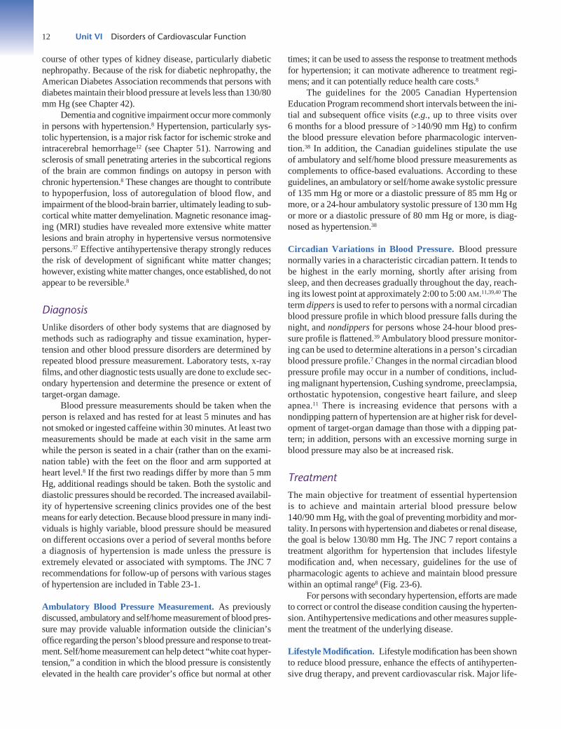
The main objective for treatment of essential hypertension is to achieve and maintain arterial blood pressure below140/90 mm Hg, with the goal of preventing morbidity and mor-tality. In persons with hypertension and diabetes or renal disease,the goal is below 130/80 mm Hg. The JNC 7 report contains atreatment algorithm for hypertension that includes lifestylemodification and, when necessary, guidelines for the use ofpharmacologic agents to achieve and maintain blood pressurewithin an optimal range8 (Fig. 23-6).
For persons with secondary hypertension, efforts are madeto correct or control the disease condition causing the hyperten-sion. Antihypertensive medications and other measures supple-ment the treatment of the underlying disease.
Lifestyle Modification. Lifestyle modification has been shownto reduce blood pressure, enhance the effects of antihyperten-sive drug therapy, and prevent cardiovascular risk. Major life-
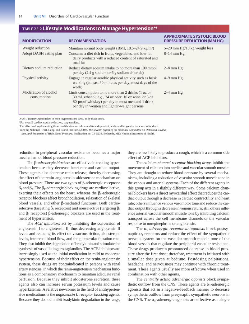
style modifications shown to lower blood pressure includeweight reduction in persons who are overweight or obese, reg-ular physical activity, adoption of the DASH eating plan,reduction of dietary salt intake, and limitation of alcohol intaketo no more than two drinks per day for most men and one drinkfor women and persons of lighter weight8 (Table 23-2).Although nicotine has not been associated with long-term ele-vations in blood pressure as in essential hypertension, it hasbeen shown to increase the risk for heart disease. The fact thatsmoking and hypertension are major cardiovascular risk fac-tors should be reason enough to encourage the hypertensivesmoker to quit. There is conflicting evidence about the directeffects of dietary fats on blood pressure. As with smoking, theinteractive effects of saturated fats and high blood pressure ascardiovascular risk factors would seem to warrant dietary mod-ification to reduce the intake of foods high in cholesterol andsaturated fats.
Pharmacologic Treatment. The decision to initiate phar-macologic treatment is based on the stage and severity of thehypertension, the presence of target-organ disease, and theexistence of other disease conditions and risk factors. TheJNC 7 has developed a pharmacologic treatment algorithmfor use in the pharmacologic treatment of hypertension8 (seeFig. 23-6). Among the drugs used in the treatment of hyper-tension are diuretics, β-adrenergic blocking agents, angiotensin-converting enzyme (ACE) inhibitors or angiotensin II receptorblockers, calcium channel blocking agents, α1-adrenoreceptorantagonists, α2-adrenergic agonists that act at the level of thecentral nervous system (CNS), and vasodilators.
Diuretics, such as the thiazides, loop diuretics, and thealdosterone antagonist (potassium-sparing) diuretics, lowerblood pressure initially by decreasing vascular volume (by sup-pressing renal reabsorption of sodium and increasing salt andwater excretion) and cardiac output. With continued therapy, a
Chapter 23 Disorders of Blood Pressure Regulation 13
Lifestyle modifications
Not at goal blood pressure (<140/90 mm Hg)(<130/80 mm Hg for patients with diabetes or chronic kidney disease)
Initial drug choices
Without compellingindications
With compellingindications
Drug(s) for thecompelling indications
Other antihypertensivedrugs (diuretics, ACEI,
ARB, BB, CCB) as needed.
Stage 1Hypertension
(SBP 140 159 or DBP90 99 mm Hg)
Thiazide-type diureticsfor most. May considerACEI, ARB, BB, CCB,
or combination.
Stage 2Hypertension
(SBP 160 or DBP 100 mm Hg)
Two-drug combination formost (usually thiazide-type diuretic and ACEI,or ARB, or BB, or CCB).
Not at goal blood pressure
Optimize dosages or add additional drugs until goal blood pressure isachieved. Consider consultation with hypertension specialist.
DBP, diastolic blood pressure; SBP, systolic blood pressure.Drug abbreviations: ACEI, angiotensin-converting enzyme inhibitor; ARB, angiotensin receptorblocker; BB, beta blocker; CCB, calcium channel blocker.
FIGURE 23-6 • Algorithm for treatmentof hypertension. (From the National Heart,Lung, and Blood Institute. [2003]. Theseventh report of the National Committeeon Detection, Evaluation, and Treatment ofHigh Blood Pressure. NIH publication no. 03-5233. Bethesda, MD: National Institutes ofHealth.)
14 Unit VI Disorders of Cardiovascular Function
reduction in peripheral vascular resistance becomes a majormechanism of blood pressure reduction.
The β-adrenergic blockers are effective in treating hyper-tension because they decrease heart rate and cardiac output.These agents also decrease renin release, thereby decreasingthe effect of the renin-angiotensin-aldosterone mechanism onblood pressure. There are two types of β-adrenergic receptors:β1 and β2. The β1-adrenergic blocking drugs are cardioselective,exerting their effects on the heart, whereas the β2-adrenergicreceptor blockers affect bronchodilation, relaxation of skeletalblood vessels, and other β-mediated functions. Both cardio-selective (targeting β1 receptors) and nonselective (targeting β1
and β2 receptors) β-adrenergic blockers are used in the treat-ment of hypertension.
The ACE inhibitors act by inhibiting the conversion ofangiotensin I to angiotensin II, thus decreasing angiotensin IIlevels and reducing its effect on vasoconstriction, aldosteronelevels, intrarenal blood flow, and the glomerular filtration rate.They also inhibit the degradation of bradykinin and stimulate thesynthesis of vasodilating prostaglandins. The ACE inhibitors areincreasingly used as the initial medication in mild to moderatehypertension. Because of their effect on the renin-angiotensinsystem, these drugs are contraindicated in persons with renalartery stenosis, in which the renin-angiotensin mechanism func-tions as a compensatory mechanism to maintain adequate renalperfusion. Because they inhibit aldosterone secretion, theseagents also can increase serum potassium levels and causehyperkalemia. A relative newcomer to the field of antihyperten-sive medications is the angiotensin II receptor blocking agents.Because they do not inhibit bradykinin degradation in the lungs,
they are less likely to produce a cough, which is a common sideeffect of ACE inhibitors.
The calcium channel receptor blocking drugs inhibit themovement of calcium into cardiac and vascular smooth muscle.They are thought to reduce blood pressure by several mecha-nisms, including a reduction of vascular smooth muscle tone inthe venous and arterial systems. Each of the different agents inthis group acts in a slightly different way. Some calcium chan-nel blockers have a direct myocardial effect that reduces the car-diac output through a decrease in cardiac contractility and heartrate; others influence venous vasomotor tone and reduce the car-diac output through a decrease in venous return; still others influ-ence arterial vascular smooth muscle tone by inhibiting calciumtransport across the cell membrane channels or the vascularresponse to norepinephrine or angiotensin.
The α1-adrenergic receptor antagonists block postsy-naptic α1 receptors and reduce the effect of the sympatheticnervous system on the vascular smooth muscle tone of theblood vessels that regulate the peripheral vascular resistance.These drugs produce a pronounced decrease in blood pres-sure after the first dose; therefore, treatment is initiated witha smaller dose given at bedtime. Postdosing palpitations,headache, and nervousness may continue with chronic treat-ment. These agents usually are more effective when used incombination with other agents.
The centrally acting adrenergic agonists block sympa-thetic outflow from the CNS. These agents are α2-adrenergicagonists that act in a negative-feedback manner to decreasesympathetic outflow from presynaptic sympathetic neurons inthe CNS. The α2-adrenergic agonists are effective as a single
TABLE 23-2 Lifestyle Modifications to Manage Hypertension*†
APPROXIMATE SYSTOLIC BLOOD MODIFICATION RECOMMENDATION PRESSURE REDUCTION (MM HG)
Weight reductionAdopt DASH eating plan
Dietary sodium reduction
Physical activity
Moderation of alcoholconsumption
DASH, Dietary Approaches to Stop Hypertension; BMI, body mass index.*For overall cardiovascular reduction, stop smoking.†The effects of implementing these modifications are dose and time dependent, and could be greater for some individuals.From the National Heart, Lung, and Blood Institute. (2003). The seventh report of the National Committee on Detection, Evalua-
tion, and Treatment of High Blood Pressure. Publication no. 03–5233. Bethesda, MD: National Institutes of Health.
5–20 mm Hg/10 kg weight loss8–14 mm Hg
2–8 mm Hg
4–9 mm Hg
2–4 mm Hg
Maintain normal body weight (BMI, 18.5–24.9 kg/m2)Consume a diet rich in fruits, vegetables, and low-fat
dairy products with a reduced content of saturated andtotal fat
Reduce dietary sodium intake to no more than 100 mmolper day (2.4 g sodium or 6 g sodium chloride)
Engage in regular aerobic physical activity such as briskwalking (at least 30 minutes per day, most days of theweek)
Limit consumption to no more than 2 drinks (1 oz or 30 mL ethanol; e.g., 24 oz beer, 10 oz wine, or 3 oz 80-proof whiskey) per day in most men and 1 drinkper day in women and lighter-weight persons

therapy for some persons, but often are used as second- orthird-line agents because of the high incidence of side effectsassociated with their use. One of the agents, clonidine, isavailable as a transdermal patch that is replaced weekly.
The direct-acting smooth muscle vasodilators promote adecrease in peripheral vascular resistance by producing relax-ation of vascular smooth muscle, particularly of the arterioles.These drugs often produce tachycardia because of an initialstimulation of the sympathetic nervous system, and salt andwater retention owing to decreased filling of the vascular com-partment. Vasodilators are most effective when used in com-bination with other antihypertensive drugs that oppose thecompensatory cardiovascular responses.
Treatment Strategies. Factors to be considered when hyper-tensive drugs are prescribed are the person’s lifestyle (i.e., some-one with a busy schedule may have problems with medicationsthat must be taken two or three times each day); demographics(e.g., some drugs are more effective in elderly or African Amer-ican persons); motivation for adhering to the drug regimen (e.g.,some drugs can produce undesirable and even life-threateningconsequences if discontinued abruptly); other disease conditionsand therapies; and potential for side effects (e.g., some drugsmay impair sexual functioning or mental acuity; others have notbeen proved safe for women of childbearing age). Particular cau-tion should be used in persons who are at risk for orthostatichypotension (e.g., those with diabetes, ANS dysfunction, andsome older individuals). Another factor to be considered is thecost of the drug in relation to financial resources. There is widevariation in the prices of antihypertensive medications, and thisfactor should be considered when medications are prescribed.This is particularly important for low-income persons with mod-erate to severe hypertension because keeping costs at an afford-able level may be the key to compliance.8
Systolic Hypertension
The JNC 7 report defined systolic hypertension as a systolicpressure of 140 mm Hg or greater and a diastolic pressure of lessthan 90 mm Hg, indicating a need for increased recognition andcontrol of isolated systolic hypertension.8 Historically, diastolichypertension was thought to confer a greater risk for cardiovas-cular events than systolic hypertension.8 However, there ismounting evidence that elevated systolic blood pressure is atleast as important, if not more so, than diastolic hypertension.41,42
There are two aspects of systolic hypertension that conferincreased risk for cardiovascular events—one is the actual ele-vation in systolic pressure and the other is the disproportionaterise in pulse pressure. Elevated pressures during systole favorthe development of left ventricular hypertrophy, increasedmyocardial oxygen demands, and eventual left heart failure. Atthe same time, the absolute or relative lowering of diastolicpressure is a limiting factor in coronary perfusion becausecoronary perfusion is greatest during diastole. Elevated pulsepressures produce greater stretch of arteries, causing damageto the elastic elements of the vessel and thus predisposing to
aneurysms and development of the intimal damage that leadsto atherosclerosis and thrombosis.42
Secondary HypertensionSecondary hypertension, which describes an elevation in bloodpressure due to another disease condition, accounts for 5% to10% of hypertension cases.43 Unlike essential hypertension,many of the conditions causing secondary hypertension can becorrected or cured by surgery or specific medical treatment.Secondary hypertension tends to be seen in persons youngerthan 30 and older than 50 years of age. Cocaine, amphetamines,and other illicit drugs can cause significant hypertension, as cansympathomimetic agents (decongestants, anorectics), erythro-poietin, and licorice (including some chewing tobaccos withlicorice as an ingredient). Obstructive sleep apnea (see Chapter52) is an independent risk factor for secondary hypertension.
Among the most common causes of secondary hyperten-sion are kidney disease (i.e., renovascular hypertension), adrenalcortical disorders, pheochromocytoma, and coarctation of theaorta. To avoid duplication in descriptions, the mechanismsassociated with elevations of blood pressure in these disordersare discussed briefly, and a more detailed discussion of specificdisease disorders is reserved for other sections of this book. Oralcontraceptive agents are also implicated as a cause of secondaryhypertension.
Renal Hypertension
With the dominant role that the kidney assumes in blood pres-sure regulation, it is not surprising that the largest single causeof secondary hypertension is renal disease. Most acute kidneydisorders result in decreased urine formation, retention of saltand water, and hypertension. This includes acute glomeru-lonephritis, acute renal failure, and acute urinary tract obstruc-tion. Hypertension also is common among persons with chronicpyelonephritis, polycystic kidney disease, diabetic nephropathy,and end-stage renal disease, regardless of cause. In older per-sons, the sudden onset of secondary hypertension often is asso-ciated with atherosclerotic disease of the renal blood vessels.
Renovascular hypertension refers to hypertension causedby reduced renal blood flow and activation of the renin-angiotensin-aldosterone mechanism. It is the most commoncause of secondary hypertension, accounting for 1% to 2% ofall cases of hypertension.44 The reduced renal blood flow thatoccurs with renovascular disease causes the affected kidney torelease excessive amounts of renin, increasing circulatinglevels of angiotensin II. Angiotensin II, in turn, acts as a vaso-constrictor to increase peripheral vascular resistance and as astimulus for increased aldosterone levels and sodium retentionby the kidney. One or both of the kidneys may be affected.When the renal artery of only one kidney is involved, the un-affected kidney is subjected to the detrimental effects of theelevated blood pressure.
There are two major types of renovascular disease: athero-sclerosis of the proximal renal artery and fibromuscular dyspla-
Chapter 23 Disorders of Blood Pressure Regulation 15
16 Unit VI Disorders of Cardiovascular Function
sia, a noninflammatory vascular disease that affects the renalarteries and branch vessels.12,44 Atherosclerotic stenosis of therenal artery accounts for 70% to 90% of cases and is seen mostoften in older persons, particularly those with diabetes, aortoiliacocclusive disease, coronary artery disease, or hypertension.Fibromuscular dysplasia is more common in women and tends tooccur in younger age groups, often persons in their third decade.45
Genetic factors may be involved, and the incidence tends toincrease with risk factors such as smoking and hyperlipidemia.
Renal artery stenosis should be suspected when hyperten-sion develops in a previously normotensive person older than50 (i.e., atherosclerotic form) or younger than 30 (i.e., fibromus-cular dysplasia) years of age, or when accelerated hypertensionoccurs in a person with previously controlled hypertension.Hypokalemia (due to increased aldosterone levels), the pres-ence of an abdominal bruit, the absence of a family history ofhypertension, and a duration of hypertension of less than 1 yearhelp to distinguish renovascular hypertension from essentialhypertension. Because renal blood flow depends on the increasedblood pressure generated by the renin-angiotensin system,administration of ACE inhibitors can cause a rapid decline inrenal function.
Diagnostic tests for renovascular hypertension may includestudies to assess overall renal function, physiologic studies toassess the renin-angiotensin system, perfusion studies to evalu-ate renal blood flow, and imaging studies to identify renal arterystenosis.12 Renal arteriography remains the definitive test foridentifying renal artery disease. Duplex ultrasonographic scan-ning, contrast-enhanced computed tomography (CT), and mag-netic resonance angiography (MRA) are other tests that can beused to screen for renovascular hypertension.12
The goal of treatment of renal hypertension is to controlthe blood pressure and stabilize renal function. Angioplasty orrevascularization has been shown to be an effective long-termtreatment for the disorder. ACE inhibitors may be used in med-ical management of renal stenosis. However, these agents mustbe used with caution because of their ability to produce markedhypotension and renal dysfunction.
Disorders of Adrenocortical Hormones
Increased levels of adrenocortical hormones also can give riseto hypertension. Primary hyperaldosteronism (excess produc-tion of aldosterone due to adrenocortical hyperplasia or ade-noma) and excess levels of glucocorticoid (Cushing disease orsyndrome) tend to raise the blood pressure12,46 (see Chapter 41).These hormones facilitate salt and water retention by the kid-ney; the hypertension that accompanies excessive levels ofeither hormone probably is related to this factor. For patientswith primary hyperaldosteronism, a salt-restricted diet oftenproduces a reduction in blood pressure. Because aldosteroneacts on the distal renal tubule to increase sodium absorption inexchange for potassium elimination in the urine, persons withhyperaldosteronism usually have decreased potassium levels.Screening tests for primary hyperaldosteronism involve thedetermination of plasma aldosterone concentration and plasma
renin activity. CT and MRI scans are used to localize thelesion. Persons with solitary adenomas are usually treated sur-gically.12 Potassium-sparing diuretics, such as spironolactone,which is an aldosterone antagonist, often are used in the med-ical management of persons with bilateral hyperplasia.12
Licorice is an extract from the roots of the Glycyrrhizaglabra plant that has been used in medicine since ancienttimes. European licorice (not licorice flavoring) is associatedwith sodium retention, edema, hypertension, and hypokalemia.Licorice, which is an effective analog of the steroid 11 β-dehydrogenase enzyme that modulates access to the aldoste-rone receptor in the kidney, produces a syndrome similar toprimary hyperaldosteronism.
Pheochromocytoma
A pheochromocytoma is a tumor of chromaffin tissue, whichcontains sympathetic nerve cells that stain with chromium salts.The tumor is most commonly located in the adrenal medulla butcan arise in other sites, such as the sympathetic ganglia, wherethere is chromaffin tissue.12,47 Although only 0.1% to 0.5% ofpersons with hypertension have an underlying pheochromocy-toma, the disorder can cause serious hypertensive crises. Thetumors are malignant 8% to 10% of the time.
Like adrenal medullary cells, the tumor cells of a pheochro-mocytoma produce and secrete the catecholamines epinephrineand norepinephrine. The hypertension that develops is a result ofthe massive release of these catecholamines. Their release maybe paroxysmal rather than continuous, causing periodic episodesof headache, excessive sweating, and palpitations. Headache isthe most common symptom and can be quite severe. Nervous-ness, tremor, facial pallor, weakness, fatigue, and weight lossoccur less frequently. Marked variability in blood pressurebetween episodes is typical. Approximately 50% of persons withpheochromocytoma have paroxysmal episodes of hypertension,sometimes to dangerously high levels. The other 50% have sus-tained hypertension, and some even may be normotensive.47
Several tests are available to differentiate hypertensiondue to pheochromocytoma from other forms of hypertension.The most commonly used diagnostic measure is the determina-tion of urinary catecholamines and their metabolites. Althoughmeasurement of plasma catecholamines also may be used, otherconditions can cause catecholamines to be elevated. After thepresence of a pheochromocytoma has been established, thetumor needs to be located. CT and MRI scans may be used forthis purpose. Radioisotopes that localize the chromaffin tissueare available. Surgical removal of operable tumors is usuallycurative.12,47 If the tumor is not resectable, treatment with drugsthat block the action or synthesis of catecholamines can be used.
Coarctation of the Aorta
Coarctation represents a narrowing of the aorta. In the adultform of aortic coarctation, the narrowing most commonlyoccurs just distal to the origin of the subclavian arteries48 (seeChapter 24). Because of the narrowing, blood flow to the lower

parts of the body and kidneys is reduced. In the infantile formof coarctation, the narrowing occurs proximal to the ductusarteriosus, in which case heart failure and other problems mayoccur. Many affected infants die within their first year of life.
In the adult form of aortic coarctation, the ejection of anincreased stroke volume into a narrowed aorta causes anincrease in systolic blood pressure and blood flow to the upperpart of the body. Blood pressure in the lower extremities maybe normal, although it frequently is low. It has been suggestedthat the increase in stroke volume and maintenance of the pres-sure to the lower part of the body is achieved through the renin-angiotensin-aldosterone mechanism in response to a decreasein renal blood flow. Pulse pressure in the legs almost always isnarrowed, and the femoral pulses are weak. Because the aorticcapacity is diminished, there usually is a marked increase inpressure (measured in the arms) during exercise, when thestroke volume and heart rate are increased. For this reason,blood pressures in both arms and one leg should be deter-mined; a pressure that is 20 mm Hg more in the arms than inthe legs suggests coarctation of the aorta. Involvement of theleft subclavian artery or an anomalous origin of the right sub-clavian may produce decreased or absent left or right brachialpulses, respectively. Palpation of both brachial pulses andmeasurement of blood pressure in both arms are important.
Treatment consists of surgical repair or balloon angio-plasty. Although balloon angioplasty is a relatively recent formof treatment, it has been used in children and adults with goodresults. However, there are few data on long-term follow-up.
Oral Contraceptive Drugs
The use of oral contraceptive pills is probably the most commoncause of secondary hypertension in young women. Women tak-ing oral contraceptive should have their blood pressure takenregularly.8 The Nurses Health Study (a prospective cohortstudy of over 70,000 nurses over 4 years between 1989 and1993) found that current users of oral contraceptives had a sig-nificant, moderately increased risk of hypertension.49 However,among this group, only 41.5 cases per 10,000 person-yearscould be attributed to oral contraceptive use.
The cause of the increased blood pressure is largelyunknown, although it has been suggested that the probablecause is volume expansion because both estrogens and syn-thetic progesterones used in oral contraceptive pills causesodium retention. Various contraceptive drugs contain differ-ent amounts and combinations of estrogen and progestationalagents, and these differences may contribute to the occurrenceof hypertension in some women but not others. Fortunately, thehypertension associated with oral contraceptives usually dis-appears after the drug has been discontinued, although it maytake as long as 3 months for this to happen.12 However, in somewomen, the blood pressure may not return to normal, and theymay be at risk for development of hypertension. The risk forhypertension-associated cardiovascular complications is foundprimarily in women older than 35 years of age and in thosewho smoke.
Malignant HypertensionA small number of persons with hypertension develop an accel-erated and potentially fatal form of the disease termed malig-nant hypertension.12,50 This usually is a disease of youngerpersons, particularly young African American men, womenwith toxemia of pregnancy, and persons with renal and colla-gen diseases.
Malignant hypertension is characterized by sudden, markedelevations in blood pressure, with diastolic values above 120 mm Hg complicated by evidence of acute or rapidly progres-sive life-threatening organ dysfunction.50 There may be intensearterial spasm of the cerebral arteries with hypertensiveencephalopathy. Cerebral vasoconstriction probably is an exag-gerated homeostatic response designed to protect the brain fromexcesses of blood pressure and flow. The regulatory mechanismsoften are insufficient to protect the capillaries, and cerebral edemafrequently develops. As it advances, papilledema (i.e., swellingof the optic nerve at its point of entrance into the eye) ensues,giving evidence of the effects of pressure on the optic nerve andretinal vessels. The patient may have headache, restlessness, con-fusion, stupor, motor and sensory deficits, and visual distur-bances. In severe cases, convulsions and coma follow.
Prolonged and severe exposure to exaggerated levels ofblood pressure in malignant hypertension injures the walls ofthe arterioles, and intravascular coagulation and fragmentationof red blood cells may occur. The renal blood vessels are par-ticularly vulnerable to hypertensive damage. Renal damagedue to vascular changes probably is the most important prog-nostic determinant in malignant hypertension. Elevated levelsof blood urea nitrogen and serum creatinine, metabolic acido-sis, hypocalcemia, and proteinuria provide evidence of renalimpairment.
The complications associated with a hypertensive crisisdemand immediate and rigorous medical treatment in an inten-sive care unit with continuous monitoring of arterial blood pres-sure. With proper therapy, the death rate from this cause can bemarkedly reduced, as can complications and additional episodes.Because chronic hypertension is associated with autoregulatorychanges in coronary artery, cerebral artery, and kidney bloodflow, care should be taken to avoid excessively rapid decreasesin blood pressure, which can lead to hypoperfusion and ischemicinjury. Therefore, the goal of initial treatment measures shouldbe to obtain a partial reduction in blood pressure to a safer, lesscritical level, rather than to normotensive levels.50
High Blood Pressure in Pregnancy
Hypertensive disorders of pregnancy complicate 5% to 10% ofpregnancies and remain a major cause of maternal and neonatalmortality and morbidity in the United States and worldwide.51–54
Most adverse events are attributable directly to the preeclampsiasyndrome, characterized by new-onset hypertension with pro-teinuria that develops in the last half of pregnancy. Women withchronic hypertension can also manifest adverse events.
Chapter 23 Disorders of Blood Pressure Regulation 17
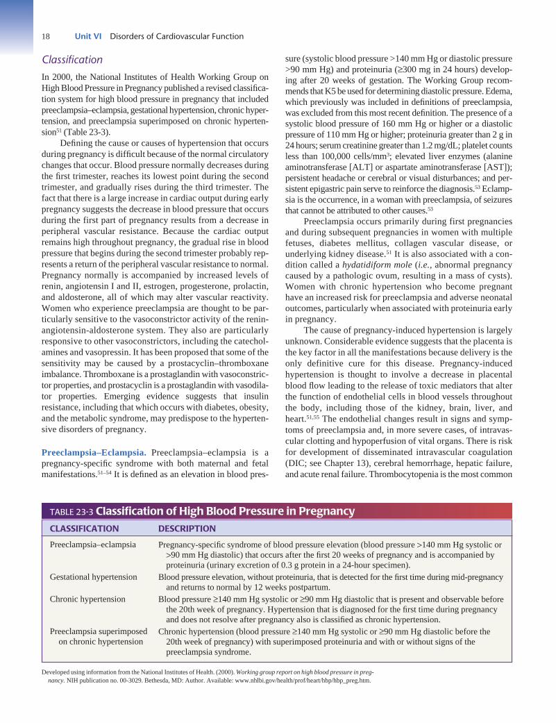
18 Unit VI Disorders of Cardiovascular Function
Classification
In 2000, the National Institutes of Health Working Group onHigh Blood Pressure in Pregnancy published a revised classifica-tion system for high blood pressure in pregnancy that includedpreeclampsia–eclampsia, gestational hypertension, chronic hyper-tension, and preeclampsia superimposed on chronic hyperten-sion51 (Table 23-3).
Defining the cause or causes of hypertension that occursduring pregnancy is difficult because of the normal circulatorychanges that occur. Blood pressure normally decreases duringthe first trimester, reaches its lowest point during the secondtrimester, and gradually rises during the third trimester. Thefact that there is a large increase in cardiac output during earlypregnancy suggests the decrease in blood pressure that occursduring the first part of pregnancy results from a decrease inperipheral vascular resistance. Because the cardiac outputremains high throughout pregnancy, the gradual rise in bloodpressure that begins during the second trimester probably rep-resents a return of the peripheral vascular resistance to normal.Pregnancy normally is accompanied by increased levels ofrenin, angiotensin I and II, estrogen, progesterone, prolactin,and aldosterone, all of which may alter vascular reactivity.Women who experience preeclampsia are thought to be par-ticularly sensitive to the vasoconstrictor activity of the renin-angiotensin-aldosterone system. They also are particularlyresponsive to other vasoconstrictors, including the catechol-amines and vasopressin. It has been proposed that some of thesensitivity may be caused by a prostacyclin–thromboxaneimbalance. Thromboxane is a prostaglandin with vasoconstric-tor properties, and prostacyclin is a prostaglandin with vasodila-tor properties. Emerging evidence suggests that insulinresistance, including that which occurs with diabetes, obesity,and the metabolic syndrome, may predispose to the hyperten-sive disorders of pregnancy.
Preeclampsia–Eclampsia. Preeclampsia–eclampsia is apregnancy-specific syndrome with both maternal and fetalmanifestations.51–54 It is defined as an elevation in blood pres-
sure (systolic blood pressure >140 mm Hg or diastolic pressure>90 mm Hg) and proteinuria (≥300 mg in 24 hours) develop-ing after 20 weeks of gestation. The Working Group recom-mends that K5 be used for determining diastolic pressure. Edema,which previously was included in definitions of preeclampsia,was excluded from this most recent definition. The presence of asystolic blood pressure of 160 mm Hg or higher or a diastolicpressure of 110 mm Hg or higher; proteinuria greater than 2 g in24 hours; serum creatinine greater than 1.2 mg/dL; platelet countsless than 100,000 cells/mm3; elevated liver enzymes (alanineaminotransferase [ALT] or aspartate aminotransferase [AST]);persistent headache or cerebral or visual disturbances; and per-sistent epigastric pain serve to reinforce the diagnosis.53 Eclamp-sia is the occurrence, in a woman with preeclampsia, of seizuresthat cannot be attributed to other causes.53
Preeclampsia occurs primarily during first pregnanciesand during subsequent pregnancies in women with multiplefetuses, diabetes mellitus, collagen vascular disease, orunderlying kidney disease.51 It is also associated with a con-dition called a hydatidiform mole (i.e., abnormal pregnancycaused by a pathologic ovum, resulting in a mass of cysts).Women with chronic hypertension who become pregnanthave an increased risk for preeclampsia and adverse neonataloutcomes, particularly when associated with proteinuria earlyin pregnancy.
The cause of pregnancy-induced hypertension is largelyunknown. Considerable evidence suggests that the placenta isthe key factor in all the manifestations because delivery is theonly definitive cure for this disease. Pregnancy-inducedhypertension is thought to involve a decrease in placentalblood flow leading to the release of toxic mediators that alterthe function of endothelial cells in blood vessels throughoutthe body, including those of the kidney, brain, liver, andheart.51,55 The endothelial changes result in signs and symp-toms of preeclampsia and, in more severe cases, of intravas-cular clotting and hypoperfusion of vital organs. There is riskfor development of disseminated intravascular coagulation(DIC; see Chapter 13), cerebral hemorrhage, hepatic failure,and acute renal failure. Thrombocytopenia is the most common
TABLE 23-3 Classification of High Blood Pressure in Pregnancy
CLASSIFICATION DESCRIPTION
Preeclampsia–eclampsia
Gestational hypertension
Chronic hypertension
Preeclampsia superimposedon chronic hypertension
Pregnancy-specific syndrome of blood pressure elevation (blood pressure >140 mm Hg systolic or>90 mm Hg diastolic) that occurs after the first 20 weeks of pregnancy and is accompanied byproteinuria (urinary excretion of 0.3 g protein in a 24-hour specimen).
Blood pressure elevation, without proteinuria, that is detected for the first time during mid-pregnancyand returns to normal by 12 weeks postpartum.
Blood pressure ≥140 mm Hg systolic or ≥90 mm Hg diastolic that is present and observable beforethe 20th week of pregnancy. Hypertension that is diagnosed for the first time during pregnancyand does not resolve after pregnancy also is classified as chronic hypertension.
Chronic hypertension (blood pressure ≥140 mm Hg systolic or ≥90 mm Hg diastolic before the 20th week of pregnancy) with superimposed proteinuria and with or without signs of thepreeclampsia syndrome.
Developed using information from the National Institutes of Health. (2000). Working group report on high blood pressure in preg-nancy. NIH publication no. 00-3029. Bethesda, MD: Author. Available: www.nhlbi.gov/health/prof/heart/hbp/hbp_preg.htm.

hematologic complication of preeclampsia. Platelet counts ofless than 100,000/mm3 signal serious disease. The cause ofthrombocytopenia has been ascribed to platelet deposition atthe site of endothelial injury. The renal changes that occurwith preeclampsia include a decrease in glomerular filtrationrate and renal blood flow. Sodium excretion may be impaired,although this is variable. Edema may or may not be present.Some of the severest forms of preeclampsia occur in theabsence of edema. Even when there is extensive edema, theplasma volume usually is lower than that of a normal preg-nancy. Liver damage, when it occurs, may range from mildhepatocellular necrosis with elevation of liver enzymes to themore ominous hemolysis, elevated liver function test results,and low platelet count (HELLP) syndrome that is associatedwith significant maternal mortality. Eclampsia, the convulsivestage of preeclampsia, is a significant cause of maternal mor-tality. The pathogenesis of eclampsia remains unclear but hasbeen attributed to both increased blood coagulability and fibrindeposition in the cerebral vessels.
The decreased placental blood flow that occurs withpreeclampsia also affects the fetus. It frequently results inintrauterine growth restriction and infants who are small forgestational age. Preeclampsia is one of the leading causes of prematurity because of frequent need for early delivery inaffected women.
Gestational Hypertension. Gestational hypertension repre-sents a blood pressure elevation without proteinuria that isdetected for the first time after mid-pregnancy.51 It includeswomen with preeclampsia syndrome who have not yet man-ifested proteinuria as well as women who do not have thesyndrome. The hypertension may be accompanied by othersigns of the syndrome. The final determination that a womandoes not have the preeclampsia syndrome is made only post-partum. If preeclampsia has not developed and blood pres-sure has returned to normal by 12 weeks postpartum, thecondition is considered to be gestational hypertension. If bloodpressure elevation persists, a diagnosis of chronic hyperten-sion is made.
Chronic Hypertension. Chronic hypertension is consideredto be hypertension that is unrelated to the pregnancy. It isdefined as a history of high blood pressure before pregnancy,identification of hypertension before 20 weeks of pregnancy,and hypertension that persists after pregnancy.51 Hypertensionthat is diagnosed for the first time during pregnancy and doesnot resolve after pregnancy also is classified as chronic hyper-tension. In women with chronic hypertension, blood pressureoften decreases in early pregnancy and increases during thelast trimester (3 months) of pregnancy, resembling preeclamp-sia. Consequently, women with undiagnosed chronic hyper-tension who do not present for medical care until the latermonths of pregnancy may be incorrectly diagnosed as havingpreeclampsia.
Preeclampsia Superimposed on Chronic Hypertension.Women with chronic hypertension are at increased risk for the
development of preeclampsia, in which case the prognosis forthe mother and fetus tends to be worse than for either condi-tion alone. Superimposed preeclampsia should be consideredin women with hypertension before 20 weeks of gestation whodevelop new-onset proteinuria; women with hypertension andproteinuria before 20 weeks of gestation; women with previ-ously well-controlled hypertension who experience a suddenincrease in blood pressure; and women with chronic hyperten-sion who develop thrombocytopenia or an increase in serumALT or AST to abnormal levels.51
Diagnosis and Treatment
Early prenatal care is important in the detection of high bloodpressure during pregnancy. It is recommended that all pregnantwomen, including those with hypertension, refrain from alcoholand tobacco use. Salt restriction usually is not recommendedduring pregnancy because pregnant women with hypertensiontend to have lower plasma volumes than normotensive preg-nant women and because the severity of hypertension mayreflect the degree of volume contraction. The exception iswomen with preexisting hypertension who have been follow-ing a salt-restricted diet.
In women with preeclampsia, delivery of the fetus iscurative. The timing of delivery becomes a difficult decisionin preterm pregnancies because the welfare of both themother and the infant must be taken into account. Bed rest isa traditional therapy. Antihypertensive medications, whenrequired, must be carefully chosen because of their potentialeffects on uteroplacental blood flow and on the fetus. Forexample, the ACE inhibitors can cause injury and even deathof the fetus when given during the second and third trimestersof pregnancy.
High Blood Pressure in Children and Adolescents
Until recently, the incidence of hypertension among childrenhas been low, with a range of 1% to 3%.56 Recent data, how-ever, indicate that the prevalence and rate of diagnosis ofhypertension in children and adolescence appear to be increas-ing.56,57 This may be due in part to increasing prevalence ofobesity and other lifestyle factors, such as decreased physicalactivity and increased intake of high-calorie, high-salt foods.
Blood pressure is known to increase from infancy to lateadolescence. The average systolic pressure at 1 day of age isapproximately 70 mm Hg and increases to approximately 85 mm Hg at 1 month of age.58 Systolic blood pressure con-tinues to increase with physical growth to about 120 mm Hgat the end of adolescence. During the preschool years, bloodpressure begins to follow a pattern that tends to be maintainedas the child grows older. This pattern continues into adoles-cence and adulthood, suggesting that the roots of essentialhypertension have their origin early in life. A familial influ-ence on blood pressure often can be identified early in life.Children of parents with high blood pressure tend to have
Chapter 23 Disorders of Blood Pressure Regulation 19
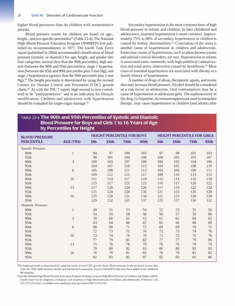
20 Unit VI Disorders of Cardiovascular Function
higher blood pressures than do children with normotensiveparents.
Blood pressure norms for children are based on age-,height-, and sex-specific percentiles59 (Table 23-4). The NationalHigh Blood Pressure Education Program (NHBPEP) first pub-lished its recommendations in 1977. The fourth Task Forcereport (published in 2004) recommended classification of bloodpressure (systolic or diastolic) for age, height, and gender intofour categories: normal (less than the 90th percentile), high nor-mal (between the 90th and 95th percentiles), stage 1 hyperten-sion (between the 95th and 99th percentiles plus 5 mm Hg), andstage 2 hypertension (greater than the 99th percentile plus 5 mmHg).59 The height percentile is determined by using the revisedCenters for Disease Control and Prevention (CDC) growthcharts.60 As with the JNC 7 report, high normal is now consid-ered to be “prehypertensive” and is an indication for lifestylemodification. Children and adolescents with hypertensionshould be evaluated for target-organ damage.59
Secondary hypertension is the most common form of highblood pressure in infants and children. In later childhood andadolescence, essential hypertension is more common. Approx-imately 75% to 80% of secondary hypertension in children iscaused by kidney abnormalities.61 Coarctation of the aorta isanother cause of hypertension in children and adolescents.Endocrine causes of hypertension, such as pheochromocytomaand adrenal cortical disorders, are rare. Hypertension in infantsis associated most commonly with high umbilical catheteriza-tion and renal artery obstruction caused by thrombosis.61 Mostcases of essential hypertension are associated with obesity or afamily history of hypertension.
A number of drugs of abuse, therapeutic agents, and toxinsalso may increase blood pressure. Alcohol should be consideredas a risk factor in adolescents. Oral contraceptives may be acause of hypertension in adolescent girls. The nephrotoxicity ofthe drug cyclosporine, an immunosuppressant used in transplanttherapy, may cause hypertension in children (and adults) after
The height percentile is determined by using the newly revised CDC growth charts. Blood pressure levels are based on new datafrom the 1999–2000 National Health and Nutritional Examination Survey (NHANES) that have been added to the childhoodBP database.
From the National High Blood Pressure Education Program Working Group on High Blood Pressure in Children and Adults. (2004).Fourth report on the diagnosis, evaluation, and treatment of high blood pressure in children and adolescents. Pediatrics 114,555–576. [On-line]. Available:www.pediatrics.org/cgi/content/full/114/S2/555.
BLOOD PRESSURE HEIGHT PERCENTILE FOR BOYS HEIGHT PERCENTILE FOR GIRLS
PERCENTILE AGE (YRS) 5th 25th 75th 95th 5th 25th 75th 95th
Systolic Pressure90th 1 94 97 100 103 97 98 101 10395th 98 101 104 106 100 102 105 10790th 3 100 103 107 109 100 102 104 10695th 104 107 110 113 104 105 108 11090th 6 105 108 111 113 104 106 109 11195th 109 112 115 117 108 110 113 11590th 10 111 114 117 119 112 114 116 11895th 115 117 121 123 116 117 120 12290th 13 117 120 124 126 117 119 122 12495th 121 124 128 130 121 123 126 12890th 16 125 128 131 134 121 123 126 12895th 129 132 135 137 125 127 130 132
Diastolic Pressure90th 1 49 51 53 54 52 53 55 5695th 54 55 58 58 56 57 59 6090th 3 59 60 62 63 61 62 64 6595th 63 64 66 67 65 66 68 6990th 6 68 69 71 72 68 69 70 7295th 72 73 75 76 72 73 74 7690th 10 73 74 76 78 73 73 75 7695th 77 79 81 82 77 77 79 8090th 13 75 76 78 79 76 76 78 7995th 79 80 82 83 80 80 82 8390th 16 78 79 81 82 78 79 81 8295th 82 83 85 87 82 83 85 86
TABLE 23-4 The 90th and 95th Percentiles of Systolic and Diastolic Blood Pressure for Boys and Girls 1 to 16 Years of Age by Percentiles for Height

bone marrow, heart, kidney, or liver transplantation. The coad-ministration of corticosteroid drugs appears to increase the inci-dence of hypertension.
Diagnosis and TreatmentThe Task Force recommended that children 3 years of agethrough adolescence should have their blood pressure takenonce each year. The auscultatory method using a cuff of anappropriate size for the child’s upper arm is recommended.59
Repeated measurements over time, rather than a single isolateddetermination, are required to establish consistent and signifi-cant observations. Children with high blood pressure should bereferred for medical evaluation and treatment as indicated.Treatment includes nonpharmacologic methods and, if neces-sary, pharmacologic therapy.
High Blood Pressure in the Elderly
The prevalence of hypertension increases with advancing ageto the extent that half of people aged 60 to 69 years and approx-imately three fourths of people 70 years and older are affected.8
The age-related rise in systolic blood pressure is primarilyresponsible for the increase in hypertension that occurs withincreasing age.
Among the aging processes that contribute to an increase inblood pressure are a stiffening of the large arteries, particularlythe aorta; decreased baroreceptor sensitivity; increased peripheralvascular resistance; and decreased renal blood flow.62 Systolicblood pressure rises almost linearly between 30 and 84 years ofage, whereas diastolic pressure rises until 50 years of age and thenlevels off or decreases.63 This rise in systolic pressure is thoughtto be related to increased stiffness of the large arteries. Withaging, the elastin fibers in the walls of the arteries are graduallyreplaced by collagen fibers that render the vessels stiffer and lesscompliant.62 Differences in the central and peripheral arteriesrelate to the fact that the larger vessels contain more elastin,whereas the peripheral resistance vessels have more smooth mus-cle and less elastin. Because of increased wall stiffness, the aortaand large arteries are less able to buffer the increase in systolicpressure that occurs as blood is ejected from the left heart, andthey are less able to store the energy needed to maintain the dia-stolic pressure. As a result, the systolic pressure increases, thediastolic pressure remains unchanged or actually decreases, andthe pulse pressure or difference between the systolic pressureand diastolic pressure widens.
Isolated systolic hypertension (systolic pressure ≥140 mmHg and diastolic pressure <90 mm Hg) is recognized as animportant risk factor for cardiovascular morbidity and mortal-ity in older persons.8 The treatment of hypertension in theelderly has beneficial effects in terms of reducing the incidenceof cardiovascular events such as stroke. Studies have shown areduction in stroke, coronary heart disease, and congestive heartfailure in persons who were treated for hypertension comparedwith those who were not.62,64
Diagnosis and Treatment
The recommendations for measurement of blood pressure inthe elderly are similar to those for the rest of the population.65
Blood pressure variability is particularly prevalent amongolder persons, so it is especially important to obtain multiplemeasurements on different occasions to establish a diagnosisof hypertension. The effects of food, position, and otherenvironmental factors also are exaggerated in older persons.Although sitting has been the standard position for bloodpressure measurement, it is recommended that blood pres-sure also be taken in the supine and standing positions in theelderly. In some elderly persons with hypertension, a silentinterval, called the auscultatory gap, may occur between theend of the first and beginning of the third phases of theKorotkoff sounds, providing the potential for underestimat-ing the systolic pressure, sometimes by as much as 50 mm Hg.Because the gap occurs only with auscultation, it is recom-mended that a preliminary determination of systolic bloodpressure be made by palpation and the cuff be inflated 30 mm Hgabove this value for auscultatory measurement of blood pres-sure. In some older persons, the indirect measurement usinga blood pressure cuff and the Korotkoff sounds has beenshown to give falsely elevated readings compared with thedirect intra-arterial method. This is because excessive cuffpressure is needed to compress the rigid vessels of someolder persons. Pseudohypertension should be suspected in older persons with hypertension in whom the radial orbrachial artery remains palpable but pulseless at higher cuffpressures.
The JNC 7 recommendations for treating hypertension inthe elderly are similar to those for the general population.8 How-ever, blood pressure should be reduced slowly and cautiously.When possible, appropriate lifestyle modification measuresshould be tried first. Antihypertensive medications should beprescribed carefully because the older person may have impairedbaroreflex sensitivity and renal function. Usually, medicationsare initiated at smaller doses, and doses are increased more grad-ually. There is also the danger of adverse drug interactions inolder persons, who may be taking multiple medications, includ-ing over-the-counter drugs.
Chapter 23 Disorders of Blood Pressure Regulation 21
IN SUMMARY, hypertension (systolic pressure ≥140 mm Hgand/or diastolic pressure ≥90 mm Hg) is one of the most com-mon cardiovascular disorders. It may occur as a primary disor-der (i.e., essential hypertension) or as a symptom of some otherdisease (i.e., secondary hypertension). The incidence of essen-tial hypertension increases with age; the condition is seen morefrequently among African Americans, and it may be associatedwith a family history of high blood pressure, metabolic syn-drome, obesity, and increased sodium intake. Causes of sec-ondary hypertension include kidney disease and adrenalcortical disorders (hyperaldosteronism and Cushing disease),

22 Unit VI Disorders of Cardiovascular Function
ORTHOSTATIC HYPOTENSIONwhich increase sodium and water retention; pheochromocy-tomas, which increase catecholamine levels; and coarctation ofthe aorta, which produces an increase in blood flow and sys-tolic blood pressure in the arms and a decrease in blood flowand systolic pressure in the legs.
Unlike disorders of other body systems that are diagnosedby methods such as radiography and tissue examination, hyper-tension and other blood pressure disorders are determined byrepeated blood pressure measurements. Uncontrolled hyperten-sion increases the risk of heart disease, renal complications,retinopathy, and stroke. Treatment of essential hypertensionfocuses on nonpharmacologic methods such as weight reduction,reduction of sodium intake, regular physical activity, and modifi-cation of alcohol intake. Among the drugs used in the treatmentof hypertension are diuretics, β-adrenergic blocking agents, ACEinhibitors, calcium channel blocking agents, α1-adrenergic block-ing agents, centrally acting α2 agonists, and vasodilating drugs.
Hypertension that occurs during pregnancy can be dividedinto four categories: preeclampsia–eclampsia, gestational hyper-tension, chronic hypertension, and preeclampsia superimposedon chronic hypertension. Preeclampsia–eclampsia is hyper-tension that develops after 20 weeks’ gestation and is accom-panied by proteinuria. This form of hypertension, which isthought to result from impaired placental perfusion along withthe release of toxic vasoactive substances that alter blood ves-sel tone and blood clotting mechanisms, poses a particularthreat to the mother and the fetus. Gestational hypertensionrepresents a blood pressure elevation without proteinuria thatis detected for the first time after mid-pregnancy and returnsto normal by 12 weeks postpartum. Chronic hypertension ishypertension that is unrelated to the pregnancy. It is character-ized by hypertension that was present before pregnancy oridentified before the 20th week of pregnancy and persists afterpregnancy.
The prevalence of hypertension in children and adolescentsappear to be increasing, partly as a result of an increase in child-hood obesity, and lifestyle factors such as physical inactivity andincreased intake of high-calorie and high-salt foods. Duringchildhood, blood pressure is influenced by growth and matura-tion; therefore, blood pressure norms have been establishedusing percentiles specific to age, height, and sex to identify chil-dren for further follow-up and treatment. Although hypertensionoccurs infrequently in children, it is recommended that children3 years of age through adolescence should have their bloodpressure taken once each year.
The most common type of hypertension in elderly personsis isolated systolic hypertension (systolic pressure ≥140 mm Hgand diastolic pressure <90 mm Hg). Its pathogenesis is relatedto the loss of elastin fibers in the aorta and the inability of the aorta to stretch during systole. Untreated systolic hyper-tension is recognized as an important risk factor for strokeand other cardiovascular morbidity and mortality in olderpersons. ■
After completing this section of the chapter, you shouldbe able to meet the following objectives:
■ Define the term orthostatic hypotension.■ Describe the cardiovascular, neurohumoral, and mus-
cular responses that serve to maintain blood pressurewhen moving from the supine to standing position.
■ Explain how fluid deficit, medications, aging, disordersof the ANS, and bed rest contribute to the developmentof orthostatic hypotension.
Orthostatic or postural hypotension, which is a physical findingand not a disease, is an abnormal drop in blood pressure onassumption of the standing position.66–68 In 1995, the Joint Con-sensus Committee of the American Autonomic Society and theAmerican Academy of Neurology defined orthostatic hypo-tension as a drop in systolic pressure of 20 mm Hg or more or a drop in diastolic blood pressure of 10 mm Hg or more within 3 minutes of standing.69 Although this is now the accepted defini-tion, it does not take into account the possibility that differentblood pressure declines may be symptomatic or asymptomatic,depending on the resting supine pressure. It also does not accountfor blood pressure changes that occur after 3 minutes of standing.Therefore, some authorities regard the presence of orthostaticsymptoms (e.g., dizziness, syncope) as being more relevant thanthe numeric decrease in blood pressure.70
ORTHOSTATIC HYPOTENSION
■ Orthostatic or postural hypotension represents anabnormal drop in blood pressure on assumption ofthe upright position due to pooling of blood in thelower part of the body.
■ Orthostatic hypotension may be accompanied by adecrease in cerebral perfusion that causes a feeling oflightheadedness, dizziness, and, in some cases, faint-ing. It poses a particular threat for falls in the elderly.
■ The fall in blood pressure is caused by conditionsthat decrease vascular volume (dehydration), impairmuscle pump function (bed rest and spinal cordinjury), or interfere with the cardiovascular reflexes(medications that decrease heart rate or causevasodilation, disorders of the ANS, effects of agingon baroreflex function).
Pathophysiology and Causative FactorsAfter the assumption of the upright posture from the supineposition, approximately 500 to 700 mL of blood is momentar-

ily shifted to the lower part of the body, with an accompany-ing decrease in central blood volume and arterial pressure.66
Maintenance of blood pressure during position change is quitecomplex, involving the rapid initiation of cardiovascular,neurohumoral, and muscular responses. When the standingposition is assumed in the absence of normal circulatory reflexesor blood volume, blood pools in the lower part of the body, car-diac output falls, blood pressure drops, and blood flow to thebrain is inadequate. As a result, symptoms of decreased bloodflow to the CNS may occur, including feelings of weakness,nausea, lightheadedness, dizziness, blurred vision, palpita-tions, and syncope (i.e., fainting).
The decrease in blood pressure that occurs on standing isusually transient, lasting through several cardiac cycles. Nor-mally, the baroreceptors located in the thorax and carotid sinusarea sense the decreased pressure and initiate reflex constrictionof the veins and arterioles and an increase in heart rate, whichbrings blood pressure back to normal (Fig. 23-7). The initialadjustment to orthostatic stress is mediated exclusively by theANS.71 Within a few minutes of standing, blood levels of anti-diuretic hormone and sympathetic neuromediators increase asa secondary means of ensuring maintenance of normal bloodpressure in the standing position. Under normal conditions, therenin-angiotensin-aldosterone system is also activated when thestanding position is assumed, and even more so in situations ofhypotensive orthostatic stress.
Muscle movement in the lower extremities also aidsvenous return to the heart by pumping blood out of the legs. Theunconscious slight body and leg movement during standing(postural sway) is recognized as an important factor in movingvenous blood back to the heart.72 Crossing the legs, whichinvolves contraction of the agonist and antagonist muscles, hasbeen shown to be a simple and effective way of increasing car-diac output and, therefore, blood pressure. When leg crossing ispracticed routinely by persons with autonomic failure, standingsystolic and diastolic pressures can be increased by approxi-mately 20/10 mm Hg.72
Causes
A wide variety of conditions, acute and chronic, are associatedwith orthostatic hypotension. Although orthostatic hypoten-sion can occur in all age groups, it is seen more frequently inthe elderly, especially in persons who are sick and frail. Anydisease condition that reduces blood volume, impairs mobility,results in prolonged inactivity, or impairs ANS function mayalso predispose to orthostatic hypotension. Adverse effects ofmedications are also commonly encountered causes of ortho-static hypotension.
Aging. Weakness and dizziness on standing are common com-plaints of elderly persons. Although orthostatic tolerance is wellmaintained in the healthy elderly, after 70 years of age there isan increasing tendency toward arterial pressure instability andpostural hypotension. Although orthostatic hypotension may beeither systolic or diastolic, that associated with aging seems
more often to be systolic.67 Several deficiencies in the circula-tory response may predispose to this problem in the elderly,including diminished ability to produce an adequate increase inthe heart rate, ventricular stroke volume, or peripheral vascularresistance; decreased function of the skeletal muscle pumps; anddecreased blood volume. Because cerebral blood flow primarilydepends on systolic pressure, patients with impaired cerebral cir-culation may experience symptoms of weakness, ataxia, dizzi-ness, and syncope when their arterial pressure falls even slightly.This may happen in older persons who are immobilized for evenbrief periods or whose blood volume is decreased owing to in-adequate fluid intake or overzealous use of diuretics.
Postprandial blood pressure often decreases in elderlypersons.73 The greatest postprandial changes occur after a
Chapter 23 Disorders of Blood Pressure Regulation 23
Assumption of the upright position
Compressionof veins
Increasedvenous return
Increasedheart rate
Increasedcardiac output
Blood pressure returns to normal
Activation ofskeletal muscle
pumps
Pooling of bloodin lower body
Decreased venousreturn to the heart
Drop inblood pressure
Decreased cardiacoutput
Vasoconstriction
Baroreceptors
FIGURE 23-7 • Mechanisms of blood control on immediateassumption of the upright position.

24 Unit VI Disorders of Cardiovascular Function
high-carbohydrate meal. Although the mechanism responsiblefor these changes is not fully understood, it is thought to resultfrom glucose-mediated impairment of baroreflex sensitivityand increased splanchnic blood flow mediated by insulin andvasoactive gastrointestinal hormones.
Reduced Blood Volume. Orthostatic hypotension often is anearly sign of reduced blood volume or fluid deficit. Whenblood volume is decreased, the vascular compartment is onlypartially filled; although cardiac output may be adequate whena person is in the recumbent position, it often decreases to thepoint of causing weakness and fainting when the person assumesthe standing position. Common causes of orthostatic hypo-tension related to hypovolemia are excessive use of diuretics,excessive diaphoresis, loss of gastrointestinal fluids throughvomiting and diarrhea, and loss of fluid volume associated withprolonged bed rest.
Bed Rest and Impaired Mobility. Prolonged bed rest pro-motes a reduction in plasma volume, a decrease in venous tone,failure of peripheral vasoconstriction, and weakness of theskeletal muscles that support the veins and assist in returningblood to the heart (see Chapter 11). Physical deconditioningfollows even short periods of bed rest. After 3 to 4 days, theblood volume is decreased. Loss of vascular and skeletal mus-cle tone is less predictable but probably becomes maximal afterapproximately 2 weeks of bed rest. Orthostatic intolerance is arecognized problem of space flight—a potential risk after re-entry into the earth’s gravitational field.
Drug-Induced Hypotension. Antihypertensive drugs andpsychotropic drugs are the most common cause of chronicorthostatic hypotension. In most cases, the orthostatic hypoten-sion is well tolerated. However, if the hypotension causeslightheadedness or syncope, the dosage of the drug is usuallyreduced or a different drug substituted.
Disorders of the Autonomic Nervous System. The sympa-thetic nervous system plays an essential role in adjustment tothe upright position. Sympathetic stimulation increases heartrate and cardiac contractility and causes constriction of periph-eral veins and arterioles. Orthostatic hypotension caused byaltered ANS function is common in peripheral neuropathiesassociated with diabetes mellitus, after injury or disease of thespinal cord, or as the result of a cerebral vascular accident inwhich sympathetic outflow from the brain stem is disrupted.The American Autonomic Society and the American Academyof Neurology have distinguished three forms of primary ANSdysfunction: (1) pure autonomic failure, which is defined as asporadic, idiopathic cause of persistent orthostatic hypoten-sion and other manifestations of autonomic failure such asurinary retention, impotence, or decreased sweating; (2)Parkinson disease with autonomic failure; and (3) multiple-system atrophy (Shy-Drager syndrome).69 The Shy-Dragersyndrome usually develops in middle to late life as orthostatichypotension associated with uncoordinated movements, uri-nary incontinence, constipation, and other signs of neurologic
deficits referable to the corticospinal, extrapyramidal, cortico-bulbar, and cerebellar systems.
Diagnosis and TreatmentOrthostatic hypotension can be assessed with the auscultatorymethod of blood pressure measurement. Measurements shouldbe made when the person is supine, after standing for 1 minute,and again after standing for 3 minutes.67 Because it takesapproximately 5 to 10 minutes for the blood pressure to stabi-lize after lying down, it is recommended that the patient besupine for this period before standing. It is strongly recom-mended that a second person be available when blood pressureis measured in the standing position to prevent injury shouldthe person become faint. The seated position may be used inpersons who are unable to stand; however, the postural bloodpressure changes may be missed.
The detection of orthostatic hypotension may requirenumerous blood pressure measurements under different condi-tions.67 The time of day is important because postural hypoten-sion is often worse in the morning when the person rises frombed. Food and alcohol can also exacerbate orthostatic hypoten-sion, as can activities that raise intrathoracic pressure (urination,defecation, coughing). An orthostatic hypotensive responsemay be immediate or delayed. Prolonged standing or a tilt tabletest may be needed to detect a delayed response. With a tilttable, the recumbent person can be moved to a head-up positionwithout voluntary movement when the table is tilted. The tilttable also has the advantage of rapidly and safely returning per-sons with a profound postural drop in blood pressure to the hor-izontal position.
The heart rate response to postural change may providevaluable information about the cause of orthostatic hypoten-sion.67 A minimal increase in heart rate (<10 beats/minute) in theface of hypotension suggests impairment of baroreflex function,whereas tachycardia (>100 beats/minute) is suggestive ofvolume depletion or orthostatic intolerance. Because of theage-related decrease in baroreflex function, the absence of anincrease in heart rate does not rule out volume depletion in theelderly person.67
Persons with a position-related drop in blood pressure suf-ficient to qualify as orthostatic hypotension should be evaluatedto determine the cause and seriousness of the condition. A his-tory should be taken to elicit information about symptoms, par-ticularly dizziness and history of syncope and falls; medicalconditions, particularly those such as diabetes mellitus that pre-dispose to orthostatic hypotension; use of prescription and over-the-counter drugs; and symptoms of ANS dysfunction, such aserectile or bladder dysfunction. A physical examination shoulddocument blood pressure in both arms and the heart rate whilein the supine, sitting, and standing positions and should note theoccurrence of symptoms. Noninvasive, 24-hour ambulatoryblood pressure monitoring may be used to determine bloodpressure responses to other stimuli of daily life, such as foodingestion and exertion.
Treatment of orthostatic hypotension usually is directedtoward alleviating the cause or, if this is not possible, toward
helping people learn ways to cope with the disorder and pre-vent falls and injuries. Medications that predispose to posturalhypotension should be avoided. Correcting the fluid deficit andtrying a different antihypertensive medication are examples ofmeasures designed to correct the cause. Measures designed tohelp persons prevent symptomatic orthostatic drops in bloodpressure include gradual ambulation to allow the circulatorysystem to adjust (i.e., sitting on the edge of the bed for severalminutes and moving the legs to initiate skeletal muscle pumpfunction before standing); avoidance of situations that encour-age excessive vasodilation (e.g., drinking alcohol, exercisingvigorously in a warm environment); and avoidance of excessdiuresis (e.g., use of diuretics), diaphoresis, or loss of body flu-ids. Tight-fitting elastic support hose or an abdominal supportgarment may help prevent pooling of blood in the lower extrem-ities and abdomen.
Pharmacologic treatment may be used when nonpharma-cologic methods are unsuccessful. A number of types of drugscan be used for this purpose.67,69 Mineralocorticoids (e.g., fludro-cortisone) can be used to reduce salt and water loss and proba-bly increase α-adrenergic sensitivity. Vasopressin-2 receptoragonists (desmopressin as a nasal spray) may be used to reducenocturnal polyuria. Sympathomimetic drugs that act directly onthe resistance vessels (e.g., phenylephrine, noradrenaline, cloni-dine) or on the capacitance vessels (e.g., dihydroergotamine)may be used. Many of these agents have undesirable side effects.Octreotide, a somatostatin analog that inhibits the release ofvasodilatory gastrointestinal peptides, may prove useful in per-sons with postprandial hypotension.
Chapter 23 Disorders of Blood Pressure Regulation 25
IN SUMMARY, orthostatic hypotension refers to an abnor-mal decrease in systolic and diastolic blood pressures that occurson assumption of the upright position. An important considera-tion in orthostatic hypotension is the occurrence of dizziness andsyncope. Among the factors that contribute to its occurrence aredecreased fluid volume, medications, aging, defective functionof the ANS, and the effects of immobility. Diagnosis of ortho-static hypotension relies on blood pressure measurements in thesupine and upright positions, and a history of symptomatology,medication use, and disease conditions that contribute to a pos-tural drop in blood pressure. Treatment includes correcting thereversible causes and assisting the person to compensate for thedisorder and prevent falls and injuries. ■
Review Exercises
1. A 47-year-old African American man who is an execu-tive in a law firm has his blood pressure taken at ascreening program and is told that his pressure is142/90 mm Hg. His father and older brother havehypertension, and his paternal grandparents had a his-tory of stroke and myocardial infarction. The patient
enjoys salty foods and routinely uses a salt shaker toadd salt to meals his wife prepares, drinks about fourbeers while watching television in the evening, andgained 15 pounds in the past year. Although his familyhas encouraged him to engage in physical activitieswith them, he states he is either too busy or too tired.
A. According to the JNC 7 guidelines, into what cate-gory does the patient’s blood pressure fall?
B. What are his risk factors for hypertension?
C. Explain how an increased salt intake might con-tribute to his increase in blood pressure.
D. What lifestyle changes would you suggest to thepatient? Explain the rationale for your suggestions.
2. A 36-year-old woman enters the clinic complainingof headache and not feeling well. Her blood pressureis 175/90 mm Hg. Her renal test results are abnormal,and follow-up tests confirm that she has a stricture ofthe left renal artery.
A. Would this woman’s hypertension be classified asprimary or secondary?
B. Explain the physiologic mechanisms underlyingher blood pressure elevation.
3. A 75-year-old woman residing in an extended carefacility has multiple health problems, including dia-betes, hypertension, and heart failure. Lately, she hasbeen feeling dizzy when she stands up, and she hasalmost fallen on several occasions. Her family is con-cerned and wants to know why this is happening andwhat they can do to prevent her from falling andbreaking her hip.
A. How would you go about assessing this woman fororthostatic hypotension?
B. What are the causes of orthostatic hypotension inelderly persons?
C. How might this woman’s medical conditions andtheir treatment contribute to her orthostatichypotension?
D. The woman tells you that she feels particularlydizzy after she has eaten, yet staff members insistthat she sit up and socialize with the other resi-dents even though she would rather lie down andrest until the dizziness goes away. Explain the pos-sible reason for her dizziness and what measuresmight be used to counteract the dizziness.
E. The woman recently had an episode of vomitingand diarrhea on an extremely hot day. She toldher family that she was so dizzy that she was sureshe would fall. Explain why her dizziness wasmore severe under these conditions and whatmight be done to alleviate the situation.

26 Unit VI Disorders of Cardiovascular Function
References
1. Smith J. J., Kampine J. P. (1990). Circulatory physiology (3rd ed., pp. 89–109). Baltimore: Williams & Wilkins.
2. Guyton A. C., Hall J. E. (2006). Textbook of medical physiology (11th ed.,pp. 204–231). Philadelphia: Elsevier Saunders.
3. Verdecchia P., Schillaci G., Porcella C. (1991). Dippers versus non-dippers. Hypertension 9(Suppl. 8), S42–S44.
4. Grim C. E., Grim C. M. (2001). Accurate and reliable blood pressure measurement in the clinic and home: The key to hypertension control.In Hollenberg N. (Ed.), Hypertension: Mechanisms and management(3rd ed., pp. 315–324). Philadelphia: Current Medicine.
5. O’Brien E. (1996). Review: A century of confusion; which bladder for accu-rate blood pressure measurement? Journal of Human Hypertension 10,565–572.
6. Pickering T. (1995). American Society for Blood Pressure Ad Hoc Panel:Recommendations for use of home (self) and ambulatory blood pressuremonitoring. Journal of Hypertension 9, 1–11.
7. Pickering T. G., Shimbo D., Haas D. (2006). Ambulatory blood-pressuremonitoring. New England Journal of Medicine 354, 2368–2374.
8. National Heart, Lung, and Blood Institute. (2003). The seventh report ofthe Joint National Committee on Detection, Evaluation, and Treatment ofHigh Blood Pressure. U.S. Department of Health and Human Services(NIH publication 03-5233). [On-line.] Available: www.nhlbi.nih.gov/guidelines/hypertension/jnc7full.pdf. Accessed April 1, 2007.
9. Vassan R. S., Larson M. G., Leip E. P., et al. (2001). Assessment of fre-quency of progression to hypertension in non-hypertensive participants inthe Framingham Heart Study: A cohort study. Lancet 358, 1682–1686.
10. American Diabetes Association. (2001). Summary of revisions for the2001 clinical practice recommendations. Diabetes Care 24(Suppl. 1), 1.
11. Staesen J. A., Wang J., Bianchi G., et al. (2003). Essential hypertension.Lancet 361, 1629–1641.
12. Kaplan N. M. (2005). Systemic hypertension. In Zipes D. P., Libby L.,Bonow R. O., et al. (Eds.), Braunwald’s heart disease (7th ed., pp. 959–985).Philadelphia: Elsevier Saunders.
13. Hunt S. C. (2003). Genetics and family history of hypertension. In Izzo J. L.,Black H. R. (Eds.), Hypertension primer (3rd ed., pp. 218–221). Dallas:American Heart Association.
14. Iliadou A., Lichtenstein P., Morgenstern R., et al. (2002). Repeated bloodpressure measurements in a sample of Swedish twins: Heritabilities andassociation with polymorphisms of the renin-angiotensin system. Journalof Hypertension 20, 1543–1550.
15. Burt V. L., Whelton P., Rocella E. J., et al. (1995). Prevalence of hyper-tension in the US population: Results from the Third National Health andNutrition Examination Survey, 1988–91. Hypertension 25, 305–313.
16. Gadegbeku C. A., Lea J. P. (2005). Update on disparities in the patho-physiology and management of hypertension: Focus on African Ameri-cans. Medical Clinics of North America 89, 921–933.
17. Gillum R. F. (1996). Epidemiology of hypertension in African Americanwomen. American Heart Journal 131, 385–395.
18. Blaustein M. P., Grim C. E. (1991). The pathogenesis of hypertension:Black-white differences. Cardiovascular Clinics 21(3), 97–114.
19. Ward K. D., Sparrow D., Landsberg L. (1996). Influence of insulin, sym-pathetic nervous system activity, and obesity on blood pressure: The Nor-mative Aging Study. Journal of Hypertension 14, 301–306.
20. Sowers J. R., Frohlich E. D. (2004). Insulin and insulin resistance: Impacton blood pressure and cardiovascular disease. Medical Clinics of NorthAmerica 88, 63–82.
21. Natali A., Ferrannini E. (2004). Hypertension, insulin resistance, and themetabolic syndrome. Endocrine and Metabolic Clinics of North America33, 417–429.
22. Wilson T. W., Grim C. E. (2000). Sodium and hypertension. In Kiple K.(Ed.), The Cambridge world history of food and nutrition (pp. 848–856).Cambridge: Cambridge University Press.
23. Katori M., Majima M. (2006). A missing link between high salt intake andblood pressure increase. Journal of Pharmacological Sciences 100, 370–390.
24. Kotchen T. A. (2005). Contributions of sodium and chloride to NaCl-induced hypertension. Hypertension 45, 849–850.
25. Elliott P., Stamler S., Nichols R., et al. (1996). INTERSALT revisited:Further analyses of 24 hour sodium excretion and blood pressure withinand across populations. British Medical Journal 312, 1249–1253.
26. Kotchen T. A., McCarron D. A. (1998). Dietary electrolytes and bloodpressure: A statement for healthcare professionals from the AmericanHeart Association Nutrition Committee. Circulation 98, 613–617.
27. Sacks F. M., Svetkey L. P., Vollmer W. M., et al. (2001). Effects on bloodpressure of reduced sodium and the Dietary Approaches to Stop Hyper-tension (DASH) diet. New England Journal of Medicine 344, 3–10.
28. Cassano P. A., Segal M. R., Vokonas P. S., et al. (1990). Body fat distri-bution, blood pressure, and hypertension: A prospective study of men inthe normative aging study. Annals of Epidemiology 1, 33–48.
29. Peiris A. N., Sothmann M. S., Hoffmann R. G., et al. (1989). Obesity, fatdistribution, and cardiovascular risk. Annals of Internal Medicine 110,867–872.
30. Rahmouni K., Correla M. L. G., Haynes W. G., et al. (2005). Obesity-associated hypertension: New insights into mechanisms. Hypertension 45,9–14.
31. Beilin L. J., Puddey I. B. (2006). Alcohol and hypertension: An update.Hypertension 47, 1035–1038.
32. Fuchs F. D., Chambless L. E., Whelton P. K., et al. (2001). Alcohol con-sumption and the incidence of hypertension. Hypertension 37, 1242–1250.
33. Klatsky A. L., Freidman G. D., Siegelaub A. B. (1977). Alcohol consump-tion and blood pressure. New England Journal of Medicine 296, 1194–1200.
34. INTERSALT Cooperative Research Group. (1988). INTERSALT: Aninternational study of electrolyte excretion and blood pressure. Results of24 hour urinary sodium and potassium excretion. British Medical Journal297, 319–328.
35. Sacks F. M., Willett W. C., Smith A., et al. (1998). Effect on blood pres-sure of potassium, calcium, and magnesium in women with low habitualintake. Hypertension 31, 131–138.
36. Neal B., MacMahon S., Chapman N. (2000). Effects of ACE inhibitors,calcium antagonists, and other blood-lowering drugs: Results of prospec-tively designed overviews of randomized trials. Blood Pressure TreatmentTrialists’ Collaboration. Lancet 356, 1955–1964.
37. Goldstein I. B., Bartzokis G., Guthrie D., et al. (2002). Ambulatory bloodpressure and brain atrophy in the healthy elderly. Neurology 59, 713–719.
38. McAlister F. A., Wooltorton E., Campbell N. R. C., for the CanadianHypertension Education Program. (2005). The Canadian HypertensionEducation Program (CHEP) recommendations: Launching a new series.CMAJ 173, 480–483.
39. Alagiakrishnan K., Masaki K., Schatz I., et al. (2001). Blood pressure dys-regulation syndrome: The case for control throughout the circadian cycle.Geriatrics 56(3), 50–60.
40. Verdecchia P., Schillaci G., Porcella C. (1991). Dippers versus non-dippers. Hypertension 9(Suppl. 8), S42–S44.
41. Griffith T. F., Klassen P. S., Franklin S. S. (2005). Systolic hypertension:An overview. American Heart Journal 149, 769–775.
42. Lloyd-Jones D. M., Evans J. C., Larson M. G., et al. (1999). Differentialimpact of systolic and diastolic blood pressure level on JNC-VI staging.Hypertension 34, 381–385.
43. Onusko E. (2003). Diagnosing secondary hypertension. American FamilyPhysician 67, 67–74.
44. Safian R. D., Textor S. C. (2001). Renal-artery stenosis. New EnglandJournal of Medicine 344, 431–444.
45. Prisant L. M., Szerlip H. M., Mulloy L. L. (2006). Fibromuscular dyspla-sia: An uncommon cause of secondary hypertension. Journal of ClinicalHypertension 81, 894–898.
46. Stern N., Tuck M. (2003). Pathology of adrenal cortical hypertension. InIzzo J. L., Black H. R. (Eds.), Hypertension primer (3rd ed., pp. 144–148).Dallas: American Heart Association.
47. Venkata C., Ram S., Fierro-Carrion G. A. (1995). Pheochromocytoma.Seminars in Nephrology 15, 126–137.
48. Roa P. S. (1995). Coarctation of the aorta. Seminars in Nephrology 15,87–105.
49. Chasan-Taber L., Willet W. C., Manson J. E. (1996). Prospective study oforal contraceptives and hypertension among women in the United States.Circulation 94, 483–489.

50. Vidt D. G. (2003). Treatment of hypertensive emergencies and urgen-cies. In Izzo J. L., Black H. R. (Eds.), Hypertension primer (3rd ed., pp.462–455). Dallas: American Heart Association.
51. Gifford R. W., Jr. (Chair). (2000). National High Blood Pressure Work-ing Group report on high blood pressure in pregnancy. NIH publicationno. 00-3029. Bethesda, MD: National Institutes of Health.
52. Roberts J. M., Pearson G., Cutler J., et al. (2003). Summary of NHLBI work-ing group on research on hypertension during pregnancy. Hypertension 41,437–445.
53. Sibai B., Dekker G., Kupfemic M. (2005). Pre-eclampsia. Lancet 365,785–799.
54. Solomon C. G., Seely E. W. (2006). Hypertension in pregnancy.Endocrinology and Metabolism Clinics 35, 157–171.
55. Sibai B. M., Lindheimer M., Hauth J., et al. (1998). Risk factors forpreeclampsia, abruptio placentae, and adverse neonatal outcomes amongwomen with chronic hypertension. New England Journal of Medicine339, 667–671.
56. Luma G. B., Spiotta R. T. (2006). Hypertension in children and adoles-cents. American Family Physician 73, 1158–1168.
57. Mitsnefes M. M. (2006). Hypertension in children and adolescents. Pedi-atric Clinics of North America 53, 493–512.
58. Bartosh S. M., Aronson A. J. (1999). Childhood hypertension. PediatricClinics of North America 46, 235–251.
59. National High Blood Pressure Education Program Working Group onHigh Blood Pressure in Children and Adolescents. (2004). The fourthreport on the diagnosis, evaluation, and treatment of high blood pressurein children and adolescents. Pediatrics 114, 555–576.
60. National Center for Health Statistics. (2000). 2000 CDC growth charts:United States. [On-line.] Available: www.cdc.gov/growthcharts. AccessedJanuary 30, 2008.
61. Behrman R. E., Kliegman R. M., Arvin A. M. (2004). Systemic hyperten-sion. In Behrman R. E., Kliegman R. M., Jenson H. B. (Eds.), Nelson text-book of pediatrics (17th ed., pp. 1592–1598). Philadelphia: ElsevierSaunders.
62. Maddens M., Imam K., Ashkar A. (2005). Hypertension in the elderly.Primary Care Clinics in Office Practice 32, 723–753.
63. Franklin S. S., Larson M. G., Khan S. A., et al. (2001). Does the relationof blood pressure to coronary heart disease risk change with aging? Circulation 103, 1245–1250.
64. Basile J. N. (2003). Treatment of elderly hypertensive: Systolic hyperten-sion. In Izzo J. L., Black H. R. (Eds.), Hypertension primer (3rd ed., pp. 446–449). Dallas: American Heart Association.
65. Dickerson L. M., Gibson M. V. (2005). Management of hypertension inolder persons. American Family Physician 71, 469–476.
66. Smith J. J., Porth C. J. M. (1990). Age and the response to orthostaticstress. In Smith J. J. (Ed.), Circulatory response to the upright posture(pp. 121–138). Boca Raton, FL: CRC Press.
67. Mukai S., Lipsitz L. A. (2002). Orthostatic hypotension. Clinics in Geri-atric Medicine 19, 253–268.
68. Bradley J. G., Davis K. A. (2003). Orthostatic hypotension. AmericanFamily Physician 68, 2393–2398.
69. American Autonomic Society and American Academy of Neurologists.(1996). Consensus statement of the definition of orthostatic hypotension,pure autonomic failure, and multiple system atrophy. Neurology 46, 1470.
70. Kochar M. S. (1990). Orthostatic hypotension. In Smith J. J. (Ed.), Circu-latory response to the upright posture (pp. 170–179). Boca Raton, FL: CRCPress.
71. Goldstein D. S. (Moderator), Robertson D., Murray E., Straus S. E., et al.,(Discussants). (2002). NIH Conference. Dysautonomias: Clinical disor-ders of the autonomic nervous system. Annals of Internal Medicine 137,753–763.
72. Van Lieshout J. J., Ten Harkel A. D. J., Weiling W. (1992). Physicalmaneuvers for combating orthostatic dizziness in autonomic failure.Lancet 339, 897–898.
73. Jansen R. W. M. M., Lipsitz L. A. (1995). Postprandial hypotension: Epi-demiology, pathophysiology, and clinical management. Annals of Inter-nal Medicine 122, 286–295.
Visit http://thePoint.lww.com for animations, journal articles, and more!
Chapter 23 Disorders of Blood Pressure Regulation 27