dialogues in diabetes - the doctor's channel · dialogues in diabetes vindico medical...
TRANSCRIPT
DIALOGUES in DIABETES
VINDICOmedical education
Supplement to
September 2013
Pathophysiology of Hyperglycemia and the Role of the Incretin Pathways in Type 2 Diabetes Mellitus
Volume 3 • Number 1 • SEPTEMBER 2013
V13-0514_Dialogues_Diabetes_Issue1_J26A.indd 1V13-0514_Dialogues_Diabetes_Issue1_J26A.indd 1 9/5/2013 4:38:19 PM9/5/2013 4:38:19 PM
FEATURED ARTICLES INTRODUCTION
The complexity of the diabetes patient population and increasingly complex treatment strategies underscore the importance that all physicians have improved understanding of diabetes and incretin pathway pathophysiology, the role of incretin-based therapies and combination therapies in the treatment of type 2 diabetes mellitus (T2DM), and the potential role of incretin-based therapies in reducing cardiovascular risk. By acquiring knowledge and competence in managing diabetes effectively, clinicians will be better able to understand the utility of these agents in current and future treatment paradigms.
Vindico Medical Education has enlisted experts in the fi eld of endocrinology to review and interpret the available research and clinical guidelines concerning the use of GLP-1 receptor agonists and DPP-4 inhibitors for the treatment of type 2 diabetes.
The articles included in this newsletter will address the pathophysiology of hyperglycemia and its role in macrovascular and microvascular diseases; the pathophysiology of the incretin pathways in T2DM; and the differences in MOA, effi cacy, and safety of incretin treatment options. There is also an expert interview addressing the current research with DPP-4 inhibitors and GLP-1 receptor agonists.
I thank the contributors for sharing their valuable knowledge and perspectives on these exciting new developments and for participating in the preparation of this issue of Dialogues in Diabetes.
Ralph A. DeFronzo, MD
Stanley Schwartz, MD, FACP, FACEAffi liate, Main Line Health SystemClinical Associate Professor of Medicine, EmeritusUniversity of PennsylvaniaPhiladelphia, Pennsylvania
Richard Pratley, MDSamuel Crockett Chair in Diabetes ResearchDirector, Florida Hospital Diabetes InstituteSenior Scientist, Translational Research InstituteProfessor, Sanford Burnham Medical Research InstituteOrlando, Florida
Ralph A. DeFronzo, MDProfessor of MedicineChief, Diabetes DivisionUniversity of Texas Health Science Center of San AntonioDeputy DirectorTexas Diabetes InstituteSan Antonio, Texas
4 Pathophysiology of Hyperglycemia
and its Role in Macrovascular and
Microvascular Disease in Type 2 Diabetes
10 Pathophysiology of the Incretin
Pathways in Type 2 Diabetes
14 Differences in Mechanisms of Action,
Effi cacy, and Safety of Incretin
Treatment Options
LEARNING OBJECTIVESAt the conclusion of this activity, participants should be able to:
• Assess the pathophysiology of hyperglycemia, its role in macrovascular and microvascular diseases, and the role of incretin pathways in type 2 diabetes mellitus.
• Examine the differences in mechanism of action, effi cacy, and safety of treatment options that target the incretin pathway.
• Incorporate evidence-based guidelines and recommendations into practice when considering the use of incretin-based therapies for type 2 diabetes.
• Examine approaches to managing the obese patient with type 2 diabetes.
• Utilize GLP-1 receptor agonists and DPP-4 inhibitors in combination with insulin and oral agents to achieve optimal glycemic control.
• Analyze the potential cardiovascular benefi ts of incretin therapies in addition to glycemic control.
17 Expert Interview with Michael A. Nauck, MD, PhD
18 CME Instructions and CME Posttest
19 CME Registration Form
EARN CME CREDIT
This activity is supported by an educational grant from
VINDICOmedical education
This continuing medical education activity is sponsored by
2 Volume 3 • Number 1 • SEPTEMBER 2013 | DIALOGUES IN DIABETES
DIALOGUES in DIABETES
Volume 3 • Number 1 • SEPTEMBER 2013
V13-0514_Dialogues_Diabetes_Issue1_J26A.indd 2V13-0514_Dialogues_Diabetes_Issue1_J26A.indd 2 9/5/2013 4:38:32 PM9/5/2013 4:38:32 PM
Created and published by Vindico Medical Education, 6900 Grove Road, Building 100, Thorofare, NJ 08086-9447. Telephone: 856-994-9400; Fax: 856-384-6680. Printed in the USA. Copyright © 2013. Vindico Medical Education. All rights reserved. No part of this publication may be reproduced without written permission from the publisher. The material presented at or in any of Vindico Medical Education continuing medical education activities does not necessarily refl ect the views and opinions of Vindico Medical Education. Neither Vindico Medical Education nor the faculty endorse or recommend any techniques, commercial products, or manufacturers. The faculty/authors may discuss the use of materials and/or products that have not yet been approved by the US Food and Drug Administration. All readers and continuing education participants should verify all information before treating patients or utilizing any product.
AccreditationVindico Medical Education is accredited by the Accreditation Council
for Continuing Medical Education to provide continuing medical
education for physicians.
Credit DesignationVindico Medical Education designates this enduring material for
a maximum of 1.25 AMA PRA Category 1 Credit(s)™. Physicians
should claim only the credit commensurate with the extent of their
participation in the activity.
This enduring material is approved for 1 year from the date of
original release, September 13, 2013 to September 13, 2014.
How To Participate in this Activity and Obtain
CME Credit:To participate in this CME activity, you must read the objectives
and articles, complete the CME posttest, and fi ll in the evaluation.
Provide only one (1) correct answer for each question. A satisfactory
score is defi ned as answering 70% of the posttest questions correctly.
Upon receipt of the completed materials, if a satisfactory score on
the posttest is achieved, Vindico Medical Education will issue an AMA
PRA Category 1 Credit(s)™ certifi cate.
ReviewersRonald A. Codario, MD, FACP, FNLA, CCMEP
Carol H. Wysham, MD
Disclosures:In accordance with the Accreditation Council for Continuing Medical
Education’s Standards for Commercial Support, all CME providers are
required to disclose to the activity audience the relevant fi nancial
relationships of the planners, teachers, and authors involved in the
development of CME content. An individual has a relevant fi nancial
relationship if he or she has a fi nancial relationship in any amount
occurring in the last 12 months with a commercial interest whose
products or services are discussed in the CME activity content over
which the individual has control.
The authors disclose that they do have signifi cant fi nancial interests
in any products or class of products discussed directly or indirectly in
this activity, including research support.
Planning Committee and Faculty members report the following relationship(s):
Lawrence Blonde, MD, FACP, FACE
Research Grant Support: To Ochsner for his role as investigator
from Eli Lilly and Company, Novo Nordisk, and Sanofi -Aventis
Speaker’s Bureau: Amylin Pharmaceuticals, Inc., Bristol-Myers
Squibb/AstraZeneca, Janssen Pharmaceuticals, Inc., Johnson &
Johnson Diabetes Institute, L.L.C., Merck & Co. Inc., Novo Nordisk,
Sanofi -Aventis, Santarus, Vivus, Inc.
Consultant: Amylin Pharmaceuticals, Inc., Eisai Inc,
GlaxoSmithKline, Janssen Pharmaceuticals, Inc., Merck & Co., Inc.,
Novo Nordisk, Pfi zer, Sanofi -Aventis, Santarus
Michael H. Davidson, MD, FACC, FACP, FNLA
Consulting Fees: AbbVie, Amgen, AstraZeneca, Daiichi-Sankyo,
Esperion, Lipidemx, Merck & Co., Inc
Speakers Bureau: Merck & Co., Inc.
Ownership Interest: Prior to July 2013, ownership interest in
Omthera Pharmaceuticals, Inc.
Ralph A. DeFronzo, MD
Consulting Fees: Amylin, Bristol-Myers Squibb, Janssen, Lexicon,
Novo Nordisk, Takeda
Contracted Research: Amylin, Bristol-Myers Squibb, Lexicon, Takeda
Speakers Bureau: Amylin, AstraZeneca, Bristol Myers Squibb,
Janssen, Novo Nordisk
Vivian A. Fonseca, MD, FRCP
Honoraria for Consulting Fees and Lectures: Abbott, AstraZeneca,
Boehringer Ingelheim, Bristol-Myers Squibb, Daiichi Sankyo, Eli Lilly,
GlaxoSmithKline, Novo Nordisk, PamLabs, Sanofi -Aventis, Takeda
Research Support (to Tulane): Abbott, Eli Lilly, Endo Barrier, Novo
Nordisk, Pan American Laboratories, Rcata, Sanofi -Aventis
Robert R. Henry, MD
Grant/Research Support: AstraZeneca, Bristol-Myers Squibb, Eli
Lilly, Sanofi -Aventis
Consultant: Boehringer Ingelheim, Intarcia, Isis, Eli Lilly, Novo
Nordisk, Roche/Genentech, Sanofi -Aventis
Advisory Board: Amgen, AstraZeneca, Boehringer Ingelheim,
Bristol-Myers Squibb, Daiichi Sankyo, Elcelyx, Eli Lilly, Intarcia,
Johnson & Johnson/Janssen, Merck, Novo Nordisk, Roche/
Genentech, Sanofi -Aventis
Michael Nauck, MD, PhD
Advisory Board: Amylin Pharmaceuticals, Inc., Berlin Chemie AG,
Boehringer lngelheim, Eli Lilly & Co., Hoffmann-La Roche Ltd.,
lntarcia Therapeutics, Inc., Janssen Global Services, LLC, Merck Sharp
& Dohme GmbH, Merck Sharp & Dohme Corp., Novo Nordisk Pharma
GmbH, Sanofi -Aventis Pharma, Takeda, Versartis, Sunnyvale
Consulting Fees: Amylin Pharmaceuticals, Inc., AstraZeneca, Berlin
Chemie AG/Menarini, Boehringer lngelheim, Bristol Myers Squibb
EMEA, Diartis Pharmaceuticals, Inc., Eli Lilly & Co., Hoffmann-
La Roche Ltd., GlaxoSmithKiine LLC, Lilly Deutschland GmbH,
MannKind Corp, Merck Sharp & Dohme GmbH,
Novartis Pharma AG, Novo Nordisk Pharma GmbH, Novo Nordisk A/S,
Bagsvaerd, Sanofi -Aventis Pharmaceuticals, Takeda, Wyeth Research
Grant/Research Support: AstraZeneca, Berlin Chemie AG/
Menarini, Boehringer lngelheim, Eli Lilly & Co.,
GlaxoSmithKiine, Lilly Deutschland GmbH, Merck Sharp & Dohme
GmbH, MetaCure Inc., Novartis Pharma AG, Novo Nordisk Pharma
GmbH, Roche Pharma AG, Toterx Inc., a Delaware Corporation
Richard Pratley, MD
Consulting Fees: AstraZeneca/BMS, Eisai, Gilead Sciences,
GlaxoSmithKline, Hanmi-Profi l, ICON Clinical Research, Janssen
Pharmaceuticals, Lexicon, Ligand Pharmaceuticals, Lilly,
Mannkind, Merck & Co., Inc, Novartis, Novo
Nordisk, ONO Pharma, Roche, Sanofi -Aventis, Takeda, Zealand
Pharmaceuticals
Speaker’s Bureau: Merck, Novo Nordisk
Contracted Research: GlaxoSmithKline. Lilly, Mann kind, Merck & Co.,
Inc., Novartis, Novo Nordisk, Pfi zer, Roche. Sanofi -Aventis, Takeda
Stanley Schwartz MD, FACP, FACE
Consulting Fees: AstraZeneca, Amylin, Bristol-Myers Squibb, Merck
& Co., Inc, Takeda
Speaker’s Bureau: Abbvie, AstraZeneca, Amylin, Bristol-Myers
Squibb, Boehringer Ingelheim, Lilly, Merck & Co., Inc., Novo,
Santarus, Takeda
Stanley Schwartz, MD, FACP, FACE
Consulting Fees: AstraZeneca, Amylin, Bristol-Myers Squibb, Merck
& Co., Inc, Takeda
Speaker’s Bureau: Abbvie, AstraZeneca, Amylin, Bristol-Myers
Squibb, Boehringer Ingelheim, Lilly, Merck & Co., Inc., Novo,
Santarus, Takeda
Reviewers report the following relationship(s):
Ronald A. Codario, MD, FACP, FNLA, CCMEP
No relevant fi nancial relationships to disclose.
Carol H. Wysham, MD
Consulting Fees: Boehringer Ingelheim, Eli Lilly, Janssen, Sanofi -
Aventis
Speaker’s Bureau: AstraZeneca, Bristol-Myers Squibb, Boehringer
Ingelheim, Eli Lilly, Janssen, Medtronic, Novo Nordisk, Sanofi -Aventis
Contract Research: Abbott, Bristol-Myers Squibb, Boehringer
Ingelheim, Eli Lilly, Intarcia, Janssen, Merck & Co., Inc., Novo
Nordisk, Sanofi -Aventis
Vindico Medical Education staff report the following relationship(s):
No relevant fi nancial relationships to disclose.
Signed disclosures are on fi le at Vindico Medical Education, Offi ce of
Medical Affairs and Compliance.
Target AudienceThe intended audience for this activity is endocrinologists and other
healthcare professionals involved in the treatment of patients with
type 2 diabetes.
Unlabeled and Investigational UsageThe audience is advised that this continuing medical education
activity may contain references to unlabeled uses of FDA-approved
products or to products not approved by the FDA for use in the
United States. The faculty members have been made aware of their
obligation to disclose such usage. All activity participants will be
informed if any speakers/authors intend to discuss either non-FDA
approved or investigational use of products/devices.
CHIEF MEDICAL EDITORRobert R. Henry, MD
Professor of Medicine
Division of Endocrinology & Metabolism
University of California, San Diego
Chief, Section of Diabetes, Endocrinology and Metabolism
Director, Center for Metabolic Research
VA San Diego Healthcare SystemSan Diego, CA
EDITORIAL BOARDLawrence Blonde, MD, FACP, FACE
Director, Ochsner Diabetes Clinical Research Unit
Department of Endocrinology
Ochsner Medical CenterNew Orleans, LA
Michael H. Davidson, MD, FACC, FACP, FNLA
Professor, Director of the Lipid Clinic
The University of Chicago, Pritzker School of MedicineChicago, IL
Ralph A. DeFronzo, MD
Professor of Medicine
Chief, Diabetes Division
University of Texas Health Science Center of San Antonio
Deputy Director
Texas Diabetes InstituteSan Antonio, TX
Vivian A. Fonseca, MD, FRCP
Professor of Medicine and Pharmacology
Tullis–Tulane Alumni Chair in Diabetes
Chief, Section of Endocrinology
Tulane University Health Sciences Center New Orleans, LA
CONTRIBUTING FACULTYMichael Nauck, MD, PhD
Head of the Diabeteszentrum Bad LauterbergHarz, Germany
Richard Pratley, MD
Samuel Crockett Chair in Diabetes Research
Director, Florida Hospital Diabetes Institute
Senior Scientist, Translational Research Institute
Professor, Sanford Burnham Medical Research InstituteOrlando, Florida
Stanley Schwartz MD, FACP, FACE
Affi liate, Main Line Health System
Clinical Associate Professor of Medicine, Emeritus
University of PennsylvaniaPhiladelphia, Pennsylvania
Medical DirectorRonald A. Codario, MD, FACP, FNLA, CCMEP
Director of Medical EducationChris Rosenberg
Medical Editor Sharon Powell
Scientifi c DirectorJennifer Frederick, PharmD, BCPS
Project CoordinatorKristin Riday
Publication DesignKimi Dolan
David BarkerTheresa McIntire
VINDICO MEDICAL EDUCATION
3Volume 3 • Number 1 • SEPTEMBER 2013 | DIALOGUES IN DIABETES
DIALOGUES in DIABETES
Volume 3 • Number 1 • SEPTEMBER 2013
V13-0514_Dialogues_Diabetes_Issue1_J26A.indd 3V13-0514_Dialogues_Diabetes_Issue1_J26A.indd 3 9/5/2013 4:38:35 PM9/5/2013 4:38:35 PM
Pathophysiology of Hyperglycemia and its Role in Macrovascular and Microvascular Disease in Type 2 DiabetesStanley Schwartz, MD, FACP, FACE
Every day in the United States more than 5,200 people are diagnosed with diabetes, 230
patients have a diabetes-related am-putation, 133 people with diabetes progress to end stage renal disease (ESRD), and 55 people with diabe-tes become blind.1 Data from 2011 indicate that 8.3% of the U.S. popu-lation have diagnosed diabetes, and approximately 35% have prediabe-tes (Figure 1).2 By 2050, it is likely that 100 million people in the United States will have diabetes, 90% of which will be undiagnosed.2-4 There is a clear loss of life expectancy as-sociated with diabetes.5 Awareness
of the pathophysiology of hypergly-cemia and its role in macrovascular and microvascular disease is required to fully appreciate the importance of treating diabetes early and aggres-sively, so that this rising epidemic can be circumvented.
Pathophysiology ofType 2 Diabetes
Insulin resistance develops early in the patient that will go on to devel-op type 2 diabetes mellitus (T2DM).6 Initially, this does not affect glucose levels, as β cells are able to com-pensate. Over time, however, the increased demand on glucose levels
leads to dysfunction, resulting in mild elevations of fasting and/or postpran-dial glucose levels that encompass prediabetes. If prediabetes is not cor-rected, glucose levels will eventually increase to more than 126 mg/dL, and glycohemoglobin levels will approach 6.5%, leading to a diagnosis of diabe-tes. The β-cell function and mass will eventually decrease over time.6
Type 2 diabetes is believed to be a multifactorial disease, infl uenced by both genetic and environmental fac-tors. Genes related to insulin resistance and abnormal β-cell secretion may be inherited. Environmental factors that contribute to insulin resistance include obesity, poor diet, and inactivity. These factors result in the insulin resistance phenotype, which includes atheroscle-rosis, obesity, hypertension, hyperinsu-linemia, and endothelial dysfunction. Patients who additionally have genes that lead to abnormal β-cell function progress to prediabetes, which mani-fests as impaired glucose tolerance and impaired fasting glucose. There are at least 8 different mechanisms of hyperglycemia that are referred to as the “ominous octet,” and they include impaired insulin secretion, increased glucagon secretion, increased hepatic glucose production, increased glucose reabsorption in the kidney, decreased peripheral glucose uptake, impaired incretin effect, increased lipolysis, and neurotransmitter dysfunction.7 As pa-tients progress, they become at risk for myocardial infarction, stroke, amputa-tion, blindness, chronic renal failure, disability, and death.
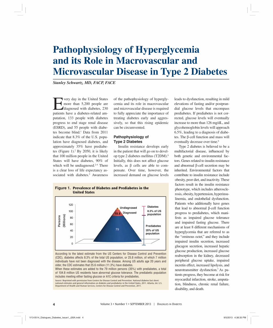
According to the latest estimate from the US Centers for Disease Control and Prevention (CDC), diabetes affects 8.3% of the total US population, or 25.8 million, of which 7 million individuals have not been diagnosed with the disease. Among US adults age 20 years and older, the CDC estimates that 25.6 million (11.3%) have diabetes. When these estimates are added to the 79 million persons (35%) with prediabetes, a total of 104.8 million US residents have abnormal glucose tolerance. The prediabetic population includes meeting either fasting glucose or A1C criteria for prediabetes. Source: Reprinted with permission from Centers for Disease Control and Prevention. National diabetes fact sheet: national estimates and general information on diabetes and prediabetes in the United States, 2011. Atlanta, GA: U.S. Department of Health and Human Services, Centers for Disease Control and Prevention, 2011.
Figure 1. Prevalence of Diabetes and Prediabetes in the United States
4 Volume 3 • Number 1 • SEPTEMBER 2013 | DIALOGUES IN DIABETES
V13-0514_Dialogues_Diabetes_Issue1_J26A.indd 4V13-0514_Dialogues_Diabetes_Issue1_J26A.indd 4 9/5/2013 4:38:35 PM9/5/2013 4:38:35 PM
Obesity and Type 2 DiabetesObesity is also increasing as
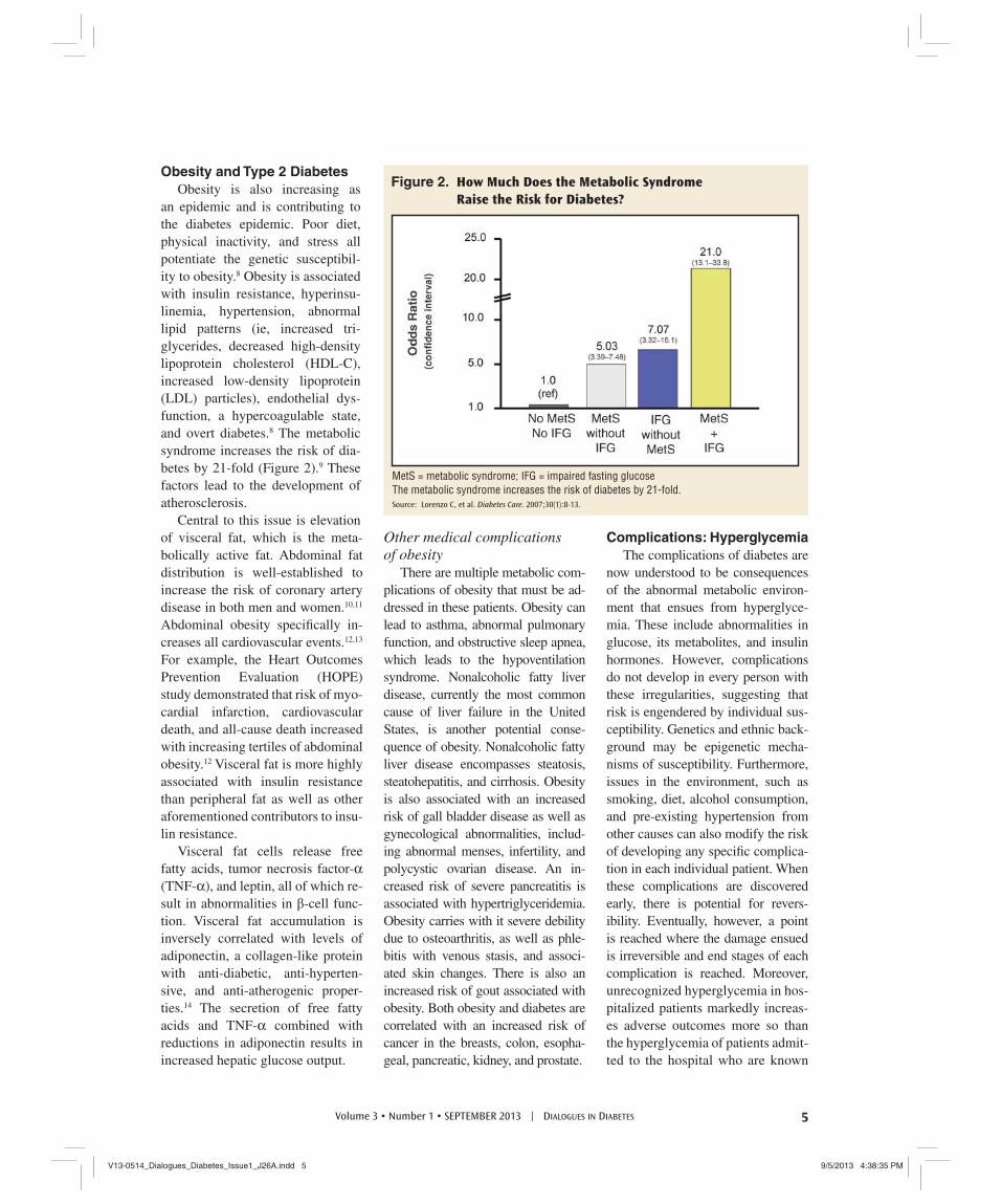
an epidemic and is contributing to the diabetes epidemic. Poor diet, physical inactivity, and stress all potentiate the genetic susceptibil-ity to obesity.8 Obesity is associated with insulin resistance, hyperinsu-linemia, hypertension, abnormal lipid patterns (ie, increased tri-glycerides, decreased high-density lipoprotein cholesterol (HDL-C), increased low-density lipoprotein (LDL) particles), endothelial dys-function, a hypercoagulable state, and overt diabetes.8 The metabolic syndrome increases the risk of dia-betes by 21-fold (Figure 2).9 These factors lead to the development of atherosclerosis.
Central to this issue is elevation of visceral fat, which is the meta-bolically active fat. Abdominal fat distribution is well-established to increase the risk of coronary artery disease in both men and women.10,11 Abdominal obesity specifi cally in-creases all cardiovascular events.12,13 For example, the Heart Outcomes Prevention Evaluation (HOPE) study demonstrated that risk of myo-cardial infarction, cardiovascular death, and all-cause death increased with increasing tertiles of abdominal obesity.12 Visceral fat is more highly associated with insulin resistance than peripheral fat as well as other aforementioned contributors to insu-lin resistance.
Visceral fat cells release free fatty acids, tumor necrosis factor-α (TNF-α), and leptin, all of which re-sult in abnormalities in β-cell func-tion. Visceral fat accumulation is inversely correlated with levels of adiponectin, a collagen-like protein with anti-diabetic, anti-hyperten-sive, and anti-atherogenic proper-ties.14 The secretion of free fatty acids and TNF-α combined with reductions in adiponectin results in increased hepatic glucose output.
Other medical complicationsof obesity
There are multiple metabolic com-plications of obesity that must be ad-dressed in these patients. Obesity can lead to asthma, abnormal pulmonary function, and obstructive sleep apnea, which leads to the hypoventilation syndrome. Nonalcoholic fatty liver disease, currently the most common cause of liver failure in the United States, is another potential conse-quence of obesity. Nonalcoholic fatty liver disease encompasses steatosis, steatohepatitis, and cirrhosis. Obesity is also associated with an increased risk of gall bladder disease as well as gynecological abnormalities, includ-ing abnormal menses, infertility, and polycystic ovarian disease. An in-creased risk of severe pancreatitis is associated with hypertriglyceridemia. Obesity carries with it severe debility due to osteoarthritis, as well as phle-bitis with venous stasis, and associ-ated skin changes. There is also an increased risk of gout associated with obesity. Both obesity and diabetes are correlated with an increased risk of cancer in the breasts, colon, esopha-geal, pancreatic, kidney, and prostate.
Complications: HyperglycemiaThe complications of diabetes are
now understood to be consequences of the abnormal metabolic environ-ment that ensues from hyperglyce-mia. These include abnormalities in glucose, its metabolites, and insulin hormones. However, complications do not develop in every person with these irregularities, suggesting that risk is engendered by individual sus-ceptibility. Genetics and ethnic back-ground may be epigenetic mecha-nisms of susceptibility. Furthermore, issues in the environment, such as smoking, diet, alcohol consumption, and pre-existing hypertension from other causes can also modify the risk of developing any specifi c complica-tion in each individual patient. When these complications are discovered early, there is potential for revers-ibility. Eventually, however, a point is reached where the damage ensued is irreversible and end stages of each complication is reached. Moreover, unrecognized hyperglycemia in hos-pitalized patients markedly increas-es adverse outcomes more so than the hyperglycemia of patients admit-ted to the hospital who are known
MetS = metabolic syndrome; IFG = impaired fasting glucoseThe metabolic syndrome increases the risk of diabetes by 21-fold.Source: Lorenzo C, et al. Diabetes Care. 2007;30(1):8-13.
Figure 2. How Much Does the Metabolic Syndrome
Raise the Risk for Diabetes?
5Volume 3 • Number 1 • SEPTEMBER 2013 | DIALOGUES IN DIABETES
V13-0514_Dialogues_Diabetes_Issue1_J26A.indd 5V13-0514_Dialogues_Diabetes_Issue1_J26A.indd 5 9/5/2013 4:38:35 PM9/5/2013 4:38:35 PM
to have diabetes.15 Therefore, early aggressive control of hyperglyce-mia is critical, and has been shown to reduce the risk of complications.16 In fact, for every 1% decrease in A1C, microvascular complications are reduced by 21%.17
Complications of hyperglycemia begin to accrue with even minimal abnormalities in either fasting hy-perglycemia or postprandial hyper-glycemia associated with prediabe-tes. Postprandial glucose elevations increase variability, a predictor of increased mortality. Postprandial elevations also increase the risk of microvascular disease, adverse pregnancy outcomes, atheroscle-rotic vascular disease, and worsened cardiovascular complications. Spe-cifi cally treating postprandial hyper-glycemia can reduce these risks.18
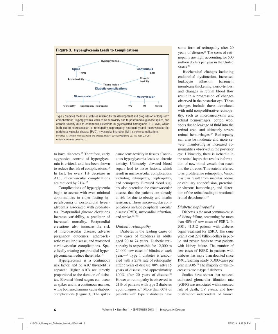
Hyperglycemia is a continuous risk factor, and no A1C threshold is apparent. Higher A1Cs are directly proportional to the duration of diabe-tes. Elevated blood sugars can occur as spikes and in a continuous manner, while both mechanisms cause diabetic complications (Figure 3). The spikes
cause acute toxicity in tissues. Contin-uous hyperglycemia leads to chronic toxicity. Ultimately, elevated blood sugars lead to tissue lesions, which result in microvascular complications including retinopathy, nephropathy, and neuropathy. Elevated blood sug-ars also potentiate the macrovascular disease that the patients are already at risk for due to obesity and insulin resistance. These macrovascular com-plications include peripheral vascular disease (PVD), myocardial infarction, and stroke.17,19-21
Diabetic retinopathyDiabetes is the leading cause of
new cases of blindness in adults aged 20 to 74 years. Diabetic reti-nopathy is responsible for 12,000 to 24,000 new cases of blindness each year.22,23 Type 1 diabetes is associ-ated with a 25% rate of retinopathy after 5 years of disease, 80% after 15 years of disease, and approximately 100% after 20 years of disease.24 However, retinopathy is observed in 21% of patients with type 2 diabetes upon diagnosis.24 More than 60% of patients with type 2 diabetes have
some form of retinopathy after 20 years of disease.25 The costs of reti-nopathy are high, accounting for 500 million dollars per year in the United States.26
Biochemical changes including endothelial dysfunction, increased leukocyte adhesion, basement membrane thickening, pericyte loss, and changes in retinal blood fl ow result in a progression of changes observed in the posterior eye. These changes include those associated with mild nonproliferative retinopa-thy, such as microaneurysms and retinal hemorrhages, cotton wool spots due to leakage of fl uid into the retinal area, and ultimately severe retinal hemorrhages.27 Retinopathy can also be moderate and more se-vere, manifesting as increased ab-normalities observed in the posterior eye. Ultimately, there is ischemia in the retinal layers that results in forma-tion of new blood vessels that reach into the vitreous. This state is referred to as proliferative retinopathy. Vision loss can result from macular edema or capillary nonperfusion, preretinal or vitreous hemorrhage, and distor-tion of the retina leading to tractional retinal detachment.22
Diabetic nephropathyDiabetes is the most common cause
of kidney failure, accounting for more than 40% of new cases of ESRD. In 2001, 41,312 patients with diabetes began treatment for ESRD. The same year, it cost 22.8 billion dollars in pub-lic and private funds to treat patients with kidney failure. The number of new cases of ESRD in patients with diabetes has more than doubled since 1991, reaching nearly 50,000 cases per year in 2005.28 The majority of this in-crease is due to type 2 diabetes.
Studies have shown that reduced estimated glomerular fi ltration rate (eGFR) was associated with increased risk of death, CV events, and hos-pitalization independent of known
Type 2 diabetes mellitus (T2DM) is marked by the development and progression of long-term complications. Hyperglycemia leads to acute toxicity due to postprandial glucose spikes, and chronic toxicity due to continuous elevations in glycosylated hemoglobin A1C level, which both lead to microvascular (ie, retinopathy, nephropathy, neuropathy) and macrovascular (ie, peripheral vascular disease [PVD], myocardial infarction [MI], stroke) complications.Brownlee M. Diabetes mellitus: theory and practice. Elsevier Science Publishing Co., Inc; 1990:279-291.
Ceriello A. Diabetes. 2005;54:1-7.
Figure 3. Hyperglycemia Leads to Complications
6 Volume 3 • Number 1 • SEPTEMBER 2013 | DIALOGUES IN DIABETES
V13-0514_Dialogues_Diabetes_Issue1_J26A.indd 6V13-0514_Dialogues_Diabetes_Issue1_J26A.indd 6 9/5/2013 4:38:36 PM9/5/2013 4:38:36 PM
risk factors. The risk of death was increased as eGFR decreased below 60 mL/min/1.73 m2. In addition, age-standardized rates of death and cardiovascular events substan-tially increased with progressively lower eGFR.29
Dialysis, one form of treatment for kidney failure, can relieve the symp-toms of kidney disease, but over time the damaged kidneys will continue to contribute to problems, such as heart disease, bone disease, arthritis, nerve damage, infertility, and malnutrition. Kidney transplant, another treatment alternative, may prove to be a more permanent solution to kidney disease for some patients. However, trans-plantation has its own risks including the risk of surgery, the risk of organ rejection, the risk of infection, and other complications from immuno-suppressant drugs.
Many of the cases of ESRD are preventable by careful control of blood glucose, blood pressure, and by early treatment of microalbumin-uria. In addition to being the earliest manifestation of nephropathy, albu-minuria is also a marker of increased cardiovascular morbidity and mor-tality for patients with diabetes. The presence of microalbuminuria is an indicator for screening for possible vascular disease and aggressive inter-vention to reduce all cardiovascular risk factors — elevated LDL, hyper-tension, smoking, and physical inac-tivity. Preliminary evidence suggests that lipid-lowering therapy may also reduce urinary protein levels.30
Diabetic neuropathyDiabetic neuropathy, the most
common neuropathy in industrialized countries, is a heterogeneous group of conditions affecting somatic and auto-nomic nerves. Approximately 50% of patients with diabetes develop neurop-athy after 25 years, and 10% develop symptomatic neuropathy. Major mor-bidities are pain, numbness, and foot
ulceration. Diabetic neuropathy is responsible for approximately 75% of all nontraumatic foot amputa-tions.31 Major signs of diabetic pe-ripheral neuropathy are not evident at the onset of disease, although some patients do experience symp-toms with mildly impaired glucose tolerance or impaired fasting glu-cose. Symptoms may occur at any time and intermittently. Periodic evaluation is essential for patients with type 2 diabetes because many are not aware of diabetic peripheral neuropathy.
Diabetic neuropathies are classi-fi ed as symmetric polyneuropathies and focal/multifocal neuropathies. Symmetric polyneuropathies include distal symmetric sensory motor neu-ropathy, autonomic neuropathy, and acute painful neuropathy. Systemic polyneuropathy is the most common form of diabetic neuropathy. This con-dition affects distal lower extremities. The longer axons of the legs are more susceptible, yet as the length of the axon damage rises, the hands, which are a function of shorter axons, will begin to develop discomfort. This is referred to as “stocking-glove” sen-sory loss and symptoms. Symptoms include paresthesias and dysesthesias of the feet and hands (predominant at night), paroxysmal lancinating pain, deep aching and muscle cramping, and autonomic dysfunction. Compli-cations of symmetric polyneuropa-thies include ulcers, Charcot arthropa-thy, dislocation, stress fractures, and ultimately amputation.
With autonomic neuropathies, which are rare, the damage to the nerves affect autonomic nerves con-trolling internal organs. Manifesta-tions may include pupillary abnor-malities, changes in heart rate, further changes in sympathetic and parasym-pathetic tone, gastroparesis, gall blad-der disease, large intestine symptoms, bladder dysfunction, and erectile dys-function. Gastrointestinal autonomic
neuropathy is characterized typically by symptoms of gastroparesis in-cluding anorexia, nausea, vomiting, undigested food many hours after eating, early satiety, and diabetic enteropathy including diarrhea and constipation. Cardiovascular auto-nomic neuropathy includes exercise intolerance and postural hypotension. Patients with diabetes can also expe-rience polyradiculopathy, including lumbar polyradiculopathy, which is often referred to as diabetic amyotro-phy, which can manifest as thigh pain followed by muscle weakness and asymmetic atrophy.
Mononeuropathy is another poten-tial complication of diabetes. This can manifest as either single nerve damage, such as the nerve of the eye, carpal-tunnel syndrome, elbow symptoms,
and unilateral foot drop. Sometimes these mononeuropathies occur simul-taneously, in a condition referred to as mononeuropathy multiplex. Mononeu-ropathies often resolve spontaneously after approximately 6 months.
Cardiac complicationsAs previously mentioned, type 2
diabetes and cardiovascular disease are closely associated. This occurs from the concordance of risk factors that accrue with obesity, the insulin resistance syndrome, and hypergly-cemia. Heart disease is increased from 2-fold to 4-fold in patients with type 2 diabetes and accounts for 80% of all diabetic mortality (75% from coronary atherosclerosis, 25% from cerebral of peripheral vascular dis-ease). Cardiovascular disease causes
The number of new cases of
end stage renal disease (ESRD) in
patients with diabetes has more
than doubled since 1991.
7Volume 3 • Number 1 • SEPTEMBER 2013 | DIALOGUES IN DIABETES
V13-0514_Dialogues_Diabetes_Issue1_J26A.indd 7V13-0514_Dialogues_Diabetes_Issue1_J26A.indd 7 9/5/2013 4:38:36 PM9/5/2013 4:38:36 PM
more than 75% of all hospitalizations for diabetic complications, and more than 50% of patients with newly di-agnosed type 2 diabetes already have pre-existing cardiovascular disease.32,33
Among adults aged 60 years and older with diabetes, 30% have coronary heart disease, 14% have congestive heart failure, 14% have had a stroke, and 21% have peripheral arterial disease.34
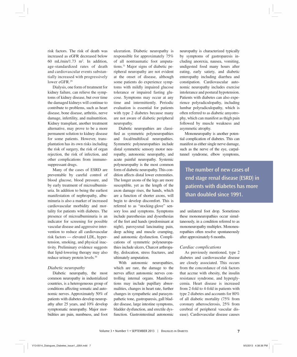
A1C levels predict coronary heart disease, with higher levels increas-ing the risk of cardiovascular events and coronary heart disease mortality (Figure 4).35 Furthermore, postpran-dial glucose levels independently in-crease and determine cardiovascular disease. This is true even for post-prandial glucose levels that are in the normal range; as they rise, elevations
in fatal heart disease and total heart disease are observed.36
Biochemical Mechanisms Behind the Macrovascular and Microvascular Complications of Diabetes
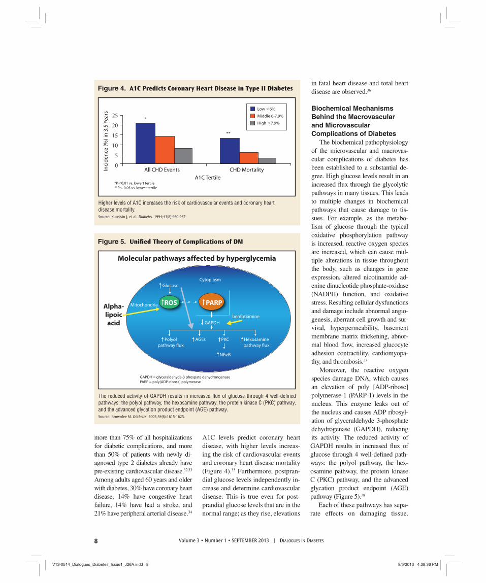
The biochemical pathophysiology of the microvascular and macrovas-cular complications of diabetes has been established to a substantial de-gree. High glucose levels result in an increased fl ux through the glycolytic pathways in many tissues. This leads to multiple changes in biochemical pathways that cause damage to tis-sues. For example, as the metabo-lism of glucose through the typical oxidative phosphorylation pathway is increased, reactive oxygen species are increased, which can cause mul-tiple alterations in tissue throughout the body, such as changes in gene expression, altered nicotinamide ad-enine dinucleotide phosphate-oxidase (NADPH) function, and oxidative stress. Resulting cellular dysfunctions and damage include abnormal angio-genesis, aberrant cell growth and sur-vival, hyperpermeability, basement membrane matrix thickening, abnor-mal blood fl ow, increased glucocyte adhesion contractility, cardiomyopa-thy, and thrombosis.37
Moreover, the reactive oxygen species damage DNA, which causes an elevation of poly [ADP-ribose] polymerase-1 (PARP-1) levels in the nucleus. This enzyme leaks out of the nucleus and causes ADP ribosyl-ation of glyceraldehyde 3-phosphate dehydrogenase (GAPDH), reducing its activity. The reduced activity of GAPDH results in increased fl ux of glucose through 4 well-defi ned path-ways: the polyol pathway, the hex-osamine pathway, the protein kinase C (PKC) pathway, and the advanced glycation product endpoint (AGE) pathway (Figure 5).38
Each of these pathways has sepa-rate effects on damaging tissue.
25
20
15
10
5
0All CHD Events
*P<0.01 vs. lowert tertile
**P< 0.05 vs. lowest tertile
A1C Tertile
Inci
de
nce
(%
) in
3.5
Ye
ars
CHD Mortality
*
**
Low <6%
Middle 6-7.9%
High >7.9%
Higher levels of A1C increases the risk of cardiovascular events and coronary heart disease mortality. Source: Kuusisto J, et al. Diabetes. 1994;43(8):960-967.
Figure 4. A1C Predicts Coronary Heart Disease in Type II Diabetes
Glucose
Mitochondria
GAPDH
benfotiamine
Polyol
pathway flux
Hexosamine
pathway flux
PKCAGEs
NFκB
Cytoplasm
PARPROSAlpha-lipoicacid
Molecular pathways affected by hyperglycemia
GAPDH = glyceraldehyde-3 phospate dehydrongenasePARP = poly(ADP-ribose) polymerase
The reduced activity of GAPDH results in increased fl ux of glucose through 4 well-defi ned pathways: the polyol pathway, the hexosamine pathway, the protein kinase C (PKC) pathway, and the advanced glycation product endpoint (AGE) pathway. Source: Brownlee M. Diabetes. 2005;54(6):1615-1625.
Figure 5. Unifi ed Theory of Complications of DM
8 Volume 3 • Number 1 • SEPTEMBER 2013 | DIALOGUES IN DIABETES
V13-0514_Dialogues_Diabetes_Issue1_J26A.indd 8V13-0514_Dialogues_Diabetes_Issue1_J26A.indd 8 9/5/2013 4:38:36 PM9/5/2013 4:38:36 PM
Increased activity of the hexos-amine pathway causes altered gene expression and increased infl am-mation.39 Increased activity of the polyol pathway causes a shift in water and ion content, which can lead to cataracts. Polyol accumula-tion, osmotic shifts, effl ux of myo-inositol, reduced ATP, decreased synthesis of reduced glutathione, reduced NADPH, and decreased Na/K ATPase activity are other con-sequences of increasing the activity of this pathway. Biochemical con-sequences of the AGE formation include crosslinks of extracellular matrix proteins, which will occur in many tissues. Advanced glycation products can also be ingested in food, especially if charred, causing similar damage to multiple tissues. Low density lipoprotein and hemo-globin can become glycosylated, altering their functions throughout the body. Lastly, increased fl ux through diacyl glycerol (DAG) and PKC activity has multiple ef-fects, including increased oxidative pathways, increased transforming growth factor β activity (which in-creases collagen and fi bronectin), increased plasminogen activator inhibitor-1 (which can affect fi bri-nolysis), and increased vascular endothelial growth factor (which can increase vascular permeability and angiogenesis). Others effects include increased nuclear factor k-light-chain-enhancer of activated β cells (NF-kB), which results in abnormal pro-infl ammatory gene expression.39
Therefore, through the basic mech-anism of increased blood sugar with fl ux through its pathway of metabo-lism, increased oxidative pathways, and increased fl ux through other al-ternate pathways, damage to multiple tissues occurs, ultimately leading to the microvascular and macrovascular complications of diabetes.
SummaryThe etiology of diabetes and the
biochemical mechanisms involved as a result of hyperglycemia underscore the importance of controlling blood sugar in the midst of the obesity and diabetes epidemics. Early aggressive control is critical, and treating postprandial hyperglycemia is just as important as treating fasting hyperglycemia. In ad-dition to treating blood sugar, it is also important that blood pressure, LDL, HDL, triglycerides, and non-HDL are treated to recommended goals. Treat-ment of these metabolic factors can reduce the risk of complications in pa-tients with diabetes.
References1. Centers for Disease Control and Preven-
tion. http://www.cdc.gov/diabetes/pubs/pdf/ndfs_2007.pdf. Accessed April 13, 2009.
2. Centers for Disease Control and Preven-tion. http://www.cdc.gov/diabetes/pubs/pdf/ndfs_2011.pdf. Accessed February 11, 2011.
3. National Diabetes Surveillance System. http://www.cdc.gov/diabetes/statistics/prev/national/fi gpersons.htm. Accessed August 9, 2013.
4. Boyle JP, et al. Popul Health Metr. 2010;8:29.
5. Kushner RF, et al. Curr Opin Endocrinol Diabetes Obes. 2013 [Epub ahead of print]
6. Kendall DM, et al. Am J Med. 2009;122(6 suppl):S37-S50.
7. Defronzo RA. Diabetes. 2009;58(4):773-795.
8. Grundy SM. J Clin Endocrinol Metab. 2005;89(6):2595-2600.
9. Lorenzo C, et al. Diabetes Care. 2007; 30(1):8-13.
10. Folsom AR, et al. Arch Intern Med. 2000; 160(14):2117-2128.
11. Canoy D. Curr Atheroscler Rep. 2010; 12(2):125-133.
12. Dagenais GR, et al. Am Heart J. 2005; 149(1):54-60.
13. Calle EE, et al. N Engl J Med. 1999; 341(15):1097-1105.
14. Matsuzawa Y. Proc Jpn Acad Ser B Phys Biol Sci. 2010;86(2):131-141.
15. Umpierrez GE, et al. J Clin Endocrinol Metab. 2002;87(3):978-982.
16. The Diabetes Control and Complications
Trial Research Group. N Engl J Med. 1993;329(14):977-986.
17. Stratton IM, et al. BMJ. 2000; 321 (7258):405-412.
18. Yamagishi S, et al. Med Hypotheses. 2005;65(1):152-154.
19. American Diabetes Association. http://www.diabetes.org/diabetes-statistics/complications.jsp.
20. Brownlee M. Elsevier Science Publishing Co, Inc; 1990:279-291. (Au: Could not verify this reference)
21. Ceriello A. Diabetes. 2005;54(1):1-7.
22. American Diabetes Association. Diabetes Care. 2001;24(suppl 1):S73-S76.
23. National Institute of Diabetes and Di-gestive and Kidney Diseases (NIDDK). Diabetes Statistics. NIH Publication No. 99-3892. March 1999 (e-text posted Sep-tember 1999).
24. UK Prospective Diabetes Study (UKPDS) Group. Lancet. 1998;352(9131):837-853.
25. Expert Committee on the Diagnosis and Clasifi cation of Diabetes Mellitus. Diabe-tes Care. 2002;25(suppl1):S90-S93.
26. Porta M, et al. Diabetologia. 2002; 45 (12):1617-1634.
27. Stratton IM, et al. Diabetologia. 2001; 44(2):156-163.
28. National Institute of Diabetes and Di-gestive and Kidney Diseases (NIDDK). USRDS 2007 Annual Data Report. http://www.usrds.org/2007/pdf/00_intro_07.pdf. Accessed August 9, 2013.
29. Go AS, et al. N Engl J Med. 2004; 351(13):1296-1305.
30. American Diabetes Association. Diabetes Care. 2001;24 (suppl 1):S69-S72.
31. Casellini CM, et al. Endocrine Pract. 2007;13(5):550-566.
32. American Diabetes Association. Diabetes Care. 1999;22(suppl 1):S56-S59.
33. Lewis GF. Can J Cardiol. 1995;11(Suppl C):24C-28C.
34. Kalyani RR, et al. Diabetes Care. 2010; 33(5):1055-1060.
35. Kuusisto J, et al. Diabetes. 1994;43(8): 960-967.
36. Donahue RP, et al. Diabetes. 1987;36(6): 689-692.
37. Sheetz MJ, et al. JAMA. 2002;288(20): 2579-2588.
38. Brownlee M. Diabetes. 2005;54(6):1615-1625.
39. Brownlee M. Nature. 2001;414(6865): 813-820.
Full references are available at www.healio.com/endocrinology/education-lab.
9Volume 3 • Number 1 • SEPTEMBER 2013 | DIALOGUES IN DIABETES
V13-0514_Dialogues_Diabetes_Issue1_J26A.indd 9V13-0514_Dialogues_Diabetes_Issue1_J26A.indd 9 9/5/2013 4:38:37 PM9/5/2013 4:38:37 PM
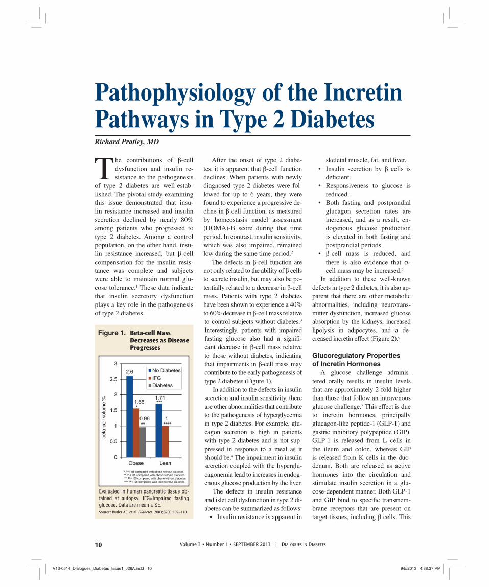
The contributions of β-cell dysfunction and insulin re-sistance to the pathogenesis
of type 2 diabetes are well-estab-lished. The pivotal study examining this issue demonstrated that insu-lin resistance increased and insulin secretion declined by nearly 80% among patients who progressed to type 2 diabetes. Among a control population, on the other hand, insu-lin resistance increased, but β-cell compensation for the insulin resis-tance was complete and subjects were able to maintain normal glu-cose tolerance.1 These data indicate that insulin secretory dysfunction plays a key role in the pathogenesis of type 2 diabetes.
After the onset of type 2 diabe-tes, it is apparent that β-cell function declines. When patients with newly diagnosed type 2 diabetes were fol-lowed for up to 6 years, they were found to experience a progressive de-cline in β-cell function, as measured by homeostasis model assessment (HOMA)-B score during that time period. In contrast, insulin sensitivity, which was also impaired, remained low during the same time period.2
The defects in β-cell function are not only related to the ability of β cells to secrete insulin, but may also be po-tentially related to a decrease in β-cell mass. Patients with type 2 diabetes have been shown to experience a 40% to 60% decrease in β-cell mass relative to control subjects without diabetes.3 Interestingly, patients with impaired fasting glucose also had a signifi -cant decrease in β-cell mass relative to those without diabetes, indicating that impairments in β-cell mass may contribute to the early pathogenesis of type 2 diabetes (Figure 1).
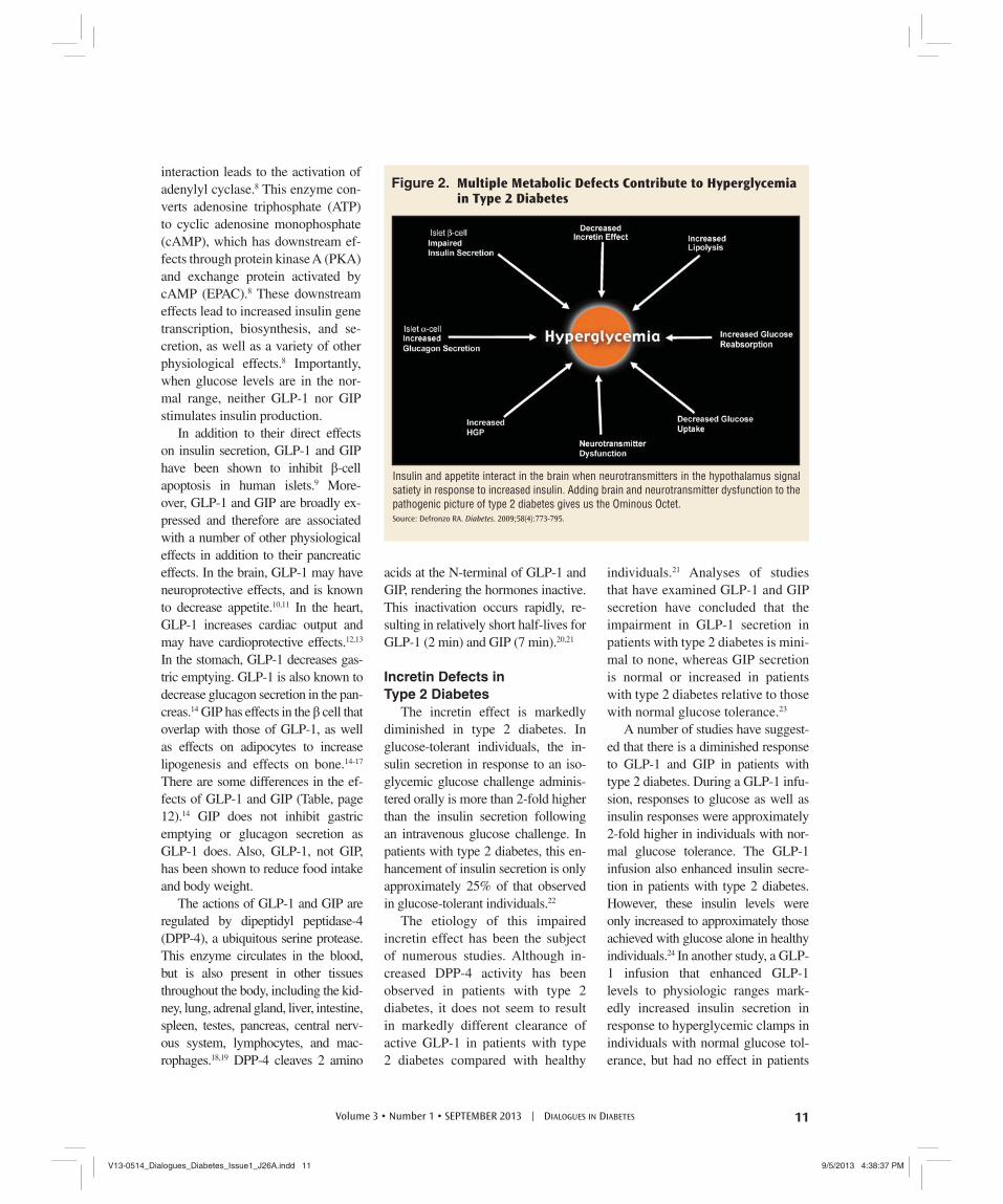
In addition to the defects in insulin secretion and insulin sensitivity, there are other abnormalities that contribute to the pathogenesis of hyperglycemia in type 2 diabetes. For example, glu-cagon secretion is high in patients with type 2 diabetes and is not sup-pressed in response to a meal as it should be.4 The impairment in insulin secretion coupled with the hyperglu-cagonemia lead to increases in endog-enous glucose production by the liver.
The defects in insulin resistance and islet cell dysfunction in type 2 di-abetes can be summarized as follows:
• Insulin resistance is apparent in
skeletal muscle, fat, and liver.• Insulin secretion by β cells is
defi cient.• Responsiveness to glucose is
reduced.• Both fasting and postprandial
glucagon secretion rates are increased, and as a result, en-dogenous glucose production is elevated in both fasting and postprandial periods.
• β-cell mass is reduced, and there is also evidence that α-cell mass may be increased.5
In addition to these well-known defects in type 2 diabetes, it is also ap-parent that there are other metabolic abnormalities, including neurotrans-mitter dysfunction, increased glucose absorption by the kidneys, increased lipolysis in adipocytes, and a de-creased incretin effect (Figure 2).6
Glucoregulatory Properties of Incretin Hormones
A glucose challenge adminis-tered orally results in insulin levels that are approximately 2-fold higher than those that follow an intravenous glucose challenge.7 This effect is due to incretin hormones, principally glucagon-like peptide-1 (GLP-1) and gastric inhibitory polypeptide (GIP). GLP-1 is released from L cells in the ileum and colon, whereas GIP is released from K cells in the duo-denum. Both are released as active hormones into the circulation and stimulate insulin secretion in a glu-cose-dependent manner. Both GLP-1 and GIP bind to specifi c transmem-brane receptors that are present on target tissues, including β cells. This
Pathophysiology of the Incretin Pathways in Type 2 DiabetesRichard Pratley, MD
Evaluated in human pancreatic tissue ob-tained at autopsy. IFG=Impaired fasting glucose. Data are mean ± SE.Source: Butler AE, et al. Diabetes. 2003;52(1):102–110.
Figure 1. Beta-cell Mass Decreases as Disease Progresses
10 Volume 3 • Number 1 • SEPTEMBER 2013 | DIALOGUES IN DIABETES
V13-0514_Dialogues_Diabetes_Issue1_J26A.indd 10V13-0514_Dialogues_Diabetes_Issue1_J26A.indd 10 9/5/2013 4:38:37 PM9/5/2013 4:38:37 PM
interaction leads to the activation of adenylyl cyclase.8 This enzyme con-verts adenosine triphosphate (ATP) to cyclic adenosine monophosphate (cAMP), which has downstream ef-fects through protein kinase A (PKA) and exchange protein activated by cAMP (EPAC).8 These downstream effects lead to increased insulin gene transcription, biosynthesis, and se-cretion, as well as a variety of other physiological effects.8 Importantly, when glucose levels are in the nor-mal range, neither GLP-1 nor GIP stimulates insulin production.
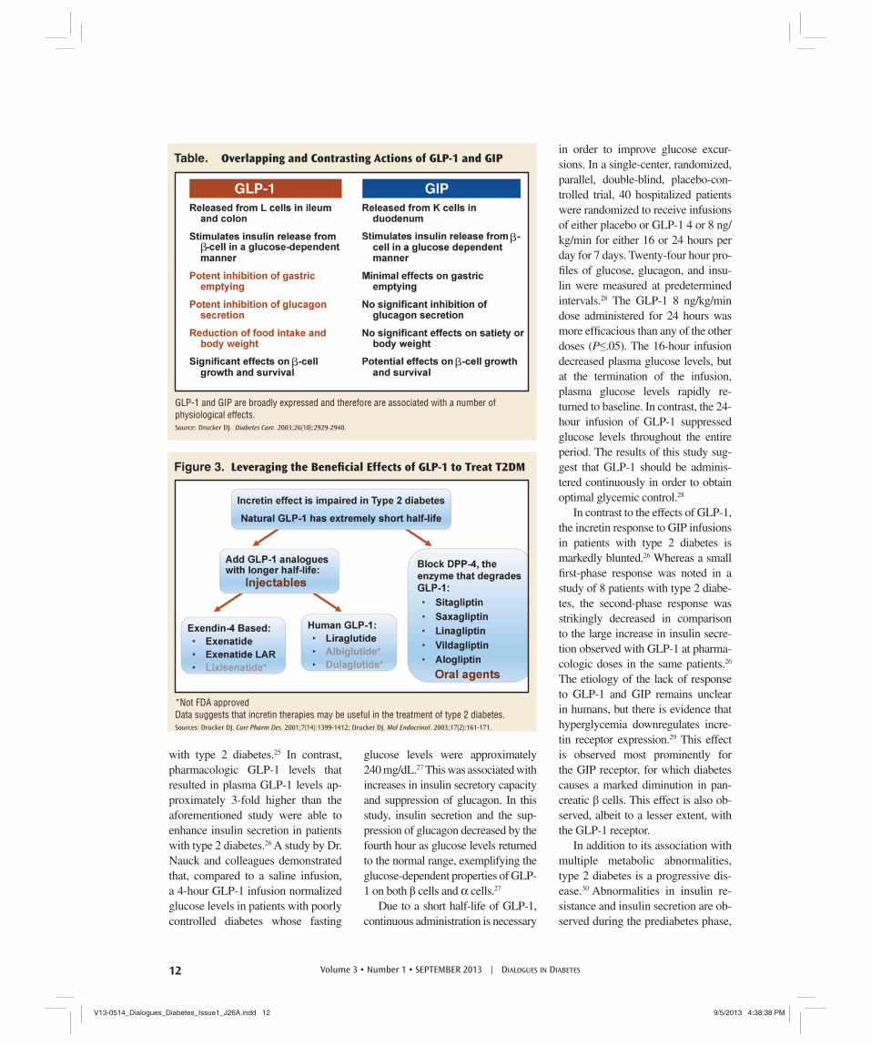
In addition to their direct effects on insulin secretion, GLP-1 and GIP have been shown to inhibit β-cell apoptosis in human islets.9 More-over, GLP-1 and GIP are broadly ex-pressed and therefore are associated with a number of other physiological effects in addition to their pancreatic effects. In the brain, GLP-1 may have neuroprotective effects, and is known to decrease appetite.10,11 In the heart, GLP-1 increases cardiac output and may have cardioprotective effects.12,13 In the stomach, GLP-1 decreases gas-tric emptying. GLP-1 is also known to decrease glucagon secretion in the pan-creas.14 GIP has effects in the β cell that overlap with those of GLP-1, as well as effects on adipocytes to increase lipogenesis and effects on bone.14-17 There are some differences in the ef-fects of GLP-1 and GIP (Table, page 12).14 GIP does not inhibit gastric emptying or glucagon secretion as GLP-1 does. Also, GLP-1, not GIP, has been shown to reduce food intake and body weight.
The actions of GLP-1 and GIP are regulated by dipeptidyl peptidase-4 (DPP-4), a ubiquitous serine protease. This enzyme circulates in the blood, but is also present in other tissues throughout the body, including the kid-ney, lung, adrenal gland, liver, intestine, spleen, testes, pancreas, central nerv-ous system, lymphocytes, and mac-rophages.18,19 DPP-4 cleaves 2 amino
acids at the N-terminal of GLP-1 and GIP, rendering the hormones inactive. This inactivation occurs rapidly, re-sulting in relatively short half-lives for GLP-1 (2 min) and GIP (7 min).20,21
Incretin Defects inType 2 Diabetes
The incretin effect is markedly diminished in type 2 diabetes. In glucose-tolerant individuals, the in-sulin secretion in response to an iso-glycemic glucose challenge adminis-tered orally is more than 2-fold higher than the insulin secretion following an intravenous glucose challenge. In patients with type 2 diabetes, this en-hancement of insulin secretion is only approximately 25% of that observed in glucose-tolerant individuals.22
The etiology of this impaired incretin effect has been the subject of numerous studies. Although in-creased DPP-4 activity has been observed in patients with type 2 diabetes, it does not seem to result in markedly different clearance of active GLP-1 in patients with type 2 diabetes compared with healthy
individuals.21 Analyses of studies that have examined GLP-1 and GIP secretion have concluded that the impairment in GLP-1 secretion in patients with type 2 diabetes is mini-mal to none, whereas GIP secretion is normal or increased in patients with type 2 diabetes relative to those with normal glucose tolerance.23
A number of studies have suggest-ed that there is a diminished response to GLP-1 and GIP in patients with type 2 diabetes. During a GLP-1 infu-sion, responses to glucose as well as insulin responses were approximately 2-fold higher in individuals with nor-mal glucose tolerance. The GLP-1 infusion also enhanced insulin secre-tion in patients with type 2 diabetes. However, these insulin levels were only increased to approximately those achieved with glucose alone in healthy individuals.24 In another study, a GLP-1 infusion that enhanced GLP-1 levels to physiologic ranges mark-edly increased insulin secretion in response to hyperglycemic clamps in individuals with normal glucose tol-erance, but had no effect in patients
Insulin and appetite interact in the brain when neurotransmitters in the hypothalamus signal satiety in response to increased insulin. Adding brain and neurotransmitter dysfunction to the pathogenic picture of type 2 diabetes gives us the Ominous Octet.Source: Defronzo RA. Diabetes. 2009;58(4):773-795.
Figure 2. Multiple Metabolic Defects Contribute to Hyperglycemia in Type 2 Diabetes
11Volume 3 • Number 1 • SEPTEMBER 2013 | DIALOGUES IN DIABETES
V13-0514_Dialogues_Diabetes_Issue1_J26A.indd 11V13-0514_Dialogues_Diabetes_Issue1_J26A.indd 11 9/5/2013 4:38:37 PM9/5/2013 4:38:37 PM
with type 2 diabetes.25 In contrast, pharmacologic GLP-1 levels that resulted in plasma GLP-1 levels ap-proximately 3-fold higher than the aforementioned study were able to enhance insulin secretion in patients with type 2 diabetes.26 A study by Dr. Nauck and colleagues demonstrated that, compared to a saline infusion, a 4-hour GLP-1 infusion normalized glucose levels in patients with poorly controlled diabetes whose fasting
glucose levels were approximately 240 mg/dL.27 This was associated with increases in insulin secretory capacity and suppression of glucagon. In this study, insulin secretion and the sup-pression of glucagon decreased by the fourth hour as glucose levels returned to the normal range, exemplifying the glucose-dependent properties of GLP-1 on both β cells and α cells.27
Due to a short half-life of GLP-1, continuous administration is necessary
in order to improve glucose excur-sions. In a single-center, randomized, parallel, double-blind, placebo-con-trolled trial, 40 hospitalized patients were randomized to receive infusions of either placebo or GLP-1 4 or 8 ng/kg/min for either 16 or 24 hours per day for 7 days. Twenty-four hour pro-fi les of glucose, glucagon, and insu-lin were measured at predetermined intervals.28 The GLP-1 8 ng/kg/min dose administered for 24 hours was more effi cacious than any of the other doses (P≤.05). The 16-hour infusion decreased plasma glucose levels, but at the termination of the infusion, plasma glucose levels rapidly re-turned to baseline. In contrast, the 24-hour infusion of GLP-1 suppressed glucose levels throughout the entire period. The results of this study sug-gest that GLP-1 should be adminis-tered continuously in order to obtain optimal glycemic control.28
In contrast to the effects of GLP-1, the incretin response to GIP infusions in patients with type 2 diabetes is markedly blunted.26 Whereas a small fi rst-phase response was noted in a study of 8 patients with type 2 diabe-tes, the second-phase response was strikingly decreased in comparison to the large increase in insulin secre-tion observed with GLP-1 at pharma-cologic doses in the same patients.26 The etiology of the lack of response to GLP-1 and GIP remains unclear in humans, but there is evidence that hyperglycemia downregulates incre-tin receptor expression.29 This effect is observed most prominently for the GIP receptor, for which diabetes causes a marked diminution in pan-creatic β cells. This effect is also ob-served, albeit to a lesser extent, with the GLP-1 receptor.
In addition to its association with multiple metabolic abnormalities, type 2 diabetes is a progressive dis-ease.30 Abnormalities in insulin re-sistance and insulin secretion are ob-served during the prediabetes phase,
*Not FDA approvedData suggests that incretin therapies may be useful in the treatment of type 2 diabetes.Sources: Drucker DJ. Curr Pharm Des. 2001;7(14):1399-1412; Drucker DJ. Mol Endocrinol. 2003;17(2):161-171.
Figure 3. Leveraging the Benefi cial Effects of GLP-1 to Treat T2DM
GLP-1 and GIP are broadly expressed and therefore are associated with a number of physiological effects.Source: Drucker DJ. Diabetes Care. 2003;26(10):2929-2940.
Table. Overlapping and Contrasting Actions of GLP-1 and GIP
�
�
�
�
12 Volume 3 • Number 1 • SEPTEMBER 2013 | DIALOGUES IN DIABETES
V13-0514_Dialogues_Diabetes_Issue1_J26A.indd 12V13-0514_Dialogues_Diabetes_Issue1_J26A.indd 12 9/5/2013 4:38:38 PM9/5/2013 4:38:38 PM
and as hyperglycemia progresses, the defects in incretin action become evi-dent. These defects contribute further to hyperglycemia and deterioration of glycemic control, leading to the development of type 2 diabetes.
In summary, there is a substantial incretin defect in type 2 diabetes. This defect does not appear to be due to impaired secretion of either GLP-1 or GIP. However, the insulinotropic responses to GIP are largely absent, which may be due to β-cell GIP re-ceptor downregulation. Insulinotro-pic responses to GLP-1 are also de-creased, but unlike the diminished response to GIP, this defect can be overcome by achieving higher than physiologic GLP-1 levels. In addi-tion, pharmacologic doses of GLP-1 suppress glucagon secretion and de-crease gastric emptying.22,25-27,31
Incretin TherapiesThese data suggest that incre-
tin therapies may be useful in the treatment of type 2 diabetes. Incre-tin-based therapies could address multiple defects in type 2 diabetes, including impairments in insulin se-cretion, hypersecretion of glucagon, and rapid gastric emptying. Other benefi ts of incretin therapies which are a consequence of their glucose-dependent actions are that they do not cause hypoglycemia and have favorable effects on body weight.
Incretin therapies can be divided into 2 categories (Figure 3). The fi rst is GLP-1 analogues, which are peptide hormones that mimic the action of GLP-1 and are resistant to DPP-4 inactivation. They are injectable therapies which can be
divided into 2 large classes: the ex-endin-4 based class which includes exenatide, exenatide long-acting release (LAR), and lixisenatide, and the human GLP-1 class, which includes liraglutide and others un-der development (eg, albiglutide, dulaglutide). The other approach to enhancing the benefi cial effect of incretin therapies in type 2 diabe-tes is to block DPP-4. A number of DPP-4 inhibitors are commercially available, including sitagliptin, sax-agliptin, linagliptin, vildagliptin, and alogliptin.32,33
In summary, the incretin system plays a key role in the regulation of blood sugar, and its effects are dimin-ished in patients with type 2 diabetes. Therapies that target the incretin sys-tem have proven to be benefi cial in the treatment of this progressive dis-ease. Healthcare professionals must be aware of the importance of the in-cretin system in the pathophysiology of type 2 diabetes.
References1. Weyer C, et al. J Clin Invest. 1999;
104(6):787-794.
2. Levy J, et al. Diabet Med. 1998; 15(4): 290-296.
3. Butler AE, et al. Diabetes. 2003;52(1): 102-110.
4. Mitrakou A, et al. Diabetes. 1990;39(11): 1381-1390.
5. Yoon KH, et al. J Clin Endocrinol Metab. 2003;88(5):2300-2308.
6. Defronzo RA. Diabetes. 2009;58(4): 773-795.
7. Nauck MA, et al. J Clin Endocrinol Metab. 1986;63(2):492-498.
8. Portha B, et al. Exp Diabetes Res. 2011;2011:376509.
9. Farilla L, et al. Endocrinology. 2003; 144(12):5149-5158.
10. McIntyre RS, et al. Behav Brain Res. 2013;237:164-171.
11. Holst JJ. Int J Obes (Lond). January 8, 2013. [Epub ahead of print]
12. Ravassa S, et al. Cardiovasc Res. 2012; 94(2):316-323.
13. Papazafi ropoulou A, et al. Mini Rev Med Chem. 2011;11(1):97-105.
14. Drucker DJ. Diabetes Care. 2003; 26(10):2929-2940.
15. Kim SJ, et al. J Lipid Res. 2010; 51(11):3145-3157.
16. Tsukiyama K, et al. Mol Endocrinol. 2006;20(7):1644-1651.
17. Cho YM, et al. Vitam Horm. 2010;84:111-150.
18. Drucker DJ. Diabetes Care. 2007; 30(6):1335-1343.
19. Baggio LL, et al. Gastroenterology. 2007;132(6):2131-2157.
20. Drucker DJ, et al. Lancet. 2006; 368(9548):1696-1705.
21. Vilsbøll T, et al. J Clin Endocrinol Metab. 2003;88(1):220-224.
22. Nauck M, et al. Diabetologia. 1986; 29(1):46-52.
23. Nauck MA, et al. Diabetologia. 2011; 54(1):10-18.
24. Quddusi S, et al. Diabetes Care. 2003; 26(3):791-798.
25. Højberg PV, et al. Diabetologia. 2009; 52(2):199-207.
26. Vilsbøll T, et al. Diabetologia. 2002; 45(8):1111-1119.
27. Nauck MA, et al. Diabetologia. 1993; 36(8):741-744.
28. Larsen J, et al. Diabetes Care. 2001; 24(8):1416-1421.
29. Xu G, et al. Diabetes. 2007;56(6):1551-1558.
30. Kendall DM, et al. Am J Med. 2009;122(6 suppl):S37-S50.
31. Laakso M, et al. Diabetologia. 2008; 51(3):502-511.
32. Drucker DJ. Curr Pharm Des. 2001; 7(14):1399-1412.
33. Drucker DJ. Mol Endocrinol. 2003; 17(2):161-171.
Full references are available at www.healio.com/endocrinology/education-lab
13Volume 3 • Number 1 • SEPTEMBER 2013 | DIALOGUES IN DIABETES
V13-0514_Dialogues_Diabetes_Issue1_J26A.indd 13V13-0514_Dialogues_Diabetes_Issue1_J26A.indd 13 9/5/2013 4:38:39 PM9/5/2013 4:38:39 PM
Type 2 diabetes mellitus (T2DM) is a common meta-bolic disorder that affects 26
million Americans and is character-ized by multiple pathophysiologic defects. Although progressive β-cell failure, insulin resistance in muscle, and insulin resistance in liver con-stitute the core metabolic/endocrine disturbances in T2DM,1 at least fi ve other abnormalities have been documented: (i) insulin resistance in the fat cell leading to accelerated lipolysis, elevated plasma free fatty acid (FFA) levels, and lipotoxic ef-
fects on the β-cell, muscle, and liver; (ii) β-cell resistance to the stimula-tory effect of GLP-1 and GIP on insulin secretion; (iii) increased glucagon secretion by the alpha cell and enhanced hepatic sensitivity to glucagon; (iv) enhanced glucose reabsorption by the kidney; and (v) brain resistance to the appetite sup-pressant effects of insulin and leptin, resulting in weight gain, insulin re-sistance, and β-cell dysfunction.
Collectively, these eight pathophysi-ologic disturbances have been re-ferred to as the Ominous Octet.2
Following ingestion of a meal or glucose load, the amount of insulin secreted by the pancreatic β-cell is from 2-fold to 3-fold greater than if the same plasma glucose profi le is reproduced by intravenous glucose and this has been referred to as the incretin effect.3 Two gastrointesti-nal hormones, glucose-dependent insulinotrophic polypeptide (GIP, secreted by the K cells in the early small intestine) and glucagon-like peptide-1 (GLP-1 secreted by the L cells in the large bowel) are respon-sible for 90% of this incretin effect4 and account for approximately half of the insulin that is secreted by nor-mal glucose tolerant individuals fol-lowing a typical mixed meal.5 Both GIP and GLP-1 are secreted within minutes after meal ingestion and this response is mediated via neu-ral connections from the stomach/upper GI tract to the hypothalamus/brain stem and back to the K and L cells via vagus nerve.6 Importantly, neither GIP nor GLP-1 cause the release of insulin unless the plasma glucose concentration is increased. GIP and GLP-1 are rapidly degraded by the enzyme dipeptidyl peptidase-4 (DPP-4) and have a half-life from 3 to 4 minutes. Thus, when food is com-pletely absorbed from the stomach and the plasma glucose concentration begins to fall, GIP and GLP-1 levels
rapidly decline, thereby removing the stimulus for insulin secretion. This glucose-dependent effect of both GIP and GLP-1 on insulin se-cretion prevents the development of postprandial hypoglycemia. GLP-1 also inhibits glucagon secretion by the pancreatic alpha cells and GLP-1, but not GIP, promotes satiety and inhibits the appetite centers in the hypothalamus leading to weight loss. GLP-1 also exerts a number of benefi cial effects on cardiovascular risk factors.7
In type 2 diabetic patients the meal-induced release of GLP-1 and GIP variably have been reported to be normal, decreased, or increased.8 On mean, no major impairment in incretin hormone secretion has been demonstrated.8 In contrast, severe resistance to the stimulatory effect of both GLP-1 and GIP on glucose-stimulated insulin secretion is well documented.9,10 However, the stim-ulatory effect of GLP-1 on insulin secretion can be overcome by infus-ing a pharmacologic dose of GLP-1 or by the subcutaneous injection of a GLP-1 receptor agonist that raises the plasma GLP-1 level into the pharmacologic range.11,12 In contrast, DPP-4 inhibitors, which cause only a modest, more physiologic increase in plasma GLP-1 (and GIP) levels, have a weak stimulatory effect of in-sulin secretion.13,14 This difference in plasma GLP-1 levels explains why the GLP-1 receptor agonists always
Differences in Mechanisms of Action, Effi cacy, and Safety of Incretin Treatment OptionsRalph A. DeFronzo, MD
Type 2 diabetes mellitus (T2DM)
is a common metabolic disorder
that affects 26 million Americans
and is characterized by multiple
pathophysiologic defects.
14 Volume 3 • Number 1 • SEPTEMBER 2013 | DIALOGUES IN DIABETES
V13-0514_Dialogues_Diabetes_Issue1_J26A.indd 14V13-0514_Dialogues_Diabetes_Issue1_J26A.indd 14 9/5/2013 4:38:39 PM9/5/2013 4:38:39 PM
produce a greater improvement in glycemic control than the DPP-4 inhibitors.15-17 This pharmacokinetic difference also explains the greater inhibition of glucagon secretion by the GLP-1 receptor agonists and their weight-reducing effect com-pared to the DPP-4 inhibitors that are weight neutral. A major attribute of the GLP-1 analogues is their du-rable effect (documented for up to 3 years) to improve β-cell function18 and maintain the reduction in AIC.19
Exenatide twice-daily was the fi rst GLP-1 receptor agonist ap-proved by the FDA and has its ma-jor mechanism of action to reduce the postprandial plasma glucose excursion.20 Half of the reduction in postprandial glucose is explained by delayed gastric emptying and the other half is explained by the inhi-bition of the basal rate of hepatic glucose production (HGP).21 Of the reduction in HGP, half is explained by the increase in plasma insulin response and half by the inhibition of glucagon secretion.21 In patients with T2DM with a starting AIC of 8.0% to 8.2%, one can expect a decrement in AIC of ~1.0% to 1.2% and a weight loss of 4 to 8 lbs over the fi rst 6 to 12 months.15,22-24 Since exenatide is given twice daily with the two largest meals and has a short biological half life, the third meal of the day will not be covered and the elevated rate of hepatic glucose pro-duction (HGP) (the primary deter-minant of the fasting plasma glucose concentration) that occurs through-out the sleeping hours will not be affected. Therefore, the reduction in AIC would be expected to be signifi -cantly less than observed with lon-ger acting GLP-1 receptor agonists, such as once-weekly exenatide and once-daily liraglutide that provide 24-hour glycemic control and have major effects on both the fasting and postprandial plasma glucose levels. GLP-1 receptor agonists
are approved for use in combina-tion with all oral antidiabetic agents and work well even in patients with long-standing T2DM.
In a head-to-head 24-week study comparing twice-daily exenatide vs. exenatide once-weekly,24 exena-tide once-weekly, as expected, pro-duced a signifi cantly greater decline in AIC than exenatide twice-daily (-1.6% vs. -0.9%, P<.01) and sig-nifi cantly greater weight loss (5.1 lbs vs. 3.1 lbs, P<.01). Similarly, a direct comparison of liraglutide vs. twice-daily exenatide demon-strated a 0.33% greater decrease in the AIC with liraglutide,23 while a one-year study demonstrated a greater decrease in AIC by 0.2% with liraglutide vs. once-weekly ex-enatide,25 although the clinical sig-nifi cance of this small difference is unclear. Weight loss was not signifi -cantly different between liraglutide and once-weekly exenatide in this study.25 Rates of nausea with once-weekly exenatide, liraglutide, and twice-daily exenatide were 14%, ~25%, and 35%, respectively23,24
and discontinuation due to gastro-intestinal adverse effects (nausea or vomiting) was less than 1% with all three GLP-1 analogues. The lower incidence of GI adverse effects with once-weekly exenatide is explained by the gradual increase in plasma ex-enatide concentration that takes 7 to 8 weeks to reach steady state levels. Because the stimulatory effect of all GLP-1 analogues on insulin secre-tion is glucose dependent, hypogly-cemia is uncommon unless they are used in combination with a sulfonyl-urea or basal insulin (once-weekly exenatide is not approved for use with basal insulin and no GLP-1 analogue is approved for use with rapid acting insulin).
Despite the superior effi cacy of the GLP-1 receptor agonists in re-ducing AIC and promoting weight loss,15-17 the DPP-4 inhibitors hold
~80% of the incretin market in the United States. Oral administration and paucity of adverse effects account for their dominant market share. DPP-4 inhibitors have a modest effect in enhancing insulin secretion, while their major mechanism of action is mediated via inhibition of glucagon secretion. As monotherapy, the DPP-4 inhibitors cause a modest reduction in AIC (0.6 to 0.7%) with a starting AIC of 8.0% to 8.2% and their durability begins to wane after the fi rst year of therapy.26,27 However, when combined with metformin, much more robust decreases in AIC are observed.28,29 Considerable data demonstrate that metformin augments GLP-1 secre-tion by the L-cells,29-31 and the elevat-ed GLP-1 levels can be maintained by the concomitant administration of a DPP-4 inhibitor.29 DPP-4 inhibi-
tors can be combined with all other classes of oral antidiabetic agents (pioglitazone, sulfonylureas, SGLT2 inhibitors) and insulin, although the reduction in AIC when combined with insulin is very modest. The only adverse effect occurring in more than 5% of individuals treated with a DPP-4 inhibitor is upper respiratory tract illness. Post-marketing reports of pancreatitis have been reported with both the DPP-4 inhibitors and GLP-1 receptor agonists, but a caus-al association has not been estab-lished. Patients should be monitored carefully for signs and symptoms of pancreatitis. In addition, GLP-1 agonist therapy causes an increased
GLP-1 receptor agonists are
approved for use in combination
with all oral antidiabetic agents
and work well even in patients with
long-standing T2DM.
15Volume 3 • Number 1 • SEPTEMBER 2013 | DIALOGUES IN DIABETES
V13-0514_Dialogues_Diabetes_Issue1_J26A.indd 15V13-0514_Dialogues_Diabetes_Issue1_J26A.indd 15 9/5/2013 4:38:39 PM9/5/2013 4:38:39 PM
incidence of thyroid-C cell tumors in rats. Human relevance has not been determined by clinical or nonclinical studies. Patients should be counseled regarding the risk and symptoms of thyroid tumors.
SummaryThe incretinomimetics – both
the GLP-1 receptor agonists and DPP-4 inhibitors – represent a sig-nifi cant advance in the treatment of patients with T2DM. DPP-4 inhib-itors have the advantage of ease of administration and lack of adverse effects, but the reduction in AIC is modest and begins to wane after the fi rst year of therapy. GLP-1 re-ceptor agonist cause a more robust and durable reduction in AIC, en-hance and preserve β-cell function for up to three years, and promote weight loss but must be given by injection and are associated with gastrointestinal adverse effects. GLP-1 receptor agonists correct six of the defects that comprise the Ominous Octet: (i) replace defi cient GLP-1 levels; (ii) over-come the severe GLP-1 resistance at the level of the β-cell, thereby augmenting insulin secretion; (iii) inhibit the elevated rates of glu-cagon secretion by the alpha cell
and reduce plasma glucagon con-centrations; (iv) increase in insulin and inhibition of glucagon secretion decrease hepatic glucose production; (v) offset the brain’s resistance to the appetite-suppressant effects of insu-lin and leptin and promote weight loss; (vi) weight loss indirectly im-proves insulin sensitivity in muscle.
References1. DeFronzo RA. Diabetes. 1988;37(6):
667-687.
2. DeFronzo RA. Diabetes. 2009;58(5): 773-795.
3. Holst JJ. Diabetologia. 2006;49(2): 253-260.
4. Drucker DJ. Cell Metab. 2006;3(3): 153-165.
5. Holst JJ. Physiol Rev. 2007; 87(4):1409-1439.
6. Plamboeck A, et al. Diabetes. 2012; 61(suppl 1):A475.
7. Chilton R, et al. Am J Med. 2011; 124(suppl 1):S35-S53.
8. Nauck MA, et al. Diabetologia. 2011; 54(1):10-18.
9. Hojberg PV, et al. Diabetologia. 2009; 52(2):199-207.
10. Vilsboll T, et al. Diabetologia. 2002; 45(8):1111-1119.
11. Rachman J, et al. Diabetologia. 1997; 40(2):205-221.
12. Fineman M, et al. Clin Pharmacokinet. 2011;50(1):65-74.
13. Raz I, et al. Diabetologia. 2006; 49(11):2564-2571.
14. Mari A, et al. J Clin Endocrinol Metab. 2005;90(8):4888-4894.
15. Aroda VR, et al. Clin Ther. 2012; 34(6):1247-1258.
16. DeFronzo RA, et al. Curr Med Res Opin. 2008;24(10):2943-2952.
17. Pratley RE, et al. Lancet. 2010; 375(9724):1447-1456.
18. Bunck MC, et al. Diabetes Care. 2011; 34(9):2041-2047.
19. Klonoff DC, et al. Curr Med Res Opin. 2008;24(1):275-286.
20. Triplitt C, et al. Exp Rev Endocrinol Metab. 2006;1(3):329-341.
21. Cervera A, et al. Am J Physiol Endocrinol Metab. 2008;294(5):E846-E852.
22. DeFronzo RA, et al. Diabetes Care. 2005;28(5):1092-1100.
23. Buse JB, et al. Lancet. 2009;374(9683): 39-47.
24. Blevins T, et al. J Clin Endocrinol Metab. 2011;96(5):1301-1310.
25. Buse JB, et al. Lancet. 2013; 381(9861):117-124.
26. Matthews DR, et al. Diabetes Obes Metab. 2010;12(9):780-789.
27. Gallwitz B, et al. Lancet. 2012; 380(9840):475-483.
28. Solis-Herrera C, et al. Diabetes Care. 2013 [Epub ahead of print]
29. Deacon CF. Diabetes Obes Metab. 2011;13(1):7-18.
30. Migoya EM, et al. Clin Pharmacol Ther. 2010;88(6):801-808.
31. Mulherin AJ, et al. Endocrinology. 2011;152(12):4610-4619.
Full references are available at www.healio.com/endocrinology/education-lab
16 Volume 3 • Number 1 • SEPTEMBER 2013 | DIALOGUES IN DIABETES
V13-0514_Dialogues_Diabetes_Issue1_J26A.indd 16V13-0514_Dialogues_Diabetes_Issue1_J26A.indd 16 9/5/2013 4:38:39 PM9/5/2013 4:38:39 PM
Expert Interview
Expert Interview with Michael A. Nauck, MD, PhD
Compare and contrast the available GLP-1 receptor agonists and their effects on AIC, post-prandial glucose, and weight loss capabilities.Michael A. Nauck, MD, PhD: The long-acting GLP-1 receptor agonists, liraglutide once-daily and exenatide once-weekly, achieve a steady plasma drug concentration, in contrast, the short-acting agonist exenatide, achieves a peak drug concentration shortly after injection followed by a fall close to zero, necessitating repeat injections. Also, long-acting GLP-1 receptor agonists appear to have a bet-ter effect on fasting, and are more effi cacious in lowering AIC compared to short-acting formulation. Weight loss, liraglutide, and exenatide once-weekly have similar ef-fects. However, in a head-to-head comparison, liraglutide appeared to be more effective than exenatide once-weekly. This may simply be a consequence of the differences in dose selection.
The FDA has approved insulin for use with lira-glutide and glargine insulin for use with exena-tide. Since exenatide appears to work faster and slightly better on postprandial glucose, would exenatide be a better combination with insulin? Dr. Nauck: In the absence of robust head-to-head studies, any attempt at a comparison is theoretical. If you combine any of these two agents with basal insulin, titrated so that the fasting glucose is in the target range, the only way to further improve AIC is by minimizing the increase in postprandial glycemia. It is my biased opinion that the short-acting agents like exenatide provide a more profound control of postpran-dial glycemia compared to the long-acting GLP-1 receptor agonists liraglutide or exenatide once-weekly.
Compare and contrast the available DPP-4 inhibitors in regard to their effi cacy, excretion, and drug interactions.Dr. Nauck: The DPP-4 inhibitors, sitagliptin, vilda-gliptin, alogliptin, saxagliptin, and linagliptin have similar effi cacy, and on average achieve a reduction in AIC be-tween 0.6% and 0.9%. Renal functional impairment may require dose reduction. For example, sitagliptin, used at 100 mg/day in patients with healthy kidneys is recom-mended at 50 mg/day in patients with moderate renal in-suffi ciency and at 25 mg/day in those with severe renal insuffi ciency. The exception is linagliptin, excreted via the biliary system, which can be dosed at 5 mg/day in all pa-tients. Regarding drug interactions, saxagliptin reacts with cytochrome P450 system. Therefore, dose reduction may be necessary if administering other drugs that also interact with this system.
How do the DPP-4 inhibitors and the GLP-1 receptor agonists compare in terms of their ad-verse effects on hypoglycemia, AIC, postprandial glucose, and weight loss? Also, can you com-ment on the perceived role of incretin-based therapies in preserving β-cell function?Dr. Nauck: The adverse effects of DPP-4 inhibitors are comparable to placebo. In contrast, vomiting, nausea, and diarrhea are the most common adverse effects with GLP-1 receptor agonists. There is good evidence that neither DPP-4 inhibitors nor GLP-1 receptor agonists can cause severe hypoglycemia, except when used in combination with other causative drugs, such as insu-lin, sulfonylureas, and nateglinide. With respect to AIC, head-to-head comparisons show more effective reduc-tion with a GLP-1 receptor agonist than a DPP-4 inhibi-tor. Generally, short-acting GLP-1 receptor agonists ap-pear to have better control of postprandial glucose than DPP-4 inhibitors. For weight loss, DPP-4 inhibitors are weight-neutral where as signifi cant reduction in weight has been reported with GLP-1 receptor agonists. Studies have failed to demonstrate a lasting beta cell effect with incretin-based therapies. Initial studies involving the is-lets of young rodents fueled this speculation, because exposure to GLP-1 receptor agonists elicits growth in their beta cell mass, which is not observed in older ani-mals. The type 2 diabetic population is more similar to the older animal group.
Concerns have been raised about risks of pan-creatitis and pancreatic cancer and thyroid can-cer with the GLP-1 receptor agonists. Can you comment on these concerns?Dr. Nauck: The available data from human and animal studies does not support a causal relationship nor an el-evated risk of cancer from GLP-1 receptor agonists, how-ever, some studies have reported up to a two-fold increase in risk of acute pancreatitis. Few studies have reported early signs of neoplastic changes in pancreatic tissue when examined with immunohistochemistry. Whether these fi ndings translate to increased cancer incidence is currently highly questionable. My personal view is that it would be diffi cult to design robust studies that can truly answer this question. In regard to thyroid cancer, un-like rodent thyroid C-cells that express a large quantity of GLP-1 receptors, and proliferate to form adenomas or even carcinomas when exposed to long-acting GLP-1 receptor agonists, human C-cells do not respond to GLP-1 or its receptor agonists, and in theory will not show a proliferative response.
17Volume 3 • Number 1 • SEPTEMBER 2013 | DIALOGUES IN DIABETES
V13-0514_Dialogues_Diabetes_Issue1_J26A.indd 17V13-0514_Dialogues_Diabetes_Issue1_J26A.indd 17 9/5/2013 4:38:39 PM9/5/2013 4:38:39 PM
1. Which of the following is NOT a characteristic of the insulin resistance phenotype?A. HypertensionB. AtherosclerosisC. HyperinsulinemiaD. Weight loss
2. Which of the following is one of the Ominous Octet of hyperglycemia?A. Decreased glucagon secretionB. Decreased hepatic glucose production C. Increased glucose reabsorption in the kidneyD. Decreased lipolysis
3. Which of the following is associated with obesity?A. Insulin resistanceB. Increased HDL-CC. Decreased LDL particlesD. Increased insulin sensitivity
4. GLP-1:A. Is released from K cells in the duodenumB. Inhibits glucagon secretionC. Inhibits insulin secretionD. Promotes increased gastric emptying
5. GIP:A. Inhibits glucagon secretionB. Suppresses appetiteC. Increases gastric emptyingD. Stimulates insulin release in a glucose dependent fashion
6. Which of the following incretin-based therapies has the highest rate of nausea?A. Twice-daily exenatideB. LiraglutideC. SitagliptinD. Linagliptin
7. Therapy with GLP-1 receptor agonists:A. Do not overcome GLP-1 resistance in the beta cellB. Have no signifi cant effect on postprandial glucoseC. Inhibit glucagon secretionD. Increase hepatic glucose production
8. Which of the following best describes the incretin defect in T2DM?A. Increased but delayed secretion of GLP-1 and GIP B. Insulinotropic responses to GIP are increasedC. Upregulation of the β-cell GIP receptorD. Decreased insulinotropic responses to GLP-1
9. Which of the following DPP-4 inhibitors has NO signifi cant renal excretion?A. LinagliptinB. SaxagliptinC. AlogliptinD. Sitagliptin
10. A 50-year-old overweight male continues to gain weight despite being on metformin 100-mg twice daily and glimepiride 4-mg once daily. His A1C is currently 7.9%. Which therapeutic strategy will help him lose weight and achieve better glycemic control?A. Increase glimepiride to 8 mg dailyB. Add once-daily insulin glargineC. Add twice-daily exenatideD. Add a DPP-4 inhibitor
CME Posttest
1. Review the activity learning objectives stated on the front cover.2. Read the articles, including the tables and illustrative materials.3. Proceed to the CME Registration Form. Type or print your name, address,
and date of birth in the spaces provided. 4. Answer each test question by circling the letter corresponding to the
correct answer or by entering it in the space provided on the Registration Form. Be sure to retain a copy of your answers for your records.
5. Complete the evaluation portion of the CME Registration Form. CME Registration Forms will be returned to you if the evaluation isnot completed.
6. CME Registration Forms will not be accepted after the expiration date. Return the CME Registration Form before the test expires to:
Vindico Medical Education PO Box 36 Thorofare, NJ 08086-0036 Or Fax to: 856-384-6680
7. The CME test will also be available online (within 1 month of mailing date) at: www.healio.com/endocrinology/education-lab
CME Instructions
18 Volume 3 • Number 1 • SEPTEMBER 2013 | DIALOGUES IN DIABETES
V13-0514_Dialogues_Diabetes_Issue1_J26A.indd 18V13-0514_Dialogues_Diabetes_Issue1_J26A.indd 18 9/5/2013 4:38:39 PM9/5/2013 4:38:39 PM
POSTTEST
1 2 3 4 5 6 7 8 9
Volume 3 • Number 1
*Time spent on this activity: Hours Minutes (reading articles and completing the learning assessment and evaluation)This information MUST be completed in order for the quiz to be scored.
Release date: September 13, 2013Expiration date: September 13, 2014
PRINT OR TYPE
Last Name First Name Degree
Mailing Address
City State Zip Code
Date of Birth (used for tracking credits ONLY)
Phone Number FAX Number E-mail
Degree: Please select one Specialty: Please select one
❏ MD ❏ PA ❏ Primary Care ❏ Cardiology
❏ PhD ❏ NP ❏ Endocrinology ❏ Research
❏ DO ❏ Other______ ❏ Other__________________________
EVALUATION (must be completed for your CME Quiz to be scored)
Please circle answers neatly and write legibly.
1. The content covered was useful and relevant to my practice. Yes No
2. The activity was presented objectively and was free of commercial bias. Yes No [Please use the additional comments fi eld below to provide further information.]
Additional comments regarding bias: ________________________________________________________________________________________________________________________
___________________________________________________________________________
3. Based on the information I learned during this activity, I feel more confi dent in treating patients within my practice. Yes No
4. Knowledge acquired from this activity will be utilized to improve outcomes in my patients. Yes No
5. Future activities concerning this subject matter are necessary. Yes No
6. I plan to make the following changes to my practice:
Y = Yes N = No 2 = I Already Do This in My Practice 1 = Not Applicable
Utilize GLP-1 receptor agonists and DPP-4 inhibitors in combination with insulin and oral agents to achieve optimal glycemic control. Y N 2 1Assess the pathophysiology of incretin pathways in type 2 diabetes mellitus . Y N 2 1Incorporate evidence-based guidelines and recommendations into practice when considering the use of incretin-based therapies for type 2 diabetes. Y N 2 1Analyze the potential cardiovascular benefi ts of incretin theapies in addition to glycemic control. Y N 2 1Other - Please explain:_____________________________________________________________________________________________________________________________________________
7. These are the barriers I face in my current practice setting that may impact patient outcomes:
Lack of evidence-based guidelines Yes NoLack of applicable guidelines for my current practice/patients Yes NoLack of time Yes NoOrganizational/institutional Yes NoInsurance/fi nancial Yes NoPatient adherence/compliance Yes NoTreatment-related adverse events Yes NoOther - Please explain:_____________________________________________________________________________________________________________________________________________
8. This activity supported achievement of each of the learning objectives. Yes No
Please explain:______________________________________________________________________________________________________________________________________________
9. I see the following number of patients per week with type 2 diabetes mellitus:
A. �10B. 10 to 25C. 26 to 50D. �50
10. Please list CE/CME topics that would be of value to you.
___________________________________________________________________________________________________________________________________________________________________________________________________________________
CME ACTIVITY REQUEST
❏ Yes, I would like the opportunity to earn CME credits through activities sponsored by Vindico Medical Education.
*Required Field
OFFICE USE ONLYEnduring material: Other
September 2013 ET-J26A
DIALOGUES in DIABETES
CME Registration Form Questions about CME? Call us at 856-994-9400 ext. 504, Fax 856-384-6680
19Volume 3 • Number 1 • SEPTEMBER 2013 | DIALOGUES IN DIABETES
V13-0514_Dialogues_Diabetes_Issue1_J26A.indd 19V13-0514_Dialogues_Diabetes_Issue1_J26A.indd 19 9/5/2013 4:38:39 PM9/5/2013 4:38:39 PM
6900 Grove Road, Bldg 100, Thorofare, NJ 08086 USAphone: 856-994-9400 • fax: 856-384-6680 • VindicoMedEd.com
DIALOGUES in DIABETES
© 2
013
Jim
Dow
dalls
/ S
cien
ce S
ourc
e
V13-0514_Dialogues_Diabetes_Issue1_J26A.indd 20V13-0514_Dialogues_Diabetes_Issue1_J26A.indd 20 9/5/2013 4:38:44 PM9/5/2013 4:38:44 PM