diagnostic imaging: clinical implications is … handout... · diagnostic imaging: clinical...
TRANSCRIPT

1
Diagnostic Imaging:Clinical Implications
Ed Mulligan, PT, DPT, OCS, SCS, ATCClinical Orthopedic Rehabilitation Education
Is Radiology Important to the PT?
JOSPT Musculoskeletal Imaging Series
December 2010 – 40:12Femoral Neck Stress Fracture in a Military TraineeIdentification of a High-Risk Anterior Tibial
September 2010 – 40:9Foot and Ankle Pain in a Young Female AthleteTibial Spine Avulsion Fractureg
Stress FractureNovember 2010 – 40:11
Hip Joint Capsule Disruption in a Young Female GymnastSpinal Schwannoma in a Young Adult
October 2010 – 40:10Insufficiency Fracture of the Pubic RamiUltrasound Assessment of the Tibialis Posterior Tendon
pAugust 2010 – 40:8
Juvenile Osteochondritis Dissecans of the KneeLower Thoracic Spine Pain in a 33-Year-Old Female
July 2010 – 40:7Fracture of the Greater Tuberosity of the Humerus
JOSPT Musculoskeletal Imaging Series
June 2010 – 40:6Kienbock's DiseaseSign of the Buttock Following Total Hip Arthroplasty
February 2010 – 40:2Enchondroma in a Running Athlete With Persistent Mid-Thigh PainFemoroacetabular Impingement in a R i Athl tMay 2010 – 40:5
Asymptomatic Spondylolisthesis and PregnancyHook of the Hamate Fracture
April 2010 – 40:4Osteochondral Lesion of the Talus
March 2010 – 40:3Diagnostic Imaging Following Cervical Spine InjuryExtreme Skeletal Adaptation to Mechanical Loading
Running AthleteJanuary 2010 – 40:1
Radial Head Fracture Following a FallDecember 2009 – 39:12
Lunate Fracture in an Amateur Soccer Player
JOSPT Musculoskeletal Imaging Series
November 2009 – 39:11Acute Dislocation of the Proximal Tibiofibular JointPatellar Tendon Rupture in a Basketball Pl
August 2009 Volume 39, No. 8 Limited Knee Extension Following Anterior Cruciate Ligament Injury
July 2009 Volume 39, No. 7 Player
October 2009 – 39:10Acute Bony Bankart Lesion and Surgical FixationAnterior Cruciate Ligament Injury and Bucket Handle Tear of the Medial Meniscus
September 2009 – 39:9 Acetabular Fracture and Protrusio Acetabuli in an Elderly Patient Following a FallThrower's Exostosis in a Collegiate Pitcher
Bipartite Patella in a Young AthleteJune 2009 Volume 39, No. 6
Osteochondral Defect of the Medial Femoral Condyle
May 2009 Volume 39, No. 5 Neck Pain and Headaches in a Patient After a Fall
April 2009 Volume 39, No. 4 Pigmented Villonodular Synovitis in a Military Trainee With Ankle Pain
JOSPT Musculoskeletal Imaging Series
March 2009 Volume 39, No. 3 Differential Diagnosis of Fibular Pain in a Patient With a History of Breast Cancer
February 2009 Volume 39, No. 2
November 2008 Volume 38, No. 11 Cauda Equina Syndrome in a Pregnant Woman Referred to Physical Therapy for Low Back PainFebruary 2009 Volume 39, No. 2
Coincidental Findings of a Vertebral Hemangioma on Magnetic Resonance Imaging
January 2009 Volume 39, No. 1Tarsometatarsal Joint Injury in a Patient Seen in a Direct-Access Physical Therapy Setting
December 2008 Volume 38, No. 12 Cervical Spondylotic Myelopathy in a Patient Presenting With Low Back Pain
October 2008 Volume 38, No. 10 Chiari Malformation in a Patient Presenting With Knee Pain
September 2008 Volume 38, No. 9 Femoral Neck Fracture in a Military Trainee
August 2008 Volume 38, No. 8 Femoral Neck Stress Fracture in a Male Runner
2
JOSPT Musculoskeletal Imaging Series
July 2008 Volume 38, No. 7 Isolated Rupture of the Teres Major Muscle
June 2008 Volume 38, No. 6 Upper Cervical Ligamentous Disruption in
March 2008 Volume 38, No. 3Trochlear Groove Spur in a Patient With Patellofemoral Pain
February 2008 Volume 38, No. 2Upper Cervical Ligamentous Disruption in a Patient With Persistent Whiplash Associated Disorders
May 2008 Volume 38, No. 5 Subcutaneous Abscess in a Patient Referred to Physical Therapy Following Spinal Epidural Injection for Lumbar Radiculopathy
April 2008 Volume 38, No. 4 Thoracic Spine Compression Fracture in a Patient With Back Pain
February 2008 Volume 38, No. 2 Proximal Tibiofibular Dislocation/Sublaxation
January 2008 Volume 38, No. 1 Slipped Capital Femoral Epiphysis in a Patient Referred to Physical Therapy for Knee Pain
Excellent Overview
Free access at http://www.jospt.org/issues/articleID.818/article_detail.aspDeyle GD, JOSPT, 2005;35:708-721
PT Scope of Practice
Recognize the need for imaging
Provide rationale and location for imaging to radiologist
Appreciate the accuracy of imaging (false positives/negatives) and the periodic lack of correlation between pathoanatomy and clinical presentation (spine)
What do you suspect? ACJ Separation
In an AP View the normal joint space is 0.3-0.8 cm and the normal coracoclavicular distance is 1.0-1.3 cm
ACJ Grading
Deformity Ligaments Instability Surgery
Type I Minor Incomplete AC none no
Type II Minor step deformity Complete AC Palpable gapping noType II Minor step deformity Incomplete CC Palpable gapping no
Type III Piano key deformity Complete AC/CC Visible gapping possible
Type IV Clavicle displaced posteriorly into trapezius Complete AC/CC; trap/deltoid tear yes
Type V CC space 100-300% Complete AC/CC; significant trap/deltoid tearing yes
Type VI inferior dislocation of clavicle - frequently locked under conjoined tendon yes
Anything wrong with the right shoulder?
Non-Displaced Displaced
Clavicular Fracture
Greenstick
3
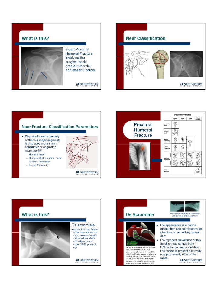
What is this?
3-part Proximal Humeral Fracture i l i hinvolving the surgical neck, greater tubercle, and lesser tubercle
Neer Classification
Neer Fracture Classification Parameters
Displaced means that any of the four major segments is displaced more than 1is displaced more than 1 centimeter or angulated more the 45°
– Humeral head– Humeral shaft - surgical neck– Greater Tuberosity– Lesser Tuberosity
Proximal Humeral Fracture
What is this?
Os acromialeresults from the failure of the acromial seconof the acromial secon-dary centers of ossifi-cation to fuse which normally occurs at about 18-20 years of age
Os Acromiale
The appearance is a normal variant than can be mistaken for a fracture on an axillary lateral
Axillary views of (R) and (L) shoulders with acromion and os acromiale
a fracture on an axillary lateral view. The reported prevalence of this condition has ranged from 1-15% in the general population. The finding is present bilaterally in approximately 62% of the cases.
Failure of fusion of the most anterior ossification center results in a preacromion, failure of fusion of the middle ossification center produces a meso‐acromion, and failure of fusion of the center located at the angle between the scapular spine and the acromion creates a meta‐acromion.
4
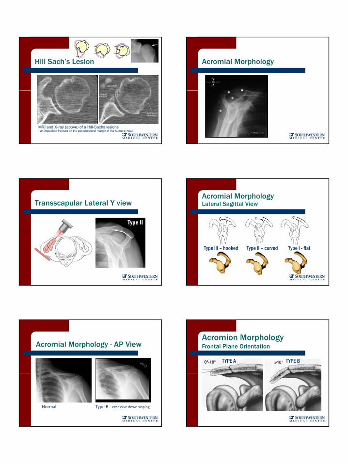
Hill Sach’s Lesion
MRI and X-ray (above) of a Hill-Sachs lesions - an impaction fracture on the posterolateral margin of the humeral head
Acromial Morphology
Transscapular Lateral Y view
Type II
Acromial Morphology Lateral Sagittal View
Type III – hooked Type II – curved Type I - flat
Acromial Morphology - AP View
Normal Type B – excessive down sloping
Acromion MorphologyFrontal Plane Orientation
TYPE A TYPE B
5
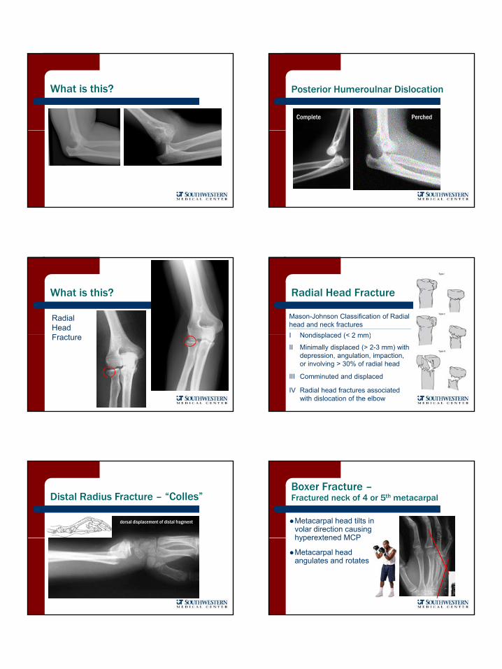
What is this? Posterior Humeroulnar Dislocation
Complete Perched
What is this?
Radial Head FFracture
Radial Head Fracture
Mason-Johnson Classification of Radial head and neck fracturesI Nondisplaced (< 2 mm)I Nondisplaced (< 2 mm)
II Minimally displaced (> 2-3 mm) with depression, angulation, impaction, or involving > 30% of radial head
III Comminuted and displaced
IV Radial head fractures associated with dislocation of the elbow
Distal Radius Fracture – “Colles”
dorsal displacement of distal fragment Metacarpal head tilts in volar direction causing hyperextened MCP
Boxer Fracture –Fractured neck of 4 or 5th metacarpal
hyperextened MCP
Metacarpal head angulates and rotates
6
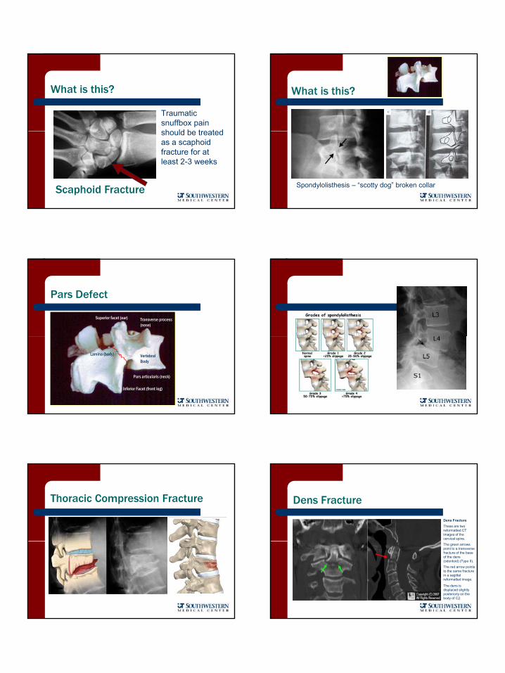
What is this?
Traumatic snuffbox pain should be treatedshould be treated as a scaphoid fracture for at least 2-3 weeks
Scaphoid Fracture
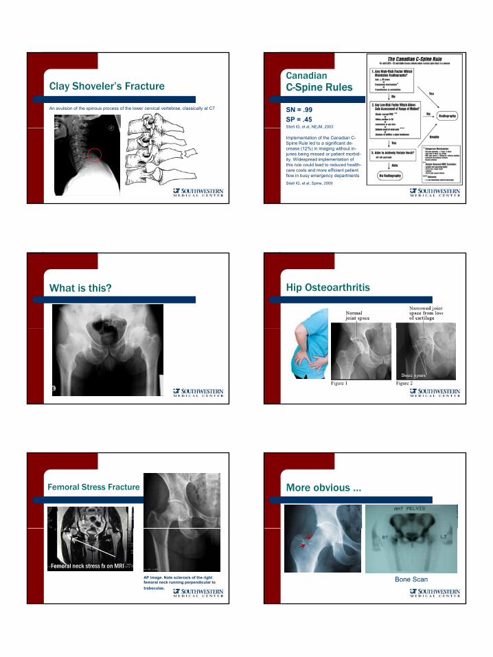
What is this?
Spondylolisthesis – “scotty dog” broken collar
Pars Defect
Transverse process (nose)
Superior facet (ear)
Pars articularis (neck)
Vertebral Body
Inferior Facet (front leg)
Lamina (body)
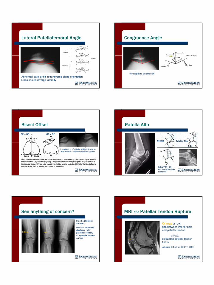
Thoracic Compression Fracture Dens Fracture
Dens FractureThese are two reformatted CT images of the cervical spine. The green arrows point to a transversefracture of the base of the dens (odontoid) (Type II).
The red arrow points to the same fracture in a sagittalreformatted image.
The dens is displaced slightly posteriorly on the body of C2.
7
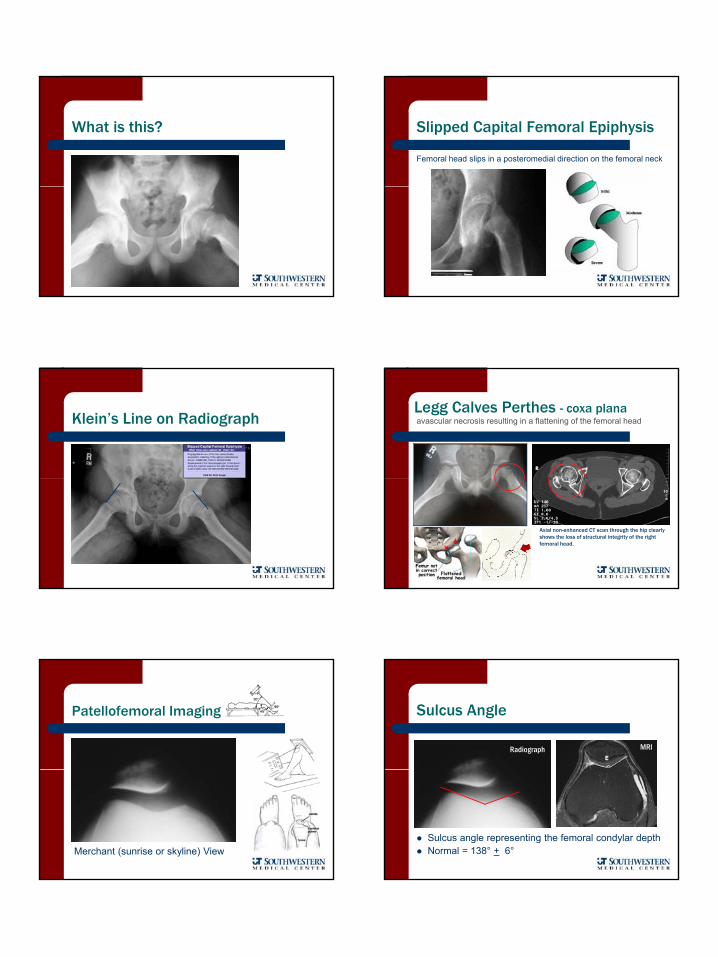
Clay Shoveler’s Fracture
An avulsion of the spinous process of the lower cervical vertebrae, classically at C7
Canadian C-Spine Rules
SN = .99SP = .45Stiell IG, et al, NEJM, 2003Stiell IG, et al, NEJM, 2003
Implementation of the Canadian C-Spine Rule led to a significant de-crease (12%) in imaging without in-juries being missed or patient morbid-ity. Widespread implementation of this rule could lead to reduced health-care costs and more efficient patient flow in busy emergency departmentsStiell IG, et al, Spine, 2009
What is this? Hip Osteoarthritis
Femoral Stress Fracture
AP image. Note sclerosis of the right femoral neck running perpendicular to trabeculae.
Femoral neck stress fx on MRI
More obvious …
Bone Scan
8
What is this? Slipped Capital Femoral Epiphysis
Femoral head slips in a posteromedial direction on the femoral neck
Klein’s Line on RadiographLegg Calves Perthes - coxa planaavascular necrosis resulting in a flattening of the femoral head
Axial non-enhanced CT scan through the hip clearly shows the loss of structural integrity of the right femoral head.
Patellofemoral Imaging
Merchant (sunrise or skyline) View
Sulcus Angle
Radiograph MRI
Sulcus angle representing the femoral condylar depthNormal = 138° + 6°
9
Lateral Patellofemoral Angle
Abnormal patellar tilt in transverse plane orientation Lines should diverge laterally
Abnormal
Congruence Angle
frontal plane orientation
Bisect Offset
G
GE > GF
G
GE = GF
Method used to measure medial and lateral displacement . Determined by a line connecting the posteriorfemoral condyles (AB) and then projecting a perpendicular line anteriorly through the deepest portion of the trochlear groove (CD) to a point where it bisected the patellar width line (EF) (left). The bisect offset isreported as the % of the patellar width lateral to the midline.
Increased % of patellar width is lateral to the midline – laterally displaced patella
Normal Patellar Alta
Patella Alta
Ratio of P:PT = 1.0More than 20% variation is abnormal
See anything of concern?
Standing bilateral AP view:
note the superiorly displaced right patella secondary to a patellar tendon rupture
Hint
Orange arrow: gap between inferior pole
d t ll t d
MRI of a Patellar Tendon Rupture
and patellar tendon
WhiteWhite arrow: distracted patellar tendon fibers
Johnson SD, et al, JOSPT, 2009
10
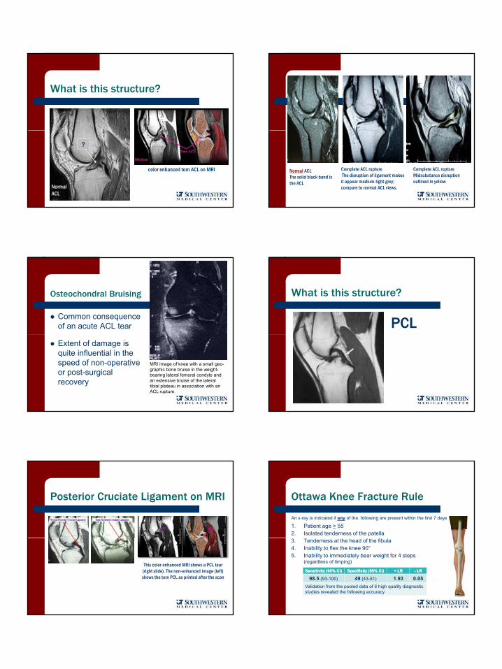
What is this structure?
color enhanced torn ACL on MRI
NormalACL
?
Normal ACL The solid black band is the ACL
Complete ACL ruptureThe disruption of ligament makes it appear medium-light grey; compare to normal ACL views.
Complete ACL ruptureMidsubstance disruption outlined in yellow
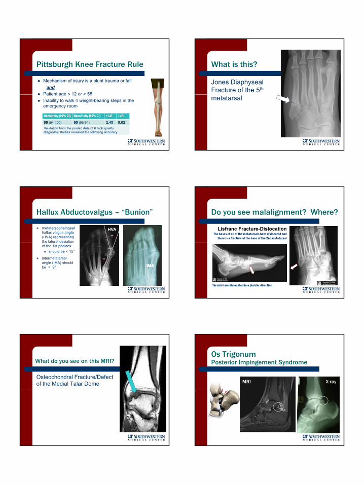
Osteochondral Bruising
Common consequence of an acute ACL tear
Extent of damage is quite influential in the speed of non-operative or post-surgical recovery
MRI image of knee with a small geo-graphic bone bruise in the weight-bearing lateral femoral condyle and an extensive bruise of the lateral tibial plateau in association with an ACL rupture.
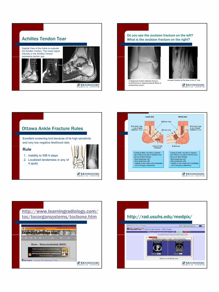
What is this structure?
PCL
Posterior Cruciate Ligament on MRI
This color enhanced MRI shows a PCL tear (right slide). The non-enhanced image (left) shows the torn PCL as printed after the scan
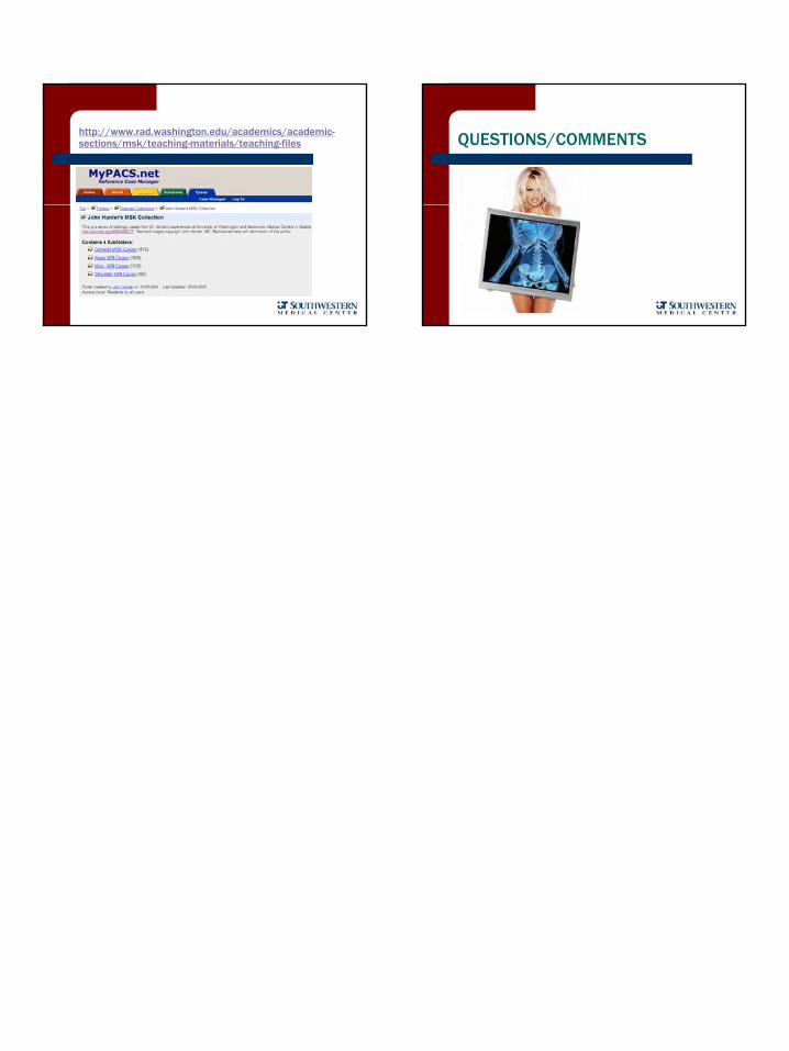
Ottawa Knee Fracture Rule
An x-ray is indicated if any of the following are present within the first 7 days
1. Patient age > 552. Isolated tenderness of the patella3. Tenderness at the head of the fibula4. Inability to flex the knee 90°5. Inability to immediately bear weight for 4 steps
(regardless of limping)
Sensitivity (95% CI)Sensitivity (95% CI) Specificity (95% CI)Specificity (95% CI) + LR+ LR -- LRLR
98.5 (93-100) 49 (43-51) 1.93 0.05Validation from the pooled data of 6 high quality diagnostic studies revealed the following accuracy
11
Pittsburgh Knee Fracture Rule
Mechanism of injury is a blunt trauma or falland
Patient age < 12 or > 55Patient age < 12 or > 55Inability to walk 4 weight-bearing steps in the emergency room
Sensitivity (95% CI)Sensitivity (95% CI) Specificity (95% CI)Specificity (95% CI) + LR+ LR -- LRLR
99 (94-100) 60 (56-64) 2.48 0.02Validation from the pooled data of 6 high quality diagnostic studies revealed the following accuracy
What is this?
Jones Diaphyseal Fracture of the 5th
metatarsal
Hallux Abductovalgus – “Bunion”
metatarsophalngeal hallux valgus angle (HVA) representing the lateral deviation
HVA
the lateral deviation of the 1st phalanx
should be < 15°
intermetatarsal angle (IMA) should be < 9° IMAIMA
Do you see malalignment? Where?
Lisfranc Fracture-DislocationThe bases of all of the metatarsals have dislocated and
there is a fracture at the base of the 2nd metatarsal
Tarsals have dislocated in a plantar direction
What do you see on this MRI?
Osteochondral Fracture/Defect of the Medial Talar Dome
Os TrigonumPosterior Impingement Syndrome
MRI X-ray
12
Achilles Tendon Tear
Sagittal View of the Ankle to evaluate the Achilles Tendon. The mixed signal intensity in the Achilles Tendon represents tendon tearrepresents tendon tear.
Do you see the avulsion fracture on the left?What is the avulsion fracture on the right?
avulsion fracture at the base of the 5th metUndisplaced medial malleolar fracture -could there be a missed proximal fibular or syndesmosis injury?
Ottawa Ankle Fracture Rules
Excellent screening tool because of its high sensitivityand very low negative likelihood ratio
Rule1. Inability to WB 4 steps2. Localized tenderness in any of
4 spots
http://www.learningradiology.com/toc/tocorgansystems/tocbone.htm
Good web sites
http://rad.usuhs.edu/medpix/
13
http://www.rad.washington.edu/academics/academic-sections/msk/teaching-materials/teaching-files QUESTIONS/COMMENTS