diagnosis and complications of cushing’s syndrome: a consensus statement
TRANSCRIPT
Diagnosis and Complications of Cushing’s Syndrome:A Consensus Statement
G. ARNALDI, A. ANGELI, A. B. ATKINSON, X. BERTAGNA, F. CAVAGNINI, G. P. CHROUSOS,G. A. FAVA, J. W. FINDLING, R. C. GAILLARD, A. B. GROSSMAN, B. KOLA, A. LACROIX,T. MANCINI, F. MANTERO, J. NEWELL-PRICE, L. K. NIEMAN, N. SONINO, M. L. VANCE,A. GIUSTINA, AND M. BOSCARO
Division of Endocrinology (G.A., B.K., T.M., M.B.), Department of Internal Medicine, University of Ancona, 60100 Ancona,Italy; Division of Internal Medicine (A.A.), Department of Clinical and Biological Science, University of Torino, 10063Torino, Italy; Regional Centre for Endocrinology and Diabetes (A.B.A.), Royal Victoria Hospital and Queen’s University,BT126BA Belfast, United Kingdom; Department of Endocrinology (X.B.), University Rene Descartes and Institut Cochin,Institut National de la Sante et de la Recherche Medicale U567 and Faculty Cochin, 75014 Paris, France; Department ofEndocrinology (F.C.), San Luca Hospital, Italian Auxological Institute, Instituto di Ricovero e Cura a Carattere Scientifico,University of Milan, 20149 Milan, Italy; National Institute of Child Health and Human Development, National Institutes ofHealth (G.P.C., L.K.N.), Bethesda, Maryland 20892-1583; Department of Psychology (G.A.F.), University of Bologna, 40127Bologna, Italy; Endocrine-Diabetes Center (J.W.F.), St. Luke’s Medical Center, Milwaukee, Wisconsin 53215; Division ofEndocrinology, Diabetology, and Metabolism (R.C.G.), University Hospital, Centre Hospitalier Universitaire Vandois, CH-1011 Lausanne, Switzerland; Department of Endocrinology (A.B.G.), St. Bartholomew’s Hospital, London EC1A 7BE,United Kingdom; Department of Medicine (A.L.), Centre hospitalier de l’Universite de Montreal, H2W1T7 Montreal, Canada;Division of Endocrinology (F.M.), Department of Medical and Surgical Sciences, University of Padova, 35128 Padova, Italy;Division of Clinical Sciences (J.N.-P.), Sheffield University, Northern General Hospital, 557AU Sheffield, United Kingdom;Department of Statistical Sciences and Department of Mental Health (N.S.), University of Padova, 35128 Padova, Italy;University of Virginia Health System (M.L.V.), Charlottesville, Virginia 22908-0746; and Department of Internal Medicine/Endocrine Section (A.G.), University of Brescia, 25125 Brescia, Italy
In October 2002, a workshop was held in Ancona, Italy, toreach a Consensus on the management of Cushing’s syn-drome. The workshop was organized by the University of An-cona and sponsored by the Pituitary Society, the EuropeanNeuroendocrine Association, and the Italian Society of Endo-crinology. Invited international participants included almost
50 leading endocrinologists with specific expertise in the man-agement of Cushing’s syndrome. The consensus statement ondiagnostic criteria and the diagnosis and treatment of com-plications of this syndrome reached at the workshop is herebysummarized. (J Clin Endocrinol Metab 88: 5593–5602, 2003)
Part I: Criteria for Diagnosis and Cure ofCushing’s Syndrome (CS)
Endogenous CS results from chronic exposure to excessglucocorticoids produced by the adrenal cortex. It may becaused by excess ACTH production (80–85%), usually by apituitary corticotroph adenoma [Cushing’s disease (CD)],less frequently by an extrapituitary tumor (ectopic ACTHsyndrome), or very rarely by a tumor secreting CRH (ectopicCRH syndrome). CS can also be ACTH-independent (15–20%) when it results from excess secretion of cortisol byunilateral adrenocortical tumors, either benign or malignant,or by bilateral adrenal hyperplasia or dysplasia (1–4).
Regardless of the etiology, some of the tumors responsible forCS can show variable secretory activity over time. In addition,tumoral cells may have maintained or lost their physiologicalregulation by hormones or growth factors or may even have
acquired new regulatory mechanisms (5). The spectrum of cor-tisol overproduction may thus vary from barely detectable tomarked oversecretion, especially in cyclical CS (2).
The suspicion of CS in a patient clearly arises in the presenceof central obesity with supraclavicular fat accumulation, a cer-vical fat pad, thinned skin, purple striae, proximal muscleweakness, fatigue, high blood pressure, glucose intolerance,acne, hirsutism, and menstrual irregularity. Neuropsycholog-ical disturbances including depression, emotional irritability,sleep disturbances, and cognitive deficits are also frequentlyobserved. Muscular atrophy and purple striae are particularlyhelpful stigmata in adults, whereas in children growth retar-dation is frequently present. The clinical phenotype is notalways florid, and suspicion should also arise with a less com-plete picture, particularly if concomitant recent weight gain,impaired glucose tolerance, and high blood pressure arepresent (1–3). Thus, some of the patients with metabolic syn-drome might require screening for CS, especially if young andresistant to conventional treatment.
Due to the variable pattern of the biochemical parametersand the nonspecificity of clinical manifestations, the diagnosisof CS is often a challenge for clinicians. This is particularly true
Abbreviations: BIPSS, Bilateral IPS sampling; BMD, bone mineraldensity; CBG, corticosteroid-binding globulin; CD, Cushing’s disease;CS, Cushing’s syndrome; CT, computed tomography; DEXA, dual en-ergy x-ray absorptiometry; DST, dexamethasone suppression test(s);IPS, inferior petrosal sinus; MRI, magnetic resonance imaging; UFC,urinary free cortisol.
0021-972X/03/$15.00/0 The Journal of Clinical Endocrinology & Metabolism 88(12):5593–5602Printed in U.S.A. Copyright © 2003 by The Endocrine Society
doi: 10.1210/jc.2003-030871
5593
The Endocrine Society. Downloaded from press.endocrine.org by [${individualUser.displayName}] on 19 March 2014. at 19:15 For personal use only. No other uses without permission. . All rights reserved.
in states of mild hypercortisolism and in some patients withadrenal incidentalomas (2). Primary care physicians may sus-pect CS and proceed to the initial biochemical screening tests.However, due to the potential complexity of investigation, fur-ther evaluation and treatment of this syndrome should be con-ducted in specialized endocrinology referral centers.
The diagnosis of endogenous CS should begin with a carefulcase history and a thorough physical examination, looking forthe characteristic features while excluding exogenous intake oforal, parenteral, inhaled, or topical corticosteroids. The extentof laboratory investigations will depend on the clinical index ofsuspicion. In addition, biochemical screening must take intoconsideration the fact that some patients have episodic or pe-riodic increases in cortisol secretion (6, 7). More specific testsrelating to the differential diagnosis should be performed onlyafter hypercortisolism has been confirmed, because they onlydifferentiate one cause of CS from another. In cases where thediagnosis of CS is suspected clinically but initial screening testsare normal, the patient should be reevaluated at a later date, andinvasive procedures should be postponed.
Diagnosis of CS
Several tests have been used extensively as first linescreening tests, but none has proven fully capable of distin-guishing all cases of CS from normal and/or obese individ-uals. More recently introduced tests have attempted to im-prove the efficiency of the initial screening and will also beconsidered here.
First-line screening tests
Twenty-four-hour urinary free cortisol (UFC). The 24-h uri-nary cortisol gives an integrated index of the free (unbound)cortisol that circulated in the blood during this period oftime. In contrast to plasma cortisol levels, which measuretotal cortisol, unbound and bound, it is not affected by factorsthat influence corticosteroid-binding globulin (CBG) levels(7, 8). Due to the possibility of intermittent hypercortisolism,if the index of suspicion is high and the first result is normal,up to three 24-h urine collections should be performed. Ifcortisol excretion results are normal in three collections, thenCS is highly unlikely, providing that renal function is normal.Mild CS is also unlikely although the alteration of the othertests can define a mild CS. Urinary creatinine may also bemeasured to verify the adequacy of the urine collection. Ifglomerular filtration rate is less than 30 ml/min, the urinarycortisol excretion is decreased and may thus be normal de-spite the presence of excessive cortisol production. In chil-dren, the urinary cortisol excretion should be corrected forbody surface area/1.72 m2. Measurement of urinary cortisolby immunoassays (RIA, immunometric assays) is influencedby various metabolites of cortisol and some synthetic glu-cocorticoids, whereas measurements using HPLC allow theseparation of various urinary glucocorticoids and metabo-lites. HPLC has a high sensitivity and specificity, but occa-sionally interfering substances, such as carbamazepin anddigoxin, can also coelute with cortisol and produce falseelevations of the UFC (9, 10). The recent introduction of massspectrometry combined with gas chromatography or HPLCmay overcome these problems; however, these techniques
are more expensive, are not widely available, and have notyet been validated extensively. UFC values can be extremelyvariable in CS. UFC values 4-fold greater than the upper limitof normal are very rare, except in CS, and therefore can beconsidered diagnostic for this condition. Milder elevations ofurinary cortisol can be found in conditions such as chronicanxiety, depression, and alcoholism, all of which are alsoknown as pseudo-Cushing states (4, 7), and in normal preg-nant women. Urinary cortisol may not identify subclinical orpreclinical CS in which hypercortisolism is still mild, and forthis reason and the others cited above, it cannot be consid-ered as a universal single screening test for the detectionof CS.
Low-dose dexamethasone suppression tests (DST). The low-dose DST are used to differentiate CS patients from thosewho do not have CS (4, 8). The overnight low-dose (1 mg)DST consists of the oral intake of 1 mg dexamethasone be-tween 2300 and 2400 h, followed by measurement of fastingplasma cortisol between 0800 and 0900 h the following morn-ing. The original criterion for normal level of suppressionwas a plasma cortisol level below 5 �g/dl (138 nmol/liter).More recently, this cut-off level has been reduced to less than1.8 �g/dl (50 nmol/liter) (10–11), greatly enhancing the sen-sitivity of the overnight DST, especially in patients with mildhypercortisolism. A serum cortisol level below 1.8 �g/dl (50nmol/liter) excludes active CS at that time. The specificity ofthe test is, however, limited, due to potential misclassifica-tion of patients with increased CBG, acute and chronic ill-ness, or pseudo-CS. Occasionally, otherwise healthy indi-viduals fail to suppress cortisol to this level also. Foroutpatient screening, the advantages of the 1 mg overnightDST are its ease of execution and low cost. The classical 2-d2 mg DST is another way to conduct the test and is used asa first line screening test in some centers (4) (see below). Forany DST, interfering conditions causing an apparent lack ofsuppression include: decreased dexamethasone absorption,drugs enhancing hepatic dexamethasone metabolism (bar-biturates, phenytoin, carbamazepine, rifampicin, mepro-bamate, aminoglutethimide, methaqualone), increased con-centration of CBG (estrogen treatment, pregnancy) andpseudo-Cushing states. Moreover, the cortisol assay musthave a sensitivity of 1 �g/dl (27.6 nmol/liter) or less (4–8).
Late-night salivary cortisol. This is a recently introduced testthat appears to be promising for the screening of CS. Cortisolconcentration in saliva is highly correlated with free plasmacortisol, independent of salivary flow rates, and stable atroom temperature for 1 wk. The normal reference ranges areassay-dependent and should be validated for each labora-tory. Late night (2300 h) salivary cortisol is a simple way toscreen for CS and could become increasingly used becauseit has been found to have high diagnostic sensitivity andspecificity (12, 13). It could be particularly useful in inves-tigating patients with cyclical CS with repeated evening mea-surements over time. Wider availability of validated com-mercial assays and larger studies will be necessary beforelate-night salivary cortisol could be considered to substitutefor UFC or low-dose DST as a first-line screening test.
5594 J Clin Endocrinol Metab, December 2003, 88(12):5593–5602 Arnaldi et al. • Consensus Statement
The Endocrine Society. Downloaded from press.endocrine.org by [${individualUser.displayName}] on 19 March 2014. at 19:15 For personal use only. No other uses without permission. . All rights reserved.
Second-line screening tests
Plasma cortisol circadian rhythm—midnight plasma cortisol.Patients with CS often have early morning serum cortisolconcentrations within or slightly above the normal range, butlack a normal circadian rhythm. Plasma cortisol levels areabove a cut-off value of 1.8 �g/dl (50 nmol/liter) whenmeasured at midnight in hospitalized, sleeping CS patients(14). This cut-off value has a very high degree of sensitivity,but its specificity was not tested. Others have adopted ahigher cut-off value (7.5 �g/dl, 207 nmol/liter) to achieve100% specificity for normal individuals and those withpseudo-Cushing (15). The test requires inpatient admissionfor a period of at least 48 h. Measurement of cortisol at othertime points appears to be relatively unhelpful (15).
Low-dose DST and combined DST-CRH test. In the classical2-d low-dose DST, the patient takes 0.5 mg dexamethasoneorally every 6 h. Urine is collected for UFC on 2 baseline daysand on the second day of dexamethasone administration, oralternatively serum cortisol is measured at 0900 h and 48 hafter the first dose. A normal response consists of a decreaseof UFC to less than 10 �g (27 nmol) per 24 h on the secondday of dexamethasone administration or of plasma cortisolto less than 1.8 �g/dl (50 nmol/liter) on the morning after thelast dose of dexamethasone (11). Use of the plasma cortisolend point results in a sensitivity and specificity of more than95% (4). Adequate urine collections and normal bioavailabil-ity and metabolism of dexamethasone are required for theprocedure to be valid. As noted above, some centers use theclassical 2-d low-dose DST as a first-line screening test (4).
The combined DST-CRH test was shown by one center to behighly accurate in distinguishing CS from pseudo-CS; it is pos-tulated that patients with pseudo-CS are thought to be underchronic CRH stimulation due to their stressful situation andshow a blunted response to exogenous CRH after dexameth-asone administration. The test is performed by giving dexa-methasone orally 0.5 mg every 6 h for 48 h, starting at 1200 h,and then administering ovine-sequence CRH (1 �g/kg) iv at0800 h (2 h after the last dose of dexamethasone). The plasmacortisol value 15 min after CRH is greater than 1.4 �g/dl (38nmol/liter) in patients with CS, but remains suppressed innormal individuals and in patients with pseudo-CS (16). Thistest would be useful when pseudo-CS is suspected. Its advan-tages over the classical 2-d low-dose DST remain to be validatedby additional studies from different centers. Its use with hu-man-sequence CRH also requires validation. Importantly, theprecision of the majority of cortisol assays in routine use at thequoted cut-off level is poor, and caution is needed in interpre-tation. Therefore, the use of highly sensitive cortisol assays isstrongly suggested for all dexamethasone suppression tests.
Differential Diagnosis of CS
If CS is confirmed, its cause will be categorized initially bydetermination of plasma ACTH values. If ACTH is not sup-pressed, ACTH-dependent causes will be investigated. Anoccult ectopic source of ACTH may mimic pituitary-depen-dent disease because such tumors may express glucocorti-coid and/or CRH and/or vasopressin receptors, and thusbehave as a misplaced pituitary in the course of all standard
dynamic tests. If ACTH is suppressed, adrenal computedtomography (CT)/magnetic resonance imaging (MRI) scan-ning will identify the type of adrenal lesion(s) responsiblefor CS.
ACTH measurement. ACTH is rapidly degraded by plasmaproteases. To prevent this, the assays for the determinationof plasma ACTH levels require collection of blood into aprechilled EDTA tube, placement in an ice water bath, andrapid delivery to the laboratory for refrigerated centrifuga-tion. Only assays such as the two-site immunoradiometricassays, which can reliably detect values less than 10 pg/ml(2 pmol/liter), should be used. ACTH concentrations belowthe level of detection or below 10 pg/ml (2 pmol/liter) at0900 h with concomitant increased production of cortisolsuggest an ACTH-independent cause of CS. However,ACTH levels may not be fully suppressed in some patientswith adrenal CS and intermittent or concomitantly relativelylow secretion of cortisol. Plasma ACTH values greater than20 pg/ml (4 pmol/liter) suggest an ACTH-dependent cause.For values between 10 and 20 pg/ml (2–4 pmol/liter), a CRHstimulation test is indicated, with measurement of plasmaACTH. ACTH levels tend to be higher in ectopic ACTH-secreting CS than in CD; however, the overlap in ACTHvalues is such that ACTH values alone rarely distinguishbetween the two conditions (1, 4).
High-dose DST. High doses of glucocorticoids partially sup-press ACTH secretion from most corticotroph adenomas(80–90%), whereas ectopic tumors are resistant to feedbackinhibition. However, some benign differentiated neuroen-docrine tumors (usually the carcinoid tumors of bronchus,thymus, and pancreas) may be sensitive to feedback inhibi-tion of ACTH like pituitary tumors. In adrenal CS, there isa lack of cortisol suppression after high-dose DST becausecortisol secretion is autonomous and the ACTH secretion isalready very low and cannot be further reduced.
There are several versions of the high-dose DST, includingthe standard 2-d oral high dose (2 mg every 6 h for eight doses),the 8-mg overnight oral, and the iv 4–7 mg tests (4).
Plasma and/or urinary cortisol levels are evaluated before,during, and/or after dexamethasone administration. Thesetests distinguish pituitary from ectopic sources of ACTH witha sensitivity varying from 60 to 80% and with high specificitywhen a cut-off of plasma cortisol suppression above 50% is used(17). The specificity can be improved using a cut-off of cortisolsuppression greater than 80%, although a specificity of 100%can never be attained. However, no variation in protocol orcriteria allows for complete discrimination between CD and anectopic source of ACTH.
CRH stimulation test. Most pituitary tumors, and also a fewectopic ACTH-secreting tumors, respond to CRH adminis-tration with an increase in plasma ACTH and cortisol levels.In adrenal CS, there is usually little or no cortisol or ACTHresponse to CRH. The test is performed by injecting iv 1�g/kg or 100 �g synthetic ovine or human CRH. There is notyet consensus on the criteria for interpreting the response toCRH test. Variability in the interpretation depends on thetype of CRH used (human vs. ovine), biochemical parametersconsidered (increase above baseline in ACTH, 35–50%; vs.
Arnaldi et al. • Consensus Statement J Clin Endocrinol Metab, December 2003, 88(12):5593–5602 5595
The Endocrine Society. Downloaded from press.endocrine.org by [${individualUser.displayName}] on 19 March 2014. at 19:15 For personal use only. No other uses without permission. . All rights reserved.
cortisol, 14–20%) and evaluated time points (ACTH, 15–30min; cortisol, 15–45 min) (18–20). However, because ectopicACTH-producing tumors can also respond to CRH, increas-ing the cut-off level of the response will not produce 100%specificity, thus preventing complete reliance on this testalone.
Desmopressin and other tests under investigation. Intravenousadministration of desmopressin (a preferential V2 and V3-vasopressin receptor agonist) 10 �g increases ACTH secre-tion in 80–90% of patients with CD and only rarely in normalindividuals or patients with pseudo-CS. Desmopressin iseasily available and inexpensive, and it does not cause sig-nificant adverse effects. However, 20–50% of ectopic ACTH-secreting tumors respond to desmopressin, thus limiting itsusefulness in distinguishing the source of excess ACTH (21).The combination of CRH and desmopressin appeared toincrease the discriminatory value above either test alone, butmore recent data cast doubt on the clinical value of thecombined test (21). Desmopressin might be useful in thedifferential diagnosis between CD and pseudo-CS (22) andin the postoperative assessment of CD (see below) (23).
In CD, GH secretagogues are more potent than CRH andvasopressin in stimulating ACTH release, particularly in pitu-itary microadenomas and, to a lesser extent, in macroadenomas(24). However, GH secretagogues can also stimulate ACTHsecretion from ectopic ACTH-secreting tumors. For these rea-sons, these substances cannot be recommended for standardclinical practice.
Opiate agonists, such as loperamide, have been shown toinhibit ACTH in most normal individuals but not in patientswith CS; opiate antagonists, such as naloxone, can stimulateACTH in patients with CS to a lesser extent than in normalindividuals. However, there appears to be overlap betweenpatients with CS and healthy individuals, and it will be nec-essary to investigate a larger number of patients, includingsome with ectopic ACTH secretion, to better evaluate the utilityof these substances (4). Additional studies using other regula-tors of ACTH, such as IL-6, or the CRH receptor antagonists,such as antalarmin, will be of interest.
Caution should be exercised when interpreting tests used fordifferential diagnosis if the history is short, if the clinical signsare modest, if there has been recent use of drugs such as ke-toconazole or metyrapone, or if UFC levels are only modestlyraised because in these situations, normal corticotroph cellsmay not be suppressed and may respond to the stimulationtests.
Pituitary MRI. A pituitary MRI with gadolinium enhance-ment should be performed in all patients with ACTH-dependent CS. This procedure will reveal a discrete pituitaryadenoma in up to 60% of patients (2, 20). In the patient witha classic clinical presentation and dynamic biochemical stud-ies compatible with pituitary CS, the presence of a focallesion (�6 mm) on pituitary MRI may provide a definitivediagnosis, and no further evaluation may be required. How-ever, it is important to realize that 10% of the general pop-ulation harbor incidental pituitary tumors disclosed on MRI,although the majority of these lesions are less than 5 mm indiameter.
Bilateral inferior petrosal sinus sampling (BIPSS). BIPSS forACTH determination should be recommended in patientswith ACTH-dependent CS whose clinical, biochemical, orradiological studies are discordant or equivocal. BIPSS hasrarely been associated with significant complications includ-ing deep vein thrombosis, pulmonary emboli, and brain stemvascular damage. The procedure should be performed whencortisol levels are elevated, indicating currently active secre-tion of ACTH by the tumor, and avoiding testing during aninactive cycled-out phase of CS. Because the results of BIPSSas well as the incidence of these adverse events are relatedto the experience of the radiology team, this procedureshould be performed only in specialized centers.
After the radiologist catheterizes both inferior petrosal si-nuses (IPSs), blood samples for ACTH are obtained in the basalstate and at 3 and 5 min (and at 10 min in some centers) afteriv ovine or human CRH (1 �g/kg or 100 �g iv) simultaneouslyfrom both IPSs and a peripheral vein. Subtraction digital an-giography should be used to verify appropriate catheter place-ment and normal petrosal sinus anatomy.
An IPS to peripheral ACTH ratio (IPS/P) greater than 2.0 inthe basal state and/or greater than 3.0 after CRH is consistentwith CD. BIPSS in experienced centers has a very high sensi-tivity (95–99%) for CD (25). However, technical factors as wellas anomalous venous drainage may result in false-negativeresults in patients with a pituitary source of ACTH. LowerIPS/P ratios suggest an ectopic ACTH-secreting tumor with aspecificity of 95–99% although patients with CD (which is �10times more common) may very rarely show similar low ratios.
The use of IPSS for the localization of pituitary microad-enoma to the right or left side of the pituitary gland is contro-versial, and its diagnostic accuracy in identifying the site of apituitary microadenoma is disputed (4, 25). However, in a re-cent study in a small series of children, BIPSS appeared to bemore accurate than other imaging studies (26).
In recent years, different sites of venous sampling (cavernousand jugular veins) have been used, but BIPPS remains the besttest for the identification of CD.
Search for occult ectopic ACTH-secreting tumors. The majorityof nonpituitary ACTH-secreting tumors are neuroendocrineneoplasms such as bronchial/thymic/pancreatic carcinoids,islet cell pancreatic tumors, medullary thyroid cancer, andpheochromocytomas. If BIPSS confirms the lack of a pituitaryACTH gradient, CT and/or MRI of the neck, thorax, andabdomen should be performed. MRI of the chest may un-cover bronchial carcinoid tumors overlooked on CT imaging.Somatostatin analog scintigraphy with 111In-pentetreotidemay identify a few occult ACTH-secreting tumors with so-matostatin receptors that were not clearly identified by CT orMRI imaging (27). The utility of positron emission tomog-raphy scanning in the search for occult ACTH-secreting tu-mors is not yet established.
Criteria for Biochemical Assessment of Remission or“Cure” of CD
It is important to treat CD as rapidly as possible to limitits long-term morbidity. Transsphenoidal surgery, the first-line treatment, offers both a possibility of prompt remissionof the condition and a return to normal. The goal of the
5596 J Clin Endocrinol Metab, December 2003, 88(12):5593–5602 Arnaldi et al. • Consensus Statement
The Endocrine Society. Downloaded from press.endocrine.org by [${individualUser.displayName}] on 19 March 2014. at 19:15 For personal use only. No other uses without permission. . All rights reserved.
treatment is the complete resection of the pituitary adenomawith correction of hypercortisolism without inducing per-manent pituitary deficiencies. There is still no widespreadagreement regarding the definition of apparent cure, and theremission rates after such surgery vary according to thecriterion used and the time of assessment. Indeed, the def-inition of cure and the prognostic effect of subtle or unrec-ognized residual hypercortisolism have a major clinical im-pact on the follow-up and therapeutic decisions in patientswith CD. Most series from major centers quote remissionrates of 70–80%, defining remission as a series of normalpostoperative cortisol levels, either as a mean of serial serumcortisol measurements, obtained throughout the day, of 5.4–10.8 �g/dl (150–300 nmol/liter), or as a UFC in the normalrange, associated with resolution of clinical stigmata. How-ever, the long-term follow-up of such patients shows a sig-nificant incidence of recurrence (�25% at 10 yr) (20, 28, 29).
Very low serum cortisol below 1.8 �g/dl (50 nmol/liter at0900 h) within 2 wk after surgery is probably the best index ofremission, but even when this is the case very occasional laterelapses have occurred (30–32). Cortisol is usually measured5–14 d after surgery and at least 24 h after the last dose ofhydrocortisone (33). With this criterion 1.8 �g/dl (�50 nmol/liter), the surgical remission rate is only 40–65% even in themost experienced hands. It should also be borne in mind thatthe 0900 h serum cortisol may fall with time after the operation,occasionally over several weeks, and that mild or cyclical hy-percortisolism may complicate the interpretation of test results(34).
Dynamic tests such as CRH or desmopressin stimulation,DST, and loperamide inhibition can be used to predict recur-rence. Recent studies have confirmed the usefulness of an earlyCRH test (20, 35), the rationale being that early responsivenessmay indicate the presence of residual tumor. The use of thedesmopressin test is also promising (20). Nevertheless, it should
be kept in mind that although each test can differentiate patientswho recur from those who do not as a group, none has suffi-ciently high diagnostic predictive value for an individualpatient.
It is becoming apparent that long-term results of transsphe-noidal surgery may not be as favorable as previously thoughtand that success rates drop during long-term follow-up. In-deed, the risk of relapse persists for at least 10 yr after surgery,if not longer. This fact, and possible ongoing subtle abnormal-ities in patients in apparent remission and with the risks ofsurgically induced hypopituitarism, warrant serious discussionregarding the results of surgery, and also emphasize the needfor stringent and indefinite follow-up of these patients.
Part I conclusions
The evaluation of patients with suspected CS is complexand expensive, and the diagnosis is often a challenge forclinicians. Most patients initially suspected of having CS willnot have this condition, and therefore efficient screeningprocedures are needed to identify the few patients who willneed additional investigation in specialized centers.
Atypical clinical presentations (mild hypercortisolism, cycli-cal CS, subclinical CS) or forms of pseudo-CS (depression, al-coholism) further complicate the assessment.
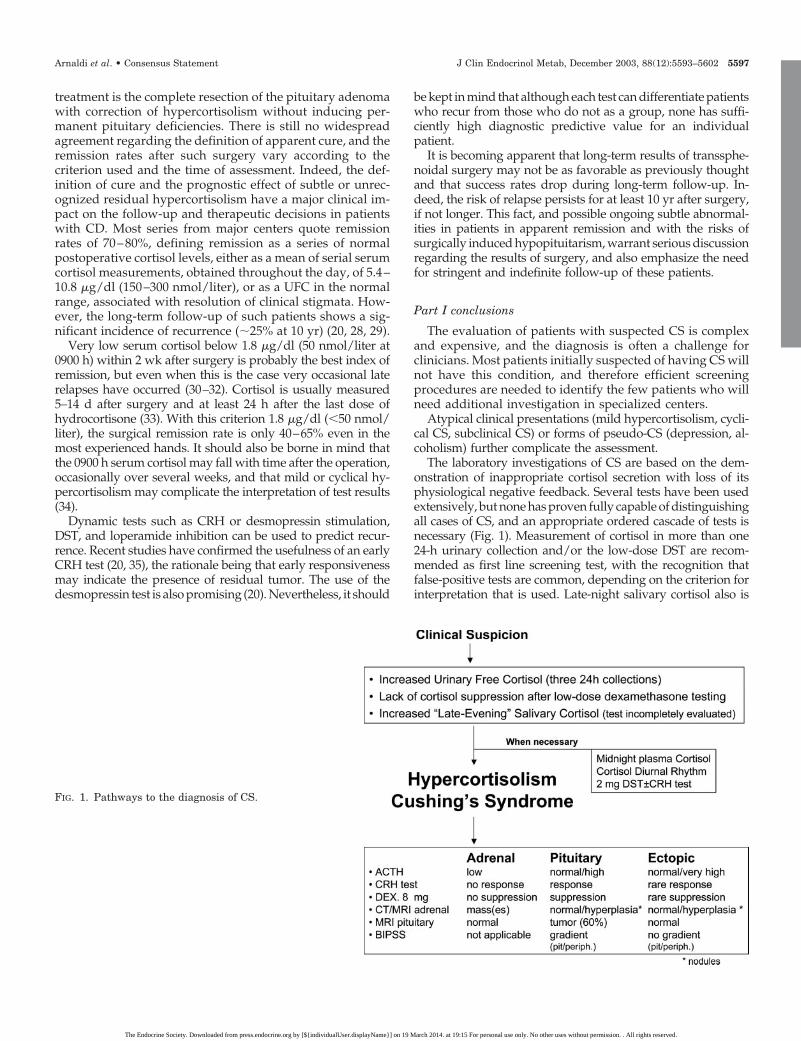
The laboratory investigations of CS are based on the dem-onstration of inappropriate cortisol secretion with loss of itsphysiological negative feedback. Several tests have been usedextensively, but none has proven fully capable of distinguishingall cases of CS, and an appropriate ordered cascade of tests isnecessary (Fig. 1). Measurement of cortisol in more than one24-h urinary collection and/or the low-dose DST are recom-mended as first line screening test, with the recognition thatfalse-positive tests are common, depending on the criterion forinterpretation that is used. Late-night salivary cortisol also is
FIG. 1. Pathways to the diagnosis of CS.
Arnaldi et al. • Consensus Statement J Clin Endocrinol Metab, December 2003, 88(12):5593–5602 5597
The Endocrine Society. Downloaded from press.endocrine.org by [${individualUser.displayName}] on 19 March 2014. at 19:15 For personal use only. No other uses without permission. . All rights reserved.
proposed as a useful screening test, although published data arestill preliminary. The diagnostic evaluation should not proceedto attempt to establish the precise etiology of hypercortisolismunless the diagnosis of CS is unequivocal. ACTH levels, theCRH stimulation test, the high-dose DST, and appropriate im-aging are the most useful noninvasive investigations for thedifferential diagnosis of CS. BIPSS for ACTH measurement isrecommended in patients with ACTH-dependent CS whoseclinical, biochemical, or radiological studies are discordant orequivocal.
There is still no widespread agreement regarding the defi-nition of cure after surgery. A very low serum cortisol early aftersurgery seems to be the best index of remission. Dynamic testscannot predict recurrence in a given individual, although theymay suggest increased or decreased risk.
Part II: Diagnosis and Treatment of Complicationsof CS
Patients with CS have a mortality rate four times higherthan age- and gender-matched subjects; this is due to thecomplications of the syndrome (36). The majority of the com-plications are correlated with direct and/or indirect effects ofglucocorticoid excess, and therefore, the primary goal in theprevention and treatment of complications is the correctionof hypercortisolism.
Cardiovascular risk factors and complications
The higher mortality rate observed in CS seems to bemainly caused by cardiovascular complications. Chronic hy-percortisolism is associated with an increased incidence ofcardiovascular risk factors such as systemic arterial hyper-tension, impaired glucose tolerance or diabetes, central obe-sity, hyperlipidemia, and hypercoagulability. In the follow-ing section, the cardiovascular effects of hypercortisolismwill be reviewed.
Hypertension. Arterial hypertension is a common feature inCS (70–80% of the patients) and may be the first sign of CS.There is no apparent difference in the prevalence in hyper-tension among patients with adrenal or pituitary CS. Patientswith severe hypercortisolism may present with hypokalemiaas well, due to the mineralcorticoid effect of cortisol. Theprevalence of hypertension increases with age (37) and,whereas it is less frequent than in adults (up to 50%), it is acommon hallmark in children and adolescents with CS (38).
Although hypertension in CS is usually mild to moderate, itmay be severe, and the physiological nocturnal decline in bloodpressure may be absent. Hypertension-induced organ damage,particularly cardiac hypertrophy, is frequent. Long-lasting ex-posure to excess circulating cortisol also may contribute directlyto left ventricular concentric remodeling in patients with CS(39). Although not routine, some use echocardiography in theinitial evaluation and the follow-up of selected patients with CSand hypertension. Similarly, ultrasound scanning of the carotidarteries may be useful for the determination of the global car-diovascular risk in CS patients.
Conventional antihypertensive therapy (thiazides, angioten-sin-converting enzyme inhibitors, and calcium antagonists aregenerally considered as first choice) may be only partially ef-
fective, whereas the additional successful use of cortisol-lowering agents may improve blood pressure control (40). Hy-pertension remits in most patients after successful treatment butmay persist, presumably because of microvessel remodelingand/or concomitant underlying essential hypertension (41).
Vascular damage is one of the multiple mechanisms in-volved in the pathogenesis of glucocorticoid-induced hyper-tension. Other possible mechanisms include: activation of therenin-angiotensin system; enhancement of cardiovascularinotropic and pressor reactivity to vasoactive substances(including catecholamines, vasopressin, angiotensin II,and erythropoietin) and suppression of vasodilatorymechanisms, including nitric oxide synthase, prostacyclin,and kinin-kallikrein. In addition, cortisol has intrinsicmineralocorticoid activity, and in the presence of veryhigh cortisol levels, the regulatory renal enzyme 11�-hydroxysteroid-dehydrogenase type 2 may not be able toinactivate the hormone, leading to hypertension and hy-pokalemia particularly in the ectopic ACTH syndrome(42). Some adrenal tumors may secrete other mineralcor-ticoids such as deoxycorticosterone.
In conclusion, hypertension is common in adults with CS andshould be treated by both correcting the hypercortisolism andusing standard practice for essential hypertension, before andafter remission of the syndrome.
Impaired glucose tolerance and diabetes. Hypercortisolism pro-motes the development of hyperglycemia and decreased car-bohydrate tolerance by increasing hepatic glycogen andglucose production and decreasing glucose uptake and uti-lization by peripheral tissues.
Epidemiological studies have shown a variable prevalence ofabnormalities of glucose metabolism: 20–50% of the patientssuffer from overt diabetes mellitus whereas impaired glucosetolerance is present in 30–60% of patients. The prevalence ofglucose metabolism abnormalities is probably underestimated,because an oral glucose tolerance test is not always performedin patients with active CS. Thus, an oral glucose tolerance testmay be useful for the assessment of global cardiovascular riskin these patients (43).
Obesity. Increased body weight is one of the earliest signs ofCS, with a characteristic redistribution of fat from peripheralto central parts of the body, mainly in the abdominal region.Glucocorticoids play a central role in the abdominal accu-mulation of body fat. An increased local generation of cor-tisol may take place in visceral adipose tissue because of highactivity of 11�-hydroxysteroid dehydrogenase type I, result-ing in high local concentrations of cortisol, by convertinginactive cortisone into active cortisol. Visceral obesity is anindependent risk factor for reduced life expectancy and alsocorrelates with increased risk for disorders such as insulinresistance and diabetes, hyperlipidemia, hypertension,and atherosclerosis of coronary, cerebral, and peripheralvessels (44).
Hyperlipidemia. In hypercortisolism, there is an increase incirculating very low-density lipoprotein and low-density li-poprotein, but not high-density lipoprotein, with consequentelevation of total triglycerides and cholesterol levels. Themechanisms for these changes are probably multifactorial,
5598 J Clin Endocrinol Metab, December 2003, 88(12):5593–5602 Arnaldi et al. • Consensus Statement
The Endocrine Society. Downloaded from press.endocrine.org by [${individualUser.displayName}] on 19 March 2014. at 19:15 For personal use only. No other uses without permission. . All rights reserved.
including direct cortisol influences on very low-density li-poprotein synthesis, free fatty acid production, and hepaticendothelial lipase activity (44–46). The insulin resistancestate induced by glucocorticoid excess is likely to play a keyrole in the determination of lipid abnormalities.
Coagulopathy. A complex derangement of the hemostatic sys-tem, characterized by both hypercoagulability and impairedfibrinolysis, is responsible for the thrombophilic state ob-served in patients with CS. Increased cortisol levels stimulatethe synthesis of several clotting factors, such as fibrinogen bythe liver, and von Willebrand factor by endothelial cells.Glucocorticoids also up-regulate the synthesis of plasmino-gen activator inhibitor type 1, the main inhibitor of the fi-brinolytic system (47, 48).
This hypercoagulability state is a crucial factor predisposingCS patients to thromboembolic events, mostly after surgery andduring IPS sampling. Therefore, patients with active CS shouldbe treated as having a prothrombotic disorder, and antithrom-botic prophylaxis should be considered. In the absence of pro-spective randomized trials, there is general agreement that pa-tients with CS should be given heparin during IPS sampling,and low-dose heparin treatment should be considered in theimmediate perioperative period.
Metabolic syndrome. In summary, patients with CS developthe manifestations of the metabolic syndrome or syndromeX, including insulin resistance, visceral adiposity, dyslipi-demia, carbohydrate intolerance, and/or diabetes mellitustype 2, coagulopathy, and hypertension as a direct or indirectconsequence of concurrent and chronic cortisol excess. Theabnormalities enhance the global cardiovascular risk that isresponsible for the increased mortality of these patients.Therefore, quantification of cardiovascular risk is important,especially if therapy does not normalize cortisol levels or thepatients relapse. Additionally, increased cardiovascular riskmay persist after the cure of CS (49).
Patients with the metabolic syndrome require treatment ofhypercortisolism as well as treatment of cardiovascular riskfactors, according to standard practice.
Osteoporosis
Pathological fractures can be the presenting manifestationof CS (50, 51). Limited data from cross-sectional studies showthat 30–50% of patients experience fractures, particularly atthe vertebral level. These are serious complications not onlybecause they can cause back pain, kyphosis, and height loss,but also because they can anticipate subsequent, nonverte-bral fractures independent of bone mineral density (BMD)(51). Using dual energy x-ray absorptiometry (DEXA), theprevalence of osteoporosis in adult patients with CS wasapproximately 50% (51).
Bone alterations cause greater morbidity in children becauseof the severe growth failure and pubertal arrest leading toreduced final adult height. Hypercortisolism in children mayalso lead to a reduction in peak bone mass, presumably in-creasing the long-term risk of osteoporosis (52). Glucocorticoidsinfluence bone and calcium metabolism at many levels, al-though the exact mechanisms are not fully understood (53). Itis thought that glucocorticoids cause loss of cortical osteocytes
and thus prevent bone repair. Inhibition of GH secretion, hy-pogonadism, and target tissue effects of growth factors, as wellas direct effects of glucocorticoids on osteoblasts, also maydecrease BMD (52, 54). Glucocorticoids decrease bone collag-enous matrix synthesis and increase its degradation. The de-crease in osteoblast number and function is reflected by de-creased serum levels of osteocalcin and alkaline phosphatase,both markers of osteoblastic function (55). Glucocorticoids in-hibit calcium absorption from the gut through a mechanismindependent of vitamin D, and also inhibit calcium reabsorp-tion by the renal tubule. PTH does not seem to play a major rolein glucocorticoid-induced osteoporosis (56).
There is some evidence to suggest that deficits in bone massmay be partially reversed after remission of hypercortisolism.Osteoblastic activity increases, as judged by increased osteo-calcin levels, within months after normalization of glucocorti-coid excess (51). Long-term studies have demonstrated an over-all improvement of BMD in patients successfully treated for CS,and even normalization of BMD after a mean period of 9 yr (57).The recovery of BMD may be explained by the preservation oftrabecular architecture despite trabecular thinning in glucocor-ticoid-induced osteoporosis. This contrasts with the loss of tra-becular bone that occurs in other forms of osteoporosis. It isreasonable to measure BMD, with DEXA, at the level of lumbarspine in all patients with CS and to consider antiresorptivetherapy in those with osteoporosis. Quantitative ultrasound hasbeen proposed as a useful tool for assessing bone microarchi-tectural involvement in CS, but its relevance cannot be ascer-tained in the absence of the availability of longitudinal data (51).Vertebral CT scan also can be used to assess osteoporosis inpatients with CS (51). Patients with severe osteopenia have ahigh risk of fracture. Therefore, in these patients, the use ofantiresorptive medication could be useful. Data suggest thatalendronate may induce a more rapid improvement in BMDthan cortisol normalization alone, probably by restoring thebalance between bone formation and bone resorption (58). Al-though there are no large prospective studies in patients withCS, additional therapeutic strategies, such as provision of ad-equate calcium and vitamin D and sex hormone replacement inmen or women with hypogonadism, are likely to have benefit.
New data on the use of PTH in glucocorticoid-induced os-teoporosis are encouraging (59) and suggest a potential role forthis anabolic therapy.
The risk of fractures persists some time after cure of hyper-cortisolism, and the decision to discontinue antiresorptive ther-apy should be based on clinical monitoring and DEXAmeasurements.
Psychological and cognitive alterations
Glucocorticoids affect behavior, mood, neural activity, anda number of specific biochemical processes in the centralnervous system. A number of psychiatric and psychologicaldisturbances may be associated with CS, regardless of itsetiology. Depending on the series, 50–80% of patients withCS meet Diagnostic and Statistical Manual of Mental Disor-ders IV criteria for major depression (60, 61). A minority ofpatients have other psychopathological manifestations in-cluding mania, anxiety, and cognitive dysfunction. If psy-chotic symptoms occur, they are likely to be a complication
Arnaldi et al. • Consensus Statement J Clin Endocrinol Metab, December 2003, 88(12):5593–5602 5599
The Endocrine Society. Downloaded from press.endocrine.org by [${individualUser.displayName}] on 19 March 2014. at 19:15 For personal use only. No other uses without permission. . All rights reserved.
of mania or severe depression. Suicidal tendency also hasbeen reported in patients with CS (61). The presence of de-pressive symptoms can be an early manifestation of CS andcorrelates with the severity of the clinical presentation (60).
In contrast to adults, hypercortisolemic children have beenreported to exhibit obsessive-compulsive behavior (62). Physi-cians should address psychiatric, cognitive, and quality of lifeissues in CS patients; this includes questioning all patientsabout anxiety, depression, and suicidal ideation with consid-eration of referral to a psychiatrist, if appropriate.
Normalization of cortisol, ideally with surgery, is the main-stay of treatment of depression. Successful medical inhibition ofsteroid production generally improves depressive as well asother disabling psychological symptoms (60, 63, 64). Althoughantidepressants are less effective, they may be the only treat-ment option while patients are undergoing evaluation that re-quires persistent hypercortisolism.
Despite cure of hypercortisolism, many patients exhibit re-sidual symptoms in the first postoperative year or even longer,including problems with social and interpersonal relationships,anxiety, irritability, and demoralization, while children mayshow a deterioration in school performance (60, 64). The long-term reversibility of psychiatric symptoms has not been well-studied. After normalization of cortisol levels, treatment mayincorporate both psychotherapeutic strategies and utilization ofpsychotropic drugs treatment (antidepressant agents such astricyclics or selective serotonin reuptake inhibitors). In cases ofsevere anxiety, benzodiazepines (e.g. clonazepam at smalldoses) may also be helpful (60).
Adults with hypercortisolism have also impaired cognitivefunction associated with reversible apparent loss of brain vol-ume. Cognitive deficits are often specific to the medial temporallobe declarative memory system. Adult patients studied 1 yrafter surgical cure show improvement in mood but no changein cognitive function, with a concomitant increased, but notnormalization, of brain volume. Longer follow-up periods areneeded to determine whether cognitive, emotional, and ana-tomical abnormalities are fully reversible (65, 66).
Physicians should advise patients and their families aboutthe persistent psychosocial and cognitive abnormalities aftersurgical remission. Counseling and the use of written educa-tional materials that detail the expected postoperative coursemay reduce demoralization and psychological distress after CSremission.
Alterations of other endocrine systems
Somatotropic axis. The somatotropic axis is negatively affectedby exogenous or endogenous (54, 67) hypercortisolism,which reduces spontaneous GH secretion as well as the GHresponse to various stimuli, although with apparently minorchanges in circulating IGF-I.
The impairment of somatic growth, observed in children, isprimarily due to the ability of glucocorticoids to inhibit directlythe development of epiphyseal cartilage in the growing longbones of children. After normalization of cortisol, children witha retarded linear growth rate should be evaluated and treatedwith GH as soon as possible, because there is a limited windowof opportunity to promote an increase in linear growth andattain a normal adult height (68, 69). If a delay of epiphyseal
closure is judged necessary, the puberty of the patients can bearrested with GnRH analogs, although the success of this strat-egy has not been established.
On the other hand, there is no agreement regarding the utilityof GH replacement therapy in adults. An impaired GH re-sponse to GH secretagogues may persist in adult patients aslong as 2 yr after successful treatment (70). GH replacementmay be considered in such cases, because GH promotes anincrease in muscle mass, a decrease in fat mass, and an increasein BMD, as well as a general feeling of well-being.
Gonadal axis. Reproductive function is often altered in CS;men usually exhibit features of hypogonadotropic hypogo-nadism, whereas women of reproductive age have oligo- oranovulation. Women with CS may have gonadal dysfunctionreminiscent of polycystic ovary syndrome, including: oligo-menorrhea or amenorrhea, adrenal hyperandrogenism withacne, and/or hirsutism and the metabolic syndrome. In somecases, mild hyperprolactinemia may also be present (71, 72).
Hormone replacement therapy during the active phase of thedisease is not usually recommended in women, because of thehigh thromboembolic risk.
There are no data about restoration of gonadal axis functionafter treatment. However, men and premenopausal womenshould be evaluated 3 months after successful treatment, and ifgonadal function has not recovered, gonadal steroid replace-ment should be considered. In women, transdermal estrogenreplacement should be considered to reduce the effects of es-trogens on liver function and perhaps the risk of thromboem-bolic disease.
Thyroid axis. Hypercortisolism suppresses thyroid function,probably through inhibition of TRH and TSH secretion andsuppression of the 5�-deiodinase enzyme that converts T4into active T3. Central hypothyroidism usually persists for atleast 3 months after surgical cure of hypercortisolism. Duringthis time, central hypothyroidism may contribute to fatigueand other postoperative symptoms. The time-course of re-covery of the hypothalamo-pituitary-thyroid axis is not wellknown. Patients with persistently subnormal free T4 valuesshould receive T4 replacement therapy, using free T4 and notTSH plasma levels as the therapeutic end point. In addition,successful cure of hypercortisolism may unmask a pre-existing primary autoimmune thyroid disease, which maypresent either as hypothyroidism or transient hyperthyroid-ism (73, 74).
Effects of subclinical hypercortisolism. Subclinical hypercorti-solism (minimal autonomous cortisol hypersecretion) ispresent in at least 10% of adrenal incidentalomas (75). Arecent case-controlled analysis showed that subclinical CS ofadrenal adenomas may be associated with features of themetabolic syndrome such as impaired glucose tolerance, in-sulin resistance, increased blood pressure, high triglyceridelevels, and increased visceral fat mass. These subjects alsodemonstrated an adverse cardiovascular and metabolic riskcompared with controls matched for gender, age, and bodymass index (76).
For these reasons, patients with adrenal incidentalomas witha small degree of cortisol oversecretion should be enrolled in a
5600 J Clin Endocrinol Metab, December 2003, 88(12):5593–5602 Arnaldi et al. • Consensus Statement
The Endocrine Society. Downloaded from press.endocrine.org by [${individualUser.displayName}] on 19 March 2014. at 19:15 For personal use only. No other uses without permission. . All rights reserved.
program of regular and careful follow-up to detect and treat allfeatures of the metabolic syndrome.
Finally, although abnormalities in biochemical markers ofbone function have been described in patients with incidenta-lomas, data on BMD in this condition are discordant.
Part II conclusions
Most patients with CS develop some manifestations of themetabolic syndrome or syndrome X, which may persist along time after remission of the hypercortisolism. The met-abolic syndrome contributes to the increased cardiovascularrisk observed in these patients and should be treated ac-cording to common standard practice. The possibility of aprothrombotic tendency also should be considered in thesepatients.
Osteoporosis is a frequent complication in CS. Consideringthe high prevalence of fractures, it is reasonable to measureBMD, with lumbar spine DEXA, and to consider antiresorptivetherapy if osteoporosis is present.
Normalization of cortisol is the mainstay for prevention andtreatment of nearly all complications of hypercortisolism. Thisis also true for depression, the major psychiatric complicationof CS, and for cognitive impairment that is associated withapparent loss of brain volume. Although conventional antide-pressants may treat major depression in the postoperative pe-riod, there is no general agreement in their effectiveness inhypercortisolemic patients. Physicians should counsel patientsand their families about the persistent psychosocial and cog-nitive abnormalities after surgical remission.
Other endocrine abnormalities of this syndrome include im-pairment of somatotropic, gonadal, and thyroid function. Chil-dren with treated CS, retarded linear growth, and biochemicalevidence of GH deficiency should be evaluated and treatedwith GH replacement as soon as possible. GH replacement maybe considered in adults. Reproductive function is often alteredin CS, and gonadal steroid replacement should be instituted inmen and women with persistent hypogonadism after correc-tion of hypercortisolism. Patients with central hypothyroidismshould receive T4 replacement therapy, using free T4, not TSH,as the therapeutic end point.
In conclusion, the complications of CS may significantly alterlife expectancy as well as life quality even in patients apparentlycured by surgery. Therefore, particular efforts should be madeby clinicians to diagnose and appropriately treat complicationsof this disorder. Appropriate laboratory and clinical examina-tions are required, with strenuous attempts to control hyper-cortisolism as well as the use of standard pharmacological treat-ment for any specific complications as necessary.
Acknowledgments
Received May 19, 2003. Accepted August 12, 2003.Address all correspondence and requests for reprints to: Dr. Giorgio
Arnaldi, Clinica di Endocrinologia Azienda Ospedaliera Umberto I, SedeTorrette 60100 Ancona, Italia. E-mail: [email protected].
Participants in the Consensus Workshop were: B. Ambrosi, A. Angeli,G. Arnaldi, E. Arvat, A. B. Atkinson, X. Bertagna, M. Boscaro, F. Casanueva,F. Cavagnini, G. P. Chrousos, R. N. Clayton, A. Colao, Y. de Keyzer, W. deHerder, G. Faglia, F. Fallo, G. A. Fava, J. W. Findling, R. C. Gaillard, M.Gasperi, A. Giustina, A. B. Grossman, B. Kola, A. Lacroix, A. Lania, R.
Lauro, P. Loli, M. Losa, T. Mancini, M. Mannelli, F. Mantero, J. Newell-Price,L. K. Nieman, R. Pasquali, F. Pecori Giraldi, R. Pivonello, M. Reincke, F.Santeusanio, M. T. Sartori, N. Sonino, G. K. Stalla, M. Terzolo, S. Tsagarakis,M. L. Vance, and R. Vettor.
References
1. Orth DN 1995 Cushing’s syndrome. N Engl J Med 332:791–8032. Boscaro M, Barzon L, Fallo F, Sonino N 2001 Cushing’s syndrome. Lancet
357:783–7913. Bertagna X, Raux-Demay MC, Giulhaume B, Girard F, Luton JP 2002 Cush-
ing’s disease. In: Melmed S, ed. The pituitary. 2nd ed. Malden, MA: Blackwell;592–612
4. Newell-Price J, Trainer P, Besser GM, Grossman AB 1998 The diagnosis anddifferential diagnosis of Cushing’s syndrome and pseudo-Cushing’s states.Endocr Rev 19:647–672
5. Lacroix A, Ndiaye N, Tremblay J, Hamet P 2001 Ectopic and abnormal hor-mone receptors in adrenal Cushing’s syndrome. Endocr Rev 22:75–110
6. Atkinson AB, Carson DJ, Hadden DR, Kennedy AL, Weaver JA, SheridanB 1985 Five cases of cyclical Cushing’s syndrome. Br Med J 29:1453–1457
7. Boscaro M, Barzon L, Sonino N 2000 The diagnosis of Cushing’s syndrome:atypical presentations and laboratory shortcomings. Arch Intern Med 160:3045–3053
8. Kaye TB, Crapo L 1990 The Cushing syndrome: an update on diagnostic tests.Ann Intern Med 112:434–444
9. Turpeinen U, Markkanen H, Valimaki M, Stenman UH 1997 Determinationof urinary free cortisol by HPLC. Clin Chem 43:1386–1391
10. Findling JW, Raff H 1999 Newer diagnostic techniques and problems inCushing’s disease. Endocrinol Metab Clin North Am 28:191–210
11. Wood PJ, Barth JH, Freedman DB, Perry L, Sheridan B 1997 Evidence for thelow dose dexamethasone suppression test to screen for Cushing’s syndrome-recommendations for a protocol for biochemistry laboratories. Ann Clin Bio-chem 34:222–229
12. Papanicolaou DA, Mullen N, Kyrou I, Nieman LK 2002 Nighttime salivarycortisol: a useful test for the diagnosis of Cushing’s syndrome. J Clin Endo-crinol Metab 87:4515–4521
13. Raff H, Raff JL, Findling JW 1998 Late-night salivary cortisol as a screeningtest for Cushing’s syndrome. J Clin Endocrinol Metab 83:2681–2686
14. Newell-Price JDC, Trainer PJ, Perry LA, Wass JAH, Grossman AB, BesserGMA 1995 Single sleeping midnight cortisol has 100% sensitivity for thediagnosis of Cushing’s syndrome. Clin Endocrinol 43:545–550
15. Papanicolaou DA, Yanovski JA, Cutler Jr GB, Chrousos GP, Nieman LK 1998A single midnight serum cortisol measurement distinguishes Cushing’s syn-drome from pseudo-Cushing states. J Clin Endocrinol Metab 83:1163–1167
16. Yanovski JA, Cutler Jr GB, Chrousos GP, Nieman LK 1993 Corticotropin-releasing hormone stimulation following low-dose dexamethasone adminis-tration. A new test to distinguish Cushing’s syndrome from pseudo-Cushing’sstates. JAMA 269:2232–2238
17. Aron DC, Raff H, Findling JW 1997 Effectiveness versus efficacy: the limitedvalue in clinical practice of high dose dexamethasone suppression testing inthe differential diagnosis of adrenocorticotropin-dependent Cushing’s syn-drome. J Clin Endocrinol Metab 82:1780–1785
18. Newell-Price J, Morris DG, Drake WM, Korbonits M, Monson JP, BesserGM, Grossman AB 2002 Optimal response criteria for the human CRH test inthe differential diagnosis of ACTH-dependent Cushing’s syndrome. J ClinEndocrinol Metab 87:1640–1645
19. Nieman LK, Oldfield EH, Wesley R, Chrousos GP, Loriaux DL, Cutler Jr GB1993 A simplified morning ovine corticotropin-releasing hormone stimulationtest for the differential diagnosis of adrenocorticotropin-dependent Cushing’ssyndrome. J Clin Endocrinol Metab 77:1308–1312
20. Invitti C, Giraldi FP, de Martin M, Cavagnini F 1999 Diagnosis and man-agement of Cushing’s syndrome: results of an Italian multicentre study. StudyGroup of the Italian Society of Endocrinology on the Pathophysiology of theHypothalamic-Pituitary-Adrenal Axis. J Clin Endocrinol Metab 84:440–448
21. Tsagarakis S, Tsigos C, Vasiliou V, Tsiotra P, Kaskarelis J, Sotiropoulou C2002 The desmopressin and combined CRH-desmopressin tests in the differ-ential diagnosis of ACTH-dependent Cushing’s syndrome: constraints im-posed by the expression of V2 vasopressin receptors in tumors with ectopicACTH secretion. J Clin Endocrinol Metab 87:1646–1653
22. Moro M, Putignano P, Losa M, Invitti C, Maraschini C, Cavagnini F 2000 Thedesmopressin test in the differential diagnosis between Cushing’s disease andpseudo-Cushing states. J Clin Endocrinol Metab 85:3569–3574
23. Colombo P, Dall’Asta C, Barbetta L, Re T, Passini E, Faglia G, Ambrosi B 2000Usefulness of the desmopressin test in the postoperative evaluation of patientswith Cushing’s disease. Eur J Endocrinol 143:227–234
24. Arvat E, Giordano R, Ramunni J, Arnaldi G, Colao A, Deghenghi R, Lom-bardi G, Mantero F, Camanni F, Ghigo E 1998 Adrenocorticotropin andcortisol hyperresponsiveness to hexarelin in patients with Cushing’s diseasebearing a pituitary microadenoma, but not in those with macroadenoma. J ClinEndocrinol Metab 83:4207–4211
25. Oldfield EH, Doppman JL, Nieman LK, Chrousos GP, Miller DL, Katz DA,
Arnaldi et al. • Consensus Statement J Clin Endocrinol Metab, December 2003, 88(12):5593–5602 5601
The Endocrine Society. Downloaded from press.endocrine.org by [${individualUser.displayName}] on 19 March 2014. at 19:15 For personal use only. No other uses without permission. . All rights reserved.
Cutler Jr GB, Loriaux DL 1991 Petrosal sinus sampling with and withoutcorticotropin-releasing hormone for the differential diagnosis of Cushing’ssyndrome. N Engl J Med 325:897–905
26. Lienhardt A, Grossman AB, Dacie JE, Evanson J, Huebner A, Afshar F,Plowman PN, Besser GM, Savage MO 2001 Relative contributions of inferiorpetrosal sinus sampling and pituitary imaging in the investigation of childrenand adolescents with ACTH-dependent Cushing’s syndrome. J Clin Endocri-nol Metab 86:5711–5714
27. Lamberts SW, de Herder WW, Krenning EP, Reubi JC 1994 A role of (labeled)somatostatin analogs in the differential diagnosis and treatment of Cushing’ssyndrome. J Clin Endocrinol Metab 78:17–19
28. Bochicchio D, Losa M, Buchfelder M 1995 Factors influencing the immediateand late outcome of Cushing’s disease treated by transsphenoidal surgery: aretrospective study by the European Cushing’s Disease Survey Group. J ClinEndocrinol Metab 80:3114–3120
29. Sonino N, Zielezny M, Fava GA, Fallo F, Boscaro M 1996 Risk factors andlong-term outcome in pituitary-dependent Cushing’s disease. J Clin Endocri-nol Metab 81:2647–2652
30. Yap LB, Turner HE, Adams CB, Wass JA 2002 Undetectable postoperativecortisol does not always predict long-term remission in Cushing’s disease: asingle centre audit. Clin Endocrinol (Oxf) 56:25–31
31. Rees DA, Hanna FW, Davies JS, Mills RG, Vafidis J, Scanlon MF 2002Long-term follow-up results of transsphenoidal surgery for Cushing’s diseasein a single centre using strict criteria for remission. Clin Endocrinol (Oxf)56:541–551
32. Chee GH, Mathias DB, James RA, Kendall-Taylor P 2001 Transsphenoidalpituitary surgery in Cushing’s disease: can we predict outcome? Clin Endo-crinol (Oxf) 54:617–626
33. McCance DR, Besser M, Atkinson AB 1996 Assessment of cure after trans-sphenoidal surgery for Cushing’s disease. Clin Endocrinol 44:1–6
34. Atkinson AB, McCance DR, Kennedy L, Sheridan B 1992 Cyclical Cushing’ssyndrome first diagnosed after pituitary surgery: a trap for the unwary. ClinEndocrinol 36:297–300
35. Avgerinos PC, Chrousos GP, Nieman LK, Oldfield EH, Loriaux DL, Cutler GB1987 The corticotropin releasing hormone test in the postoperative evaluation ofpatients with Cushing’s syndrome. J Clin Endocrinol Metab 65:906–913
36. Etxabe J, Vazquez JA 1994 Morbidity and mortality in Cushing’s disease: anepidemiological approach. Clin Endocrinol (Oxf) 40:479–484
37. Mantero F, Boscaro M 1992 Glucocorticoid-dependent hypertension. J SteroidBiochem Mol Biol 43:409–413
38. Magiakou MA, Mastorakos G, Zachman K, Chrousos GP 1997 Blood pressurein children and adolescents with Cushing’s syndrome before and after surgicalcare. J Clin Endocrinol Metab 82:1734–1738
39. Fallo F, Budano S, Sonino N, Muiesan ML, Agabiti-Rosei E, Boscaro M 1994Left ventricular structural characteristics in Cushing’s syndrome. J Hum Hy-pertens 8:509–513
40. Fallo F, Paoletta A, Tona F, Boscaro M, Sonino N 1993 Response of hyper-tension to conventional antihypertensive treatment and/or steroidogenesisinhibitors in Cushing’s syndrome. J Intern Med 234:595–598
41. Fallo F, Sonino N, Barzon L, Pistorello M, Pagotto U, Paoletta A, Boscaro M1996 Effect of surgical treatment on hypertension in Cushing’s syndrome. Am JHypertens 9:77–80
42. Danese RD, Aron DC 1994 Cushing’s syndrome and hypertension. EndocrinolMetab Clin North Am 23:299–324
43. Biering H, Knappe G, Gerl H, Lochs H 2000 Prevalence of diabetes in acro-megaly and Cushing syndrome. Acta Med Austriaca 27:27–31
44. Melanson KJ, McInnis KJ, Rippe JM, Blackburn G, Wilson PF 2001 Obesityand cardiovascular disease risk: research update. Cardiol Rev 9:202–207
45. Taskinen MR, Nikkila EA, Pelkonen R, Sane T 1983 Plasma lipoproteins,lipolytic enzymes, and very low density lipoprotein triglyceride turnover inCushing’s syndrome. J Clin Endocrinol Metab 57:619–626
46. Friedman TC, Mastorakos G, Newman TD, Mullen NM, Horton EG, CostelloR, Papadopoulos NM, Chrousos GP 1996 Carbohydrate and lipid metabolismin endogenous hypercortisolism: shared features with metabolic syndrome Xand NIDDM. Endocr J 43:645–655
47. Boscaro M, Sonino N, Scarda A, Barzon L, Fallo F, Sartori MT, Patrassi GM,Girolami A 2002 Anticoagulant prophylaxis markedly reduces thromboemboliccomplications in Cushing’s syndrome. J Clin Endocrinol Metab 87:3662–3666
48. Casonato A, Pontata E, Boscaro M, Sonino N, Sartorello F, Ferasin S, GirolamiA 1999 Abnormalities of von Willebrand factor are also part of the prothromboticstate of Cushing’s syndrome. Blood Coagul Fibrinolysis 10:145–151
49. Colao A, Pivonello R, Spiezia S, Faggiano A, Ferone D, Filippella M, Mar-zullo P, Cerbone G, Siciliani M, Lombardi G 1999 Persistence of increasedcardiovascular risk in patients with Cushing’s disease after five years of suc-cessful cure. J Clin Endocrinol Metab 84:2664–2672
50. Manelli F, Giustina A 2000 Glucocorticoid-induced osteoporosis. Trends En-docrinol Metab 11:79–85
51. Kaltsas G, Manetti L, Grossman AB 2002 Osteoporosis in Cushing’s syn-drome. Front Horm Res 30:60–72
52. Leong GM, Mercado-Asis LB, Reynolds JC, Hill SC, Oldfield EH, ChrousosGP 1996 The effect of Cushing’s disease on bone mineral density, body com-
position, growth, and puberty: a report of an identical adolescent twin pair.J Clin Endocrinol Metab 81:1905–1911
53. Canalis E, Giustina A 2001 Glucocorticoid-induced osteoporosis: summary ofa workshop. J Clin Endocrinol Metab 86:5681–5685
54. Giustina A, Veldhuis JD 1998 Pathophysiology of the neuroregulation ofgrowth hormone secretion in experimental animals and the human. EndocrRev 19:717–797
55. Osella G, Terzolo M, Reimondo G, Piovesan A, Pia A, Termine A, PaccottiP, Angeli A 1997 Serum markers of bone and collagen turnover in patients withCushing’s syndrome and in subjects with adrenal incidentalomas. J Clin En-docrinol Metab 82:3303–3307
56. Rubin MR, Bilezikian JP 2002 Clinical review 151: the role of parathyroidhormone in the pathogenesis of glucocorticoid-induced osteoporosis—a re-examination of the evidence. J Clin Endocrinol Metab 87:4033–4041
57. Manning PJ, Evans MC, Reid IR 1992 Normal bone mineral density followingcure of Cushing’s syndrome. Clin Endocrinol (Oxf) 36:229–234
58. Di Somma C, Colao A, Pivonello R, Klain M, Faggiano A, Tripodi FS, MerolaB, Salvatore M, Lombardi G 1998 Effectiveness of chronic treatment withalendronate in the osteoporosis of Cushing’s disease. Clin Endocrinol (Oxf)48:655–662
59. Lane NE, Sanchez S, Modin GW, Genant HK, Pierini E, Arnaud CD 2000Bone mass continues to increase at the hip after parathyroid hormone treat-ment is discontinued in glucocorticoid-induced osteoporosis: results of a ran-domized controlled clinical trial. J Bone Miner Res 15:944–951
60. Sonino N, Fava GA 2001 Psychiatric disorders associated with Cushing’ssyndrome. Epidemiology, pathophysiology and treatment. CNS Drugs 15:361–373
61. Dorn LD, Burgess ES, Dubbert B, Kling M, Gold PW, Chrousos GP 1995Psychopathology in patients with endogenous Cushing syndrome: “atypical”or “melancholic” features. Clin Endocrinol (Oxf) 43:433–442
62. Magiakou MA, Mastorakos G, Oldfield EH, Gomez MT, Doppman JL, CutlerJr GB, Nieman LK, Chrousos GP 1994 Cushing’s syndrome in children andadolescents. Presentation, diagnosis, and therapy. N Engl J Med 331:629–636
63. Jeffcoate WJ, Silverstone JT, Edwards CR, Besser GM 1979 Psychiatric man-ifestations of Cushing’s syndrome: response to lowering of plasma cortisol. QJ Med 48:465–472
64. Dorn LD, Burgess ES, Friedman TC, Dubbert B, Gold PW, Chrousos GP 1997The longitudinal course of psychopathology in Cushing syndrome after cor-rection of hypercortisolism. J Clin Endocrinol Metab 82:912–919
65. Bourdeau I, Bard C, Noel B, Leclerc I, Cordeau MP, Belair M, Lesage J,Lafontaine L, Lacroix A 2002 Loss of brain volume in endogenous Cushing’ssyndrome and its reversibility after correction of hypercortisolism. J ClinEndocrinol Metab 87:1949–1954
66. Forget H, Lacroix A, Cohen H 2002 Persistent cognitive impairment followingsurgical treatment of Cushing’s syndrome. Psychoneuroendocrinology 27:367–383
67. Giustina A, Wehrenberg WB 1992 The role of glucocorticoids in the regulationof growth hormone secretion. Trends Endocrinol Metab 3:306–311
68. Lebrethon MC, Grossman AB, Afshar F, Plowman PN, Besser GM, SavageMO 2000 Linear growth and final height after treatment for Cushing’s diseasein childhood. J Clin Endocrinol Metab 85:3262–3265
69. Magiakou MA, Mastorakos G, Chrousos GP 1994 Final stature in patientswith endogenous Cushing syndrome. J Clin Endocrinol Metab 79:1082–1085
70. Feldt-Rasmussen U, Abs R, Bengtsson BA, Bennmarker H, Bramnert M,Hernberg-Stahl E, Monson JP, Westberg B, Wilton P, Wuster C 2002 KIMSInternational Study Board on behalf of KIMS Study Group. Growth hormonedeficiency and replacement in hypopituitary patients previously treated foracromegaly or Cushing’s disease. Eur J Endocrinol 146:67–74
71. Kaltsas GA, Korbonits M, Isidori AM, Webb JA, Trainer PJ, Monson JP,Besser GM, Grossman AB 2000 How common are polycystic ovaries and thepolycystic ovarian syndrome in women with Cushing’s syndrome? Clin En-docrinol 53:493–500
72. Lado-Abeal J, Rodriguez-Arnao J, Newell-Price JD, Perry LA, Grossman AB,Besser GM, Trainer PJ 1998 Menstrual abnormalities in women with Cush-ing’s disease are correlated with hypercortisolemia rather than raised circu-lating androgen levels. J Clin Endocrinol Metab 83:3083–3088
73. Colao A, Pivonello R, Faggiano A, Filippella M, Ferone D, Di Somma C,Cerbone G, Marzullo P, Fenzi G, Lombardi G 2000 Increased prevalence ofthyroid autoimmunity in patients successfully treated for Cushing’s disease.Clin Endocrinol 53:13–19
74. Stratakis C, Magiakou MA, Mastorakos G, Oldfield E, Chrousos GP 1997Thyroid function in children with Cushing disease before and after trans-sphenoidal surgery. J Pediatr 131:905–909
75. Mantero F, Terzolo M, Arnaldi G, Osella G, Masini AM, Ali A, GiovagnettiM, Opocher G, Angeli A 2000 A survey on adrenal incidentaloma in Italy.Study Group on Adrenal Tumors of the Italian Society of Endocrinology. J ClinEndocrinol Metab 85:637–644
76. Terzolo M, Pia A, Ali A, Osella G, Reimondo G, Bovio S, Daffara F, ProcopioM, Paccotti P, Borretta G, Angeli A 2002 Adrenal incidentaloma: a new causeof the metabolic syndrome? J Clin Endocrinol Metab 87:998–1003
5602 J Clin Endocrinol Metab, December 2003, 88(12):5593–5602 Arnaldi et al. • Consensus Statement
The Endocrine Society. Downloaded from press.endocrine.org by [${individualUser.displayName}] on 19 March 2014. at 19:15 For personal use only. No other uses without permission. . All rights reserved.