developmental biology, genetics, and teratology … biology, genetics, and teratology (dbgt) branch...
TRANSCRIPT
Developmental Biology, Genetics, and Teratology (DBGT) Branch
NICHD
Report to the NACHHD Council September 2006
U.S. Department of Health and Human Services
National Institutes of Health
National Institute of Child Health and Human Development
The information in this document is no longer current. It is intended for reference only.

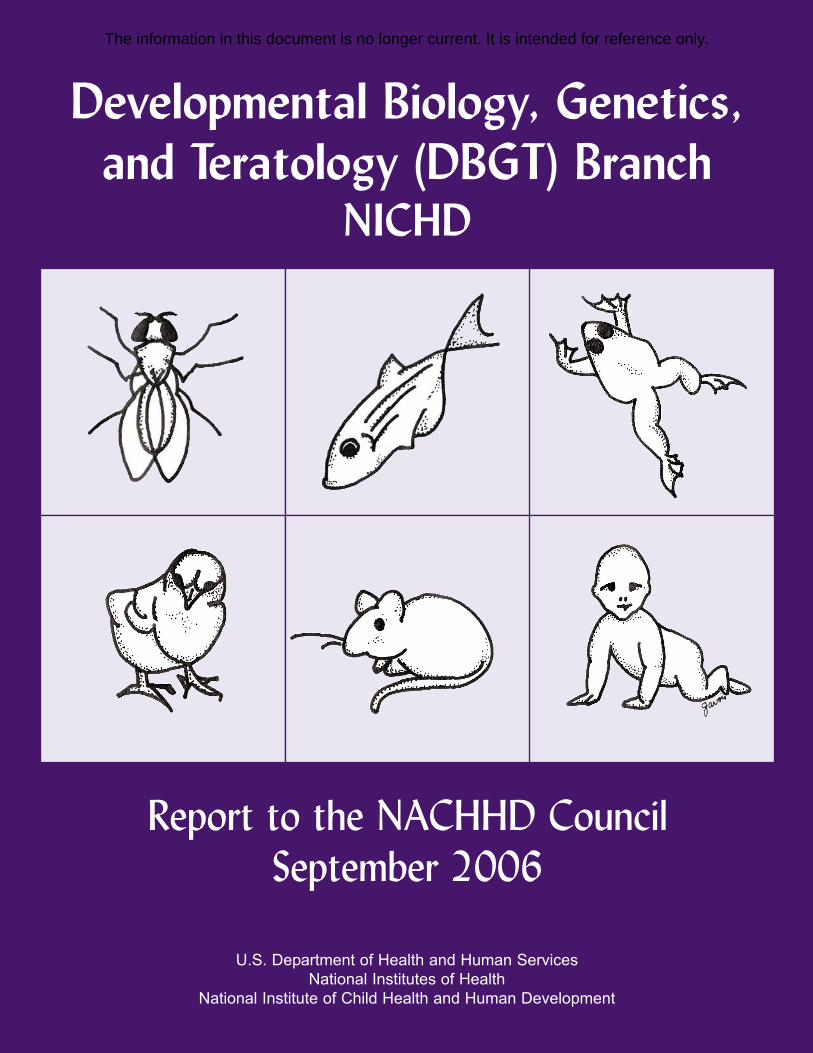
Cover Image: The figures illustrate several of the animal model organisms used in
research supported by the DBGT Branch including: the fruit fly, Drosophila (top, left); the
zebrafish, Danio (top, middle); the frog, Xenopus (top, right); the chick, Gallus (bottom,
left); and the mouse, Mus (bottom, middle). The human baby (bottom, right) represents
the translational research on human birth defects.
Drawings by Lorette Javois, Ph.D., DBGT Branch
The information in this document is no longer current. It is intended for reference only.
TABLE OF CONTENTS
EXECUTIVE SUMMARY .......................................................................................................... 1
BRANCH PROGRAM AREAS .......................................................................................................... 1 BRANCH FUNDING TRENDS.......................................................................................................... 2 HIGHLIGHTS OF RESEARCH SUPPORTED AND BRANCH ACTIVITIES.............................................. 3 FUTURE DIRECTIONS FOR THE DBGT BRANCH ........................................................................... 4
HIGHLIGHTS OF RESEARCH SUPPORTED ....................................................................... 4
THE DEVELOPMENTAL GENETICS AND GENOMICS PROGRAM...................................................... 5 THE EARLY EMBRYONIC DEVELOPMENT PROGRAM .................................................................... 7 THE DEVELOPMENTAL NEUROBIOLOGY PROGRAM ..................................................................... 9 THE ORGANOGENESIS PROGRAM ............................................................................................... 14 DEVELOPMENTAL IMMUNOBIOLOGY PROGRAM......................................................................... 17 THE REPRODUCTIVE IMMUNOLOGY PROGRAM .......................................................................... 19 TRAINING AND CAREER DEVELOPMENT PROGRAMS.................................................................. 20
HIGHLIGHTS OF BRANCH ACTIVITIES........................................................................... 22
BIRTH DEFECTS INITIATIVE ....................................................................................................... 22 RESEARCH INITIATIVES IN DEVELOPMENT THAT USE ANIMAL MODELS.................................... 23
FUTURE DIRECTIONS FOR THE DBGT BRANCH .......................................................... 30
DEVELOPING A BRANCH PLAN FOR THE FUTURE ....................................................................... 30 SUMMARY OF PANEL DISCUSSIONS............................................................................................ 30 DBGT BRANCH PLANS FOR FUTURE ACTIVITIES ...................................................................... 37
FIGURES AND TABLES ...............................................................FIGURES AND TABLES-1
APPENDIX A: DBGT BRANCH PERSONNEL.................................................................. A-1
APPENDIX B: DBGT BRANCH ACTIVITIES, FISCAL YEAR 2002 THROUGH FISCAL YEAR 2006................................................................................................................. A-3
APPENDIX C: DBGT BRANCH SOLICITATIONS, FISCAL YEAR 2002 THROUGH FISCAL YEAR 2006........................................................................................... A-5
APPENDIX D: DATABASES AND RESOURCES SUPPORTED BY THE DBGT BRANCH AND THE NICHD ..................................................................................... A-6
APPENDIX E: PUBLICATIONS BY DBGT STAFF, FISCAL YEAR 2002 THROUGH FISCAL YEAR 2006........................................................................................... A-8
APPENDIX F: DBGT BRANCH EXPERT PANEL............................................................ A-9
The information in this document is no longer current. It is intended for reference only.

The information in this document is no longer current. It is intended for reference only.
EXECUTIVE SUMMARY
According to a recent report from the March of Dimes, birth defects (as defined broadly to include both structural and functional/metabolic abnormalities) continue to be a major public health concern: “Every year, an estimated 7.9 million children—6 percent of total births worldwide—are born with a serious birth defect of genetic or partially genetic origin.”1 Even in the United States, the birth defects prevalence is 4.8 percent of live births.2 Birth defects remain a leading cause of infant mortality. In addition, structural abnormalities, such as neural tube defects (NTDs), congenital heart defects, craniofacial anomalies, and abnormalities of the musculoskeletal, digestive, respiratory, and urogenital systems, contribute significantly to disabilities of infancy, childhood, adolescence, and adult life. The emotional stress on families and afflicted individuals and the economic impact are enormous. Although the incidence of neural tube defects has declined in recent years due, in large part, to folate supplementation of grains, the incidence of many other types of structural abnormalities remains unchanged.
A longstanding goal of the National Institute of Child Health and Human Development (NICHD) is to support and encourage research on the underlying mechanisms of normal development as well as the molecular susceptibility and etiology of human birth defects, namely though the programs of the Developmental Biology, Genetics, and Teratology (DBGT) Branch. The mission of the Branch is to support a comprehensive national effort to increase understanding of the biological processes controlling both normal and abnormal development. It is through increased knowledge that effective strategies to prevent birth defects will ultimately become possible. One of the messages resulting from the Institute’s strategic planning process on developmental biology was the importance of improving knowledge and understanding of the underlying mechanisms associated with the formation of structural birth defects. Advances in developmental and molecular biology, genetics, and other biotechnologies and disciplines, supported both by the Branch and by other entities, continue to provide medical science with an armamentarium of tools to dissect and understand the complex biological and genetic mechanisms responsible for birth defects.
BRANCH PROGRAM AREAS
One of the Branch’s major research priorities is to understand the causes of structural birth defects and primary immunodeficiencies, birth defects of the immune system. Most of the research projects supported by the Branch are primarily basic science in nature and take advantage of opportunities offered by a variety of animal models. The Branch’s Birth Defects Initiative also fosters interactions between basic scientists and clinicians who have a common interest in birth defects to build on their respective strengths and fill the gaps in knowledge about how both genetic and environmental perturbations of normal processes result in developmental abnormalities.
1 Christianson, A., Howson, C.P., & Modell, B. (2006). March of Dimes Global Report on Birth Defects: The Hidden Toll of Dying and Disabled Children. White Plains, NY: March of Dimes. 2 Ibid, Appendix B.
Executive Summary 1
The information in this document is no longer current. It is intended for reference only.
Along the continuum of development, DBGT supports studies essentially starting with gastrulation and proceeding through early patterning and the formation of organ rudiments. Specific Branch program areas include research and training in early development, basic and clinical developmental genetics, genomics, organogenesis, developmental neurobiology, reproductive and developmental immunology, and factors in teratogenesis. The content and the responsibilities of the DBGT Branch relate to basic and clinical investigations identified in the six program categories listed below (the percentage of grants in the Branch’s holdings for each category appear in parentheses; see Figure 1 and Figure 2 for fiscal year 2005 holdings):
• Developmental Genetics and Genomics Program seeks to identify and characterize the genes, genetic networks, and epigenetic factors that control developmental processes and to understand how alterations in these components lead to structural birth defects (20.6 percent).
• Early Embryonic Development Program aims to elucidate the cellular, molecular, and physical mechanisms that direct the formation of the embryonic plan of a complex, multicellular organism (18.4 percent).
• Organogenesis Program attempts to determine the mechanisms underlying the normal development of organ primordia against which aberrations of these processes can be better understood (25.5 percent).
• Developmental Neurobiology Program strives to better understand the mechanisms controlling the early pattern of the developing central nervous system (CNS), the processes of neurogenesis, axonal guidance, and neural crest differentiation (23.6 percent).
• Developmental and Reproductive Immunology Program aims to understand the development of the immune system and the postnatal consequences of abnormal development as well as the immunological basis for maternal-fetal tolerance and the maintenance of pregnancy (10.0 percent). This program contains portfolios in Developmental Immunobiology, Neonatal Infection, and Reproductive Immunology.
• Factors in Teratogenesis Program seeks to assess adverse genetic and/or environmental influences on development and to arrive at mechanisms by which developmental aberrations are produced (1.9 percent). (Note: in Figure 1, Figure 2, Figure 6, and Figure 7, this program is subsumed under the Organogenesis Program.)
BRANCH FUNDING TRENDS
Support for Branch research is primarily provided through standard research project funding mechanisms, including R01s, R03s, R21s, R37s, and P01s. Funding for these mechanisms comprises more than 85 percent of the Branch’s budget. Most of the rest of the Branch’s budget supports a variety of training (T32s, F32s) and career development awards (K08s, K23s, and K24s). (See Figure 3 and Figure 4 for details on fiscal year 2005 holdings.) The DBGT Branch does not fund any centers programs. The Branch funds one contract for the distribution of mouse models for the study of NTDs and co-funds a limited number of other contracts and interagency agreements (see Figure 3, Figure 4, Appendix C, and Appendix D).
Executive Summary 2
The information in this document is no longer current. It is intended for reference only.
Since the Branch’s report to the National Advisory Child Health and Human Development (NACHHD) Council in 2002, the budget trend has been downward, as expected in light of the current National Institutes of Health (NIH) appropriation. Between 1997 and 2002, the Branch’s budget rose from $64 million to slightly more than $103 million. From 2002 to 2005, the Branch’s budget dropped by approximately 9 percent to $94 million, or to a level comparable to the 2001 budget (Figure 5 and Figure 7). This decrease was accompanied by a 20-percent decrease in Branch holdings from 462 projects in 2002, to the current level of 369 projects (Figure 6). These figures suggest that the average cost per project has increased over the years. The costs of most of the Branch’s programs have decreased to a certain degree, although the Organogenesis Program has remained fairly stable (Figure 6 and Figure 7). Support for research training has experienced a less severe downward trend during this same time period (Figure 8 and Figure 9).
HIGHLIGHTS OF RESEARCH SUPPORTED AND BRANCH ACTIVITIES
The Branch continues to support many of the foremost biomedical investigators, including Nobel Laureates, Lasker Prize winners, Presidential New Investigator Awardees, and Howard Hughes Medical Institute investigators, as they conduct research in the field of developmental biology. This research, along with that of a cadre of promising new investigators, has produced advances on such important developmental topics as axis formation, left-right asymmetry, genes associated with neurogenesis, stimulatory and inhibitory factors associated with axonal guidance, characterization of the segmentation clock associated with somite formation, as well as new findings related to limb outgrowth and patterning that challenge long-standing developmental dogma. Many of the studies supported by the Branch address fundamental questions associated with critical developmental processes. Studies of the hereditary basis of human NTDs are progressing toward identifying genes that predispose humans to spina bifida and examining the effects of environmental conditions to assess the complex relationship between genetic susceptibility and environment. Similarly, studies on the mutations and complex interactions of the Wiskott-Aldrich Syndrome Protein with other molecules are helping to explain the pathology associated with this X-linked primary immunodeficiency. The Highlights of Research Supported section of this report provides only a small sample of the exciting research being supported by the DBGT Branch.
A judicious use of funds and efficient partnering with other NIH Institutes and Centers has enabled the Branch to stimulate research and provide infrastructure to support cutting-edge research in developmental biology and birth defects. Through leadership roles in the Trans-NIH Zebrafish Coordinating Committee, the Trans-NIH Xenopus Working Group, and the Mouse Mutagenesis Project, the Branch has leveraged funds from other sources to support projects that generate new mutants, develop genetic tools, and provide genomic resources that benefit the Branch mission of better understanding the causes of birth defects. Participation in other trans-NIH programs, such as the Knockout Mouse Project, the NIH Roadmap, and the Neuroscience Blueprint, also ensure that the interests of the DBGT Branch and the NICHD are well-served. These activities are described in the section titled Highlights of Branch Activities.
Executive Summary 3
The information in this document is no longer current. It is intended for reference only.
FUTURE DIRECTIONS FOR THE DBGT BRANCH
As resources for funding become more limited, it is increasingly important to carefully and intelligently formulate plans for the future. In keeping with the new policy established by the Institute’s leadership, the DBGT Branch elicited the recommendations of a panel of experts to help in this planning process. In February 2006, the Branch convened the expert panel to develop a set of recommendations for addressing a series of questions related to scientific opportunities, public health issues related to the Branch’s mission, and areas that should be de-emphasized. The committee was composed of scientists, advocates, and members of the NACHHD Council (see Appendix F for a list of panel members).
The panel’s recommendations covered topics related to scientific endeavors as well as infrastructure and training. The panel felt it was necessary to maintain an emphasis on investigator-initiated projects as the most effective way to delve into understanding developmental processes. In addition, it recommended that the Branch promote research on quantitative aspects of development, such as non-syndromic birth defects, that may be caused by gene dosage, systems biology of development, and cellular differentiation. The panel also firmly believed that enhanced infrastructure was needed, such as improving and maintaining model organism databases, establishing and maintaining registries for human birth defects, and supporting the technologies required to enhance investigators’ abilities to mine data. The panel also considered interdisciplinary training and curriculum development in new research areas, such as quantitative biology and systems biology, as high priorities.
The DBGT Branch believes that supporting these new research areas, along with the areas that it has been supporting, will help to ensure significant advances in understanding the mechanisms that control embryonic development. Branch staff considered the panel’s recommendations in informing and developing the Future Directions for the DBGT Branch section of this report.
HIGHLIGHTS OF RESEARCH SUPPORTED
The NICHD understands that development is a continuum, which is initiated at fertilization, proceeds through embryonic and fetal development, and continues postnatally through adolescence. For the sake of tracking grants and projects, the Institute divides development into categories. The areas of development handled by the DBGT Branch essentially start with gastrulation and proceed through early patterning and the formation of organ rudiments. Earlier developmental events (e.g., fertilization and early cleavage) and mechanisms related to the reproductive process are the purview of the Institute’s Reproductive Sciences Branch.
The majority of the grants supported by the DBGT Branch are investigator-initiated and focus primarily on basic mechanisms regulating embryonic development. Most use animal models for this research, but other projects have a human focus. With more than 360 projects in the Branch’s portfolio, this section is not meant to be a complete compendium of the projects supported. Instead, the section provides only a brief overview of each program’s focus and
Highlights of Research Supported 4
The information in this document is no longer current. It is intended for reference only.
offers limited highlights of its supported projects. This report briefly discusses training mechanisms supported by the Branch in a separate section.
THE DEVELOPMENTAL GENETICS AND GENOMICS PROGRAM
This program focuses on research, training, and the dispersal of scientific knowledge regarding the genetic determinants of development by supporting studies of the basic biological processes underlying both normal development and the formation of developmental defects. The program represents several areas, including birth defects and models of human disease, genetic regulation of diverse fundamental cellular processes, epigenetic and genomic regulation of gene expression, genetic networks and mutations, developmental signaling, and genetic maps.
For instance, in the area of birth defects and models of disease, researchers are studying congenital defects and their causes in human populations. In addition, by screening animal models for mutations that resemble human clinical conditions, researchers are providing new research tools for further study. Several projects also seek to identify the genetic basis of human congenital defects or are combining the use of human and model animal information to study the causes of these defects. Projects include studies of the following human conditions: hereditary angioneurotic edema, congenital heart defects, familial patent ductus arteriosis, X-linked chondroplasia punctata, glycerol kinase deficiency, oro-facial clefts, oculo-oto-facial dysplasia, branchio-oto-renal syndrome, congenital diaphragmatic hernia, and Rothmund-Thomson syndrome.
Program efforts rely on animal and human stem cells to elucidate fundamental processes that regulate cell-fate commitment and differentiation—work that will have a significant impact on regenerative medicine for both developmental effects and degenerative diseases. Importantly, all of the areas supported by this program strive to utilize information from both human and animal models to unravel the complex genetic and epigenetic regulation of development.
The remainder of this section highlights some of the recent findings from program-supported research in some of these areas.
Epigenetic and Genomic Regulation of Gene Expression DNA methylation has a profound effect on gene expression and plays a key role in the acquisition of functional epigenetic modifications to DNA. Methylation serves as a tag, enabling regulatory factors to distinguish sequences that should be transcribed from those that should not be. Modification of DNA by methylation can prevent transcriptional initiation and ensure the silencing of genes on the inactive X chromosome and imprinted genes. Perturbation of methylation patterns can lead to congenital anomalies, growth defects, and immunological defects.
Using the mouse as a model system, investigators demonstrated that an oocyte-specific DNA methyltransferase variant, Dnmt-1o, is required to maintain methylation patterns at imprinted loci. In mutant embryos, the defects occur prior to the blastocyst stage, and these defects are maintained in stem cell lines derived from these embryos. Recent studies indicate that some
Highlights of Research Supported 5
The information in this document is no longer current. It is intended for reference only.
human embryonic stem cell lines have altered imprinting, and investigators are concerned that this defect could significantly impair the utility of these cells for therapeutic transplantation. Therefore, all of the NIH-approved lines are now being analyzed for methylation patterns at several known imprinted loci. Other investigators are using computational and bioinformatics approaches to identify DNA methylation sites, which may impact developmental processes; these studies are conducted in both human and model organism sequences.
Developmental Patterning and Signaling Pioneering work using the invertebrate model, Drosophila melanogaster, revealed several transcription factor-gene complexes that are responsible for organizing the body plan. Homologous genes have been discovered in all animals, including humans, and mutations in these genes are responsible for a number of developmental anomalies. These findings demonstrate the importance of using animal models to study human disorders.
This program has also supported work on the molecular mechanism by which initial polarity is established after fertilization. Through work in the simple model organism, Caenorhabditis elegans, researchers have discovered new polarity proteins, their binding partners, and the signaling pathways by which they act. This work is likely to have a significant impact on understanding developmental defects in pre-blastocyst human embryos.
The program also supports several projects studying the function and regulation of Hox, T-box, Six/Eya, and Polycomb genes in both animal models and in human populations. For example, use of conditional alleles from different members of the T-box family has allowed researchers to demonstrate unique functions for single T-box genes in the development of the allantois, mammary gland, and heart. Comparison of several embryonic patterning genes in non-model organisms reveals that the regulation of segmentation and limb development can vary across animals by subtle, but significant, genomic differences. Signaling factor pathways that interact with these regulatory protein networks are also a focus of the portfolio. For example, construction of chimeric tyrosine kinase receptors reveals differences in the usage of intracellular pathways for different ligand systems and in the generation of certain tissues. Mutational analysis of the transforming growth factor (TGF) pathway has revealed specific heart and eye defects.
Genomic and Gene-Expression Informatics This program has supported the development of databases as repositories for genomic and gene-expression information from a large number of developmental animal models used by the NICHD and by NIH-wide research communities. These include chick, Dictyostelium, frog, and mouse embryo; in addition, the Branch helps to support the rat and zebrafish databases, for which other NIH Institutes have the primary responsibility. (See Appendix D for a listing of resources supported by the DBGT Branch and the NICHD.)
As projects mature from the development phase, supported by the R01 mechanism, to the established resource phase, they will require different strategies for evaluation and maintenance. For example, the National Human Genome Research Institute (NHGRI) recently converted the databases that it supports (e.g., fly, C. elegans, mouse, and zebrafish) from R01 to P41 type grants. The resources supported by the DBGT Branch comprise an essential component of the
Highlights of Research Supported 6
The information in this document is no longer current. It is intended for reference only.
Institute’s holdings and provide important resources and data dissemination for NICHD and NIH research programs. The fact that once these important resources become established, they are expensive to maintain is a major concern; however, in the long run, this publicly available data helps to reduce the overall cost of research while making the research process more efficient— but only as long as investigators continue to deposit their genomic/genetic data in these databases.
In addition to databases, this program supports the acquisition of extensive genomic and gene-expression data from a large number of normal and mutant animal models. For example, RNAibased mutagenesis in C. elegans is allowing scientists to identify all genes required for embryonic development and to construct a digital phenotype map (phenome) for identifying the functional clusters of these genes. Similarly, a genome-wide mutagenesis, expression, and functional analysis project in Dictyostelium is underway to reveal genes involved in growth and development. Researchers are also developing a genetic map for linkage studies of an emerging model organism, Xenopus tropicalis, and several projects are analyzing genomic structure and function in the mouse genome. Web-based databases (independent of the animal model databases described above) make public the information gathered by these large-scale projects.
THE EARLY EMBRYONIC DEVELOPMENT PROGRAM
The Early Embryonic Development Program supports research to elucidate the genetic, molecular, cellular, and physical mechanisms that transform the fertilized egg cell into an embryo with defined axes, primary germ layers, and organ primordia. The mechanisms involved in this transformation operate when the embryo consists of only hundreds or thousands of cells. They rely on cellular interactions, physical/mechanical tension or traction, localized expression of growth factors, and activation of specific intracellular signaling pathways. These mechanisms affect the target cell by activating specific sets of genes that control the cell’s properties. The physical aspects of cell-cell mechanisms, cell-matrix mechanisms, and cell movements also play important roles in the formation of the body plan. When these mechanisms operate normally, they direct the target cell to differentiate and migrate in a characteristic way so that the appropriate organs form in the correct location. Altering these mechanisms perturbs early developmental events. Because these events establish the embryo’s global pattern, even slight alterations in them usually lead to drastic defects or embryonic lethality.
The Early Embryonic Development Program supports projects to examine the events and mechanisms that produce the embryo’s first pattern, and to identify and characterize the genes and factors controlling these events and mechanisms. Studies of developmental events include specification of the embryonic axes, cell-fate determination and cell-type differentiation, and the cell migrations that bring organ primordia to their proper locations. Program developmental mechanisms include cell-cell signaling, intracellular signal transduction, and cell-matrix interactions. These events create an embryo with an axial pattern and with correctly positioned organ primordia.
Highlights of Research Supported 7
The information in this document is no longer current. It is intended for reference only.
Axes Formation Proper embryonic development depends upon the establishment of the embryo’s cardinal axes: the early forming anterior-posterior and dorsal-ventral axes, and the later forming left-right axis. Decades of research supported by the Early Embryonic Development Program identified the cellular processes that control the formation of the embryo’s first two axes: the site at which the sperm fertilizes the egg, the orientation of resulting cell cleavage planes, and the localization of RNAs within the egg. These cellular asymmetries are translated into localized gene expression, which in turn, controls the formation of different structures along these primary axes. In addition to elucidating cellular mechanisms, these examinations also provided enormous amounts of information about the signaling molecules and developmental genes that control cell migration and cell-fate specification. This research also showed that the same molecules and genes are used repeatedly during subsequent developmental events, and that the left-right axis was not established by the cellular mechanisms that form the first two axes. Recent studies show that an animal’s left-right axis forms by surprising cellular mechanisms, described below.
Left-Right Asymmetry Even after the anterior-posterior and dorsal-ventral axes are established, the embryo remains bilaterally symmetrical. During the neural plate stages, precursor cells of the visceral organs form as bilateral populations of cells. Following neurulation, the groups of cells destined to form the unpaired organs (e.g., digestive system and heart) migrate to the midline and combine to form a single organ, which then either migrates to its characteristic lateral location (e.g., liver and spleen) or orients itself with respect to the left-right axis (e.g., intestine and heart). For these events to occur properly, the migrating cells must identify the midline and distinguish between the left and right sides of the embryo. Defects in these processes result in improper organ placement. In humans, failure to form the third axis correctly causes several medical conditions, which involve missing, duplicated, and abnormally located internal organs. For example, in situs inversus, visceral organs are located and oriented in the mirror image arrangement, leading to heart defects. In heterotaxy, the organs are located in a random arrangement, leading to numerous serious medical complications.
Recent studies show that the first step in the process of establishing the embryonic midline involves an unexpected cellular event. During a brief window of time in the late neural plate stage, motile cilia projecting from the surface of certain cells move extracellular fluid from the right side to the left side of the embryonic midline. This motion plays a critical role in establishing the embryo’s left-right axis. Although the exact cellular mechanisms by which this motion exerts its influence are currently under investigation, preliminary studies indicate that this event is propagated either by a population of sensory cilia or gap junctions. Branch-supported studies to clarify this issue are ongoing.
Even though the mechanism by which this cellular event is propagated remains unclear, other studies have already identified its molecular outcome. The findings show that the motion causes the cells on the left side of the embryo to express genes that are not expressed by cells on the right side, including nodal and PitX2. This laterally localized gene expression is subsequently transferred to the surrounding tissues by several extracellular signaling pathways, including the Wnt, Fgf, and Notch pathways. Fortunately, program-supported studies examining other developmental events have already elucidated the way in which these signaling pathways exert
Highlights of Research Supported 8
The information in this document is no longer current. It is intended for reference only.
their influence on cell-surface receptors, intracellular transduction pathways, and ultimately on cell-specific migration and gene expression. Thus, in a short period of time a few studies have provided a detailed understanding of the formation of the left-right axis. Much of this progress is possible because many early developmental mechanisms utilize the same genetic pathways. The synergistic effects of studying several developmental events and stages have enabled rapid progress in understanding these complex events.
By studying the cellular, molecular, and genetic mechanisms of several early developmental events, the Early Embryonic Development Program provides a comprehensive understanding of events required for normal development and of processes that can lead to birth defects.
THE DEVELOPMENTAL NEUROBIOLOGY PROGRAM
Many structural neurological birth defects are caused by problems that occur during early CNS development. A major focus of the Developmental Neurobiology Program is supporting basic research that contributes to understanding how the nervous system develops under both normal and abnormal conditions. In the context of understanding the mechanisms that control nervous system formation, program goals are to:
• Determine how nerve cell progenitors divide, migrate, and begin to differentiate into specific types of nerve cells;
• Understand pathfinding strategies of these immature, developing neurons for getting to the correct location and project processes that make appropriate connections with their targets;
• Decipher how synapses between appropriate targets form; and • Elucidate how superfluous synapse and neurons are eliminated.
Formation of the nervous system begins shortly after gastrulation, when the cells of the dorsal ectoderm are induced by the underlying mesoderm to become the neural plate, which extends the length of the embryo. From this very simple neural plate, the molecular and mechanical processes associated with convergence and extension morphogenetic activities begin a course of events that ultimately results in a highly stereotyped, complexly organized and integrated nervous system. During neurulation, the neural tube is formed, and the embryonic CNS is patterned along the three major axes. Neural progenitor cells and their offspring derive positional information from within these boundaries. Thus, neurons and glia are generated from this undifferentiated neuroepithelium and acquire the diversity of cell types that make up the adult CNS. These new cells migrate to specific positions within the developing nervous system and send out processes to specific targets. Once specific patterns of connectivity are generated, synapses are formed; hormonal and trophic factors influence the survival, differentiation, and selective elimination of these connections.
While embryonic development of the CNS remains a complex process, recent advances in genetics, genomics, and proteomics now allow for a more in-depth analysis of the processes underlying its development. In addition, genes, genetic networks, and the timing of their activation and inactivation are also conserved, in large part, across species, making it possible to predict some of the underlying mechanisms of human development based on studies that use animal models. With newly developed tools, investigators are now better able to examine the
Highlights of Research Supported 9
The information in this document is no longer current. It is intended for reference only.
underlying mechanisms of neural development and are learning that similar developmental strategies are used multiple times during CNS development.
Neurogenesis The CNS develops from a group of cells that is initially homogeneous in its developmental specification. Current understanding is that the nervous system is established through progressive stages of regional development, in two major phases: early and late. In both phases, interactions between neighboring tissues are sequential and are important in specifying regionally distinct structures. Early stages include differentiation of the neuroectoderm and segmental patterning. Later stages include the determination of specific neuronal and glial phenotypes and the increased specialization of CNS structures. In addition, both the early and late phases of neural differentiation are controlled by a multitude of transcription factors that act as switching molecules by binding to DNA and activating or repressing gene expression, signaling factors, and growth factors. For example, basic Helix-Loop-Helix (bHLH) transcription factors are essential for the development of multiple neuronal lineages in both the CNS and peripheral nervous system, and their expression is precisely controlled both spatially and temporally.
One gene of particular interest, single-minded (sim), acts as a master genetic switch directing cells to form the midline region of the CNS, and then continuing to direct development of specific sets of midline neural and glial cell types. sim was initially identified in Drosophila, but its important role in neural development led to the identification of two mammalian Sim genes, Sim1 and Sim2. CNS midline cells in Drosophila and floor plate cells of the vertebrate neural tube have similar functions in that they act as signaling centers to direct axon guidance, the formation of adjacent tissues, and cell-fate specification of cell types. sim both activates midline gene expression and represses lateral gene expression in the midline cells. Preliminary studies of the mouse genes, sim1 and sim2, strongly suggest conserved biochemical and functional roles.
Neural Patterning Early neural pattern formation along the anterior-posterior and dorsal-ventral axes is a major focus of the Developmental Neurobiology Program portfolio. Anterior-posterior patterning of the nervous system occurs soon after neural induction, when adjacent tissues produce signals that turn on regulatory genes in discrete domains of the neural plate. Along the anterior-posterior axis, signals divide this presumptive neural tissue into four major areas: forebrain, midbrain, hindbrain, and spinal cord. The working hypothesis is that neural tissues are initially anteriorly defined, and that the actions and interactions of multiple signaling pathways posteriorize these tissues in a progressive manner by encoding positional values along the anterior-posterior axis.
Dorsoventral patterning occurs when mesodermal structures beneath the neural tube secrete diffusible factors resulting in the specialization of cells in the ventral half of the neural tube. This cell-fate determination occurs during and following neural tube closure and involves the action of two opposing signaling pathways: sonic hedgehog (Shh)—ventrally from the notochord—and bone morphogenic protein (BMP)—dorsally from the boundary of neural and nonneural ectoderm, and later from the roof plate. Shh may work by repressing the expression of genes that encode dorsal neural tube transcription factors, which would otherwise be expressed throughout the neural tube. In zebrafish BMP mutants, neural crest, dorsal sensory neurons, and
Highlights of Research Supported 10
The information in this document is no longer current. It is intended for reference only.
interneurons display aberrant phenotypes ranging from complete loss to dramatic expansion of cell populations.
Investigators have identified, in Drosophila, many of the genes in the signaling cascade downstream of hedgehog (Hh), and all these genes have homologues in mammals. At the end of the pathway is a putative transcription factor, cubitus interruptus (ci). Hh induces ci protein and antagonizes the negative regulation of ci by the proteins Patched and protein kinase A. In mammals, the homologues of ci are the three Gli genes: Gli1, Gli2, and Gli3. Current work aims to determine whether the three mouse Gli gene products have similar functions to the fly ci protein, and whether they function in an analogous signaling pathway. Observations that humans with the dominant Greig cephalopolysyndactyly syndrome have limb and craniofacial defects and spina bifida due to mutation in GLI3, and that mouse Gli3 mutants display similar abnormalities, suggest an important role for these genes and their products.
Regionalization of the CNS primordia and the definition of distinct neural progenitor cell domains along the major axes occur through the interactions of different signaling molecules, such as Wnts, fibroblast growth factors, and retinoic acid, and through the regionalized expression of hox genes. Because cells of the embryo must respond to cues in both the anterior-posterior and dorsal-ventral axes, it is important to understand how this positional information is integrated at the level of a single gene in establishing the CNS plan in mammals. Studies have shown that Pax3 may be an effector gene that functions to integrate and coordinate these cues.
By taking advantage of the explosion of genetic and genomic information in a wide variety of animal models, including but not limited to Drosophila, zebrafish, Xenopus, and mouse, researchers have made tremendous progress in identifying genes and defining the cascades that are likely to mediate neural patterning. Although this work has yet to identify all the individual genes involved in various aspects of neural development, researchers are actively identifying and placing genes and their products in appropriate genetic networks and biochemical pathways. Completion of these activities will ultimately advance understanding of how neural patterning evolves and how neural identity is determined. The next step is to understand how cells become committed to their specific fates, and how regulatory genes are specifically involved in these individual cascades. Neural patterning has become one of the most rapidly evolving areas of developmental neuroscience.
Left-Right Asymmetry In contrast to the significant progress being made toward unraveling the molecular genetic basis for asymmetric development of visceral organs, the study of how cortical asymmetries are generated or perturbed in neurological disorders is a relatively new area for the Developmental Neurobiology Program. Using the zebrafish model, researchers have found components of the Nodal signaling pathway to be specifically expressed on the left side of the developing zebrafish brain. Fish lacking this transient expression later show a randomization in the left-right positioning of the pineal gland. The implication of this finding and the identification of other CNS left-right asymmetries are only now being investigated.
Highlights of Research Supported 11
The information in this document is no longer current. It is intended for reference only.
Axonal Guidance, Pathfinding, and Trophic Factors Understanding how connections form with appropriate targets is another major focus of the Developmental Neurobiology Program. For correct connections to form, neuronal precursors must migrate to their correct locations and extend processes into the extracellular environment. Investigators are identifying new factors that influence this process all the time, and understanding the regulation of growth cone responses to multiple guidance cues is critical for ultimate understanding of nervous system development. During the highly directed process of axonal pathfinding, the axons are repeatedly confronted with points at which they must “select” the appropriate axonal pathways. Correct pathway selection requires the presence of spatially and temporally orchestrated cues at each choice point, and the responsiveness of individual neural growth cones to only a specific set of cues. External guidance cues in the microenvironment can be short-range and contact-mediated, long-range and diffusible, attractive, or repulsive and can include extracellular cell-surface molecules, cell adhesion molecules, and trophic factors. Interestingly, an individual factor can either inhibit or facilitate guidance depending on the age of the growth cone and its environmental milieu.
One extrinsic cue, known to direct motor axons at individual choice points, is the zebrafish unplugged gene. In unplugged null mutants, two pioneering motor growth cones reach the choice point, but make inappropriate pathway decisions. The unplugged gene encodes a muscle-specific kinase (MuSK)-like receptor tyrosine kinase. Although these kinases have been studied extensively in the context of synapse formation, they are now recognized as important for axonal pathway selection as well.
Research to explore other receptor tyrosine kinases, of which the Eph receptors are the largest known subfamily, and their ligands, the ephrins, continues even as their critical role in the guidance of axons to their targets continues to unfold. Ligands signal through their Eph receptors by direct cell-cell contact, a mechanism that provides the potential for bi-directional signaling with a forward signal through the tyrosine kinase receptor, and a reverse signal through the ligand. Investigators have biochemically demonstrated this reverse signaling and postulate that it plays an important role in axonal pathfinding as well as in related developmental processes, such as guidance of cell migration.
Other work has identified a number of additional secreted and transmembrane molecules—such as semiphorins, plexins, and components of the Slit-Robo signaling cascade—as playing critical roles in shaping the circuitry of the nervous system. Research has also shown that neurotrophic factors are essential for ensuring the correct development of the nervous system; specifically, they modulate responses of growth cones to other guidance cues via cytoplasmic signaling pathways, which regulate the dynamics of cytoskeletal components. For example, two neurotrophins—nerve growth factor and brain-derived nerve factor—have long been known to be attractants for sensory growth cones in culture. Recently, this response was shown to require local activation of the specific neurotrophin trk receptors and receptor autophosphorylation, which initiates cytoplasmic signaling. Further examination of the cytoplasmic signaling pathways indicates that activation of PI3 kinase is required for sensory growth cones to turn toward an attractive cue during pathfinding.
Highlights of Research Supported 12
The information in this document is no longer current. It is intended for reference only.
Neural Tube Defects (NTDs) NTDs are one of the most frequent and severe developmental anomalies of the CNS. Spina bifida, a developmental malformation resulting from abnormal or incomplete closure of the caudal end of the neural tube, has received much attention in the developmental biology field. Within the Branch’s larger Birth Defects Initiative, study of NTDs resides within the Developmental Neurobiology Program. The underlying causes of NTDs are poorly understood; however, animal models, such as mouse and zebrafish, are assisting researchers in deciphering the mechanisms that underlie both abnormal and normal neurodevelopment. For example, recent mice studies in which each of the three disheveled (Dv1) genes were inactivated indicated that this gene and the evolutionarily conserved Wnt/wingless signal transduction pathway play important roles during neural tube closure.
The Branch currently supports studies on the hereditary basis of human NTDs by identifying genes that predispose humans to spina bifida and other NTDs. Preliminary work is underway to assess the genetic contribution of affected candidates using detailed phenotypic descriptions, newly developed statistical techniques, and rapid genetic-marker genotyping for a thorough genomic screen. Studies that examine the effects of environmental conditions complement studies of individuals to tease out the complex relationships between genetic susceptibility and environment. These researchers also are creating a data resource to examine the potential genetic determinants of spina bifida in a large, well-characterized sample of approximately 500 families, which consist of a proband affected with spina bifida, along with the biological parents and unaffected siblings. This resource will be useful for testing identified candidate loci, as well as for evaluating putative susceptibility loci. Recent studies in humans and in animal models suggest that specific biochemical and developmental pathways may control neural tube development, providing a starting point for potential new candidate susceptibility loci.
Neural Crest Studies The vertebrate neural crest is a migratory embryonic cell population that forms at the border between the neural plate and future epidermis. Neural crest cells delaminate from the neuroepithelium in a rostrocaudal wave and migrate throughout the embryo to form a wide range of derivatives, including head, sensory, sympathetic and enteric neurons, glia, melanocytes, smooth muscle, dermis, connective tissue, cartilage and bone, pigment cells, and, together with cranial placodes, the peripheral nervous system. Using modern labeling techniques in transparent zebrafish embryos, it is now possible to image, with subcellular resolution, the complete neural crest migration pathway. These technologies allow researchers not only to understand neural crest migration, but also to assess the molecular basis of neuronal guidance.
The Developmental Neurobiology Program supports a variety of studies on neural crest because abnormal crest migration is implicated in a wide variety of structural birth defects. A number of studies have already identified molecules associated with neural crest development and lineage decisions. For example, the transcription factor, Foxd3, is sufficient to specify neural crest. These studies are generalizable to understanding the biological processes of establishing and/or maintaining multipotent cell properties. Use of the Cre/LoxP system has also demonstrated the important roles of a variety of signaling factors in normal neural crest development. For example, findings show that Wnts are involved in the induction of neural crest cells in embryos, although the molecular nature of the Wnt-signaling downstream targets remains unclear.
Highlights of Research Supported 13
The information in this document is no longer current. It is intended for reference only.
BMPs are also known to regulate formation of the neural crest and the development of both neurons and glia. Within individual neural lineages, BMPs control progressive developmental decisions, which are reflected in changes in cellular responses over time. In the peripheral nervous system of zebrafish, BMPs increase neuronal differentiation of neural crest cells and later promote the maturation of enteric and sympathetic neurons along lineage-specific pathways.
THE ORGANOGENESIS PROGRAM
The ability to detect spatial and temporal patterns in gene expression and to test gene function using targeted disruption or misexpression of specific genes has enhanced understanding of the roles that specific genes play during the development of organs and organ systems. Events, such as specification of the organ primordia, inductive signaling, outgrowth, and patterning, are routinely investigated at the level of molecular genetic mechanisms. One theme that has emerged from these studies is the conserved role of growth factors, signaling molecules, and signaling pathways across vastly different animal species and organs. This universality of molecular mechanisms is giving rise to a template for organ morphogenesis. The DBGT Branch Organogenesis Program supports research to examine:
• Development of the limb, including cartilage and bone primordia; • Somitogenesis, including development of skeletal and muscular elements; and • Establishment of the gut, heart, lung, pituitary, kidney, and other organ primordia.
Limb Development One child in every 200 is born with developmentally generated limb anomalies, and such abnormalities pose long-term suffering and morbidity for many affected individuals. Significant advances in molecular biology and the elucidation of genetic networks involved in limb development have provided a better understanding of both normal limb development and skeletal dysplasias. Research on the development of the vertebrate limb, funded by the Organogenesis Program, has combined mouse model genetic insights with physical manipulations of the developing chick limb bud to generate fundamental new paradigms. Since sonic hedgehog (Shh) was identified as a necessary component of limb morphogenesis 12 years ago, many studies have underscored the general long-range signaling ability of Shh, its essential functions during many aspects of embryogenesis, and its roles in the maintenance of stem cells and in vertebrate disease conditions. Recent work on early limb development, also funded by the Organogenesis Program, has provided new insights into the role of Shh in limb outgrowth and in anterior-posterior patterning of the limb, leading researchers to re-examine the way proximal-distal patterning is regulated in the limb bud. From this work, three significant findings (described below) now challenge long-standing theories about how limbs develop.
THE SHH-FIBROBLAST GROWTH FACTOR FEEDBACK LOOP AND REGULATION OF LIMB OUTGROWTH Classical grafting experiments in the chick limb bud demonstrated that outgrowth and patterning depend on reciprocal interactions between the apical ectodermal ridge (AER), a specialized epithelial structure at the distal edge of the limb bud, and the limb bud mesenchyme. The molecular components associated with these interactions include Shh, which is produced by the zone of polarizing activity (ZPA) in the posterior mesenchyme, and several members of the fibroblast growth factor (Fgf) family, which emanate from the AER. A Shh-Fgf feedback loop
Highlights of Research Supported 14
The information in this document is no longer current. It is intended for reference only.
operates early during limb development until embryonic day six. Shh maintains the Fgfs indirectly by up-regulating Gremlin, a BMP antagonist, which in turn prevents BMPs from down-regulating Fgf; this process is necessary for the continued production of Shh, thereby completing the loop.
An important question posed by researchers was what down-regulates the entire feedback loop— a critical step in the ultimate control of limb size. For example, if the feedback loop is artificially maintained, additional phalanges are produced resulting in a longer-than-normal limb. Recent work funded by the Organogenesis Program examined the roles of Shh, Gremlin, and Fgf4 to see if any of these could maintain the other genes in the loop after they would normally be down-regulated. Examination of the molecular events revealed that posterior Shh-expressing ZPA cells and their descendents cannot express Gremlin. Early in limb outgrowth, the absence of Gremlin is not problematic because the width of the limb bud is such that the Shh protein diffuses across the limb bud and activates target genes. As limb growth progresses, however, the proliferation of Shh-producing descendents creates a barrier between the source of Shh in the posterior ZPA and more anterior cells capable of expressing Gremlin. By embryonic day six, the Gremlin-competent cells are too far from the source of Shh to produce Gremlin. So, this barrier essentially triggers the breakdown of the Shh-Fgf feedback loop and the cessation of limb outgrowth. These findings have also provided new molecular insights into the phenomenon of regulative growth, whereby normal limbs can develop despite removal of significant amounts of limb bud tissue early in limb outgrowth.
SHH AND ANTERIOR-POSTERIOR PATTERNING IN THE LIMB The prevailing model for anterior-posterior specification of limb structures is one in which Shh, secreted by cells of the ZPA, forms a spatial gradient from posterior to anterior across the limb bud. In theory, cells interpret the level of Shh they are exposed to and differentiate accordingly, with high concentrations specifying posterior and low concentrations specifying anterior. Recent work funded by the Organogenesis Program, which originally set out to map the fate of ZPA/Shh-producing cells, unexpectedly revealed that the anterior-posterior sequence of digits is specified not only by the level of Shh that cells are exposed to, but also by the duration of their exposure. Descendents of Shh-producing cells directly contributed to digits three through five in the mouse, arising from cells that are exposed to the maximal concentration of Shh protein. However, the duration of exposure is critical to specifying digit identity, in that more anterior cells cease Shh expression at earlier stages than those that contribute to the posterior-most digit five. The cells in digit two never expressed Shh but were found to be dependent on low concentrations of Shh achieved by a spatial diffusion gradient.
REASSESSMENT OF THE PROGRESS ZONE MODEL FOR PROXIMAL-DISTAL LIMB PATTERNING During proximal-distal limb bud outgrowth, cells proliferate at a constant rate throughout the limb bud, but remain undifferentiated in a zone just beneath the AER. In the long-standing Progress Zone (PZ) Model, the length of time cells spend in the PZ determines proximal-distal cell fate, so that cells leaving the PZ earlier in development would form more proximal limb structures (e.g., the humerus), while cells leaving the PZ later would form more distal structures (e.g., the digits). For almost 30 years, the PZ Model has been cited as the prevailing model for the specification of proximal-distal cell fates. However, work supported by the Organogenesis Program has provided new insights into these events and brings the PZ Model into question.
Highlights of Research Supported 15
The information in this document is no longer current. It is intended for reference only.
In a series of program-supported studies, researchers labeled early limb bud cells and examined their progeny’s fate at later stages, either with or without AER removal. Labeled cells from the distal tip of the early limb bud colonized distal skeletal elements if the limb bud was intact. However, following AER removal, researchers unexpectedly could not find labeled progeny in the most distal-forming elements, suggesting that these distal cells did not contribute to skeletogenesis following AER removal. A combination of cell death and lack of cell proliferation following AER removal seem to play a causal role in the truncation of the limb outgrowth. Additional cell labeling studies performed during early limb development at different proximal-distal levels suggest that basic proximal-distal segments are specified much earlier in the developing limb bud than suggested by the PZ Model. This work found that, as development continued, a progressive restriction in the distal mesenchyme occurred such that cells were increasingly determined to form a narrower range of distal structures.
These and other new studies support a view that challenges the long-accepted PZ Model. While these recent findings have generated considerable controversy, some researchers argue that the results might be explained by a looser interpretation of the PZ Model. There is little doubt, however, that insights gleaned and questions raised by these experiments will stimulate further advances in understanding proximal-distal limb development.
Somitogenesis Somitogenesis is the fundamental process during vertebrate embryogenesis whereby the anterior-posterior body axis is progressively divided into repeating segmental elements, or somites. The initially uniform field of cells comprising the presomitic mesoderm (PSM) is progressively subdivided from anterior to posterior into metameric blocks of paraxial mesoderm that differentiate into the bone, cartilage, and tendons of the trunk; skeletal muscles of the body wall and limbs; and the dermis of the back. Somitogenesis is embedded into the global formation of the anterior-posterior axis such that somite formation is finely balanced with the rate of axis elongation. The somitic scaffold also supports the proper patterning of the peripheral nervous system and circulatory system as they develop along the anterior-posterior axis.
ESTABLISHING THE PERIODICITY OF SOMITES The striking periodicity or regular recurrence of somite production and distribution led researchers to hypothesize that an oscillator or segmentation clock acted in the PSM to trigger somitogenesis. The first molecular evidence for this segmentation clock was the observation of the periodic expression of the chick gene Hairy1 in the PSM. More recently, research has identified other “cyclic genes” in the Notch and Wnt signaling pathways whose rhythmic expression in the PSM parallels the segmentation process. The presence of the segmentation clock in fish, frogs, birds, and mammals suggests a conserved developmental mechanism in vertebrates. The Organogenesis Program is funding ongoing work on how local signaling interactions between a cell and its immediate neighbors give rise to an emergent, higher-level of organization within the PSM.
COUPLING OF MECHANISMS ASSOCIATED WITH SEGMENTATION AND ELONGATION OF THE ANTERIOR-POSTERIOR AXIS Research also has demonstrated that the posterior progression of PSM determination (the determination front) is regulated by two dynamic, mutually inhibitory gradients. First,
Highlights of Research Supported 16
The information in this document is no longer current. It is intended for reference only.
transcription of Fgf8 mRNA is restricted to the growing posterior tip of the embryo. The progressive degradation of the mRNA results in a caudal-rostral gradient of FGF8 protein, which prevents the initiation of the segmentation program. A rostral-caudal gradient of retinoic acid (RA) relieves this inhibition either by antagonizing the action of FGF8 directly, or by activating the cyclic genes. As a result of this dynamic interaction, somitogenesis is coupled to the elongation of the anterior-posterior axis. RA signaling is also associated with synchronizing bilateral somitogenesis and modulating the left-right signaling machinery that establishes handedness in vertebrates. Finally, ongoing work also suggests a coupling between the segmentation clock and the activation or maintenance of Hox genes during axis formation. In the PSM, regionalization is established early and relies mostly on the Hox genes, ultimately patterning the somitic mesoderm into cervical, thoracic, lumbar, sacral, and caudal regions.
Analysis of the genetic networks that govern somitogenesis is central to understanding vertebrate development and has broad implications for the complex properties of biological circuits. Unraveling these fundamental questions of developmental biology is no longer an issue of satisfying intellectual curiosity; rather, understanding how the segmentation clock functions and other related processes is of considerable clinical relevance because mutations in the human homolog of delta (a cyclic gene) result in abnormal segmentation of the vertebral column, a condition known as spondylocostal dysostosis syndrome.
DEVELOPMENTAL IMMUNOBIOLOGY PROGRAM
This program covers basic, applied, and clinical studies in developmental genetics and the ontogeny of the immune system. The long-term goal of this program is to translate the basic knowledge, insights, and understanding from supported studies into new approaches and strategies for the effective diagnosis, treatment, and prevention of developmental disorders of immunity. Researchers have already identified a wide variety of Primary Immunodeficiency (PI) diseases, each caused by defective or missing genes that may effect the normal development and function of T cells, B cells, or other components of the immune system. This program looks at both normal development of the immune system and abnormalities in developmental processes that result in these birth defects of the immune system.
Ontogeny of the Immune System It is well known that T-cell precursors from the bone marrow migrate to the thymus, where they undergo further differentiation. The stages of T-cell development are identified by the expression of specific cell-surface markers, such as T-cell receptor (TCR), CD3, and CD4/CD8. A series of interactions between immature thymocytes and thymic epithelial cells causes thymocytes to undergo a succession of positive and negative selection processes, based to a great degree on their affinities for various MHC I and MHC II molecules, which help them become mature T cells. Because this developmental process is so complex, NICHD-supported investigators have developed a two-cell-type selection culture system to study the cellular interactions that T lymphocytes undergo in the thymus. In this system, thymocytes, which are engineered to stop development at the immature double-positive CD4+/CD8+ stage, are cocultured with clonal lines of thymic epithelial cells, which are looking for lines of epithelial cells that will support the selection of the immature double-positive thymocytes into single-positive
Highlights of Research Supported 17
The information in this document is no longer current. It is intended for reference only.
thymocytes. Microarray analysis has identified 18 genes in several thymic epithelial cell lines that could be influential in supporting positive selection. Studies are currently underway to identify the functions of these genes and to test for their involvement in positive selection using short interfering RNA to knock-down levels of gene activity. These studies should increase understanding of the selection and development of the T-cell system and may provide insight into developing more efficient therapies for a number of disease conditions.
Primary Immunodeficiency (PI) Diseases Scientists have defined upwards of 100 genetic types of PI diseases, which typically involve defective or missing genes that affect the development and function of a number of cells associated with the immune system, including T cells, B cells, phagocytes, neutrophils, or platelets. Some mutated genes causing PI diseases may be X-linked, meaning males are clinically affected, but females are silent carriers. Although treatments are available for most PI diseases, the challenge is to develop more effective, practical, cost-effective therapies and screening methods for newborns.
In addition to supporting research on PI diseases, the NICHD also co-funds, with the National Institute on Allergy and Infectious Diseases (NIAID), the Primary Immunodeficiency Research Consortium, called USIDNet Consortium, a coalition of the world’s most prominent investigators in the field of PI diseases. The Consortium is charged with helping to prioritize and coordinate research directions and to develop new resources to study these rare disorders.
The remainder of this section focuses on two PI diseases and the results of research efforts funded by the Developmental Immunobiology Program.
SEVERE COMBINED IMMUNODEFICIENCY (SCID) SCID results from a genetic defect in the development and/or function of both T cells and B cells. Although different mutations can cause SCID, one common cause is adenosine deaminase (ADA) deficiency. Patients with ADA deficiencies are profoundly lymphopenic and suffer from a wide variety of infections that can be fatal if not treated with enzyme replacement therapy or bone marrow transplants. While researchers have actively studied the disease for some time, they still do not know the exact mechanism by which loss of ADA inhibits lymphocyte development. Because this human condition is rare, many studies on the effects of ADA loss on thymocyte development rely on a murine system. However, recent work suggests that important differences may exist between human and mouse regarding the loss of ADA and thymocyte development. Using cultures of human cells and human/mouse chimeric fetal thymic organ cultures, a model for human thymocyte development is emerging. Evidence suggests that β selection, the developmental checkpoint associated with T-cell receptor lineage decisions, occurs gradually throughout several phenotypic stages of development, and that the intermediate stages of development comprise a long window during which competition occurs between the γδ T-cell receptors and the pre-T cell receptors. Whichever receptor is expressed first and is functional directs the lineage fate of the developing thymocyte. Guided by knowledge of normal development, researchers should gain a better understanding of why human thymocytes fail to develop under ADA-deficient conditions, which will ultimately lead to alternative therapies.
Highlights of Research Supported 18
The information in this document is no longer current. It is intended for reference only.
WISKOTT-ALDRICH SYNDROME (WAS) WAS is an X-linked, recessive disorder in which patients exhibit complex immunological and hematological abnormalities that affect the development and function of their B cells, T cells, platelets, and hematopoietic cells. Studies of WAS have focused on Wiskott-Aldrich Syndrome Protein (WASP) mutation hotspots and genotype/phenotype correlations. The most typical mutations are amino acid substitutions, and alterations in the formation of protein splice variants are the second-most prevalent abnormality. In a study of 270 unrelated families, researchers found six major mutational hotspots—three within the coding region of the protein, and three involved in the formation of splice variants. These six hotspots are noted in 26 percent of families studied.
WASP has a very complex structure and its various functions are the result of interactions with many other proteins. Studies of these interactions are providing new insights into the disease mechanisms seen in WAS. The most-often studied function of WASP is its role in actin polymerization and actin cytoskeleton rearrangement. With regard to the immune system, WASP has a role in integrating cellular signals that lead to the nuclear translocation of various regulatory molecules. Absence of WASP in this context leads to decreased accumulation of calcineurin, WASP-interacting protein (WIP), and other molecules associated with calcium mobilization, which can influence the course of WAS.
Studies show that another protein, called WICH, interacts with WASP and plays a role in B-cell migration. Blocking WASP-WICH interactions decreases B-cell migration to the chemokine, stromal cell-derived factor-1. Evidence suggests that the WASP-WICH interaction plays a role in regulating integrin affinity in B cells. Studies have also identified a calcium-binding protein, CIB, that interacts with the N-terminus of WASP and with the cytoplasmic tail of a platelet-specific integrin—an interaction essential for the integrin activation of platelets. Mutations that cause WASP to have a reduced affinity for CIB result in the impaired platelet aggregation seen in WAS patients. In addition, WASP plays a role in monocyte chemotaxis by interacting with a family of mammalian verprolins, such as WIP and WIP-related protein (WIRE). WASP and mammalian verprolins function as a unit in monocyte chemotaxis to establish cell polarization. Blocking the binding of WASP to these verprolins impairs cell polarization, but not actin polymerization. Impaired chemotaxis helps to explain the recurrent infections experienced in patients with WAS. All of these studies demonstrating interactions between WASP and so many other proteins indicate the complexity of Wiskott-Aldrich Syndrome and its myriad manifestations.
THE REPRODUCTIVE IMMUNOLOGY PROGRAM
In this program, projects investigate the immunobiology of the placenta and maternal-fetal interactions during pregnancy in humans and animal models. A number of these projects center on maternal-fetal tolerance by identifying the underlying immunologic and/or genetic mechanisms that protect the fetus from maternal rejection. In general, studies supported by this program focus on defining the proper immunologic milieu to allow for the successful completion of pregnancy.
Highlights of Research Supported 19
The information in this document is no longer current. It is intended for reference only.
Maternal-Fetal Tolerance Many of the projects supported by the Reproductive Immunology Program investigate the immunologic mechanisms that protect the fetus from maternal rejection. Fetal protection appears to be multifactorial and possibly involves: modulated or unique expression of MHC/HLA molecules; hormonal changes associated with pregnancy; expression of non-MHC cell-surface molecules; epigenetic factors, such as methylation; and the specific functions of cells and cytokines at the utero-placental interface. MHC class II genes are silenced in trophoblasts cells, and the placenta is the only tissue to express the nonclassical MHC class I antigens, HLAE, F, and G, simultaneously, meaning these antigens may be important for the unique immune environment present during pregnancy. Understanding the basic mechanisms of maternal-fetal tolerance has important implications not only for the successful completion of pregnancy, but also for developing therapies for immunologic forms of infertility and miscarriage.
The primate placenta is unique in its selective expression of nonclassical MHC class I molecules. The co-expression of HLA-E and HLA-G in human trophoblasts suggests a role in maternal-fetal tolerance, although the functional relevance of the unusual expression of these MHC molecules remains unclear. Using a non-human primate model, investigators are looking at the expression of the rhesus homologues of MHC class I molecules, Mamu-E and Mamu-AG, to determine if these molecules play a role in modulating the maternal response to pregnancy. The differential localization of Mamu-AG and Mamu-E in the cytotrophoblasts of the chorionic villi suggests that Mamu-E may have interactions with fetal cells within the villous stroma. Extravillous cytotrophoblasts are Mamu-AG-positive, while decidua is Mamu-AG-negative, suggesting that the MHC molecule has the potential to interact with maternal immune cells.
Functions of Pregnancy-Specific Glycoproteins (PSGs) PSGs are a family of proteins secreted into the maternal circulation by the placenta from the time of implantation until birth. PSGs induce secretion of anti-inflammatory cytokines, thus helping to establish an immune environment compatible with a successful pregnancy. Investigators have recently shown that anti-PSG antibodies or vaccination with PSGs induces abortion in mice and monkeys and reduces fertility in non-pregnant monkeys. In addition, reduced levels of PSGs in the maternal circulation are associated with threatened miscarriage, intrauterine growth retardation, and fetal hypoxia. The immunoregulatory role of PSGs is also consistent with the suppression of cell-mediated immunity (e.g., rheumatoid arthritis and multiple sclerosis), and with the strengthening of humoral immunity (e.g., systemic lupus erythematosis) during pregnancy.
Consequently, it would seem that PSGs impact several important aspects related to women’s health. However, a better understanding of their functions and modes of action at the cellular and molecular level are still required for the design of possible therapeutic interventions.
Highlights of Research Supported 20
The information in this document is no longer current. It is intended for reference only.
TRAINING AND CAREER DEVELOPMENT PROGRAMS
In addition to supporting research grants, the DBGT Branch supports numerous training and career development awards that provide training in the latest research methodologies (see Figure 3, Figure 4, Figure 8, and Figure 9). For example, the Branch supports 29 institutional training grants (T32s) at the nation’s most prestigious and successful training programs located at universities, medical schools, and research institutions. This support represents approximately 20 percent of the NICHD’s institutional training grants. However, compared to numbers in the Branch’s last report to the NACHHD Council in 2002, the number of T32s supported by the Branch has declined by approximately 19 percent. The Branch uses this mechanism to support approximately 125 graduate students and 41 postdoctoral fellows, representing about 34 percent and 12 percent, respectively, of all graduate students and postdoctoral students that the NICHD supports with T32s.
The DBGT Branch also supports an additional 30 postdoctoral fellows on individual fellowships (F32s), about 37 percent of the NICHD total; these fellows are training at the top developmental biology laboratories in the country. As was the case with the T32s, the number of fellows supported by the Branch also declined since 2002, dropping from 42 to the current level of 30— approximately a 28-percent reduction. The Branch also supports advanced training, via individual career development awards (K series), for 24 researchers per year, approximately 9 percent of the Institute’s total. The number of individual K awards supported by the Branch has remained relatively stable since the Branch’s last report to the NACHHD Council in 2002. (Note: The DBGT Branch does not use the institutional K mechanism [K12].) The K award mechanism enables medical professionals, mostly M.D.s, to learn and perform basic and clinical research. Collectively, the training and career development programs supported by the Branch are helping to produce future generations of researchers to perform basic developmental biology research and clinical research on the causes of birth defects.
In addition to these various training programs, the DBGT Branch holds a workshop every three years that is specifically designed for the postdoctoral fellows who are supported by individual National Research Service Award Fellowships (F32s). The most recent workshop was held April 6 to 8, 2005. The purpose of these meetings is to bring together those who represent the future of developmental biology in an atmosphere of camaraderie to meet their peers, engage senior scientists, learn about the NIH system, and discuss issues related to career development.
At the most recent meeting, most sessions were devoted to the topics of organogenesis and patterning, developmental genetics, developmental neurobiology and developmental signaling. Each session consisted of a keynote address by a senior developmental biologist, followed by short presentations on the individual fellows’ research projects. The keynote speakers for the 2005 workshop were Billie Swalla (University of Washington), Stephen Johnson (Washington University at St. Louis), Catherine Krull (University of Michigan), and Philippe Soriano (Fred Hutchinson Cancer Center). In the second component of the workshop, NIH officials provided an overview of the NIH system for support of extramural research. In the third part of the workshop, previous NICHD postdoctoral fellows, as well as the keynote speakers and NIH staff, shared their career experiences during a round table discussion. Also present were representatives from academia, teaching, biotechnology, journal editorial staff, and science
Highlights of Research Supported 21
The information in this document is no longer current. It is intended for reference only.
regulation venues. Participants reviewed their career tracks and offered strategies for success in both academic and non-academic settings. Over the past 14 years, these workshops have been very successful in providing young investigators with food for thought as they move toward the independent phases of their career.
HIGHLIGHTS OF BRANCH ACTIVITIES
BIRTH DEFECTS INITIATIVE
More than five years ago, the DBGT Branch began its Birth Defects Initiative with the stated mission of capitalizing on the revolutionary discoveries of the Human Genome Project and the extraordinary advances in biochemistry, genetics, and molecular and developmental biology to identify the genes, environmental factors, gene/environment interactions, and underlying mechanisms responsible for birth defects. The Branch has now created a network of researchers and settings where basic scientists could interact closely and synergistically with clinicians to enhance the translation of basic research findings into clinical applications. (See Figure 10 for the national distribution of the charter projects, and Table 1 for investigators and titles of projects associated with this Initiative.)
Birth Defects Network In 2000, the first Birth Defects Network Request for Application (RFA)—sponsored by the NICHD and co-funded by the National Institute of Dental and Craniofacial Research (NIDCR), the National Institute on Environmental Health Sciences (NIEHS), and the Environmental Protection Agency (EPA)—funded 11 R01 research grants that encouraged collaborative, interdisciplinary, and innovative genetic epidemiological studies; the NICHD supported six of these grants. These studies exploited and integrated the extraordinary advances in developmental genetics, functional genomics, and high-throughput biotechnology with cutting-edge mathematical, methodological, and statistical tools to evaluate data and determine the etiology, prevalence, distribution, and genetic susceptibility of birth defects in various populations. These projects covered a broad range of conditions including NTDs, orofacial clefts, and congenital heart defects.
A year later, a second RFA—co-sponsored by the NICHD and NIEHS—funded five P01 grants designed to integrate basic, translational, and clinical approaches for elucidating the molecular mechanisms and genetic bases of human malformations (the NICHD supported four grants). Using relevant animal models and recent advances in structural, functional, and comparative genomics, proteomics, and biotechnology, these projects are dissecting the complex genetics, developmental processes, and molecular mechanisms responsible for several human structural birth defects. These program projects are investigating congenital heart malformations, lung hypoplasia, NTDs, craniofacial, and skeletal anomalies. Each program project consists of at least three component projects, including basic studies using animal models, and at least one clinical or translational project. In 2004, following the model established by the 2001 RFA, the
Highlights of Branch Activities 22
The information in this document is no longer current. It is intended for reference only.
NICHD issued a Program Announcement (PA) soliciting additional P01 applications to augment and expand the Birth Defects Network.
As a component of and contributor to the Birth Defects Initiative, the DBGT Branch brings investigators funded through these solicitations, as well as through other select, related grants, together to participate in annual meetings; in this forum, they discuss the progress of their research, exchange ideas, share resources, and foster collaborations that are relevant to the research goals of the Initiative. Four such meetings have resulted in a network of investigators who are interested in multidisciplinary approaches to enhancing the community’s understanding of the epidemiology, etiology, pathogenesis, developmental biology, and genetics of structural birth defects. The Branch and other Initiative contributors intend to continue expanding this Network to include newly funded P01 grants through the ongoing PA and, by invitation, newly funded investigator-initiated R01s in relevant areas. Dr. Lorette Javois coordinates the Branch’s activities in the Birth Defects Initiative.
Branch Participation in the NICHD Global Network for Women’s and Children’s Health Research The NICHD Global Network for Women’s and Children’s Health Research, initiated in 2000 with co-funding from the Bill and Melinda Gates Foundation, has ten sites in developing countries that carry out research in pregnancy and early childhood development. In one important project within the Network, Dr. Jeffrey Murray, from the Department of Pediatrics at the University of Iowa Medical College, is funded in collaboration with the South American Birth Defects Registry (ECLAMC) and the Centrinho Clinic in Bauru, Brazil, as an NICHD Global Network Research Unit. This project aims to measure the impact of interventional use of folic acid supplementation on cleft lip/palate and on NTDs. The effort is also measuring the impact having a child with clefting has on subsequent maternal/infant/family health, specifically during the first two years of the baby’s life. Dr. Lorette Javois serves as the scientific collaborator for this cooperative agreement and as a member of the NICHD Global Network Team.
RESEARCH INITIATIVES IN DEVELOPMENT THAT USE ANIMAL MODELS
Comparative Genetics of Structural Birth Defects In 2003, a third RFA, which was related to the Birth Defects Initiative and resulted from Branch strategic planning, Understanding Structural Birth Defects in Animal Models Using Comparative Genomics and Proteomics Approaches, was released by the NICHD and the NIDCR. The solicitation encouraged applications for new R01s and R21s, as well as supplements to existing R01s, to broaden the range, power, and utility of the recently acquired genomic sequences for human, mouse, zebrafish, fly, and worm, as well as any other genomes, such as X. tropicalis, that were becoming available. The goal was to stimulate studies of functional and comparative genomics to compare genes, gene products, or pathways known to be important and well understood in one animal to other, less well-characterized models in order to determine if general developmental principles apply across species. The Branch expected that the solicitation would encourage researchers from different animal model communities to collaborate and to collectively explore those genes, proteins, networks and modifications that have universal
Highlights of Branch Activities 23
The information in this document is no longer current. It is intended for reference only.
importance. The NICHD and NIDCR funded nine projects, and the National Heart, Lung, and Blood Institute funded a tenth application. As hoped, projects included a wide range of animal models, as well as a wide range of structural defects, including, but not limited to, heart, limb, nervous system, and craniofacial.
Mouse Models of Birth Defects One of the main goals of modern developmental biology is to identify the genes involved in embryonic development and the formation of birth defects, and to determine the precise roles that those genes play in developmental processes. DBGT Branch staff have been instrumental in identifying and implementing strategies to achieve this goal, beginning in 1998, when Branch staff helped the NIH convene a large meeting of distinguished scientists to recommend activities that would help determine the function of mammalian genes. The outcome of the meeting was the recommendation that the NIH establish research centers to generate mutant mice with phenotypes that mimic human birth defects and diseases. To help accomplish this goal, Dr. Steven Klein of the DBGT Branch led the effort, with six other Institutes, to issue an RFA related to mouse mutagenesis and phenotyping facilities. This RFA established projects at Baylor College of Medicine, Sloan-Kettering Institute, and Harvard Medical School that were specifically designed to identify genes that control embryonic development. (A subsequent RFA, issued in collaboration with the NICHD Reproductive Sciences Branch, funded projects to generate and characterize mutations that effect fertility.) Because many early embryonic mutations usually prevent birth, these projects have devised ingenious strategies to indicate when an embryo has died before birth, to define the time and cause of death, and to identify and map the gene responsible for the defect. In addition, because most human birth defects are caused by recessive mutations, these projects employ complex genetic engineering to propagate recessive mutations in the affected mice.
Over the past five years, these facilities have produced and phenotyped thousands of new models of human birth defects and have identified, mapped, and cloned hundreds of genes involved in developmental processes. Researchers have widely distributed these mutant animals and their phenotypic and genetic information throughout the scientific community, enabling extensive examinations of the cellular, molecular, and genetic causes of many human birth defects, including those that cause altered fertility, those that are related to growth and metabolism, and those within specific organ systems.
One example of an informative model is the Charlie Chaplin (CC) mutant, which researchers identified based on a characteristic gait caused by hind limb defects. Examinations of this mutant showed that its defects were similar to those seen in Saethre-Chotzen syndrome, an inherited disorder that affects 1 in 25,000 live births and causes limb and craniofacial abnormalities. The most common limb defects are brachydactyly and syndactyly, and the most common craniofacial defect is premature fusion of the skull bones. Examination of the CC mutant showed that its defects resulted from premature skeletal ossification. Genetic studies revealed a mutation in the Twist2 gene, previously shown to be involved in mesoderm formation in Drosophila and Xenopus, was responsible for this defect; the normal inhibitory interactions with Runx2 are prevented so that bone formation occurs at an earlier developmental stage. Thus, this mouse mutant led to a detailed understanding of the genetic and molecular causes of Saethre-Chotzen syndrome and provided new insights into related birth defects, such as Crouzon
Highlights of Branch Activities 24
The information in this document is no longer current. It is intended for reference only.
syndrome. Examinations of numerous other models are having a major impact on understanding many categories of birth defects.
Knockout Mouse Project (KOMP) For many years, mouse mutants with phenotypes that mimic human traits have served as critical research tools for understanding the genetics underlying mammalian biology. The fact that construction of genetic and physical maps of the mouse genome was a goal included in the initial plan for the Human Genome Project belies the importance of the mouse as a model organism. The Human Genome Project not only generated these maps, but also generated a high-quality, finished sequence of the mouse genome (strain C57BL/6). The Mammalian Gene Collection Project—another major genomic resource for mouse research—included 15,325 full open reading frame (ORF) cDNA clones as of July 2005, representing 11,501 individual mouse genes. The goal of the Mammalian Gene Collection Project is to produce at least one full-ORF cDNA clone for each of the approximately 18,000 currently well-defined mouse genes by 2007.
To complement the mouse genome sequence and full-length cDNA collection, researchers need a defined genetic resource that can be used to elucidate gene function. To this end, attendees at an international meeting convened in the fall of 2003 strongly supported the establishment of a focused, large-scale international effort to produce a publicly available, comprehensive collection of mouse knockout strains, i.e., a library of mice containing a null mutation for every gene in the mouse genome. In addition to coordinating efforts with those of a European initiative that has similar goals, the NIH initiative has two major facets: the acquisition of extant knockout mice from the private sector, and the subsequent production of new knockout mice. Dr. Lorette Javois coordinates the NICHD’s participation in these efforts.
PARTICIPATION IN THE KNOCKOUT MOUSE PROCUREMENT In September 2005, under the guidance of Dr. James Battey, director of the National Institute of Deafness and Other Communication Disorders (NIDCD), and Dr. Francis Collins, director of the NHGRI, 19 NIH Institutes and Centers have combined their resources to issue contracts to Deltagen and Lexicon Genetics. Through these contracts, the companies will provide NIH and its scientific partners with access to approximately 250 lines of extensively characterized knockout mice. For each line, the contractors will provide not only the mouse line itself, but also detailed, objective data of the impact of the specific gene deletion on the mouse’s phenotype, including appearance, health, fitness, behavior, ability to reproduce, and radiological and microscopic data. These contracts provide NIH with irrevocable, perpetual, worldwide, royalty-free licenses to use and distribute these knockout mice lines to academic and non-profit researchers via NIH-funded mouse repositories. To date, 27 of the 39 knockout mice selected as being of interest to NICHD researchers were among the approximately 250 lines acquired in the initial contract purchase.
PARTICIPATION IN THE TRANS-NIH KOMP To build upon the acquisition of knockout mice available from the private sector, the NIH is also co-funding the KOMP. NIH leadership convened the Trans-NIH KOMP Working Group in late 2004 under the guidance of NHGRI; the group comprises representatives from 18 Institutes and Centers. In March 2005, the Trans-NIH KOMP Working Group held a workshop to update its guiding concept—developing a comprehensive knockout mouse resource—and to assess the
Highlights of Branch Activities 25
The information in this document is no longer current. It is intended for reference only.
status of the field. This workshop reaffirmed recommendations from the 2003 meeting—a comprehensive collection of null mutants would be an important tool for dissecting mammalian gene function and would be a critical complement to the conditional mutant resource being constructed elsewhere.
Additionally, workshop participants endorsed several other points:
• Researchers preferred a resource fully based on the C57BL/6 strain, if technology were available to do so;
• Given that knockouts existed for nearly 10,000 of the approximately 20,000 to 25,000 genes in the mouse genome, isolation of mutations in a minimum of 10,000 remaining mouse genes was needed to complete the current resource;
• Because only 3,000 to 4,000 of the nearly 10,000 genes already knocked out are represented in mice currently residing in research laboratories around the world, an effort to collect and preserve as many as possible of these existing mutants as embryos was a high priority.
In the fall of 2005, the Trans-NIH KOMP Working Group issued three RFAs to establish a KOMP that would address the goals identified at various meetings during the last two years. The NICHD also participates in the Neuromouse Project, a component of the Neuroscience Blueprint initiative, which is supported by the Institutes that fund neuroscience research. The Neuromouse Project has similar goals as KOMP but focuses on knockouts affecting the nervous system. The activities of these two projects are very closely coordinated to eliminate duplication of efforts.
Xenopus Genetics and Genomics Xenopus is a major model for early embryonic development because it uses the same developmental processes and genetic networks as humans, while also being accessible to examination and experimental manipulation. Recent advances made this important model amenable to genetic analyses, which increase its value for elucidating the role of genes in developmental processes. Because of Xenopus’ properties and potential, the DBGT Branch has championed its use and established the NIH Xenopus Initiative, which coordinates the resources and expertise of numerous NIH Institutes and Centers to produce reagents and data needed for Xenopus research. The Trans-NIH Xenopus Working Group, under the direction of Dr. Steven Klein, accomplishes its goals by working closely with the research community, national resource centers, genome sequencing centers, and several private companies. The Initiative has produced a large array of genetic and genomic tools including cDNA libraries and cDNA sequences, genomic libraries and genomic sequences, and transgenic and mutant lines.
CDNA LIBRARIES AND SEQUENCES The library and sequence components of the Xenopus Initiative enable researches to coordinate the production of cDNA libraries, arrange the sequencing of Expressed Sequence Tags (ESTs) from these libraries, coordinate the production of gene-oriented clusters from the ESTs, and oversee projects to select and sequence full-length cDNA clones.
Highlights of Branch Activities 26
The information in this document is no longer current. It is intended for reference only.
These efforts established a collection of 100 cDNA libraries of most developmental stages, organs, and tissues that researchers used to sequence Xenopus ESTs. These ESTs form the nucleus of the National Center for Biotechnology Information (NCBI)3 Xenopus database of ESTs (dbEST); the database currently contains more than one million X. tropicalis ESTs and approximately 500,000 X. laevis ESTs. In fact, the library for X. tropicalis has the third-most ESTs for any organism, next to human and mouse. Gene-oriented clusters derived from ESTs and identified full-length clones are important for testing gene function. So far, researchers have completely sequenced more than 10,000 genes (visit http://xgc.nci.nih.gov/ for more information), and all the clones and sequence information are publicly available through the IMAGE Consortium distributors.
The Trans-NIH Xenopus Working Group also arranged for the Affymetrix Company to make an oligonucleotide microarray using these data. The current array represents more than 14,400 transcripts, and the research community uses the array extensively to assay global changes in gene expression. Affymetrix is currently developing a second-generation Xenopus chip that will include more than 60,000 genes from both X. laevis and X. tropicalis. Using these microarrays, scientists have identified new genes that control a variety of normal developmental processes, and that, when mutated, lead to developmental defects.
GENOMIC LIBRARIES AND SEQUENCES Libraries and sequence components assist researchers in coordinating the production of large insert libraries, arranging for these libraries to be sequenced and mapped; and orchestrating for the U.S. Department of Energy’s Joint Genome Institute to use the sequence and mapping data to complete sequencing the Xenopus genome.
Branch efforts have led to the production of four bacterial artificial chromosome (BAC) libraries, which are used for end sequencing and fingerprint mapping. These sequence data and maps have already proved to be extremely valuable for characterizing the Xenopus genome. Researchers have also used the data to assemble smaller scaffolds, which have been useful in developing a complete genomic sequence. Finally, researchers use clones from these libraries for sequencing of syntenic regions targeted by the ENCODE project for cross species comparison (visit http://www.genome.gov/Pages/Research/ENCODE/ for more information). Thus, these large insert libraries have been enormously beneficial for characterizing the Xenopus genome, for enabling the completion of the genomic sequence, and for allowing direct comparison of specific developmental genes from different species.
TRANSGENIC AND MUTANT LINES The Trans-NIH Xenopus Working Group oversees several projects to create and characterize transgenic and mutant Xenopus with phenotypes to serve as models of developmental defects. Researchers are required to disseminate their protocols and mutants to other investigators to ensure rapid adoption by the Xenopus research community.
These projects established, for the first time, procedures for the efficient mutagenesis of Xenopus and showed that developmental mutants could be identified, characterized and propagated. The scientists established a variety of mutagenesis strategies, including chemical and insertional
3 The NCBI is part of the National Library of Medicine.
Highlights of Branch Activities 27
The information in this document is no longer current. It is intended for reference only.
techniques. Additionally, they devised successful strategies for rapidly screening large numbers of embryos to identify those with altered development. To date, the work has identified and characterized dozens of new developmental mutants, which fall into several categories, including those with defects in growth, patterning, and organogenesis.
The availability of sequence data and genomic resources for Xenopus is making this traditionally important model of embryonic development even more useful for elucidating the complex genetic networks that control development. The conservation of developmental genes across species means that these new resources have broad impact upon understanding normal and abnormal development and the causes of birth defects.
Zebrafish Research The zebrafish, Danio rerio, is a valuable animal model of vertebrate development and disease primarily because it is a single species on which researchers can conduct both experimental and genetic analyses of early development. The embryo presents numerous advantages over other model systems: it is small, transparent, and contains a relatively small number of cells, which are identifiable early in development and easily accessible to marking, observation, and manipulation. Further, the zebrafish’s small size and short generation time (three to four months per generation) make it easy to propagate large populations and characterize many mutations. These attributes have allowed research to construct detailed fate maps and cell lineages, transplant and experimentally manipulate individual cells, characterize specific cell migrations, and monitor differentiation in living vertebrate embryos.
The increased number of investigators who use this model prompted the NIH to take an active interest in promoting the development of resources to foster its use. In 1997, the NIH Director formed the Trans-NIH Zebrafish Coordinating Committee (TZCC), a working group now composed of representatives from 18 NIH Institutes and Centers that have an interest in promoting zebrafish as a research model. The Committee is co-chaired by DBGT Branch chief, Dr. Tyl Hewitt, and by Dr. Rebekah Rasooly, from the National Institute of Diabetes and Digestive and Kidney Diseases.
RESEARCH SOLICITATIONS AND RESOURCE DEVELOPMENT The TZCC has played an active role in advocating for the zebrafish as an important model of vertebrate development and disease research. To this end, the Committee sponsored two RFAs that provided nearly $32 million in support for the development of resources needed by the zebrafish research community. The first RFA was for genomic resources; the second RFA was for mutagenesis screens and phenotyping tools for the community. In addition, the TZCC issued a PA to solicit investigator-initiated applications using the zebrafish as a model of development and disease in 1998, and reissued the PA in 2001. As a follow up to the mutagenesis screens and phenotyping tools RFA, the Committee issued another PA in 2001 to continue soliciting mutagenesis and screening tools. When it became clear that a critical need existed for group review of non-hypothesis driven, tool-development proposals within a single framework, the Committee issued a PA with special review (PAR) in 2002 to target the development of genomics data and tools. These efforts complemented the genomic sequencing effort launched by the Wellcome Trust at the Sanger Institute in 2001. Ongoing dialog with the zebrafish research community also suggested a continued need not only for tools, but also for high-priority
Highlights of Branch Activities 28
The information in this document is no longer current. It is intended for reference only.
resources. Therefore, the TZCC reissued the PAR in 2005 to include the development of tools and high-priority resources (visit http://grants.nih.gov/grants/guide/pa-files/PAR-05-080.html for more information). Drs. Deborah Henken and Lorette Javois shepherded all the solicitation efforts through development, publication, and receipt, with the exception of the initial genomics RFA issued in 1998.
In addition to promoting the zebrafish as a model organism for studies of development and disease, the DBGT Branch and the NICHD also co-fund several important zebrafish resources for the community, including the common community database (Zebrafish Information Network or ZFIN—at http://zfin.org) and the stock center (Zebrafish International Resource Center or ZIRC—at http://zfin.org/zirc/home/guide.php). Both of these resources evolved from cores of a long-standing DBGT-supported P01.
ZEBRAFISH GENE COLLECTION Another important resource co-funded by the Branch and the NICHD is the Zebrafish Gene Collection, an ancillary project to the Trans-NIH Mammalian Gene Collection Project that includes a collaboration with the Genome Institute of Singapore. In 2002, the TZCC began an effort to collect at least 10,000 unique, full-length zebrafish cDNAs. These reagents are valuable for helping researchers enumerate genes, improve the accuracy of gene identification, and facilitate molecular genetic research. Full-length transcripts also will provide information about the 5' untranslated regions of genes for construction of morpholinos, the primary tool for gene expression “knockdown” in zebrafish. The collection of full-length clones will be invaluable for any researcher studying a particular gene, or interested in developing arrays or other tools to study multiple genes. Through the efforts of the DBGT Branch and the NICHD, the Affymetrix Company has already used data from this resource to develop microarrays for zebrafish research.
The Zebrafish Gene Collection has already produced full-length clones for approximately 7,500 unique genes. As in the case for the resources related to Xenopus, all data and reagents (libraries, ESTs, and full-length clones) produced by this effort are publicly available through the NCBI (visit http://zgc.nci.nih.gov) and the IMAGE Consortium distributors.
Highlights of Branch Activities 29
The information in this document is no longer current. It is intended for reference only.
FUTURE DIRECTIONS FOR THE DBGT BRANCH
DEVELOPING A BRANCH PLAN FOR THE FUTURE
As funding for biomedical research becomes more limited, it is incumbent upon the NICHD and its components to become more proactive in formulating plans for the future, thus ensuring the best use of resources. Accordingly, and in keeping with the NICHD leadership model of a more transparent planning process, the Branch solicited the recommendations of an expert panel to help develop a plan for future scientific activities of the Branch.
In February 2006, the DBGT Branch convened a one-day meeting of a panel composed of scientists, advocates, and representatives from the NACHHD Council (see Appendix F for a list of panel members). Panel members received, in advance, extensive information regarding the research and training projects supported by the Branch as well as related fiscal information. In the context of this information, meeting discussions between the Branch staff and the members of the panel centered on a set of questions, established by the Institute, with the goal of developing a set of recommendations. (The questions are included throughout this section.) Following the meeting, Branch staff carefully considered the recommendations of the panel and developed a set of goals that it plans to address over the next four years.
The first portion of this section provides a summary of the topics discussed at the meeting. The expert panel made a number of important recommendations covering topics related not only to scientific endeavors, but also to enhancing the infrastructure and the training requirements needed to adequately address the demands of future research endeavors. The latter portion of the section lists the activities proposed for the future of the Branch.
SUMMARY OF PANEL DISCUSSIONS
Question #1: What are the most important scientific opportunities that the DBGT Branch should pursue over the next four years? What infrastructure enhancements will be needed to support research priorities? What areas of training support needs to be emphasized in order to meet the demands of future research endeavors?
The panel agreed that the field of developmental biology was self-propagating in that investigators were continually moving the field forward by following up on findings and generating new courses to pursue. Consequently, there was consensus that the Branch should continue its emphasis on investigator-initiated projects because these represent the most effective way to answer the fundamental questions associated with the processes underlying development. The panel also suggested that the Branch continue its various mutagenesis programs to identify additional structural defects, notably those for mouse, Xenopus, and zebrafish. Additionally, panel members recommended pursuing several areas related to scientific opportunities and to infrastructure and training to advance the mission of the Branch.
Future Directions for the DBGT Branch 30
The information in this document is no longer current. It is intended for reference only.
NON-SYNDROMIC BIRTH DEFECTS AND GENE DOSAGE Scientists have tended to approach genetics by knocking out a single gene and seeing what happens. But sometimes human disease and birth defects are explained not by removing anything, but by tweaking the system in small ways. Consequently, the panel felt that the field should give more thought to non-syndromic birth defects, which may be the consequence of gene dosage. The panel agreed that this area presented a unique opportunity for investigation by developmental biologists over the next few years. However, there was some discussion regarding approach, in that development of an allelic series of a hypomorph (e.g., a collection of hypomorphic mice with different levels of gene activity) would be difficult to fund. But, members noted that using a bank of morpholinos in a model such as zebrafish could be a useful alternative. Non-syndromic birth defects were viewed as an important research opportunity.
SYSTEMS BIOLOGY AND DEVELOPMENT Members indicated that “systems biology” offered another important opportunity to integrate various levels of information for understanding how biological systems function. In contrast to the way most research is conducted, systems biology does not break a system down into its parts and then analyze them in isolation. Rather, systems biology takes a holistic approach to examine the various interactions among the genome and proteome and their complex assortment of networks and metabolic and signaling pathways. The idea behind systems biology is that studying the interactions and relationships (genetic, molecular, and physical) between the parts of a biological system can derive a model showing how the whole system operates. This approach relies on high-throughput technologies to quantify changes in behavior within a perturbed system. Such technologies include microarray analysis, to look at changes in mRNAs, and mass spectroscopy, to identify proteins and to analyze changes in the quantity of or modifications in proteins.
Systems biology research is often expensive and hard to do properly. In addition to data collection and database formation and management, the effort should also properly consider methods for mining data and conducting modeling, analysis, and computation. It is also an approach that one person cannot do alone. Nor is it enough to only look at isolated molecular, genetic, or cellular interactions. Knowledge of downstream quantitative and qualitative implications of interactions as well as their resultant physical and mechanical repercussions and concomitant feedback compensatory/regulatory loops is needed to get a complete picture of developmental processes. Systems biology calls for a truly interdisciplinary approach with input from statisticians, computational biologists, mathematicians, engineers, and physicists. The input provided by these various disciplines is important for developing and modeling systems so that the predicted behavior meshes with the physical reality (i.e., phenotype) seen in a developing system. This type of research is comparatively new in the development field, and it frequently does not do well in review. However, many in the field see it as the “wave of the future” for understanding how biological systems function.
UNDERSTANDING CELLULAR DIFFERENTIATION Panel members considered the control of cellular differentiation as another unique area for research. The ability to do a comprehensive analysis of proteomes makes differentiation questions easier to address. Although the differentiation of specific cell types is frequently viewed as the purview of the categorical Institutes, while the formation of organ rudiments and
Future Directions for the DBGT Branch 31
The information in this document is no longer current. It is intended for reference only.
their patterning more within the Branch’s domain, partnering with other Institutes may be a way to move the field forward.
Imaging cells as they undergo differentiation is a related topic that the panel discussed. In the past, advanced imaging technologies allowed investigators to develop models that show morphogenetic movements in the temporal development of organ systems, and to capture gene expression data for inclusion in databases. There is no doubt that emerging technologies will enable scientists to visualize developmental processes and to monitor cellular and molecular changes at a level of resolution previously not possible. Since the formation of the National Institute Biomedical Imaging and Bioengineering (NIBIB), much of the research associated with imaging now falls under that Institute’s portfolio. However, because there is an interest in this topic within the NICHD, the panel suggested that the Institute explore efforts to partner with NIBIB in some capacity.
ANIMAL MODELS The panel applauded the Branch’s support for the development and use of animal models and emphasized the importance of continued support for the use of animal models to study development. The panel noted that, during the past few years, researchers had done a lot of work in mouse, Xenopus, and zebrafish and that the findings have only begun to be translated bidirectionally—as translational research stimulates new studies in basic science. However, the members suggested that the Branch consider supporting other systems for development as well, such as Planaria for regeneration and Tribolium for segmentation (pattern formation). Staff pointed out that the Branch has always been open to the use of appropriate model systems, including bat, guinea pig, hemicordates, leech, planaria, and sea urchin, and added that it will continue to be supportive in this regard. The panel also pointed out that research using a variety of animal models would help to inform the area of evolutionary biology (Evo-Devo), while at the same time addressing areas of development and regeneration. With the technologies available, developing a useful model might only take a few years rather than the decades as it has in the past. Consequently, the panel noted, the time is ripe to champion the development of new model systems.
DATABASES AND REPOSITORIES The panel was concerned that the Institute and its grantees had not invested sufficiently in shared resources, notably of human samples, databases, and databanks. Members suggested that it might be time to use what is known about basic processes by applying it to human disease through the development of banks/repositories of human samples, perhaps in collaboration with other organizations/agencies. Along with developing registries and repositories, the panel identified the need to develop improved methods for phenotypic analysis, including syndrome delineation and natural history of conditions. Developing registries to collect cases and networks to provide data on phenotype and families would prove vital for linkage analyses and, ultimately, for mapping genes responsible for specific birth defects and for the eventual elucidation of gene function at the molecular level. The panel endorsed the value of having a registry or database in which researchers could map the human birth defects that occur during embryogenesis.
Future Directions for the DBGT Branch 32
The information in this document is no longer current. It is intended for reference only.
Staff pointed out that the Institute had small footholds in human tissue banking, such as the “human brain bank” (supported by the NICHD Mental Retardation and Developmental Disabilities Branch), which accepts a variety of tissues in addition to brain. Some other efforts at human tissue banking, undertaken as parts of grants, are in their infancy and are not yet available to the general public. Branch staff also noted that its partnership with the NIAID in a consortium dealing with PI diseases includes a repository of DNA for patients with these conditions. In addition, the Branch supports several model organism databases, most notably for mouse, zebrafish, and Xenopus, that are striving to become interactive both with each other and with human databases. Such interactivity was a goal established by the NHGRI for all model organism databases, a goal fully supported by the DBGT Branch. There was general agreement that the Branch should continue its support for the model organism databases and strive to develop and support databases, registries, and repositories for human structural birth defects.
MODIFIER GENE RESEARCH The panel also considered identification of modifier genes as a potential area for scientific exploration. Modifiers are genes that affect the manifestation of disease genes by influencing the penetrance of the disease gene and influencing the expression of the phenotype for a specific genotype. Modifier gene research has been extremely difficult to get through the review process because of the high level of associated risk. Some members of the panel felt that the use of knockout models alone would not be entirely successful in reaping the benefits of this area because investigators might not realize that the primary gene interacted with another complement of genes. However, the panel noted that including mutagenesis projects in the mix of approaches might be useful because the mutation rate is high enough that identification of modifiers is within the realm of reality. Other members of the panel also noted that the systems biology approach, with developmental biologists collaborating with engineers and mathematicians, might result in a new approach for pulling out modifiers, downstream networks, etc.
STEM CELLS The panel encouraged the Branch to promote stem cell research, especially studies of stem cell differentiation. Developmental biologists need to be involved in deciphering the processes and factors that control stem cell differentiation because of their expertise in studying the differentiation of embryonic cells. Involving developmental biologists in elucidating the basic processes of stem cell differentiation would lend greater credibility to the science.
Staff pointed out that, although the Branch does not have a specific initiative devoted to stem cell biology, it does support studies to characterize and define embryonic and postnatally derived stem cells from different species, to identify factors that stimulate and trigger the proliferation of stem cells towards various cell lineages, and to foster the use of stem cells as a tool for studying the processes of differentiation and cell lineage determination. The Institute also issued a PA to supplement existing R01s for the use of human embryonic stem cells, and many of these supplements have gone to DBGT Branch grantees. Several NICHD Branches, especially the Reproductive Sciences Branch, share interest in the support of stem cell research. In addition, most other Institutes support research to elucidate the process by which stem cells differentiate into cells, as relevant to their missions.
Future Directions for the DBGT Branch 33
The information in this document is no longer current. It is intended for reference only.
ISSUES RELATED TO THE USE OF MICE There was concern about the limited availability of genetically altered mice and the difficulties associated with importing them once they are made available by the investigators who developed them. Between intellectual property issues and importation issues involved in moving animals between institutions, it can sometimes take up to two years before an investigator can use these animals in experiments. Panel members noted that a big impediment to importing mice stemmed from the fact that each local institution has its own culture, issues, and policies. This means that a cultural change at the national level will have to take place in order to remove these barriers to importation, which will be a difficult undertaking. However, transportation of frozen embryos is now possible and, once implanted in a foster mother, the mice are available in a few weeks without the problems associated with moving mice through quarantine. However, many institutions do not have the capability to import frozen embryos. The panel indicated that funding infrastructure to make such importation possible would both save money and facilitate research. Members added that the infrastructure problem might be resolved with proper training on how to ship embryos and implant them into foster mothers, a process that would involve developing a training program and establishing incentives. Branch staff indicated that establishing training courses on this topic would probably require collaboration with the NICHD Reproductive Sciences Branch and the National Center for Research Resources.
TERATOLOGY There was concern about the seeming lack of teratology research supported by the DBGT Branch. Members explained that a broad set of effects could be examined and could be related to the Barker Hypothesis, such as how maternal diet affects disease 40 to 50 years later. Additionally, estrogen disruptors found in the environment can have profound effects. Some of these compounds can affect meiosis in mice, and animals with prenatal exposure to bisphenol A (BPA) develop obesity and the murine equivalent of attention deficit disorder. Therefore, the panel indicated that teratology was much broader than just congenital malformations.
During discussion of this topic, Branch staff mentioned that some of the effects of estrogen disruptors were the purview of other parts of the NICHD and other NIH Institutes. Concurrently, staff explained that the review of topics related to teratology has been problematic because many of these projects have been more phenomenological than mechanistic or hypothesis-driven. The difficulties were compounded when the Center for Scientific Review (CSR) disbanded the ALTOX Study Sections, which normally reviewed the bulk of these projects. Since the meeting, DBGT Branch staff has discussed the issue with CSR leadership, who agree that the absence of these study sections has become problematic. To reach a middle ground on resolving this issue, CSR is developing a plan to review applications on developmental toxicology and related topics together in Special Emphasis Panels, enabling evaluation by individuals having the appropriate expertise.
There was also some discussion regarding the small size of the teratology portfolio. Branch staff explained that the portfolio and efforts seem artificially small because most projects were redistributed to other appropriate programs within the Branch based on the science and the developmental process being perturbed. Finally, the consensus was that the major emphasis of DBGT Branch research should be on elucidating normal developmental processes and that major environmental influences would be better served by the NIEHS and the EPA.
Future Directions for the DBGT Branch 34
The information in this document is no longer current. It is intended for reference only.
INFRASTRUCTURE ISSUES An important part of the discussion centered on the infrastructural enhancements in the extramural community needed to support future research initiatives. All of the scientific opportunities discussed had infrastructure issues related to them. In addition to those already addressed, the members emphasized the need to develop tools for high-throughput data analysis, such as those necessary for systems biology, and to make these tools readily available to the developmental biology research community.
TRAINING OPPORTUNITIES Another important issue discussed by the panel was the training support needed to meet the demands of future research endeavors. Training came up several times during the meeting, although frequently it was in the context of Institute policy or the efficiency of different training mechanisms.
Even though the T32 mechanism for graduate students was seen as an important means of training, the panel added that the mechanism needed a way to attract the best and the brightest from undergraduate institutions, while also cautioning that far too many undergraduate students were uninformed about what developmental biology really is. This topic led to a discussion of Academic Research Enhancement Award (AREA) grants (R15s). Members identified the AREA mechanism as a fantastic means of giving undergraduates a strong research background and suggested that a Branch initiative focused on AREA grants might help to strengthen the pool of outstanding applicants for training grants in the areas of developmental biology.
The panel also suggested that the Branch consider targeting training programs to specific research areas, such as systems biology or quantitative aspects of development. The members believed that the developmental biology research community will respond if the Branch indicates that a training program is needed in certain areas. The panel added that indicating a specific area also would stimulate training program directors to modify existing programs to stimulate training in new research areas. With reference to the previous discussion on infrastructure, the panel added that training investigators on how to analyze high-throughput data, such as microarrays, and standardizing data analysis techniques is also important.
Question #2: Given that the research supported by the DBGT Branch is predominantly basic in nature, what are the most important public health issues that need to be addressed in the next four years?
The main public health issues facing the DBGT Branch are structural birth defects and PI diseases. During the discussion of these issues and what the Branch can do to address them over the course of the next few years, it became apparent that a number of the same issues were raised during the discussion of question #1.
Future Directions for the DBGT Branch 35
The information in this document is no longer current. It is intended for reference only.
In addition, the panel recommended the sharing of resources as an important strategy at a time when funding is limited. For example, the panel members explained that instead of developing new Centers, the existing Centers of other Institutes or NICHD-supported Centers Programs or P01s could be augmented to retarget funding for a broader focus. This approach would also help to facilitate the transition from basic science to clinical studies and back again.
TRANSLATIONAL/INTERVENTIONAL RESEARCH The panel was concerned that translational research often tended to jump prematurely from basic studies directly to the bedside, instead of logically progressing over a series of incremental steps. One possible alternative the members described was to examine interventions or prevention in animal models, a step in the direction of translation that would also enable the Branch to contribute to public health issues. One example presented by the panel was the use of interventions in non-syndromic and non-recessive gene disorders. Panel members felt that the field had placed a disproportionate focus on recessive, single-gene disorders, in which a gene product is completely absent, so that therapy could focus either on reintroducing the missing gene product (as in the lysosomal storage diseases) or intervening downstream of the genetic insult. In contrast, for most non-syndromic disorders, as well as for syndromic disorders that arise from gene dosage effects, no gene products are actually missing, meaning that therapy could be directed toward subtly altering the regulation of the affected genes. Interventions, such as folic acid supplementation during pregnancy for reducing NTDs, may work in just this way, so members felt that Branch-supported research could lead to other such interventions.
The panel recommended that researchers also use screens for small molecules to look for molecules that make a condition either better or worse. This approach could also provide more information about developmental pathways and perhaps even help in finding modifier genes.
The panel also suggested that the Branch foster interactions between basic and clinical scientists who have a common interest in birth defects to promote translational research. Fostering these interactions could be accomplished by making contacts with clinicians associated with organizations, such as the Teratology Society, March of Dimes, American Society of Human Genetics, American College of Medical Genetics, and the organizers of the David W. Smith Workshop on Malformations and Morphogenesis. In particular, the panel noted that the Birth Defects Initiative needed to be nurtured and emphasized because it was relatively new.
CURRICULUM DEVELOPMENT The issue of training also was discussed in the context of question #2. The panel noted a lack of interest at medical schools for teaching about birth defects. Embryology courses in medical school are only one month long, students do not see syndromes, and they rarely discuss them with patients. Training is needed for medical students, interns, and residents, especially those in obstetrics-gynecology. Panel members also mentioned that, while it may be true that clinical researchers are not well trained in developmental biology, students in developmental biology also do not know much about human development. Members of the panel explained that encouraging changes in medical school and graduate school curricula may be a way of addressing these issues.
Future Directions for the DBGT Branch 36
The information in this document is no longer current. It is intended for reference only.
Question #3: Which areas of the Branch’s portfolio require less emphasis because progress will continue without further stimulation and are there any areas that should no longer be supported?
As mentioned earlier, there was general agreement that developmental biology is an area that is relatively self-directed. Investigators tend to follow leads or pick up on other investigator’s findings and move the field forward very quickly. The panel generally agreed that it had answered question #3 by what it had not said during the discussions of question #1. The panel discussed stimulating research in several areas, such as systems biology and cell differentiation, implying that other areas did not need a lot of emphasis because they could manage on their own.
The panel had very few suggestions regarding areas that should no longer be supported. One member commented that, in the area of developmental neurobiology, there may be overlap with other Institutes interested in the neurosciences, such as National Institute of Neurological Disorders and Stroke (NINDS) and the National Institute of Mental Health.
Neonatal infection and reproductive immunology, although recognized as still important research areas for the NICHD, were identified as areas that could be de-emphasized by the DBGT Branch or reassigned to other Branches, such as the Pregnancy and Perinatology Branch. The panel also felt that the only immunology topic that fit well into the Branch’s mission was the development of the immune system and its associated “birth defects,” the PI diseases.
DBGT BRANCH PLANS FOR FUTURE ACTIVITIES
The discussions with the expert panel were thoughtful, interesting, and far-ranging in scope. As the DBGT Branch developed its plan for future activities, the perspective and insights of the panel provided some unexpected and useful comments, particularly regarding infrastructure and training. Although the panel indicated that it wanted the Branch to maintain an emphasis on investigator-initiated projects as the most effective way to garner understanding of developmental processes, it also recommended that the Branch promote new areas of research on topics such as quantitative aspects of development, systems biology of development, and cellular differentiation. The panel also firmly recommended that the Branch enhance infrastructure, including improving and maintaining model organism databases, establishing and maintaining registries for human birth defects, and supporting the technologies required to enhance investigators’ abilities to mine data. Interdisciplinary training and curriculum development in new research areas, such as quantitative biology and systems biology, were also priorities.
Some of the panel’s suggestions did not fall entirely within the purview of the Branch, such as Institute policy related to training mechanisms, CSR restructuring of study sections, or logistical issues related to the importation of mice. Even though these issues are not part of the Branch’s final plan, Branch staff will bring them to the attention of the appropriate NIH and NICHD staff in an effort to effect change.
In developing its future directions, the DBGT Branch focused on identifying goals that would advance the field of developmental biology and help to elucidate the causes of birth defects.
Future Directions for the DBGT Branch 37
The information in this document is no longer current. It is intended for reference only.
Given the current and predicted fiscal limitations, achieving these goals will be challenging and require a degree of creativity. However, Branch staff believes that these ideas will facilitate further progress by the developmental biology research community and the publication of this Branch report will help to direct the community.
Branch Plans Related to Question #1 The DBGT Branch will consider:
• Maintaining and enhancing research related to: o Investigator-initiated projects o Mutagenesis projects in various animal models, to identify additional structural defects o Further development of the Birth Defects Initiative
• Promoting research on: o Quantitative aspects of development (e.g., non-syndromic birth defects/gene dosage) o Systems biology of development (e.g., interdisciplinary approaches to development that
include input from biologists, statisticians, computational biologists, mathematicians, engineers, and physicists)
o Modifier genes o Physics/mechanics of development o Basic mechanisms of cellular differentiation (including stem cells) o Imaging to study cellular differentiation (possibly starting with a state-of-the-science
workshop on the topic)
• Promoting infrastructure enhancements to: o Improve and maintain model organism databases o Support the development of registries and repositories to promote phenotyping and
mapping of human birth defects o Support the development of additional animal models to study development o Develop the means for investigators to identify all gene expression changes associated
with specific phenotypes o Enhance investigators’ ability to mine data by: → Supporting development of tools for data analysis → Making tools available to the developmental biology research community → Establishing facilities to perform analyses
o Explore partnerships with facilities already established by other Institutes or within NICHD-supported Centers or P01s that may be beneficial for members of the developmental biology community (Note: Since the panel meeting, Branch staff has been negotiating with the NINDS to develop an agreement allowing some Branch investigators use NINDS microarray facilities.)
• Creating opportunities for: o Interdisciplinary training programs on new research areas, such as quantitative aspects of
development and systems biology o Undergraduate training programs in developmental biology using AREA grants
Future Directions for the DBGT Branch 38
The information in this document is no longer current. It is intended for reference only.
Branch Plans Related to Question #2 The DBGT Branch will consider:
• Focusing on translational and interventional research to: o Gain a broad perspective of the function of developmental pathways from multiple
organisms o Devise strategies to intervene in non-syndromic and recessive disorders using animal
models (e.g., screen for small molecules that will influence the progress of a condition) o Solicit applications to devise and use strategies to intervene and prevent conditions in
animal models
• Addressing infrastructure needs by: o Establishing repositories of well-defined phenotypes and samples o Investigating the development of collaborations with existing CDC and state birth defects
registries o Augmenting existing NICHD Center Programs to include developmental biology
components o Continuing development of the Birth Defects Initiative and emphasizing sharing of cores
and resources with other investigators → Emphasize sharing of cores and resources with other investigators → Foster interactions with clinicians associated with organizations that advocate birth
defects research
• Emphasizing curriculum development/training to: o Develop strategies to teach medical students about birth defects research and to teach
graduate students about human birth defects o Solicit applications to develop interdisciplinary programs and curricula in systems
biology and other targeted areas o Convene workshops on human birth defects for graduate students who receive
developmental biology T32 support
Branch Plans Related to Question #3 The DBGT Branch will consider:
• Examining overlap with other Institutes that support neurobiology research • Exploring opportunities and options for the Branch’s holdings in neonatal infection and
reproductive immunology, including possibly relocating these projects to other NICHD Branches
• Maintaining the research program on developmental immunobiology and Primary Immunodeficiency Diseases
Future Directions for the DBGT Branch 39
The information in this document is no longer current. It is intended for reference only.
The next few years will be a challenge for the Branch as it moves to develop new research areas and partner with groups to enhance the infrastructural and training needs of the developmental biology community. The Branch is very excited by both where it is now and by the direction in which it is moving. Based on the many positive comments made by the panel on Branch-supported projects and activities, and focusing on the Branch’s plans for the future, the Branch is, indeed, well positioned and moving in the right direction to gain a better understanding of developmental processes and the causes of birth defects.
Future Directions for the DBGT Branch 40
The information in this document is no longer current. It is intended for reference only.
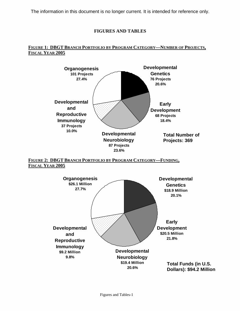
FIGURES AND TABLES
Figures and Tables-1
FIGURE 1: DBGT BRANCH PORTFOLIO BY PROGRAM CATEGORY—NUMBER OF PROJECTS, FISCAL YEAR 2005
Organogenesis Developmental 101 Projects Genetics
27.4% 76 Projects 20.6%
Developmental Early and Development
Reproductive 68 Projects Immunology 18.4%
37 Projects 10.0%
Total Number of Projects: 369
Developmental Neurobiology
87 Projects 23.6%
FIGURE 2: DBGT BRANCH PORTFOLIO BY PROGRAM CATEGORY—FUNDING, FISCAL YEAR 2005
Organogenesis Developmental $26.1 Million Genetics
27.7% $18.9 Million 20.1%
Early Developmental Development
$20.5 Million and 21.8% Reproductive
Immunology $9.2 Million Developmental
9.8% Neurobiology $19.4 Million Total Funds (in U.S.
20.6% Dollars): $94.2 Million
The information in this document is no longer current. It is intended for reference only.
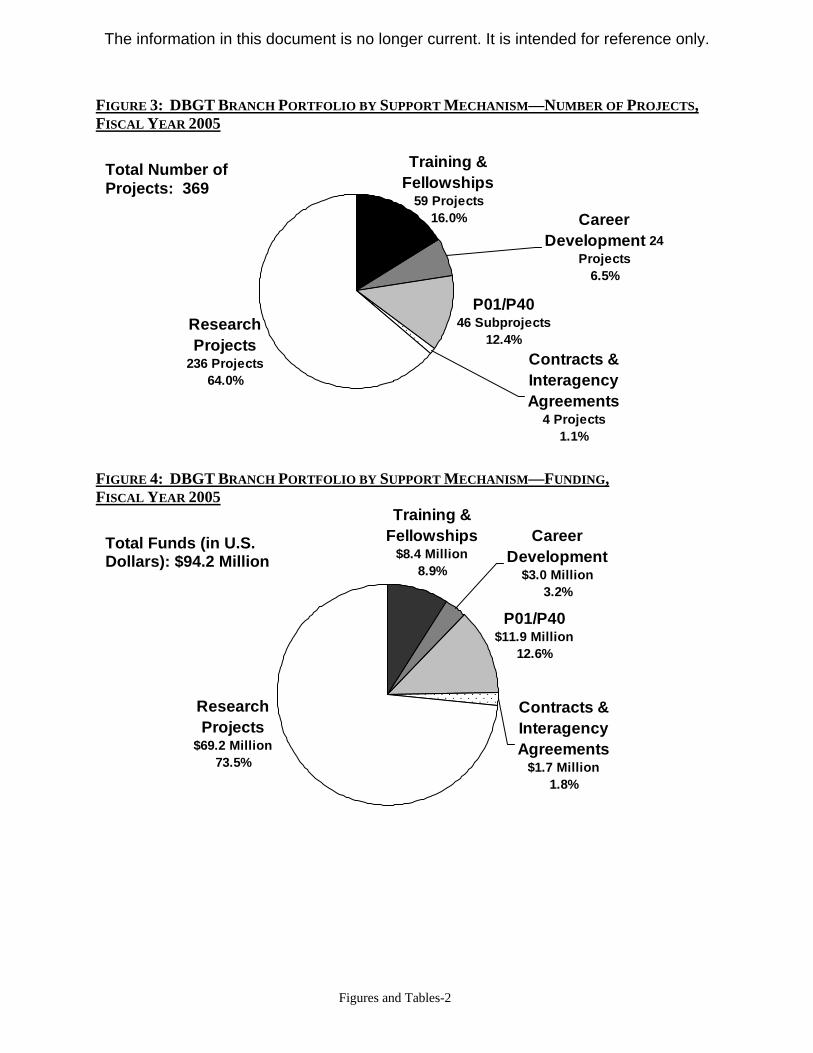
FIGURE 3: DBGT BRANCH PORTFOLIO BY SUPPORT MECHANISM—NUMBER OF PROJECTS, FISCAL YEAR 2005
Contracts & Interagency Agreements
4 Projects 1.1%
P01/P40 46 Subprojects
12.4% Research Projects
236 Projects 64.0%
Training & Fellowships
59 Projects 16.0% Career
Development 24 Projects
6.5%
Total Number of Projects: 369
FIGURE 4: DBGT BRANCH PORTFOLIO BY SUPPORT MECHANISM—FUNDING, FISCAL YEAR 2005
Total Funds (in U.S. Dollars): $94.2 Million
Research Projects
$69.2 Million 73.5%
Training & Fellowships Career
$8.4 Million Development 8.9% $3.0 Million
3.2%
P01/P40 $11.9 Million
12.6%
Contracts & Interagency Agreements
$1.7 Million 1.8%
Figures and Tables-2
The information in this document is no longer current. It is intended for reference only.

FIGURE 5: DBGT BRANCH FUNDS IN CURRENT AND CONSTANT DOLLARS, FISCAL YEAR 1996 THROUGH FISCAL YEAR 2005
$62.5 $62.4 $67.5
$77.4 $85.4
$93.1 $103.5 $104.7 $101.9
$94.2
$62.5 $61.3 $65.0 $72.4
$77.1 $81.2 $87.7 $85.2
$80.2 $71.8
$0
$20
$40
$60
$80
$100
$120
Am
ount
(In M
illio
ns o
f U.S
. Dol
lars
)
Current Dollars Constant Dollars
1996 1997 1998 1999 2000 2001 2002 2003 2004 2005 Fiscal Year
FIGURE 6: DBGT BRANCH PROJECTS BY PROGRAM CATEGORY—NUMBER OF PROJECTS, FISCAL YEAR 2001 THROUGH FISCAL YEAR 2005
Total Number of Grants 0 50 100 150 200 250 300 350 400 450 500
85
97
88
74
76
104
102
97
80
110
100
87
49
46
42
38
3768
120
116
2001
2002
83
101
Fisc
al Y
ear
2003
2004
97
396
369
104
2005 101
Developmental Genetics Early Development Developmental Neurobiology Developmental and ReproductiOrganogenesis
Figures and Tables-3
The information in this document is no longer current. It is intended for reference only.
441
ve Im
462
434munology
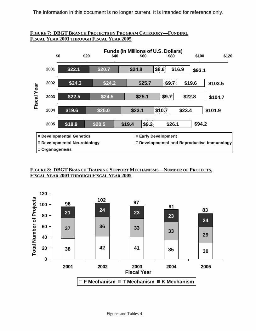
FIGURE 7: DBGT BRANCH PROJECTS BY PROGRAM CATEGORY—FUNDING, FISCAL YEAR 2001 THROUGH FISCAL YEAR 2005
Funds (In Millions of U.S. Dollars) $0 $20 $40 $60 $80 $100 $120
$24.3
$22.5
$19.6
$18.9
$20.7
$24.2
$24.5
$25.0
$20.5
$24.8
$25.7
$25.1
$23.1
$19.4
$22.1
$9.2
$10.7
$9.7
$9.7
$8.6 2001 $16.9 $93.1
2002
2003
2004
$19.6 $103.5
Fisc
al Y
ear
$22.8 $104.7
$23.4 $101.9
$26.1 $94.22005
Developmental Genetics Early Development Developmental Neurobiology Developmental and Reproductive Immunology Organogenesis
FIGURE 8: DBGT BRANCH TRAINING SUPPORT MECHANISMS—NUMBER OF PROJECTS, FISCAL YEAR 2001 THROUGH FISCAL YEAR 2005
120
Tota
l Num
ber o
f Pro
ject
s 102
24 9796100 91
23 83
37 36 33
21 2380
60
40 29
24
33
42 4120 38 35 30 0
2001 2002 2003 2004 2005 Fiscal Year
F Mechanism T Mechanism K Mechanism
Figures and Tables-4
The information in this document is no longer current. It is intended for reference only.
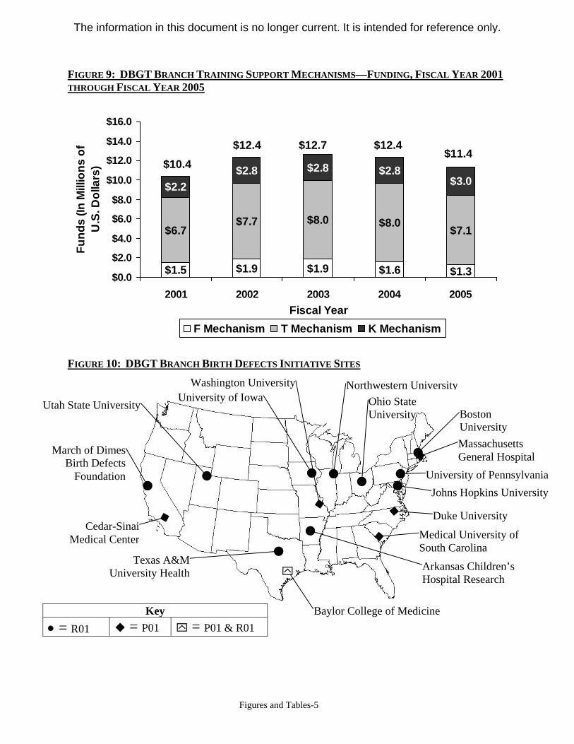
FIGURE 9: DBGT BRANCH TRAINING SUPPORT MECHANISMS—FUNDING, FISCAL YEAR 2001 THROUGH FISCAL YEAR 2005
$1.5 $1.9 $1.9 $1.6 $1.3
$6.7 $7.7 $8.0 $8.0 $7.1
$2.2 $2.8 $2.8 $2.8
$3.0
$0.0
$2.0
$4.0
$6.0
$8.0
$10.0
$12.0
$14.0
$16.0
Fund
s (In
Mill
ions
of
U.S
. Dol
lars
)
$11.4 $12.4$12.7$12.4
$10.4
2001 2002 2003 2004 2005 Fiscal Year
F Mechanism T Mechanism K Mechanism
FIGURE 10: DBGT BRANCH BIRTH DEFECTS INITIATIVE SITES
Key • = R01 � = P01 ` = P01 & R01
Washington University University of Iowa
March of Dimes Birth Defects
Foundation
Utah State University
Duke University
Medical University of South Carolina
Arkansas Children’s Hospital Research
Cedar-Sinai Medical Center
`
Baylor College of Medicine
Texas A&M University Health
Boston University Massachusetts General Hospital
University of Pennsylvania Johns Hopkins University
Ohio State University
Northwestern University
Figures and Tables-5
The information in this document is no longer current. It is intended for reference only.
TABLE 1: DBGT BRANCH BIRTH DEFECTS INITIATIVE PROJECTS
(Unless otherwise noted, projects listed below receive primary support from the NICHD.)
Principle Investigator Institution Project Title Genetic Epidemiology R01s Belmont, John W. Baylor College of
Medicine Genetic studies of common congenital heart defects
Eng, Charis Ohio State University Ret receptor polymorphisms & hirschprung disease
Hobbs, Charlotte a Arkansas Children's Hospital Research Institute
Genes, micronutrients, and homeobox related malformations
Kessler, John A. Northwestern University Role of chromosome 13q genes in neural tube defects
Manson, Jeanne University of Pennsylvania
Molecular epidemiology of hypospadias (Primary Support from the U.S. Environmental Protection Agency)
Mitchell, Allen Boston University Medical Campus
Pharmacogenetic determinants of human birth defects (Primary Support from the National Institute of Environmental Health Sciences)
Mitchell, Laura Texas A&M University Health Science Center
Maternal, fetal, and environmental causes of birth defects
Munger, Ronald G. Utah State University Nutrient biomarkers, genes, and orofacial clefts Murray, Jeffrey C. University of Iowa Comprehensive sequence evaluation of cleft lip
and palate (Primary Support from National Institute of Environmental Health Sciences)
Scott, Alan Johns Hopkins University
Snp discovery and analysis in craniofacial birth defects (Primary Support from National Institute of Dental and Craniofacial Research)
Shaw, Gary March of Dimes Birth Defects Foundation
Gene-environment interaction and human malformations (Primary Support from U.S. Environmental Protection Agency)
Program Projects Donahoe, Patricia K. Massachusetts General
Hospital Comparative genomics to correct human lung hypoplasia
Gitlin, Jonathan David Washington University Mechanisms of growth and the overgrowth syndromes
Gourdie, Robert G. Medical University of South Carolina
Patterning by invasive mesenchyme in the embryonic heart
Lee, Brendan Baylor College of Medicine
Genetic studies of common congenital heart defects (Primary Support from National Institute of Environmental Health Sciences)
McClay, David R. Duke University Neural tube defects Rimoin, David Cedars-Sinai Medical
Center The skeletal dysplasias
Figures and Tables-6
The information in this document is no longer current. It is intended for reference only.
APPENDIX A: DBGT BRANCH PERSONNEL
A. Tyl Hewitt, Ph.D., joined the staff of the DBGT Branch as a health scientist administrator in 1991. He was appointed as Branch chief in August 1997. Prior to joining the Branch, he was on the faculty of the Wilmer Eye Institute at the Johns Hopkins University School of Medicine. His postdoctoral training in connective tissue biochemistry at the National Institute of Dental Research (NIDR, now NIDCR) followed completion of his dissertation research at Emory University that focused on changes in cell-surface properties during limb development. In addition to his duties as Branch chief, he also currently serves as acting deputy director of the Center for Developmental Biology and Perinatal Medicine.
James N. Coulombe, Ph.D., joined the staff of the Branch in 2006. He received his doctorate from the University of California, Irvine, studying migration and differentiation of the neural crest. His postdoctoral research at the Oregon Health Sciences University examined target influences on neuropeptide expression in autonomic neurons. He joined the faculty of the Uniformed Services University of the Health Sciences teaching in the School of Medicine and the Graduate Neurosciences Program, and directing a laboratory studying neuropeptide expression during neuronal development. Dr. Coulombe continued to study influences on neurotransmitter expression as a staff scientist in the Neural Development Section of the NINDS. Immediately prior to joining the Branch, Dr. Coulombe served as a program director at NINDS. His current responsibilities include administration of programs on developmental genetics and developmental immunobiology.
Deborah B. Henken, Ph.D., joined the staff of the DBGT Branch in 1996. She is responsible for administering research in developmental neurobiology and related topics in genetics. She came to the NICHD after completing the NIH Grants Associate Program and has the distinction of being the Program’s last graduate. Prior to being in the Program, she was an intramural scientist at NINDS, where she studied biological responses to virus infection in the nervous system. A graduate of Swarthmore College in Pennsylvania, she received her doctorate degree from Dalhousie University, Halifax, Nova Scotia, in the area of nervous system regeneration and plasticity. Dr. Henken currently chairs the NICHD Neuroscience Steering Committee and is a past chair of the NIH Staff Training in Extramural Programs Committee. She is also the NICHD representative to a number of trans-NIH neuroscience initiatives, including the NIH Joint Neuroscience Training Program and Neuroscience Blueprint Committees, among others.
Lorette C. Javois, Ph.D., joined the staff of the DBGT Branch in 2000 and is responsible for administering the program on organogenesis, including the areas of limb development, somitogenesis, chondrogenesis, and myogenesis. She received her doctorate in biology from Purdue University, where she studied pattern formation using the developing vertebrate limb as a model system. She continued to study aspects of pattern formation using the coelenterate, hydra, while doing her postdoctoral training at the Developmental Biology Center at the University of California, Irvine. Prior to coming to the NICHD, she spent 14 years on the biology faculty at the Catholic University of America in Washington, D.C., conducting research on cellular and molecular pattern formation and teaching developmental biology and anatomy. Dr. Javois coordinates the Branch’s Birth Defects Initiative, is active on the TZCC, and serves as the
Appendix-1
The information in this document is no longer current. It is intended for reference only.
NICHD’s representative on a number of trans-NIH projects including KOMP and the Neuromouse Project Working Groups.
Steven L. Klein, Ph.D., was trained as a developmental neurobiologist and a developmental biologist. Prior to joining the DBGT Branch, he was on the faculty of the department of anatomy and cell biology at the University of Virginia School of Medicine, where he studied the cellular and molecular events involved in embryonic pattern formation, cell migration, cell interactions, and cell-fate determination. He is now responsible for the Branch’s Early Embryonic Development Program. Additionally, he leads the trans-NIH initiatives on Mouse Mutagenesis and Phenotyping for Developmental Defects, and on Xenopus Genetics and Genomics. He is also the chair of the NICHD’s Training Policy Committee, which helps to oversee the Institute’s training and career development programs.
Sally Moody, Ph.D., was trained in neuroscience, developmental neurobiology, and developmental biology. She served on an interagency personnel agreement assignment in the DBGT Branch for two years during which she managed the Developmental Genetics and Genomics Program. Her assignment ended January 2006. Prior to joining the DBGT Branch, she was on the faculty of the department of anatomy and cell biology at the University of Virginia School of Medicine. She is currently a full professor in the department of anatomy and cell biology at the George Washington University of School of Medicine and Health Sciences. Her laboratory studies the cellular and molecular events involved in early nervous system induction and patterning, and in cell-fate specification in the retina.
Appendix-2
The information in this document is no longer current. It is intended for reference only.
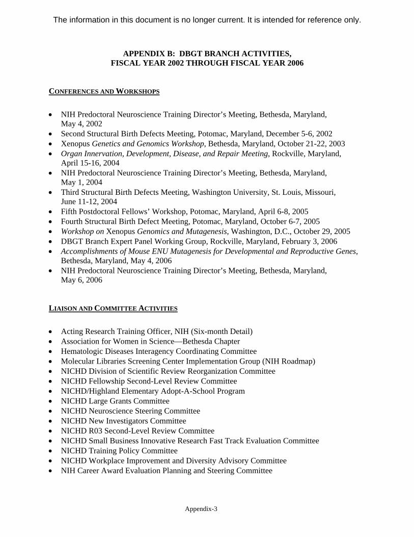
APPENDIX B: DBGT BRANCH ACTIVITIES, FISCAL YEAR 2002 THROUGH FISCAL YEAR 2006
CONFERENCES AND WORKSHOPS
• NIH Predoctoral Neuroscience Training Director’s Meeting, Bethesda, Maryland, May 4, 2002
• Second Structural Birth Defects Meeting, Potomac, Maryland, December 5-6, 2002 • Xenopus Genetics and Genomics Workshop, Bethesda, Maryland, October 21-22, 2003 • Organ Innervation, Development, Disease, and Repair Meeting, Rockville, Maryland,
April 15-16, 2004 • NIH Predoctoral Neuroscience Training Director’s Meeting, Bethesda, Maryland,
May 1, 2004 • Third Structural Birth Defects Meeting, Washington University, St. Louis, Missouri,
June 11-12, 2004 • Fifth Postdoctoral Fellows’ Workshop, Potomac, Maryland, April 6-8, 2005 • Fourth Structural Birth Defect Meeting, Potomac, Maryland, October 6-7, 2005 • Workshop on Xenopus Genomics and Mutagenesis, Washington, D.C., October 29, 2005 • DBGT Branch Expert Panel Working Group, Rockville, Maryland, February 3, 2006 • Accomplishments of Mouse ENU Mutagenesis for Developmental and Reproductive Genes,
Bethesda, Maryland, May 4, 2006 • NIH Predoctoral Neuroscience Training Director’s Meeting, Bethesda, Maryland,
May 6, 2006
LIAISON AND COMMITTEE ACTIVITIES
• Acting Research Training Officer, NIH (Six-month Detail) • Association for Women in Science—Bethesda Chapter • Hematologic Diseases Interagency Coordinating Committee • Molecular Libraries Screening Center Implementation Group (NIH Roadmap) • NICHD Division of Scientific Review Reorganization Committee • NICHD Fellowship Second-Level Review Committee • NICHD/Highland Elementary Adopt-A-School Program • NICHD Large Grants Committee • NICHD Neuroscience Steering Committee • NICHD New Investigators Committee • NICHD R03 Second-Level Review Committee • NICHD Small Business Innovative Research Fast Track Evaluation Committee • NICHD Training Policy Committee • NICHD Workplace Improvement and Diversity Advisory Committee • NIH Career Award Evaluation Planning and Steering Committee
Appendix-3
The information in this document is no longer current. It is intended for reference only.
• NIH Extramural Program Management Committee Workgroup on Advisory Council Activities
• NIH Joint Neuroscience Training Program • NIH Neuroscience Blueprint Coordinating Committee • NIH Neuroscience Blueprint Subcommittees
o Neuromouse o Neurobiology of Disease Course o Microarrays o Neurobiology Training o Neurodevelopment Planning o Gene Expression Nervous System Atlas (GENSAT)
• NIH New Investigators Committee • NIH Training Advisory Committee • Research Training Activities Coordinator, NIH (Six-month Detail) • Staff Training in Extramural Programs Committee • Trans-NIH Coordinating Committee for Lymphedema and Lymphatic Biology • Trans-NIH Evaluation Workgroup for the Multidisciplinary Clinical Research Career
Development K12 Program • Trans-NIH Genomics Working Group • Trans-NIH Knockout Mouse Project (KOMP) Working Group • Trans-NIH Mouse Transcriptome Project • Trans-NIH Xenopus Working Group • Trans-NIH Zebrafish Coordinating Committee
Appendix-4
The information in this document is no longer current. It is intended for reference only.
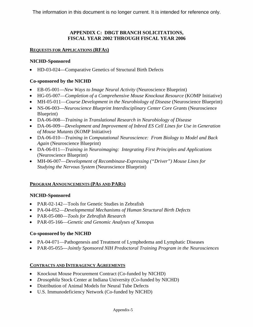
APPENDIX C: DBGT BRANCH SOLICITATIONS, FISCAL YEAR 2002 THROUGH FISCAL YEAR 2006
REQUESTS FOR APPLICATIONS (RFAS)
NICHD-Sponsored
• HD-03-024—Comparative Genetics of Structural Birth Defects
Co-sponsored by the NICHD
• EB-05-001—New Ways to Image Neural Activity (Neuroscience Blueprint) • HG-05-007—Completion of a Comprehensive Mouse Knockout Resource (KOMP Initiative) • MH-05-011—Course Development in the Neurobiology of Disease (Neuroscience Blueprint) • NS-06-003—Neuroscience Blueprint Interdisciplinary Center Core Grants (Neuroscience
Blueprint) • DA-06-008—Training in Translational Research in Neurobiology of Disease • DA-06-009—Development and Improvement of Inbred ES Cell Lines for Use in Generation
of Mouse Mutants (KOMP Initiative) • DA-06-010—Training in Computational Neuroscience: From Biology to Model and Back
Again (Neuroscience Blueprint) • DA-06-011—Training in Neuroimaging: Integrating First Principles and Applications
(Neuroscience Blueprint) • MH-06-007—Development of Recombinase-Expressing (“Driver”) Mouse Lines for
Studying the Nervous System (Neuroscience Blueprint)
PROGRAM ANNOUNCEMENTS (PAS AND PARS)
NICHD-Sponsored
• PAR-02-142—Tools for Genetic Studies in Zebrafish • PA-04-052—Developmental Mechanisms of Human Structural Birth Defects • PAR-05-080—Tools for Zebrafish Research • PAR-05-166—Genetic and Genomic Analyses of Xenopus
Co-sponsored by the NICHD
• PA-04-071—Pathogenesis and Treatment of Lymphedema and Lymphatic Diseases • PAR-05-055—Jointly Sponsored NIH Predoctoral Training Program in the Neurosciences
CONTRACTS AND INTERAGENCY AGREEMENTS
• Knockout Mouse Procurement Contract (Co-funded by NICHD) • Drosophila Stock Center at Indiana University (Co-funded by NICHD) • Distribution of Animal Models for Neural Tube Defects • U.S. Immunodeficiency Network (Co-funded by NICHD)
Appendix-5
The information in this document is no longer current. It is intended for reference only.
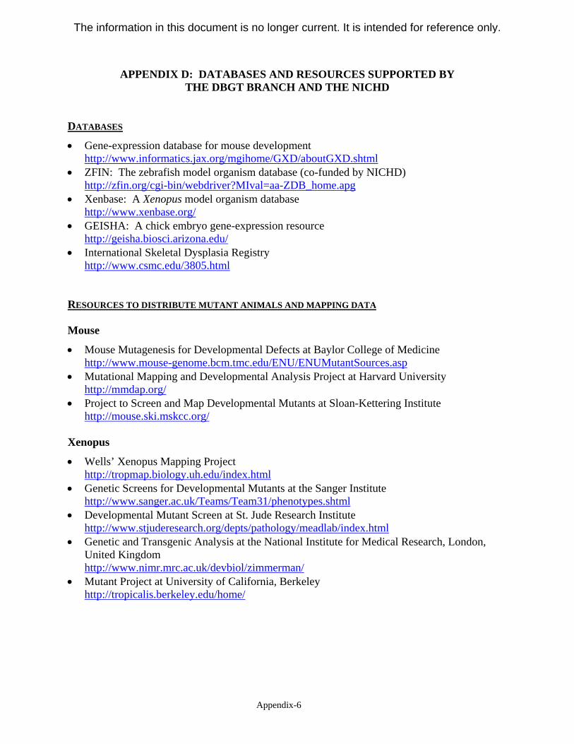
APPENDIX D: DATABASES AND RESOURCES SUPPORTED BY THE DBGT BRANCH AND THE NICHD
DATABASES
• Gene-expression database for mouse development http://www.informatics.jax.org/mgihome/GXD/aboutGXD.shtml
• ZFIN: The zebrafish model organism database (co-funded by NICHD) http://zfin.org/cgi-bin/webdriver?MIval=aa-ZDB_home.apg
• Xenbase: A Xenopus model organism database http://www.xenbase.org/
• GEISHA: A chick embryo gene-expression resource http://geisha.biosci.arizona.edu/
• International Skeletal Dysplasia Registry http://www.csmc.edu/3805.html
RESOURCES TO DISTRIBUTE MUTANT ANIMALS AND MAPPING DATA
Mouse
• Mouse Mutagenesis for Developmental Defects at Baylor College of Medicine http://www.mouse-genome.bcm.tmc.edu/ENU/ENUMutantSources.asp
• Mutational Mapping and Developmental Analysis Project at Harvard University http://mmdap.org/
• Project to Screen and Map Developmental Mutants at Sloan-Kettering Institute http://mouse.ski.mskcc.org/
Xenopus
• Wells’ Xenopus Mapping Project http://tropmap.biology.uh.edu/index.html
• Genetic Screens for Developmental Mutants at the Sanger Institute http://www.sanger.ac.uk/Teams/Team31/phenotypes.shtml
• Developmental Mutant Screen at St. Jude Research Institute http://www.stjuderesearch.org/depts/pathology/meadlab/index.html
• Genetic and Transgenic Analysis at the National Institute for Medical Research, London, United Kingdom http://www.nimr.mrc.ac.uk/devbiol/zimmerman/
• Mutant Project at University of California, Berkeley http://tropicalis.berkeley.edu/home/
Appendix-6
The information in this document is no longer current. It is intended for reference only.
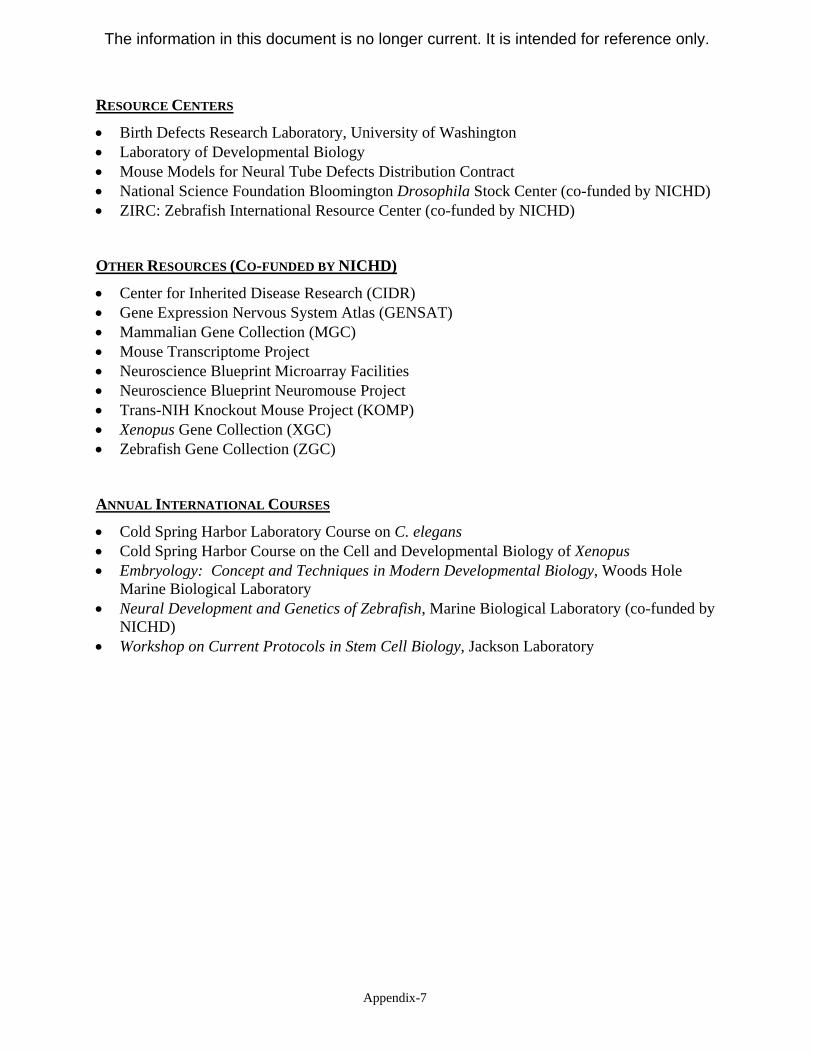
RESOURCE CENTERS
• Birth Defects Research Laboratory, University of Washington • Laboratory of Developmental Biology • Mouse Models for Neural Tube Defects Distribution Contract • National Science Foundation Bloomington Drosophila Stock Center (co-funded by NICHD) • ZIRC: Zebrafish International Resource Center (co-funded by NICHD)
OTHER RESOURCES (CO-FUNDED BY NICHD)
• Center for Inherited Disease Research (CIDR) • Gene Expression Nervous System Atlas (GENSAT) • Mammalian Gene Collection (MGC) • Mouse Transcriptome Project • Neuroscience Blueprint Microarray Facilities • Neuroscience Blueprint Neuromouse Project • Trans-NIH Knockout Mouse Project (KOMP) • Xenopus Gene Collection (XGC) • Zebrafish Gene Collection (ZGC)
ANNUAL INTERNATIONAL COURSES
• Cold Spring Harbor Laboratory Course on C. elegans • Cold Spring Harbor Course on the Cell and Developmental Biology of Xenopus • Embryology: Concept and Techniques in Modern Developmental Biology, Woods Hole
Marine Biological Laboratory • Neural Development and Genetics of Zebrafish, Marine Biological Laboratory (co-funded by
NICHD) • Workshop on Current Protocols in Stem Cell Biology, Jackson Laboratory
Appendix-7
The information in this document is no longer current. It is intended for reference only.
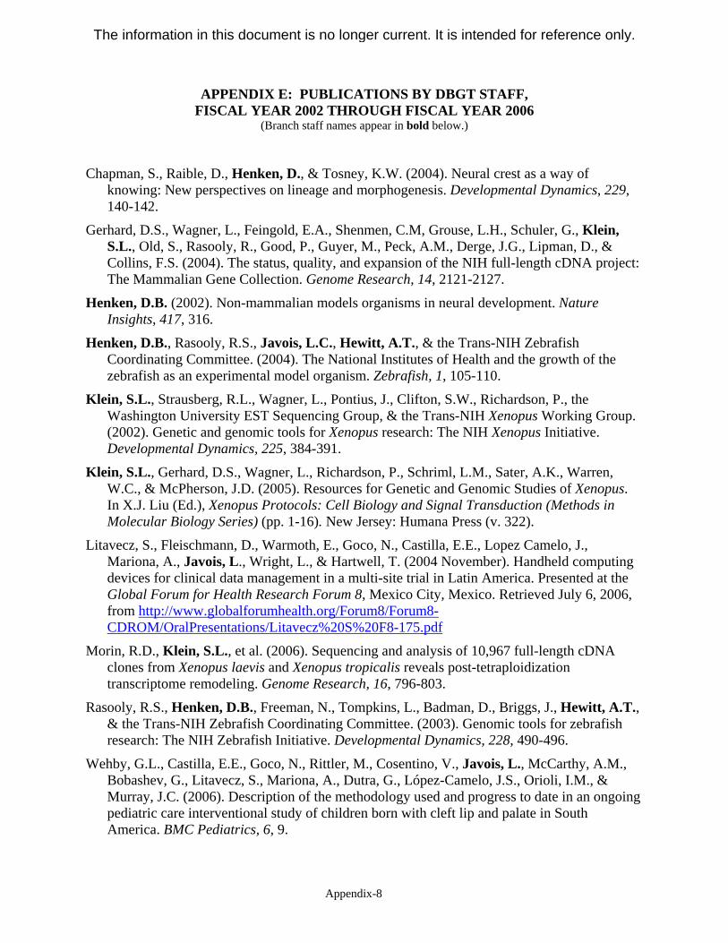
APPENDIX E: PUBLICATIONS BY DBGT STAFF, FISCAL YEAR 2002 THROUGH FISCAL YEAR 2006
(Branch staff names appear in bold below.)
Chapman, S., Raible, D., Henken, D., & Tosney, K.W. (2004). Neural crest as a way of knowing: New perspectives on lineage and morphogenesis. Developmental Dynamics, 229, 140-142.
Gerhard, D.S., Wagner, L., Feingold, E.A., Shenmen, C.M, Grouse, L.H., Schuler, G., Klein, S.L., Old, S., Rasooly, R., Good, P., Guyer, M., Peck, A.M., Derge, J.G., Lipman, D., & Collins, F.S. (2004). The status, quality, and expansion of the NIH full-length cDNA project: The Mammalian Gene Collection. Genome Research, 14, 2121-2127.
Henken, D.B. (2002). Non-mammalian models organisms in neural development. Nature Insights, 417, 316.
Henken, D.B., Rasooly, R.S., Javois, L.C., Hewitt, A.T., & the Trans-NIH Zebrafish Coordinating Committee. (2004). The National Institutes of Health and the growth of the zebrafish as an experimental model organism. Zebrafish, 1, 105-110.
Klein, S.L., Strausberg, R.L., Wagner, L., Pontius, J., Clifton, S.W., Richardson, P., the Washington University EST Sequencing Group, & the Trans-NIH Xenopus Working Group. (2002). Genetic and genomic tools for Xenopus research: The NIH Xenopus Initiative. Developmental Dynamics, 225, 384-391.
Klein, S.L., Gerhard, D.S., Wagner, L., Richardson, P., Schriml, L.M., Sater, A.K., Warren, W.C., & McPherson, J.D. (2005). Resources for Genetic and Genomic Studies of Xenopus. In X.J. Liu (Ed.), Xenopus Protocols: Cell Biology and Signal Transduction (Methods in Molecular Biology Series) (pp. 1-16). New Jersey: Humana Press (v. 322).
Litavecz, S., Fleischmann, D., Warmoth, E., Goco, N., Castilla, E.E., Lopez Camelo, J., Mariona, A., Javois, L., Wright, L., & Hartwell, T. (2004 November). Handheld computing devices for clinical data management in a multi-site trial in Latin America. Presented at the Global Forum for Health Research Forum 8, Mexico City, Mexico. Retrieved July 6, 2006, from http://www.globalforumhealth.org/Forum8/Forum8CDROM/OralPresentations/Litavecz%20S%20F8-175.pdf
Morin, R.D., Klein, S.L., et al. (2006). Sequencing and analysis of 10,967 full-length cDNA clones from Xenopus laevis and Xenopus tropicalis reveals post-tetraploidization transcriptome remodeling. Genome Research, 16, 796-803.
Rasooly, R.S., Henken, D.B., Freeman, N., Tompkins, L., Badman, D., Briggs, J., Hewitt, A.T., & the Trans-NIH Zebrafish Coordinating Committee. (2003). Genomic tools for zebrafish research: The NIH Zebrafish Initiative. Developmental Dynamics, 228, 490-496.
Wehby, G.L., Castilla, E.E., Goco, N., Rittler, M., Cosentino, V., Javois, L., McCarthy, A.M., Bobashev, G., Litavecz, S., Mariona, A., Dutra, G., López-Camelo, J.S., Orioli, I.M., & Murray, J.C. (2006). Description of the methodology used and progress to date in an ongoing pediatric care interventional study of children born with cleft lip and palate in South America. BMC Pediatrics, 6, 9.
Appendix-8
The information in this document is no longer current. It is intended for reference only.
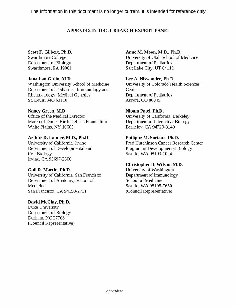
APPENDIX F: DBGT BRANCH EXPERT PANEL
Scott F. Gilbert, Ph.D. Swarthmore College Department of Biology Swarthmore, PA 19081
Jonathan Gitlin, M.D. Washington University School of Medicine Department of Pediatrics, Immunology and Rheumatology, Medical Genetics St. Louis, MO 63110
Nancy Green, M.D. Office of the Medical Director March of Dimes Birth Defects Foundation White Plains, NY 10605
Arthur D. Lander, M.D., Ph.D. University of California, Irvine Department of Developmental and Cell Biology Irvine, CA 92697-2300
Gail R. Martin, Ph.D. University of California, San Francisco Department of Anatomy, School of Medicine San Francisco, CA 94158-2711
David McClay, Ph.D. Duke University Department of Biology Durham, NC 27708 (Council Representative)
Anne M. Moon, M.D., Ph.D. University of Utah School of Medicine Department of Pediatrics Salt Lake City, UT 84112
Lee A. Niswander, Ph.D. University of Colorado Health Sciences Center Department of Pediatrics Aurora, CO 80045
Nipam Patel, Ph.D. University of California, Berkeley Department of Interactive Biology Berkeley, CA 94720-3140
Philippe M. Soriano, Ph.D. Fred Hutchinson Cancer Research Center Program in Developmental Biology Seattle, WA 98109-1024
Christopher B. Wilson, M.D. University of Washington Department of Immunology School of Medicine Seattle, WA 98195-7650 (Council Representative)
Appendix-9
The information in this document is no longer current. It is intended for reference only.