development of a sensitive, selective electrochemical immunoassay for progesterone in cow's...
TRANSCRIPT
Development of a sensitive, selectiveelectrochemical immunoassay for progesterone incow's milk based on a disposable screen-printed
amperometric biosensor
R. M. Pemberton,a J. P. Harta* and J. A. Foulkesb
aFaculty of Applied Sciences, University of the West of England, Frenchay Campus, Coldharbour Lane,Bristol, BS16 1QY, U.K.
bRidgeway Science Ltd., Rodmore Mill Farm, Alvington, Gloucestershire, GL15 6AH, U.K.
(Received 18 September 1997; in revised form 6 November 1997)
AbstractÐA disposable amperometric biosensor, based on a screen-printed carbon electrode (SPCE) coatedwith antibodies, has been prepared and evaluated for measuring progesterone in cow's milk. This immuno-sensor was employed in a competitive assay involving progesterone labelled with alkaline phosphatase. In-itially, phenylphosphate was used as the enzyme substrate and the product of the reaction, phenol, wasdetected by chronoamperometry at +0.70 V vs saturated calomel (SCE). However, this approach provedunsatisfactory, since an unidenti®ed component of cow's milk adsorbed to the electrode and underwent avariable oxidation at this potential. This resulted in a calibration curve which did not have the character-istic sigmoidal shape. An alternative enzyme substrate, 4-aminophenolphosphate, was therefore examined,the product of the reaction, 4-aminophenol, being detectable at +0.20 V vs SCE; at this lower potential,the interferent in milk did not undergo oxidation. Consistently low background currents were obtained,enabling construction of a sigmoidal-shaped calibration curve for progesterone concentration in the milksamples investigated. Progesterone levels determined by the proposed electrochemical immunoassay com-pared favourably with a colorimetric immunoassay method. # 1998 Elsevier Science Ltd. All rightsreserved
Key words: screen-printed, antibodies, milk progesterone, competitive immunoassay, chronoamperometry.
INTRODUCTION
In the cattle breeding industry, where arti®cial inse-
mination techniques are employed, the successful
prediction of oestrus onset leads to considerablecost saving in herd management [1]. One way of
detecting oestrus onset is to monitor the progester-one levels in milk, optimum fertility rates being
achieved when insemination is performed three days
after the progesterone level has fallen to below5 ng mlÿ1 of whole milk [2, 3]. On-site monitoring of
cow's milk for progesterone levels is highly desir-
able, both for convenience and for economicreasons and such measurements have been con-
ducted to date using immunoassay methods invol-
ving colorimetry [4]. The present study describes an
alternative approach involving electrochemical
measurement with a view to developing a disposa-
ble amperometric biosensor.
Electrochemical immunoassays have been investi-
gated for analytes other than progesterone. For
example, electrochemical detection, following liquid
chromatography or ¯ow injection, has been per-
formed on solutions which have undergone a com-
petitive assay [5±7], and similar approaches have
been employed with voltammetric detection [8].
Recently, workers have reported immobilisation
onto membranes connected over a carbon transdu-
cer, then removed after each analysis [9]. In all of
these cases, the biological element was fabricated
separately from the transducer. In contrast, a ®nal
biosensor for use on the farm should ideally be
Electrochimica Acta, Vol. 43, No. 23, pp. 3567±3574, 1998# 1998 Published by Elsevier Science Ltd. All rights reserved
Printed in Great Britain0013±4686/98 $19.00+0.00PII: S0013-4686(98)00104-2
*Author to whom correspondence should be addressed.
3567
robust, reliable and low cost, in which case a moreintegrated fabrication would be desirable. Hence,
with a minimum of instrumentation and manipu-lation steps, the whole biosensor, consisting of thetransducer with the immobilised biorecognition el-
ement could be disposed of after a single use.A previous preliminary study reported the fabri-
cation of a biosensor based on a monoclonal anti-
progesterone antibody immobilised directly onto ascreen-printed carbon electrode [10] and demon-strated that it might be feasible to monitor pro-
gesterone levels over the range required in cattlebreeding applications. The device employed alkalinephosphatase-labelled progesterone in a competitiveenzyme immunoassay and was e�ective in aqueous
bu�ered solutions but had not been applied to de-terminations in cow's milk.In the present study, the possibility was explored
of determining progesterone in milk samples with adetection limit of 5 ng mlÿ1 or lower. As milk is acomplex mixture of biomolecules, many of which
are electroactive, it is perhaps not surprising thatproblems can arise when trace determinations arerequired. Our approaches towards overcoming such
di�culties and achieving the desired selectivity andsensitivity for a progesterone biosensor aredescribed in this paper.
EXPERIMENTAL
Chemicals and reagents
Unless stated otherwise, chemicals were AR-
grade, purchased from Merck, Poole, Dorset. Allsolutions were prepared in water drawn from aMilli-Q Water Puri®cation System (Millipore U.K.,Watford, Herts.). Bu�er solutions: carbonate coat-
ing bu�er (CB), pH 9.6 containing 0.015 mol dmÿ3
Na2CO3 and 0.035 mol dmÿ3 NaHCO3;100 mmol dmÿ3 diethanolamine±HCl bu�er (DB),
pH 7.2, or pH 9.8 containing 10ÿ2 mol dmÿ3
MgCl2. Phenol (Fisons Scienti®c Equipment,Loughborough) and 4-aminophenol (Sigma-Aldrich
Company, Poole, Dorset) were dissolved, respect-ively, in water or 0.1 mol dmÿ3 NaOH before ®naldilution in bu�er.The puri®ed IgG fraction of a polyclonal rabbit
antibody against sheep IgG was purchased fromSigma-Aldrich (rIg). Crude culture ¯uid containinga sheep monoclonal antibody (mAb) against
progesterone [11], and a solution containing pro-gesterone labelled with alkaline phosphatase (AP-prog) were kindly provided by Ridgeway Science.
Unlabelled progesterone (formula wt = 314.5), p-nitrophenylphosphate and phenylphosphate (diso-dium salt) were purchased from Sigma-Aldrich; 4-
aminophenylphosphate (monosodium salt) was pur-chased from ICN Biomedicals, Thame, Oxon.Samples of cow's milk without progesterone and
with progesterone were obtained by sampling at
various times throughout the oestrus cycle. Thesewere kindly provided by Ridgeway Science.
Preparation of sensors
Screen-printed carbon electrodes (SPCEs), havinga 3 mm� 3 mm working area were printed fromD14 carbon ink onto PVC substrate at Gwent
Electronic Materials (Mamhilad, Gwent) asdescribed previously [12]. SPCEs were coated indivi-dually with antibodies according to the optimum
conditions established previously [10]. Brie¯y, mAb,in DB, was immobilised onto the working area viaa layer of rIg adsorbed previously in CB. These
antibody-coated SPCEs (mAb-SPCEs) were used onthe day of preparation.
Cyclic voltammetry and chronoamperometry
All electrochemical procedures were carried out
using an E612 VA Scanner and an E611 VADetector (Metrohm, Herisau), combined with a JJInstuments chart recorder (Fareham, Hampshire).In order to allow operation within small volumes of
enzyme substrate solution, a dual electrode setupwas adopted throughout, using a screen-printedworking electrode in conjunction with a saturated
calomel counter/reference electrode [10]. Thisallowed cyclic voltammetric and chronoampero-metric analyses to be performed using 0.5 ml
volumes of analyte solution contained within coni-cal polypropylene cells.
Calibration studies on cow's milk
Progesterone standards were prepared initially as
a 1 mg mlÿ1 solution in methanol. Various aliquotsof this were transferred to glass tubes and evapor-ated under N2, before subsequent dilution into buf-fer solution or cow's milk.
Colorimetric and chronoamperometric calibrationstudies were carried out using freshly-preparedmAb-SPCEs. These were ®rst rinsed in DB, pH 7.2,
before the addition of 8 ml volumes of a mixturecomprising 3 ml unlabelled progesterone (standardsolution or milk sample) plus 5 ml AP-prog sol-
ution.For colorimetric determinations, mAb-SPCEs
were then rinsed twice in DB, pH 9.8, dried andlocated individually into 96-well immunoassay plate
microwells (Dynatech Immulon, Farenheit LabSupplies, Milton Keynes). Each well then received120 ml pH 9.8 DB containing 1 mg mlÿ1 p-nitrophe-
nylphosphate, prior to 60 min incubation at RT.The presence of p-nitrophenol was determined, fol-lowing removal of mAb-SPCEs from the wells, by
measuring absorbance at 405 nm with an AnthosHTII plate reader (Labtech International, Uck®eld,East Sussex).
For calibration by chronoamperometry, the bio-sensors were washed in DB, pH 9.8, then placedinto 0.5 ml DB, pH 9.8, or into the same bu�ercontaining 1 mg mlÿ1 phenylphosphate or 4-amino-
R. M. Pemberton et al.3568
phenylphosphate, as speci®ed. Following a 15 minincubation period, chronoamperometry was per-
formed by stepping from open circuit to +700 mV(substrate, phenyl phosphate) or +200 mV (sub-strate, 4-aminophenylphosphate). The current±time
response was recorded and peak current (ip)measured; calibration graphs were constructed byplotting ip against progesterone concentration.
RESULTS AND DISCUSSION
Electrochemical immunoassay for progesterone in
cow's milk using phenylphosphate as substrate
In a previous preliminary study [10], an electro-chemical immunoassay for the measurement of pro-gesterone was described and optimised usingprogesterone standards in aqueous bu�er solutions;
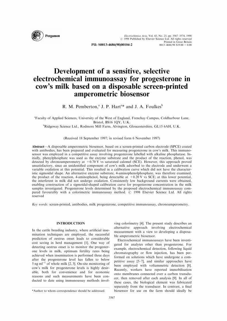
the scheme for this competitive assay is shown inFig. 1. Our present studies began by performingthis immunoassay, involving our antibody based
screen-printed carbon electrodes (mAb-SPCEs), onmilk samples containing known concentrations ofprogesterone and reading these against a calibration
curve constructed from standard progesterone sol-utions in bu�er. Figure 2(a) shows the typical sig-moidal shape expected for a competitive
immunoassay. The current responses obtained forthe milk samples were referred to this plot and thecalculated progesterone concentrations are shown inTable 1, together with the actual concentrations.
Clearly, there is some discrepancy at all but the
lowest concentrations.
As the calibration graph in Fig. 2(a) had been
constructed from data obtained on bu�ered pro-
gesterone standards, the possibility was explored
that, despite the inclusion of a wash step (Fig. 1),
some matrix e�ects from the milk were responsible
for the discrepancy shown in Table 1. Therefore, a
calibration plot was constructed from a single pro-
gesterone-free milk sample, divided into aliquots,
and spiked with increasing known concentrations of
the analyte [Fig. 2(b)]. There are two main features
to note: ®rst all the currents are of greater magni-
tude than those obtained for corresponding concen-
trations shown in Fig. 2(a); secondly, an increase in
current appears for concentrations between 10 and
50 ng mlÿ1.A possible explanation for this ``V''-shaped cali-
bration curve was that at concentrations above
10 ng mlÿ1 (unlabelled progesterone) some seques-
tration had occurred during step 1 (Fig. 1), result-
ing in more enzyme-labelled progesterone being
bound to mAb and a corresponding increase in
phenol production. In order to test this hypothesis,
the assay was repeated using a colorimetric rather
than an electrochemical readout, in order to focus
on the antigen±antibody interaction. Figure 3
shows that a characteristic dose±response curve was
obtained for progesterone standards prepared in
milk, indicating the absence of sequestration.
Fig. 1. Competitive electrochemical enzymeimmunoassay for progesterone. Step 1 = competitive binding; step 2 = wash;
step 3 = product formation; step 4 = chronoamperometry. [Prog.AP = alkaline phosphatase-labelled progesterone;
Prog = unlabelled progesterone; ArOH= dephosphorylated phenol or 4-aminophenol].
Development of an electrochemical immunoassay 3569

An alternative possibility was that the ``V'' shape
observed in Fig. 2(b) related to the electrochemicalmeasurement and that since the currents were
obtained by chronoamperometry at an applied po-
tential of +0.7 V vs SCE, some electroactive com-
ponent of milk was being oxidised at the same
potential as the enzymatically produced phenol
(Fig. 1, step 4). In order to test this hypothesis, cyc-lic voltammetry was performed in the absence of
phenylphosphate (and therefore phenol) on mAb-
SPCEs following the incubation (with milk and
bu�er containing the same progesterone concen-
trations) and wash steps. Figure 4(a) shows that thevoltammogram for an mAb-SPCE exposed to milk
exhibits a large unresolved anodic peak around
+0.6 to +0.7 V vs SCE. This contrasts with vol-
tammograms for a mAb-SPCE [Fig. 4(b); a smallpeak occurs in this potential region, due to the
mAb/antigen coating] and a mAb-free SPCE
[Fig. 4(c)] exposed to bu�er, demonstrating that the®rst peak must have resulted from an adsorbed
electroactive compound present in milk. Adsorption
of the interfering substance in milk might beexpected to occur mainly at the SPCE itself and not
on the Ab-paratope (antigen binding site) which is
borne out by the results for milk shown in Fig. 3.This e�ect on its own would explain the observed
increases in the magnitude of current in milk over
bu�er, but not the ``V''-shaped plot shown inFig. 2(b).
The next consideration was that the nature of the
problem related to the molecular size of the electro-
active interference in milk, i.e., the ``V'' e�ect mayarise because displacement of a large antigen-
enzyme conjugate by a smaller unlabelled antigen
would allow more of the interferent to di�usethrough to the SPCE surface. In order to test this
possibility, the immunoassay was performed on
milk samples as described above, but phenylpho-sphate was eliminated from step 3 so that no phe-
nol was present during step 4 (Fig. 1). Figure 5
indeed shows that currents increased with increasingunlabelled progesterone (closed circles) and that
Fig. 2. Chronoamperometric calibration plots for standard
progesterone solutions prepared in (a) DB, pH 7.2 and (b)
cow's milk. ip values are means (2s.e.m.) for n= 3 elec-
trodes.
Fig. 3. Colorimetric calibration plot for standard pro-
gesterone solutions prepared in cow's milk. Absorbance
values are means (2s.e.m.) for n= 3 microwells/mAb-
SPCEs.
Table 1.
Comparison of actual values for progesterone concen-
tration in milk samples with values obtained by chron-
oamperometric calibration in bu�er
Progesterone concentration (ng/ml)
Sample Actual Obtained from calibration curve
A 1.2 1.4
B 1.0 1.4
C 27.0 7.9
D 31.6 6.3
E 9.7 35.5
F 6.3 28.2
R. M. Pemberton et al.3570

this phenomenon did not occur following incu-
bation in bu�er (open circles).
At this stage, an attempt was made to eliminate
the electroactive interferent from the SPCE surface
prior to the measurement step. Although the inter-ferent was unidenti®ed it was probably hydrophobic
in nature as it seemed to be retained during thewash step with aqueous bu�ers. Therefore, a varietyof solvents were introduced into the wash step
(Fig. 1, step 2) including methanol, dimethylforma-mide and 1.4-dioxan, and also a solution of Tween;none of these eliminated the chronoamperometric
background current.
Electrochemical immunoassay for progesterone in
cow's milk using 4-aminophenylphosphate as enzyme
substrate
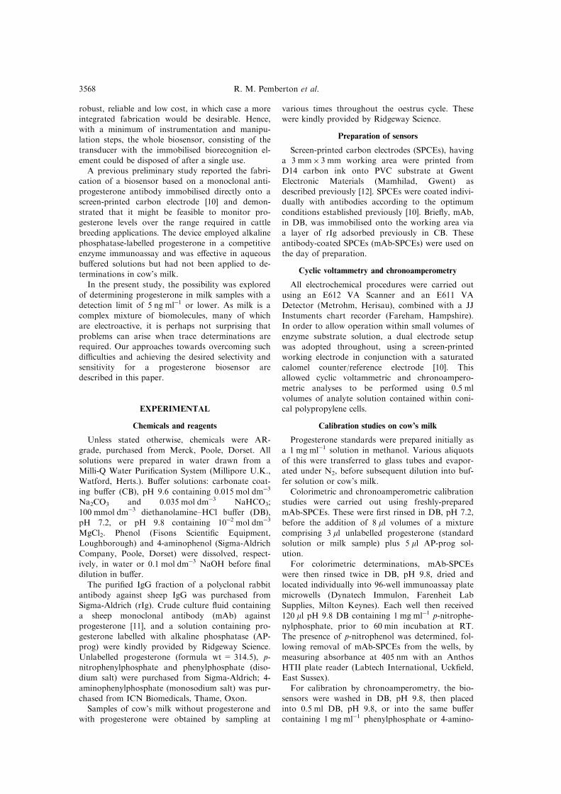
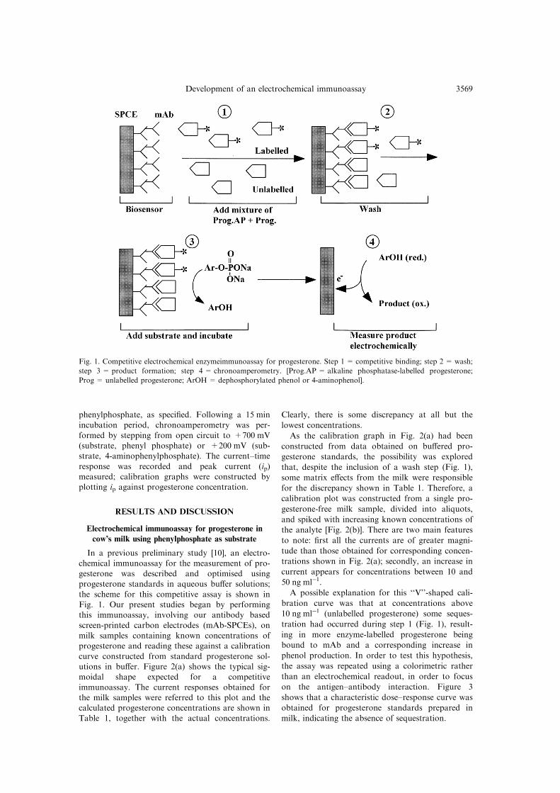
Since the electroactive interferent was oxidised at
around +0.6 V vs SCE [Fig. 4(a)], the possibilitywas considered that operating the immunosensor atpotentials well below this value could achieve thedesired selectivity. This approach would require an
alternative enzyme substrate.4-aminophenylphosphate, which is converted to
4-aminophenol by the antigen-label, alkaline phos-
phatase, was a good candidate. Figure 6(a) showsthe cyclic voltammogram of 4-aminophenol andFig. 6(b) its precursor 4-aminophenylphosphate;
Fig. 6(c) and (d) show the voltammograms of phe-nol and phenylphosphate for comparison. It is clearthat the oxidation peak for 4-aminophenol occurs
at a much lower potential than phenol and a valueof +0.2 V vs SCE was selected since it would pro-vide maximum sensitivity but minimum interferencefrom the milk [see Fig. 4(a)]. This potential was
Fig. 4. Cyclic voltammograms performed in DB, pH 9.8 containing 10ÿ2 mol dmÿ3 MgCl2 for mAb-SPCEs incubated in
(a) AP-prog in milk or (b) AP-prog in bu�er and (c) untreated SPCEs (mAb-free). Scan rate = 20 mV sÿ1.
Fig. 5. Chronoamperometric background response (mean
ip2s.e.m.) for mAb-SPCEs (n= 3) incubated with AP-
prog plus standard progesterone solutions prepared in
milk (*) or in bu�er (w).
Development of an electrochemical immunoassay 3571
also su�ciently low that the 4-aminophenylpho-
sphate precursor would not undergo oxidation at
the SPCE [Fig. 6(b)].
In order to establish that 4-aminophenol could be
detected by chronoamperometry over a wide con-
centration range, without passivating the SPCEs, a
calibration study was performed using individual
SPCEs for each measurement. A linear calibration
plot was indeed obtained over the concentration
range 10ÿ2 to 10ÿ4 mol dmÿ3 when an applied po-
tential of +0.2 V was employed. Initial evaluation
of mAb-SPCEs for susceptibility to milk interfer-
ents at +0.2 V was performed by examining back-
ground currents following incubation of the
immunosensor in milk, with increasing unlabelled
progesterone concentrations (enzyme substrate was
absent from the bu�er during the chronoampero-
metric measurement step). Background currents
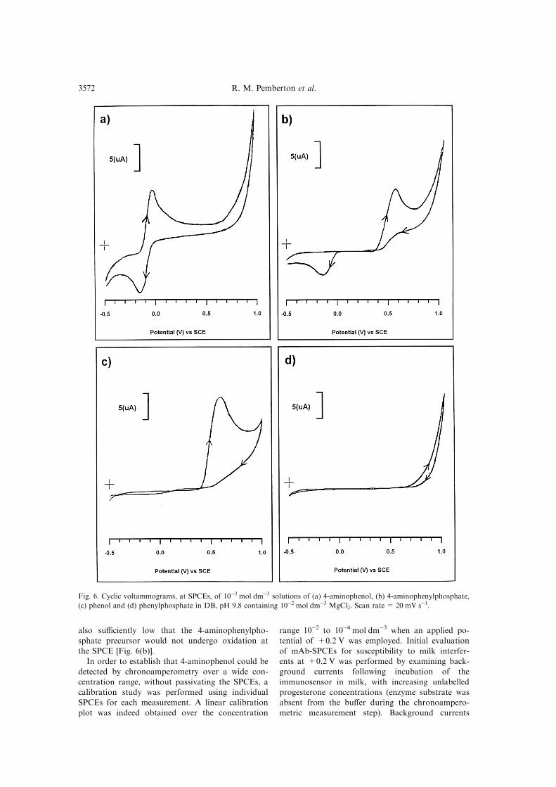
Fig. 6. Cyclic voltammograms, at SPCEs, of 10ÿ3 mol dmÿ3 solutions of (a) 4-aminophenol, (b) 4-aminophenylphosphate,
(c) phenol and (d) phenylphosphate in DB, pH 9.8 containing 10ÿ2 mol dmÿ3 MgCl2. Scan rate = 20 mV sÿ1.
R. M. Pemberton et al.3572
were generally less than 300 nA and, most impor-
tantly, did not vary over the concentration range 0±500 ng mlÿ1 unlabelled progesterone.
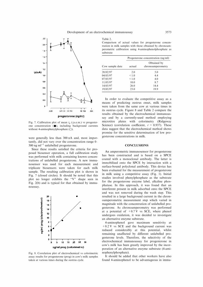
Since these results satis®ed the criteria for pro-
posed biosensor operation, a full calibration studywas performed with milk containing known concen-
trations of unlabelled progesterone. A new immu-
nosensor was used for each measurement and
triplicate biosensors were taken for each milk
sample. The resulting calibration plot is shown in
Fig. 7 (closed circles). It should be noted that thisplot no longer exhibits the ``V'' shape seen in
Fig. 2(b) and is typical for that obtained by immu-
noassay.
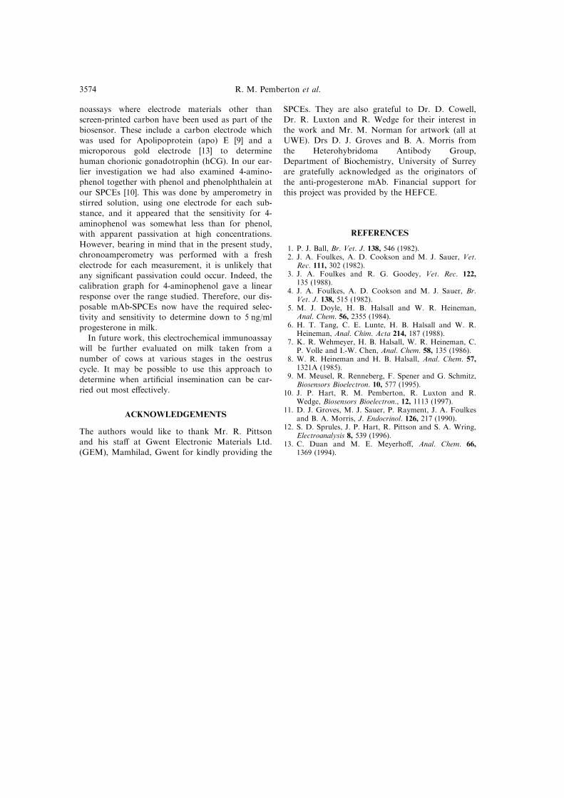
In order to evaluate the competitive assay as a
means of predicting oestrus onset, milk sampleswere taken from the same cow at various times inits oestrus cycle. Figure 8 and Table 2 compare theresults obtained by the electrochemical immunoas-
say and by a currently-used method employingmicrotitre plates with colorimetry (RidgewayScience) (correlation coe�cient, r= 0.937). These
data suggest that the electrochemical method showspromise for the sensitive determination of low pro-gesterone concentrations in milk.
CONCLUSIONS
An amperometric immunosensor for progesteronehas been constructed and is based on a SPCEcoated with a monoclonal antibody. The latter is
immobilised onto the SPCE by interaction with asurface-bound polyclonal antibody. This device hasbeen evaluated for the measurement of progesteronein milk using a competitive assay (Fig. 1). Initial
studies involved phenylphosphate as the substratefor the progesterone enzyme label, alkaline phos-phatase. In this approach, it was found that an
interferent present in milk adsorbed onto the SPCEand was not removed during the wash step. Thisresulted in a large background current in the chron-
oamperometric measurement step which varied inmagnitude with the concentration of unlabelled pro-gesterone. As chronoamperometry was performedat a potential of +0.7 V vs SCE, where phenol
undergoes oxidation, it was decided to investigatean alternative enzyme substrate.4-aminophenol gave maximum sensitivity at
+0.2 V vs SCE and the background current wasreduced considerably at this potential, whilstremaining una�ected by di�erent unlabelled pro-
gesterone levels. Therefore, the selectivity of theelectrochemical immunoassay for progesterone incow's milk has been greatly improved by the incor-
poration of an alternative enzyme substrate (4-ami-nophenylphosphate).It should be added that other workers have also
found 4-aminophenol to be advantageous in immu-
Fig. 7. Calibration plot of mean ip (2s.e.m.) vs progester-
one concentration (*), including background currents
without 4-aminophenylphosphate (w).
Fig. 8. Correlation plot of electrochemical vs colorimetric
assay results for progesterone (prog) in cow's milk samples
taken at various times during the oestrus cycle.
Table 2.
Comparison of actual values for progesterone concen-
tration in milk samples with those obtained by chronoam-
perometric calibration using 4-aminophenolphosphate as
substrate
Progesterone concentration (ng/ml)
Cow sample date actual
Obtained by
chronoamperometry
28/02/97 2.0 5.0
04/03/97 <1.0 4.4
07/03/97 <1.0 4.0
11/03/97 10.0 8.7
14/03/97 26.0 14.4
19/03/97 23.0 19.9
Development of an electrochemical immunoassay 3573
noassays where electrode materials other thanscreen-printed carbon have been used as part of the
biosensor. These include a carbon electrode whichwas used for Apolipoprotein (apo) E [9] and amicroporous gold electrode [13] to determine
human chorionic gonadotrophin (hCG). In our ear-lier investigation we had also examined 4-amino-phenol together with phenol and phenolphthalein at
our SPCEs [10]. This was done by amperometry instirred solution, using one electrode for each sub-stance, and it appeared that the sensitivity for 4-
aminophenol was somewhat less than for phenol,with apparent passivation at high concentrations.However, bearing in mind that in the present study,chronoamperometry was performed with a fresh
electrode for each measurement, it is unlikely thatany signi®cant passivation could occur. Indeed, thecalibration graph for 4-aminophenol gave a linear
response over the range studied. Therefore, our dis-posable mAb-SPCEs now have the required selec-tivity and sensitivity to determine down to 5 ng/ml
progesterone in milk.In future work, this electrochemical immunoassay
will be further evaluated on milk taken from a
number of cows at various stages in the oestruscycle. It may be possible to use this approach todetermine when arti®cial insemination can be car-ried out most e�ectively.
ACKNOWLEDGEMENTS
The authors would like to thank Mr. R. Pittsonand his sta� at Gwent Electronic Materials Ltd.(GEM), Mamhilad, Gwent for kindly providing the
SPCEs. They are also grateful to Dr. D. Cowell,Dr. R. Luxton and R. Wedge for their interest in
the work and Mr. M. Norman for artwork (all atUWE). Drs D. J. Groves and B. A. Morris fromthe Heterohybridoma Antibody Group,
Department of Biochemistry, University of Surreyare gratefully acknowledged as the originators ofthe anti-progesterone mAb. Financial support for
this project was provided by the HEFCE.
REFERENCES
1. P. J. Ball, Br. Vet. J. 138, 546 (1982).2. J. A. Foulkes, A. D. Cookson and M. J. Sauer, Vet.
Rec. 111, 302 (1982).3. J. A. Foulkes and R. G. Goodey, Vet. Rec. 122,
135 (1988).4. J. A. Foulkes, A. D. Cookson and M. J. Sauer, Br.
Vet. J. 138, 515 (1982).5. M. J. Doyle, H. B. Halsall and W. R. Heineman,
Anal. Chem. 56, 2355 (1984).6. H. T. Tang, C. E. Lunte, H. B. Halsall and W. R.
Heineman, Anal. Chim. Acta 214, 187 (1988).7. K. R. Wehmeyer, H. B. Halsall, W. R. Heineman, C.
P. Volle and I.-W. Chen, Anal. Chem. 58, 135 (1986).8. W. R. Heineman and H. B. Halsall, Anal. Chem. 57,
1321A (1985).9. M. Meusel, R. Renneberg, F. Spener and G. Schmitz,
Biosensors Bioelectron. 10, 577 (1995).10. J. P. Hart, R. M. Pemberton, R. Luxton and R.
Wedge, Biosensors Bioelectron., 12, 1113 (1997).11. D. J. Groves, M. J. Sauer, P. Rayment, J. A. Foulkes
and B. A. Morris, J. Endocrinol. 126, 217 (1990).12. S. D. Sprules, J. P. Hart, R. Pittson and S. A. Wring,
Electroanalysis 8, 539 (1996).13. C. Duan and M. E. Meyerho�, Anal. Chem. 66,
1369 (1994).
R. M. Pemberton et al.3574