development of a particle concentration fluorescence immunoassay for the quantitative determination...
TRANSCRIPT
882 J. Agrlc. ~ood chem. IOOS, 41, 682-688
Development of a Particle Concentration Fluorescence Immunoassay for the Quantitative Determination of IgG in Bovine Milk
Jack N. Lasso,* Angela Kummer, Eunice Li-Chan, and Shuryo Nakai
Department of Food Science, University of British Columbia, 6650 Northwest Marine Drive, Vancouver, British Columbia V6T 124, Canada
A rapid solid-phase particle concentration fluorescence immunoassay (PCFIA) was developed to quantitate the total IgG concentration in bovine milk. The assay is based on agglutination of antibody covalently bound to carboxyl-polystyrene particles with the antigen, which is in turn detected by a fluorescent labeled antibody by epifluorometry. Optimum assay performance was obtained using 0.25 9% w/v dilution of 0.8 pm carboxyl-polystyrene particles coated with 250 pg/mL of rabbit anti-bovine IgG at pH 4.5, two-step incubation of 30 and 20 min, respectively, and a 1:lOOO dilution of the fluorescein isothiocyanate (F1TC)-labeled rabbit anti-bovine IgG as the fluorescence signal. PCFIA is simple to perform, sensitive (5 ng of IgG/mL), accurate, repeatable, and faster than most immunoassays.
INTRODUCTION
Immunoglobulins G (IgG1, IgGd are the major immune components of bovine milk that protect the gut mucosa against invading microorganisms (Yolken et al., 1985; Ebina et al., 1985; Ma et al., 1987; Michalek et al., 1987; Kobayashi et al., 1991). The quantitative determination of IgG concentration in bovine milk is a prerequisite for any potential application of bovine milk IgG. Available methods for the determination of IgG concentration in serum are either of low sensitivity, e.g., radial immun- odiffusion (RID) and nephelometry, or time-consuming with a low turnaround e.g., ELISA, chemiluminescence immunoassay, and immunonephelometry (Montagne et al., 1991).
Experience in our laboratory has shown that the quantitative determination of total IgG in bovine milk by ELISA, using commercial IgG as standard, is subject to sample matrix interference and plate source variations resulting in lower values compared to RID (Kummer et al., 1992). To overcome this problem, a milk sample whose concentration had been determined by RID was used to construct the ELISA standard curve. Under many prac- tical situations, in research or industry, this approach is time-consuming.
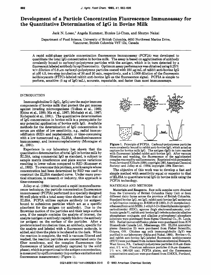
Jolley et al. (1984) introduced a rapid immunofluores- cence technique, the particle concentration fluorescence immunoassay (PCFIA) performed in a specially designed 96-well plate with a total assay time shorter than that of ELISA. PCFIA utilizes capture antibody (or antigen) bound to submicron particles which act as a specific adsorbent for the antigen (or antibody). Due to the Brownian motion of the particles and their large surface area, if the sample contains the analyte of interest, the analyte (antigen or antibody) rapidly binds to the antibody (or antigen) on the surface of the particles. After a predetermined incubation time, an antibody, specific for the analyte and labeled with a fluorescent molecule, is added, and then the plate is incubated in the dark. When the reaction is complete, the well is vacuum filtered and washed, the reaction particles are concentrated onto the filter membrane, and the complex fluorescence (the fluorescence of labeled antibody captured by the solid phase), which is proportional to the antigen concentration, is measured by epifluorometry (top surface excitation and fluorescence measurement) (Figure 1).
Figure 1. Principle of PCFIA. Carboxyl-polystyrene particles were covalently bound to rabbit anti-bovine IgG, which acted as capture for bovine milk IgG. The antigen (IgG) in turn reacted with fluorescent labeled rabbit anti-bovine IgG. After vacuum filtration and washing, the fluorescence of the agglutinated complex was read by epifluorometry. Reprinted with permission from Jervis and Kilbum (1991; copyright 1991 American Chemical Society) and Jolley et al. (1984; copyright 1984 Elsevier).
The objective of this work was to develop a fast and simple method with sensitivity equal or superior to that of ELISA to quantitate total IgG in bovine milk using the PCFIA technology. MATERIALS AND METHODS
Materials and Reagents. Raw milk samples were obtained from the University of British Columbia Dairy Unit or from different dairy farms across the province of British Columbia. Standard bovine IgG, rat IgG, rabbit anti-bovine IgG antiserum or IgG fraction (catalog no. B 8395 or B 1395), 2-(N-morpho1ino)- ethanesulfonic acid (MES), l-ethy1-3-[3-(dimethylamino)propyl]- carbodiimide) (EDC), agarose (high electroendosmosis), oval- bumin (gradeV), rabbit anti-bovine IgG (wholemolecule) alkaline phosphatase conjugate, and alkaline p-nitrophenyl phosphate substrate were purchased from Sigma Chemical Co., St. Louis, MO. Radial immunodiffusion plates were purchased from ICN Biomedicals Canada Ltd., Mississauga, ON. ELISA microtiter plates (Immulon 11) were purchased from Fisher Scientific, Ottawa, ON. Chicken egg yolk immunoglobulin (IgY) was purified in our laboratory (Akita and Nakai, 1992). Bovine T-GI and rabbit anti-bovine IgG-labeled fluorescein isothiocyanate (FITC) were purchased from Jackson Immunochemical Research, West Grove, PA. Carboxyl-polystyrene particles (0.8-rm diam- eter, 5 % w/v), Fluoricon reference particles (0.8-wm diameter, 1% w/v, 5901620 nm), PCFIA plates, and the fluorescence concentration analyzer were purchased from IDEXX, Portland, ME.
0021-8561/93/1441-0682$04.00/0 0 1993 American Chemical Society
PCFIA for IgG In Bovlne Milk
Preparation of Milk Samples and Standard IgG. Milk samples were diluted to 1:1000 in isotonic potassium phosphate bufferedsaline (PBS), pH 7.4, containing0.05% Tween 20 (PBS- Tween) and 0.25% ovalbumin. When necessary, raw milk samples were defatted by centrifugation at 14000g for 10 min. Standard bovine IgG concentration was determined at 280 nm in a Shimadzu 160 UV-visible spectrophotometer using absorp- tivity equal to 13.8 for IgG (Sober et al., 1970).
Skim milk powder, reconstituted to 9 % solids in distilled water, was boiled for 30 min (in an Erlenmeyer flask with a lid to minimize volume loss) in a water bath to destroy active IgG and used as diluent for standard IgG.
Radial Immunodiffusion. To prepare radial immunodif- fusion gels, 200 pL of 2.4 mg/mL rabbit anti-bovine IgG antiserum mixed with 1.8 mL of PBS buffer at 55 "C in a water bath was added to 5 mL of 1 % agarose containing 0.02 % sodium azide in PBS, mixed, and poured on immunodiffusion plates. The plates were allowed to solidify at room temperature for 15 min and then transferred to a moisture chamber for storage at 4 "C for at least 24 h before use. Wells of 3-mm diameter were perforated on the agarose using a template. Three microliters of milk samples or standards with concentration in the range 0.0575-0.92 mg/mL were applied to each well and the plates incubated in a moist chamber at room temperature. The diameter of the halo after 24 h read with a caliper (Mitutoyo) was related to the antigen concentration,using a standard curve run with each plate. ELISA. Flat-bottom polystyrene microtiter ELISA plates
were coated with 100 pL of 1 pg/mL rabbit anti-bovine IgG in carbonate buffer, pH 9.6, and incubated for 60 min at 37 "C. After the wells were washed twice with PBS-Tween, 250 pL of blocking solution (0.25 % ovalbumin in PBS-Tween) was applied to the wells for 30 min at 37 "C. The blocking solution was removed, and 100 pL of sample or IgG standard was added to the wells in triplicate. Bovine IgG standards were prepared between 1 and 100 ng/mL in PBS-Tween. The plates were incubated for 60 min at 37 "C. After three washes with PBS-Tween, 100 pL of rabbit anti-bovine alkaline phosphatase conjugate diluted to 0.43 pg/mL in PBS-Tween was added to each well and the plates were incubated for 60 min at 37 "C. The wells were washed three times with PBS-Tween and once with deionized distilled water beforeaddition of lOOpLof substrate (0.5 mg/mLofp-nitrophenyl phosphate in diethanolamine buffer, pH 9.8). After 60 min of incubation at 37 "C, the color developed was measured at 405 nm using a reference filter a t 655 nm with a Bio-Rad plate reader (Model 450, Bio-Rad, Richmond, CA). Absorbance values from ELISA plates were transformed, using the equation y = (ax/b + x ) + c, where y is the absorbance and x is the concentration, to IgG concentration using the computer program ELIZA (ELIZA release 2.79, Leading Edge Research, Vancouver, BC).
Particle Concentration Fluorescence Immunoassay. Coating of Carboxyl-Polystyrene Particles with Anti-Bovine ZgG. Carboxyl-polystyrene particles (0.8-pm diameter) were coated with 250 pg/mL of rabbit anti-bovine IgG antiserum in MES buffer a t pH 4.5 according to the manufacturer's instruct,ions as follows. To prepare 20 mL of coated particles a t 250 pg/mL IgG, add the following to a 30-mL siliconated glass centrifuge tube: 7 mL of 0.1 M MES buffer, pH 4.5; 2 mL of 2.5 mg/mL rabbit anti-bovine IgG (antiserum is added to provide a final concen- tration of 250 pg/mL IgG in a total volume of 20 mL; therefore, the MES buffer is added accordingly); 1.0 mL of carboxyl- polystyrene particles (5% w/v); and 5 mg of EDC. Mix by pipetting with narrow pipet tip several times and incubate at room temperature for 2 h; vortex periodically. Centrifuge at 4340g for 20 min. Remove the supernatant carefully. Add 20 mL of PBS buffer containing 0.2% sodium azide and 1% ovalbumin; mix well by pipeting and vortexing. Centrifuge at 4340g for 20 min. Remove supernatant carefully and resuspend the particles in 20 mL of PBS containing 0.2% sodium azide and 0.25% ovalbumin to give a final concentration of 250 pg/mL IgG and 0.25% w/v particles. Add 100 pL of Fluoricon reference particles (1% w/v, 590/620 nm), and store the particles at 4 "C until use.
PCFZA Procedure for Bovine Milk ZgG. PCFIA microtiter plates were first washed with PBS buffer followed by a 30-8 vacuum filtration. The wells were fiied with 20pL of the standard or the diluted milk samples. Standards containing from 4.8 to
J. Agrlc. Food Chem., Vol. 41, No. 4, l9Q3 689
920 ng/mL IgG were prepared in PBS-Tween containing 0.25 % ovalbumin or in PBS-Tween containing 0.25% ovalbumin and boiled milk. All buffers were filtered through a 0.22-pm filter, All samples and standards were run in triplicate. Twenty microliters of rabbit anti-bovine IgG coated carboxyl-polystyrene particles containing the reference particles was added to each well containing a sample. The plate was gently tapped along the side and incubated for 30 min at room temperature (21 "C). Then 20 pL of fluorescein isothiocyanate (F1TC)-labeled rabbit anti- bovine IgG at 1:1000 dilution in PBS-Tween containing 0.25% ovalbumin was added to each well and gently mixed, and the plate was incubated for 20 min in the dark at room temperature. The plate was inserted into the fluorescence concentration analyzer, vacuum filtered, washed with PBS buffer and vacuumed three times. The epifluorescence of each well was read using the instrument sample channel 485/535 nm and reference channel 590/620 nm (gain 10). The data were collected on a PC computer interfaced with the fluorescence concentration analyzer and expressed as the ratio of fluorescence intensity measured at 535 and 620 nm. Milk IgG concentrations were calculated from a standard curve run with each plate. In case Fluoricon reference particles were not added during coating of carboxyl-polystyrene particles, results are reported as relative fluorescence units. The computer program ELIZA (release 2.79) was used to draw standard curves, and Sigma Plot (version 4.1) was used for statistical analysis.
Determination of Optimal Concentration of Rabbit Anti- Bovine ZgG Bound to the Carboxyl-Polystyrene Particles. Carboxyl-polystyrene particles were coated with different con- centrations of affinity-purified rabbit anti-bovine IgG (50,100, 250, 500, and 1000 pg/mL) in MES buffer a t pH 4.5. IgG concentrations from five different milk samples were determined.
Determination of Optimal Incubation Times. IgG standards were incubated with the antibody coated on carboxyl-polystyrene particles for varying lengths of time from 0 to 60 min and incubated with FITC-conjugated antibody from 0 to 45 min followed by vacuum filtration, washings, and reading.
Specificity, Sensitivity, and Reliability of PCFZA. The specificity of the assay was evaluated by using rabbit anti-bovine IgG to determine the concentrations of bovine ~ G I , rat IgG, human IgA, chicken IgY, and casein whose concentrations were previously determined by absorbance at 280 nm. The concen- tration of IgG in reconstituted milk powder boiled for 30 min and used as diluent was also determined by RID.
Intra- and Inter-Assay Variability. To estimate the intra- and inter-assay variability, randomly pooled milk samples were assayed in five sets of triplicates on a single plate and in one set of triplicates on three different plates at different days.
Analytical Recovery and Effect of Sample Dilution. Different concentrations of commercially purified IgG between 28 and 230 pg/mL were spiked into milk samples. Milk samples were diluted from 1:lOOO to 1:8000 and IgG concentrations determined by PCFIA as described above.
Effect of Fat on the Accuracy of PCFZA. To study the effect of milk fat on the epifluorescence results, raw milk samples from two different cows were skimmed and IgG concentrations determined on both the raw and skimmed fractions.
Effect of Casein. To evaluate the contribution of casein to IgG values, defatted milk samples from four different cows were acidified to pH 4.6 with 2 N HC1 to minimize the volume change. IgG concentrations were determined in skim milk and the whey fraction by PCFIA and RID. Whey pH was adjusted to pH 7.4, casein added back as 33 % ,66 % , or 100 % , and IgG concentration determined by PCFIA.
Matrix Effect. IgG standard in PBST containing 0.25% ovalbumin with or without boiled milk was run with milk samples to estimate the matrix effect on IgG determination.
Correlation of RID with PCFZA and ELZSA. To compare PCFIA and RID or ELISA and RID, a milk sample was diluted to80%,60%,50%,40%,30%,20%,and10% with boiledmilk and IgG concentrations determined by PCFIA, RID, and ELISA using commercial IgG as standard. RESULTS
Optimal Concentration of Rabbit Anti-Bovine IgG, Optimal Incubation Times, and Optimal Concentra-
884 J. Agric. Food Chem., Vol. 41, No. 4, 1993
Table I. Optimal Concentration of Rabbit Anti-Bovine IgG Antiserums
antibody concn (mean f SD), mg/mL milk sample 50 pg/mL 100 pg/mL 250 pg/mL RID
1 0.30f 0.01 0.27 f 0.00 0.31 f 0.01 0.32 2 0.24f 0.00 0.23 f 0.00 0.27 f 0.03 0.29 3 0.23f0.00 0.26f0.00 0.27f0.01 0.29 4 0.21 f 0.01 0.23 f 0.00 0.24 f 0.00 0.27 5 0.18f0.00 0.20f0.02 0.20f0.01 0.20
4 Carboxyl-polystyrene particles were coated with 50, 100, and 250 pg/mL of rabbit anti-bovine IgG, respectively. Standard curves were run at each concentration of the capture antibody. Five different milk samples were analyzed ( N = 3) for IgG content (mg/mL) at each concentration of the capture antibody and the results compared with RID.
35000 1
Loss0 et el.
30000 P I 20000
15000
10000
5000
0
t /////
0 0 0 1 0 2 0 3 0 4 0 5 0 6 0 7 IgG Concentration. mg/ml
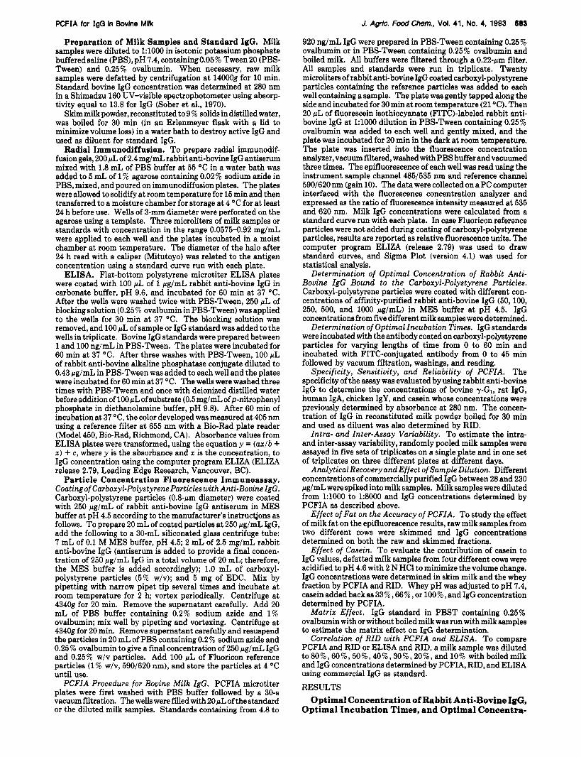
Figure 2. Optimal incubation time. Antigen at different concentrations was incubated with the antibody bound on carboxyl-polystyrene particles followed by a second incubation with FITC-labeled rabbit anti-bovine IgG for 0 and 0 min (a), 5 and 0 min (O), 5 and 5 min (v), 10 and 10 min (v), 20 and 15 min (w), 20 and 20 min (O) , and 30 and 20 min (A). The standard curve a t each incubation time was computed. tion of FITC-Labeled Antibody. A concentration of 250 pg/mL was chosen for subsequent analysis because it gave values closer to RID values than the other concen- trations (Table I). Higher concentrations of antiserum (500 or lo00 pg/mL) increased the fluorescence background but did not improve the accuracy of the assay (data not shown).
From Figure 2,30 min for the first incubation time and 20 min for the second incubation time were selected for further experiments. Longer incubation times did not increase the epifluorescence values.
A 1:lOOO dilution was found optimal to achieve linear fluorescence increase and was therefore used for all subsequent experiments.
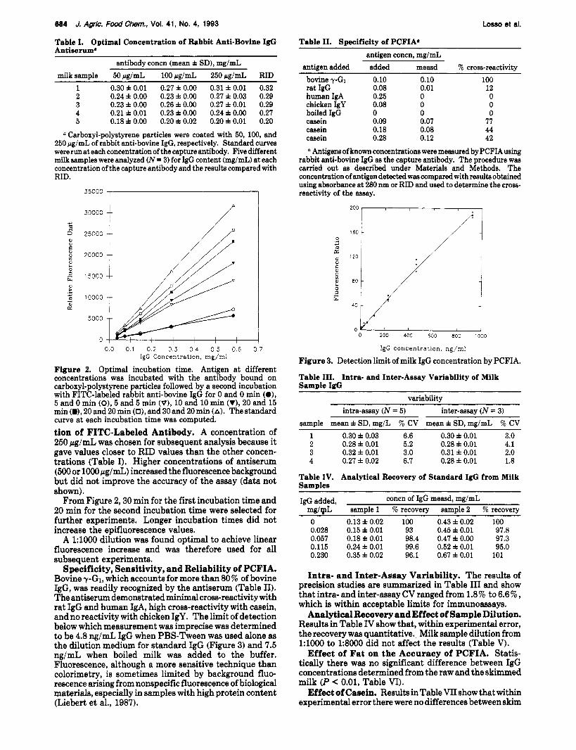
Specificity, Sensitivity, and Reliability of PCFIA. Bovine yG1, which accounts for more than 80% of bovine IgG, was readily recognized by the antiserum (Table 11). The antiserum demonstrated minimal cross-reactivity with rat IgG and human IgA, high cross-reactivity with casein, and no reactivity with chicken IgY. The limit of detection below which measurement was imprecise was determined to be 4.8 ng/mL IgG when PBS-Tween was used alone as the dilution medium for standard IgG (Figure 3) and 7.5 ng/mL when boiled milk was added to the buffer. Fluorescence, although a more sensitive technique than colorimetry, is sometimes limited by background fluo- rescence arising from nonspecific fluorescence of biological materials, especially in samples with high protein content (Liebert et al., 1987).
Table 11. Specificity of PCFIA. antigen concn, mg/mL
antigen added added measd % cross-reactivity bovine ?-GI rat IgG human IgA chicken IgY boiled IgG casein casein casein
0.10 0.08 0.25 0.08 0 0.09 0.18 0.28
0.10 100 0.01 12 0 0 0 0 0 0 0.07 77 0.08 44 0.12 42
a Antigens of known concentrations were measured by PCFIAusing rabbit anti-bovine IgG as the capture antibody. The procedure was carried out as described under Materials and Methods. The concentration of antigen detected was compared with results obtained using absorbance at 280 nm or RID and used to determine the cross- reactivity of the assay.
200 , 1
160 I I
/ I i
1 1
0 200 400 600 800 1000
IgG concentrat lon, ng/ml
Figure 3. Detection limit of milk IgG concentration by PCFIA.
Table 111. Intra- and Inter-Assay Variability of Milk Sample I&
variability intra-assay (N = 5) inter-assay ( N = 3)
sample mean f SD, mg/L % CV mean f SD, mg/mL 76 CV 1 0.30 f 0.03 6.6 0.30 f 0.01 3.0 2 0.28 f 0.01 5.2 0.28 f 0.01 4.1 3 0.32 f 0.01 3.0 0.31 f 0.01 2.0 4 0.27 f 0.02 6.7 0.28 f 0.01 1.8
Table IV. Analytical Recovery of Standard IgG from Milk Samples
IgG added, concn of IgG measd, mg/mL mg/mL sample 1 % recovery sample 2 % recovery 0 0.13 f 0.02 100 0.43 f 0.02 100 0.028 0.15 i 0.01 93 0.45 f 0.01 97.8 0.057 0.18 f 0.01 98.4 0.47 f 0.00 97.3 0.115 0.24 f 0.01 99.6 0.52 f 0.01 95.0 0.230 0.35 f 0.02 96.1 0.67 f 0.01 101
Intra- and Inter-Assay Variability. The results of precision studies are summarized in Table I11 and show that intra- and inter-assay CV ranged from 1.8% to 6.6%, which is within acceptable limits for immunoassays.
Analytical Recovery and Effect of Sample Dilution. Results in Table IV show that, within experimental error, the recovery was quantitative. Milk sample dilution from 1:lOOO to 1:8000 did not affect the results (Table V).
Effect of Fat on the Accuracy of PCFIA. Statis- tically there was no significant difference between IgG concentrations determined from the raw and the skimmed milk (P < 0.01, Table VI).
Effect of Casein. Results in Table VI1 show that within experimental error there were no differences between skim
PCFIA for IgG In Bovlne Milk
Table V. Effect of Sample Dilution IgG concn, corrected IgG concn,
dilution ma/mL mdmL
J. A g k . Food Chem., Vd. 41, No. 4, 1993 685
0.5 I
~~~ ~
1:lOOo 0.31 1:2000 0.16 1:4000 0.08 1:8OOo 0.04
0.31 0.32 0.32 0.32
Table VI. Effect of Fat on I& Determination raw mean f SD, skimmed mean f SD,
sample mg/mL sample mdmL 1 0.20 f 0.03 1 0.20 f 0.03 2 0.14 f 0.00 2 0.13 f 0.00
Table VII. Effect of Casein on Bovine 1%: Concentrationa
sample skim RIDb Wc pH7.4 W + V2Cd W + ~/ sC W + C W at
1 0.17 0.17 0.15 0.14 0.13 0.15 0.15 2 0.26 0.26 0.25 0.23 0.22 0.23 0.24 3 0.16 0.16 0.15 0.15 0.15 0.13 0.14 4 0.14 0.14 0.12 0.11 0.11 0.11 0.12
Milligrams per milliliter. Four different milk samples were defatted, and IgG concentration was determined by PCFIA and RID. Casein was precipitated from each sample at pH 4.6, and IgG concentration in whey was determined by PCFIA. Whey pH was adjusted to pH 7.4; casein was added back in increments of 0.09%, 0.18%, and 2.8%; and IgG concentration was again determined by PCFIA. RID, radial immunodiffusion. W, whey. C, casein.
Table VIII. Diluent Effecte sample PBST-M-OV PBST-OV
1 0.23 0.21 2 0.36 0.34 3 0.16 0.17 4 0.10 0.11 5 0.38 0.36
Milk sample concentrations (mg/mL) were calculated from a standard curve constructed with boiled milk (fist column) or without boiled milk (second column). PBST-M-OV, phosphate-buffered salinecontainiig0.05% Tween20, boiledmilk, and0.25% ovalbumin. PBST-OV, phosphate-buffered saline containing 0.05% Tween 20 and 0.25% ovalbumin. milk and whey and between whey with added increments of casein. The specificity of the antiserum was not evident when casein was analyzed alone since it contributed some fluorescence by nonspecific binding that would translate into false positive IgG concentration (Table 11). This nonspecific binding phenomenon, which is also observed when coated carboxyl-polystyrene particles we incubated with FITC-labeled rabbit anti-bovine IgG in the absence of antigen, has been reported by Islam et al. (1990). But in the presence of milk IgG, casein was outcompeted by IgG for binding to antibody on latex particles and did not contribute any increase in fluorescence. The results show that casein in micellar form, as it is found in the milk, would not contribute any fluorescence.
Matrix Effect. As shown in Table VIII, IgG standard curves could be run in PBS-Tween containing ovalbumin with or without boiled milk without significant difference in IgG results.
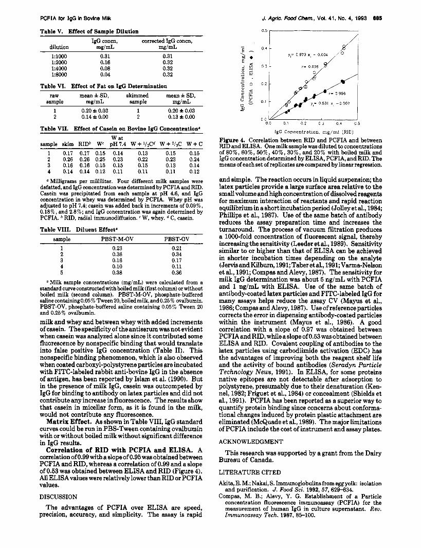
Correlation of RID with PCFIA and ELISA. A correlation of 0.99 with a slope of 0.95 was obtained between PCFIA and RID, whereas a correlation of 0.99 and a slope of 0.53 was obtained between ELISA and RID (Figure 4). All ELISA values were relatively lower than RID or PCFIA values.
DISCUSSION
The advantages of PCFIA over ELISA are speed, precision, accuracy, and simplicity. The assay is rapid
o o v ' I 0 0 0 1 02 0 3 0 4 0 5
IgG Concentration, mg/ml (RID)
Figure 4. Correlation between RID and PCFIA and between RID and ELISA. One milk sample was diluted to concentrations of 8O%, 60%, 50%, 40%, 3076, and 20% with boiled milk and IgG concentration determined by ELISA, PCFIA, and RID. The means of each set of replicates are compared by linear regression.
and simple. The reaction occurs in liquid suspension; the latex particles provide a large surface area relative to the small volume and high concentration of dissolved reagents for maximum interaction of reactants and rapid reaction equilibrium in a short incubation period (Jolley et al., 1984; Phillips et al., 1987). Use of the same batch of antibody reduces the assay preparation time and increases the turnaround. The process of vacuum filtration produces a 1000-fold concentration of fluorescent signal, thereby increasing the sensitivity (Leeder et al., 1989). Sensitivity similar to or higher than that of ELISA can be achieved in shorter incubation times depending on the analyte (Jervis and Kilburn, 1991;Taber et al., 1991; Varma-Nelson et al., 1991; Compas and Alevy, 1987). The sensitivity for milk IgG determination was about 5 ng/mL with PCFIA and 1 ng/mL with ELISA. Use of the same batch of antibody-coated latex particles and FITC-labeled IgG for many assays helps reduce the assay CV (Mayus et al., 1986; Compas and Alevy, 1987). Use of reference particles corrects the error in dispensing antibody-coated particles within the instrument (Mayus et al., 1986). A good correlation with a slope of 0.97 was obtained between PCFIAand RID, while a slope of 0.53 was obtained between ELISA and RID. Covalent coupling of antibodies to the latex particles using carbodiimide activation (EDC) has the advantages of improving both the reagent shelf life and the activity of bound antibodies (Seradyn Particle Technology News, 1991). In ELISA, for some proteins native epitopes are not detectable after adsorption to polystyrene, presumably due to their denaturation (Ken- nel, 1982; Friguet et al., 1984) or concealment (Shields et al., 1991). PCFIA has been reported as a superior way to quantify protein binding since concerns about conforma- tional changes induced by protein plastic attachment are eliminated (McQuade et al., 1989). The major limitations of PCFIA include the cost of instrument and assay plates.
ACKNOWLEDGMENT
Bureau of Canada.
LITERATURE CITED
Akita, E. M.; Nakai, S. Immunoglobulinsfrom egg yolk: isolation and purification. J. Food Sci. 1992, 57, 629-634.
Compas, M. B.; Alevy, Y. G. Establishment of a Particle concentration fluorescence immunoassay (PCFIA) for the measurement of human IgG in culture supernatant. Rev. Immunoassay Tech. 1987, 85-100.
This research was supported by a grant from the Dairy
888 J. Agrlc. FoodChem., Vol. 41, No. 4, 1993
Ebina, T.; Sato, A.; Umezu, K.; Ishida, N.; Ohyama, S.; Oizumi, A.; Aikawa, K.; Katagi, S.; Katsushima, N.; Imai, A.; Kitaoka, S.; Suzuki, H.; Konno, T. Prevention of rotavirus infection by oral administration of colostrum containing anti-human ro- tavirus antibody. Med. Microbiol. Zmmunol. 1985,174,177- 185.
Friguet, B.; Djavadi-Ohaniance, L.; Golberg, M. E. Some mon- oclonal antobodies raised with a native protein bind prefer- entially to denatured antigen. Mol. Zmmunol. 1984,21,673- 677.
Islam, F.; Urade, Y.; Wanatabe, Y.; Hayaishi, 0. A particle concentration fluorescence immunoassay for prostaglandin D synthetase in the rat central nervous system. Arch. Biochem. Biophys. 1990,277, 290-295.
Jervis, E.; Kilburn, D. Rapid immunoassay technique for process monitoring of animal cell fermentations. Biotechnol. Prog. 1991, 7,28-32.
Jolley, M. E.; Wang, C-H. J.; Ekenberg, S. J.; Zuelke, M. S.; Kelso, D. M. Particle concentration fluorescence immunoassay (PCFIA): a new, rapid immunoassay technique with high sensitivity. J. Zmmunol. Methods 1984, 67, 21-35.
Kennel, S. J. Binding of MAb to protein antigen in fluid phase or bound to solid supports. J. Zmmunol. Methods 1982, 55, 1-12.
Kobayashi, T.; Ohmori, T.; Yanai, M.; Kawanishi, G.; Yoshikai, Y.; Nomoto, K. Protective effect of orally administering immune milk on endogenous infection in X-irradiated mice. Agric. Biol. Chem. 1991,55, 2265-2272.
Kummer, A,; Kitts, D. D.; Li-Chan, E.; Losso, J. N.; Skura, B. J.; Nakai, S. Quantitation of bovine IgG in milk using a Enzyme- Linked Immunosorbent Assay (ELISA). Food Agric. Zmmu- nol. 1992, 4 , 93-102.
Leeder, J. S.; Dosch, H.-M.; Harper, P. A.; Lam, P.; Spielberg, S. P. Fluorescence-based viability assay for studies of reactive drug intermediates. Anal. Biochem. 1989,177, 364-372.
Ma, J. K.-C.; Smith, R.; Lehner, T. Use of monoclonal antibodies in local passive immunization to prevent colonization of human teeth by Streptococcus mutans. Infect. Zmmun. 1987, 55,
Mayus, J.; Macke, K.; Shakelford, P.; Kim, J.; Nahru, M. Human IgG subclass assays using a novel assay method. J. Zmmunol. Methods 1986,88,65-73.
1274-1278.
Losso et ai.
McQuade, T. J.; Pitts, T. W.; Tarpley, W. G. A rapid solution immunoassay to quantify binding of the human immunode- ficiency virus envelope glycoprotein to soluble CD4. Biochem. Biophys. Res. Commun. 1989,163,172-176.
Michalek, S. M.; Gregory, R. L.; Harmon, C. C.; Katz, J.; Richardson, G.; Hilton, T.; Filler, S.; McGhee, J. R. Protection of gnotobiotic rata against dental caries by passive immuni- zation with bovine antibodies to Strep. mutans. Infect. Zmmun. 1987,55, 2341-2347.
Montagne, P.; Gavriloff, C.; Humbert, G.; Cuilliere, M. L.; Duheille, J.; Linden, G. Microplate-enhanced nephelometric immunoassay of immunoglobulins G in cow’s milk. Lait 1991, 71, 493-499.
Phillips, D. J.; Wells, T. W.; Reimer, C. B. Estimation of association constants of 42 monoclonal antibodies to human IgG epitopes using a fluorescent sequential saturation assay. Zmmunol. Lett. 1987,17, 159-168.
Seradyn Particle Technology News. Adsorption and covalent coupling of proteins to CML and PS microparticles. 1991,2, 1-8.
Shields, M. J.; Siegel, J. N.; Clark, C. R.; Hines, K. K.; Potempa, L. A.; Gewurz, H.; Anderson, B. An appraisal of polystyrene- (ELISA) and nitrocellulose-based (ELIFA) enzyme immu- noassay systems using monoclonal antibodies reactive toward antigenically distinct forms of human C-reactive protein. J. Zmmunol. Methods 1991, 141, 253-261.
Sober,H. A.;Harte,R.A.;Sober,E.K.HandbookojBiocltemktry. Selected Data for Molecular Biology, 2nd ed.; The Chemical Rubber Co.: Cleveland, OH, 1970; p (2-80.
Taber, L. D.; O’Brien, P.; Bowsher, R. R.; Sportsman, J. R. Competitive particle concentration fluorescence immunoassay for measuring 5,10-dideaza-5,6,7,&tetrahydrofolic acid (Lom- etrol) in serum. Clin. Chem. 1991, 37, 254-260.
Varma-Nelson, P.; Nelson, J. E.; Belknap, S. Particle concen- tration fluorescence immunoassay of gentamicin. Ther. Drug Monit. 1991, 13, 260-262.
Yolken, R. H.; Losonsky, G. A.; Vonderfecht, S.; Leister, F.; Wee, S. B. Antibody to human rotavirus in cow’s milk. N . Engl. J. Med. 1985, 312, 605-610.
Received for review August 10,1992. Revised manuscript received December 15, 1992. Accepted January 25, 1993.