development of a chemiluminescence immunoassay for the detection of hepatitis b surface antigen
TRANSCRIPT
Am&a Chmca Acta, 266 (1992) 201-204 Elsevxer Science Pubbshers B V , Amsterdam
201
Development of a chemiluminescence immunoassay for the detection of hepatitis B surface antigen
Ian Weeks and J Stuart Woodhead
Department of Me&al Bmhemmy, Unwerszty of Wales, College of Medwne, Heath Park, Cadff CF4 4xN (UK)
Ivan Lucas
Molecular Light Technology Research Lusted, Card@ Bumess Technology Centre, Senghenydd Road Cardff CF2 4AY (UK)
(Received 3rd February 1992, reused manuscnpt recewed 9th Apn11992)
A chenulummescent acndmmm salt has been used to label a monoclonal antlbody to hepatms B surface antigen (HBsAgl Smce the antibody recogmses a repeatmg epltope on HBsAg the same antibody m Its unlabelled form has been used to coat the wells of a 96-well mxrotltre plate These reagents have been used as the basis of a two-site mununochemdummomet assay (ICMA) for the detectlon of HBsAg m senun. The assay cons&s of a smgle 90 mmute mcubation at 37°C followed by a wash step and then quantltatton of chemdummescence mtenslty m a nucrot&e plate lummometer The countmg tune IS 1 second per well and the assay has a sensltmty of dettion of OWngml-’
Keywords Chemdummescence, Immunoassay, Acndmmm salts, Hepatitis B antigen, Lummometer
The screenmg of blood donatlons for the pres- ence of hepatltls B surface antigen (HBsAg) 1s a vital step m mmnmsmg the transnnsslon of the hepatltls B vuus by blood transfusion Screenmg m a gtven regonal laboratory often requires the testmg of several hundred thousand donations annually In view of the h&-throughput of such tests it 1s desirable that the assay used should be sunple, rapid and robust, without any compro- nuse m analytical sensltlvlty Conventionally, most testmg mvolves the use of enzyme-hnked un- munosorbent assay (ELISA) [l] In such systems,
samples are mcubated wth the appropriate en- zyme-labelled ant&&y m antibody-coated 96-well mlcrotltre plates Followmg the unmunochermcal reaction and removal of unbound labelled antI- body by washmg, enzyme substrate solutions must be made up and added to the wells, followmg which a further Incubation must be performed The enzyme-substrate reactlon must then be stopped by addition of another reagent pnor to measurement of the colourunetrlc endpomt
Chermhunmescent unmunoassays for HBsAg have been described [2] but suffer poor sensltlvlty due to the problems associated vvlth the use of ~solummol labels Chemdummescent unmunoas- says usmg acndmnun salts as labels have become established as some of the most sensitive, robust
Corrargondence to I Weeks, Department of Medical Blo- chermstry, Umversity of Wales, College of Medicme, Heath Park, Caxhff CF4 4XN (UK)
0003-X70/92/$05 00 8 1992 - Elsevler hence Pubhshers B V All nghts reserved
202 Z Weeks et al /Anal. Chum Acta 266 (Z992) 201-204
assays currently available [3] However, most reh- able measuring instruments for chemdurmnes- cence (lummometers) have been deslgned to ac- comodate conventional 12 x 75 mm reactlon cu- vettes H&throughput assays benefit from the use of nucrotltre plates smce this permits easy manrpulatlon of up to 96 samples when used m conJunction with multichannel pipettes and auto- mated mlcroplate washers
Recently a nucrotltre plate lummometer has been produced by Laboratormm Berthold (Wdd- bad) which 1s ideally suited to a vanety of cheml- lummescence and blolummescence apphcatlons mcludmg tmmunoassays based on the use of chemllummescent acrldmmm salts Here we de- scribe the prehmmary development of a two-site mununochemllummometerlc assay (ICMA) for HBsAg which is much sunpler and has a more rapid end-pomt detection system than ELISA
Lummometer A mlcrotltre plate lummometer was generously
loaned by Laboratormm Prof Dr Berthold, (Wddbad) This mstrument IS capable of perform- mg photon countmg on each well of a 96-well nucrotltre plate m the appropnate sequence and IS equipped with two reagent injectors for use when onboard mltlatlon of chemllummescent re- actions 1s required as m the case of acrldmmm salts The instrument was programmed to inject an acldlc solution of hydrogen peroxide (50 ~1) from the first injector followed by an alkali/ surfactant nuxture (50 ~1) These reagents were purchased from Cuba-Corning Dragnostlcs, (Hal- stead) as Magic Lite Initiator Reagents 1 and 2 Under these condltlons the chemllummescent re- actron 1s complete wlthm 1 second and measure- ments were made as total photon counts (relative light units, RLU) integrated over a one second period
MATERIALS
METHODS Acrldmnun labellmg luts were obtamed from
Molecular Light Technology Research Limited, Cardlff, UK White mlcrotltre plates (M119W) were obtained from Dynatech Laboratories, Bllhnghurst Monoclonal antibody to HBsAg (MIH9705) was a gift from Medlx Blotech, Foster City HBsAg was obtamed from Scripps Labora- tories (San Diego, CA> Normal serum was ob- tamed from Scantlbodles Inc (Santee, CA) Bovme serum albumin and bovme gamma-globu- lins were from Sigma Chenucals (Poole) All other chemicals were from BDH-Merck (Poole)
Buffers The followmg buffers were used m this work coating buffer was aqueous sodmm bicarbonate solution (0 05 mol l-‘, pH 9 61, assay buffer was a mtire of sodmm dlhydrogen or- thophosphate and dlsodmm hydrogen orthophos- phate (0 1 mol 1-l) contammg 0 15 mol 1-l sodnun chloride, 0 02% (w/v> sodium azlde, 0 1% (w/v) bovme serum albumm and 40 mg 1-l bovme gamma-globulin, pH 7 4, wash buffer consisted of a nuxture of sodmm dlhydrogen orthophosphate and dlsodmm hydrogen orthophosphate (0 05 mol 1-l) contammg 0 15 mol 1-l sodmm chloride and 0 1% (v/v) of the surfactant Tween 20
The monoclonal antibody (50 pg) was labelled urlth a chemllummescent acndmmm salt accord- mg to published procedures [31 Microwell plates were coated wrth unlabelled antibody by dlspens- mg 50 ~1 of an appropriate solution of the antl- body (see below) m coating buffer mto each well The plates were covered with polythene film and left to stand overnight at room temperature The plates were washed three tunes with wash buffer using an automated nucroplate washer and then 200 ~1 of assay buffer dispensed mto each well The plates were mcubated for 3 h at 37°C and washed a further three tunes as described above Batches of plates were stored at 4°C until re- quired
HBsAg standards were prepared by dllutmg the stock solution of antigen m assay buffer or normal serum as required
Optunrsatwn of mode and tune of rncubatwn Two modes of incubation were mvestlgated Firstly 50 ~1 of HBsAg standards were mcubated m the coated plate for 1 h at 37”C, the plate was given two washes and then a solution of acn- dmmm labelled antibody m assay buffer added
I Weeks et al /AnaL Chtm Acta 266 (1992) 201-204 203
(50 ~1, 2 5 ng, 5 X lo6 RLU) The plate was mcubated for a further 1 h at 37“C, washed four times and placed m the lummometer for mea- surement of chemdummescence Secondly, HBs- Ag standard (50 ~~11 and labelled antibody (50 ~1) were added snnultaneously and the plate mcu- bated for 2 h at 37°C The plate was washed four trmes and then placed m the lummometer for measurement of chemllummescence
The mcubatlon tune of the sunultaneous assay was varied over the range 45, 90 and 180 mm
Optunwatwn of anthody concentratwns Coat- mg antIbody concentrations were vaned over the range 3 25, 6 5, 13, 26 and 52 pg ml-’ Labelled antibody was vaned over the range 125,2 5 and 5 ng per well
Optmed assay protocol The final assay used plates coated at a concentration of 13 pg mi-’ and labelled antibody at 2 5 ng (5 x lo6 RLU) per well 50 ~1 of sample or standard were added to each well followed by 50 ~1 of labelled anti- body solution The plate was covered and mcu- bated for 90 mm at 37°C The plate was then washed four tunes usmg the automated plate washer and transferred to the nucrotltre plate lummometer where each well was counted for one second Results were expressed as relative hght umts
RESULTS AND DISCUSSION
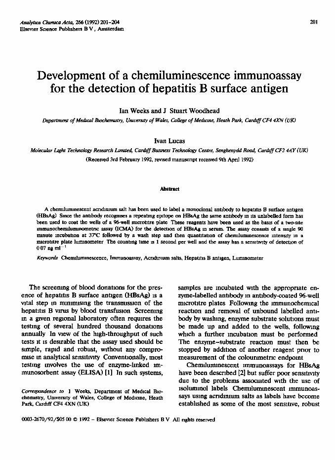
It was found that the simultaneous mcubatlon yielded a better signal-to-noise (S/N) ratlo at a given concentration of HBsAg than the sequen- tial mcubatron In view of this fmdmg and the fact that the sunultaneous protocol is technrcally snnpler to perform, the simultaneous assay was adopted for all further experunents It was found that a 90-mm mcubatlon gave an optunal S/N ratio smce although increased mcubatlon times yielded hgher specfilc bmdmg, a substantial m- crease m non-specific bmdmg was also observed (Ag 1)
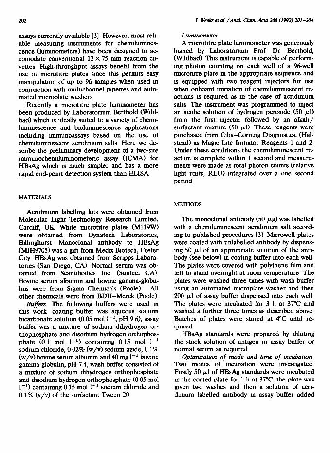
Bmdmg of HBsAg was found to increase qvith mcreasmg coating antibody concentration (Fig 2), however the mcrease was mmmal at concen- trations above 13 pg ml-’ and so this concentra- tlon was used routinely to coat the plates
Fig 1 Effect of mubatron time on the sunultaneous assay The ordmate axis (B - Bo) represents the a&wty bound to the plate m RLJ_J wth subtractIon of non-specific bmdmg ( n ) 45’, (+) 90’, (*) 180’
Fig 2 Effect of changmg the concentration of the antibody coatmgsoluuon B-Bo~sasforFu 1 (X)325,(0)65,(*) 13, (+) 26, (M) 52 ng ml-’
1000~ 01 IHBsA~ w/ml
ICI
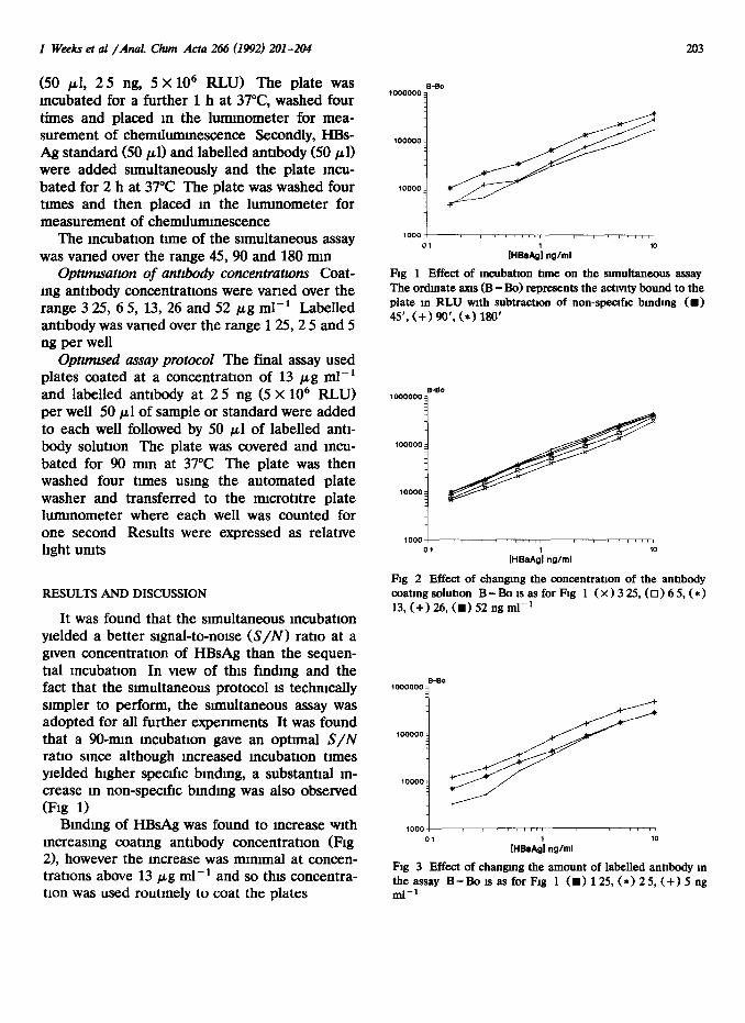
Fig 3 Effect of changmg the amount of labelled antibody m the assay B-Bo 1s as for Fig 1 (m) 125, (*) 25, (+) 5 ng ml-’
204 I Weeks et al /And Chum Acta 246 (1992) 201-204
,000 c 01
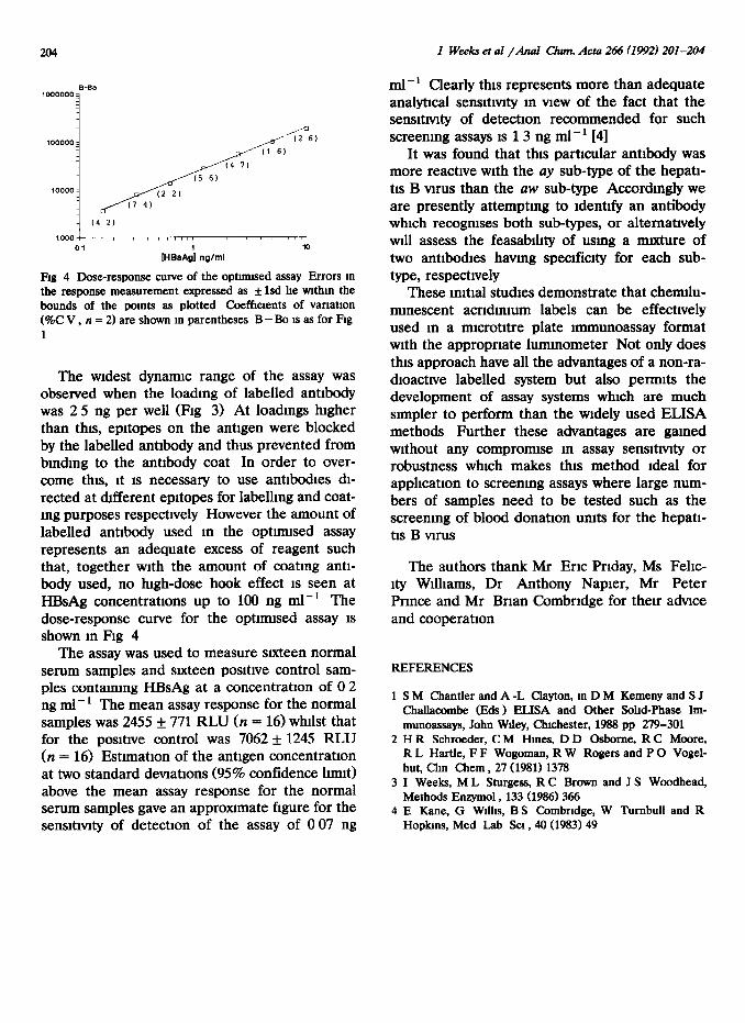
FU 4 Dose-response curve of the optmnsed assay Errors m the response measurement expressed as f lsd he wthm the bounds of the pomts as plotted Coefficients of vanatlon (%C V , n = 2) are shown m parentheses B - Bo 1s as for Fig 1
The wrdest dynanuc range of the assay was observed when the loadmg of labelled antibody was 2 5 ng per well (Ag 3) At loadings higher than thus, epttopes on the antrgen were blocked by the labelled antibody and thus prevented from bmdmg to the antibody coat In order to over- come thus, rt IS necessary to use antibodies dr- rected at different eprtopes for labelhng and coat- mg purposes respectively However the amount of labelled antibody used 111 the optrmrsed assay represents an adequate excess of reagent such that, together wrth the amount of coating antr- body used, no hrgh-dose hook effect is seen at HBsAg concentrations up to 100 ng ml-l The dose-response curve for the opttmtsed assay IS shown m Fig 4
The assay was used to measure srxteen normal serum samples and sncteen posttrve control sam- ples contammg HBsAg at a concentration of 0 2 ng ml-’ The mean assay response for the normal samples was 2455 + 771 RLU (n = 16) whtlst that for the posrtrve control was 7062 f 1245 RLU (n = 16) Estlmatron of the antigen concentratton at two standard devratrons (95% confidence lnmt) above the mean assay response for the normal serum samples gave an apprownate figure for the sensttrvrty of detection of the assay of 0 07 ng
ml-’ Clearly this represents more than adequate analytical sensttrvtty m vrew of the fact that the sensmvrty of detection recommended for such screenmg assays is 13 ng ml-’ [41
It was found that this particular antibody was more reactive with the uy sub-type of the hepatt- tts B vnus than the aw sub-type Accordingly we are presently attemptmg to Identify an antibody which recogmses both sub-types, or alternatively will assess the feasabthty of usmg a mrxture of two antibodies havmg specrfictty for each sub- type, respectrvely
These mttral studies demonstrate that chemrlu- mmescent acrrdmmm labels can be effectively used m a nucrotttre plate immunoassay format with the appropriate lummometer Not only does this approach have all the advantages of a non-ra- dtoacttve labelled system but also permits the development of assay systems which are much simpler to perform than the wtdely used ELISA methods Further these advantages are gamed without any compromtse m assay sensttrvtty or robustness which makes this method ideal for applrcatron to screening assays where large num- bers of samples need to be tested such as the screening of blood donation units for the hepatr- trs B vnus
The authors thank Mr Eric Prrday, MS Fehc- rty Wtlhams, Dr Anthony Napier, Mr Peter Prmce and Mr Brian Combrrdge for then advtce and cooperation
REFERENCES
1 S M Chantler and A -L Clayton, m D M Kemeny and S J Challacombe (Eds) ELISA and Other Sohd-Phase Im- munoassays, John Whey, ChIchester, 1988 pp 279-301
2 H R Schroeder, CM Hmes, D D Osborne, R C Moore, R L Hartle, F F Wogoman, R W Rogers and P 0 Vogel- hut, Chn Chem , 27 (1981) 1378
3 I Weeks, ML Sturgess, R C Brown and J S Woodhead, Methods Enzymol , 133 (1986) 366
4 E Kane, G W&s, B S CombrIdge, W Tumbull and R Hopkms, Med Lab SCI , 40 (1983) 49