development and validation of an indirect enzyme immunoassay for detection of antibody to brucella...
TRANSCRIPT
veterinary microbiology
Veterinary Microbiology 52 (1996) 165-173 ELSEVIEE.
Development and validation of an indirect enzyme immunoassay for detection of antibody to Brucella
abortus in milk
K. Nielsen a3 * , P. Smith a, D. Gall a, B. Perez b ‘f C. Cosma ‘, P. Mueller d, J. Trottier e, G. Cote e, L. Boag , J. Bosse a
a Agriculture and Agri-Food Canada, Animal Diseases Research Institute, 3851 Fallowfield Road. Nepean, Ontario, Canada K2H 8P9
b Sermcio Agricola y Gandera, Osorno. Chile ’ Secertaria de Agricultura Ganaderia y Pesca, Sermcio National de Sanidad Animal, Buenos Aires.
Argentina d 3400 Casavant Blvd. W, Saint Hyacinthe, Quebec, Canada J2S 8E3
e Gouvernement du Quebec, Ministere de l’Agriculture, des Pecheries de Ailmentation, 2700 Rue Einstein, C2-105, Sainte Fey Quebec. Canada JIP 3B8
f Health of Animals Laboratories. 110 Stone Road, Guelph, Ontario, Canada
Received 21 November 1995: accepted 5 February 1996
Abstract
An indirect enzyme immunoassay for detection of antibody to Brucella abortus in bovine milk was developed and validated using 6238 milk samples from Canadian herds (brucellosis free) and 202 samples from herds infected with B. abortus (from Argentina and Chile). The assay utilized
lipopolysaccharide as the antigen, immobilized on a polystyrene matrix, whole milk to test and a mouse monoclonal antibody, specific for an epitope of bovine IgG,, conjugated with horseradish peroxidase.
The sensitivity of the assay was 95.2% + 3.7% at a confidence limit of 95% for samples from B. abortus infected herds obtained from Chile and 98.7% + 0.3% at a confidence limit of 95% for samples from similar herds in Argentina.
Of the negative milk samples tested, 77 gave a result above the threshold value of 0.200 optical density units. When the 77 false positive samples were retested using 7.5 mM (final concentration) of EDTA and ethyleneglycol-bis-aminoether-N, N, N’, N’-tetraacetic acid (EGTA), the number of false positive reactions was reduced to 3, giving a diagnostic specificity of 99.95%. The divalent
* Corresponding author. Tel.: + I-613-9989320; fax: + l-613-9528072.
0378-l 135/96/$15.00 Copyright 0 1996 Elsevier Science B.V. All rights reserved.
PII SO378-1135(96)00059-4
166 K. Nielsen et al. / Veterinary Microbio1og.v 52 (1996) 165-173
cation chelating agents did not affect positive reactions and the sensitivity remained the same. Based on control samples included with each assay, the performance of the assay was consistent.
Kqv~~&s: Enzyme immunoassay; Milk; Brucella abortus; Validation
1. Introduction
Most milk testing for antibody to B. abortus is currently done with the Brucella milk ring test. The milk ring test works on the principle that antibody attached to fat globules will rise to the surface of the milk and be concentrated in the cream layer. Stained B. abortus whole cell antigen added to the milk will bind with antibody and be concen- trated in the cream layer as well. This creates a colour deeper than that of the skim portion and is considered positive. The milk ring test is a good screening test for cattle; however, milk samples taken shortly after parturition, near the end of the lactation cycle and from mastitic quarters may give false positive reactions and it is of limited use for beef cattle (MacMillan, 1990).
The sensitivity and specificity of enzyme immunoassays (ELISA) used for the serological diagnosis of bovine brucellosis have been improved considerably (Nielsen et al., 1995). The addition of divalent cation chelating agents at critical points in the indirect ELISA increased specificity from 95% to 99.9% when testing 10,000 sera from Canadian cattle with no loss in sensitivity (100%) (Nielsen et al., 1995). Because of the expense of obtaining serum samples from individual animals, a reasonable alternative would be to test milk samples for antibody.
Milk samples, when available, have several advantages in that they may be obtained in a non-invasive fashion; quarter, individually pooled or bulk samples may be tested and sampling is inexpensive. Several ELISAs for detection of milk antibody to B. abortus have been reported (Thoen et al., 1979, 1980, 1983, Heck et al., 1980, Boraker et al., 1981, Bruner et al., 19831, however, most of these assays used relatively crude or
undefined antigens and polyclonal anti-species-enzyme reagents for detection. In gen- eral, these assays were not much better than the milk ring test. The newer generation of indirect ELISAs use more defined antigens and monoclonal anti-species-enzyme conju- gates to enhance accuracy.
We report the development and evaluation of an indirect ELISA for detection of antibody to B. abortus in whole milk samples. This assay uses purified lipopolysaccha- ride as the antigen, undiluted (or diluted 1:l) milk or whey for testing and a monoclonal antibody, specific for an epitope of bovine IgG,, conjugated with horseradish peroxi- dase, for a detection reagent.
2. Materials and methods
2. I. Milk samples
Milk samples were obtained from the provincial Milk Quality Control Laboratory of the Ministry of Agriculture at St. Foy, Quebec, and Health of Animals Laboratories, Guelph, Ontario. A total of 6238 single samples from a large number of herds, tested by
K. Nielsen et al. / Veterinary Microbiology 52 (1996) 165-l 73 167
the brucellosis milk ring test were included in the study. These samples were shipped frozen without preservation. Milk samples from herds infected with B. abortus were obtained from Osomo, Chile, and from Buenos Aires, Argentina. The milk samples were shipped containing a preservative ’ at 4°C and then stored at - 20°C until tested. In all samples, the casein had precipitated. No attempt was made to remove the precipitate prior to testing. A positive milk sample of Canadian origin was treated with the same preservative and tested sequentially in the ELISA for 3 weeks to ascertain the effects of weeks to ascertain the effect of this treatment on antibody activity. From Chile, 77 milk
samples were obtained from individual cattle of herds from which B. abortus had been isolated. The samples were tested in the ELISA and then centrifuged. The whey was found to be positive in the buffered plate antigen test, the tube agglutination test and the 2-mercaptoethanol modification of the tube agglutination test. Individual cattle samples from Argentina (n = 12.5) that had matching serum samples were used. All sera were positive in the buffered plate antigen test, the complement fixation test and the indirect ELISA for serum antibody. The milk samples from Chile and Argentina had travelled poorly and were unfit for testing in the milk ring test.
Five positive milk samples of Canadian origin were diluted in negative milk to
simulate milk from an infected animal mixed in a bulk tank (undiluted to 1:3200) and tested in the indirect ELISA for antibody activity.
2.2. Indire’ct ELISA for antibody in milk
The indirect ELISA was performed similarly to the method previously reported for serum antibody to B. abortus (Nielsen et al., 1995). Briefly, polystyrene plates ’ were passively coated with 100 pl of 1 pg/ml B. abortus smooth lipopolysaccharide dissolved in 0.06 M carbonate buffer, pH 9.6 overnight at 22-25°C (Nielsen et al., 1995). The following day, the plates were frozen at -20°C. For use, antigen coated plates were thawed at 37°C for 45 min and then washed four times with 0.01 M phosphate buffer containing 0.15 M sodium chloride and 0.05% Tween 20, pH 7.2 (PBST). After the final wash cycle, 100 p,l of undiluted milk sample was added to duplicate wells in the plate. Sera were used as controls: a strong positive serum (BAC’+ > that resulted in an optical density of approximately 1 .O in 10 min of substrate/chromogen development; an intermediate positive serum (BAC+) that gave an
optical density of about 0.50 and a negative serum (BAC-) that gave an optical density value of 0.10 or less. Also included were a buffer control (no serum), BACc, and a positive milk sample (BAM’+) that gave results similar to the BAC”. A negative milk sample (BAM-) was also included. Plates were shaken on an orbital shaker for 2 min and reactions were allowed to proceed for an additional 28 min at 25°C. This was followed by an additional four wash cycles with PBST and addition of 100 pl of appropriatlely diluted murine monoclonal antibody specific for bovine IgG,, conjugated with horseradish peroxiase. This reaction was allowed to continue for 1 h at 25°C after which the plate was washed four times with PBST and substrate/chromogen (100 pl of
’ Bro-Tab, CRC Enterprises (Systems Plus), P.O. Box 393, New Hamburg, Ontario.
’ NUNC Z!-69620, GIBCO-BRL, Burlington, Ontario.
168 K. Nielsen et al. / Veterinary Microbiology 52 (1996) 165-I 73
0.05 M citrate buffer, pH 4.5 containing 0.5 pl 3% hydrogen peroxide and 2.5 ~1 40 mM azino-ethylbenz-thiazoline-sulfonic acid per well) was added and the plate was shaken for 4 min after which optical density values were assessed at 414 nm in a spectrophotometer. A computer program then calculated the time required for the BAC’+ to achieve an optical density of 1.0 and the plate was re-read at this time
(Wright et al., 1985). Any milk samples thought to give a positive reaction were retested using 50 ~1 of
undiluted milk and 50 ~1 of PBST, pH 6.3 and containing 15 mM of each of EDTA and EGTA. The remainder of the test procedure was identical.
The optical densities of the serum and milk control samples included on each 96 well plate were collected for the last 60 assay plates of 229 runs.
2.3. Data analysis
The mean of the 99th percentile of the negative samples tested was calculated. This value was used as an initial cut-off value between positive and negative samples.
Sensitivity and specificity values were calculated using standard statistical methods.
Based on the data obtained, receiver operator characteristics (ROC) were determined for combined sensitivity and specificity determinations at various cut-off optical densi- ties.
The optical densities of all the control reagents were collected for the last 60 plates, the mean optical density values of the control samples done in quadruplicate samples and the standard deviations were calculated.
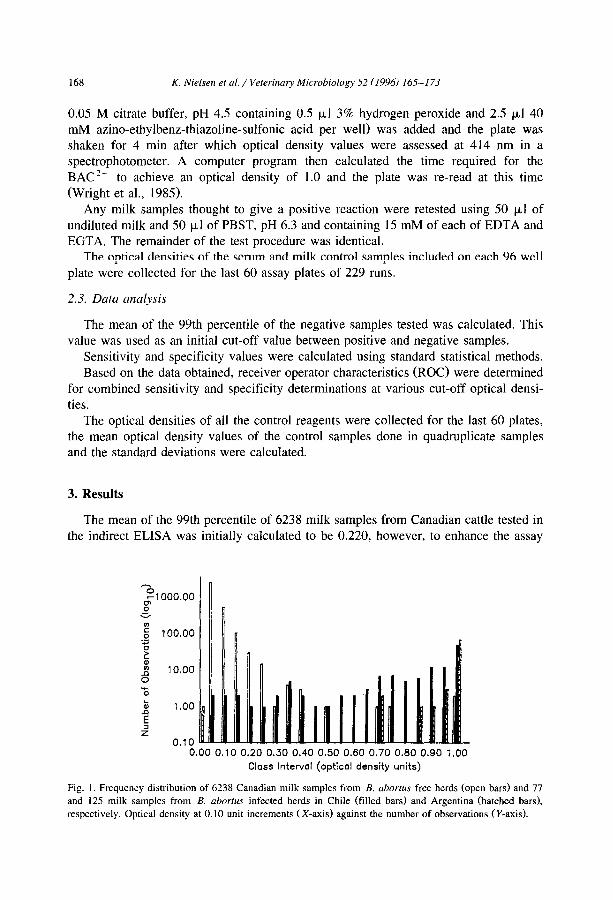
3. Results
The mean of the 99th I lercentile of 6238 milk samples from Canadian cattle tested in the indirect ELISA was ii nitially calculated to be 0.220, however, to enhance the assay
^o -1000.00
0” 3
0.00 0.10 0.20 0.30 0.40 0.50 0.60 0.70 0.60 0.90 1.00
Class Interval (opticol density units)
Fig. 1. Frequency distribution of 6238 Canadian milk samples from B. abortus free herds (open bars) and 77
and 125 milk samples from B. abortus infected herds in Chile (filled bars) and Argentina (hatched bars), respectively. Optical density at 0.10 unit increments (X-axis) against the number of observations (Y-axis).
K. Nielsen et al. / Veterinary Microbiology 52 (1996) 165-l 73 169
/- lOO--
so*- Specificity (%)
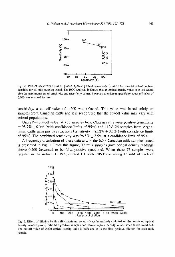
Fig. 2. Percent sensitivity (y-axis) plotted against percent specificity (x-axis) for various cut-off optical
densities for all milk samples tested. The ROC analysis indicated that an optical density value of 0.110 would
give the maximum sum of sensitivity and specificity values, however, to enhance specificity, a cut-off value of
0.200 was selected for use.
sensitivity, a cut-off value of 0.200 was selected. This value was based solely on samples from Canadian cattle and it is recognized that the cut-off value may vary with animal populations.
Using this cut-off value, 76/77 samples from Chilean cattle were positive (sensitivity = 98.7% :k 0.3% (with confidence limits of 95%) and 119/125 samples from Argen- tinian cattle gave positive reactions (sensitivity = 95.2% + 3.7% (with confidence limits of 95%). The combined sensitivity was 96.5% + 2.5% at a confidence limit of 95%.
A frequency distribution of these data and of the 6238 Canadian milk samples tested is presented in Fig. 1. From this figure, 77 milk samples gave optical density readings above 0.200 (assumed to be false positive reactions). When these 77 samples were retested in the indirect ELISA, diluted 1:l with PBST containing 15 mM of each of
1.2
0.0 0 400 800 1200 1600 2000 2400 2800 3200
Reciprocal dilution
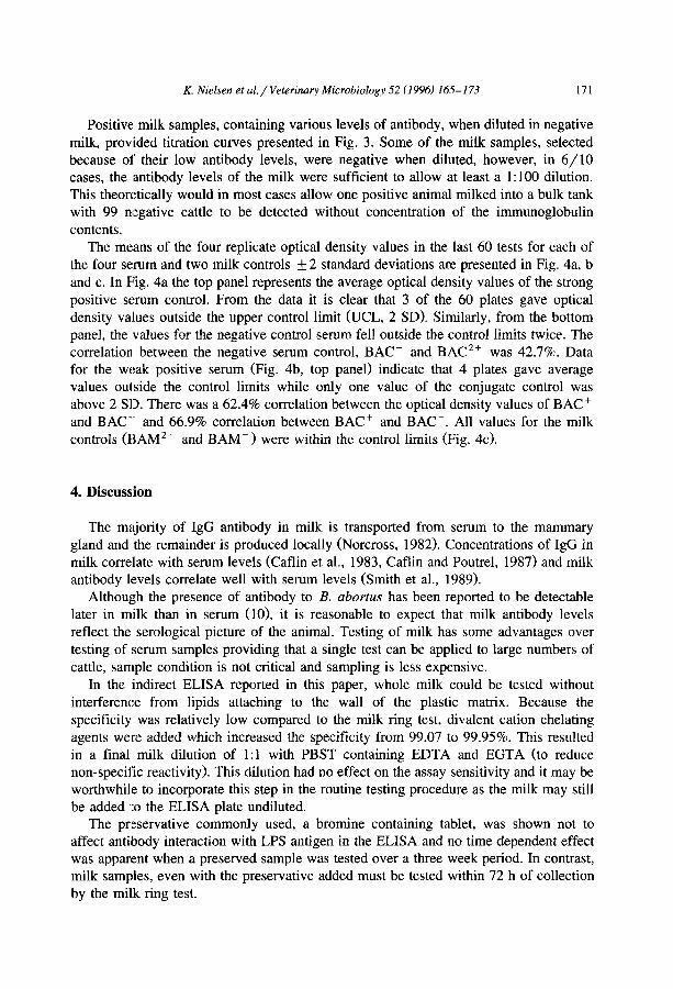
Fig. 3. Effect of dilution (with milk containing no anti-Brucella antibody), plotted on the x-axis on optical
density values (y-axis). The five positive samples had various optical density values when tested undiluted.
The cut-off value of 0.200 optical density units is indicated as is the final positive dilution for each milk
sample.
170 K. Nielsen et al. / Veterinary Microbiology 52 (1996) 165-l 73
EDTA and EGTA, the number of false positive reactions were reduced to 3, for a final specificity determination of 99.95% _+ 0.05% with confidence limits of 95%. This compares with 27 false positive reactions encountered with the milk ring test for a
specificity value of 99.28%. The ROC analysis of the combined data (Fig. 2) indicated that for maximum
combined sensitivity and specificity, a cut-off value of 0.110 would be appropriate, however, the specificity would be below that of the milk ring test. Using the higher cut-off value of 0.200, specificity (99.8%) was nearly at its maximum and the sensitivity was only slightly diminished (96.5%).
The bromine based preservative used in this study did not effect the antibody-antigen interaction of a positive milk sample when tested over a three week period. Thus when the antibody level in the milk was titrated there was no difference between a sample stored at -20°C and or at 4’C with the preservative.
ua
RAQc++
La
ua BAQc+
La.
02
O-_-- UCL
aAQcC
-02 I&L
1.8
12 BA.M++ 1
02
0.8
0.4
02 ua
08 Law-
.02-
PLATE NUMBER
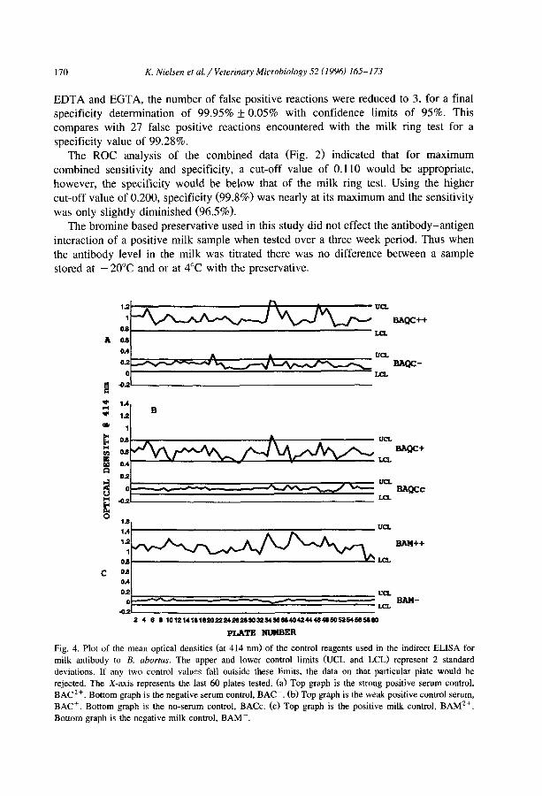
Fig. 4. Plot of the mean optical densities (at 414 nm) of the control reagents used in the indirect ELISA for
milk antibody to B. abortus. The upper and lower control limits (UCL and LCL) represent 2 standard
deviations. If any two control values fall outside these limits, the data on that particular plate would be
rejected. The X-axis represents the last 60 plates tested. (a) Top graph is the strong positive serum control, BAC2+. Bottom graph is the negative serum control, BAC-. (b) Top graph is the weak positive control serum,
BAC+. Bottom graph is the no-serum control, BACc. (c) Top graph is the positive milk control, BAM*+.
Bottom graph is the negative milk control, BAM-.
K. Nielsen et d/Veterinary Microbiology 52 (1996) 165-173 171
Positive milk samples, containing various levels of antibody, when diluted in negative milk, provided titration curves presented in Fig. 3. Some of the milk samples, selected because OS their low antibody levels, were negative when diluted, however, in 6/10 cases, the antibody levels of the milk were sufficient to allow at least a 1:lOO dilution. This theoretically would in most cases allow one positive animal milked into a bulk tank with 99 nlegative cattle to be detected without concentration of the immunoglobulin
contents. The means of the four replicate optical density values in the last 60 tests for each of
the four serum and two milk controls f 2 standard deviations are presented in Fig. 4a, b and c. In Fiig. 4a the top panel represents the average optical density values of the strong
positive serum control. From the data it is clear that 3 of the 60 plates gave optical density values outside the upper control limit @JCL, 2 SD>. Similarly, from the bottom panel, the values for the negative control serum fell outside the control limits twice. The correlation between the negative serum control, BAC- and BAC*+ was 42.7%. Data for the weak positive serum (Fig. 4b, top panel) indicate that 4 plates gave average values outside the control limits while only one value of the conjugate control was above 2 SD. There was a 62.4% correlation between the optical density values of BAC+ and BAC’ and 66.9% correlation between BAC+ and BAC-. All values for the milk controls @AM 2 + and BAM-) were within the control limits (Fig. 4~).
4. Discussion
The majority of IgG antibody in milk is transported from serum to the mammary gland and the remainder is produced locally (Norcross, 1982). Concentrations of IgG in milk correlate with serum levels (Caflin et al., 1983, Caflin and Poutrel, 1987) and milk antibody llsvels correlate well with serum levels (Smith et al., 1989).
Although the presence of antibody to B. abortus has been reported to be detectable later in milk than in serum (lo), it is reasonable to expect that milk antibody levels reflect the serological picture of the animal. Testing of milk has some advantages over testing of serum samples providing that a single test can be applied to large numbers of cattle, sample condition is not critical and sampling is less expensive.
In the indirect ELISA reported in this paper, whole milk could be tested without interference from lipids attaching to the wall of the plastic matrix. Because the specificity was relatively low compared to the milk ring test, divalent cation chelating agents were added which increased the specificity from 99.07 to 99.95%. This resulted in a final milk dilution of 1: 1 with PBST containing EDTA and EGTA (to reduce non-specific reactivity). This dilution had no effect on the assay sensitivity and it may be worthwhile to incorporate this step in the routine testing procedure as the milk may still be added ‘to the ELISA plate undiluted.
The preservative commonly used, a bromine containing tablet, was shown not to affect antibody interaction with LPS antigen in the ELISA and no time dependent effect was apparent when a preserved sample was tested over a three week period. In contrast, milk samples, even with the preservative added must be tested within 72 h of collection by the milk ring test.
172 K. Nielsen et al. / Veterinary Microbiology 52 (1996) 165-173
The ELISA is able to detect a single positive milk sample diluted in 100 negative samples (Forschner and Buenger, 1986). This finding is confirmed here, however it was also demonstrated that some milk samples with low antibody levels were only positive when tested at dilutions less than 1:lO (Fig. 3). Similarly, in the milk ring test, assessment of low positive reactions is frequently difficult and screening of bulk milk samples without preconcentration of the immunoglobulin fraction may result in false
negative reactions (Forschner and Buenger, 1986). The sensitivity of the milk ring test has been shown to be considerably less than that of the indirect ELISA (Kerkhofs et al., 1990) and has been reported as low as 88.5% (Nicoletti, 1969) and 89% (Hunter and Allen, 1972), both using samples from animals from which B. abortus had been isolated. Because of the condition of the milk samples from B. abortus infected herds used in this study, a direct sensitivity comparison between the milk ring test and the indirect ELISA was not possible. However, the indirect ELISA appeared to increase the sensitivity and the specificity of brucellosis surveillance over the current milk test in use, the milk ring test based on results obtained with samples tested in this study and data on milk ring test sensitivity from the literature.
The quality control charts (Fig. 4a, b and c> indicate that the indirect ELISA is functioning consistently over time with minor fluctuations in the day to day performance of the assay. The floating mean and the dynamic specification limits of two standard deviations ensure assay uniformity by accounting for minor operator variations and environmental conditions. Plates that have values outside the control limits are rejected and the test results are not used.
In conclusion, we have developed and validated an indirect ELISA for detection of bovine milk antibody to B. abortus which appears to have a higher sensitivity and specificity than the milk ring test currently in use.
References
Boraker, D.K., Stinebring, W.R. and Kunkel, J.R., 1981. BrucELISA: an enzyme-antibody immunoassay for
detection of Brucella abortus antibodies in milk: correlation with the Brucella ring test and with shedding
of viable organisms. J. Clin. Microbial., 14: 396.
Bruner, J.A., Thoen. C.O. and Hall, M.R., 1983. Evaluation of poly-B in an ELISA for detection of brucella
antibodies in cow’s milk. ASM Abstract. 80: 83 (#E21).
Caflin, J.P. and Poutrel, B., 1987. Physiological and pathological factors influencing bovine immunoglobulin
G2 concentration in milk. J. Dairy Sci., 71: 2035.
Caflin, J.P., Poutrel. B. and Rainard, P., 1983. Physiological and pathological factors influencing bovine immunoglobulin Gl concentration in milk. J. Dairy Sci., 66: 2161.
Forschner, V.E. and Buenger, I., 1986. Detection of IBR/IPV, EBL and brucellosis antibodies in samples of
bulk milk with ELISA using a simple method for concentration of antibodies. Dtsch. Tieraertzl.
Wochenschr., 93: 112.
Heck, F.C., Williams, J.D., Pruett, J., Sanders, R. and Zink, D.L., 1980. Enzyme linked immunosorbent assay for detection of antibodies to Brucella abortus in bovine milk and serum. Am. J. Vet. Res., 41: 2082.
Hunter, D. and Allen, J., 1972. An evaluation of milk and blood tests used to diagnose brucellosis. Vet. Rec., 91: 310.
Kerkhofs, P., Botton, Y., Thiange, P., Dekeyser, P. and Limet, J.N., 1990. Diagnosis of bovine brucellosis by enzyme immunoassay of milk. Vet. Microbial., 24: 73.
K. Nielsen et al. / Veterinary Microbiology 52 (1996) 165-l 73 173
MacMillan, A., 1990. Conventional serological tests. In: ed. Nielsen, K.H. and Duncan, J.R., Animal
Brucellosis. CRC Press, Boca Raton, FL.
Nicoletti, P., 1969. Further evaluation of serologic tests procedures used to diagnose brucellosis. Am. J. Vet.
Res., 30: 1811.
Nielsen, K.Hi., Kelly, L., Gall, D., Balsevicius, S., Bosse, J., Nicoletti, P. and Kelly, W., 1995. Comparison of
enzyme immunoassays for the diagnosis of bovine brucellosis. Prev. Vet. Med. (in press).
Norcross, N.L., 1982. Secretion and composition of colostrum and milk. J. Am. Vet. Med. Assoc., 181: 1057.
Smith, B.P., Oliver, D.G., Singh, P., 1989. Detection of Salmonella dublin mammary gland infection in carrier
cows, usutg an enzyme-linked immunosorbent assay for antibody in milk and serum. Am. J. Vet. Res., 50:
1352.
Thoen, C.0, Bruner, J.A., Luchsinger, D.W. and Pietz, D.E., 1983. Detection of Brucella antibodies of
different immunoglobulin classes in cow’s milk by enzyme-linked immunosorbent assay. Am. J. Vet. Res.,
44: 306.
Thoen, CO., Hopkins, M.P. and Pietz, D.E., 1980. Detection of Brucella antibodies in cow’s milk using an
aut0mate.d enzyme-linked immunosorbent assay. Proc. 2nd Int. Symp. Vet. Lab. Diag., 7: 228.
Thoen, CO., Pietz, D.E., Armbrust, A.L. and Harrington, R., Jr., 1979. Enzyme immunoassay for detection of
Brucella antibodies in cow’s milk. J. Clin. Microbial., 10: 222.
Wright, P.F., Kelly, W.A. and Gall, D.G., 1985. Application of a timing protocol to the reduction of inter-plate
variability in the indirect enzyme immunoassay for detection of anti-Brucella antibody. J. Immunoassay, 6:
189.