detection of eight β-thalassemia mutations using a dna enzyme immunoassay
TRANSCRIPT
lnt J Clin Lab Res (1996) 26: 136-139 �9 Springer-Verlag 1996
A. C o l o s i m o �9 G. Nove l l i �9 A. Cav icch in i B. Da l lap icco la
Detection of eight thalassemia mutations using a DNA enzyme immunoassay
Received: 28 February 1996/Accepted: 25 March 1996
Abstract We describe the use of a polymerase chain re- action-based method followed by a DNA enzyme immu- noassay for the simultaneous detection of the eight most common ,6-thalassemia mutations in the Mediterranean population. The method is specific, sensitive, and easily applicable in routine clinical laboratories for the molecu- lar diagnosis of,6-thalassemia patients and at risk couples.
Key words ,6-Thalassemia �9 Polymerase chain reaction �9 DNA enzyme immunoassay �9 Screening
Introduction
~ T h a l a s s e m i a (,6-thai) is a heterogenous, autosomal reces- sive disease resulting from reduced or absent production of the ,6-chain of human hemoglobin. '6-Thai is essentially characterized by hypochromic and hemolytic anemia [ 1 ]. The disorder has been reported from almost all populations and ethnic groups around the world, with a higher preva- lence in areas endemic for malaria, including the Mediter- ranean, North Africa, Middle East, India, southeast Asia, and southern China [2]. More than 130 different mutations in the ,6-globin locus have been identified [3], which cause defects in any of the steps of/~-globin gene expression and give rise t o / ~ - or ]Y-thai. There are a few common muta- tions and a variable number of rare mutations in each at- risk ethnic group. For example, while in the Singaporean population 11 different mutations account for approxi- mately 95% of the ,6-globin mutations [4], among Medi-
A. Colosimo - G. Novelli (~) . B. Dallapiccola Cattedra di Genetica Umana e Medica, Universith Tor Vergata di Roma, Via di Tot Vergata 135. 1-00133 Rome, Italy
A. Cavicchini Sorin Biomedica, Saluggia, Italy
B. Dallapiccola Ospedale CSS, IRCCS San Giovanni Rgtondo, Italy
terraneans, 8 mutations account for more than 92% of their defects [5].
A wide variety of polymerase chain reaction (PCR)- based techniques are available for the detection of these mutations. These include: restriction enzyme analysis [6], amplification refractory mutations system (ARMS) [7], dot-blot hybridization with allele-specific oligonucleotide probes [8, 9], reverse dot-blot analysis [ 10, 1 1], allele-spe- cific PCR by color amplification [12], multiplex amplifi- cation refractory mutation system [l 3], denaturing gradi- ent gel electrophoresis [141, chemical cleavage of mis- matched duplexes [ 15], heteroduplex analysis with univer- sal DNA heteroduplex generator molecules [16], and di- rect DNA sequencing [4]. Despite efficiency, reliability, and ease of use, these methods are expensive, t ime-con- suming, and not ideal for the rapid screening of a large number of samples in a routine clinical laboratory.
We set up a microtiter based method which uses PCR in conjunction with DNA e n z y m e immunoassay (DEIA) [ 17] and allows the analysis o f the eight most common ,6- thai mutations accounting for about 92% of ,6-globin al- leles in the Italian population [5]. The method is specific, sensitive, rapid, simple, non-radioactive, and appropriate for clinical laboratories involved in routine screening.
Materials and methods
Sample preparation
Genomic DNA was isolated from peripheral blood cells of normal. carrier, and where available, affected subjects, in which the presence or absence of the ,B-thai mutations had been previously documented by ARMS and restriction enzyme analysis [6, 7]. About 200 ng of each sample were used for a PCR, using two pairs of primers which amplify two different segments of the /~-glohin gene (Table 1 ) [9]. The first pair of primers, R95 and R94. amplifies a 720-base pair (bp) fragment, including the/3'+-87 (C--+G), ,6~ (C-+T),/~6 (-A), fl~ nt 1 (G---+A), ]YIVSI nt6 (T-+C), ]3'+IVSI nt 1 I0 (G-+A), and /Y'IVSII nt 1 (G-+A) mutations. The second pair of primers, R 92 and R61, amplifies a 487-bp fragment, spanning the /~IVSII nt745 (C---+G) mutation. The amplifications were performed in a DNA ther- mal cycler (Perkin Elmer-Roche, USA) with 30 denaturation cycles
A. Colosimo et al.: Detection of/3-thalassemia mutations with DNA enzyme immunoassay
Table 1 ]3-Thatassemia (/3-thal)-specific primers R 95: 5"-TAAGCCAGTGCCAGAAGAGCC-3 ' {pos. position) R 94: 5 ' - TC CTA TG A CA TGAACTTAAC-Y
R 92: 5'-TGCATATAAATTGTAACTGAT-3' R 61 : 5 ' -CACTGACCTCCCACATTCCC-3"
137
From -158 to-138 IVSII: from pos. 62 to pos. 42
IVSII: from pos. 680 to pos. 699 From pos. 60 to pos. 40 3' to the polyA signal
Table 2 /3-Thai-specific oligonucleotide probes
/3 + -87 (C-+G)
/~6(-A)
,/3~ ( C--)T}
/JqVSI ntl (G-+A}
,6~IVSI nI6 (T-)C)
,6'+IVSI nt I l0 {G--+A)
fl~ nt I (G-)A}
,6'+IVSII nt745 (C--)G}
M: 5'-GAGCCACACGCTAGGGTG-3" N: 5'-CAACCCTAGGGTGTGGCT-3'
M: 5'-TGACTCCTGGGAGAAGTCT-3' N: 5'-GACTTCTCCTCAGGAGTCA-3'
M: 5"-AGAACCTCTAGGTCCAAGG-3 ' N: 5'-CCTTGGACCCAGAGGTTCT-3"
M: 5'-CCTGGGCACATTGGTATCA-3' N: 5'-TGATACCAACCTGCCCAGG-3'
M: 5'-GCAGGTTGGCATCAAGGTT-3' N: 5 ' -AACCTTGATACCAACCTGC-3 '
M: 5'CTGCCTATTAGTCTATTTT-3' N: 5"-AAAATAGACCAATAGGCAG-3'
M: 5"-AACTTCAGGATGAGTCTAT-3' N: 5'-ATAGACTCACCCTGAAGTT-3'
M: 5'-ACAATCCAGGTACCATTCT-3" N: 5'-AGAATGGTAGCTGGATTGT-3'
at 94~ for I ram, annealing at 52~ for I min, and primer exten- sion at 72~ for I nlin. The PCR mixture includes a mixture ofde- oxynucleotide triphosphates (200 btM). 50 pM of each primer. 2 units of Super Taq (HT Biotechnology, UK), 1.5 mM magnesium chlo- ride, 50 mM potassium chloride, 10 mM TRIS-HCI (pH 9), 0.1% Tri- ton X-100, and 0.01% gelatin in a total volume of 100 btl. Products were loaded onto a 1.8% agarose gel and run in TRIS-borate buffer for 30 min. Gets were then stained with ethidium bromide to check the size and the quality of amplification.
DE1A analysis
Each biotinylated fl-thal-specific oligonucleotide probe (50 ng/well) was immobilized onto streptavidin-coated microtiter plates (ETI- IEMA, Sorin Biomedica, Italy) in 100 btl of TE buffer (10 mM TRIS, HCI, I mM EDTA, pH8), at 4~ for 16 h. These probes recognize the 16 /3-thai alleles, wild type and mutant: /3+-87 (C--+G), ,6~ (C-+T),/3~ (-A), ]3~IVSI nt I (G-->A), fl+IVSI nt6 (T- +C), ,6'+IVSI nt 1 I 0 (G- +A) and fl~ nt I (G--+A), JTIVSII nt 745 (C-+G) (Ta- ble 2). Following adsorption, the plates were washed three times with 200 btl of washing buffer (6.7 mM phosphate buffer, pH 6.5, 0.13 M sodium chloride, 40 mg of 2-ethylmercurithio-5-benzoxalcarboxyl- ic acid sodium salt, 1% Tween 20). About 5 btl of crude denatured ( 10 rain at 96~ PCR products were added to each well into 100 t.tl of hybridization buffer (I x SSC, 2x Denhardt's solution, i0 mM TRIS-HCI pH 7.5, I mM EDTA) and then incubated for I h at 37 ~ for/3~ (C-+T), at 50~ for/3+-87 (C--->G), if'6 (-A), ~IVSI nt 1 (G-+A), and /3~ nt6 (T-+C), and at 45~ for /3+IVSI n t l l 0 (G-+A),/3~ nt 1 (G-+A), and ,/3+IVSII nt745 (C-+G). Unbound components were removed by washing three times with washing so- lution. Hybrids between wild type and mutant fl--thal probes were identified by adding 100 I11 of a standard preparation of anti-DNA monoclonal antibody (mAb27-14-D9, Sorin Biomedica) ( 1 h at room temperature), which recognizes double-stranded DNA and 100 btl of horseradish peroxidase-labelled rabbit anti-mouse IgG antibody (Sorin Biomedica) diluted 2,000-fold in a phosphate buffer contain-
ing 1% fetal calf serum ( I h at room temperature). Single reactions were detected by adding I00 gl of ETI-IEMA chromogen/substrate mixture (27 g/I tetramethylbenzidine. 0.1 mM hydrogen peroxide), and incubating in the dark for 30 rain at room temperature. After stopping the reaction with 200 ul of 1 mM sulfuric acid, the ,/3-thai genotype of each amplified sample was determined by examining the absorbanee of each well at 450 nm (ETI-SYSTEM Fast Reader, Sorin Biomedica).
Results
In order to determine the optimal condit ions for the detec- tion of wild type or mutated fi-thal alleles the following variables were extensively investigated: amount of probe, amount of added PCR product, and hybridization temper- ature. The amount of immobil ized probe ranged from 10 to 100 ng/100 gl hybridizat ion volume. Opt imum perfor- mance was obtained with 50 ng of capture, normal or mu- tant, probe per well for each tested fi-thal allele. Different aliquots (from 5 to 50 btl) of added PCR products were in- vestigated, but the amount did not influence assay perfor- mance. Aliquots of 5 bl, l of single amplif icat ion products were selected for the test. The hybridization temperature was the critical parameter. Three sets of temperatures were selected for efficient hybridizat ion signals with both mu- tated and wild type probes: 50 ~ for ,/3+-87 (C-+G), /~~ (-A), fl~ nt 1 (G- +A), and ,6'+IVSI nt6 (T--~C) alleles, 45 ~ for /YlVSI nt 1 I0 (G---->A), ]3~ nt 1 (G--->A), and ]3'+IVSll nt745 (C--+G), and 37 ~ for the ,6~ (C-+T) mu- tation. Deviations of ___2 ~ from the selected temperature resulted in non-specific hybridization.
To verify the general applicabil i ty and specificity of this DEIA-based method, genomic DNA from 50 fl-thal pa- tients (courtesy of C. Rosatelli, Cagliari, Italy), previously characterized by the ARMS technique [7], and genomic DNA from 20 normal individuals were used. There was complete agreement between the results obtained by the two methods. As negative control we used unrelated PCR products of non-complementary sequences (exon 10 of the cystic fibrosis t ransmembrane regulator gene and human papilloma virus) on the microtiter plates coated with the ,&thai probes. In all cases we obtained negative signals, in- dist inguishable from background. A cut-off value, based on the mean for the negative control (0 .125+3SD) was 0.320_+0.04 (mean_+SD and was used for ,6+-87 (C--->G), ,~6 (-A), /~IVSI n t l (G->A), ]3'+IVSI nt6 (T-+C), /3+IVSI nt 110 (G->A), /3~ nt 1 (G-+A), and ,/~-IVSII nt745 (C--->G), while a cut-off of 0.13 was obtained for ]3~ (C--~T). There were no false-posit ive results at the selected cut-off values.
138
A
G
H
1 2 3 4 5 6 7
~ O ~ "
@,| @
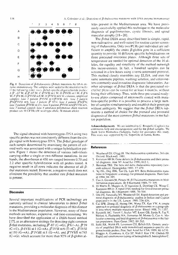
Fig. 1 Detection of/~-thalassemia Ifi-thal) mutations by DNA en- zyme immunoassay. The samples were added to the microtiter wells in ~he tolh)wing order: ro~'s. B-thai-specific oligonucleotide probes: A /,r -87 W; B ff-87 M: C fl~ nt I W; D/3~ nt I M; E/.f'6 W; F/~~ M; G/~[VSI nt6 W: H ]3'+IVSI nt6 M; columns, amplified samples: lane I patient /3~ nt I/]3'+IVSi nt6 hme 2 patient /~6/]5~[VSI nt6; lane 3 patient /~-87/+ laJte 4 patient ,/J~176 lane 5 patient/Y'IVSI nt I/+; lane 6 patient ~-'+[VSI nt6//~lVSl nt6. hme 7 normal control: lane 8 unrelated polymcrase chain reaction product (ex I0 CFTR) (W wild type allele, M mutant allele)
A. Colosimo et al.: Detection of ,~-thalasscmia inutations with DNA enzyme immunoassay
8 leles present in the Medi ter ranean area. We have previ- ously successful ly appl ied this technique to the molecular d iagnosis of papi l lomavirus , cys t ic fibrosis, and spinal muscular atrophy [ 1 8 - 2 0 ] .
The fl-thal DEIA assay desc r ibed here is s imple , rapid, non-radioact ive , and wel l -sui ted for routine carr ier screen- ing of thalassemia. Only two PCRs per individual are suf- f icient to amplify the entire /3-globin gene in a sufficient quanti ty to provide 16 different spec i f ic hybr id iza t ions on three precoated microti tre plates . Al though three sets of temperature are needed for op t imal detect ion of the 16 al- leles, the rapidi ty and s impl ic i ty of the method outweigh this inconvenience. In fact, a hundred samples can be screened in a few hours using a s emi -au toma ted protocol . This method closely resembles any ELISA, and uses the same automatic pipettes, washing solution, and co lor ime- ters commonly used in routine d iagnos t i c laboratories . An- other advantage of/~-thal DEIA is that the precoated mi- crot i ter plates can be stored for at least 4 months, without losing their efficiency. Thus, wi th three pre-coated ready- to-use plates composed of mic rowe l l s coated with muta- t ion-speci f ic probes it is poss ib le to process a large num- ber of samples s imul taneous ly and establ ish their genotype without ambiguity. We suggest that the ,~thal DEIA rep- resents a method of choice for the prenatal and postnatal d iagnosis of the most common/~- tha l mutat ions in the Ital- ian populat ion.
The signal obta ined with heterozygous DNA using two specific probes was not consis tent ly different from the sin- gle signal with homozygous DNA. The/~-thal genotype of each sample de te rmined by examining the pattern of col- ored wells was associa ted with a unique hybr idizat ion pat- tern. Figure 1 shows the detect ion of various individuals carrying ei ther a s ingle or two different mutations. In our hands, the absorbance at 450 nm ranged between 0.70 and 2.2 after specif ic hybr id iza t ion with all probes tested. A negative result in all rows indicates the absence of all ,8- thal mutat ions tested. However , a negative result does not e l iminate the poss ib i l i ty that another rare ~ t h a l mutat ion is present.
Discussion
Several important modi f ica t ions of PCR technology are current ly ut i l ized in cl inical laborator ies to detect ~-that mutations, p rovid ing a molecu la r d iagnosis o f this d isease in the Medi ter ranean populat ion. However , many of these methods are tedious, expensive , and t ime-consuming. We have descr ibed the appl ica t ion of a DEIA-based method [17], as an a l ternat ive strategy for detect ing eight common ]3-thal mutat ions, including/7+-87 (C--+G),/7~ ( -A) , /7~ (C--+T), ,/~--IVSI nt 1 ( G - > A ) , / ~ I V S I nt6 (T- +C), fl+IVSI nt 110 (G->A) , /3~ nt I (G--~A), and ]5'+IVSII nt745 (C--~G), which account for more than 92% of /3-globin al-
Acknowledgements We arc indebted to C. Rosatelli (Cagliari) t'or continuous help and encouragement and t\)r the/3-thai samples. We thank Sorin Biomedica (Saluggia, Italy) for assistance this study. This work was supported by Ist. Superiore di Sanit'a, Roma.
References
I. Weatherall D J. Clcgg JB. The thalassaemia syndromes, 3rd edn. Oxlk)rd: Blackwell. 1981.
2. Kazazian HH Jr. Gene defects in/~-thalassemia and their prena- tal diagnosis. Ann NY Acad Sci 1990: 612: 1.
3. Huisman THJ. The beta and delta thalassemia repository (sev- enth edition). Hemoglobin 1993: 17:479.
4. Ng ISL, Ong JBK, Tan CL, Law HY. Beta-thalassaemia muta- tions in Singapore: a strategy for prenatal diagnosis. Hum Gen- et 1994; 94: 385.
5. Cao A, Gossens M, Pirastu M. ,g-Thalassemia mutations in Med- iterranean populations. Br J Haematol 1989; 71: 309.
6. Di Marzo R, Maggio A, D'Agostino S, Dowling CE, Wong C. Kazazian HH Jr. A rapid DNA method for first-trimester prena- tal diagnosis. Br J Haematol 1988; 70: 504.
7. Old JM, Varawalla NY, Weatherall DJ. Rapid detection and pre- natal diagnosis of ~thalassemia: studies in Indian and Cypriot populations in the UK. Lancet i990: 336: 834.
8. Cat SPB, Zhang JZ, Huang DH, Wang ZX, Kan YW. A simple approach to prenatal diagnosis of/3-thalassemia in a geograph- ic area where multiple mutations occur. Blood 1988; 71: 1357.
9. Rosatelli MC, Tuveri T, Scalas MT, Leoni GB, Sardu R, Fa'a V, Meloni A, Pischedda MA, Demurtas M, Monni G, Cao A. Mo- lecular screening and fetal diagnosis of/~-thatassemia in the Ital- ian population. Hum Genet 1992; 89: 585.
10. Saiki RK, Walsh PS, Levenson CH, Erlich HA. Genetic analy- sis of amplified DNA with immobilized sequence-specific oli- gonucleotide probes. Proc Natl Acad Sci USA 1989; 86: 6230.
II. Maggio A. Giambona A, Cat SP. Wall J, Kan YW, Chehab FF. Rapid and simultaneous typing of hemoglobin S, hemoglobin C.
A. Colosimo et al.: Detection ol + fl-thalassemia mutations with DNA enzyme immunoassay
and seven Mediterranean ]3-thalassemia mutations by covalent 16. reverse dot-blot analysis: application to prenatal diagnosis in Sicily. Blood 1993; 81:239.
12. Chehab FF, Kan YW. Detection of sickle cell anemia mutation 17. by color DNA amplification. Lancet 1990: 335: 15.
13. Fortina P, Dotti G, Conant R. Monokian O. Parrella T, Hitch- cock W, Rappaport E, Schwartz E, Surrey S. Detection of the 18. most common mutations causing ]3-thalassemia in Mediterra- neans using a multiplex amplification refractory mutation system (MARMS). PCR Methods Applic 1992: 2: 163.
14. Losekoot M, Fodde R, Harteveld CL, Heeren van, Giordano PC. Bernini LF. Denaturing gradient gel electrophoresis and direct 19. sequencing of PCR amplified genomic DNA: a rapid and reli- able diagnostic approach to/~-thalassemia. Br J Haematol 1990; 76: 269.
15. Dianzani I, Camaschella C, Saglio G, Forrest S, Ramus S. Cotton RHG. Simultaneous screening lk)r fi-thalassemia muta- 20. tions by chemical cleavage of mismatch. Genomics 1991: I l: 48.
139
Sava,,e DA, Wood NAP, Bidwell JL. Fitches A. Old JM, Hui KM. Detection of/3-thalassemia mut:~tions using DNA hetero- duplex generator molecules. Br J Haematol 1995: 90: 564. Mantero G, Zonaro A, Albertini A, Bcrtolo P, Primi D. DNA en- zyme immunoassay: general method for detecting products of polymerase chain reaction. Clin Chem 1991: 37: 422. Sangiuolo F. De Santis L, Cavicchini A, Angetoni U, Romani- ni C, Novelli G, Dallapiccola B. A new method tk~r direct anal- ysis of polymerase chain reaction-amplified human papilloma- vires using DNA enzyme immunoassay. Int J Clin Lab Res 1994: 24: 223. Sangiuolo F, Maceratesi P, Mesoraca A, Botta A, Cavicchini A, Novelli G, Dallapiccola B. Simultaneous detection of AF508, G 542X, N 1303K, G 551 D. and 1717-1G--->A cystic fibrosis al- leles by a multiplex DNA enzyme immunoassay, lnt J Clin Lab Res 1995: 25: 142. Novelli G, Capon F, Levato C, Cavicchini A, Dallapiccola B. DNA enzyme immunoassay for improved molecular detection in spinal muscular atrophies. Clin Chem 1996: 42:643