detecting envelope stress by monitoring β-barrel assemblymbib.med.harvard.edu/pages/micro201/cell...
TRANSCRIPT

Article
Detecting Envelope Stressby Monitoring b-Barrel AssemblySeung-Hyun Cho,1,2,4 Joanna Szewczyk,1,2,4 Christina Pesavento,3,4 Matylda Zietek,3 Manuel Banzhaf,3
Paula Roszczenko,1,2 Abir Asmar,1,2 Geraldine Laloux,2 Ann-Kristin Hov,3 Pauline Leverrier,2 Charles Van der Henst,1,2
Didier Vertommen,2 Athanasios Typas,3,* and Jean-Francois Collet1,2,*1WELBIO2de Duve Institute
Universite catholique de Louvain, Avenue Hippocrate 75, Brussels 1200, Belgium3European Molecular Biology Laboratory, Genome Biology Unit, Meyerhofstrasse 1, 69117 Heidelberg, Germany4Co-first authors*Correspondence: [email protected] (A.T.), [email protected] (J.-F.C.)
http://dx.doi.org/10.1016/j.cell.2014.11.045
SUMMARY
The cell envelope protects bacteria from their sur-roundings. Defects in its integrity or assembly aresensed by signal transduction systems, allowing cellsto rapidly adjust. The Rcs phosphorelay responds toouter membrane (OM)- and peptidoglycan-relatedstress in enterobacteria. We elucidated how the OMlipoprotein RcsF, the upstream Rcs component,senses envelope stress and activates the signalingcascade. RcsF interacts with BamA, the majorcomponent of the b-barrel assembly machinery. Ingrowing cells, BamA continuously funnels RcsFthrough the b-barrel OmpA, displaying RcsF on thecell surface. This process spatially separates RcsFfrom the downstream Rcs component, which weshow is the inner membrane protein IgaA. The Rcssystem is activated when BamA fails to bind RcsFand funnel it to OmpA. Newly synthesized RcsF thenremains periplasmic, interacting with IgaA to activatethe cascade. Thus RcsF senses envelope damage bymonitoring the activity of the Bam machinery.
INTRODUCTION
The cell envelope of Gram-negative bacteria consists of two
membranes, separated by a viscous periplasm that contains
the peptidoglycan (PG). The envelope is a permeability and
structural barrier, which is essential for cell shape and growth,
and serves as interface to the environment. To monitor their sur-
roundings, bacteria use a number of signaling cascades that
transduce the information from their envelope to their decision
center (cytoplasm).
Bacteria also constantlymonitor the growth and assembly sta-
tus of their envelope. As this compartment is devoid of energy,
transport and assembly of its structural subunits are controlled
by multicomponent protein machineries, which span the en-
velope and utilize energy from the cytoplasm (Silhavy et al.,
2010). These machineries must tightly coordinate their function,
as assembly of the different envelope layers is interlinked (com-
1652 Cell 159, 1652–1664, December 18, 2014 ª2014 Elsevier Inc.
ponents synthesizing one layer often reside in or interact with the
other layer [Typas et al., 2012]) and coupled to growth rate. It is
therefore vital for bacteria to detect when envelope assembly is
perturbed and to rapidly fix or contain the damage.
Many signaling systems can sense envelope perturbations in
E. coli and mount a repair and/or a preventive response to mini-
mize the damage. The best understood system is the sE stress
response, which uses three proteins to sense accumulation of
unassembled OM porins (OMPs) and LPS in the periplasm
(Lima et al., 2013; Walsh et al., 2003). In response to these in-
sults, sE directs the transcription of genes that facilitate OMP
and LPS assembly, transport, and turnover (Rhodius et al.,
2006) and of small RNAs that block the expression of various
OMPs and of the most abundant OM lipoprotein, Lpp (Gogol
et al., 2011; Guo et al., 2014). In contrast to sE, other signal trans-
duction systems associated with envelope damage surveillance
in E. coli are less well understood, and in most cases, the direct
activating signal is obscure.
The Rcs phosphorelay is one of the most complex bacterial
signaling systems. It is induced mostly by OM and cell wall dam-
age (Evans et al., 2013; Farris et al., 2010; Laubacher and Ades,
2008; Majdalani and Gottesman, 2005). In response to these
cues, Rcs controls the expression of genes involved in motility,
biofilm formation, virulence, and periplasmic quality control (Maj-
dalani and Gottesman, 2005). The complexity of the system,
both at the input and output level, has been an obstacle in dis-
secting it and fully addressing its physiological role. Unlike
typical two-component systems consisting of an inner mem-
brane (IM) sensor histidine kinase (HK) and a cytoplasmic
response regulator (RR), the Rcs system has at least six compo-
nents (Figure 1A). In addition to RcsC (HK) and RcsB (RR), the
system contains an intermediate IM phosphorelay protein,
RcsD, an auxiliary nonphosphorylatable transcription factor
RcsA, and two proteins that act upstream of the phosphorelay
cascade and are associated with signal sensing, YrfF and
RcsF (Cano et al., 2002; Castanie-Cornet et al., 2006). YrfF is
an IM protein, mostly characterized in Salmonella Typhimurium,
which downregulates the Rcs pathway by an unknown mecha-
nism (Domınguez-Bernal et al., 2004). Deletion of the gene is le-
thal, unless the Rcs phosphorelay is also inactivated (Cano et al.,
2002). yrfF has been renamed to igaA in S. Typhimurium and we
use the same nomenclature for the E. coli gene in this paper.
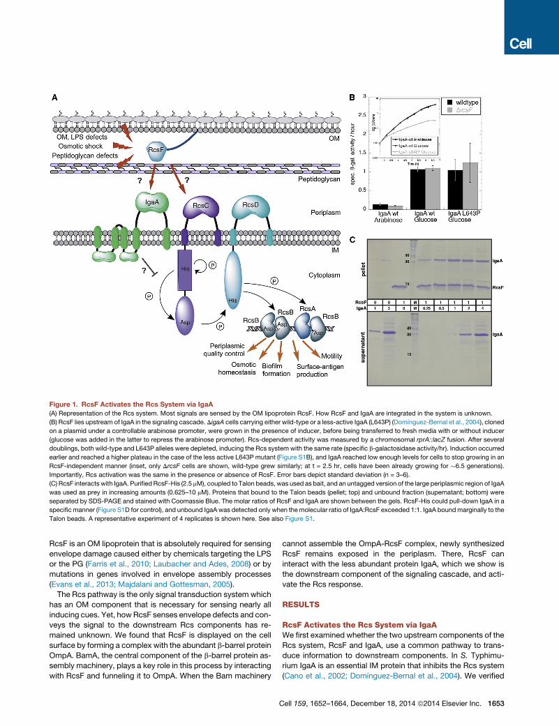
Figure 1. RcsF Activates the Rcs System via IgaA
(A) Representation of the Rcs system. Most signals are sensed by the OM lipoprotein RcsF. How RcsF and IgaA are integrated in the system is unknown.
(B) RcsF lies upstream of IgaA in the signaling cascade. DigaA cells carrying either wild-type or a less-active IgaA (L643P) (Domınguez-Bernal et al., 2004), cloned
on a plasmid under a controllable arabinose promoter, were grown in the presence of inducer, before being transferred to fresh media with or without inducer
(glucose was added in the latter to repress the arabinose promoter). Rcs-dependent activity was measured by a chromosomal rprA::lacZ fusion. After several
doublings, both wild-type and L643P alleles were depleted, inducing the Rcs systemwith the same rate (specific b-galactosidase activity/hr). Induction occurred
earlier and reached a higher plateau in the case of the less active L643P mutant (Figure S1B), and IgaA reached low enough levels for cells to stop growing in an
RcsF-independent manner (inset, only DrcsF cells are shown, wild-type grew similarly; at t = 2.5 hr, cells have been already growing for �6.5 generations).
Importantly, Rcs activation was the same in the presence or absence of RcsF. Error bars depict standard deviation (n = 3–6).
(C) RcsF interacts with IgaA. PurifiedRcsF-His (2.5 mM), coupled to Talon beads, was used as bait, and an untagged version of the large periplasmic region of IgaA
was used as prey in increasing amounts (0.625–10 mM). Proteins that bound to the Talon beads (pellet; top) and unbound fraction (supernatant; bottom) were
separated by SDS-PAGE and stained with Coomassie Blue. The molar ratios of RcsF and IgaA are shown between the gels. RcsF-His could pull-down IgaA in a
specific manner (Figure S1D for control), and unbound IgaA was detected only when themolecular ratio of IgaA:RcsF exceeded 1:1. IgaA boundmarginally to the
Talon beads. A representative experiment of 4 replicates is shown here. See also Figure S1.
RcsF is an OM lipoprotein that is absolutely required for sensing
envelope damage caused either by chemicals targeting the LPS
or the PG (Farris et al., 2010; Laubacher and Ades, 2008) or by
mutations in genes involved in envelope assembly processes
(Evans et al., 2013; Majdalani and Gottesman, 2005).
The Rcs pathway is the only signal transduction system which
has an OM component that is necessary for sensing nearly all
inducing cues. Yet, howRcsF senses envelope defects and con-
veys the signal to the downstream Rcs components has re-
mained unknown. We found that RcsF is displayed on the cell
surface by forming a complex with the abundant b-barrel protein
OmpA. BamA, the central component of the b-barrel protein as-
sembly machinery, plays a key role in this process by interacting
with RcsF and funneling it to OmpA. When the Bam machinery
C
cannot assemble the OmpA-RcsF complex, newly synthesized
RcsF remains exposed in the periplasm. There, RcsF can
interact with the less abundant protein IgaA, which we show is
the downstream component of the signaling cascade, and acti-
vate the Rcs response.
RESULTS
RcsF Activates the Rcs System via IgaAWe first examined whether the two upstream components of the
Rcs system, RcsF and IgaA, use a common pathway to trans-
duce information to downstream components. In S. Typhimu-
rium IgaA is an essential IM protein that inhibits the Rcs system
(Cano et al., 2002; Domınguez-Bernal et al., 2004). We verified
ell 159, 1652–1664, December 18, 2014 ª2014 Elsevier Inc. 1653
that igaA is also essential in E. coli and using an established pipe-
line for large-scale testing of genetic interactions (Typas et al.,
2008), we showed that, among a collection of knockout mutants
for all E. coli nonessential genes (Baba et al., 2006), an igaA dele-
tion was viable only when combined with deletions of rcsB, rcsC,
and rcsD (Figure S1A available online). Thus, E. coli and S. Typhi-
murium igaA play similar roles. Importantly, deletion of rcsF did
not suppress igaA lethality, implying that IgaA lies downstream
of RcsF in the signaling cascade. In agreement with this config-
uration, depletion of igaA activated the Rcs system indepen-
dently of RcsF (Figures 1B and S1B).
We next testedwhether IgaA and RcsF are physically linked by
expressing a tagged version of the only periplasmic domain of
IgaA (IgaAperi; �32 kDa) in the periplasm and pulling down its
interaction partners after crosslinking with DTSSP (3,30-dithio-bis[sulfosuccinimidylpropionate]), which cannot cross the IM.
RcsF was identified by both mass spectrometry (MS) and west-
ern blot (Figure S1C and Table S1). Likewise, a purified tag-less
version of IgaAperi and a soluble His-tagged version of RcsF
directly interacted, forming a complex with a 1:1 stoichiometry
(Figures 1C and S1D). These results support a model in which
RcsF activates the Rcs system by interacting with IgaA, likely
alleviating its inhibitory effect on the signaling cascade.
By Forming a Complex with the b-Barrel Proteins OmpAand BamA, RcsF Is Occluded from IgaAWild-type RcsF turns on the signaling cascade only upon enve-
lope stress, suggesting that RcsF is physically occluded from IM
IgaA under steady-state growth. This physical occlusion is tightly
interconnected with the OM location of RcsF, as rerouting RcsF
to the IM (RcsFIM) or expressing it as a soluble periplasmic pro-
tein (RcsFperi), constitutively activates the Rcs system (Farris
et al., 2010; Tao et al., 2012). We therefore looked for the under-
lying occlusion mechanism.
RcsF is composed of a 31-residue intrinsically disordered
N-terminal linker (S17-T47), which connects its globular domain
(P48-K134; referred to as ‘‘signaling domain’’) to the lipidated
cysteine residue anchoring the protein to the OM (Figure S2A)
(Leverrier et al., 2011). We first tested whether this linker is
cleaved under stress, releasing RcsF in the periplasm, by frac-
tionating cells exposed to various Rcs-inducing stresses. RcsF
was never detected in the soluble fraction (Figure S2B). We
then determinedwhether RcsFwas occluded from IgaA by being
sequestered by other proteins. To find proteins interacting with
RcsF, we performed in vivo DTSSP crosslinking in both DrcsF
and wild-type cells. Three RcsF-containing protein complexes
were detected (Figure 2A; marked as 1, 2, and 3). To identify
them, the RcsF-interacting complexes were immunoprecipi-
tated and analyzed by MS after reversing the crosslinks. We
identified BamA, the core protein for b-barrel assembly, and
the b-barrels OmpA, OmpC and OmpF as potential RcsF inter-
acting partners (Table S2A). We further verified these interac-
tions by analyzing the immunoprecipitated samples by western
blot using antibodies specific for the interacting proteins. We
confirmed that the �115 kDa band (complex 1) contained
BamA (�100 kDa) (Figure S2C). Only OmpA (38 kDa) could be
detected in the �55 kDa complex (complex 2) (Figure S2C),
but not OmpC (40 kDa) and OmpF (39 kDa) (data not shown),
1654 Cell 159, 1652–1664, December 18, 2014 ª2014 Elsevier Inc.
suggesting that OmpA was the major interaction partner
involved in this complex. Consistently, upon depleting the es-
sential BamA or deleting ompA, the respective complexes dis-
appeared (Figure 2B). Using an lpp deletionmutant, we identified
that the protein involved in complex 3 (�25 kDa) was Lpp (8 kDa),
the most abundant OM lipoprotein (Figure S2D).
We further verified the specificity of these interactions by site-
specific photocrosslinking, inserting the crosslinkable amino acid
p-benzoyl-L-phenylalanine (pBpa) at 25 specific positions in
RcsF. Thereby we could map the interaction interface of RcsF
with its binding partners more precisely, and lower the risk of
nonspecific interactions (pBpa can only form covalent bonds
with residues at a very close proximity [3 A], whereas DTSSP
has a 12 A spacer). We selected 21 residues located on the sur-
face of the signaling domain and 4 located in the N-terminal linker
(Figure S3A). Following UV-exposure, 6/25 variants formed the
previously observed 55 kDa complex with OmpA (Figures 2C
and S3B). The identity of OmpA was confirmed by MS after im-
munoprecipitating RcsFK40pBPA-OmpA (OmpC and OmpF were
not detected in the sample, Table S2B). High complex levels
were observed when pBpa was inserted in the N-terminal linker
and at the tip of the signaling domain of RcsF (Figure S3C).
Four of these variants could also form the 115 kDa complex cor-
responding to BamA-RcsF (Figures 2C and S3D), which was
confirmed with a BamA antibody (Figure S3D). As none of the
25 pBpa-containing variants was found in complex with Lpp,
and as Lpp is the most abundant protein in E. coli, which could
lead to nonspecific interactions, we decided not to follow up on
this interaction. Altogether, these results indicated that RcsF in-
teracts specifically with BamA and OmpA. Importantly, the levels
of the RcsFK40pBPA-OmpA complex were�30%–40%of these of
free RcsF, indicating that 25%–30% of total RcsF is bound to
OmpA (Figures S3E and S3F). Given that photocrosslinking effi-
ciency at optimal conditions can reach 40% (Zhang et al.,
2011), we concluded that most RcsF is in complex with OmpA.
The Bam Machinery Assembles the RcsF-OmpAComplex and Is Key for the Sensing Role of RcsFThe interactions of RcsF with BamA and OmpA suggested that
in nonstress conditions RcsF is occluded from IgaA by interact-
ing with OM proteins, but that these interactions are disturbed
upon envelope stress, enabling RcsF to interact with IgaA. If
OmpA and BamA occlude RcsF from IgaA under nonstress con-
ditions, then the Rcs system should be activated when bamA or
ompA are knocked down/out. We found that an ompA deletion
induced the Rcs system by �3-fold, with induction being
dependent on RcsF (Figure 2D). As OmpC and OmpF were
also identified as RcsF partners, we tested the effect of deleting
ompC or ompF on Rcs activity. Whereas the system was only
marginally induced in the ompC mutant, the ompF deletion
had no impact (Figure 2D). When ompC and ompF deletions
were combined together or with ompA, synergistic effects
were observed, but the absence of OmpA was clearly the
most important contributor of the three to the activation of
Rcs (Figure 2D), consistently with our interaction data (Figures
2 and S3). In contrast to the omp mutants, the Rcs system
was fully induced in the bamA knockdown (bamA101) mutant
(Figure 2D). In this strain, BamA levels decrease �5-fold without
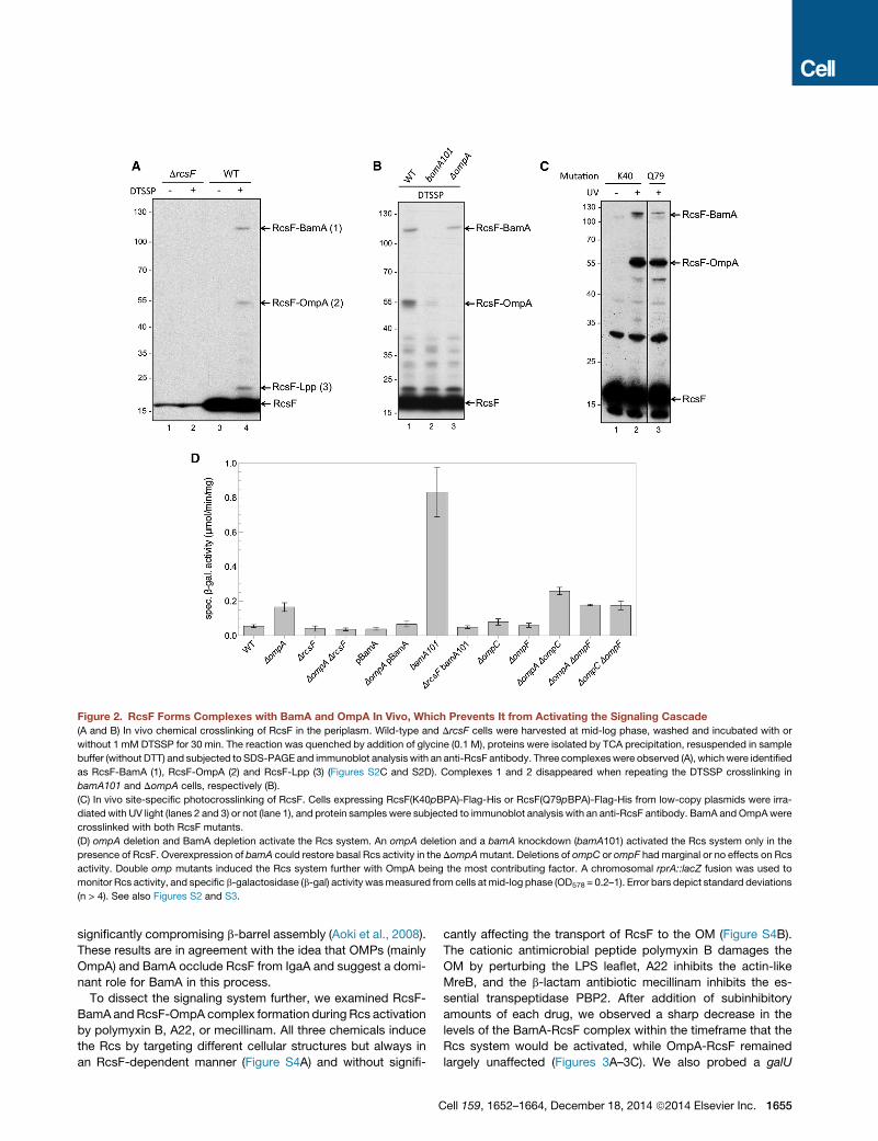
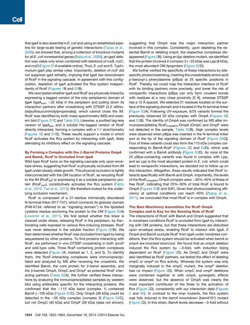
Figure 2. RcsF Forms Complexes with BamA and OmpA In Vivo, Which Prevents It from Activating the Signaling Cascade
(A and B) In vivo chemical crosslinking of RcsF in the periplasm. Wild-type and DrcsF cells were harvested at mid-log phase, washed and incubated with or
without 1 mM DTSSP for 30 min. The reaction was quenched by addition of glycine (0.1 M), proteins were isolated by TCA precipitation, resuspended in sample
buffer (without DTT) and subjected to SDS-PAGE and immunoblot analysis with an anti-RcsF antibody. Three complexeswere observed (A), whichwere identified
as RcsF-BamA (1), RcsF-OmpA (2) and RcsF-Lpp (3) (Figures S2C and S2D). Complexes 1 and 2 disappeared when repeating the DTSSP crosslinking in
bamA101 and DompA cells, respectively (B).
(C) In vivo site-specific photocrosslinking of RcsF. Cells expressing RcsF(K40pBPA)-Flag-His or RcsF(Q79pBPA)-Flag-His from low-copy plasmids were irra-
diated with UV light (lanes 2 and 3) or not (lane 1), and protein samples were subjected to immunoblot analysis with an anti-RcsF antibody. BamA andOmpAwere
crosslinked with both RcsF mutants.
(D) ompA deletion and BamA depletion activate the Rcs system. An ompA deletion and a bamA knockdown (bamA101) activated the Rcs system only in the
presence of RcsF. Overexpression of bamA could restore basal Rcs activity in the DompAmutant. Deletions of ompC or ompF had marginal or no effects on Rcs
activity. Double omp mutants induced the Rcs system further with OmpA being the most contributing factor. A chromosomal rprA::lacZ fusion was used to
monitor Rcs activity, and specific b-galactosidase (b-gal) activity wasmeasured from cells at mid-log phase (OD578 = 0.2–1). Error bars depict standard deviations
(n > 4). See also Figures S2 and S3.
significantly compromising b-barrel assembly (Aoki et al., 2008).
These results are in agreement with the idea that OMPs (mainly
OmpA) and BamA occlude RcsF from IgaA and suggest a domi-
nant role for BamA in this process.
To dissect the signaling system further, we examined RcsF-
BamA and RcsF-OmpA complex formation during Rcs activation
by polymyxin B, A22, or mecillinam. All three chemicals induce
the Rcs by targeting different cellular structures but always in
an RcsF-dependent manner (Figure S4A) and without signifi-
C
cantly affecting the transport of RcsF to the OM (Figure S4B).
The cationic antimicrobial peptide polymyxin B damages the
OM by perturbing the LPS leaflet, A22 inhibits the actin-like
MreB, and the b-lactam antibiotic mecillinam inhibits the es-
sential transpeptidase PBP2. After addition of subinhibitory
amounts of each drug, we observed a sharp decrease in the
levels of the BamA-RcsF complex within the timeframe that the
Rcs system would be activated, while OmpA-RcsF remained
largely unaffected (Figures 3A–3C). We also probed a galU
ell 159, 1652–1664, December 18, 2014 ª2014 Elsevier Inc. 1655
Figure 3. RcsF-BamA Is More Sensitive
than RcsF-OmpA to Envelope Stress
(A–C) RcsF-BamA and RcsF-OmpA complex for-
mation upon treatment with different cues sensed
by RcsF (see also Figure S4A). (A) Cells were
treated with 0.5 mg/ml polymyxin B when they
reached an OD600 of 0.4, and samples were
collected 10 min later, crosslinked with DTSSP
and immunoblotted with an anti-RcsF antibody.
(B and C) Cells were treated with mecillinam
(0.3 mg/ml) or A22 (5 mg/ml) when they reached an
OD600 of 0.2, samples were collected at indicated
time points after stress induction, and were sub-
jected to DTSSP crosslinking and immunoblot. In
all three stresses the RcsF-BamA complex dis-
appeared when the Rcs system was activated
(Figure S4A), whereas the RcsF-OmpA complex
remained largely unaffected.
(D) BamA overexpression shifts all RcsF to BamA.
In vivo DTSSP crosslinking of wild-type cells
harboring an empty vector (pET3a) or a vector
expressing BamA (pBamA). In all panels, DTSSP
crosslinking and immunoblot were done as in
Figure 2A, and a representative experiment is
shown (n = 3–4). See also Figure S4.
mutant that cannot produce UDP-D-glucose, a precursor for
LPS and other surface-exposed sugars, and in which the Rcs
system is constitutively turned on in an RcsF-dependent manner
(Figure S4C) (Girgis et al., 2007). Similarly, the impact on the
BamA-RcsF complex was stronger (Figure S4D). Thus, the
BamA-RcsF complex was more responsive than the OmpA-
RcsF complex, regardless of the stress applied. RcsF seems
to be in a ‘‘locked’’ conformation with OmpA, which is not disrup-
ted upon stress.
BamA is required for assembly of b-barrel proteins in the OM,
including OmpA (Hagan et al., 2011). We postulated that BamA
1656 Cell 159, 1652–1664, December 18, 2014 ª2014 Elsevier Inc.
is also required for assembling the
OmpA-RcsF complex. In this model,
the BamA-RcsF complex would be an
intermediate in the RcsF-OmpA forma-
tion during the assembly of the latter in
the OM. Consistently decreasing BamA
levels (bamA101) led to lower OmpA-
RcsF levels (Figure 2B). Moreover, over-
expressing BamA alone, without the
other components of the Bam machinery
(BamA alone cannot assemble OMPs
[Hagan et al., 2010]), resulted in signifi-
cantly higher levels of the BamA-RcsF
complex, while the OmpA-RcsF com-
plex almost disappeared (Figure 3D).
BamA overexpression also restored
basal Rcs activity in the DompA mutant
(Figure 2D). These results indicated
that: (1) overexpressed, nonfunctional
BamA can act as a sink for RcsF, pre-
venting Rcs activation, and (2) a func-
tional Bam machinery is required to
assemble the OmpA-RcsF complex, with BamA funneling
RcsF to OmpA.
Newly Synthesized RcsF Monitors the Activity of theBam MachineryWe established that the BamA-RcsF interaction is key in the abil-
ity of RcsF to activate the Rcs system and in the assembly of the
OmpA-RcsF complex. However, it remained unclear if the two
events are connected, i.e., does formation of the OmpA-RcsF
complex play a role in the ability of RcsF to sense stress? We
reasoned that as only active BamA can form the OmpA-RcsF
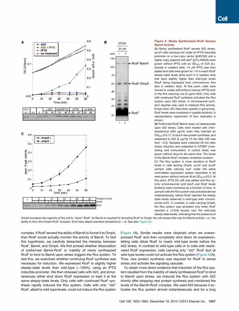
Figure 4. Newly Synthesized RcsF Senses
BamA Activity
(A) Newly synthesized RcsF senses A22 stress.
DrcsF cells carrying rcsF under an IPTG-inducible
promoter on a low-copy vector (pNG162) and a
higher-copy plasmid with lacIq (pTrc-HIS2A) were
grown without IPTG until an OD578 of 0.05 (b.i.
sample in western blot). 15 mM IPTG was then
added and cells were grown for 1 hr to reach RcsF
steady-state levels (time point 0 in western blot)
that were slightly higher than wild-type levels
(RcsF being expressed from chromosome; first
lane in western blot). At this point, cells were
moved to media with/without inducer (IPTG) and/
or the Rcs inducing cue (2 mg/ml A22). Only cells
with continued RcsF synthesis activated the Rcs
system upon A22 stress. A chromosomal rprA::
lacZ reporter was used to measure Rcs activity.
Empty dots: OD; filled dots: specific b-gal activity.
RcsF levels were monitored in parallel (bottom). A
representative experiment of four replicates is
shown.
(B) Preformed RcsF-BamA does not disassociate
upon A22 stress. Cells were treated with chlor-
amphenicol (300 mg/ml) when they reached an
OD600 of 0.17, to block new protein synthesis, and
subjected to A22 (5 mg/ml) 10 min later (OD was
then �0.2). Samples were collected 20 min after
stress induction and subjected to DTSSP cross-
linking and immunoblot. A control strain was
grown without drug for the same time. The levels
of the BamA-RcsF complex remained constant.
(C) The Rcs system is more sensitive to RcsF
levels in cells lacking OmpA. DrcsF and DrcsF
DompA cells carrying rcsF under the same
controllable expression system described in (A)
were grown without inducer till an OD578 of 0.3. At
this point, IPTG (20 mM) was added and Rcs ac-
tivity (chromosomal rprA::lacZ) and RcsF levels
(bottom) were monitored as a function of time. In
DompA cells the Rcs systemwas activated almost
instantaneously, before RcsF reached the steady
state levels observed in wild-type (with chromo-
somal rcsF). In contrast, in cells carrying OmpA,
the Rcs system was activated only when RcsF
reached a >3-fold excess over the wild-type
steady state levels, indicating that the presence of
OmpA increases the capacity of the cell to ‘‘store’’ RcsF. As BamA is required for funneling RcsF to OmpA, the cell senses this way the BamA activity– i.e., the
ability to form the OmpA-RcsF complex. Error bars depict standard deviations (n = 4). See also Figure S5.
complex, if RcsF sensed the ability of BamA to funnel it to OmpA,
then RcsF would actually monitor the activity of BamA. To test
this hypothesis, we carefully dissected the interplay between
RcsF, BamA, and OmpA. We first probed whether dissociation
of preformed BamA-RcsF or inability of newly synthesized
RcsF to bind to BamA upon stress triggers the Rcs system. To
test this, we examined whether continuing RcsF synthesis was
necessary for induction. We expressed RcsF to slightly higher
steady-state levels than wild-type (�130%), using an IPTG
inducible promoter. We then stressed cells with A22, and simul-
taneously either shut down RcsF expression or kept it at the
same steady-state levels. Only cells with continued RcsF syn-
thesis rapidly induced the Rcs system. Cells with only ‘‘old’’
RcsF, albeit to wild-type levels, could not induce the Rcs system
C
(Figure 4A). Similar results were obtained when we overex-
pressed RcsF and then completely shut down its expression,
letting cells dilute RcsF to nearly wild-type levels before the
A22 stress. In contrast to wild-type cells or to cells with reacti-
vated RcsF expression, cells carrying only ‘‘old’’ RcsF (but at
wild-type levels) could not activate the Rcs system (Figure S5A).
Thus, new protein synthesis was required for RcsF to sense
stress and activate the signaling cascade.
To obtain more direct evidence that induction of the Rcs sys-
tem resulted from the inability of newly synthesized RcsF to bind
to BamA upon stress, we induced the Rcs system with A22
shortly after stopping new protein synthesis and monitored the
levels of the BamA-RcsF complex. We used A22 because it ac-
tivates the Rcs system almost instantaneously and for a long
ell 159, 1652–1664, December 18, 2014 ª2014 Elsevier Inc. 1657
period of time (Figure S4A). In contrast, mecillinam-mediated
activation was slow, while polymyxin B-mediated activation
was short-lived (Figure S4A) (Farris et al., 2010). The BamA-
RcsF complex levels remained unchanged when protein synthe-
sis was stopped before A22 addition (Figure 4B), whereas they
decreased when protein synthesis was ongoing (Figure 3B).
This suggests that the newly arriving RcsF cannot bind to
BamA when cells are stressed, leading to the activation of the
system. Consistently the BamA-RcsF complex disappeared
faster than OmpA-RcsF after adding A22 or polymyxin B under
ongoing protein synthesis (Figures 3A–3B). This is because
BamA could presumably keep funneling RcsF to OmpA to a
certain degree, preventing OmpA-RcsF from disappearing with
dilution-like kinetics. We also stopped new protein synthesis
by chilling the cells, then added polymyxin B and probed
RcsF-BamA and RcsF-OmpA complex formation after 10 min.
Both complexes remained intact (Figure S5B), which is in agree-
ment with the inability of preformed RcsF-BamA to respond to
stress.
These results indicated that constant synthesis of RcsF is
required for RcsF to act as a sensor, and supported a model in
which activation results from newly synthesized RcsF being un-
able to bind BamA. However, it remained unclear whether irre-
versible sequestration of RcsF by BamA was sufficient to keep
the Rcs system off or whether continuous funneling of RcsF to
OmpA was also required. To discriminate between these two
possibilities, we compared the levels of RcsF that were required
to activate the system in DompA and wild-type strains. While
both strains have similar BamA levels (Figure S5C), BamA can
funnel RcsF to OmpA only in the wild-type, thereby theoretically
increasing its capacity for RcsF. We found that in the strain lack-
ing ompA, the Rcs system was induced at a fraction (<80%) of
wild-type RcsF levels, whereas wild-type cells could tolerate a
�3-fold increase in RcsF levels before inducing the Rcs system
(Figure 4C). Thus, by funneling RcsF to OmpA, BamA increases
its capacity for RcsF and maintains the Rcs system in an off
state. This means that RcsF can monitor the capacity of BamA
to assemble the OmpA-RcsF complex, which is presumably
affected during stress. Since active BamA is required for
OmpA-RcsF assembly (Figure 3D), RcsF senses this way the ac-
tivity of the Bam machinery.
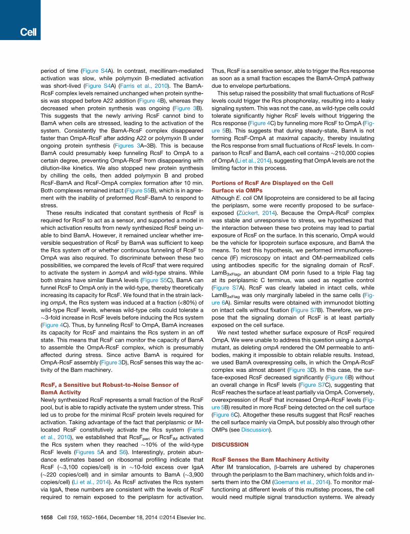
RcsF, a Sensitive but Robust-to-Noise Sensor ofBamA ActivityNewly synthesized RcsF represents a small fraction of the RcsF
pool, but is able to rapidly activate the system under stress. This
led us to probe for the minimal RcsF protein levels required for
activation. Taking advantage of the fact that periplasmic or IM-
located RcsF constitutively activate the Rcs system (Farris
et al., 2010), we established that RcsFperi or RcsFIM activated
the Rcs system when they reached �10% of the wild-type
RcsF levels (Figures 5A and S6). Interestingly, protein abun-
dance estimates based on ribosomal profiling indicate that
RcsF (�3,100 copies/cell) is in �10-fold excess over IgaA
(�220 copies/cell) and in similar amounts to BamA (�3,900
copies/cell) (Li et al., 2014). As RcsF activates the Rcs system
via IgaA, these numbers are consistent with the levels of RcsF
required to remain exposed to the periplasm for activation.
1658 Cell 159, 1652–1664, December 18, 2014 ª2014 Elsevier Inc.
Thus, RcsF is a sensitive sensor, able to trigger the Rcs response
as soon as a small fraction escapes the BamA-OmpA pathway
due to envelope perturbations.
This setup raised the possibility that small fluctuations of RcsF
levels could trigger the Rcs phosphorelay, resulting into a leaky
signaling system. This was not the case, as wild-type cells could
tolerate significantly higher RcsF levels without triggering the
Rcs response (Figure 4C) by funneling more RcsF to OmpA (Fig-
ure 5B). This suggests that during steady-state, BamA is not
forming RcsF-OmpA at maximal capacity, thereby insulating
the Rcs response from small fluctuations of RcsF levels. In com-
parison to RcsF and BamA, each cell contains �210,000 copies
of OmpA (Li et al., 2014), suggesting that OmpA levels are not the
limiting factor in this process.
Portions of RcsF Are Displayed on the CellSurface via OMPsAlthough E. coli OM lipoproteins are considered to be all facing
the periplasm, some were recently proposed to be surface-
exposed (Zuckert, 2014). Because the OmpA-RcsF complex
was stable and unresponsive to stress, we hypothesized that
the interaction between these two proteins may lead to partial
exposure of RcsF on the surface. In this scenario, OmpA would
be the vehicle for lipoprotein surface exposure, and BamA the
means. To test this hypothesis, we performed immunofluores-
cence (IF) microscopy on intact and OM-permeabilized cells
using antibodies specific for the signaling domain of RcsF.
LamB3xFlag, an abundant OM porin fused to a triple Flag tag
at its periplasmic C terminus, was used as negative control
(Figure S7A). RcsF was clearly labeled in intact cells, while
LamB3xFlag was only marginally labeled in the same cells (Fig-
ure 6A). Similar results were obtained with immunodot blotting
on intact cells without fixation (Figure S7B). Therefore, we pro-
pose that the signaling domain of RcsF is at least partially
exposed on the cell surface.
We next tested whether surface exposure of RcsF required
OmpA. We were unable to address this question using a DompA
mutant, as deleting ompA rendered the OM permeable to anti-
bodies, making it impossible to obtain reliable results. Instead,
we used BamA overexpressing cells, in which the OmpA-RcsF
complex was almost absent (Figure 3D). In this case, the sur-
face-exposed RcsF decreased significantly (Figure 6B) without
an overall change in RcsF levels (Figure S7C), suggesting that
RcsF reaches the surface at least partially via OmpA. Conversely,
overexpression of RcsF that increased OmpA-RcsF levels (Fig-
ure 5B) resulted in more RcsF being detected on the cell surface
(Figure 6C). Altogether these results suggest that RcsF reaches
the cell surface mainly via OmpA, but possibly also through other
OMPs (see Discussion).
DISCUSSION
RcsF Senses the Bam Machinery ActivityAfter IM translocation, b-barrels are ushered by chaperones
through the periplasm to the Bammachinery, which folds and in-
serts them into the OM (Goemans et al., 2014). To monitor mal-
functioning at different levels of this multistep process, the cell
would need multiple signal transduction systems. We already
Figure 5. RcsF Is a Sensitive but Robust-to-Noise Sensor of BamA Activity(A) Only a fraction of RcsF is required to be in the periplasm for Rcs activation. DrcsF cells carrying RcsFIM or RcsFperi under an IPTG-inducible promoter on a low-
copy vector (pSC202) and a higher-copy plasmid encoding lacIq (pREP4) were grown for three generations in LB and before adding inducer (100 mM IPTG). RcsF
protein levels (bottom) and Rcs activity (top left; chromosomal rprA::lacZ fusion) were closely monitored onward. Note that in this setup, no matter how much
IPTGwas added, or when added, rcsF expression remained undetectable until cells reached an OD578 of�0.6. Quantification of RcsFIM or RcsFperi protein levels
at the time point of Rcs activation is shown at the top right. Error bars depict standard deviation (n = 3). The time point of activation was considered as the point at
which a linear curve fitted on specific b-gal activity versus time crossed the basal activity, minus 3 min required for b-gal synthesis and folding. For quantifying
RcsF levels, we always ensured that the signal detected from cells expressing RcsFIM or RcsFperi (40 mg) was within linear range by loading a titration of total
protein extracts fromwild-type cells (2.5–20 mg). An example western blot is shown (bottom). Empty dots: OD; filled dots: specific b-gal activity. The full gel can be
seen in Figure S6.
(B) The capacity of BamA to form the OmpA-RcsF complex is not maxed out in wild-type cells. Increasing RcsF expression resulted into more OmpA-RcsF
complex being formed, but the levels of the BamA-RcsF complex remained largely unchanged. Thus, in nonstressed cells, BamA has the ability to funnel more
RcsF toOmpA. In lane 1 thewild-type levels of theOmpA-RcsF andBamA-RcsF complexes are shown. In lane 2, RcsFwas expressed inDrcsF cells carrying rcsF
under an IPTG-inducible promoter on a low-copy vector, pSC202. In the absence of lacIq, the RcsF steady-state levels were�3- to 4-fold higher than in the wild-
type. DTSSP crosslinking and immunoblot were performed as described in Figure 2A, and a representative experiment is shown (n = 3).
know that accumulation of unassembled OMPs in the periplasm
is the primary signal for the sE stress response (Walsh et al.,
2003). We now report that the activity of the Bam machinery is
monitored by the Rcs system through RcsF. BamA interacts
with RcsF and, when active, funnels it to OmpA. When bound
to BamA or OmpA, RcsF is occluded from IgaA and cannot acti-
vate the Rcs system. Yet, BamA cannot sequester all RcsF mol-
ecules and funneling of newly synthesized RcsF to OmpA is
necessary for maintaining the Rcs system off. This is especially
important because preformed BamA-RcsF does not disasso-
ciate upon stress, and only newly arriving RcsF can sense stress.
Thus, this constant flow of RcsF fromBamA to OmpA is what de-
fines the availability of BamA and what RcsF is sensing. Stress
conditions impair BamA availability for newly arriving RcsF,
which ends up facing the periplasm, free to activate the Rcs
cascade (Figure 7).
In addition to its well-known activity in OMPs assembly, we
report that BamA funnels RcsF to OmpA and other OMPs. Since
functional Bam machinery is required for both events, we sug-
gest that they are coupled (Figure 7), which implies that RcsF
senses by default both activities. Further structure-function anal-
ysis will be required for deciphering if and how the two events are
connected and how RcsF intervenes.
C
An interesting feature of the Rcs system is that RcsF is in�10-
fold excess over its downstream partner IgaA (Li et al., 2014),
despite the two forming a 1:1 complex (Figure 1C). This results
into only a fraction of RcsF being required for fully activating
the Rcs system (Figure 5A). The cell presumably maintains
RcsF in excess over IgaA to efficiently monitor the Bam machin-
ery, which is present at a similar copy/cell ratio as RcsF (Li et al.,
2014). At the same time, the steady-state RcsF levels are kept
low enough to prevent activation of the Rcs system by small
fluctuations. Indeed, a �3-fold increase in RcsF levels was
required for the Rcs system to be activated without stress (Fig-
ure 4C). An interesting hypothesis that we are currently pursuing
is that RcsF levels are optimized for high sensitivity and low
noise.
How can PG and OM stress affect Bam activity? Although
PG perturbations could affect the journey of RcsF bound to
the lipoprotein-specific chaperone LolA through the porous
PG layer, we did not see RcsF accumulating in the periplasm
upon mecillinam or A22 treatment. On the other hand, transport
of the bulkier BamA may be more impaired, creating a bottle-
neck in BamA availability/activity. Alternatively the POTRA do-
mains of BamA that extend deep into the periplasm could be
affected by changes in PG integrity, with direct consequences
ell 159, 1652–1664, December 18, 2014 ª2014 Elsevier Inc. 1659
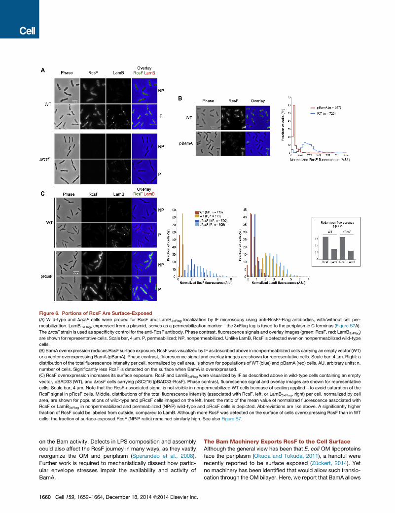
Figure 6. Portions of RcsF Are Surface-Exposed
(A) Wild-type and DrcsF cells were probed for RcsF and LamB3xFlag localization by IF microscopy using anti-RcsF/-Flag antibodies, with/without cell per-
meabilization. LamB3xFlag, expressed from a plasmid, serves as a permeabilization marker—the 3xFlag tag is fused to the periplasmic C terminus (Figure S7A).
The DrcsF strain is used as specificity control for the anti-RcsF antibody. Phase contrast, fluorescence signals and overlay images (green: RcsF, red: LamB3xFlag)
are shown for representative cells. Scale bar, 4 mm. P, permeabilized; NP, nonpermeabilized. Unlike LamB, RcsF is detected even on nonpermeabilized wild-type
cells.
(B) BamA overexpression reduces RcsF surface exposure. RcsFwas visualized by IF as described above in nonpermeabilized cells carrying an empty vector (WT)
or a vector overexpressing BamA (pBamA). Phase contrast, fluorescence signal and overlay images are shown for representative cells. Scale bar: 4 mm. Right: a
distribution of the total fluorescence intensity per cell, normalized by cell area, is shown for populations of WT (blue) and pBamA (red) cells. AU, arbitrary units; n,
number of cells. Significantly less RcsF is detected on the surface when BamA is overexpressed.
(C) RcsF overexpression increases its surface exposure. RcsF and LamB3xFlag were visualized by IF as described above in wild-type cells containing an empty
vector, pBAD33 (WT), and DrcsF cells carrying pSC216 (pBAD33-RcsF). Phase contrast, fluorescence signal and overlay images are shown for representative
cells. Scale bar, 4 mm. Note that the RcsF-associated signal is not visible in nonpermeabilized WT cells because of scaling applied—to avoid saturation of the
RcsF signal in pRcsF cells. Middle, distributions of the total fluorescence intensity (associated with RcsF, left, or LamB3xFlag, right) per cell, normalized by cell
area, are shown for populations of wild-type and pRcsF cells imaged on the left. Inset: the ratio of the mean value of normalized fluorescence associated with
RcsF or LamB3xFlag in nonpermeabilized and permeabilized (NP/P) wild-type and pRcsF cells is depicted. Abbreviations are like above. A significantly higher
fraction of RcsF could be labeled from outside, compared to LamB. Although more RcsF was detected on the surface of cells overexpressing RcsF than in WT
cells, the fraction of surface-exposed RcsF (NP/P ratio) remained similarly high. See also Figure S7.
on the Bam activity. Defects in LPS composition and assembly
could also affect the RcsF journey in many ways, as they vastly
reorganize the OM and periplasm (Sperandeo et al., 2008).
Further work is required to mechanistically dissect how partic-
ular envelope stresses impair the availability and activity of
BamA.
1660 Cell 159, 1652–1664, December 18, 2014 ª2014 Elsevier Inc.
The Bam Machinery Exports RcsF to the Cell SurfaceAlthough the general view has been that E. coli OM lipoproteins
face the periplasm (Okuda and Tokuda, 2011), a handful were
recently reported to be surface exposed (Zuckert, 2014). Yet
no machinery has been identified that would allow such translo-
cation through the OM bilayer. Here, we report that BamA allows
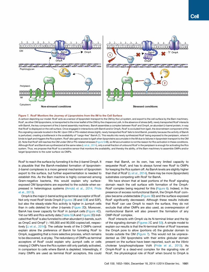
Figure 7. RcsF Monitors the Journey of Lipoproteins from the IM to the Cell Surface
A cartoon depicting our model: RcsF acts as a sensor of lipoprotein transport to the OM by the Lol system, and export to the cell surface by the Bammachinery.
RcsF, as other OM lipoproteins, is transported to the inner leaflet of theOMby the chaperone LolA. In the absence of stress (left), newly transported RcsF interacts
with BamA, the key component of the b-barrel assembly machinery. BamA assembles a complex between RcsF and OmpA, an abundant b-barrel protein, in way
that RcsF is displayed on the cell surface. Once engaged in interactionswith BamA and/or OmpA, RcsF is occluded from IgaA, the downstream component of the
Rcs signaling cascade located in the IM. Upon OM or PG-related stress (right), newly transported RcsF fails to bind BamA, possibly because the activity of BamA
is perturbed, creating a bottleneck in the availability of ‘‘cargo-free’’ BamA (1). This results into newly synthesized RcsF being exposed to the periplasm, where it
binds to IgaA and triggers the Rcs system. RcsF also gains access to IgaAwhen lipoproteins accumulate in the IM due to failures in lipoprotein transport to theOM
(2). Note that RcsF still reaches the OM under OM or PG-related stresses (Figure S4B), so IM accumulation is not the reason for Rcs activation in these conditions.
Although RcsF and BamA are synthesized at the same rates (Li et al., 2014), only a small fraction of unbound RcsF in the periplasm is enough for activating the Rcs
system. Thus, we propose that RcsF is a sensitive sensor that monitors the availability, and thereby the ability, of the Bam machinery to assemble OMPs and/or
target lipoproteins to the outer surface via OMPs.
RcsF to reach the surface by funneling it to the b-barrel OmpA. It
is plausible that the BamA-mediated formation of lipoprotein-
b-barrel complexes is a more general mechanism of lipoprotein
export to the surface, but further experimentation is needed to
establish this. As the Bam machine is highly conserved among
Gram-negative bacteria, this would explain why surface-
exposed OM lipoproteins are exported to the outside when ex-
pressed in heterologous systems (Arnold et al., 2014; Pride
et al., 2013).
OmpA is themajor b-barrel acting as terminal acceptor of RcsF.
Not only most RcsF binds OmpA (Figures 2B and S3E and S3F),
but also the steady-state Rcs activity is higher in DompA cells
than in cells deleted for other b-barrels (Figure 2D). Moreover,
BamA has lower capacity for RcsF in DompA cells (Figure 4C).
Yet our MS and Rcs-activity data (Table S2A and Figure 2D) indi-
cated thatRcsF is also funneled to other abundant b-barrels, such
as OmpC and OmpF (�165,000 and 90,000 copies/cell, respec-
tively [Li et al., 2014]). The cellular levels of the 3 OMPs cannot
explain alone the preference of BamA for funneling RcsF to
OmpA, suggesting that a more selective process, which remains
to be discovered, is at play. The redundancy of OMPs as terminal
acceptors of RcsF could explain why DompA cells or cells
missing 2 OMPs have the Rcs system still only partially activated,
in comparison to cells where BamA is depleted. Interestingly, if
many OMPs are used as terminal RcsF acceptors, this could
C
mean that BamA, on its own, has very limited capacity to
sequester RcsF, and has to always funnel new RcsF to OMPs
for keeping the Rcs system off. As BamA levels are slightly higher
than that of RcsF (Li et al., 2014), there may be more (lipoprotein)
substrates competing with RcsF for BamA.
We have shown that at least portions of the RcsF signaling
domain reach the cell surface with formation of the OmpA-
RcsF complex being required for this (Figure 6). Indeed, in the
presence of excess nonfunctional BamA, the OmpA-RcsF com-
plex became undetectable (Figure 3D) and the surface-exposed
RcsF significantly decreased. Although these results indicate
that RcsF can use OmpA to reach the surface, they do not
exclude that other OMPs are also used, as overexpression of
nonfunctional BamA will also prevent the formation of any
OMP-RcsF complex.
RcsF interacts with OmpA via its N-terminal linker and the tip
of the signaling domain (Figures 2C and S3). A simple model to
explain our results is that the N-terminal linker of RcsF traverses
the OmpA pore to allow (portions of) the globular domain to
locate outside the OM (Figure 7). This would not be unprece-
dented as OM lipoproteins with their entire globular domain
present on the surface have been reported, such as the Vibrio
cholerae lysophospholipase VolA (Pride et al., 2013). As
OmpA-RcsF is a dead-end complex for the signaling role of
RcsF, the physiological role of RcsF when bound to OmpA is
ell 159, 1652–1664, December 18, 2014 ª2014 Elsevier Inc. 1661
enigmatic. Additional work will be required to clarify how OmpA
and RcsF interact and the role of RcsF in this complex.
How can RcsF use a b-barrel such asOmpA to access the sur-
face? OM porins act as gates for peptides coming from outside
(Housden et al., 2013) and for periplasmic proteins secreted by
the cell, such as YebF (Prehna et al., 2012). The lipoprotein
LptE was also recently shown to reside inside the b-barrel
LptD, presumably acting as a controllable plug for the LPS as-
semblymachinery (Freinkman et al., 2011). Thus, it is not uncom-
mon that a b-barrel pore can accommodate a polypeptide.
OmpA, in one of its two known conformations, forms a 16-
stranded b-barrel structure with a large pore (Reusch, 2012).
This conformation could accommodate a disordered segment
such as the RcsF linker. In its second conformation, which has
been proposed to be an intermediate state, OmpA assumes a
2-domain structure, with a smaller N-terminal b-barrel and a
C-terminal periplasmic domain interacting with the PG (Reusch,
2012). In this conformation, the b-barrel diameter is too small for
a polypeptide, but an OmpA-RcsF interaction at this stage could
be an important intermediate for the funneling of RcsF from
BamA.
Finally, we detected an RcsF-Lpp complex (Figure S2D). As
this interaction was not recapitulated with any of the 25 pBpa-
containing RcsF variants, we deduced that it might be indirect.
This would be consistent with the very high abundance of Lpp
and its shared localization with RcsF at both OM leaflets (Cowles
et al., 2011). In addition, the absence of Lpp did not affect the
RcsF-BamA and RcsF-OmpA interactions (Figure S2D). It re-
mains to be tested if Lpp has any direct effect on Rcs signaling.
Integrating Envelope Stresses: RcsF Monitors theJourney of Lipoproteins through the EnvelopeThere are �100 lipoproteins in E. coli. The vast majority is local-
ized in the OM. Although the function of most is unknown, some
are components of essential OM assembly machineries (Silhavy
et al., 2010) and others regulate core envelope processes (Para-
dis-Bleau et al., 2010; Typas et al., 2010; Uehara et al., 2010).
Thus lipoprotein targeting is vital for the cell.
OM lipoproteins are escorted across the periplasm by the
essential chaperone LolA (Okuda and Tokuda, 2011). RcsF
senses defects in: (1) phosphatidylglycerol biosynthesis (Shiba
et al., 2004), which is required for lipoprotein maturation; and
(2) in the LolA-mediated transport of lipoproteins to the OM, pre-
sumably because it gets stuck in the IM when LolA’s function is
impaired (Tao et al., 2012), gaining access to IgaA. Rcs activation
resulting from RcsF accumulation in the IM leads to higher lolA
expression, creating a feedback loop to fix the damage (Tao
et al., 2012).
For RcsF, and at least a few other lipoproteins, the journey
does not end at the inner leaflet of the OM, as they are finally
translocated to the cell surface. As we have shown here, it is
the Bammachine that mediates the export of RcsF to the surface
by inserting it into b-barrels such asOmpA.Malfunctioning of this
process results into newly translocated RcsF remaining exposed
in the periplasm, where it can reach IgaA and trigger the signaling
cascade (Figure 7). Therefore, RcsF also monitors the ability of
BamA to insert OM lipoproteins to OMPs. Altogether this means
that Rcs can sense the entire lipoprotein journey across the en-
1662 Cell 159, 1652–1664, December 18, 2014 ª2014 Elsevier Inc.
velope, frommaturation to OM exposure, adjusting the envelope
composition in response to failures at any step.
Rcs, a Complex Signal Transduction SystemRcs is one of the most complex signaling systems known in bac-
teria with key steps remaining unresolved. We have shown that
RcsF interacts with the large periplasmic domain of IgaA, which
likely triggers the signaling cascade. As the two membranes are
separated by�200 A, it remains to be determined how this inter-
action occurs. RcsF has an intrinsically disordered 31 amino
acid-long N-terminal linker. It is likely that, when extended, this
region allows RcsF to reach the large periplasmic domain of
IgaA. The OM lipoprotein LpoB uses a similar configuration to
access its IM counterpart, PBP1B (Egan et al., 2014).
It also remains to be proven whether the RcsF-IgaA interaction
is sufficient for conveying the signal downstream and activating
the Rcs cascade; our genetic data that put IgaA downstream of
RcsF strongly suggest so. How IgaA itself mechanistically con-
trols the Rcs phosphorelay, whether it directly interacts with
the other IM components RcsC and RcsD, and whether it plays
additional roles in the cell remain unknown and will all be fields of
future research. Moreover, further work will be required to eluci-
date how the few genetic perturbations that activate the Rcs sys-
tem independently of RcsF (Majdalani and Gottesman, 2005,
2007) are sensed by the system.
CONCLUSIONS
We elucidated how the OM lipoprotein RcsF senses stress and
talks to the downstream signaling cascade. RcsF monitors the
activity of the machinery for OM b-barrel assembly, Bam, trig-
gering the signaling cascadewhen Bam ismalfunctioning. More-
over, we identified the formation of complexes between RcsF
and the b-barrel OmpA as a novel mechanism for lipoprotein
translocation through the bacterial OM. We propose that this
may be a conserved system for lipoprotein export. Although
many of the molecular details of both processes described
here remain to be fully elucidated, these findings generate a
number of intriguing hypotheses on the mechanisms that the
cell uses to sense the activity of the protein machineries that
build its envelope.
EXPERIMENTAL PROCEDURES
Bacterial Strains, Media, and Plasmids
Cells were grown in LB at 37�C and, when necessary, growthmedia were sup-
plemented with spectinomycin (50–100 mg/ml), ampicillin (100–200 mg/ml),
chloramphenicol (20–25 mg/ml), or kanamycin (50 mg/ml). The bacterial strains
and plasmids used in this study are listed in Tables S3 and S4, respectively,
and information on their construction is provided in Extended Experimental
Procedures.
In Vitro RcsF-IgaA Binding
RcsF with a C-terminal 6-Histidine tag (RcsF-His) and an untagged version of
the periplasmic IgaA domain were purified as described in Extended Experi-
mental Procedures. RcsF-His (0.15 nmol) was coupled to 20 ml Talon beads
and washed with PD buffer (25 mM Tris [pH 7.5], 200 mM NaCl, 10% glycerol)
to remove residual RcsF-His. IgaA was then added to the RcsF (2.5 mM) con-
taining Talon beads in a concentration range: 0.375–10 mM (assay volume =
60 ml; 0.625–10 mM range is shown in Figure 1C). The RcsF-IgaA suspension
was incubated for 15 min at room temperature and pelleted by brief centrifu-
gation. Half of the supernatant was aspirated to quantify unbound IgaA by
SDS-PAGE. The pellet was washed with 500 ml PD buffer and half was also
analyzed by SDS-PAGE to quantify the pulled-down fraction of IgaA.
In Vivo DTSSP Crosslinking
In vivo chemical crosslinking experiments were performed as described by
Thanabalu et al. (1998) with some modifications. The detailed procedures
are described in Extended Experimental Procedures.
In Vivo Site-Specific Photocrosslinking
Site-specific photocrosslinking was performed essentially as described by
Okuda et al. (2012) with some modifications. The detailed procedures are
described in Extended Experimental Procedures.
SUPPLEMENTAL INFORMATION
Supplemental Information includes Extended Experimental Procedures, seven
figures, and four tables and can be foundwith this article online at http://dx.doi.
org/10.1016/j.cell.2014.11.045.
AUTHOR CONTRIBUTIONS
S.H.C., J.S., C.P., A.T., and J.F.C. conceived this study; S.H.C., J.S., C.P.,
M.Z., M.B., A.T., and J.F.C. designed research; S.H.C., J.S., C.P., M.Z.,
P.R., A.A., G.L., M.B., A.-K.H., P.L., C.V.d.H., D.V., and A.T. performed all ex-
periments (S.H.C., J.S. performed all DTSSP experiments, except for the pull
down-MS for IgaA-peri which were performed by C.P. and M.Z.; S.H.C. per-
formed all photocrosslinking experiments; C.P., M.Z., and A.K. performed all
b-gal assays; C.P. performed the quantitative western blotting; M.B. and
A.K. performed the in vitro binding assays; P.R., A.A., and G.L. performed
the IF microscopy; J.S. performed the dot blotting; A.T. performed the igaA
suppressor screen); S.H.C., J.S., C.P., M.Z., P.R., A.A., G.L., M.B., D.V.,
A.T., and J.F.C. interpreted the data; A.T. and J.F.C. wrote the manuscript;
S.H.C., J.S., C.P., A.A., G.L. prepared the figures; S.H.C., J.S. and C.P. edited
the manuscript; A.T. and J.F.C. supervised all aspects of the project.
ACKNOWLEDGMENTS
We are grateful to C.A. Gross, K.C. Huang, and N. Ruiz for critically reading the
manuscript and providing feedback. We thank H. Mori (Nara, Japan), S. Got-
tesman (NIH, USA), D. Kahne (Harvard, USA), and T. Silhavy (Princeton, USA)
for providing strains. We thank the members of the two labs for helpful discus-
sions, A. Boujtat, G. Herinckx, A. Koumoutsi, andM. Pruteanu for assistance in
experiments and D. Colau for antibody purification. M.S. was performed at the
Proteomics Core Facilities of EMBL and of the de Duve Institute. J.S. is a FRIA
research fellow, C.P. is an EMBO long-term fellow, P.L. is a ‘‘Chargee de Re-
cherche’’ and J.F.C. is ‘‘Maıtre de Recherche’’ of the FRS-FNRS. This work
was supported by grants from the FRS-FNRS and the Interuniversity Attraction
Pole Program-Belgian Science Policy (network P7/44) to JFC, and the Sofja
Kovalevskaja Award of the Alexander von Humboldt Foundation and EMBL in-
ternal funding to A.T.
Received: July 11, 2014
Revised: October 6, 2014
Accepted: November 24, 2014
Published: December 18, 2014
REFERENCES
Aoki, S.K., Malinverni, J.C., Jacoby, K., Thomas, B., Pamma, R., Trinh, B.N.,
Remers, S., Webb, J., Braaten, B.A., Silhavy, T.J., and Low, D.A. (2008). Con-
tact-dependent growth inhibition requires the essential outer membrane pro-
tein BamA (YaeT) as the receptor and the inner membrane transport protein
AcrB. Mol. Microbiol. 70, 323–340.
Arnold, M.F., Caro-Hernandez, P., Tan, K., Runti, G., Wehmeier, S., Scocchi,
M., Doerrler, W.T., Walker, G.C., and Ferguson, G.P. (2014). Enteric YaiW is
C
a surface-exposed outer membrane lipoprotein that affects sensitivity to an
antimicrobial peptide. J. Bacteriol. 196, 436–444.
Baba, T., Ara, T., Hasegawa, M., Takai, Y., Okumura, Y., Baba, M., Datsenko,
K.A., Tomita, M., Wanner, B.L., and Mori, H. (2006). Construction of Escheri-
chia coli K-12 in-frame, single-gene knockout mutants: the Keio collection.
Mol. Syst. Biol. 2, 0008.
Cano, D.A., Domınguez-Bernal, G., Tierrez, A., Garcia-Del Portillo, F., and Ca-
sadesus, J. (2002). Regulation of capsule synthesis and cell motility in Salmo-
nella enterica by the essential gene igaA. Genetics 162, 1513–1523.
Castanie-Cornet, M.P., Cam, K., and Jacq, A. (2006). RcsF is an outer mem-
brane lipoprotein involved in the RcsCDB phosphorelay signaling pathway in
Escherichia coli. J. Bacteriol. 188, 4264–4270.
Cowles, C.E., Li, Y., Semmelhack, M.F., Cristea, I.M., and Silhavy, T.J. (2011).
The free and bound forms of Lpp occupy distinct subcellular locations in Es-
cherichia coli. Mol. Microbiol. 79, 1168–1181.
Domınguez-Bernal, G., Pucciarelli, M.G., Ramos-Morales, F., Garcıa-Quinta-
nilla, M., Cano, D.A., Casadesus, J., and Garcıa-del Portillo, F. (2004). Repres-
sion of the RcsC-YojN-RcsB phosphorelay by the IgaA protein is a requisite for
Salmonella virulence. Mol. Microbiol. 53, 1437–1449.
Egan, A.J., Jean, N.L., Koumoutsi, A., Bougault, C.M., Biboy, J., Sassine, J.,
Solovyova, A.S., Breukink, E., Typas, A., Vollmer, W., and Simorre, J.P.
(2014). Outer-membrane lipoprotein LpoB spans the periplasm to stimulate
the peptidoglycan synthase PBP1B. Proc. Natl. Acad. Sci. USA 111, 8197–
8202.
Evans, K.L., Kannan, S., Li, G., de Pedro, M.A., and Young, K.D. (2013). Elim-
inating a set of four penicillin binding proteins triggers the Rcs phosphorelay
and Cpx stress responses in Escherichia coli. J. Bacteriol. 195, 4415–4424.
Farris, C., Sanowar, S., Bader, M.W., Pfuetzner, R., and Miller, S.I. (2010).
Antimicrobial peptides activate the Rcs regulon through the outer membrane
lipoprotein RcsF. J. Bacteriol. 192, 4894–4903.
Freinkman, E., Chng, S.S., and Kahne, D. (2011). The complex that inserts lipo-
polysaccharide into the bacterial outer membrane forms a two-protein plug-
and-barrel. Proc. Natl. Acad. Sci. USA 108, 2486–2491.
Girgis, H.S., Liu, Y., Ryu, W.S., and Tavazoie, S. (2007). A comprehensive ge-
netic characterization of bacterial motility. PLoS Genet. 3, 1644–1660.
Goemans, C., Denoncin, K., and Collet, J.F. (2014). Folding mechanisms of
periplasmic proteins. Biochim. Biophys. Acta 1843, 1517–1528.
Gogol, E.B., Rhodius, V.A., Papenfort, K., Vogel, J., and Gross, C.A. (2011).
Small RNAs endow a transcriptional activator with essential repressor func-
tions for single-tier control of a global stress regulon. Proc. Natl. Acad. Sci.
USA 108, 12875–12880.
Guo, M.S., Updegrove, T.B., Gogol, E.B., Shabalina, S.A., Gross, C.A., and
Storz, G. (2014). MicL, a new sE-dependent sRNA, combats envelope stress
by repressing synthesis of Lpp, the major outer membrane lipoprotein. Genes
Dev. 28, 1620–1634.
Hagan, C.L., Kim, S., and Kahne, D. (2010). Reconstitution of outer membrane
protein assembly from purified components. Science 328, 890–892.
Hagan, C.L., Silhavy, T.J., and Kahne, D. (2011). b-Barrel membrane protein
assembly by the Bam complex. Annu. Rev. Biochem. 80, 189–210.
Housden, N.G., Hopper, J.T., Lukoyanova, N., Rodriguez-Larrea, D., Wojdyla,
J.A., Klein, A., Kaminska, R., Bayley, H., Saibil, H.R., Robinson, C.V., and
Kleanthous, C. (2013). Intrinsically disordered protein threads through the bac-
terial outer-membrane porin OmpF. Science 340, 1570–1574.
Laubacher, M.E., and Ades, S.E. (2008). The Rcs phosphorelay is a cell enve-
lope stress response activated by peptidoglycan stress and contributes to
intrinsic antibiotic resistance. J. Bacteriol. 190, 2065–2074.
Leverrier, P., Declercq, J.P., Denoncin, K., Vertommen, D., Hiniker, A., Cho,
S.H., and Collet, J.F. (2011). Crystal structure of the outer membrane protein
RcsF, a new substrate for the periplasmic protein-disulfide isomerase DsbC.
J. Biol. Chem. 286, 16734–16742.
Li, G.W., Burkhardt, D., Gross, C., and Weissman, J.S. (2014). Quantifying ab-
solute protein synthesis rates reveals principles underlying allocation of
cellular resources. Cell 157, 624–635.
ell 159, 1652–1664, December 18, 2014 ª2014 Elsevier Inc. 1663
Lima, S., Guo, M.S., Chaba, R., Gross, C.A., and Sauer, R.T. (2013). Dual mo-
lecular signals mediate the bacterial response to outer-membrane stress. Sci-
ence 340, 837–841.
Majdalani, N., and Gottesman, S. (2005). The Rcs phosphorelay: a complex
signal transduction system. Annu. Rev. Microbiol. 59, 379–405.
Majdalani, N., and Gottesman, S. (2007). Genetic dissection of signaling
through the Rcs phosphorelay. Methods Enzymol. 423, 349–362.
Okuda, S., and Tokuda, H. (2011). Lipoprotein sorting in bacteria. Annu. Rev.
Microbiol. 65, 239–259.
Paradis-Bleau, C., Markovski, M., Uehara, T., Lupoli, T.J., Walker, S., Kahne,
D.E., and Bernhardt, T.G. (2010). Lipoprotein cofactors located in the outer
membrane activate bacterial cell wall polymerases. Cell 143, 1110–1120.
Prehna, G., Zhang, G., Gong, X., Duszyk, M., Okon, M., McIntosh, L.P.,
Weiner, J.H., and Strynadka, N.C. (2012). A protein export pathway involving
Escherichia coli porins. Structure 20, 1154–1166.
Pride, A.C., Herrera, C.M., Guan, Z., Giles, D.K., and Trent, M.S. (2013). The
outer surface lipoprotein VolA mediates utilization of exogenous lipids by Vib-
rio cholerae. MBio 4, e00305–e00313.
Reusch, R.N. (2012). Biogenesis and functions of model integral outer mem-
brane proteins: Escherichia coli OmpA and Pseudomonas aeruginosa OprF.
FEBS J. 279, 893.
Rhodius, V.A., Suh, W.C., Nonaka, G., West, J., and Gross, C.A. (2006).
Conserved and variable functions of the sigmaE stress response in related ge-
nomes. PLoS Biol. 4, e2.
Shiba, Y., Yokoyama, Y., Aono, Y., Kiuchi, T., Kusaka, J., Matsumoto, K., and
Hara, H. (2004). Activation of the Rcs signal transduction system is responsible
for the thermosensitive growth defect of an Escherichia coli mutant lacking
phosphatidylglycerol and cardiolipin. J. Bacteriol. 186, 6526–6535.
Silhavy, T.J., Kahne, D., and Walker, S. (2010). The bacterial cell envelope.
Cold Spring Harb. Perspect. Biol. 2, a000414.
Sperandeo, P., Lau, F.K., Carpentieri, A., De Castro, C., Molinaro, A., Deho, G.,
Silhavy, T.J., and Polissi, A. (2008). Functional analysis of the protein machin-
ery required for transport of lipopolysaccharide to the outer membrane of Es-
cherichia coli. J. Bacteriol. 190, 4460–4469.
Tao, K., Narita, S., and Tokuda, H. (2012). Defective lipoprotein sorting induces
lolA expression through the Rcs stress response phosphorelay system.
J. Bacteriol. 194, 3643–3650.
1664 Cell 159, 1652–1664, December 18, 2014 ª2014 Elsevier Inc.
Thanabalu, T., Koronakis, E., Hughes, C., and Koronakis, V. (1998). Substrate-
induced assembly of a contiguous channel for protein export from E.coli:
reversible bridging of an inner-membrane translocase to an outer membrane
exit pore. EMBO J. 17, 6487–6496.
Typas, A., Nichols, R.J., Siegele, D.A., Shales, M., Collins, S.R., Lim, B., Bra-
berg, H., Yamamoto, N., Takeuchi, R., Wanner, B.L., et al. (2008). High-
throughput, quantitative analyses of genetic interactions in E. coli. Nat.
Methods 5, 781–787.
Typas, A., Banzhaf, M., van den Berg van Saparoea, B., Verheul, J., Biboy, J.,
Nichols, R.J., Zietek, M., Beilharz, K., Kannenberg, K., von Rechenberg, M.,
et al. (2010). Regulation of peptidoglycan synthesis by outer-membrane pro-
teins. Cell 143, 1097–1109.
Typas, A., Banzhaf, M., Gross, C.A., and Vollmer, W. (2012). From the regula-
tion of peptidoglycan synthesis to bacterial growth and morphology. Nat. Rev.
Microbiol. 10, 123–136.
Uehara, T., Parzych, K.R., Dinh, T., and Bernhardt, T.G. (2010). Daughter cell
separation is controlled by cytokinetic ring-activated cell wall hydrolysis.
EMBO J. 29, 1412–1422.
Walsh, N.P., Alba, B.M., Bose, B., Gross, C.A., and Sauer, R.T. (2003).
OMP peptide signals initiate the envelope-stress response by activating
DegS protease via relief of inhibition mediated by its PDZ domain. Cell
113, 61–71.
Zhang, M., Lin, S., Song, X., Liu, J., Fu, Y., Ge, X., Fu, X., Chang, Z., and Chen,
P.R. (2011). A genetically incorporated crosslinker reveals chaperone cooper-
ation in acid resistance. Nat. Chem. Biol. 7, 671–677.
Zuckert, W.R. (2014). Secretion of bacterial lipoproteins: through the cyto-
plasmic membrane, the periplasm and beyond. Biochim. Biophys. Acta
1843, 1509–1516.
Note Added in Proof
While this paper was under revision, Konovalova et al. reported the surface
exposure of portions of RcsF via OM ß-barrels.
Konovalova, A., Perlman, D.H., Cowles, C.E., and Silhavy, T.J. (2014). Trans-
membrane domain of surface-exposed outer membrane lipoprotein RcsF is
threaded through the lumen of b-barrel proteins. Proc. Natl. Acad. Sci. USA
111, E4350–E4358.