desempenho muscular isocinÉtico do complexo … · nascimento, l.r. . 1 lucas rodrigues nascimento...
TRANSCRIPT
Lucas Rodrigues Nascimento
DESEMPENHO MUSCULAR ISOCINÉTICO DO
COMPLEXO DO OMBRO DE INDIVÍDUOS
COM HEMIPARESIA CRÔNICA
Belo Horizonte
2011
Nascimento, L.R. . 1
Lucas Rodrigues Nascimento
DESEMPENHO MUSCULAR ISOCINÉTICO DO
COMPLEXO DO OMBRO DE INDIVÍDUOS
COM HEMIPARESIA CRÔNICA
Belo Horizonte
Escola de Educação Física, Fisioterapia e Terapia Ocupacional
Universidade Federal de Minas Gerais
2011
Dissertação apresentada ao Programa de Pós-Graduação em Ciências da Reabilitação da Escola de Educação Física, Fisioterapia e Terapia Ocupacional da Universidade Federal de Minas Gerais como requisito parcial à obtenção do título de Mestre em Ciências da Reabilitação Linha de Pesquisa: Estudos do desempenho motor e funcional humano. Orientadora: Prof. Luci Fuscaldi Teixeira-Salmela, PhD, Professora Titular, Departamento de Fisioterapia, Escola de Educação Física, Fisioterapia e Terapia Ocupacional, UFMG. Co-orientadora: Prof. Glória Elizabeth Carneiro Laurentino, PhD, Professora Adjunta, Departamento de Fisioterapia, Centro de Ciências da Saúde, UFPE.
Nascimento, L.R. . 2
PREFÁCIO
De acordo com as normas estabelecidas pelo Colegiado do Programa de Pós-
Graduação em Ciências da Reabilitação da UFMG, a estrutura deste trabalho é
composta por três partes. A primeira parte é composta por uma introdução com o
objetivo de apresentar a revisão bibliográfica sobre o tema, a problematização e a
justificativa do estudo, bem como por uma descrição detalhada do método utilizado
para realização do trabalho. A segunda parte é composta por um artigo em que os
resultados e a discussão são apresentados, redigidos de acordo com as normas
preconizadas pelo periódico para o qual este trabalho será posteriormente enviado
para publicação (Clinical Biomechanics – ISSN: 0268-0033). Por fim, na terceira
parte do trabalho, são apresentadas as considerações finais relacionadas aos
resultados encontrados.
Nascimento, L.R. . 3
Nascimento, L.R. . 4
Nascimento, L.R. . 5
AGRADECIMENTOS
À minha orientadora, Luci Fuscaldi Teixeira-Salmela, uma exímia professora e
pesquisadora. Simplesmente um exemplo aos interessados em pesquisa científica.
Agradeço pelo modo acolhedor como sempre me recebeu e por se tornar uma
verdadeira mãe durante esses anos, não apenas conduzindo a orientação de um
trabalho, mas reafirmando princípios de ética, respeito e sinceridade. Mãe, amiga,
guia, sempre disponível. Obrigado por acreditar em meu potencial e acreditar na
realização dessa dissertação.
À Louise Ada, pelas brilhantes considerações na concepção da idéia do estudo e por
todas as orientações relacionadas à redação dos artigos científicos. Obrigado
principalmente por acreditar em mim e ampliar minhas possibilidades de
crescimento.
À Glória Elizabeth Carneiro Laurentino, por suas correções e considerações ao
longo desse trabalho. É sempre gratificante conviver com uma pessoa competente
na profissão e amavelmente divertida.
À Família Teixeira-Salmela, em especial àqueles que contribuíram direta ou
indiretamente para a conclusão desse trabalho: Aline Alvin Scianni, Gustavo de
Carvalho Machado, Jennifer Granja Peixoto, John Henry Salmela, Marina de Barros
Pinheiro, Renata Cristina Magalhães Lima. Agradeço imensamente a oportunidade
de conviver, ouvir, aprender e me orgulhar de vocês.
Ainda em família...
Agradeço imensamente à Christina Danielli Coelho de Morais Faria, um
exemplo de dedicação, disponibilidade, educação e inteligência. É sempre
uma honra trabalhar a seu lado e ouvir suas considerações.
À Janaine Cunha Polese, mais que uma companheira de pesquisa ao longo
dos passados e dos próximos anos, uma verdadeira amiga capaz de abraçar
e lutar pelos seus sonhos e pelos sonhos dos que com ela estão. São
Nascimento, L.R. . 6
invejáveis sua garra e disponibilidade para fazer as coisas acontecerem.
Percebi que não estava sozinho e que a caminhada seria mais amena quando
ouvi você dizer “esse projeto também é meu”. Hoje, confirmo... “esse projeto é
NOSSO”. Muito Obrigado! Bjoo Cuore!
Aos colegas de mestrado... foi um prazer conviver com uma turma diferenciada em
termos sociais e surpreendentemente brilhante no meio acadêmico. É inspirador e
motivador conviver com pessoas tão inteligentes; admiro cada um de vocês.
Obrigado a todos pelos momentos juntos, em especial à Camila Mourão, Henrique
Gomes, Luciana Mundim, Natália Bittencourt, Paulinha TO, Susan Lage, Sabrina
Baracho, Sílvia Lanziotti da Silva e Rita de Cássia Miguel.
A Renan Alves Resende, que mais que um amigo, tornou-se um verdadeiro irmão.
Muito obrigado por sua ajuda e suas considerações; é gratificante e raro poder
conviver com alguém capaz de promover e sustentar discussões com tão alto nível
de conhecimento. Agradeço fortemente por me proporcionar tamanho crescimento
pessoal e profissional. Definitivamente “a gente não faz amigos, reconhece-os”.
A todos os professores responsáveis pela minha formação acadêmica. Espero
sempre poder retribuir repassando esses ensinamentos adquiridos ao longo de
anos. Em especial, agradeço àqueles me motivaram para a área acadêmica, Daniela
Matos Garcia Oliveira e Geraldo Fabiano de Souza Moraes, e àqueles que
contribuíram diretamente durante esses dois anos com meu crescimento acadêmico
e profissional, permitindo a conclusão desse projeto: Ana Maria Sette Câmara,
Elyonara Mello de Figueiredo, Fátima Rodrigues-de-Paula, João Marcos Domingues
Dias, Marisa Cotta Mancini e Sérgio Teixeira da Fonseca.
Aos alunos de iniciação científica da Família Teixeira-Salmela: Giselle Silva Faria,
Marina Resende Godoy e Marluce Lopes Basílio. Agradecimentos especiais à Lívia
Cristina Guimarães Caetano por todo auxílio e acompanhamento durante as coletas.
Acredito bastante no potencial de todos vocês.
Aos funcionários dos Departamentos de Fisioterapia e Terapia Ocupacional da
UFMG pela disposição e ajuda contínua, em especial a: Gilvana Gomes de Souza,
Nascimento, L.R. . 7
Marilane Soares, Margaret Amaral de Morais, Pollyana Maria Francisco Gomes,
Richard Marques Perdigão e Rivamar Conceição de Souza.
A todos os indivíduos que voluntariamente participaram desse estudo. Espero ter, de
alguma forma, contribuído para melhorar um pouco a vida de vocês. Esse trabalho é
a somatória de cada um de vocês. Muito Obrigado!
Aos melhores amigos do mundo, por me acompanharem e me incentivarem durante
toda essa fase. Obrigado por me proporcionarem os melhores momentos de
diversão e descontração na companhia de vocês. Vocês estão sempre: Adam
Edwards Glória, Daniel Felipe Faria, Fernanda Maria Xavier da Silva e Flávia Pereira
Cartacho. Em especial, à Inalda Cunha Burni... agradeço todos os dias por terem
colocado esse anjo em minha vida; amo pra sempre!
A todos os meus familiares que me apoiaram e continuam me incentivando nesse
vício incurável que é a pesquisa científica. Espero que tenham compreendido minha
ausência por muitas vezes, mas me lembro de cada um de vocês e absorvo forças
para sempre continuar. Agradecimentos especiais aos meus avós Reginaldo e
Florinda; Silvério e Maria José – sei que estão sempre olhando por mim.
Agradeço incansavelmente aos meus pais Luiz Gonzaga Nascimento e Maria Célia
Rodrigues Nascimento pelo exemplo de amor e família que guiou a minha formação
pessoal e profissional. Obrigado por sempre acreditarem em mim e não pouparem
esforços para que eu possa caminhar em busca dos meus objetivos. Ao meu irmão,
Guilherme Rodrigues Nascimento, por todo carinho e admiração... ohaeim. Amo
vocês!
Nascimento, L.R. . 8
RESUMO
Introdução: Fraqueza muscular é descrita como uma das principais deficiências
após o Acidente Vascular Encefálico e apresenta correlações importantes com
disfunções motoras relacionadas aos membros superiores. Ainda são escassas
informações sobre a magnitude da fraqueza muscular em condições dinâmicas e
sobre os déficits residuais específicos do membro superior parético em relação à
musculatura gleno-umeral e escapular. Objetivo: O presente estudo comparou o
desempenho muscular isocinético do complexo do ombro entre indivíduos com e
sem histórico de AVE, bem como o padrão de distribuição do déficit residual durante
movimentos gleno-umerais e escapulares para melhor compreender os
determinantes das disfunções motoras e funcionais do membro superior parético.
Método: Doze indivíduos com hemiparesia crônica e 12 indivíduos saudáveis foram
recrutados. Medidas concêntricas de pico de torque e trabalho durante os
movimentos de rotação medial/lateral de ombro, flexão/extensão de ombro e
protração/retração escapulares foram aleatoriamente obtidas por meio do
Dinamômetro Isocinético Biodex na velocidade angular de 60º/s. Para o grupo de
indivíduos com hemiparesia, foi também calculado o déficit residual relacionado ao
trabalho isocinético nos movimentos de protração escapular, rotação lateral e flexão
de ombro. ANOVA de medidas repetidas foi utilizada para investigar efeitos
principais e de interação entre grupos e lados e para comparar os valores de déficit
residual entre os três movimentos avaliados. Resultados: ANOVA revelou que
indivíduos com hemiparesia demonstraram redução significativa na capacidade de
gerar e manter força no ombro parético, inclusive durante movimentos
predominantemente escapulares, mas não apresentaram redução significativa no
ombro contralateral. O padrão de distribuição do déficit residual foi similar em todos
os movimentos avaliados. Conclusões: Os achados do estudo sugeriram que a
fraqueza escapular pode contribuir significativamente para um pior desempenho do
complexo do ombro e que indivíduos com hemiparesia podem possivelmente se
beneficiar de exercícios de fortalecimento direcionados à musculatura escapular, em
adição à musculatura gleno-umeral. Fisioterapeutas, de modo geral, devem focar
atenção do atendimento no membro superior parético dessa população.
Palavras-chave: Doença cerebrovascular; hemiparesia; complexo do ombro; força muscular; dinamometria isocinética.
Nascimento, L.R. . 9
ABSTRACT
Introduction: Muscular weakness is the most common impairment following stroke
and has been shown to be significantly related to upper limb function. Information is
still scarce regarding the magnitude of the weaknesses during dynamic conditions
and the specific residual deficits of the paretic upper limb regarding the glenohumeral
and the scapulothoracic muscles. Purpose: The present study compared the
isokinetic muscular performance of the shoulder complex between individuals with
and without stroke, as well as the patterns of distribution of the residual deficits
during shoulder and scapular movements to better understand the determinants of
the motor and functional upper limb impairments. Method: Twelve chronic stroke
survivors and 12 healthy control subjects were recruited. Concentric measures of
peak torque and work during the movements of shoulder external/internal rotations,
shoulder flexion/extension, and scapular protraction/retraction, were randomly
obtained by the Biodex isokinetic dynamometer at the angular velocity of 60º/s. For
the individuals with stroke, the residual deficits related to the isokinetic work for the
movements of scapular protraction, shoulder external rotationa, and shoulder flexion
were also calculated. Repeated-measure ANOVAs were employed to investigate the
main and interaction effects between the groups and sides and to compare the
values of the residual deficits between the three evaluated movements. Results: The
ANOVAS revealed that individuals with stroke demonstrated significant decreases in
strength of the paretic shoulder, including during predominantly scapular movements,
but no statistically significant decreases were observed for the non-paretic shoulder.
The patterns of distributions of the residual deficits were similar for all assessed
movements. Conclusions: The findings suggested that scapular weaknesses might
significantly contribute to reduced performance of the shoulder complex. Individuals
with chronic stroke may benefit from strengthening exercises directed to the scapular
muscles, in addition to the glenohumeral muscles. Physical therapists should begin
to focus their attention on the paretic upper limbs.
Key Words: Cerebrovascular disease; hemiparesis; shoulder complex; muscular strength; isokinetic.
Nascimento, L.R. . 10
SUMÁRIO
1 – INTRODUÇÃO...................................................................................... 12
1.1 – Objetivos........................................................................................... 19
1.1.1 – Objetivo Geral.................................................................................. 19
1.1.2 – Objetivos Específicos...................................................................... 19
2 – MATERIAIS E MÉTODO....................................................................... 22
2.1 – Delineamento do Estudo................................................................. 22
2.2 – Amostra............................................................................................. 22
2.2.1 – Cálculo Amostral............................................................................. 23
2.3 – Instrumentos e Medidas.................................................................. 24
2.3.1 – Dinamômetro Isocinético................................................................. 24
2.3.2 – Medidas de Desfecho...................................................................... 25
2.4 – Procedimentos.................................................................................. 27
2.4.1 - Procedimentos da avaliação isocinética.......................................... 27
2.5 – Análise dos Dados........................................................................... 32
3 – REFERÊNCIAS BIBLIOGRÁFICAS..................................................... 35
4 – ARTIGO................................................................................................. 45
5 – CONSIDERAÇÕES FINAIS.................................................................. 68
APÊNDICE A.............................................................................................. 70
APÊNDICE B.............................................................................................. 73
ANEXO A.................................................................................................... 75
ANEXO B.................................................................................................... 76
Nascimento, L.R. . 11
1 – INTRODUÇÃO
Nascimento, L.R. . 12
1 INTRODUÇÃO
O complexo do ombro apresenta o maior grau de mobilidade dentre as
articulações do corpo humano (1;2). Em condições normais, as articulações agem
em padrão consistente e coordenado para permitir ao membro superior (MS) uma
execução de movimentos adequada durante atividades motoras e funcionais. Essa
execução é dependente da coordenação muscular, responsável simultaneamente
por permitir o desempenho adequado do movimento e promover estabilização
dinâmica de todas as articulações envolvidas no complexo do ombro (3;4).
Os membros superiores (MMSS) possuem capacidade única de
movimentação, o que permite que a mão alcance diferentes pontos no espaço e
realize atividades não somente de precisão, mas também de força (5). Estão
envolvidos em uma ampla variedade de tarefas que os requerem para produzir
diferentes configurações articulares ou sequenciamento dos movimentos articulares
para executar determinada função (6). De modo geral, seres humanos são capazes
de gerar padrões de movimento estritamente coordenados ao ambiente e
direcionados ao seu objetivo.
A execução adequada desses movimentos requer uma ação coordenada –
uma vez que diversos componentes musculoesqueléticos são temporariamente
organizados em um padrão de movimento coordenado – e, requer, também, a
percepção de informações sobre o mundo e o corpo, o que permite seleção e
adaptação apropriadas das ações executadas (7). Essa extensa gama de
movimentos só é possível devido às peculiaridades existentes no complexo do
ombro, que é capaz de fornecer uma base estável e ao mesmo tempo móvel para

Nascimento, L.R. . 13
desempenhar as diversas demandas impostas por atividades cotidianas, laborais ou
esportivas (5;8;9).
Tradicionalmente, o complexo do ombro é considerado instável em seu
aspecto anatômico de união óssea, entretanto uma análise adequada dos aspectos
anatômicos e biomecânicos articulares demonstra um sistema eficiente, capaz de
promover equilíbrio dinâmico entre estabilidade e mobilidade local (2;5;10). O
sistema de estabilização gleno-umeral (GU) apresenta elementos-chave dentre os
quais se destacam: a concavidade anatômica da cavidade glenóide associada ao
lábio glenoidal, a compressão dinâmica da cabeça umeral na glenóide, importantes
estruturas ligamentares e o equilíbrio escápulo-umeral (10;11). Uma análise
biomecânica clássica descreve que durante a movimentação adequada do úmero, a
cabeça umeral apresenta-se centralizada em relação à cavidade glenóide de modo
que o somatório de forças locais aja em uma linha perpendicular à superfície
côncava da cavidade denominada linha central da glenóide – alinhada 10º
posteriormente ao plano escapular (10).
Quando é necessária estabilidade nos movimentos dos MMSS, músculos
escapulares alinham a cavidade glenóide em relação às forças de reação articulares
locais mantendo o equilíbrio escápulo-umeral (10;11). Ou seja, os movimentos
escapulares alinham a cavidade glenóide com a cabeça do úmero, maximizando a
congruência articular e promovendo uma base estável para o movimento umeral
(12). O alinhamento escápulo-umeral adequado associado às forças de compressão
local promovidas principalmente por ligamentos e músculos do manguito rotador
mantém a relação ideal entre cabeça umeral e cavidade glenóide (10-12).
Perspectivas contemporâneas ressaltam, ainda, as características dos
sistemas biológicos, mais precisamente das propriedades do sistema músculo-

Nascimento, L.R. . 14
esquelético na geração e transmissão de forças por meio de um mecanismo de
compressão local e distribuição contínua de tensão, definido como tensegridade
(13;14). Quando o complexo do ombro é analisado seguindo as leis de tensegridade,
compreende-se que as escápulas flutuam na tensão da rede de tecido muscular e
conectivo do sistema biológico. Nesse modelo, o ombro torna-se inerentemente
estável e modifica sua posição apenas quando um dos elementos do sistema é
encurtado ou alongado, devido a modificações de tensão decorrentes de forças
locais. A tensão contínua presente nos tecidos moles estabiliza a articulação em
cada momento dos movimentos (15;16). Dessa forma, músculos e estruturas
conectivas – ligamentos e cápsulas – oferecem resistência elástica às forças de
deformação. Quando a escápula é deslocada em um sentido que tensiona alguma
dessas estruturas, uma força elástica provinda da estrutura deformada tende a
trazer a escápula de volta à posição inicial, o que ocorre por meio de dissipação de
energia armazenada em tecidos conectivos no momento da deformação. Nesse
contexto, músculos escapulares agem não apenas para manter um adequado
alinhamento entre cavidade glenóide e cabeça umeral, mas determinam movimentos
escapulares importantes para a manutenção de uma relação comprimento-tensão
adequada dos músculos do complexo do ombro (16;17).
Associados aos torques e forças passivas gerados por músculos e estruturas
conectivas, estão as forças ativas provindas das pontes cruzadas formadas entre
actina e miosina durante a contração muscular para permitir movimentação
escapular necessária e aumentar a amplitude de movimento de elevação dos
MMSS, durante o ritmo escápulo-umeral (16). Desequilíbrios musculares entre o
somatório de forças que influenciam a movimentação da escápula podem, portanto,
Nascimento, L.R. . 15
estar relacionadas a alterações de movimentos clinicamente observadas em
pacientes com limitação de movimentação dos MMSS (16).
Segundo Cools et al. (8), a fraqueza em um ou mais músculos escapulares ou
da articulação GU pode determinar um desequilíbrio muscular nas forças que agem
na região escapular, culminando em alterações na cinemática local. Considerando
que esses músculos trabalham em sinergismo para assegurar o desempenho
adequado das funções escapulares, qualquer disfunção pode resultar na diminuição
da efetividade da estabilização determinando alterações em estrutura e funções do
corpo, além de minimizar o uso funcional dos MMSS (5;18;19). Esse menor uso dos
MMSS é observado em determinados grupos de indivíduos, dentre eles os
acometidos por acidente vascular encefálico (AVE) (20). Nessa condição de saúde,
a fraqueza muscular é uma das principais deficiências, estando associada a
importantes déficits funcionais, tanto relacionados aos membros superiores quanto
inferiores (20;21).
Considerado um problema de saúde mundial e representando a principal
causa de morte no Brasil e a terceira causa de morte nos Estados Unidos (22;23), o
AVE é conceituado como uma síndrome clínica decorrente de uma redução do
suprimento sanguíneo para estruturas encefálicas caracterizado por rápido
desenvolvimento de sinais focais ou globais de perturbação das funções encefálicas
(24). Descreve-se, entretanto, a partir da década de 70, uma tendência ao declínio
da mortalidade por doenças cardiovasculares, como o AVE, e um maior número de
indivíduos enfrentam as sequelas decorrentes (20;25). Após a ocorrência do AVE,
observa-se que mais de 80% dos sobreviventes apresentam hemiparesia, 15%
demonstram afasia e 35% apresentam sinais de depressão (26;27).
Aproximadamente 70% dos indivíduos que apresentam paresia na extremidade
Nascimento, L.R. . 16
superior mantêm algum tipo de limitação funcional; número expressivo considerando
que o MS é necessário para a realização da maioria das atividades de vida diária
(AVD) e que a fraqueza muscular é descrita como diretamente responsável pelo
comprometimento da função nesses indivíduos (20;21;28;29).
Apesar da contemporânea mudança de paradigma na reabilitação neurológica
que ressalta a importância do fortalecimento muscular e treinos contexto-
dependentes na melhora funcional de indivíduos com hemiparesia (30) e do
aumento do uso clínico do fortalecimento muscular como estratégia de intervenção
(29;31;32), ainda é escasso o conhecimento sobre o desempenho muscular desses
indivíduos, especialmente em relação aos MMSS e região escapular.
Usualmente, a avaliação do desempenho muscular é realizada por meio de
mensuração estática, impossibilitando adequada compreensão da dinâmica
muscular utilizada na realização dos movimentos funcionais (33-36). Estudos
relataram que indivíduos com hemiparesia apresentam fraqueza muscular
predominante no membro contralateral à lesão encefálica (34-36), sendo a
capacidade de gerar força significativamente menor quando comparada à de
indivíduos sem lesão neurológica (36). Os resultados demonstraram, ainda, que a
fraqueza muscular característica não apresenta um padrão típico de distribuição (34)
– anteriormente descrito como progressivo de proximal para distal – e está
significativamente correlacionada ao desempenho funcional dos MMSS (34;36).
Segundo Andrews e Bohannon (37) uma análise do desempenho motor do membro
ipsilateral em relação à lesão encefálica e de indivíduos sem histórico de AVE deve
ser considerada como um importante critério de comparação uma vez que estudos
prévios sugerem alterações motoras e funcionais bilaterais decorrentes do AVE
(37;38).
Nascimento, L.R. . 17
Dada a natureza dinâmica das atividades funcionais, considera-se importante
incluir a descrição do desempenho muscular em condições dinâmicas por meio de
contrações isotônicas ou isocinéticas (39). Dessa forma, recentemente o
desempenho muscular do ombro de diferentes populações passou a ser avaliado
por meio de dinamometria isocinética (40). Embora o instrumento permita quantificar
de modo objetivo medidas de desempenho muscular em populações específicas, a
avaliação isocinética dos músculos escapulares foi utilizada prioritariamente para
análise das relações musculares entre atletas com e sem sintomas de impacto na
articulação GU (8;9;41), evidenciando que o MS afetado apresenta um significativo
decréscimo na geração de força durante o movimento de protração escapular em
altas velocidades. Nessa população foi evidenciada, também, uma menor proporção
na relação protração/retração no membro acometido (8;9) e na comparação com o
grupo controle (9).
Embora os resultados acima possam sugerir que indivíduos acometidos por
AVE possam apresentar um desequilíbrio muscular importante nos MMSS, não
foram encontrados estudos que fizeram uso de dinamometria isocinética para
avaliação do desempenho muscular dos MMSS de indivíduos com hemiparesia. Kim
et al. (39) apenas avaliaram a confiabilidade das medidas de desempenho muscular
isocinético durante os movimentos de flexão de ombro, flexão e extensão de
cotovelo em 10 indivíduos que apresentavam hemiparesia decorrente de AVE. Os
participantes foram avaliados em três diferentes velocidades angulares (30º/s, 75º/s
e 120º/s) em relação ao torque, velocidade e potência. Os resultados demonstraram
que o torque no MS parético foi, no mínimo, duas vezes menor do que no membro
contralateral. Além disso, foi observado um aumento sistemático no déficit de torque
e potência relacionado ao aumento da velocidade, sendo que cerca de 90% dos
Nascimento, L.R. . 18
indivíduos foram incapazes de gerar torque do MS nas velocidades angulares mais
altas. Os coeficientes de confiabilidade apresentaram-se excelentes para as
medidas de torque e potência (ICC:0,82-0,98) e variaram de bom a excelente nas
diferentes velocidades (ICC: 0,63-0,99) (39).
É recomendado que estratégias de reabilitação destinadas a melhorar a
função motora após AVE devam se basear na compreensão da natureza das
deficiências assim como na importância relativa de suas contribuições às
incapacidades observadas (42). Embora o movimento escapular seja necessário
para um ritmo escápulo-umeral adequado e para permitir a elevação dos MMSS em
amplitudes fisiológicas, ainda é desconhecida a magnitude da contribuição das
alterações escapulares em relação às gleno-umerais para a limitação de movimento
do MS parético. Embora mensurações isométricas indiquem que indivíduos com
hemiparesia apresentam fraqueza muscular nos MMSS, essas medidas apenas
informam sobre uma fraqueza generalizada na extremidade superior provendo
pouca informação sobre a contribuição da deficiência na região escapular para a
limitação funcional dos MMSS nessa população.
Considerando o fato de o movimento escapular e a dinâmica muscular do
ombro serem necessárias para adequada função dos MMSS, e a escassez de dados
referentes ao desempenho muscular do complexo do ombro de indivíduos com
hemiparesia, torna-se fundamental uma caracterização adequada para melhor
compreensão das possíveis alterações e direcionamento de intervenções clínicas
destinadas a melhora do uso do MS parético. Para tanto, este estudo utilizou
mensurações dinâmicas do desempenho muscular dos MMSS de indivíduos com
hemiparesia crônica visando: (i) avaliar a magnitude da fraqueza muscular no
complexo do ombro dessa população comparada a um grupo controle de indivíduos
Nascimento, L.R. . 19
sem AVE e (ii) compreender se a natureza da deficiência estava relacionada a
movimentos predominantemente gleno-umerais ou similarmente distribuída em
movimentos predominantemente escapulares. Dessa forma, os resultados poderão
informar sobre a magnitude da fraqueza muscular em ambos os MMSS e identificar
os movimentos que potencialmente contribuem para a limitação funcional dos
mesmos.
1.1 Objetivos
1.1.1 Objetivo Geral
Avaliar e caracterizar o desempenho muscular (pico de torque e trabalho
normalizados pela massa corporal) dos grupos musculares relacionados aos
movimentos de protração e retração escapulares, flexão, extensão e rotações medial
e lateral de ambos os ombros em indivíduos com e sem seqüelas motoras
decorrentes de AVE utilizando a dinamometria isocinética.
1.1.2 Objetivos Específicos
§ Caracterizar as medidas de desempenho muscular isocinético dos grupos
musculares relacionados à protração e retração escapulares, flexão, extensão
e rotações medial e lateral do ombro em indivíduos com hemiparesia crônica;
§ Comparar as medidas de desempenho isocinético dos grupos musculares
relacionados à protração e retração escapulares, flexão, extensão e rotações
medial e lateral do ombro entre indivíduos com hemiparesia crônica e
indivíduos sem histórico de AVE;
Nascimento, L.R. . 20
§ Comparar as medidas de desempenho isocinético dos grupos musculares
relacionados à protração e retração escapulares, flexão, extensão e rotações
medial e lateral do ombro entre o MS parético e o não-parético;
§ Comparar o padrão de distribuição do Déficit Residual (DR) de força –
trabalho isocinético – em diferentes movimentos do complexo do ombro: A –
movimento predominantemente gleno-umeral (rotação externa do ombro), B –
movimento predominantemente escapular (protração escapular) e C –
movimento associado (flexão do ombro).
Nascimento, L.R. . 21
2 – MATERIAIS E MÉTODO
Nascimento, L.R. . 22
2 MATERIAIS E MÉTODO
2.1 Delineamento do estudo
Foi conduzido um estudo exploratório (43), realizado no Laboratório de
Desempenho Motor e Funcional Humano do Departamento de Fisioterapia da
Escola de Educação Física, Fisioterapia e Terapia Ocupacional da Universidade
Federal de Minas Gerais (UFMG) – Belo Horizonte, Minas Gerais, Brasil.
2.2 Amostra
Foi recrutada, na comunidade em geral, uma amostra composta por
indivíduos voluntários que apresentaram os seguintes critérios de inclusão:
diagnóstico clínico de AVE unilateral associado à hemiparesia superior a seis
meses, determinada por diferença superior a 15% na força de preensão palmar
entre MMSS avaliada pelo Dinamômetro Jamar® (44;45); idade igual ou superior a
20 anos; ausência de dor que impossibilitasse a realização dos testes; amplitude de
movimento suficiente para a realização dos testes; ausência de alterações cognitivas
significativas identificadas pelo Mini-exame do estado mental (MEEM) (46;47);
classificação motora de acometimento dos MMSS em leve ou moderado (entre 30 e
65 pontos), segundo a escala de Fugl-Meyer para itens relacionados à função
motora do MS (48-50); tônus muscular de flexores de cotovelo inferior a quatro,
segundo a escala de Ashworth modificada (51;52) e ausência de outras deficiências
neurológicas ou ortopédicas não relacionadas ao AVE.
Foi selecionado um grupo controle composto por indivíduos sem histórico de
AVE ou quaisquer outras condições que comprometessem a estrutura e função dos
MMSS. Este grupo foi pareado ao grupo de indivíduos com hemiparesia em relação
Nascimento, L.R. . 23
às principais variáveis atributo – idade, sexo, dominância e nível de atividade física –
coletadas na ficha de avaliação. Foram excluídos do estudo os indivíduos incapazes
de compreender ou realizar os testes e movimentos propostos.
Todos participantes leram e assinaram o termo de consentimento livre e
esclarecido - TCLE (APÊNDICE A), aprovado pelo Comitê de Ética em Pesquisa
(COEP) da UFMG – Parecer nº. ETIC 0539.0.203.000-09 (ANEXO A).
2.2.1 Cálculo amostral
O único estudo encontrado que apresentou alguma medida de desempenho
isocinético de músculos do MS de indivíduos com hemiparesia foi o conduzido por
Kim et al. (39). Entretanto, como não foram apresentadas as medidas de
variabilidade das variáveis de desempenho muscular, os dados não puderam ser
utilizados como referência para o cálculo amostral do presente estudo.
Dessa forma, na ausência de estudos com avaliação isocinética de MMSS em
populações similares a desse estudo, o cálculo amostral foi inicialmente estimado de
acordo com o estudo conduzido por Faria e Teixeira-Salmela (53) que apresentaram
dados referentes à avaliação isométrica do ombro de indivíduos com hemiparesia.
Neste estudo, os autores avaliaram a força muscular isométrica dos flexores de
ombro de 55 voluntários com hemiparesia crônica comparando os lados parético e
não-parético, sendo reportados valores de 22,1(±10,8) e 30,7(±10,3) kgf,
respectivamente. O cálculo do tamanho de efeito indicou valor equivalente a 0,75
(d=0,75). Considerando um nível de significância estabelecido em α=0.05 e um
poder estatístico de 80%, estimou-se, inicialmente um n amostral equivalente a 16
indivíduos. A estimativa foi realizada utilizando o instrumento estatístico G*Power
versão 3.0.10 (54).
Nascimento, L.R. . 24
Para avaliar a adequação do presente estudo aos dados propostos, um
estudo piloto foi conduzido para definição do n amostral final, considerando os
mesmos parâmetros acima descritos. Os resultados indicaram necessidade de n
amostral mínimo equivalente a nove indivíduos, por grupo, para análise do desfecho
primário do estudo – comparação do desempenho muscular entre lados e entre
grupos. Visando maior poder estatístico para o desfecho secundário do estudo,
considerou-se a possibilidade de aumento em 30% no n amostral, totalizando 12
indivíduos em cada grupo.
2.3 Instrumento e medidas
2.3.1 Dinamômetro Isocinético
As variáveis de desempenho muscular foram avaliadas pelo dinamômetro
isocinético Biodex System 3 Pro® (Biodex Medical System Inc., Shirley, NY, EUA). O
equipamento é composto por uma cadeira, um dinamômetro e um microcomputador
para o processamento dos dados (55). A dinamometria isocinética permite
quantificação rápida e confiável de variáveis relacionadas ao desempenho muscular
em diferentes velocidades, incluindo medidas de pico de torque e trabalho
isocinético. (39;40;55). Os sistemas isocinéticos são baseados no princípio de que o
braço de alavanca se move a uma velocidade angular constante previamente
determinada, por maior que seja a força no giro, ou momento, aplicada pelo usuário.
Assim, uma variação da força muscular do indivíduo produz variação da resistência
e não o aumento de velocidade como ocorre nos exercícios isotônicos (56;57). O
teste é caracterizado pela realização de contrações musculares máximas, mantendo
o membro em movimento sob uma velocidade constante e predeterminada, sendo

Nascimento, L.R. . 25
que o registro básico da medida consiste em uma sequência de números que
representam a magnitude da força exercida pelo segmento distal do corpo se
movendo contra um sensor de força (55-57). Os dados do instrumento permitem
quantificar não somente o pico de torque, mas também o trabalho muscular
(quantidade de força gerada ao longo de determinado deslocamento articular)
(56;57).
A velocidade angular selecionada para todos os movimentos foi equivalente a
60º/s (ou 12,2cm/s, considerando os movimentos lineares de escápula), no modo
concêntrico-concêntrico. Essa escolha foi baseada no estudo de Kim et al. (39) que
observaram que cerca de 90% dos indivíduos com hemiparesia foram incapazes de
executar os movimentos do membro parético em velocidades acima de 75º/s. O
teste foi composto por cinco contrações máximas recíprocas para cada membro e
grupo muscular (4).
2.3.2 Medidas de Desfecho
Foram consideradas como medidas de desfecho primário as variáveis pico de
torque (T) e trabalho (W), ambas normalizadas pela massa corporal. O pico de
torque é definido como o produto da massa multiplicado pela aceleração e o
comprimento do braço de alavanca, fornecendo informação sobre o valor máximo de
torque produzido durante determinado movimento (58). Esta variável foi
operacionalizada como o máximo de força exercida na alavanca do dinamômetro,
em Newton-metro (Nm), e apresentada normalizada pela massa corporal (Kg) e
multiplicada por 100, sendo descrita, assim, na forma de porcentagem da massa
corporal (55;59). Embora o pico de torque seja considerado um excelente indicador
do nível máximo de força apresentado pelo indivíduo, esta variável não considera a
Nascimento, L.R. . 26
habilidade de manutenção dessa força durante determinada amplitude de
movimento (58). Para tanto, foi mensurado o trabalho isocinético, variável capaz de
indicar a habilidade dos indivíduos em gerar e sustentar a força produzida durante
dada amplitude de movimento (58). Esta variável foi definida como o máximo de
força exercida em cada ponto ao longo de toda a amplitude disponibilizada na
alavanca do dinamômetro e normalizada pela massa corporal. Dessa forma, foi
calculada pela área abaixo da curva de força X deslocamento, dada em Joules (J). O
valor desta variável foi, também, apresentado na forma de porcentagem da massa
corporal (Kg) e multiplicado por 100. Tais procedimentos de normalização são
necessários para comparação dos dados entre indivíduos com diferentes valores de
massa corporal (59). Estas variáveis foram utilizadas para comparar o desempenho
muscular isocinético entre os membros parético e não-parético, bem como entre os
grupos (indivíduos com hemiparesia e controle).
Como desfecho secundário, o trabalho isocinético foi utilizado para calcular o
DR de força entre os três diferentes movimentos do complexo do ombro
selecionados para caracterização do padrão de distribuição da deficiência de força
no MS parético (movimento predominantemente gleno-umeral, movimento
predominantemente escapular e movimento associado). Para tanto, os dados do MS
parético foram normalizados considerando como referência: (1) membro MS não-
parético e (2) grupo controle, de acordo com as seguintes fórmulas (60):
(1) DR = 100 – (parético / não-parético * 100)
(2) DR = 100 – (parético / controle * 100)
Nascimento, L.R. . 27
2.4 Procedimentos
Inicialmente, os indivíduos foram informados sobre os propósitos do estudo e
convidados a assinar o TCLE. Em seqüência, participaram de uma entrevista
individual para coleta de dados clínicos, demográficos e características
antropométricas (APÊNDICE B). Para verificação dos critérios de inclusão, os
indivíduos foram avaliados em relação ao estado cognitivo por meio de entrevista
direta com uso do MEEM, tônus muscular dos flexores de cotovelo bilateralmente
pela escala de Ashworth modificada, função motora dos MMSS pela escala de Fugl-
Meyer e força de preensão palmar pelo dinamômetro Jamar. Para esta medida, o
participante, foi posicionado assentado em uma cadeira com encosto reto, sem
suporte para os braços, ombro aduzido e neutralmente rodado, cotovelo flexionado a
90º, antebraço em posição neutra, punho entre 0 e 30º de extensão, e 0 a 15º de
desvio ulnar (61). Foi considerada a média de três medidas consecutivas, com um
período de descanso de 20 segundos entre as mesmas (62).
2.4.1 Procedimentos da avaliação isocinética
Para a avaliação do desempenho muscular, o dinamômetro isocinético foi
calibrado antes da realização dos testes, de acordo com as instruções do manual do
fabricante e todos os testes foram realizados pelos mesmos examinadores. As
mensurações foram realizadas em ambos os MMSS dos indivíduos com
hemiparesia e dos indivíduos do grupo controle, iniciando a coleta pelo MS não-
parético no grupo de indivíduos com hemiparesia e pelo membro dominante –
definido por relato do participante – no grupo controle. Previamente à realização
efetiva da avaliação, os indivíduos realizaram um aquecimento com movimentos
balísticos de MMSS, supervisionados por um fisioterapeuta. Posteriormente,
Nascimento, L.R. . 28
receberam adequada explicação sobre os procedimentos de teste e participaram de
um teste submáximo para familiarização com o instrumento e adequação do
movimento correto a ser avaliado (4). Cada teste foi composto por cinco contrações
máximas recíprocas com repouso entre medidas equivalente a, no mínimo, 90
segundos na velocidade angular de 60º/s (ou 12,2 cm/s considerando os
movimentos lineares de escápula) (4). A sequência dos movimentos avaliados foi
determinada de maneira aleatória. Correções de gravidade foram utilizadas em
todos os movimentos, exceto durante os movimentos de protração e retração
escapulares, uma vez que tais movimentos ocorrem no plano horizontal (8). Durante
todos os testes, os indivíduos receberam estímulos verbais para a realização e
manutenção de força voluntária máxima (4;39;39). Ao final de cada teste, os valores
obtidos foram armazenados no computador para posterior análise, sendo o
processamento e normalização dos dados feitos pelo software do próprio
dinamômetro utilizado (63).
Para avaliação isocinética durante os movimentos de protração e retração
escapulares, o teste foi realizado utilizando o módulo para movimentos em cadeia
cinemática fechada dos MMSS (8;55) Este módulo permite translações anteriores e
posteriores do MS testado com a mão fixa em uma manete que desliza sobre um
trilho. Esta manete é interligada por cabos ao dinamômetro, que interpreta o
deslocamento angular de seu sensor e o transforma em deslocamento linear (cm)
(55). Os indivíduos foram avaliados em posição assentada, mantendo abdução de
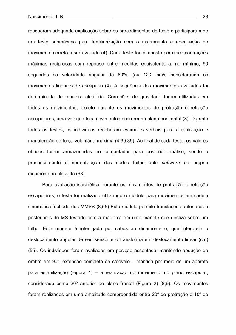
ombro em 90º, extensão completa de cotovelo – mantida por meio de um aparato
para estabilização (Figura 1) – e realização do movimento no plano escapular,
considerado como 30º anterior ao plano frontal (Figura 2) (8;9). Os movimentos
foram realizados em uma amplitude compreendida entre 20º de protração e 10º de
Nascimento, L.R. . 29
retração, para minimizar compensações de tronco, limitadas também pela presença
de tiras de contenção cruzando o tronco (63).
Figura 1: Aparato estabilizador do cotovelo
Figura 2: Posicionamento para avaliação isocinética nos movimentos de protração e retração escapulares.
Nascimento, L.R. . 30
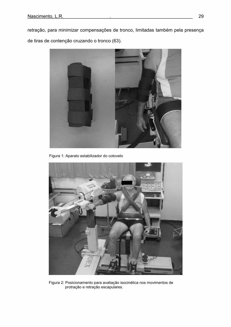
O posicionamento para a avaliação dos movimentos de flexão e extensão do
ombro foi realizado de acordo com estudo conduzido por Kim et al. (39) e normas
descritas no manual do fabricante (39;63). Os indivíduos foram posicionados
assentados, estando o eixo de rotação do dinamômetro alinhado ao acrômio. Os
movimentos foram realizados perfazendo um arco de movimento de 90º, entre 70º
de flexão e 20º de extensão de ombro, sendo esta amplitude baseada nos valores
propostos no estudo de Kim et al. (39) e adaptada após um estudo piloto para
atender às características dos participantes deste estudo (Figura 3).
Figura 3: Posicionamento para avaliação isocinética nos movimentos de flexão e extensão de ombro.
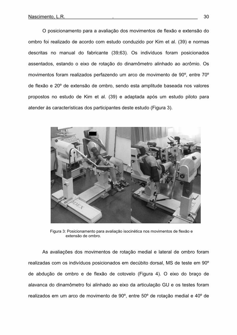
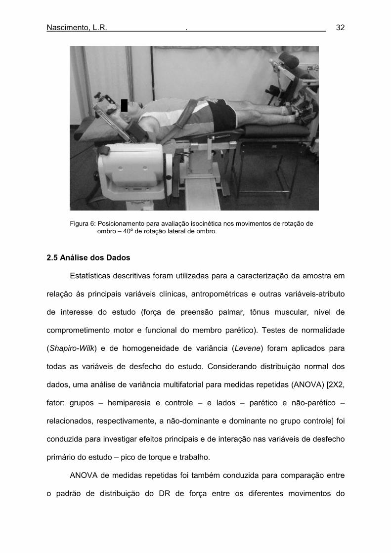
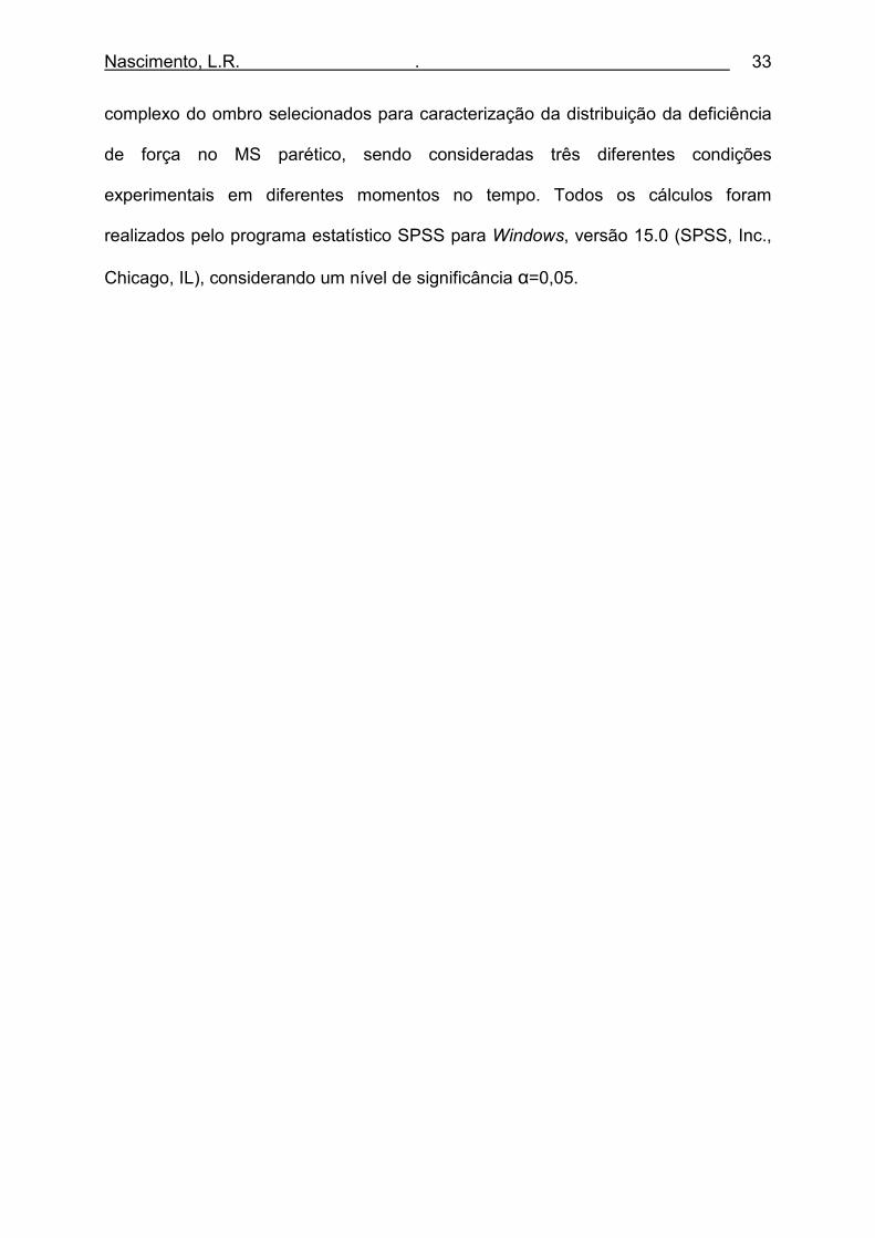
As avaliações dos movimentos de rotação medial e lateral de ombro foram
realizadas com os indivíduos posicionados em decúbito dorsal, MS de teste em 90º
de abdução de ombro e de flexão de cotovelo (Figura 4). O eixo do braço de
alavanca do dinamômetro foi alinhado ao eixo da articulação GU e os testes foram
realizados em um arco de movimento de 90º, entre 50º de rotação medial e 40º de
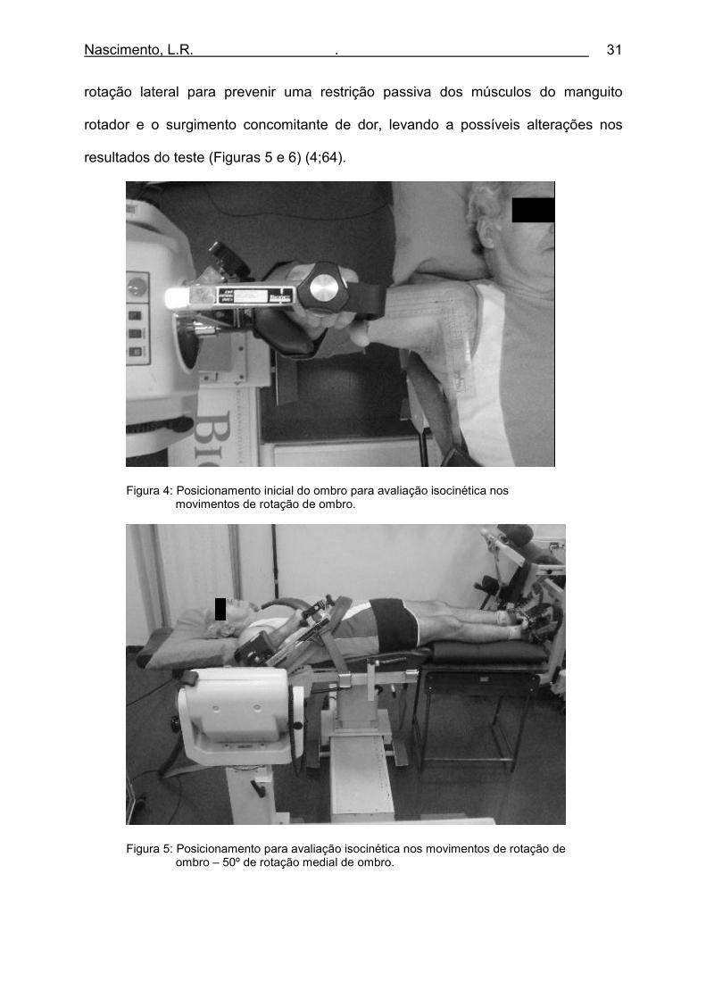
Nascimento, L.R. . 31
rotação lateral para prevenir uma restrição passiva dos músculos do manguito
rotador e o surgimento concomitante de dor, levando a possíveis alterações nos
resultados do teste (Figuras 5 e 6) (4;64).
Figura 4: Posicionamento inicial do ombro para avaliação isocinética nos movimentos de rotação de ombro.
Figura 5: Posicionamento para avaliação isocinética nos movimentos de rotação de ombro – 50º de rotação medial de ombro.
Nascimento, L.R. . 32
Figura 6: Posicionamento para avaliação isocinética nos movimentos de rotação de ombro – 40º de rotação lateral de ombro.
2.5 Análise dos Dados
Estatísticas descritivas foram utilizadas para a caracterização da amostra em
relação às principais variáveis clínicas, antropométricas e outras variáveis-atributo
de interesse do estudo (força de preensão palmar, tônus muscular, nível de
comprometimento motor e funcional do membro parético). Testes de normalidade
(Shapiro-Wilk) e de homogeneidade de variância (Levene) foram aplicados para
todas as variáveis de desfecho do estudo. Considerando distribuição normal dos
dados, uma análise de variância multifatorial para medidas repetidas (ANOVA) [2X2,
fator: grupos – hemiparesia e controle – e lados – parético e não-parético –
relacionados, respectivamente, a não-dominante e dominante no grupo controle] foi
conduzida para investigar efeitos principais e de interação nas variáveis de desfecho
primário do estudo – pico de torque e trabalho.
ANOVA de medidas repetidas foi também conduzida para comparação entre
o padrão de distribuição do DR de força entre os diferentes movimentos do
Nascimento, L.R. . 33
complexo do ombro selecionados para caracterização da distribuição da deficiência
de força no MS parético, sendo consideradas três diferentes condições
experimentais em diferentes momentos no tempo. Todos os cálculos foram
realizados pelo programa estatístico SPSS para Windows, versão 15.0 (SPSS, Inc.,
Chicago, IL), considerando um nível de significância α=0,05.
Nascimento, L.R. . 34
3 – REFERÊNCIAS BIBLIOGRÁFICAS
Nascimento, L.R. . 35
3 REFERÊNCIAS BIBLIOGRÁFICAS (1) Johnson MP, McClure PW, Karduna AR. New method to assess scapular
upward rotation in subjects with shoulder pathology. J Orthop Sports Phys
Ther 2001 Feb;31(2):81-9.
(2) Donatelli R. Physical theray for the shoulder. 3 ed. Philadelfia: Churchill
Livingstone; 1997.
(3) Ebaugh DD, McClure PW, Karduna AR. Three-dimensional scapulothoracic
motion during active and passive arm elevation. Clin Biomech (Bristol , Avon
) 2005 Aug;20(7):700-9.
(4) Moraes GF, Faria CD, Teixeira-Salmela LF. Scapular muscle recruitment
patterns and isokinetic strength ratios of the shoulder rotator muscles in
individuals with and without impingement syndrome. J Shoulder Elbow Surg
2008 Jan;17(1 Suppl):48S-53S.
(5) Gomes PF, Teixeira-Salmela LF, Sesselmann M. Desenvolvimento de um
sistema de medição para análise da cinemática escapular Escola de
Educação Física, Fisioterapia e Terapia Ocupacional da Universidade
Federal de Minas Gerais; 2009.
(6) Carr J, Shepherd R. Reabilitação neurológica: otimizando o desempenho
motor. 1 ed. Barueri: Manole; 2008.
(7) Warren WH. The dynamics of perception and action. Psychol Rev 2006
Apr;113(2):358-89.
Nascimento, L.R. . 36
(8) Cools AM, Witvrouw EE, Declercq GA, Vanderstraeten GG, Cambier DC.
Evaluation of isokinetic force production and associated muscle activity in
the scapular rotators during a protraction-retraction movement in overhead
athletes with impingement symptoms. Br J Sports Med 2004 Feb;38(1):64-8.
(9) Cools AM, Witvrouw EE, Mahieu NN, Danneels LA. Isokinetic Scapular
Muscle Performance in Overhead Athletes With and Without Impingement
Symptoms. J Athl Train 2005 Jun;40(2):104-10.
(10) Matsen FA, III, Chebli C, Lippitt S. Principles for the evaluation and
management of shoulder instability. J Bone Joint Surg Am 2006
Mar;88(3):648-59.
(11) Lippitt S, Matsen F. Mechanisms of glenohumeral joint stability. Clin Orthop
Relat Res 1993 Jun;(291):20-8.
(12) Phadke V, Camargo PR, Ludewig PM. Scapular and rotator cuff muscle
activity during arm elevation: a review of normal function and alterations with
shoulder impingement. Rev Bras Fisioter 2009;13(1):1-9.
(13) Levin S. Tensegrity: the new biomechanics. In: Hutson M, Ellis R, editors.
Textbook of musculoskeletal medicine.Oxford: Oxford University Press;
2005. p. 69-80.
(14) Ingber DE. The architecture of life. Sci Am 1998 Jan;278(1):48-57.
(15) Levin SM. Putting the shoulder to the wheel: a new biomechanical model for
the shoulder girdle. Biomed Sci Instrum 1997;33:412-7.
Nascimento, L.R. . 37
(16) Nascimento LR, Bittencourt NFN, Resende RA, Teixeira-Salmela LF,
Fonseca ST. Biomecânica aplicada ao voleibol: análise do complexo do
ombro e implicações para avaliação e desempenho. Terapia Manual
2010;8(40):in press.
(17) Gajdosik RL. Passive extensibility of skeletal muscle: review of the literature
with clinical implications. Clin Biomech (Bristol , Avon ) 2001 Feb;16(2):87-
101.
(18) Voight ML, Thomson BC. The Role of the Scapula in the Rehabilitation of
Shoulder Injuries. J Athl Train 2000 Jul;35(3):364-72.
(19) Organização Mundial de Saúde. CIF: Classificação internacional de
funcionalidade, incapacidade e saúde. 1 ed. São Paulo: Edusp; 2003.
(20) Harris JE, Eng JJ. Paretic upper-limb strength best explains arm activity in
people with stroke. Phys Ther 2007 Jan;87(1):88-97.
(21) Ouellette MM, LeBrasseur NK, Bean JF, Phillips E, Stein J, Frontera WR, et
al. High-intensity resistance training improves muscle strength, self-reported
function, and disability in long-term stroke survivors. Stroke 2004
Jun;35(6):1404-9.
(22) Lessa I. Epidemiologia das doenças cerebrovaculares no Brasil. Rev Soc
Cardiol 1999;9(4):509-17.
(23) Kaiser E. Aspectos epidemiológicos nas doenças coronariana e
cerebrovascular. SOCERJ 2004;17(1):11-8.
Nascimento, L.R. . 38
(24) Royal College of Physicians. National clinical guidelines for stroke. 2 ed.
London: Intercollegiate Stroke Working; 2004.
(25) Uemura K, Pisa Z. Trends in cardiovascular disease mortality in
industrialized countries since 1950. World Health Stat Q 1988;41(3-4):155-
78.
(26) LeBrasseur NK, Sayers SP, Ouellette MM, Fielding RA. Muscle impairments
and behavioral factors mediate functional limitations and disability following
stroke. Phys Ther 2006 Oct;86(10):1342-50.
(27) Nakayama H, Jorgensen HS, Raaschou HO, Olsen TS. Compensation in
recovery of upper extremity function after stroke: the Copenhagen Stroke
Study. Arch Phys Med Rehabil 1994 Aug;75(8):852-7.
(28) Wade DT. Measuring arm impairment and disability after stroke. Int Disabil
Stud 1989 Apr;11(2):89-92.
(29) Moraes GF, Nascimento LR, Glória AE, Teixeira-Salmela LF, Paiva CM,
Lopes TA, et al. A influência do fortalecimento muscular no desempenho
motor do membro superior parético de indivíduos acometidos por acidente
vascular encefálico. Acta Fisiatr 2008;15(4):245-8.
(30) Carr JH, Shepherd RB. The changing face of neurological rehabilitation. Rev
Bras Fisioter 2006;10(2):147-56.
(31) Morris SL, Dodd KJ, Morris ME. Outcomes of progressive resistance
strength training following stroke: a systematic review. Clin Rehabil 2004
Feb;18(1):27-39.
Nascimento, L.R. . 39
(32) Harris JE, Eng JJ. Strength training improves upper-limb function in
individuals with stroke: a meta-analysis. Stroke 2010 Jan;41(1):136-40.
(33) Mercier C, Bertrand AM, Bourbonnais D. Comparison of strength
measurements under single-joint and multi-joint conditions in hemiparetic
individuals. Clin Rehabil 2005 Aug;19(5):523-30.
(34) Mercier C, Bourbonnais D. Relative shoulder flexor and handgrip strength is
related to upper limb function after stroke. Clin Rehabil 2004 Mar;18(2):215-
21.
(35) Bertrand AM, Mercier C, Bourbonnais D, Desrosiers J, Gravel D. Reliability
of maximal static strength measurements of the arms in subjects with
hemiparesis. Clin Rehabil 2007 Mar;21(3):248-57.
(36) Boissy P, Bourbonnais D, Carlotti MM, Gravel D, Arsenault BA. Maximal grip
force in chronic stroke subjects and its relationship to global upper extremity
function. Clin Rehabil 1999 Aug;13(4):354-62.
(37) Andrews AW, Bohannon RW. Short-term recovery of limb muscle strength
after acute stroke. Arch Phys Med Rehabil 2003 Jan;84(1):125-30.
(38) Milot MH, Nadeau S, Gravel D, Requiao LF. Bilateral level of effort of the
plantar flexors, hip flexors, and extensors during gait in hemiparetic and
healthy individuals. Stroke 2006 Aug;37(8):2070-5.
(39) Kim M, Kothari DH, Lum PS, Patten C. Reliability of dynamic muscle
performance in the hemiparetic upper limb. J Neurol Phys Ther 2005
Mar;29(1):9-17.
Nascimento, L.R. . 40
(40) Meeteren J, Roebroeck ME, Stam HJ. Test-retest reliability in isokinetic
muscle strength measurements of the shoulder. J Rehabil Med 2002
Mar;34(2):91-5.
(41) Cools AM, Geerooms E, Van den Berghe DF, Cambier DC, Witvrouw EE.
Isokinetic scapular muscle performance in young elite gymnasts. J Athl Train
2007 Oct;42(4):458-63.
(42) Canning CG, Ada L, Adams R, O'Dwyer NJ. Loss of strength contributes
more to physical disability after stroke than loss of dexterity. Clin Rehabil
2004 May;18(3):300-8.
(43) Portney L, Watkins M. Foundations of clinical research: applications to
practice. 3 ed. Upper Saddle River, NJ: Prentice Hall; 2009.
(44) Mathiowetz V, Kashman N, Volland G, Weber K, Dowe M, Rogers S. Grip
and pinch strength: normative data for adults. Arch Phys Med Rehabil 1985
Feb;66(2):69-74.
(45) Caporrino FA, Faloppa F, Santos JBG, Réssio C, Soares FHC, Nakachima
LR, et al. Estudo populacional da força de preensão palmar com
dinamômetro Jamar. Rev Bras Ortop 1998;33(2):150-4.
(46) Bertolucci PHF, Brucki SMD, Campacci SR, Juliano Y. O mini-exame do
estado mental em uma população geral. Arq Neuropsiquiatr 1994;52(1):1-7.
(47) Brucki SMD, Nitrini R, Caramelli P, Bertolucci PHF, Okamoto IH. Sugestões
para o uso do mini-exame do estado mental no Brasil. Arq Neuropsiquiatr
2003;61(3B):777-81.
Nascimento, L.R. . 41
(48) Maki T, Quagliato EMAB, Cacho EVA, Paz LPS, Nascimento NH, Inoue
MMEA, et al. Estudo de confiabilidade da aplicação da escala de Fugl-
Meyer no Brasil. Rev Bras Fisioter 2006;10(2):177-83.
(49) Michaelsen SM, Levin MF. Short-term effects of practice with trunk restraint
on reaching movements in patients with chronic stroke: a controlled trial.
Stroke 2004 Aug;35(8):1914-9.
(50) Michaelsen SM, Dannenbaum R, Levin MF. Task-specific training with trunk
restraint on arm recovery in stroke: randomized control trial. Stroke 2006
Jan;37(1):186-92.
(51) Brashear A, Zafonte R, Corcoran M, Galvez-Jimenez N, Gracies JM, Gordon
MF, et al. Inter- and intrarater reliability of the Ashworth Scale and the
Disability Assessment Scale in patients with upper-limb poststroke spasticity.
Arch Phys Med Rehabil 2002 Oct;83(10):1349-54.
(52) Teixeira-Salmela LF, Olney SJ, Brouwer B. Mecanismos e medidas de
espasticidade. Rev Fisioter Uni São Paulo 1998;5(1):4-19.
(53) Faria I, Michaelsen SM, Teixeira-Salmela LF. Função do membro superior
em hemiparéticos crônicos: análise através da classificação internacional de
funcionalidade, incapacidade e saúde. Escola de Educação Física,
Fisioterapia e Terapia Ocupacional da Universidade Federal de Minas
Gerais; 2008.
(54) Faul F, Erdfelder E, Lang AG, Buchner A. G*Power 3: a flexible statistical
power analysis program for the social, behavioral, and biomedical sciences.
Behav Res Methods 2007 May;39(2):175-91.
Nascimento, L.R. . 42
(55) Anjos MTS, Fonseca ST. Análise das propriedades musculares em
indivíduos com e sem postura de protrusão de ombros Escola de Educação
Física, Fisioterapia e Terapia Ocupacional da Universidade Federal de
Minas Gerais; 2006.
(56) Dvir Z. Isocinética: avaliações musculares, interpretações e aplicaçõe
clínicas. 1 ed. São Paulo: Manole; 2002.
(57) Lima LAO, Paula FR. Desempenho muscular de indivíduos na fase inicial da
Doença de Parkinson. Escola de Educação Física, Fisioterapia e Terapia
Ocupacional da Universidade Federal de Minas Gerais; 2008.
(58) Brown LE, Weir JP. ASEP procedures recommendation I: accurate
assessment of muscular strength and power. JEP 2001;4(3):1-21.
(59) Gaines JM, Talbot LA. Isokinetic strength testing in research and practice.
Biol Res Nurs 1999 Jul;1(1):57-64.
(60) Alon G. Defining and measuring residual deficits of the upper extremity
following stroke: a new perspective. Top Stroke Rehabil 2009
May;16(3):167-76.
(61) Innes E. Handgrip strength testing: a review of literature. Aust Occup Ther J
1999;46(3):120-40.
(62) Figueiredo IM, Sampaio RF, Mancini MC, Silva FCM, Souza MAP. Teste de
força de preensão utilizando o dinamômetro Jamar. Acta Fisiatr
2007;14(2):104-10.
Nascimento, L.R. . 43
(63) Biodex Medical Systems. SYSTEM 3 PRO: Application / Operation Manual.
New York, USA.
(64) Mendonça LM, Bittencourt NFN, Anjos MTS, Silva AA, Fonseca ST.
Avaliação muscular isocinética da articulação do ombro em atletas da
seleção brasileira de voleibol sub-19 e sub-21 masculino. Rev Bras Med
Esporte 2010;16(2):107-11.
Nascimento, L.R. . 44
4 – ARTIGO

Nascimento, L.R. . 45
4 ARTIGO
Isokinetic muscular performance and patterns of distribution of the residual
deficits of the shoulder complex in individuals with chronic stroke
ABSTRACT
Background: The present study compared the isokinetic performance of the shoulder complex between individuals with and without stroke, as well as the patterns of distribution of the residual deficits during shoulder and scapular movements to better understand the determinants of the motor and functional upper limb impairments. Methods: Twelve chronic stroke survivors and 12 healthy control subjects were recruited. Concentric measures of peak torque and work during the movements of shoulder external/internal rotation, shoulder flexion/extension, and scapular protraction/retraction, were randomly obtained by the isokinetic Biodex dynamometer at the speed of 60º/s. For individuals with stroke, the residual deficits related to the isokinetic work for the movements of scapular protraction, shoulder external rotation, and shoulder flexion were also calculated. Repeated-measure ANOVAs were employed to investigate the main and interaction effects between the groups and sides and to compare the values of the residual deficits between the three evaluated movements. Findings: The ANOVAS revealed that individuals with stroke demonstrated significant decreases on the paretic shoulder, including during predominantly scapular movements, but no statistically significant decreases were observed on the non-paretic shoulder. The patterns of distributions of the residual deficits were similar for all assessed movements. Interpretation: The findings suggested that scapular weaknesses might significantly contribute to reduced performance of the shoulder complex. Individuals with chronic stroke may benefit from strengthening exercises directed to the scapular muscles, in addition to the glenohumeral muscles. Physical therapists should begin to focus their attention on the paretic upper limbs. Key Words: Cerebrovascular disease; Hemiparesis; Shoulder complex; Muscular strength. _________________________________________________________________________ Autores: Lucas R Nascimento, Luci F Teixeira-Salmela, Janaine C Polese, Christina DCM Faria, Glória EC Laurentino. Periódico: Clinical Biomechanics ISSN: 0268-0033 Endereço eletrônico: http://www.clinbiomech.com/ (ver ANEXO B)
Nascimento, L.R. . 46
1. Introduction
Stroke is one of the leading causes of disability worldwide and has significant
impacts on physical, emotional and social life (Harris and Eng, 2007; Murtezani et al.,
2009). It has been argued that rehabilitation strategies designed to improve motor
functions after stroke should rely upon the understanding of the nature of these
impairments, as well as knowledge of the relative importance of their contributions to
disability (Canning et al., 2004). Muscular weakness is the most common impairment
following stroke and has been shown to significantly relate to motor functions of the
upper limbs (Ada et al., 2006; Harris and Eng, 2007; Teixeira-Salmela et al., 1999).
Considering that upper limb movements are required for most activities of daily living,
studies aimed to improve the understanding of impairments and disabilities related to
upper limb functions are necessary to clarify rehabilitation goals which should be
achieved by physical therapists (Harris and Eng 2007).
The shoulder complex exhibits the greatest amount of motion in the human
body. This mobility is the result of the combined and constrained motions of two main
joints: The glenohumeral and scapulothoracic (Faria et al., 2009; Sharkey et al.,
1994). Weaknesses in one or more of the scapulothoracic or glenohumeral muscles
may cause imbalances in the force couples around the shoulder complex, leading to
abnormal kinematics (Cools et al., 2004; Nascimento et al., 2010). Since these
muscles are constrained to act as a single unit, any dysfunctions in one muscle may
result in instability, which in turn, may decrease movements during upper limb
activities (Gomes et al., 2010; Voight and Thomson, 2000). Although scapular
movements are necessary to guarantee appropriate scapulohumeral rhythm and
allow adequate range of motion during arm elevation (Gomes et al., 2010), the
magnitude of the contributions of scapular weaknesses to the loss of function of the

Nascimento, L.R. . 47
upper limb in relation to glenohumeral joint contributions of individuals with
hemiparesis is still unknown.
Isometric strength measurements outlined that individuals with stroke
demonstrated prominent weaknesses contralateral to the brain injury and reduced
maximal force generating capacities of the non-paretic limbs (Gerrits et al., 2009;
Mercier et al., 2005). However, these measures only indicate general weaknesses of
the glenohumeral muscles and do not provide information regarding the contributions
of possible scapular deficits during upper limb movements. Information is still scarce
regarding the magnitude of weaknesses during dynamic conditions and the specific
residual deficits of the paretic upper limb regarding the glenohumeral and the
scapulothoracic muscles, which may negatively contribute to upper limb performance
following stroke.
Therefore, the aims of this study were to evaluate the dynamic strength of the
upper limb muscles to characterize the magnitude of the weaknesses in both
shoulders of individuals with stroke compared to healthy participants, as well as, to
understand if the nature of these impairments were predominant during
glenohumeral movements, or similarly distributed to the scapulothoracic movements.
Thus, the patterns of distribution of the residual deficits of the paretic upper limb
related to isokinetic work were measured for the stroke group during predominantly
glenohumeral and scapular movements, as well as during movements which involved
both joints. These findings may provide information regarding the magnitudes of
weaknesses in both upper limbs of individuals with stroke and in which movements
may contribute to the losses of function of the entire shoulder complex. These might
help target specific interventions for individuals with stroke.
Nascimento, L.R. . 48
2. Methods
2.1 Participants
Twelve chronic stroke survivors (stroke group) and 12 healthy control subjects
(control group) matched by age, gender, upper limb dominance, and levels of
physical activity, were recruited from the general community of the city of Belo
Horizonte, Brazil. Stroke survivors were included according to the following criteria:
were ≥20 years; had a time since the onset of a unilateral stroke greater than six
months; had no pain or contractures of the upper limb joints which could prevent the
test procedures; had no cognitive deficits, determined by the Mini-mental state
examination (Brucki et al., 2003); had mild or moderate upper limb motor
impairments, based upon the Fugl-Meyer - upper limb motor scores between 30 to
65 (Maki et al., 2006); muscular tone of the elbow flexors ≤3, according to the
Modified Ashworth Scale (Teixeira-Salmela et al., 1998); and had no other
neurological or orthopedic disorders. All participants provided consent prior to their
evaluation, based upon ethical approval from the University research review board.
2.2 Procedures
Physical assessments and interviews were initially conducted with all
individuals for collection of anthropometric, demographic, and clinical data, such as
the time since the onset of the stroke, the paretic side, grip strength, muscular tonus,
motor recovery, and the amount and quality of use of the paretic upper limb.
2.3 Muscular Performance Assessment
Measures of peak torque and work of the glenohumeral and scapular muscles
were obtained on the isokinetic dynamometer, Biodex Medical System 3 Pro (Biodex
Medical Systems, Shirley, NY). The movements of shoulder external/internal
rotations, shoulder flexion/extension, and scapular protraction/retraction were
Nascimento, L.R. . 49
randomly evaluated by the same physical therapist, following previously described
protocols (Cools et al., 2004; Kim et al., 2005; Mendonça et al., 2010). Calibrations
were performed immediately before the tests, following the manufacturer’s
recommendations and the axis of the dynamometer was aligned with the axis of the
glenohumeral joint (Dvir, 2002; Moraes et al., 2008). Gravity corrections were
employed during the tested movements, except for the scapular
protraction/retractions, since these movements occurred in the horizontal plane
(Cools et al., 2004) and all the data were normalized by body mass.
Adaptations were carried out as necessary regarding the test positioning and
range of motion to minimize possible compensatory movements (Hsu et al., 2002).
For the scapular protraction-retraction movements, the closed chain attachment was
fixed to the dynamometer in the horizontal position. The dynamometer shaft was
rotated 30º and the participants were assessed in a seated position with their arms in
the scapular plane (Cools et al., 2004; Cools et al., 2007). The elbow was kept
extended by a stabilizing device and the trunk was stabilized by two crossed straps.
The range of motion was limited to 20º of the scapular protraction to 10º of the
scapular retraction, to better allow comparisons between subjects and avoid
excessive compensatory movements.
The shoulder flexion-extension movements were also assessed in the seated
position with the elbow in extension. The tests were performed within an excursion of
90°, starting from 20º of shoulder extension to 70º of flexion. Adjustments were
made, as necessary, for comfort and to allow the trough to clear the body (Kim et al.,
2005). To evaluate shoulder rotations, the participants were lying in the supine
position to reduce scapular movements, with 90º of shoulder abduction and elbow
flexion. The tests were performed within an arc of 90º, between 40º of external

Nascimento, L.R. . 50
rotation, and 50º of internal rotation (Moraes et al., 2008). This range of motion was
chosen to prevent passive restriction of the rotator cuff and the concurrent onset of
pain (Mendonça et al., 2010).
After a brief explanation of the testing procedures, the participants were asked
to execute three sub-maximal trials to familiarize themselves with the device and the
test protocols. The tests consisted of five maximal concentric-concentric repetitions
for the selected speed of 60º/s or 12,2cm/s, considering the scapular linear
movements and muscular groups. For the stroke group, the non-paretic side was
always tested first, while for the control group, it was the dominant side. During all
tests, the participants received standardized verbal commands. All data were
recorded and stored for future analyses.
2.4 Outcome Measures
Measures of peak torque (Nm) and work (J) were selected for analyses and
used to compare differences in performance between the paretic and non-paretic
sides, as well as between the stroke and control groups. Peak torque is defined as
the product of the mass, acceleration and the lever arm length and provided
information regarding the greatest torque output of the tested limb (Brown and Weir,
2001). Although peak torque measures are considered to be excellent indicators of
the subject’s maximum strength levels, they do not take into account the range of
motion. For this reason, it was also important to analyze the accomplished work,
which revealed the individual’s ability to produce and sustain the torque throughout a
determined range of motion (Brown and Weir, 2001).
The patterns of the distribution of the work residual deficits were calculated
during the following movements: Predominantly glenohumeral (shoulder external
rotation); predominantly scapular (protraction), and associated glenohumeral and
Nascimento, L.R. . 51
scapular (shoulder flexion), as follows: Residual deficits = 100 – (paretic / non-paretic
* 100). The residual deficits focused on the premise that performance of the non-
paretic side of each individual would be the preferred clinical gold standard reference
for the performance of the paretic one (Alon, 2009). In case of finding any
weaknesses on the non-paretic upper extremity, when compared to control subjects,
secondary analyses were conducted and the control group was used as a reference
for the performance of the paretic upper extremity, based upon the following formula:
Residual deficits = 100 – (paretic / control * 100).
2.5 Data Analyses
Descriptive statistics, tests for normality (Shapiro-Wilk), and homogeneity of
variance (Levene) were carried out for all outcome variables, using SPSS for
Windows 15.0 (SPSS, Chicago, IL). A multifactorial repeated measure 2X2 ANOVA
was employed to investigate the main and interaction effects between the groups and
sides for the primary outcome variables (peak torque and work), with a significance
level of α<0.05. Repeated measure ANOVAs were also employed to compare the
residual deficits between the evaluated movements, i.e, predominantly scapular,
predominantly gleonohumeral, and associated glenohumeral and scapular
movements for the stroke group; first using the non-paretic upper limb as a reference
and, secondly, using the control group.
3. Results
3.1 Participants´ Characteristics
The stroke group was composed of 12 individuals (six men) with a mean age
of 52 years (SD: 10.5), ranging from 32 to 67 years, and a mean time since the onset
of the stroke of 10 years (SD: 4.9). The control group was comprised of 12 volunteers
with a mean age of 51.8 years (SD: 11.8), ranging from 30 to 66 years, matched by
Nascimento, L.R. . 52
age, gender, hand dominance, and levels of physical activity. Their demographic,
clinical and anthropometric characteristics are shown in Table 1.
3.2 Strength Measures
Table 2 summarizes the descriptive data and the results from the statistical
analyses for the strength measures of peak torque and work for both groups and
sides regarding the six tested movements. The ANOVA revealed significant group by
side interactions for all evaluated movements: Shoulder external/internal rotations
(17.11≤F≤19.15; df=1; p≤0.01), shoulder flexion/extension (5.64≤F≤21.47; df=1;
0.01≤p≤0.02) and scapular protraction/retraction (6.46≤F≤16.45; df=1; 0.01≤p≤0.02).
These results indicated that the differences between the sides were significant only
for the stroke group. The statistical power ranged from 0.80 to 0.98 for the peak
torque and from 0.79 to 0.91 for the isokinetic work.
3.3 Patterns of Distribution of the Residual Deficits
The initial analyses used the non-paretic shoulder as a reference to compare
the patterns of distribution of the residual deficits. No statistical significant differences
were found for the investigated movements (F=0.50; df=2; p=0.60), with a mean
residual deficit of 44% (95%CI=27 to 61) for shoulder external rotation, 39%
(95%CI=22 to 56) for scapular protraction, and 39% (95%CI=27 to 50) for shoulder
flexion. The secondary analyses were conducted using the control group as a
reference and, again, no statistical differences were found (F=3.26; df=2; p=0.08),
with a mean residual deficit of 52% (95%CI=34 to 69) for external shoulder rotation,
58% (95%CI= 42 to 73) for scapular protraction, and 51% (95%CI= 35 to 66) for
shoulder flexion. These results indicated that the patterns of the residual deficits were
similar between the three evaluated movements for the stroke group.
Nascimento, L.R. . 53
4. Discussion
A detailed assessment of the muscular performance of the shoulder complex
of individuals with hemiparesis is of key relevance, given the fact that brain lesions
and tissue modifications of the musculoskeletal system may affect muscular strength.
The consequent impairments can not only jeopardize the ability to perform functional
activities, but also the level of social participation among individuals with hemiparesis
(Dehail et al., 2008; Goljar et al., 2010). The present study compared the isokinetic
performance of the shoulder complex between individuals with and without stroke, as
well as the patterns of distribution of the residual deficits during shoulder and
scapular movements to better understand the determinants of motor impairments
and functional limitations. No other studies which evaluated the isokinetic muscular
performance of the hemiparetic shoulder complex were found, which made it difficult
to compare with the present results.
The statistical analyses revealed significant interaction effects between the
groups and sides for all of the evaluated movements and for both strength outcomes.
This indicated that only the paretic side demonstrated significant decreases in the
ability to produce and sustain the torque throughout a determined range of motion. It
has been reported that the loss of voluntary strength could be the result of impaired
neural activation, most likely because of impaired excitatory inputs from the damaged
hemisphere (Gerrits et al., 2009; Horstman et al., 2008). However, it has been
suggested that the sedentary lifestyle of the individuals with stroke, plays a
considerable role in determining the loss of concentric torque, since more physically
active individuals demonstrate greater relative concentric torque values for both sides
(Eng et al., 2009). Because the individuals in the present study were included after a
substantially longer period after stroke, the contributions of disuse atrophy and
Nascimento, L.R. . 54
peripheral muscular adaptations, such as contractures and modifications of
mechanical stiffness of the muscles, could be more appropriate to explain losses of
muscular strength.
There are conflicting conclusions regarding the magnitude of the weaknesses
on the non-paretic side, when analyzing previous studies which employed strength
measurements of the upper (Canning et al., 2004; Mercier and Bourbonnais, 2004)
and lower limbs (Eng et al., 2009; Gerrits et al., 2009). Surprisingly, the results of the
present study demonstrated no statistically significant decreases in the ability to
generate force in the non-paretic upper limb. However, lower values and greater
variability were observed for all the measures. These findings suggested that
weaknesses of the non-paretic upper limb appeared not to be as clinically relevant,
when compared to the paretic upper limb. This may be explained by the fact that
although the individuals with stroke reduce their global frequency of daily activities,
they usually perform most of their motor tasks exclusively with their non-paretic upper
limbs, as adaptive mechanisms.
This may allow for sufficient physical stresses to be applied to the
musculoskeletal system, preventing significant weaknesses. These findings
suggested that intervention programs designed to improve upper limb strength
should really focus on the paretic upper limbs to modify structural strength
parameters and then, allow for the emergence of appropriate functions. Therefore,
functional activities, including bimanual activities, should be associated with strength
training to allow the acquired skills to be incorporated in daily life tasks, by applying
constant physical stresses to the biological tissues to prevent negative adaptations
and progressive strength losses (Mueller and Maluf 2002; Nascimento et al., 2009).
Nascimento, L.R. . 55
Previous studies (Cahalan et al., 1991; Shklar and Dvir, 1995) reported that
the shoulder extensor muscles were the strongest group operating on the
glenohumeral joint, contrary to the present results, which demonstrated greater
torque and work output during shoulder flexion. There are two plausible explanations
for the observed differences. First, they might be attributable to the incorporation of
gravity correction procedures, employed in the present study and not in other studies,
which evaluated the isokinetic performance of the shoulder complex. Secondly, in the
present study, the initial range of motion was set at 20º of shoulder extension,
whereas, in previous studies, it was set at 0°. From the 20º of shoulder extension,
might have caused absorption and storage of elastic energy as a function of the
stretching of the shoulder flexors and the subsequent conversion and release of this
energy to perform shoulder flexion and enhance the peak torque and work values.
Similar results were reported by Dehail et al. (2008) in individuals with paraplegia,
when the starting range of motion was set at 35º of shoulder extension. These
findings suggested that the demands imposed by the activity were met by efficiently
using the passive properties of the musculoskeletal system (Fonseca et al., 2010;
Nascimento et al., 2010).
The present results also found weaknesses of the paretic shoulder during the
external and internal rotations. Although it has been suggested that individuals with
hemiparesis demonstrate significant weaknesses of the rotator cuff muscles
(Bertrand et al., 2007; Lum et al., 2003), previous studies which dynamically
quantified the magnitudes of these impairments were not found, probably due to
difficulties in the adaptations of the subjects’ positioning. This study employed
adaptive and feasible methods to measure strength during shoulder rotations in
individuals with hemiparesis, since the lying position was described as comfortable
Nascimento, L.R. . 56
by all participants, who did not relate any pain or discomfort during the test
procedures.
The present results pointed out that weaknesses of the glenohumeral muscles
may be one of the main factors which affect upper limb performance and corroborate
previous studies which reported that during arm elevation, the glenohumeral and
scapulothoracic motions occurred synchronously in about a 2:1 overall ratio, with the
glenohumeral motion occurring alone during the first 30º of arm elevation (Gomes et
al., 2010; Sharkey et al., 1994). The identified abnormalities in the glenohumeral
muscles of individuals with stroke may determine force imbalances around the joint,
affect the dynamic stabilization strategies, and prevent the initiation of movements,
which require arm elevation during daily life activities.
The participants also demonstrated significant deficits related to isokinetic
work, which suggested that even if the movement is initiated, insufficient ability to
sustain torque may compromise movement performance. Thus, individuals with
chronic stroke might get into a vicious cycle, in which muscular weaknesses limit arm
elevation and subsequent inactivity leads to increases in these weaknesses.
Considering that after the first 30º of arm elevation, scapular activity is also required
and that these individuals usually cannot perform activities at higher degrees of arm
elevation, weaknesses of the scapular muscles may be developed due to inactivity
and disuse. This suggests that weaknesses of the scapular muscles may be other
significant impairments affecting upper limb performance of individuals with chronic
stroke.
Evaluations of the scapulothoracic joint have commonly been neglected in the
area of rehabilitation, even with the current evidence indicating that strong scapular
muscles are among the major pre-requisites for optimal stability and functional
Nascimento, L.R. . 57
movements of the upper limbs (Cools et al., 2007). The results of the present study
demonstrated significant reductions in the ability to generate and maintain force
generation during scapular movements in individuals with hemiparesis. Since the
scapula plays a critical role in controlling the position of the glenoid fossa, relatively
small changes in the actions of the scapular muscles may affect the alignment and
forces involved in the movements around the shoulder complex (Cools et al., 2002;
Phadke et al., 2009). These findings suggest that muscular imbalances of the
scapulothoracic joint may affect and negatively contribute to upper limb performance
of individuals with stroke during their daily living activities. Cools et al. (2004)
reported significant side differences regarding protraction strength with athletes who
used overhead movements and had impingement symptoms and reinforced the idea
that muscular weaknesses of the scapular muscles may be related to shoulder
dysfunctions.
This current perspective was also reinforced by results obtained with the
secondary analyses of the present study. Comparisons of patterns of the residual
deficit distributions demonstrated no differences between the three evaluated
movements. These findings indicated that individuals with stroke demonstrated
significant weaknesses of the paretic shoulder, including scapular movements which
suggested that scapular weaknesses might significantly contribute to reduced
performance of the shoulder complex. Therefore, clinical evaluations and
interventions of the scapular muscles should be recommended, since these muscles
are constrained to act in synergisms with the glenohumeral muscles.
It is also believed that appropriate balance and coordination between these
muscular groups, involved in the same functional activities, may improve their
synergic actions and promote better ability to perform a given motor activity (Faria et
Nascimento, L.R. . 58
al., 2009; Moraes et al., 2008). The present results suggested that individuals with
chronic stroke might benefit from strengthening exercises directed to the scapular
muscles in addition to the glenohumeral muscles, and that physical therapists should
initially focus their attention on the paretic upper limbs.
Some limitations of this study were related to the reduced range of motion
used to assess the scapular protraction/retraction movements and the lack of
scapular kinematic analyses during the associated movements of shoulder flexion.
However, the limited range of motion was determined to minimize possible
compensatory movements of the trunk and the utilization of stronger muscular
groups to perform the scapular movements. One could argue regarding the relatively
small sample size. However, this was not believed to be the case, since this sample
was previously calculated and the statistical power was high for the primary outcome
measures. Future studies with larger samples of subjects with different levels of
upper limb impairments are necessary to better control for some confounding factors
and enhance generalizability of the present findings to the whole stroke population.
Since the present results reflected the concentric muscular performance of
individuals with mild or moderate upper limb impairments, caution should be taken in
extrapolating the results to individuals with severe chronic stroke, or other types of
muscular contraction.
5. Conclusions
The findings of the present study indicated that individuals with chronic stroke
classified as mild or moderate regarding upper limb impairments, demonstrated
significant weaknesses of the paretic shoulder, including during predominantly
scapular movements and non-significant decreases in strength of the non-paretic
upper limb, when compared to healthy controls. The patterns of the residual deficits
Nascimento, L.R. . 59
were similar for all assessed movements, which suggested that scapular
weaknesses might be related to reduced performance of the shoulder complex.
These findings suggested that individuals with chronic stroke might benefit from
strengthening exercises directed to the scapular, in addition to the glenohumeral
muscles. Physical therapists should focus their attention more on the paretic upper
limb. Randomized clinical trials are then necessary to verify the efficacy of muscular
strengthening of the scapular and glenohumeral muscles on outcomes related to the
domains of body structures and functions of the hemiparetic shoulder, as well as on
the functional activities performed with the paretic upper limb.
Acknowledgements
The authors are thankful to Dr. Louise Ada and Dr. John Henry Salmela for their
precious contributions to the development of the study’s research questions and to
copy-editing the manuscript.
References
Ada, L., Dorsch, S., Canning, C.G. 2006. Strengthening interventions increase
strength and improve activity after stroke: A systematic review. Aust.J.Physiother. 52,
241-248.
Alon, G. 2009. Defining and measuring residual deficits of the upper extremity
following stroke: A new perspective. Top.Stroke Rehabil. 16, 167-176.
Bertrand, A.M., Mercier, C., Bourbonnais, D., Desrosiers, J., Gravel, D. 2007.
Reliability of maximal static strength measurements of the arms in subjects with
hemiparesis. Clin.Rehabil. 21, 248-257.
Brown, L.E., Weir, J.P. 2001. ASEP procedures recommendation I: Accurate
assessment of muscular strength and power. JEP. 4, 1-21.
Nascimento, L.R. . 60
Brucki, S.M.D., Nitrini, R., Caramelli, P., Bertolucci, P.H.F., Okamoto, I.H. 2003.
Sugestões para o uso do mini-exame do estado mental no Brasil.
Arq.Neuropsiquiatr. 61, 777-781.
Cahalan, T.D., Johnson, M.E., Chao, E.Y. 1991. Shoulder strength analysis using the
Cybex II isokinetic dynamometer. Clin.Orthop.Relat Res. 271, 249-257.
Canning, C.G., Ada, L., Adams, R., O'Dwyer, N.J. 2004. Loss of strength contributes
more to physical disability after stroke than loss of dexterity. Clin.Rehabil. 18, 300-
308.
Cools, A.M., Geerooms, E., Van den Berghe, D.F., Cambier, D.C., Witvrouw, E.E.
2007. Isokinetic scapular muscle performance in young elite gymnasts. J.Athl.Train.
42, 458-463.
Cools, A.M., Witvrouw, E.E., Danneels, L.A., Vanderstraeten, G.G., Cambier, D.C.
2002. Test-retest reproducibility of concentric strength values for shoulder girdle
protraction and retraction using the Biodex isokinetic dynamometer.
Isokinet.Exerc.Sci. 10, 129-136.
Cools, A.M., Witvrouw, E.E., Declercq, G.A., Vanderstraeten, G.G., Cambier, D.C.
2004. Evaluation of isokinetic force production and associated muscle activity in the
scapular rotators during a protraction-retraction movement in overhead athletes with
impingement symptoms. Br.J.Sports Med. 38, 64-68.
Dehail, P., Gagnon, D., Noreau, L., Nadeau, S. 2008. Assessment of agonist-
antagonist shoulder torque ratios in individuals with paraplegia: A new interpretative
approach. Spinal Cord. 46, 552-558.
Nascimento, L.R. . 61
Dvir, Z. 2002. Isocinética: avaliações musculares, interpretações e aplicaçõe
clínicas., 1 ed. São Paulo, Manole.
Eng, J.J., Lomaglio, M.J., Macintyre, D.L. 2009. Muscle torque preservation and
physical activity in individuals with stroke. Med.Sci.Sports Exerc. 41, 1353-1360.
Faria, C.D., Teixeira-Salmela, L.F., Gomes, P.F. 2009. Applicability of the
coactivation method in assessing synergies of the scapular stabilizing muscles.
J.Shoulder.Elbow.Surg. 18, 764-772.
Fonseca, S. T., Souza, T. R., Ocarino, J. M., Goncalves, G. P., Bittencourt, N. F. N.
2010, Applied biomechanics of soccer. In Athletic and sport issues in
musculoskeletal rehabilitation, 1 ed. D. J. Magee et al., eds., St. Louis MO: Saunders
Elsevier, pp. 12.1-12.20.
Gerrits, K.H., Beltman, M.J., Koppe, P.A., Konijnenbelt, H., Elich, P.D., de, H.A.,
Janssen, T.W. 2009. Isometric muscle function of knee extensors and the relation
with functional performance in patients with stroke. Arch.Phys.Med.Rehabil. 90, 480-
487.
Goljar, N., Burger, H., Vidmar, G., Marincek, C., Krizaj, J., Chatterji, S., Raggi, A.,
Leonardi, M., Bickenbach, J.E. 2010. Functioning and disability in stroke.
Disabil.Rehabil. 32(Suppl 1), S50-S58.
Gomes, P.F., Sesselmann, M., Faria, C.D., Araujo, P.A., Teixeira-Salmela, L.F. 2010.
Measurement of scapular kinematics with the moire fringe projection technique.
J.Biomech. 43, 1215-1219.
Nascimento, L.R. . 62
Harris, J.E., Eng, J.J. 2007. Paretic upper-limb strength best explains arm activity in
people with stroke. Phys.Ther. 87, 88-97.
Horstman, A.M., Beltman, M.J., Gerrits, K.H., Koppe, P., Janssen, T.W., Elich, P., de,
H.A. 2008. Intrinsic muscle strength and voluntary activation of both lower limbs and
functional performance after stroke. Clin.Physiol Funct.Imaging. 28, 251-261.
Hsu, A.L., Tang, P.F., Jan, M.H. 2002. Test-retest reliability of isokinetic muscle
strength of the lower extremities in patients with stroke. Arch.Phys.Med.Rehabil. 83,
1130-1137.
Kim, M., Kothari, D.H., Lum, P.S., Patten, C. 2005. Reliability of dynamic muscle
performance in the hemiparetic upper limb. J.Neurol.Phys.Ther. 29, 9-17.
Lum, P.S., Burgar, C.G., Shor, P.C. 2003. Evidence for strength imbalances as a
significant contributor to abnormal synergies in hemiparetic subjects. Muscle Nerve.
27, 211-221.
Maki, T., Quagliato, E.M.A.B., Cacho, E.V.A., Paz, L.P.S., Nascimento, N.H., Inoue,
M.M.E.A., Viana, M.A. 2006. Estudo de confiabilidade da aplicação da escala de
Fugl-Meyer no Brasil. Rev.Bras.Fisioter. 10, 177-183.
Mendonça, L.M., Bittencourt, N.F.N., Anjos, M.T.S., Silva, A.A., Fonseca, S.T. 2010.
Avaliação muscular isocinética da articulação do ombro em atletas da seleção
brasileira de voleibol sub-19 e sub-21 masculino. Rev.Bras.Med.Esporte. 16, 107-
111.

Nascimento, L.R. . 63
Mercier, C., Bertrand, A.M., Bourbonnais, D. 2005. Comparison of strength
measurements under single-joint and multi-joint conditions in hemiparetic individuals.
Clin.Rehabil. 19, 523-530.
Mercier, C., Bourbonnais, D. 2004. Relative shoulder flexor and handgrip strength is
related to upper limb function after stroke. Clin.Rehabil. 18, 215-221.
Moraes, G.F., Faria, C.D., Teixeira-Salmela, L.F. 2008. Scapular muscle recruitment
patterns and isokinetic strength ratios of the shoulder rotator muscles in individuals
with and without impingement syndrome. J.Shoulder.Elbow.Surg. 17(1 Suppl), 48S-
53S.
Mueller, M.J., Maluf, K.S. 2002. Tissue adaptation to physical stress: A proposed
"Physical Stress Theory" to guide physical therapist practice, education, and
research. Phys.Ther. 82, 383-403.
Murtezani, A., Hundozi, H., Gashi, S., Osmani, T., Krasniqi, V., Rama, B. 2009.
Factors associated with reintegration to normal living after stroke. Med.Arh. 63, 216-
219.
Nascimento, L.R., Bittencourt, N.F.N., Resende, R.A., Teixeira-Salmela, L.F.,
Fonseca, S.T. 2010. Biomecânica aplicada ao voleibol: Análise do complexo do
ombro e implicações para avaliação e desempenho. Terapia Manual. 8, in press.
Nascimento, L.R., Glória, A.E., Habib, E.S. 2009. Effects of constraint-induced
movement therapy as a rehabilitation strategy for the affected upper limb of children
with hemiparesis: a systematic review. Rev.Bras.Fisioter. 13, 97-102.
Nascimento, L.R. . 64
Phadke, V., Camargo, P.R., Ludewig, P.M. 2009. Scapular and rotator cuff muscle
activity during arm elevation: a review of normal function and alterations with
shoulder impingement. Braz.Phys.Ther.J. 13, 1-9.
Sharkey, N.A., Marder, R.A., Hanson, P.B. 1994. The entire rotator cuff contributes to
elevation of the arm. J.Orthop.Res. 12, 699-708.
Shklar, A., Dvir, Z. 1995. Isokinetic strength relationships in shoulder muscles.
Clin.Biomech. 10, 369-373.
Teixeira-Salmela, L.F., Olney, S.J., Brouwer, B. 1998. Mecanismos e medidas de
espasticidade. Rev.Fisioter.Uni.São Paulo. 5, 4-19.
Teixeira-Salmela, L.F., Olney, S.J., Nadeau, S., Brouwer, B. 1999. Muscle
strengthening and physical conditioning to reduce impairment and disability in
chronic stroke survivors. Arch.Phys.Med.Rehabil. 80, 1211-1218.
Voight, M.L., Thomson, B.C. 2000. The role of the scapula in the rehabilitation of
shoulder injuries. J.Athl.Train. 35, 364-372.
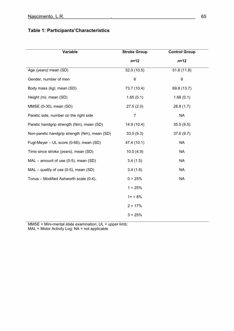
Nascimento, L.R. . 65
Table 1: Participants’Characteristics
Variable Stroke Group
n=12
Control Group
n=12
Age (years) mean (SD) 52.0 (10.5) 51.8 (11.8)
Gender, number of men 6 6
Body mass (kg), mean (SD) 73.7 (10.4) 69.8 (13.7)
Height (m), mean (SD) 1.65 (0.1) 1.68 (0.1)
MMSE (0-30), mean (SD) 27.5 (2.0) 28.8 (1.7)
Paretic side, number on the right side 7 NA
Paretic handgrip strength (Nm), mean (SD) 14.9 (10.4) 35.5 (9.5)
Non-paretic handgrip strength (Nm), mean (SD) 33.0 (9.3) 37.6 (9.7)
Fugl-Meyer – UL score (0-66), mean (SD) 47.4 (10.1) NA
Time since stroke (years), mean (SD) 10.0 (4.9) NA
MAL – amount of use (0-5), mean (SD) 3.4 (1.5) NA
MAL – quality of use (0-5), mean (SD) 3.4 (1.6) NA
Tonus – Modified Ashworth scale (0-4), 0 = 25%
1 = 25%
1+ = 8%
2 = 17%
3 = 25%
NA
MMSE = Mini-mental state examination; UL = upper limb; MAL = Motor Activity Log; NA = not applicable
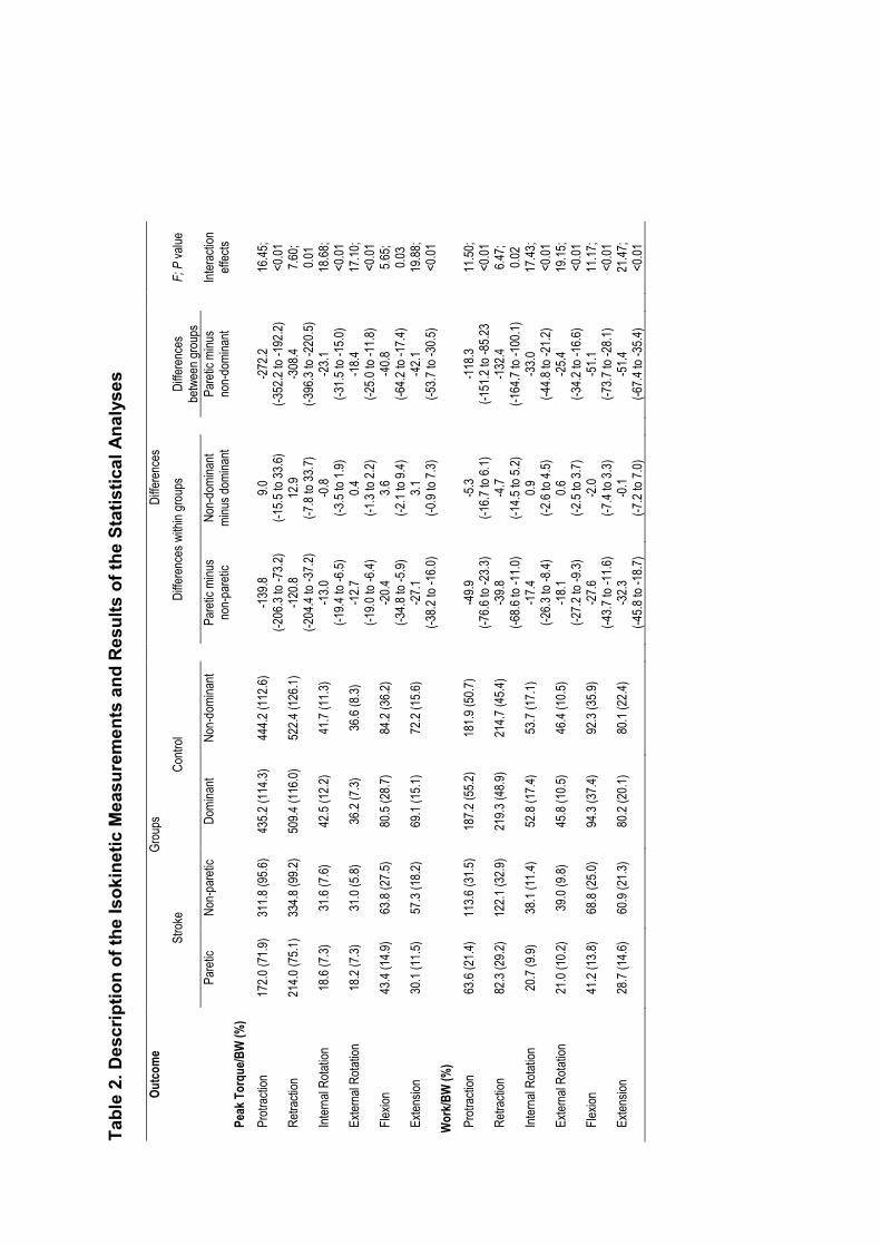
Tab
le 2. D
escription of the Isokinetic Measu
remen
ts and Res
ults of the Statistical Analyses
Out
com
e Groups
Differences
Stroke
Control
Differences within groups
Differences
betwee
n groups
F; P value
Pa
retic
Non-pa
retic
Dominant
Non-do
minant
Paretic minus
non-pa
retic
Non-do
minant
minus dom
inant
Paretic minus
non-do
minant
Interaction
effects
Peak
Tor
que/
BW (%
)
Protraction
172.0 (71.9)
311.8 (95.6)
435.2 (114.3)
444.2 (112.6)
-139.8
(-206
.3 to -7
3.2)
9.0
(-15.5 to 33.6)
-272.2
(-352
.2 to -1
92.2)
16.45;
<0.01
Retra
ction
214.0 (75.1)
334.8 (99.2)
509.4 (116.0)
522.4 (126.1)
-120.8
(-204
.4 to -3
7.2)
12.9
(-7.8 to 33.7)
-308.4
(-396
.3 to -2
20.5)
7.60;
0.01
Internal Rotation
18.6 (7
.3)
31.6 (7
.6)
42.5 (1
2.2)
41.7 (1
1.3)
-13.0
(-19.4 to -6
.5)
-0.8
(-3.5 to 1.9)
-23.1
(-31.5 to -1
5.0)
18.68;
<0.01
External Rotation
18.2 (7
.3)
31.0 (5
.8)
36.2 (7
.3)
36.6 (8
.3)
-12.7
(-19.0 to -6
.4)
0.4
(-1.3 to 2.2)
-18.4
(-25.0 to -1
1.8)
17.10;
<0.01
Flexion
43.4 (1
4.9)
63.8 (2
7.5)
80.5 (2
8.7)
84.2 (3
6.2)
-20.4
(-34.8 to -5
.9)
3.6
(-2.1 to 9.4)
-40.8
(-64.2 to -1
7.4)
5.65;
0.03
Ex
tension
30.1 (1
1.5)
57.3 (1
8.2)
69.1 (1
5.1)
72.2 (1
5.6)
-27.1
(-38.2 to -1
6.0)
3.1
(-0.9 to 7.3)
-42.1
(-53.7 to -3
0.5)
19.88;
<0.01
Wor
k/BW
(%)
Protraction
63.6 (2
1.4)
113.6 (31.5)
187.2 (55.2)
181.9 (50.7)
-49.9
(-76.6 to -2
3.3)
-5.3
(-16.7 to 6.1)
-118.3
(-151
.2 to -8
5.23
11.50;
<0.01
Retra
ction
82.3 (2
9.2)
122.1 (32.9)
219.3 (48.9)
214.7 (45.4)
-39.8
(-68.6 to -1
1.0)
-4.7
(-14.5 to 5.2)
-132.4
(-164
.7 to -1
00.1)
6.47;
0.02
Internal Rotation
20.7 (9
.9)
38.1 (1
1.4)
52.8 (1
7.4)
53.7 (1
7.1)
-17.4
(-26.3 to -8
.4)
0.9
(-2.6 to 4.5)
-33.0
(-44.8 to -2
1.2)
17.43;
<0.01
External Rotation
21.0 (1
0.2)
39.0 (9
.8)
45.8 (1
0.5)
46.4 (1
0.5)
-18.1
(-27.2 to -9
.3)
0.6
(-2.5 to 3.7)
-25.4
(-34.2 to -1
6.6)
19.15;
<0.01
Flexion
41.2 (1
3.8)
68.8 (2
5.0)
94.3 (3
7.4)
92.3 (3
5.9)
-27.6
(-43.7 to -1
1.6)
-2.0
(-7.4 to 3.3)
-51.1
(-73.7 to -2
8.1)
11.17;
<0.01
Extension
28.7 (1
4.6)
60.9 (2
1.3)
80.2 (2
0.1)
80.1 (2
2.4)
-32.3
(-45.8 to -1
8.7)
-0.1
(-7.2 to 7.0)
-51.4
(-67.4 to -3
5.4)
21.47;
<0.01
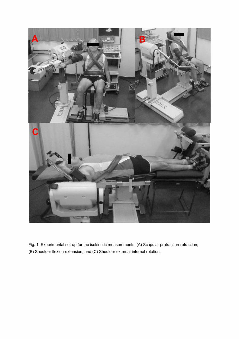
Fig. 1. Experimental set-up for the isokinetic measurements: (A) Scapular protraction-retraction;
(B) Shoulder flexion-extension; and (C) Shoulder external-internal rotation.
A B
C
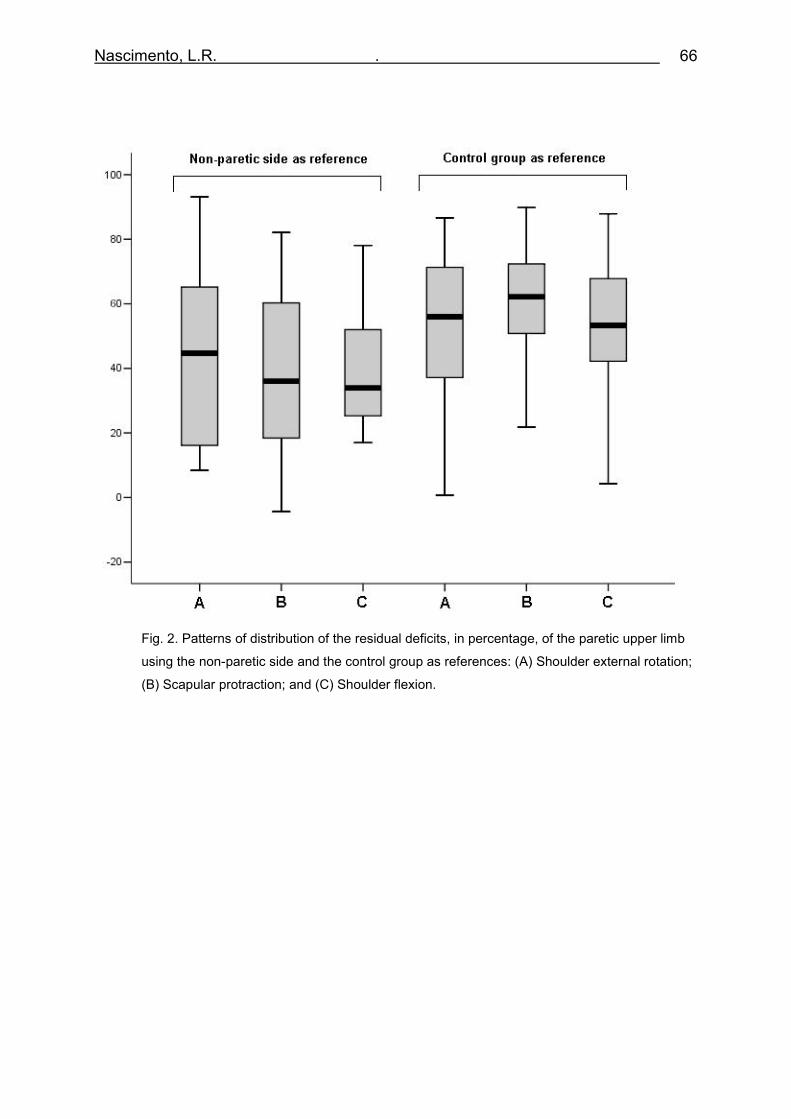
Nascimento, L.R. . 66
Fig. 2. Patterns of distribution of the residual deficits, in percentage, of the paretic upper limb
using the non-paretic side and the control group as references: (A) Shoulder external rotation;
(B) Scapular protraction; and (C) Shoulder flexion.
Nascimento, L.R. . 67
5 – CONSIDERAÇÕES FINAIS
Nascimento, L.R. . 68
5 CONSIDERAÇÕES FINAIS
O presente estudo investigou o desempenho muscular isocinético (pico de
torque e trabalho) dos grupos musculares relacionados aos movimentos de
protração e retração escapulares, flexão, extensão e rotações medial e lateral de
ambos os ombros em indivíduos com e sem sequelas motoras decorrentes de AVE.
Foi analisado, ainda, o padrão de distribuição do déficit residual relacionado ao
trabalho isocinético durante um movimento predominantemente gleno-umeral
(rotação lateral de ombro), um movimento predominantemente escapular (protração)
e um movimento associado (flexão de ombro) para melhor compreender os
determinantes das deficiências motoras e funcionais relacionadas ao MS parético.
A análise dos dados indicou efeitos de interação entre grupos e lados em
todas as medidas de desfecho primário do estudo, demonstrando que apenas o MS
parético apresentou diminuição significativa na capacidade de gerar e manter força
muscular durante os movimentos. Esses resultados sugerem que os profissionais da
área de reabilitação devam priorizar o MS parético durante o processo de
intervenção, com objetivo de modificar parâmetros estruturais relacionados à força
muscular para, então, permitir a emergência de adequadas habilidades funcionais.
Adicionalmente à fraqueza muscular caracterizada em movimentos
predominantemente gleno-umerais, o presente estudo demonstrou que indivíduos
com hemiparesia crônica apresentaram diminuição na capacidade de gerar e
sustentar a força produzida durante movimentos predominantemente escapulares.
Em acordo, a comparação entre os padrões de distribuição do déficit residual do
trabalho isocinético não demonstrou diferenças significativas entre as três diferentes
condições avaliadas. Tais achados sugerem que as alterações em musculatura
Nascimento, L.R. . 69
escapular após o AVE podem influenciar negativamente o desempenho funcional do
complexo do ombro dessa população durante atividades de vida diária e devem ser
considerados no processo de avaliação. Dessa forma, é possível hipotetizar que
indivíduos com hemiparesia crônica possam se beneficiar de programas de
fortalecimento muscular direcionados à musculatura escapular, em adição aos
exercícios usualmente direcionados à musculatura gleno-umeral.
Ensaios clínicos aleatorizados são, então, necessários para verificar a eficácia
de programas de fortalecimento muscular progressivo no complexo do ombro de
indivíduos com hemiparesia em variáveis relacionadas os domínios de estrutura e
função do corpo, bem como seus efeitos na capacidade de realizar atividades
funcionais com o MS parético.
Nascimento, L.R. . 70
APÊNDICE A
Termo de Consentimento Livre e Esclarecido
Pesquisador: Lucas Rodrigues Nascimento, Esp. (031) 3313-7949 Orientadora: Prof. Luci Fuscaldi Teixeira-Salmela, PhD. (031) 3409-7403 TÍTULO DO PROJETO DESEMPENHO MUSCULAR ISOCINÉTICO DO COMPLEXO DO OMBRO DE INDIVÍDUOS HEMIPARÉTICOS CRÔNICOS.
Você está sendo convidado a participar de um projeto de pesquisa a ser
desenvolvido no Departamento de Fisioterapia da Escola de Educação Física
Fisioterapia e Terapia Ocupacional da Universidade Federal de Minas Gerais, para
avaliar a capacidade de produzir força nos músculos do braço de indivíduos que
sofreram derrame e comparar com a força de indivíduos que não sofreram derrame.
Endereço: Departamento de Fisioterapia: Avenida Antônio Carlos, 6627, 3o andar – Campus Pampulha - Belo Horizonte – Minas Gerais - CEP: 31270-901 - Telefone: (31) 3409-4783
Este estudo irá avaliar a força músculos que realizam os principais
movimentos dos seus ombros e o nível de utilização dos mesmos nas suas
atividades cotidianas.
Uma entrevista inicial será administrada para coleta dos seus dados clínicos e
pessoais. Será realizado um exame físico para avaliar suas condições antes dos
testes e um questionário contendo perguntas sobre a utilização dos seus braços
durante as atividades que você habitualmente realiza.
A força muscular de alguns músculos dos seus braços será avaliada de modo
dinâmico para melhor refletir as situações diárias de sua vida. Para tanto, você será
adequadamente posicionado em um aparelho denominado dinamômetro isocinético
e fará a familiarização com o instrumento antes de realizar os testes. Após esse
período, você irá fazer quatro movimentos específicos com o seu braço em duas
diferentes velocidades. Durante os procedimentos, sua imagem poderá ser
registrada por meio de fotos ou vídeos, sem identificação do seu rosto, para
posterior apresentação em apresentações e publicações de natureza técnico-
científicas relacionadas ao projeto de pesquisa.
Nascimento, L.R. . 71
Todos os testes serão realizados no Laboratório de Performance Humana do
Departamento de Fisioterapia da Escola de Educação Física, Fisioterapia e Terapia
Ocupacional da Universidade Federal de Minas Gerais.
Para manter a confidencialidade dos seus registros, o investigador Lucas
Rodrigues Nascimento colocará uma identificação numérica nos seus questionários
e em suas avaliações, sendo que apenas o investigador e a sua orientadora terão
conhecimento do nome correspondente a esta identificação, ou seja, sua identidade
não será revelada. Seus dados serão apresentados em relatórios estatísticos
agrupados sem qualquer identificação individual. Os dados serão armazenados sob
cuidado e manejo restrito aos pesquisadores responsáveis, para uso exclusivo desta
pesquisa, com guarda em local apropriado no Depto. de Fisioterapia da UFMG por
cinco anos, após o qual serão descartados.
Os testes e procedimentos adotados não apresentam riscos específicos além
daqueles presentes no seu dia-a-dia. Durante o teste, você pode sentir cansaço e
caso isto aconteça, períodos de repouso serão permitidos entre um teste e outro.
Qualquer tipo de desconforto vivenciado durante os testes deve ser revelado, para
que os pesquisadores tomem as devidas providências com o objetivo de minimizá-
lo.
Você não obterá benefícios imediatos ao participar dessa pesquisa. Na
realidade, você estará contribuindo para a nossa melhor compreensão de como a
força muscular dos seus braços se apresenta em indivíduos que sofreram derrame.
Com isso, poderemos planejar estratégias de tratamento mais adequadas para
pessoas que apresentam disfunções relacionadas a essa doença. Você não
receberá nenhuma forma de pagamento pela participação no estudo. Custos de
transporte para o local dos testes e seu retorno poderão, se necessários, ser
arcados pelos pesquisadores.
A sua participação no estudo é voluntária e você tem o direito de retirar-se por
qualquer motivo e a qualquer momento, sem necessidade de justificativa prévia.
Garantimos a ausência de prejuízos acadêmicos e sociais caso você se recuse ou
desista de participar dessa pesquisa.
Depois de ter lido as informações acima, se for de sua vontade participar, por favor, preencha o consentimento abaixo.
Nascimento, L.R. . 72
DECLARAÇÃO E ASSINATURA
Eu, _______________________________________________________
li e entendi toda a informação repassada sobre o estudo, sendo que os objetivos, procedimentos e linguagem técnica satisfatoriamente explicados. Tive tempo suficiente, para considerar as informações acima e tive a oportunidade de tirar todas as minhas dúvidas. Estou assinando este termo voluntariamente e tenho direito de agora, ou mais tarde, discutir qualquer dúvida que venha a ter com relação à pesquisa com:
Lucas Rodrigues Nascimento (31) 3313-7949 (31) 9292-7878
Prof. Luci Fuscaldi Teixeira-Salmela (31) 3409-7403
Comitê de Ética em Pesquisa da UFMG (31) 3409-4592 Endereço: Avenida Antônio Carlos, 6627, Pampulha, BH/MG Campus – UFMG – Unidade Administrativa II – 2º andar. Assinando esse termo de consentimento, estou indicando que concordo em participar deste estudo. _________________________ Assinatura do Participante Data: ______________________ End: ______________________ Responsáveis __________________________ ___________________________ Lucas Rodrigues Nascimento Luci Fuscaldi Teixeira-Salmela Pesquisador Orientadora

Nascimento, L.R. . 73
APÊNDICE B
Ficha de Avaliação
1. DADOS DEMOGRÁFICOS
Nome:_____________________________Sexo:______Telefone:_____________
Endereço:_________________________________________________________
Nome acompanhante: ________________________Telefone: _______________
Endereço:_________________________________________________________
Data de Nascimento:________Idade (anos): _____Naturalidade:______________
Estado civil: ______________Mora com: ________________________________
Escolaridade:______________________________________________________
Formação: __________________________Ocupação: _____________________
Em geral,o senhor diria que a sua saúde é:
( )Excelente ( )Muito boa ( )Boa ( )Razoável ( )Ruim
QP: ______________________________________________________________
Altura:_______________ Peso:__________________ IMC:________________
2. DADOS CLÍNICOS DO AVE
( ) Um episódio de AVE ( ) Mais de um episódio de AVE
Data: _______________ ( ) Data da última: _______________
Tempo de evolução (meses):________ Tempo de evolução (meses):________
( ) Isquêmico ( ) Hemorrágico ( ) Isquêmico ( ) Hemorrágico
( ) HP esquerda ( ) HP direita ( ) HP esquerda ( ) HP direita
Tempo de estadia hospitalar: _______ Tempo de estadia hospitalar: _______
Reabilitação: Fisio: ______________ TO: ___________________
Fono: ______________ Outro: _________________
Nascimento, L.R. . 74
3. DADOS CLÍNICOS GERAIS
Membro superior dominante pré-AVE: ( ) D ( ) E
Membro superior dominante pós-AVE: ( ) D ( ) E
Uso de órtese em membro superior: ( ) Sim ( ) Não Qual: _______
Número de medicamentos em uso: _____
Descrição: ________________________________________________________
Numero de doenças associadas: _____
Descrição: ________________________________________________________
Atividade física: ( ) Sim ( ) Não Qual: __________ Freqüência: ________
( ) Déficit visual ( ) Déficit auditivo ( ) Afasia Tipo: _____________ ( ) Disartria
4. DADOS CLÍNICOS ESPECÍFICOS
Força Muscular de preensão palmar D: ____ _____ _____ = _____ N
Força Muscular de preensão palmar E: ____ _____ _____ = _____ N
Tônus de Flexores de Cotovelo (Ashworth): _____
Pontuação motora na Escala de Fugl-Meyer: ______ pontos MMSS
Pontuação MAL-BRASIL: ______ qualidade do uso
______ quantidade de uso
Comentários:
___________________________________________________________________
___________________________________________________________________
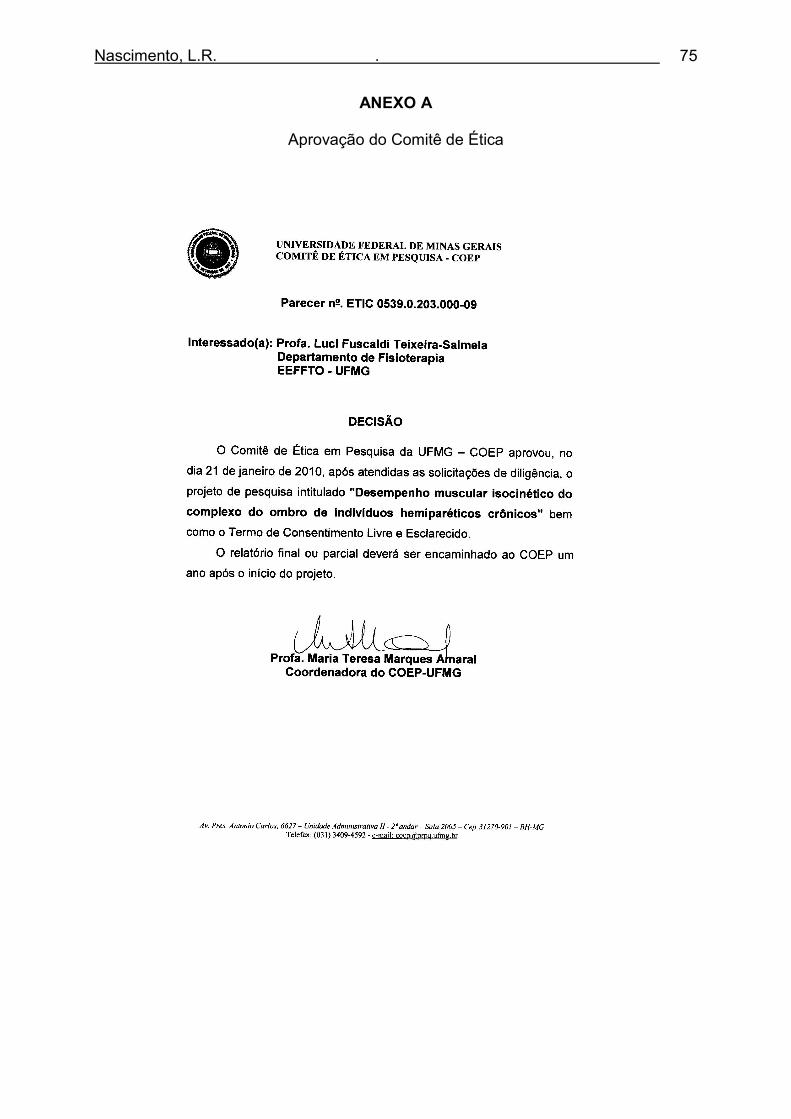
Nascimento, L.R. . 75
ANEXO A
Aprovação do Comitê de Ética
Nascimento, L.R. . 76
ANEXO B
Normas para submissão de manuscrito no Clinical Biomechanics
Aims Clinical Biomechanics aims to strengthen the link between clinic and laboratory by publishing biomechanics research which helps to explain the causes of musculoskeletal disorders and provides knowledge contributing to improved management. Scope Clinical Biomechanics explores all facets of musculoskeletal biomechanics with an emphasis on clinical management. The role of basic and medical science is recognized in a clinical context. The readership of the journal closely reflects its contents, being a balance of scientists, engineers and clinicians. Authorship All authors should have made substantial contributions to all of the following: (1) the conception and design of the study, or acquisition of data, or analysis and interpretation of data, (2) drafting the article or revising it critically for important intellectual content, (3) final approval of the version to be submitted. Instructions for Authors Contributions falling into the following categories will be considered for publication and are accepted on the understanding that they have not been published previously, not are under consideration for publication in any other journal. Papers - scientific reports within the scope of the journal. The length should not normally exceed 4000 words with around six figures/tables (large data tables and multi-part figures are generally best placed in Supplementary Data - see below). Reports focused on validity/reliability of methods in the absence of an experimental application are not acceptable. Reports on model development should address a specific question of clinical interest or report a novelty not yet understood. Reports of implant tests should involve a clinical application, not solely a laboratory test. Submissions are screened by an editorial panel; if considered suitable for the journal, two or more peer reviewers will be allocated. Only a proportion of scientifically acceptable papers can be accepted for publication, so authors should be aware that submissions requiring extensive revisions are unlikely to be offered the opportunity to revise and resubmit. In cases where the original reviewers disagree, the editor may opt to obtain further opinion. Appeals can only be considered where the authors can identify an irregularity in the review process: it is not acceptable simply to state that the reviewers' concerns can be addressed. Authors are invited to submit to the journal online http://ees.elsevier.com/clbi/. You will be guided through the creation and uploading of the various files. Once the uploading is done, the system automatically generates an electronic (PDF) proof, which is then used for reviewing. All correspondence, including notification of the Editor's decision and requests for revisions, will be by e-mail. Enquiries about the suitability of potential articles should be sent to the Editor: Prof Kim Burton, Clinical Biomechanics, 30 Queen Street, Huddersfield HD1 2SP, UK Tel: +44(0)1484 535200; fax: +44(0)1484 435744; e-mail: [email protected] When submitting a paper you are expecting a number of colleagues to review your work. As a matter of courtesy you should ensure your manuscript is neatly presented as well as complying with the journal's requirements. Submissions will be returned immediately without review if they do not follow all these guidance notes. • English language; double spaced; single sided; page-numbered and line-numbered.

Nascimento, L.R. . 77
• A title page including name(s) of author(s), qualifications, institute and correspondence addresses should be provided. Also provide a word count for the abstract and the main text (excluding reference list), and give the number of Tables and Figures. • When compiling the author list for a manuscript, please list only those members of the team who have made a significant contribution to the work. To assist the Editor in accepting a list of more than five authors, a statement detailing the part played by each author must be included in the cover letter. The difficulties facing authors whose native language is not English is appreciated. Nevertheless, it is the authors' responsibility to ensure correct use of English (through a scientific translator or similar). It is also the responsibility of the author to check the manuscript carefully for errors prior to submission. The Journal has a list of topics used to classify papers. During the online submission process, authors must select as many as are relevant to their paper. These classifications are included in issue 1 of each volume, and as a PDF file on the Journal's homepage http://www.elsevier.com/wps/find/journaldescription.cws_home/30397/description#description • An accompanying cover letter should include: (a)information on any duplicate publication elsewhere of any part of the work; (b)a statement of any commercial relationships which may lead to a conflict of interests; (c)a statement that the typescript has been read and agreed by all authors; (d) name, address and e-mail of the corresponding author. (e)a reference to any closely related paper you have previously published in Clinical Biomechanics. •The Abstract should start on a new page, and must be in structured format. The following section headings (in italics) should each start a new line: Background, Methods, Findings, Interpretation. Please give an idea of the effect size of the results of hypothesis tests rather than simply quoting the statistical significance. The interpretation paragraph should explain how the findings add to understanding of the topic and outline the clinical implications. Only universally accepted and understood abbreviations are allowed in the Abstract (e.g. CT, MR), but no specialties or author-defined abbreviations (e.g. OA, osteoarthritis; TKR, total knee replacement etc). References are not permitted. The abstract should not exceed 250 words in total. Keywords should be added for indexing. •The main text should be divided into appropriate headings, e.g. Introduction, Methods, Results, Discussion, Conclusions. Subheadings may also be used, and review papers may use other formats. The technical basis of new experiments should be fully detailed; previously used methods should also be described briefly, together with reference to previous publications. Statistical methods should be detailed where appropriate. Footnotes are not permitted. •Ensure all acronyms/abbreviations are defined at first use. The use of many abbreviations in the text makes reading difficult and tiring: keep to a minimum. For products ensure the source details are complete (company, city, country) [All US addresses must include USA]. •Authors must suggest two or more referees although the choice is left to the Editors. Please supply the address and e-mail address. Papers will be reviewed by at least two referees and their comments will be made known to the corresponding author. •In a separate file labelled "Conflict of Interest Statement" all authors must disclose any financial and personal relationships with other people or organisations that could inappropriately influence (bias) their work. Examples of potential conflicts of interest include employment, consultancies, stock ownership, honoraria, paid expert testimony, patent applications/registrations, and grants or other funding. •All sources of funding should be declared as an acknowledgement at the end of the text. Authors should declare the role of the study sponsors, if any, in the study design, in the collection, analysis and interpretation of data: in the writing of the manuscript; and in the decision to submit the manuscript for publication. If the study sponsors had no such involvement, the authors should so state. References: Must follow the Harvard style and should be listed alphabetically at the end of the text. Please consult an issue of the journal for the details of how references should be formatted. Text: All citations in the text should be referenced: 1. Single author - the author's name (without initials unless there is ambiguity) and the year of publication; 2. Two authors - both authors' names and the year of publication; 3. Three or more authors - first author's name followed by 'et al.' and the year of publication.
Nascimento, L.R. . 78
In-text citation styles: Citations may be made directly (or parenthically). Groups of references should be listed first alphabetically, then chronologically. Multiple citations to a single point are generally not required and can impact on readability: if unavoidable, they must come at the end of a sentence. References should be restricted to those that are retrievable through normal library sources. References to conference proceedings, internal reports and theses are only appropriate when they have been published and readily can be retrieved. Otherwise the reference should be in-text as (Author name, year, personal communication). Around 30 references is typical for original papers, though review papers will be more extensively referenced. Tables These must be provided as a separate file. Each table should begin on a separate page and should be numbered as Table 1, Table 2 etc., each with its fully explanatory title above the table with footnotes (if any) beneath. Vertical rules and shading should be avoided. Figures The final reproduction will be either single or double column; single column is preferred: please scale your originals accordingly. Ensure legibility of all components, and avoid excessive "white space". All figures to be referred to as Figure 1, Figure 2 etc. Legends to figures to be listed together on a separate page. Figures and Tables must be constructed and labelled in such a way that they may be understood without reference to the text. Scientific measurements Avoid the +/- symbol both in tables and text - use for example "mean xx (SD yy)". Ensure statistical abbreviations are in correct case and style (e.g., capital italic for P). Use n for number. SI units must be used. Conventions for abbreviations can be found in Units, Symbols and Abbreviations (available from the Royal Society of Medicine, www.rsmpress.co.uk ). Confidence intervals are preferred over just P values; their use is described in Statistics with Confidence (BMJ Books, 2000). Ethics Work on human beings that is submitted to Clinical Biomechanics should comply with the principles laid down in the Declaration of Helsinki; Recommendations guiding physicians in biomedical research involving human subjects. Adopted by the 18th World Medical Assembly, Helsinki, Finland, June 1964, amended by the 29th World Medical Assembly, Tokyo, Japan, October 1975, the 35th World Medical Assembly, Venice, Italy, October 1983, and the 41st World Medical Assembly, Hong Kong, September 1989. For all studies involving human or animal participants. The manuscript should contain a statement that the work has been approved by the appropriate ethical committees related to the institution(s) in which it was performed and that subjects gave informed consent to the work. Studies involving experiments with animals must state that their care was in accordance with institution guidelines. Patients' and volunteers' names, initials, and hospital numbers should not be used.