dermatological infections in newborn.. dr.padmesh
TRANSCRIPT
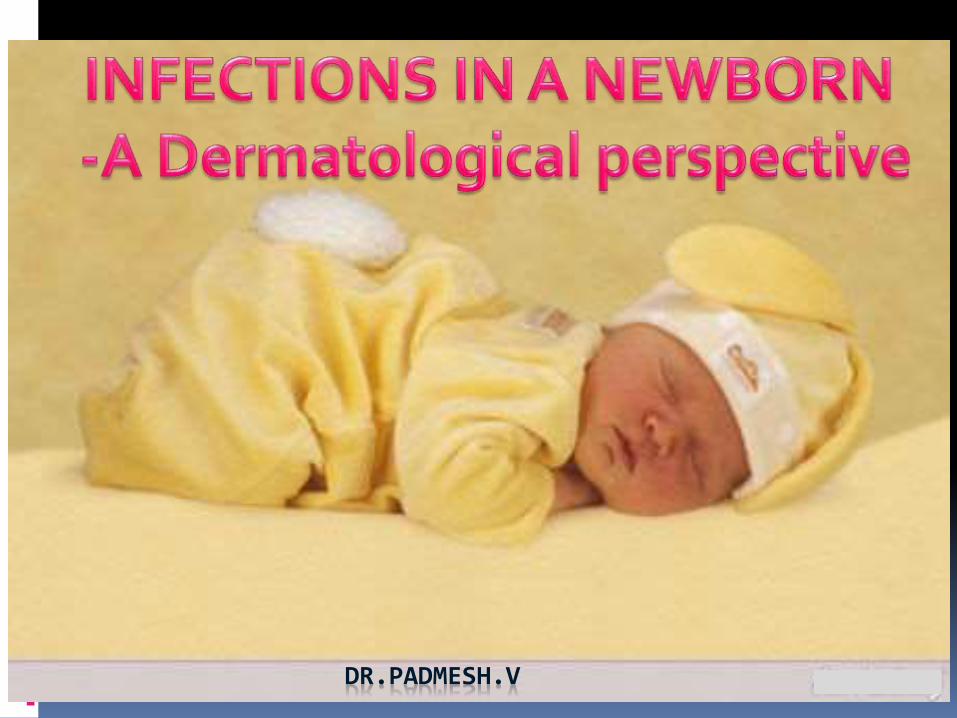
DR.PADMESH.V
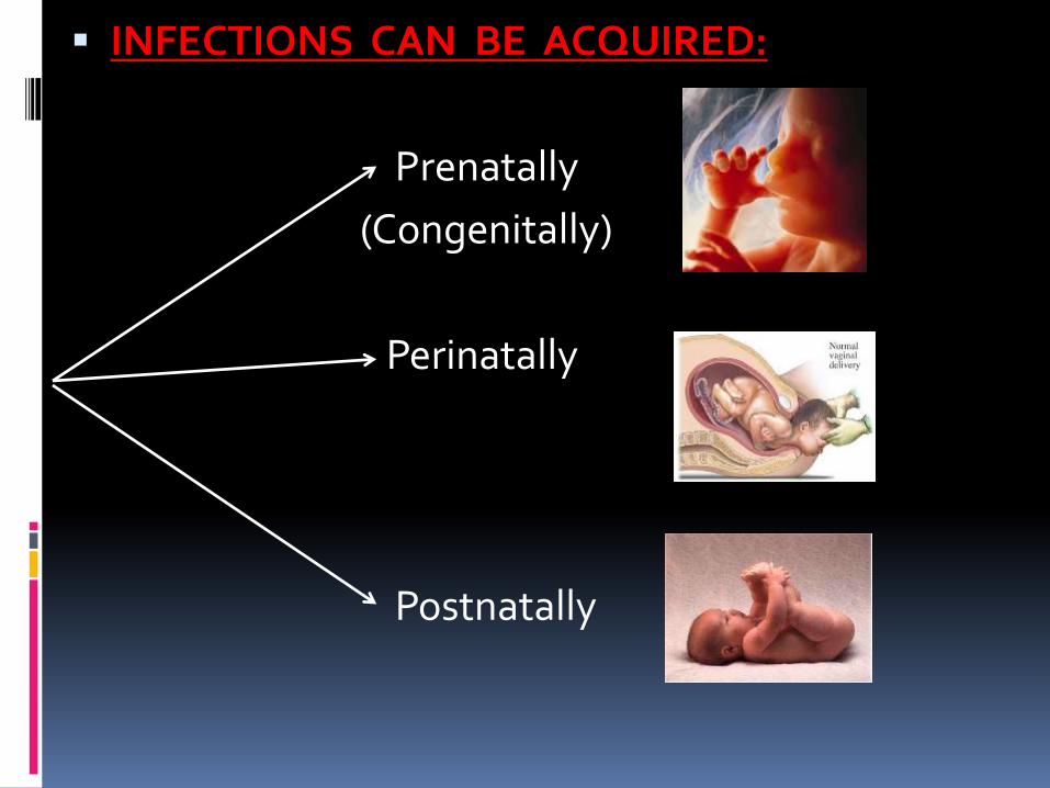
INFECTIONS CAN BE ACQUIRED:
Prenatally
(Congenitally)
Perinatally
Postnatally
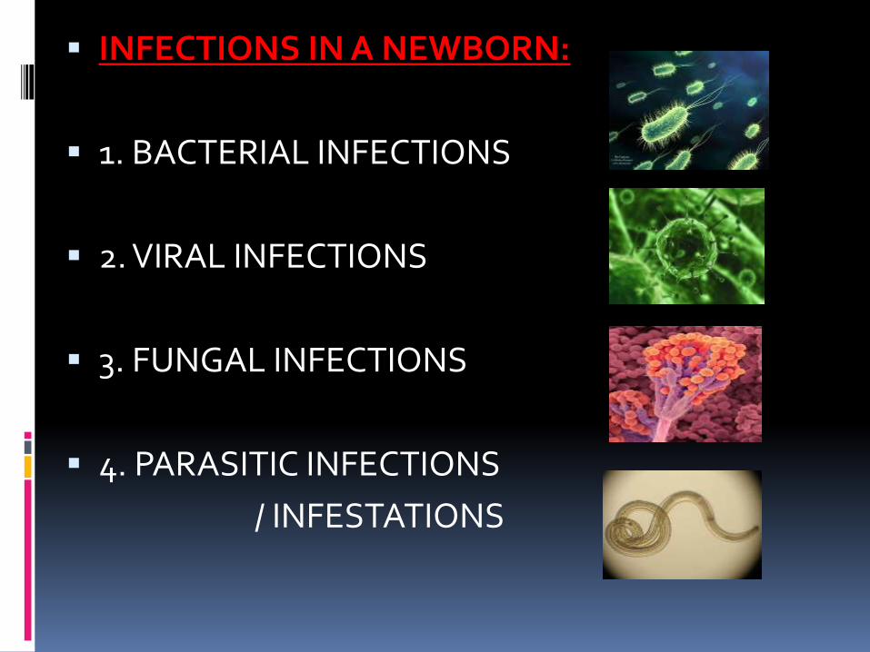
INFECTIONS IN A NEWBORN:
1. BACTERIAL INFECTIONS
2. VIRAL INFECTIONS
3. FUNGAL INFECTIONS
4. PARASITIC INFECTIONS
/ INFESTATIONS
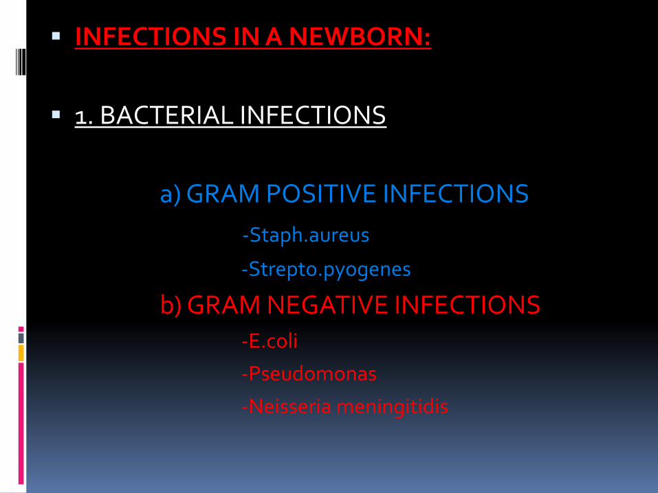
INFECTIONS IN A NEWBORN:
1. BACTERIAL INFECTIONS
a) GRAM POSITIVE INFECTIONS
-Staph.aureus
-Strepto.pyogenes
b) GRAM NEGATIVE INFECTIONS-E.coli
-Pseudomonas
-Neisseria meningitidis
GRAM POSITIVE
INFECTIONS
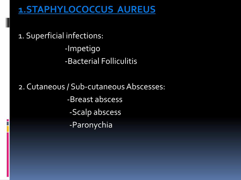
1.STAPHYLOCOCCUS AUREUS
1. Superficial infections:
-Impetigo
-Bacterial Folliculitis
2. Cutaneous / Sub-cutaneous Abscesses:
-Breast abscess
-Scalp abscess
-Paronychia
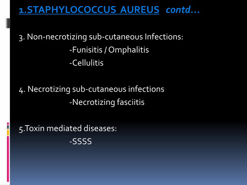
1.STAPHYLOCOCCUS AUREUS contd…
3. Non-necrotizing sub-cutaneous Infections:
-Funisitis / Omphalitis
-Cellulitis
4. Necrotizing sub-cutaneous infections
-Necrotizing fasciitis
5.Toxin mediated diseases:
-SSSS
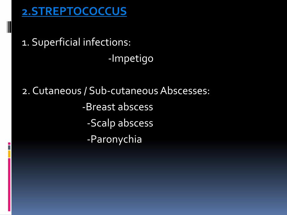
2.STREPTOCOCCUS
1. Superficial infections:
-Impetigo
2. Cutaneous / Sub-cutaneous Abscesses:
-Breast abscess
-Scalp abscess
-Paronychia
2.STREPTOCOCCUS contd…
3. Non-necrotizing sub-cutaneous Infections:
-Funisitis / Omphalitis
-Cellulitis -ERYSIPELAS
4. Necrotizing sub-cutaneous infections
-Necrotizing fasciitis
5.Toxin mediated diseases:
-STSS
IMPETIGO: Latin word impetere (to assail)
Constitutional signs +
2nd week of life.
1. Non-bullous Impetigo:
-Sub-corneal.
-Erythematous, honey colored crusted plaques.
2. Bullous Impetigo:
-Due to toxins which cause epidermolysis & bullae.
-Coagulase Positive Staph.aureus. From phage group 2.
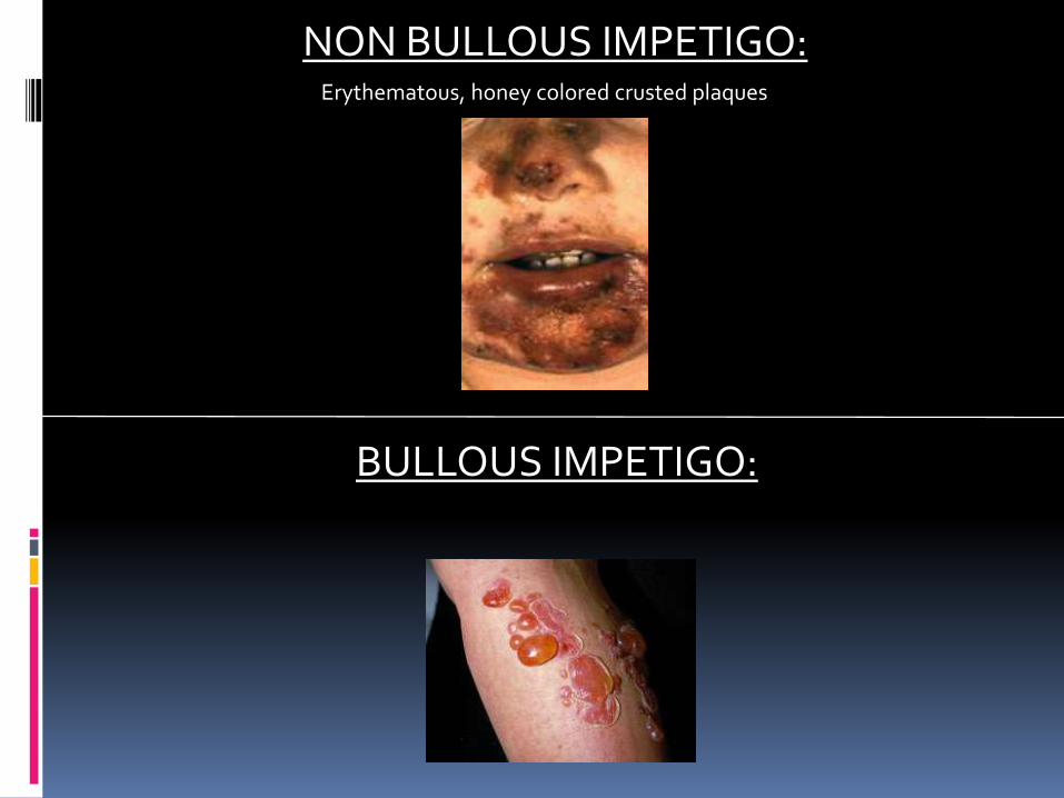
NON BULLOUS IMPETIGO:Erythematous, honey colored crusted plaques
BULLOUS IMPETIGO:
Treatment of Impetigo:
-Localized non-bullous impetigo with no constitutional signs:
Oral lactamase resistant antibiotics (Cephalexin, Cloxacillin)
-Bullous Impetigo:
IV Nafcillin, Oxacillin, Methicillin, Clindamycin,Cefazolin.
Duration: 10 days
May change to oral therapy after severity decreases.
Topical antibiotics not suitable.
BREAST ABSCESS:
-Etiology: Staph.aureus; (Also by Gp B Strepto, Ecoli, Salmonella)
-Full term newborn.
-Most common: 2-3 weeks
-M = F in the first 2 weeks. After that, F > M.
-Clinical Features:
-Initially: Breast enlargement, erythema, induration, tenderness
-Fluctuation
-Most common complication: Cellulitis (5-10%)
BREAST ABSCESS: contd…
-Treatment:
1. If fluctuation hasn’t developed:
Systemic anti-staphylococcal antibiotics.
2. If fluctuation is present:
Drain pus by needle aspiration or by I & D.
Systemic anti-staphylococcal antibiotics
3. If baby is ill, or if gram negative bacilli are seen:
Aminoglycoside or Cefotaxime
Duration of treatment: 7-14 days according to severity.
PARONYCHIA: Localised inflammation of the nail fold.
Initial separation of skin from the nail fold.
Secondary infection (Staphylococcus aureus or Streptococcus pyogenes) follows.
Treatment: Oral / IV antibiotics.
I & D +/-
D/D: Gram negative organisms or Candida.
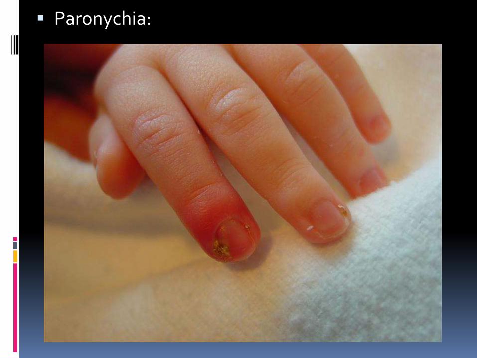
Paronychia:
FUNISITIS / OMPHALITIS:
-Onset : 3rd PND (average)
-Etiology: Staph.aureus, Strpto.pyogenes, Gm Neg
-Funisitis:
-Inflammation of umbilical cord.
-Increased secretions and foul odour.
-Omphalitis:
-Infection of umbilical cord.
-Periumbilical erythema, edema, tenderness.
-With or without discharge.
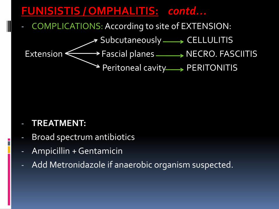
FUNISISTIS / OMPHALITIS: contd…- COMPLICATIONS: According to site of EXTENSION:
Subcutaneously CELLULITIS
Extension Fascial planes NECRO. FASCIITIS
Peritoneal cavity PERITONITIS
- TREATMENT:
- Broad spectrum antibiotics
- Ampicillin + Gentamicin
- Add Metronidazole if anaerobic organism suspected.
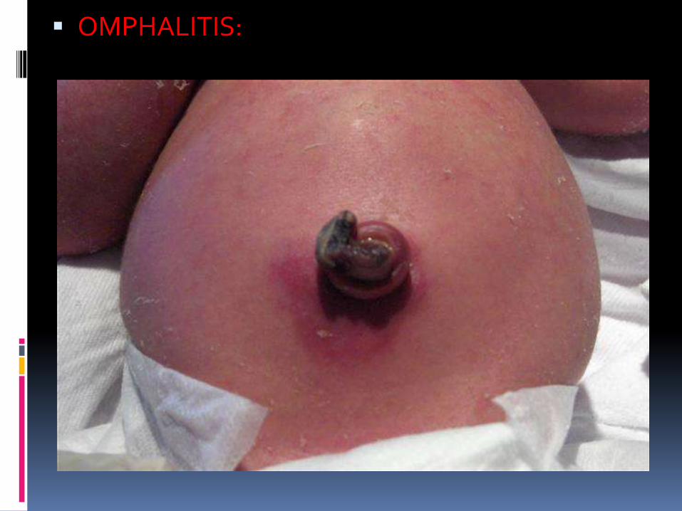
OMPHALITIS:
CELLULITIS:
-Infection and inflammation of loose connective tissue.
-Limited involvement of dermis. Relative sparing of epidermis.
-A break in skin predisposes to cellulitis.
-ETIOLOGY:
-Staph.aureus ( more localized; may suppurate)
-Strepto.pyogenes (Spread rapidly; Distinction not clear)
-Other streptococci, Hemophilus influenza, E.coli
CELLULITIS: contd..
- Clinical Findings:
- Edema,warmth,erythema,tenderness.
- Lateral margins are indistinct
- Application of pressure may cause pitting.
- Treatment:
Beta lactamase stable, anti-staphylococcal antibiotic like methicilin, oxacillin plus aminoglycoside,
or Cefotaxime.
Duration: 10 days
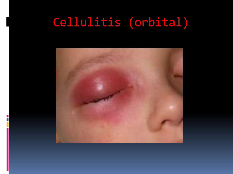
Cellulitis (orbital)
NECROTISING FASCIITIS- Etiology:
Polymicrobial.
- Subcutaneous tissue infection .
- Involves deep layer of superficial fascia, but spares adjacent epidermis, deep fascia and muscle.
- Clinical Findings:
Initial: Local swelling, erythema ,tenderness,local rise in temperature.
Later: Bullae, Darkening of affected tissues, gangrene
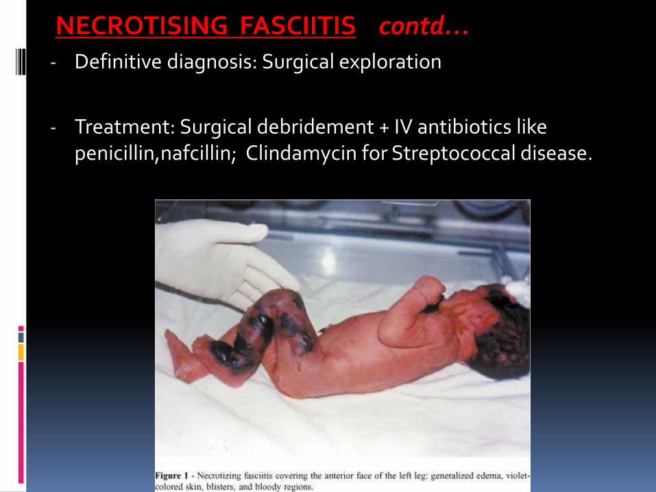
NECROTISING FASCIITIS contd…- Definitive diagnosis: Surgical exploration
- Treatment: Surgical debridement + IV antibiotics like penicillin,nafcillin; Clindamycin for Streptococcal disease.
SSSS:- ETIOLOGY:
Phage group II Staph, particularly strains 71, 55 .
- Staphylococcal EPIDERMOLYTIC TOXIN mediated disease.
- Cutaneous tenderness & superficial widespread blistering and / or desquamation.
- Begins as generalized macular erythema, becomes wrinkled, followed by desquamation over 2-5 days.
SSSS: contd…
- TREATMENT:
IV Nafcillin / Methicillin
Minimize handling of the child
Use of emollients
- Healing occurs without scarring in 10-14 days.
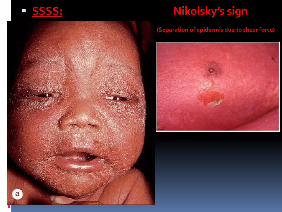
SSSS: Nikolsky’s sign
(Separation of epidermis due to shear force)
ERYSIPELAS: ‘St Anthony's fire’ ETIOLOGY: Polymicrobial; Streptococcal.
Superficial form of Cellulitis
Involves dermis and upper subcutaneous tissue.
Prominent lymphatic involvement.
Clinically:
Small area of burning and redness - followed by shiny red indurated tender plaque with peau d’ orange appearance and elevated sharply demarcated margins.
Treatment: Broad spectrum antibiotics
(Penicillin, methicillin plus aminoglycoside , or cefotaxime)
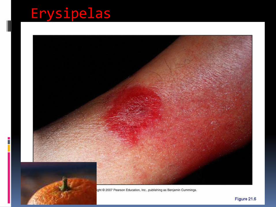
Erysipelas
SREPTOCOCCAL TOXIC SHOCK SYNDROME:
-Etiology: Strepto.pyogenes; TOXIN mediated disease.
-Acute onset of shock and multisystem organ failure.
-Rare in newborns.
-Often follows a minor,focal skin or soft tissue infection
Clinically: Localized swelling, erythema,pain. May have vesicles/bullae.
SREPTOCOCCAL TOXIC SHOCK SYNDROME:
contd…
- Management:
- Aggressive fluid resuscitation
- Early surgical exploration & debridement
- Inotropic agents
- IV antibiotics: Penicillin / Clindamycin
OTHER GRAM POSITIVE INFECTIONS:
- Coagulase Negative Staphylococcus (CONS):
-Staph epidermidis – commonest cause of indwelling catheter
related infections.
- Treatment: Vancomycin.
- Mycobacterium tuberculosis:
- Congenital TB
- Skin manifestations are unusual: scaly,erythematous, umbilicated papules & subcutaneous nodules.
GRAM NEGATIVE INFECTIONS
GRAM NEGATIVE INFECTIONS:
Gram negative sepsis is common in newborns, but cutaneousmanifestations are uncommon.
Can cause a variety of skin lesions in association with other infections,like:
-Impetigo
-Abscess
-Paronychia
-Cellulitis
-Necrotizing funisitis
-Necrotizing fasciitis
ECTHYMA GANGRENOSUM: Etiology: Pseudomonas aeruginosa
Necrotic skin lesions
Painful red or purpuric macule; Later develops a pustular or vesicular centre.
Rapidly ulcerates.
Risk factors: Prematurity, prolonged illness, neutropenia, bowel surgery,
Treatment: Extended spectrum penicillin( piperacillin) or Ceftazidime in combination with aminoglycoside.
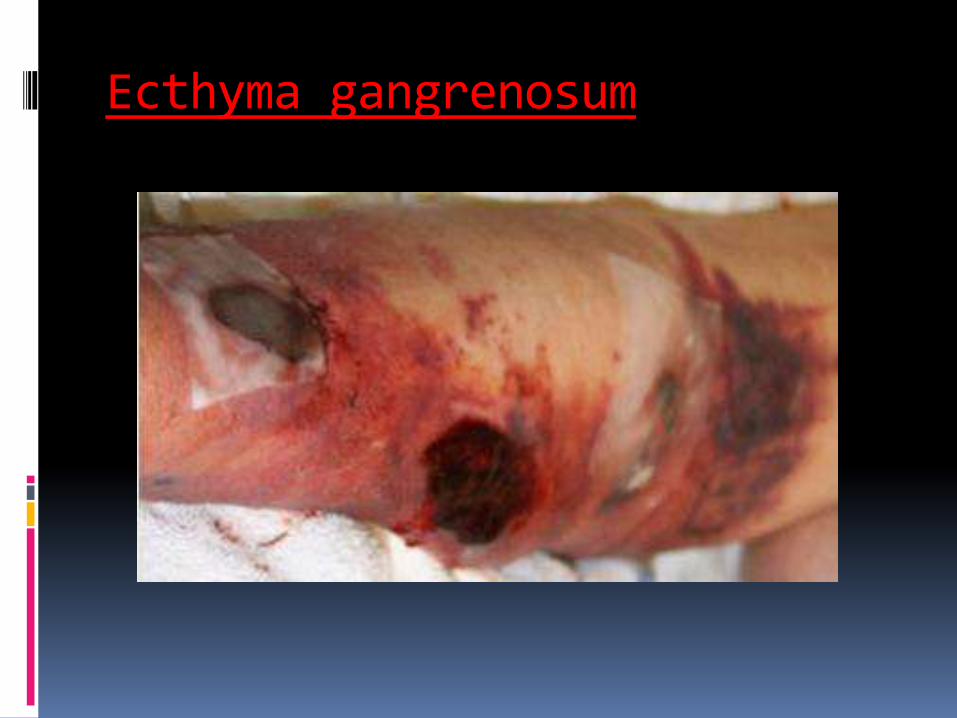
Ecthyma gangrenosum
PURPURA FULMINANS:
Etiology: Neisseria meningitidis, Gp B Streptococcus, occasionally with other gram positive/negative bacteria
Life threatening condition
Acute onset of progressive cutaneous hemorrhage & necrosis caused by dermal vascular thrombosis and DIC.
Clinically:
Cutaneous erythema, petechiae develops first
Evolve rapidly into painful,indurated,well-demarcated irregularly bordered purpuric papules/plaques.
Thin layer of advancing erythematous border.
PURPURA FULMINANS:Late findings:
Vesicles, bullae.
Firm eschar forms later,then sloughs.
Distal extremities most severely affected, usually symmetrically.
Extra cutaneous: Shock, DIC
Treatment:
Respiratory and cardiovascular support
Broad spectrum IV antibiotics –penicillin
Vit K, FFP to correct coagulation disorders.
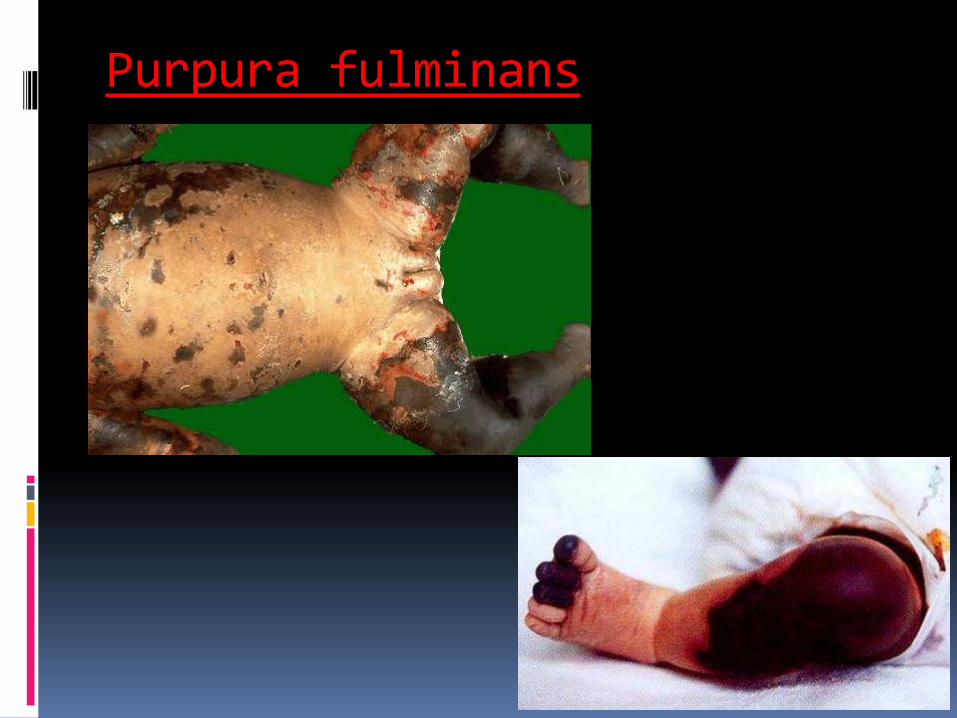
Purpura fulminans
SYPHILIS: Treponema pallidum
Transmission rate in untreated syphilis in pregnancy: 100%.
Fetal or perinatal death -in 40% of affected infants.
Survivors manifest features:
-Early signs appear during the 1st 2 yr of life
-Late signs appear gradually during the 1st 2 decades.
66% of infected infants - asymptomatic at birth.
Identified only by routine prenatal screening;
If untreated, symptoms develop within weeks or months.
SYPHILIS: contd..SKIN MANIFESTATIONS:
-Variable; Palms,soles,perioral,anogenital regions.
-Mucous membrane involvement: Snuffles.
-Condyloma lata
-Papulosquamous eruption
EXTRA CUTANEOUS:
Early: LBW, HSM, elevated liver enzymes, anemia, jaundice,
osteochondritis, pseudoparalysis of Parrot.
Late: Hutchinson’s teeth, Saddle nose; Clutton’s joints;
HUTCHINSON’S TRIAD: Defects in incisors, Interstitial Keratitis,
Sensorineural hearing loss.
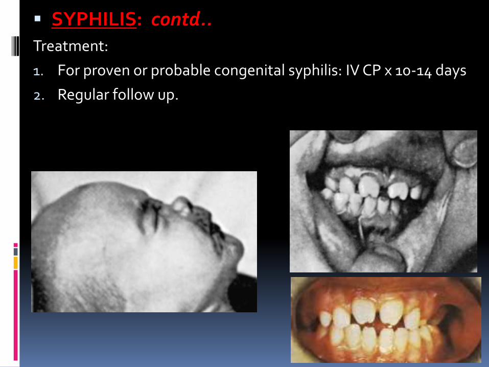
SYPHILIS: contd..Treatment:
1. For proven or probable congenital syphilis: IV CP x 10-14 days
2. Regular follow up.
VIRAL
INFECTIONS
TORCH INFECTIONS:
T - Toxoplasmosis
O - Others: Hepatitis B, VZV ,HIV, Parvovirus B19
Syphilis.
R - Rubella
C - Cytomegalovirus
H - Herpes Simplex
TORCH INFECTIONS:
T - Toxoplasmosis
O - Others: Hepatitis B, VZV ,HIV, Parvovirus B19
Syphilis.
R - Rubella
C - Cytomegalovirus
H - Herpes Simplex

HERPES SIMPLEX VIRUS: HSV type 1,2
1. INTRA UTERINE HERPES SIMPLEX:
Fetal death +
If not, congenital anomalies
SKIN manifestations CNS manifestations
-Vesicular lesions -Microcephaly (>50%)
-Scar -Chorioretinitis (60%)
Diagnosis: Histology(Tzanck preparation) ,
Antibody-specific stains, Culture, Serology.
Treatment: Acyclovir IV
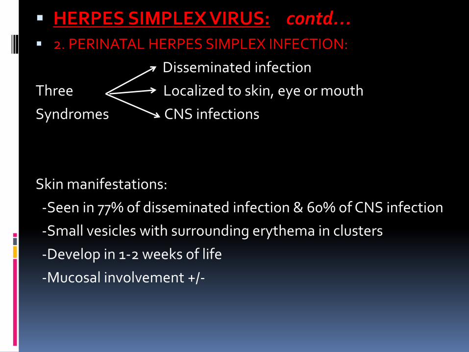
HERPES SIMPLEX VIRUS: contd… 2. PERINATAL HERPES SIMPLEX INFECTION:
Disseminated infection
Three Localized to skin, eye or mouth
Syndromes CNS infections
Skin manifestations:
-Seen in 77% of disseminated infection & 60% of CNS infection
-Small vesicles with surrounding erythema in clusters
-Develop in 1-2 weeks of life
-Mucosal involvement +/-
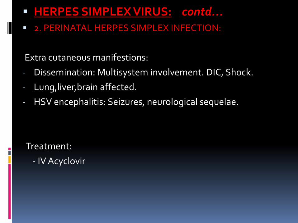
HERPES SIMPLEX VIRUS: contd… 2. PERINATAL HERPES SIMPLEX INFECTION:
Extra cutaneous manifestions:
- Dissemination: Multisystem involvement. DIC, Shock.
- Lung,liver,brain affected.
- HSV encephalitis: Seizures, neurological sequelae.
Treatment:
- IV Acyclovir
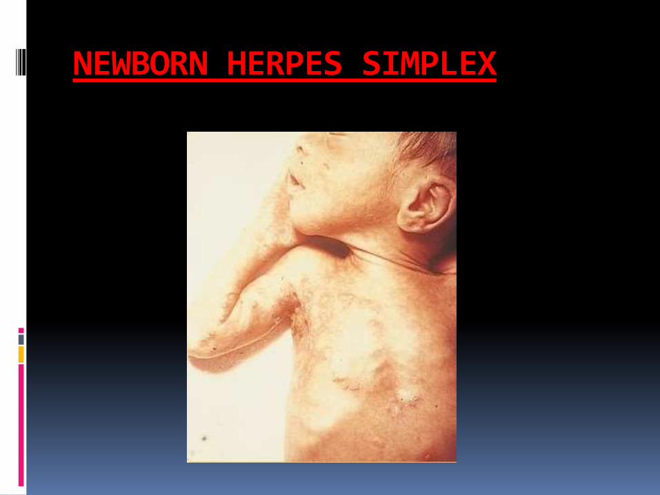
NEWBORN HERPES SIMPLEX
VARICELLA:
Varicella zoster virus
THREE distinct disorders:
1. Fetal varicella syndrome
2. Neonatal varicella
3. Infantile herpes zoster
VARICELLA: Fetal /Cong. varicella syndrome:
0 to 9% incidence after maternal infection during pregnancy.
1st Trimester attack rate: 2.2%
Greatest risk: First 20 weeks of gestation.
Clinical features:
Common: (>50%)
-Skin: Cicatricial lesions, corresponding to dermatomes
-CNS: Limb paresis
-Eye: Chorioretinitis
-Skeletal: Hypoplasia of extremities
VARICELLA: Fetal /Cong. varicella syndrome:
Uncommon clinical features: (<50%)
-CNS: Hydrocephalus/Microcephaly,Seizures, MR.
-Eye: Cataract, Optic Nerve atrophy
Diagnosis: CVS / Amniocentesis: IgM / Culture
Prevention:
1.VZIG to mother – within 5 days (2 days)
2.Acyclovir to mother
Treatment of affected infant is supportive if no active lesion.
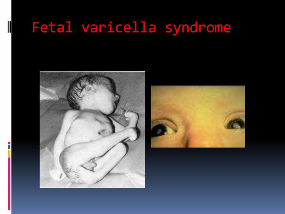
Fetal varicella syndrome
VARICELLA: Neonatal varicella: If mother has varicella before or immediately after delivery.
If maternal varicella 5 days before, to 2 days after delivery: High risk for disseminated disease in newborn.
Infant may develop lesions 1-16 days after birth.
Clinical findings:
SKIN:
-Variable
-Small pink macules that become papular,then vesicular.
-Necrotic or hemorrhagic lesions may be seen.
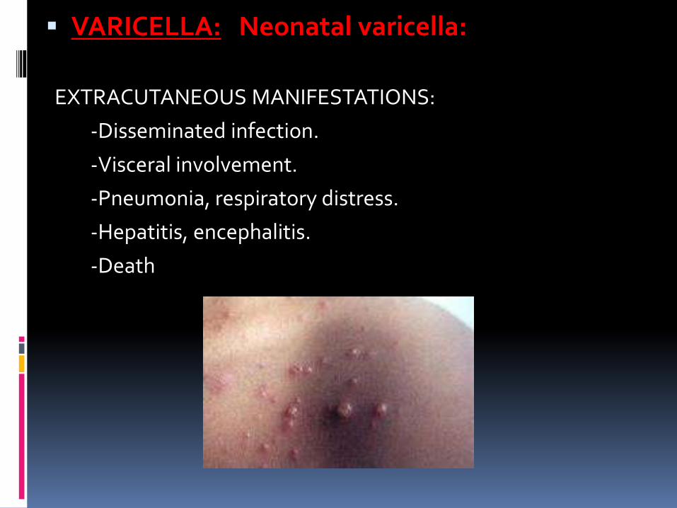
VARICELLA: Neonatal varicella:
EXTRACUTANEOUS MANIFESTATIONS:
-Disseminated infection.
-Visceral involvement.
-Pneumonia, respiratory distress.
-Hepatitis, encephalitis.
-Death
VARICELLA: Neonatal varicella: contd…PREVENTION AND TREATMENT:
1. Delaying delivery (for transfer of maternal antibodies into fetus)
Usually after 5-7 days of onset of maternal illness
2. High risk newborn: VZIG (125 units)
3. Affected newborn: IV Acyclovir for atleast 5 days
4. Prophylactic Acyclovir
5. Avoid direct contact of infant with maternal skin lesions.
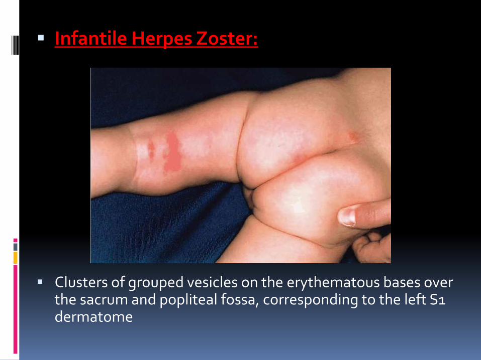
VARICELLA: Infantile Herpes Zoster:
Risk more if exposure to virus is in 2nd half of pregnancy (2%).
VZ Virus persists in sensory dorsal root ganglia. Reactivation leads to localized skin/nerve involvement.
Dermatome affected according to ganglion in which reactivation occurred.
SKIN FINDINGS: Discrete papular lesions in dermatomes.
Later become vesicular & crusting occurs.
TREATMENT:
- IV Acyclovir
-Supportive therapy
Infantile Herpes Zoster:
Clusters of grouped vesicles on the erythematous bases over the sacrum and popliteal fossa, corresponding to the left S1 dermatome
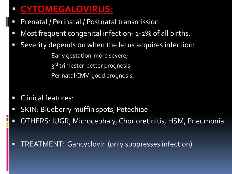
CYTOMEGALOVIRUS: Prenatal / Perinatal / Postnatal transmission
Most frequent congenital infection- 1-2% of all births.
Severity depends on when the fetus acquires infection:
-Early gestation-more severe;
-3rd trimester-better prognosis.
-Perinatal CMV-good prognosis.
Clinical features:
SKIN: Blueberry muffin spots; Petechiae.
OTHERS: IUGR, Microcephaly, Chorioretinitis, HSM, Pneumonia
TREATMENT: Gancyclovir (only suppresses infection)
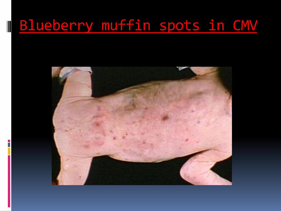
Blueberry muffin spots in CMV
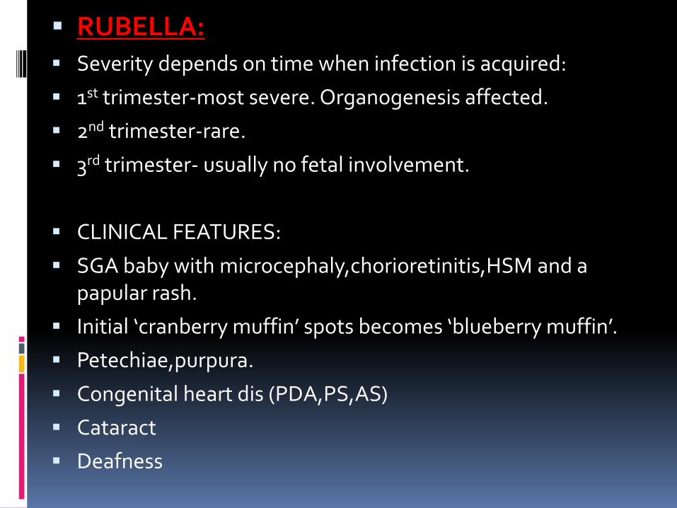
RUBELLA: Severity depends on time when infection is acquired:
1st trimester-most severe. Organogenesis affected.
2nd trimester-rare.
3rd trimester- usually no fetal involvement.
CLINICAL FEATURES:
SGA baby with microcephaly,chorioretinitis,HSM and a papular rash.
Initial ‘cranberry muffin’ spots becomes ‘blueberry muffin’.
Petechiae,purpura.
Congenital heart dis (PDA,PS,AS)
Cataract
Deafness
RUBELLA: contd… Psychomotor dysfunction
Pneumonia
PREVENTION / TREATMENT:
Universal immunization
Prenatal screening
No specific antiviral treatment.
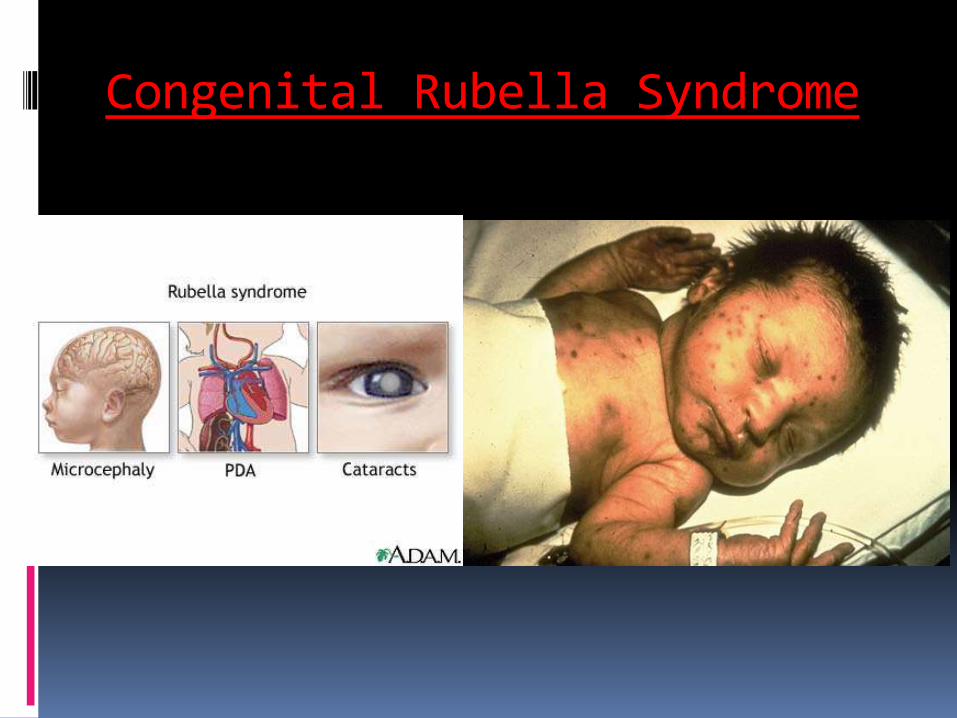
Congenital Rubella Syndrome
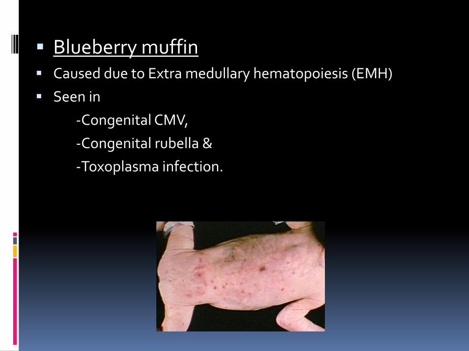
Blueberry muffin Caused due to Extra medullary hematopoiesis (EMH)
Seen in
-Congenital CMV,
-Congenital rubella &
-Toxoplasma infection.
HUMAN PARVOVIRUS B19:
High morbidity & mortality
Miscarriage & Hydrops fetalis.
Persistent anemia
Eye defects, B/L cleft lip & palate, micrognathia,etc.
TREATMENT: IVIG
ENTEROVIRUS: Include Poliovirus, Coxsackie A&B, Echoviruses.
2-38/1000 births
Clinical features:
Fever, irritability, poor feeding
Maculopapular / macular / petechial / vesiculopustular rash
Treatment:
Supportive care
IVIG if severe.
Anti-picorna agent: PLECONARIL under clinical trial.
HUMAN PAPILLOMA VIRUS: Low risk subtypes: HPV 6,11
High risk: HPV 16,18,31,33 – associated with anogenital cancer
HPV 30 – associated with Oral, Laryngeal &
anogenital cancer
High risk for exposure during vaginal delivery.
CLINICAL FEATURES:
Condyloma acuminata
Sites in infant: perianal skin,glans,vulva,vaginal introitus
Laryngeal papillomas
TREATMENT: Less painful methods in infants;
Podophyllin resin, Trichloroacetic acid, imiquimod cream
Prophylaxis: HPV vaccine
HIV: In most newborns, physical examination at birth is normal.
Initial features may be
-Subtle, such as lymphadenopathy and HSM, or
-Nonspecific, such as failure to thrive, chronic or recurrent diarrhea, interstitial pneumonia, or oral thrush.
Perinatal transmission : Most common
HIV: contd…
Various opportunistic infections: PCP, Candidiasis, TB, HSV, Herpes zoster, Toxoplasmosis, Cryptococcosis etc
Malignancies associated: Non-Hodgkin lymphoma, primary CNS lymphoma, leiomyosarcoma.
Kaposi’s sarcoma rare in children.
SKIN MANIFESTATIONS: Due to immunosuppression.
FUNGAL INFECTIONS
CANDIDIASIS: Most common fungal pathogen of newborn.
C.albicans- 75%
Others: C.tropicalis, C.parapsilosis
TYPES:
1. Congenital Candidiasis
2. Systemic Candidiasis
3. Invasive fungal dermatitis
4.Localized neonatal candidiasis
CANDIDIASIS: Contd…1.CONGENITAL CANDIDIASIS:
-Acquired in-utero.
-Presents in the first few days of life.
SKIN: Monomorphous erythematous papulo vesicular eruption
Followed by pustules, late crusting & desquamation.
OTHERS: Systemic dissemination is rare. Seen in LBW babies.
Prognosis is excellent.
CANDIDIASIS: Contd…2. SYSTEMIC CANDIDIASIS:
- Candida in an otherwise sterile body fluid.
- 2-4% of VLBW babies.
- Skin manifestations in 50-60%
- May be acquired in-utero or postnatally.
- SKIN: Burnlike dermatitis, desquamation, progressive diaper dermatitis, cutaneous abscess at site of IV cannula
- OTHERS: Apnea, bradycardia, abd.distension, temperature instability, stool occult blood positivity.
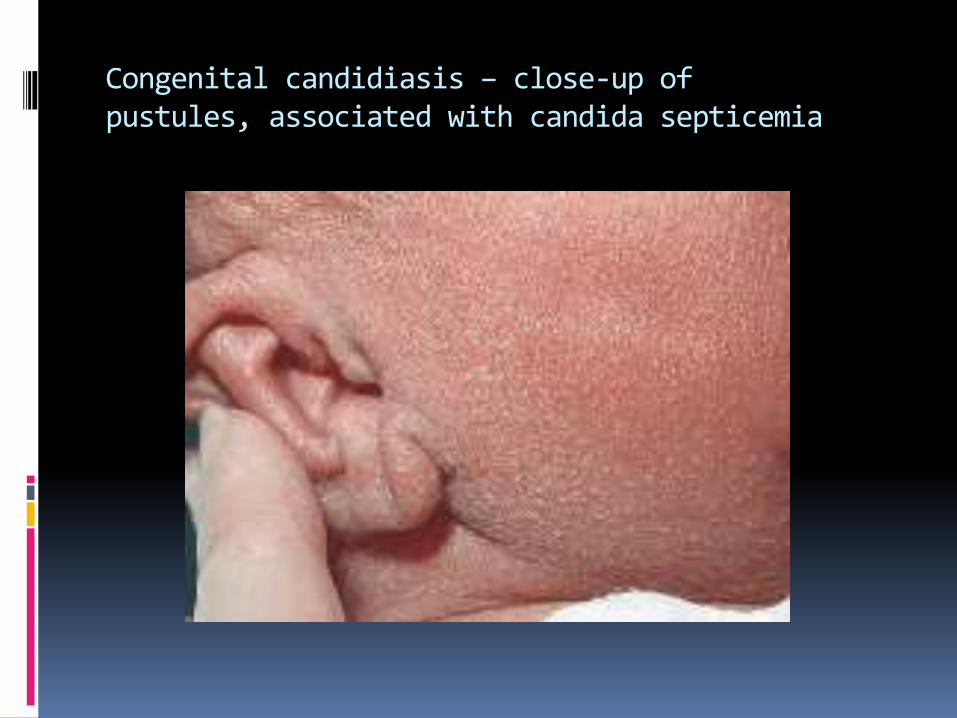
Congenital candidiasis – close-up ofpustules, associated with candida septicemia
CANDIDIASIS: Contd…3. INVASIVE FUNGAL DERMATITIS:
-Erosive crusting lesions in VLBW babies
-Primary skin condition—leads to secondary systemic disease.
-OTHER ORGANISMS: Aspergillus, Trichosporum
-Risk factors: Extreme prematurity, vaginal delivery, steroid administration, hyperglycemia.
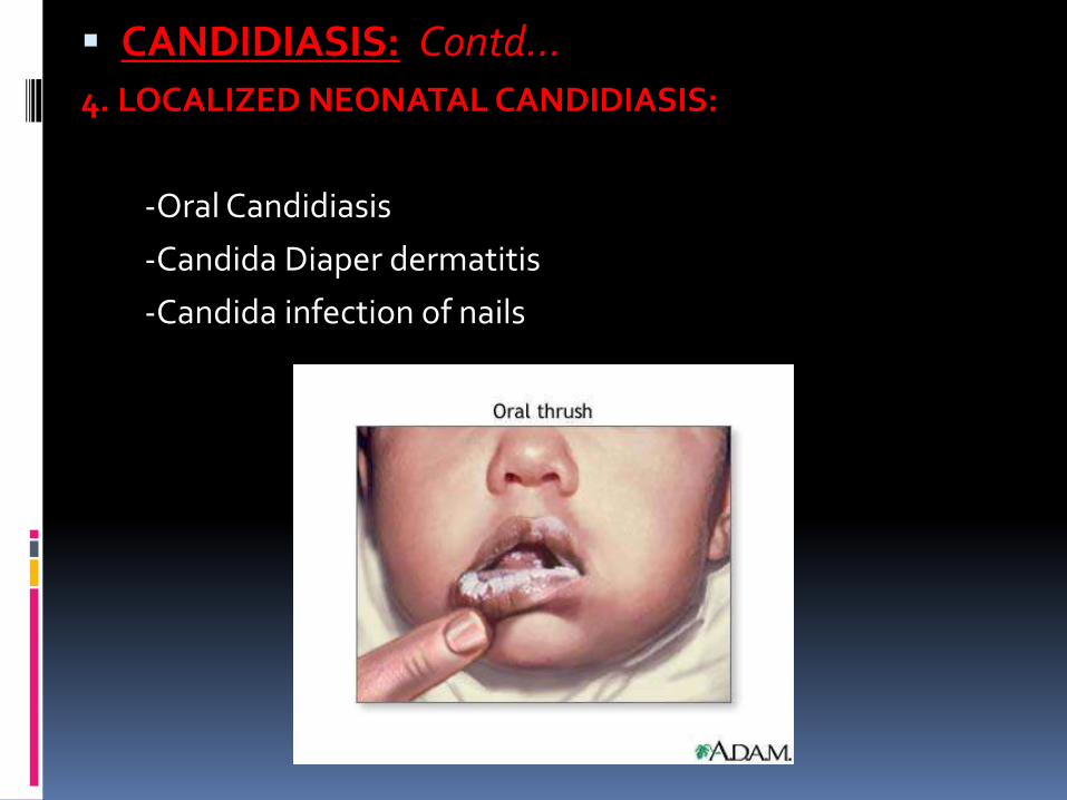
CANDIDIASIS: Contd…4. LOCALIZED NEONATAL CANDIDIASIS:
-Oral Candidiasis
-Candida Diaper dermatitis
-Candida infection of nails
CANDIDIASIS: Contd…TREATMENT:
1. Localized forms – Topical agents.
Thrush – Nystatin solution QID
-Resistant thrush- Oral fluconazole or Itraconazole
2. Diaper dermatitis – Imidazole cream. Nystatin. Combination with 1% hydrocortisone cream.
3. Congenital Candidiasis:
Term infants: Topical agents
Preterm/ VLBW: Systemic Treatment:
IV Amphotericin B
5-Fluorocytosine (5-FC)
Fluconazole
OTHER INFESTATIONS
SCABIES: Sarcoptes scabiei var.hominis
Congenital scabies not seen.
SKIN: Erythematous vesiculo papular eruptions.
Mainly in axillae,neck,palms,soles,head.
-Burrow
Clinical features: Irritability,insomnia,poor feeding.
TREATMENT:
5% Permethrin- entire body; 8-12 hrs; Reapplication after 1 wk.
Antihistamines-hydroxyzine
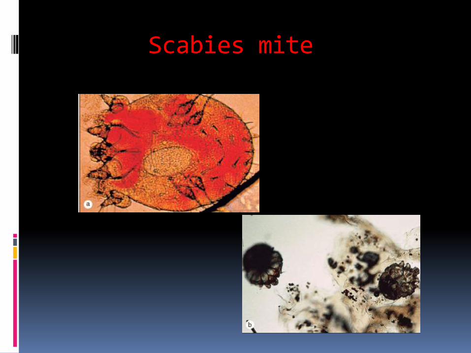
Scabies mite
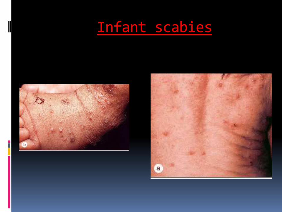
Infant scabies
PARASITIC INFECTIONS
TOXOPLASMOSIS: Antenatal/Perinatal infection
Untreated maternal infections in 1st trimester- 17% fetuses are infected; Severe ;
Untreated maternal infection in 3rd trimester- 65% fetuses are infected; Mild or inapparent at birth.
CLINICAL FEATURES:
Triad of chorioretinitis, hydrocephalus, cerebral calcifications
Seizures;
Squint.
No specific skin manifestations. Rash+
TOXOPLASMOSIS: contd… TREATMENT:
More than half of congenitally infected infants are considered normal in the perinatal period, but almost all develop ocular involvement later in life if they are not treated during infancy.
Pyrimethamine
Sulfadiazine
Folinic acid
Treatment for atleast 1 year.

TOXOPLASMOSIS: contd…
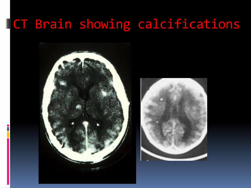
CT Brain showing calcifications