dermatological aspects of angiogenesis
TRANSCRIPT

TOPICAL REVIEW
Dermatological aspects of angiogenesis
P . V E L A S C O A N D B . L A N G E - A S S C H E N F E L D TDepartment of Dermatology, University of Kiel, Schittenhelmstraße 7, 24105 Kiel, Germany
Accepted for publication 14 August 2001
Summary Neovascularization is vital for the growth of tumours, providing a lifeline for sustenance and waste
disposal. Tumour vessels can grow by sprouting, intussusception or by incorporating bone marrow-derived endothelial precursor cells into growing vessels. Recent advances in vascular biology have
identified some key factors that control vascular growth, and have led to the hypothesis that in
normal tissues vascular quiescence is maintained by the dominant influence of endogenousangiogenesis inhibitors over angiogenic stimuli. In contrast, increased secretion of angiogenic
factors and the down-regulation of endogenous angiogenesis inhibitors induce tumour
angiogenesis. Vascular quiescence in the skin seems to be primarily maintained by a balancebetween the endogenous angiogenesis inhibitors thrombospondin 1 and thrombospondin 2 and the
potent proangiogenic factor vascular endothelial growth factor A. Inhibiting tumour growth by
controlling angiogenesis is an intriguing approach with great potential for the treatment of vasculartumours such as haemangioma, Kaposi’s sarcoma and solid cutaneous tumours such as squamous
cell carcinoma, melanoma and basal cell carcinoma. In this review, the role of angiogenesis and
more recent topics such as lymphangiogenesis in cutaneous tumour growth, invasion andmetastasis will be discussed.
Key words: angiogenesis inhibitors, angiogenesis, lymphangiogenesis, skin, tumour, vascular
endothelial growth factor
Human skin is a richly vascularized organ consisting of
a superficial vascular plexus that is found directly
beneath the epidermis and a deep vascular plexuslocated at the dermal-subcutaneous border. These
vascular beds are tightly interconnected and play an
important role in vital physiological processes of theskin such as the supply of nutrients, controlling the
influx of immune cells, hair growth and thermoregu-
lation. The development of the blood vasculature is acomplex process that begins when progenitor cells
(angioblasts) differentiate into endothelial cells, co-
alesce and form the first vessels in the embryo(vasculogenesis). These embryonic vascular tubes give
rise to new vessels by branching or sprouting (angio-
genesis) that in turn can elongate, enlarge, mature orregress (vascular remodelling) in response to different
stimuli (reviewed by Carmeliet and Jain1). Together,
these processes are vital in the developing embryo but
are mostly absent in the adult.In normal and diseased skin, the angiogenic stimulus
seems to originate from the epidermis and not the
dermis.2 This makes intuitive sense as the avascularepidermis would require mechanisms designed to meet
the high metabolic demands associated with hyperpro-
liferation in healing wounds, inflammation or neo-plasms. Many potent proangiogenic factors such as
basic fibroblast growth factor (FGF-2)3–5 and chemok-
ines such as interleukin (IL)-86,7 have been described incutaneous angiogenesis. However, vascular endothelial
growth factor (VEGF)-A is regarded as the major
angiogenesis factor in many cutaneous diseases, par-ticularly in tumour growth.8 VEGF-A is a homodimeric
heparin-binding glycoprotein occurring in at least four
isoforms as a result of alternative splicing.9 It binds twotype III tyrosine kinase receptors primarily expressed
Correspondence: Dr. med. Bernhard Lange-Asschenfeldt.
E-mail: [email protected]
British Journal of Dermatology 2002; 147: 841–852.
� 2002 British Association of Dermatologists 841
on vascular endothelial cells, VEGF receptor (VEGFR)-1, also termed fms-like-tyrosine kinase (Flt)-1, and
VEGFR-2,10–12 also known as kinase-insert domain-
containing receptor or fetal liver kinase-1, as well asthe neuropilin receptors.13 Several experimental mod-
els highlight the importance of VEGF-A as a potent
proangiogenic and tumour growth factor (reviewed byFerrara and Gerber14), and VEGF-A up-regulation is
implicated in promoting tumour invasiveness.15–17
VEGF-A secreted by tumour cells can induce the releaseof matrix metalloproteinases (MMPs) by endothelial
cells.18 Elevated levels of membrane type 1 MMP,
MMP-2 and MMP-919 have been demonstrated todegrade the extracellular matrix, paving the way for
the invasion of endothelial cells into the surrounding
tissue. Furthermore, MMPs release sequestered angio-genic factors such as FGF-2 and VEGF-A from extra-
cellular stores, thereby increasing their bioavailability
(Table 1). Conversely, MMP activity is important forthe generation of endogenous angiogenesis inhibitors
such as endostatin, which is a cleavage product of
collagen XVIII.20
Recently, the angiopoietin growth factor family has
been shown to modulate vascular remodelling, leak-
age21 and tumour growth.22 Angiopoietin 1 (Ang-1),normally found on pericytes, binds the tyrosine-protein
kinase receptor (Tie)-2 primarily expressed on endot-
helial cells and stabilizes the blood vessel. Angiopoietin2 (Ang-2) is a competitive antagonist to Ang-1 and
counters this stability. The vessel can undergo angio-
genesis in the presence of a proangiogenic factor, orregress in its absence.23,24 Ang-2 and VEGFR-2 have
been shown to be up-regulated in the very early stages
of tumour growth and to be continuously overex-pressed in the lesions, suggesting that these molecules
work together to induce new vessel growth andremodel the tumour vasculature.25
It is generally believed that in quiescent vessels, a
balance between proangiogenic and antiangiogenicfactors maintains homeostasis. Experimental data sug-
gest that the �angiogenic switch�, the change from a
quiescent tumour cell population to cells that caninduce angiogenesis, is a rate-limiting step in the
progression from a premalignant minimal lesion to a
malignant invasive lesion. It is dependent on theup-regulation of angiogenic factors and the down-
regulation of inhibitors.26 In the skin, aside from
follicular-related angiogenesis in certain phases of thehair cycle,27 the blood vessels remain quiescent and
neovascularization is generally not seen. It has been
suggested that the normal dermal matrix, in particularthe basement membrane surrounding microvascular
endothelial cells, may function as a natural inhibitor of
angiogenesis, thereby ensuring the quiescence of ves-sels in healthy skin. Recent evidence suggests that
thrombospondin (TSP)-1 and TSP-2 are major physio-
logical inhibitors of skin angiogenesis.28,29 TSP-1 andTSP-2 are members of a family of matrix glycoproteins
and are deposited in the dermoepidermal basement
membrane,30 thereby contributing to the antiangio-genic barrier that separates the avascular epidermis
from the vascularized dermis.28 Furthermore, these
endogenous angiogenesis inhibitors,31–34 as well asendostatin,20 angiostatin,35 vasostatin36 and IL-12,37
have been identified as inhibitors of tumour angiogen-
esis and tumour growth in vivo (Table 2).Early metastasis to the lymph nodes as well as
the presence of dilated peritumour lymphatic vessels
in melanoma and other tumours38 led to rising interestin investigations of tumour lymphangiogenesis.
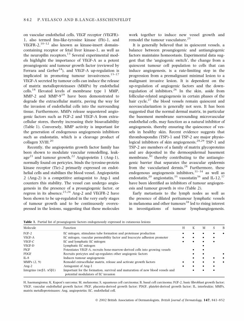
Table 1. Partial list of proangiogenic factors endogenously expressed in cutaneous lesions
Molecule Function H K M S B
FGF-2 EC mitogen, stimulates tube formation and proteinase production • • • • •VEGF-A EC mitogen, vascular permeability factor and leucocyte adhesion promoter • • • •VEGF-C EC and lymphatic EC mitogen • • • •VEGF-D Lymphatic EC mitogen •PlGF Potentiates VEGF-A, recruits bone-marrow-derived cells into growing vessels • •PDGF Recruits pericytes and up-regulates other angiogenic factors • • •IL-8 Induces tumour angiogenesis • • • •MMPs (2, 9) Remodel extracellular matrix, release and activate growth factors • • • • •Ang-2 Antagonist of Ang-1 • • • •Integrins (avb3, a5b1) Important for the formation, survival and maturation of new blood vessels and
potential modulators of EC invasion• • •
H, haemangioma; K, Kaposi’s sarcoma; M, melanoma; S, squamous cell carcinoma; B, basal cell carcinoma; FGF-2, basic fibroblast growth factor;
VEGF, vascular endothelial growth factor; PlGF, placenta-derived growth factor; PDGF, platelet-derived growth factor; IL, interleukin; MMPs,
matrix metalloproteinases; Ang, angiopoietin; EC, endothelial cell.
8 4 2 P . V E L A S C O A N D B . L A N G E - A S S C H E N F E L D T
� 2002 British Association of Dermatologists, British Journal of Dermatology, 147, 841–852
VEGF-C was the first lymphangiogenesis factor des-
cribed.39,40 It binds to VEGFR-3, also known as Flt-4,
normally expressed exclusively on the lymphaticendothelium, and to VEGFR-2.39,40 VEGF-C has re-
cently been demonstrated to promote tumour lymph-
angiogenesis, angiogenesis and metastasis.41–44 Therecent discovery of specific markers for lymphatic
vessels such as the lymphatic vessel endothelial hya-
luronan receptor-145 and the homeobox genePROX146 promises to facilitate studies of lymphangio-
genesis in tumour biology.
Angiogenesis in vascular tumoursof the skin
There are several described benign cutaneous vascular
neoplasms such as pyogenic granuloma and angio-keratoma as well as malignant vascular tumours such
as angiosarcoma, yet most studies concerning angio-
genesis in cutaneous vascular tumours have beenperformed using haemangioma and Kaposi’s sarcoma
(KS) as models.
Haemangioma is the most common neoplasm ofinfancy. This endothelial tumour undergoes a prolifer-
ative phase followed by an involution phase leading to
the spontaneous regression of the lesion (involutedhaemangioma). Normally haemangioma presents no
medical threat but sometimes the fast-growing nature
of this tumour can interfere with normal functions. Themost common complications are circulatory problems
arising from high shunt volumes or vision impairment
resulting from the physical obstruction of the eye.Furthermore, trauma of extensive haemangiomas
can cause pronounced bleeding. In some cases haem-
angiomas can develop into severe diseases such as
Kasabach–Meritt syndrome where dysfunction of the
coagulation system can be lethal.
Proliferating haemangiomas are composed of denselypacked endothelial cells with little connective tissue
and barely discernible vessel lumina. This hyperprolif-
erative endothelial cell population has recently beendemonstrated to be clonal, originating from a progen-
itor cell that could have a mutated angiogenesis
regulation gene. A second group has suggested thatthe clonal cell population could arise from one or a few
placental endothelial cells that have migrated into the
fetal circulation from chorionic villi (reviewed byMarchuk47).
Proliferating haemangiomas overexpress a plethora
of angiogenesis activators (Table 1). Among them areFGF-2,48 monocyte chemoattractant protein-149 and
VEGF-A.5 However, low expression levels of VEGF-A in
haemangioma-derived endothelial cells and in prolifer-ating lesions have recently been reported. This study
identifies the angiopoietins as potential modulators in
the pathological growth and vascular remodellingevident in haemangioma.50 The group reports up-
regulation of Tie-2 expression both in the haemangi-
oma-derived endothelial cells and in the proliferatinglesions.50 Notably, previous studies have linked Tie-2
expression abnormalities to other venous malforma-
tions.51
A small number of thin-walled vessels that resemble
normal capillaries surrounded by regions of fibrofatty
tissue characterizes involuting and involuted haeman-gioma. Interestingly, during this phase the expression
levels of the endogenous angiogenesis suppressors
tissue inhibitor of MMP (TIMP)-1 and interferon(IFN)-b are up-regulated when compared with the
proliferative phase.48,52
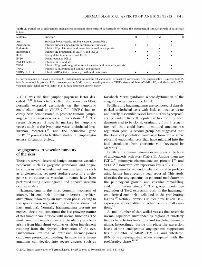
Table 2. Partial list of endogenous angiogenesis inhibitors demonstrated successfully to reduce the experimental tumour growth of cutaneous
lesions
Molecule Function H K M S B
Ang-1 Stabilizes blood vessels, inhibits vascular permeability •Angiostatin Inhibits tumour angiogenesis, mechanism is unclear • • • •Endostatin Inhibits EC proliferation and migration as well as apoptosis •Interferon-a Inhibits the production of VEGF-A and FGF-2 • • • •IL-12 Up-regulates interferon-c and IP-10 • • •IP-10 Down-regulates FGF-2 • •Platelet factor 4 Inhibits FGF-2 and VEGF •TSP-1 Inhibits EC growth, migration, tube formation and induces apoptosis • •TSP-2 Inhibits EC migration and tumour angiogenesis •TIMPs (1, 2…) Inhibit MMP activity, tumour growth and metastasis • • •
H, haemangioma; K, Kaposi’s sarcoma; M, melanoma; S, squamous cell carcinoma; B, basal cell carcinoma; Ang, angiopoietin; IL, interleukin; IP,
interferon inducible protein; TSP, thrombospondin; MMP, matrix metalloproteinase; TIMPs, tissue inhibitor of MMPs; EC, endothelial cell; VEGF,vascular endothelial growth factor; FGF-2, basic fibroblast growth factor.
D E R M A T O L O G I C A L A S P E C T S O F A N G I O G E N E S I S 8 4 3
� 2002 British Association of Dermatologists, British Journal of Dermatology, 147, 841–852

Clinically, corticosteroid therapy and the angiogen-esis inhibitor IFN-a are used as therapeutic agents for
life-threatening haemangioma. However, because of
side-effects and inconsistent success rates, new therap-ies are still necessary. Both endogenous and exogenous
angiogenesis inhibitors have been reported successfully
to reduce the experimental tumour growth of haeman-gioma (Table 2). Angiostatin has been found to inhibit
murine haemangioendothelioma growth.53 TIMP-254
and a VEGF-toxin conjugate,55 as well as syntheticcompounds such as TNP-470 (a derivative of fumagil-
lin)56 and batimastat (a synthetic MMP inhibitor)57
have been reported effectively to reduce experimentaltumour growth.
Understanding the cellular and biochemical mecha-
nisms that control the growth and spontaneousregression of haemangioma would provide critical
insights into aberrant angiogenesis and the mecha-
nisms that exist to override this defect. It seemsimportant to investigate whether at later stages in
childhood, endogenous angiogenesis inhibitors are
naturally up-regulated in order to counteract theangiogenic stimuli necessary for growth during child-
hood. This could explain the spontaneous regression of
haemangioma.KS is an angiogenic tumour affecting mostly the skin
and mucosa. It is found endogenously among several
ethnic groups but is more commonly associated withimmunological disorders arising from organ trans-
plants and human immunodeficiency virus (HIV)
infection.58,59 It is vascular in origin with tumoursconsisting mainly of hyperproliferative spindle-shaped
tumour cells, inflammatory cell infiltrate and numer-
ous blood vessels, and is frequently accompanied byextensive oedema.60
Many factors and cytokines that promote angiogen-
esis, lymphangiogenesis and vascular remodelling areknown to be overexpressed in KS cells and lesions
(Table 1). Among them are VEGF-A,61 FGF-2,3 IL-662
and IL-8.6 Recently, IL-8 has been identified as animportant angiogenesis factor and autocrine growth
factor in KS, as the functional inhibition of this
proinflammatory cytokine using a neutralizing anti-body resulted in a significant inhibition of experimental
tumour growth.6 Additionally, strong overexpressionof Ang-2, Tie-1 and Tie-2 highlights a potential role of
the angiopoietin family in the pathobiology of both KS
and angiosarcoma.63 Lastly, it has been suggested thatVEGF-C acts as a paracrine growth factor for KS
because its receptors VEGFR-2 and VEGFR-3 have been
demonstrated to be overexpressed by KS tumour
cells.64 In vitro, VEGF-C has been shown to stimulatethe migration and proliferation of KS cells.65 As the
expression of VEGF-C and VEGFR-3 is normally con-
fined to the lymphatic endothelium, it has beensuggested that KS cells might be of lymphatic origin.64
There is convincing evidence that the angiogenic
activity of KS cells is linked to viral infection. Humanherpesvirus 8 (HHV-8) expression is found in both
endogenous and induced KS and is thought to stimu-
late the release of inflammatory cytokines that canconvert nascent endothelial cells into KS-like spindle
cells.66 Furthermore, HHV-8-transformed endothelial
cells overexpress VEGF-A, VEGF-C and VEGF-D.67
However, KS is more common and aggressive in
conjunction with HIV infection. In fact, HIV-1-infected
T cells secrete a transactivator of viral gene expression(Tat), a protein that promotes the migration, invasion,
growth and adhesion of the spindle cells and tumour-
related endothelial cells in vitro.68 Tat evokes apronounced angiogenic effect on endothelial cells when
combined with inflammatory cytokines68 such as IL-
1b, tumour necrosis factor-a and IFN-c, and synergizeswith FGF-2 in vivo.69 Evidently, KS is a model for
altered angiogenesis as a result of either viral infection
or immunosuppressive therapy. Additional investiga-tions of this tumour might elucidate this link and reveal
its potential relevance in other cutaneous tumours.
Highly vascularized tumours such as KS are goodcandidates for therapeutic angiogenesis inhibition. In
fact, many experimental approaches have been inves-
tigated to treat KS with both endogenous and syntheticangiogenesis inhibitors (Table 2). TSP-1,70 IFN-a and
IFN-b71 have been shown to inhibit angiogenesis and
diminish tumour growth in KS. These endogenousangiogenesis inhibitors are thought to suppress the
proangiogenic Tat protein.71,72 Furthermore, IFN-aand IFN-b directly suppress the migration of endothel-ial cells and their organization into capillary-like struc-
tures.72 IFN-b also down-regulates the expression of
FGF-2 and lowers the production of MMP-2.71 TSP-1reportedly binds FGF-273 and modulates the production
of MMP-2 and TIMP-2.73 Other natural compounds
such as antineoplastic urinary protein74 have beenshown to diminish experimental KS growth. Further-
more, gene therapy approaches have been createdwhereby IFN- or angiostatin-producing cells are im-
planted into KS-bearing mice, resulting in inhibition of
both angiogenesis and tumour growth.73,75 Recently,the HIV proteinase inhibitors indinavir and saquinavir
have been shown to exert a potent antiangiogenic and
antitumour effect on experimental KS, suggesting that
8 4 4 P . V E L A S C O A N D B . L A N G E - A S S C H E N F E L D T
� 2002 British Association of Dermatologists, British Journal of Dermatology, 147, 841–852

these molecules could be used in the treatment of otherHIV-related tumours and in non-HIV KS.76 Finally,
several synthetic compounds such as TNP-470,77
IM862,78 N-acetylcysteine,79,80 SU541681 and tha-lidomide82 have been found to act as angiogenesis
inhibitors in KS and angiosarcoma tumour growth.83
Angiogenesis in squamous cell carcinoma
Squamous cell carcinoma (SCC) is a malignant cancer
of the epidermal keratinocytes with a destructive
growth pattern and the ability to metastasize. Itsdevelopment is a multistep process, in which cells
accumulate genetic alterations thereby gradually con-
verting from normal to malignant cells. In humans,60% of cutaneous SCCs develop from actinic keratoses.
These premalignant lesions exhibit up to 20% probab-
ility of becoming malignant84 and carry similar geneticchanges also found in SCCs, such as mutations in the
p53 tumour suppressor gene.85
The VEGF family has been shown to play a funda-mental role in the growth and invasion of SCC. The
experimental overexpression of VEGF-A alone in a non-
aggressive SCC clone could induce tumour growth andinvasion in vivo.17 Moreover, blocking VEGFR-2 func-
tion using a neutralizing antibody resulted in the
inhibition of angiogenesis and invasion of malignantSCC.86 VEGFR-2 expression has been shown to be
transiently up-regulated in actinic keratosis and to be
continuously overexpressed in malignant lesions, sug-gesting a key role of the receptor in SCC tumorigenic-
ity.86 Meanwhile, VEGF-A expression levels in
premalignant and malignant lesions have been repor-ted to be similar.87 In these tumour biopsies, increased
neovascularization has been observed only in the later
stages of the tumour, indicating a late onset of theangiogenic switch.87 Yet, in a transgenic model, the
angiogenic switch in SCC was reported to occur early,
before a malignant lesion was evident.26 Interestingly,a recent report shows that VEGFR-2 up-regulation in
experimental tumours occurs earlier than the up-
regulation of VEGF-A. The group concludes that aslow levels of VEGF-A can normally be found in
tumours, the rate-limiting step for tumour angiogenesis
is the induction of VEGFR-2.25
TSP-1 and TSP-2 expression in the basement mem-
brane separating the dermis from the epidermis is lostin invasive SCC, suggesting that the down-regulation of
TSP-1 and TSP-2 is a requisite step for SCC invasion.29
Interestingly, some SCC cell lines have a deletion inchromosome 15, where the TSP-1 gene has been
mapped.32 Adding a copy of chromosome 15 or TSP-1to one of these cell lines resulted in tumour growth
suppression. These experiments provide convincing
evidence that the aberrant expression of this endog-enous angiogenesis inhibitor is an important aspect of
SCC.32 Furthermore, Streit et al. demonstrated that the
experimental growth of SCC is significantly inhibitedwhen the cells overexpress TSP-1 and ⁄ or TSP-2.33,34
In fact, cell-based antiangiogenic gene therapy of SCC
has resulted in the successful inhibition of experimentaltumour growth using TSP-2-and angiostatin-produ-
cing cells.88
Recently, Ang-1-overexpressing SCC cells wereshown to have diminished tumour growth.89 No major
changes in the vascular density were found in these
tumours, suggesting that other vascular mechanismscould be responsible for the observed inhibition. Inter-
estingly, a higher number of mature vessels composed
of endothelial cells closely associated with pericytes wasreported in these tumours. Previous studies have
shown that mature vessels have a diminished ability
to sprout and form new capillaries90–92 and, further-more, have been found in other tumours subjected to
antiangiogenesis treatment.93
Angiogenesis in melanoma
Cutaneous malignant melanoma is known for its poor
prognosis and high resistance to treatment as well as
its steadily increasing incidence rate. Distinct sequen-tial steps from a common acquired melanocytic naevus
to a dysplastic naevus leading to a radial growth phase
primary melanoma characterize the progression oftumour malignancy. This primary phase is followed
by a vertical growth phase that is linked to high
angiogenic activity and can result in metastasis.94
Melanoma tends to spread via the lymphatic vessels to
the area surrounding the tumour and to the draining
lymph nodes. In later stages, melanoma cells metasta-size distal from the primary tumour mainly to the lung,
cerebrum and liver. Conventional chemotherapy treat-
ments currently available are not very promising andmany research efforts address potential alternative
therapies.
Many potent angiogenesis factors such as VEGF-A,95,96 FGF-2,4 IL-8,7 placenta-derived growth factor97
and Ang-222 have been observed in human melanomacells and tumours (Table 1). VEGF-A-transfected mel-
anomas are characterized by increased angiogenesis
and tumour growth.95 Furthermore, melanoma celllines that develop low metastatic experimental tumours
D E R M A T O L O G I C A L A S P E C T S O F A N G I O G E N E S I S 8 4 5
� 2002 British Association of Dermatologists, British Journal of Dermatology, 147, 841–852
express low levels of VEGF-A compared with highmetastatic tumour xenografts.15 Importantly, several
receptors previously thought to be exclusively ex-
pressed on endothelial cells such as VEGFR-1, VEGFR-2 and Tie-2 are also expressed on different tumour
cells. Therefore, growth factors formerly described as
specific endothelium modulators are less particularthan previously believed. In fact, in addition to proan-
giogenic roles, VEGF-A, FGF-2 and IL-8 function as
autocrine and paracrine growth factors in melanoma.Moreover, functional blocking studies of these factors
using neutralizing antibodies resulted in the inhibition
of melanoma metastasis.7 Recent experiments impli-cate the Tie-2 signal pathway as an important angi-
ogenesis modulator in melanoma. Blocking the Tie-2
signal using dominant-negative receptor domains98
and using an adenoviral vector to deliver recombinant
soluble Tie-2 resulted in the inhibition of experimental
melanoma growth.99 Several reports indicate that theexpression of the integrin avb3 correlates with the
vertical growth phase of melanoma and could play a
role in the progression of the tumour.100 Functionalblocking studies using antibodies directed against the
avb3 integrin inhibited the experimental growth of
melanoma.101 The integrin family of cell adhesionreceptors mediates cellular interaction with the extra-
cellular matrix and is required for angiogenesis.102
Several experimental approaches to treat melanomausing angiogenesis inhibitors have been reported. The
endogenous inhibitors TSP-1,103 TSP-1 fragments,104
TSP-2,88 IL-12,105 angiostatin,106 endostatin107 andother compounds such as TNP-470,108 thalidomide,109
SU6668,110 SR25989111 and batimastat112 have been
reported effectively to inhibit experimental melanomagrowth. However, Kruger et al. recently found that
batimastat treatment of T-cell lymphoma promoted
liver metastasis, presumably by inhibiting the genera-tion of endostatin by MMPs.113 This report demon-
strates the unexpected side-effects that can result from
the therapeutic use of these novel compounds.The importance of lymphangiogenesis in the spread
of melanoma has recently been reported. VEGF-C is
known to induce lymphatic and vascular endothelialcell proliferation in vivo and is expressed in human
melanoma cells.41,114 Skobe et al. recently reported anincrease in the number of both intratumour lymphatic
and blood vessels using a VEGF-C-overexpressing
melanoma model.41 The authors and other groups42,43
have shown that overexpression of VEGF-C in different
tumour models induced a significant increase of lung
and lymph node metastasis, presumably through the
tumour-associated lymphatic vessels. However, over-expression of VEGF-C did not result in an increase in
the experimental growth of melanoma41 or breast
tumours.42 Finally, VEGF-D has also been implicated ininducing lymphangiogenesis in melanoma. VEGF-D is
expressed in the lesions115 and blocking its activity
diminished experimental metastasis, suggesting thatthis factor might be involved in the spread of melan-
oma.116
Angiogenesis in basal cell carcinoma
Basal cell carcinoma (BCC) is the most common
cutaneous cancer found in humans. This well-vascu-
larized and slow-growing tumour generally follows arelatively benign course, although in some cases BCC
can become aggressive, invade deeper skin structures
and even metastasize. However, few studies haveaddressed the role of angiogenesis in BCC.
VEGF-A is normally absent in BCC. Yet, in contrast to
nascent BCC tumour cells, IL-6-transfected cells dem-onstrated accelerated growth and tended to invade the
area around the xenotransplant in mice. Tumour cells
in these lesions expressed VEGF-A,16 thereby providingfurther evidence that the expression of VEGF-A is
important for tumour growth and tumour invasion.
Macroscopic telangiectasias are a hallmark of BCC.These superficially visible blood vessels allow for a
relatively simple method to observe and quantify blood
vessels during tumour development, making BCC apossible model to study the effects of angiogenesis
inhibition on tumour microcirculation.117 Further
research will clarify the role of angiogenesis in BCCand elucidate whether the onset of the angiogenic
switch is linked to the increasingly aggressive pheno-
type sometimes evident in this tumour.
Angiogenesis in cutaneous lymphomas
Although few studies address the role of angiogenesis
in lymphoproliferative diseases of the skin, a correlationbetween microvascular density (MVD) and growth
dynamics of cutaneous lymphomas has recently been
described. Vacca et al. have shown that the MVD andthe expression of angiogenesis inducers such as MMP-2
and MMP-9 undergo significant up-regulation in rela-
tion to advancing stages of mycosis fungoides.118
Similar results relating vascular density and the
aggressiveness of cutaneous B-cell lymphomashave been reported.119 Interestingly, little is known
about angiogenesis factors or inhibitors in cutaneous
8 4 6 P . V E L A S C O A N D B . L A N G E - A S S C H E N F E L D T
� 2002 British Association of Dermatologists, British Journal of Dermatology, 147, 841–852
lymphomas. The lack of alternative treatment optionsfor cutaneous lymphoma and the long duration of this
disease call for new therapy concepts. Angiogenesis
inhibitors could be used in the early stages of thedisease in order to keep the tumour in a dormant state,
preventing the onset of the angiogenic switch. To date,
there is no evidence of drug resistance to angiogenesisinhibitors, making them an appealing choice for
long-term treatments particularly in slow-developing
tumours.120
Angiogenesis as a significant prognosticfactor?
In the last decade, several studies have revealed acorrelation between an increase in intratumour MVD
and different parameters of tumour aggressiveness,
metastasis and patient survival.121 However, there arecontradictory reports showing no such correla-
tion.122,123 These discrepancies could be a result of
the methodology selected by different researchers orvariations in the tumour stage of investigated samples.
In the skin, haemangioma and pyogenic granuloma
are examples of extremely well-vascularized fast-grow-ing cutaneous tumours that are nonetheless benign
and non-invasive. Additionally, the MVD in premalig-
nant keratoacanthoma and SCC is similar, showing norelationship with tumour progression.123–125 These
observations question the validity of MVD as a reliable
parameter for the grade of malignancy or prognosis incutaneous tumours. In melanoma, the expression
patterns of factors responsible for angiogenesis and
invasion such as VEGF-A, FGF-2 and IL-8 are a usefulprognostic tool126 that could perhaps improve future
clinical diagnosis for different malignancies. Moreover,
lymphatic vessel density and lymphatic growth factorexpression might some day be a valuable prognostic
tool for patient outcome.127
Angiogenesis inhibitors in clinical trials
There are currently several ongoing clinical trials using
antiangiogenic substances either alone or in combina-
tion with conventional therapies targeting differentcutaneous tumours such as melanoma, KS and T-cell
lymphoma. These trials are based on different experi-
mental strategies for angiogenesis inhibition. Somecompounds interfere with proangiogenic ligands, their
receptors or downstream signals (SU5416). Others up-regulate or deliver endogenous angiogenesis inhibitors
(IL-12) or directly target the tumour vasculature
(thalidomide). Previous trials have reported a partialresponse in the treatment of KS using thalidomide128
and stabilization of melanoma patients using carboxy-
amidotriazole, an inhibitor of calcium-mediated signaltransduction.129,130 Table 3 lists substances currently
undergoing clinical trials. However, most of the com-
pounds listed are in phase II trials and the outcome forcutaneous tumours has not yet been evaluated (more
detailed and up-to-date information is available at
www.nci.nih.gov/clinical_trials).Interestingly, several angiogenesis inhibitors have
proven unsuccessful in clinical trials. These failures
could result from potential problems such as dosage,formulation or drug delivery strategies. Moreover,
during tumour growth a variety of angiogenic mole-
cules is produced by both stroma and tumour cells.Blocking the function of one of these molecules may
not successfully eradicate the tumour as other factors
may be up-regulated and counter the inhibitory effect.Perhaps a mixture of compounds blocking several
angiogenic pathways might be the best therapeutic
approach.Other factors may account for the discrepancy
between experimental results and the outcome of
human clinical trials. Preclinical data are gathered inimmune compromised mice with specially selected fast-
growing tumour cell lines. The short experimental
treatment often commences before or at the time oftumour cell inoculation, whereas oncology treatment
in humans involves drug administration over several
years beginning once the tumour has been detected.
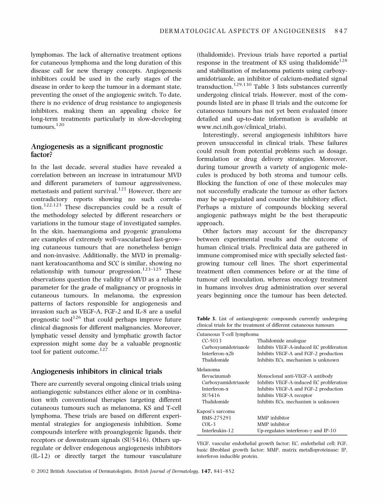
Table 3. List of antiangiogenic compounds currently undergoing
clinical trials for the treatment of different cutaneous tumours
Cutaneous T-cell lymphoma
CC-5013 Thalidomide analogue
Carboxyamidotriazole Inhibits VEGF-A-induced EC proliferationInterferon-a2b Inhibits VEGF-A and FGF-2 production
Thalidomide Inhibits ECs, mechanism is unknown
Melanoma
Bevacizumab Monoclonal anti-VEGF-A antibodyCarboxyamidotriazole Inhibits VEGF-A-induced EC proliferation
Interferon-a Inhibits VEGF-A and FGF-2 production
SU5416 Inhibits VEGF-A receptorThalidomide Inhibits ECs, mechanism is unknown
Kaposi’s sarcoma
BMS-275291 MMP inhibitor
COL-3 MMP inhibitorInterleukin-12 Up-regulates interferon-c and IP-10
VEGF, vascular endothelial growth factor; EC, endothelial cell; FGF,
basic fibroblast growth factor; MMP, matrix metalloproteinase; IP,interferon inducible protein.
D E R M A T O L O G I C A L A S P E C T S O F A N G I O G E N E S I S 8 4 7
� 2002 British Association of Dermatologists, British Journal of Dermatology, 147, 841–852
Furthermore, patients eligible for clinical trials areoften afflicted with advanced stages of the disease that
include metastases. Novel antiangiogenesis treatments
have been met with great enthusiasm and extremelyhigh expectations of complete tumour eradication. Yet,
little is known about the potential side-effects of long-
term treatments, and extensive toxicity studies mustsoon follow. Potential side-effects, such as impaired
wound healing, remain to be evaluated and recent
studies addressing this question have reported conflict-ing results. Mice treated with TNP-470 exhibited
delayed wound healing,131 while endostatin and vas-
ostatin treatment did not affect cutaneous woundhealing at tumour-inhibiting doses.93,132
Finally, new experimental strategies seek to influence
other vascular parameters such as leakage or thematuration status of blood vessels in order to diminish
the sprouting capability of capillaries. Furthermore,
new approaches have targeted the tumour vasculaturebased on the assumption that most endothelial cells in
tumours express distinct markers (�zip codes�). Identi-
fying this heterogeneous group of surface molecules ondifferent endothelial cell populations133 could be useful
for more precise drug targeting strategies.134
Implications of angiogenesis in dermatology
Many cutaneous diseases could potentially benefit from
antiangiogenic treatments. Potential targets include
the vascular tumours discussed and less commontumours such as angiokeratoma. Moreover, therapeu-
tic neovascularization and its inhibition is not only
restricted to neoplastic skin diseases. Blood vessels areinvolved in a variety of cutaneous diseases and are
often of essential diagnostic value. Different types of
vessel growth can be observed in diseased skin. Forexample, telangiectasias are smaller forms of neovas-
cularization and are a hallmark of certain pathologies
such as rosacea or cutaneous autoimmune disease.Dilated, elongated and tortuous blood vessels are a
typical feature of inflammatory diseases such as
psoriasis. Subsequent investigations will elucidate thepotential value of treating inflammatory disorders of
the skin by blocking angiogenesis. Other potential
targets include fast-growing hypertrophic scars andkeloids that could be equally dependent on a vascular
supply. Furthermore, the administration of angiogen-esis factors on chronically impaired healing wounds
such as diabetic ulcers might stimulate the formation
of granulation tissue and thereby improve the overallwound healing process. It has been recently reported
that angiogenesis plays an important role in ultravi-olet-induced skin ageing135 and hair growth.27 Fur-
ther studies are necessary to reveal whether modulating
angiogenesis can successfully influence these processes.Ongoing and future clinical studies will reveal the
potential of antiangiogenic and proangiogenic therapies
for the treatment of cutaneous pathologies.Skin angiogenesis is easily visualized and facilitates
the use of techniques such as surgically created
windows or intravital microscopy to quantify bloodvessel growth and to study the function of blood vessels
in tumours. Both neovascularization and angiogenesis
inhibition can be directly observed in the skin. Thischaracteristic has made the skin an attractive organ for
the development of experimental assays such as
carcinogenesis and orthotopic tumour growth. Theseexperimental approaches can be combined with thera-
peutic trials to test the effects of substances such as
angiogenesis inhibitors. Dermatologists face the chal-lenge to develop drug delivery systems for locally admin-
istered angiogenesis factors and inhibitors avoiding
systemic side-effects. Lastly, only the dermatologist canidentify the wide range of cutaneous diseases that could
potentially benefit from this novel treatment concept.
Acknowledgments
The authors thank Prof. Enno Christophers, Department
of Dermatology, University of Kiel, Prof. Michael Detmar,
Department of Dermatology, Massachusetts GeneralHospital and Medical School, and Prof. Mihaela Skobe
Derald H. Ruttenberg Cancer Center, Mount Sinai School
of Medicine, New York for critical and helpful advice.
References
1 Carmeliet P, Jain RK. Angiogenesis in cancer and other diseases.
Nature 2000; 407: 249–57.2 Malhotra R, Stenn KS, Fernandez LA et al. Angiogenic properties
of normal and psoriatic skin associate with epidermis, not der-
mis. Lab Invest 1989; 61: 162–5.
3 Ensoli B, Nakamura S, Salahuddin SZ et al. AIDS–Kaposi’s sar-coma-derived cells express cytokines with autocrine and para-
crine growth effects. Science 1989; 243: 223–6.
4 Graeven U, Rodeck U, Karpinski S et al. Modulation of angio-
genesis and tumorigenicity of human melanocytic cells by vas-cular endothelial growth factor and basic fibroblast growth
factor. Cancer Res 2001; 61: 7282–90.
5 Chang J, Most D, Bresnick S et al. Proliferative hemangiomas:analysis of cytokine gene expression and angiogenesis. Plast
Reconstr Surg 1999; 103: 1–9, discussion 10.
6 Masood R, Cai J, Tulpule A et al. Interleukin 8 is an autocrine
growth factor and a surrogate marker for Kaposi’s sarcoma. ClinCancer Res 2001; 7: 2693–702.
8 4 8 P . V E L A S C O A N D B . L A N G E - A S S C H E N F E L D T
� 2002 British Association of Dermatologists, British Journal of Dermatology, 147, 841–852
7 Rofstad EK, Halsor EF. Vascular endothelial growth factor, in-
terleukin 8, platelet-derived endothelial cell growth factor, andbasic fibroblast growth factor promote angiogenesis and meta-
stasis in human melanoma xenografts. Cancer Res 2000; 60:
4932–8.
8 Detmar M. The role of VEGF and thrombospondins in skin an-giogenesis. J Dermatol Sci 2000; 24 (Suppl. 1): S78–84.
9 Houck KA, Ferrara N, Winer J et al. The vascular endothelial
growth factor family: identification of a fourth molecular speciesand characterization of alternative splicing of RNA. Mol End-
ocrinol 1991; 5: 1806–14.
10 Terman BI, Carrion ME, Kovacs E et al. Identification of a new
endothelial cell growth factor receptor tyrosine kinase. Oncogene1991; 6: 1677–83.
11 De Vries C, Escobedo J, Ueno H et al. The fms-like tyrosine kinase,
a receptor for vascular endothelial growth factor. Science 1992;
255: 989–91.12 Quinn TP, Peters KG, De Vries C et al. Fetal liver kinase 1 is a
receptor for vascular endothelial growth factor and is selectively
expressed in vascular endothelium. Proc Natl Acad Sci USA1993; 90: 7533–7.
13 Soker S, Gollamudi PS, Fidder H et al. Inhibition of vas-
cular endothelial growth factor (VEGF)-induced endothelial
cell proliferation by a peptide corresponding to the exon7-encoded domain of VEGF165. J Biol Chem 1997; 272:
31582–8.
14 Ferrara N, Gerber HP. The role of vascular endothelial growth
factor in angiogenesis. Acta Haematol 2001; 106: 148–56.15 Potgens AJ, Lubsen NH, van Altena MC et al. Vascular per-
meability factor expression influences tumor angiogenesis in
human melanoma lines xenografted to nude mice. Am J Pathol
1995; 146: 197–209.16 Staibano S, Boscaino A, Salvatore G et al. The prognostic sig-
nificance of tumor angiogenesis in nonaggressive and aggressive
basal cell carcinoma of the human skin. Hum Pathol 1996; 27:695–700.
17 Detmar M, Velasco P, Richard L et al. Expression of vascular
endothelial growth factor induces an invasive phenotype in
human squamous cell carcinomas. Am J Pathol 2000; 156:159–67.
18 Zucker S, Mirza H, Conner CE et al. Vascular endothelial growth
factor induces tissue factor and matrix metalloproteinase pro-
duction in endothelial cells: conversion of prothrombin tothrombin results in progelatinase A activation and cell prolif-
eration. Int J Cancer 1998; 75: 780–6.
19 Vu TH, Shipley JM, Bergers G et al. MMP-9 ⁄ gelatinase B is a keyregulator of growth plate angiogenesis and apoptosis of hyper-
trophic chondrocytes. Cell 1998; 93: 411–22.
20 O’Reilly MS, Boehm T, Shing Y et al. Endostatin: an endogenous
inhibitor of angiogenesis and tumor growth. Cell 1997; 88:277–85.
21 Thurston G, Rudge JS, Ioffe E et al. Angiopoietin-1 protects the
adult vasculature against plasma leakage. Nat Med 2000; 6:
460–3.22 Pomyje J, Zivny JH, Stopka T et al. Angiopoietin-1, angiopoietin-
2 and Tie-2 in tumour and non-tumour tissues during growth of
experimental melanoma. Melanoma Res 2001; 11: 639–43.
23 Maisonpierre PC, Goldfarb M, Yancopoulos GD et al. Distinct ratgenes with related profiles of expression define a TIE receptor
tyrosine kinase family. Oncogene 1993; 8: 1631–7.
24 Holash J, Maisonpierre PC, Compton D et al. Vessel cooption,regression, and growth in tumors mediated by angiopoietins and
VEGF. Science 1999; 284: 1994–8.
25 Vajkoczy P, Farhadi M, Gaumann A et al. Microtumor growth
initiates angiogenic sprouting with simultaneous expression ofVEGF, VEGF receptor-2, and angiopoietin-2. J Clin Invest 2002;
109: 777–85.
26 Hanahan D, Folkman J. Patterns and emerging mechanisms of
the angiogenic switch during tumorigenesis. Cell 1996; 86:353–64.
27 Yano K, Brown LF, Detmar M. Control of hair growth and fol-
licle size by VEGF-mediated angiogenesis. J Clin Invest 2001;107: 409–17.
28 Detmar M. Molecular regulation of angiogenesis in the skin.
J Invest Dermatol 1996; 106: 207–8.
29 Hawighorst T, Velasco P, Streit M et al. Thrombospondin-2 playsa protective role in multistep carcinogenesis: a novel host anti-
tumor defense mechanism. Embo J 2001; 20: 2631–40.
30 Wight TN, Raugi GJ, Mumby SM et al. Light microscopic im-
munolocation of thrombospondin in human tissues. J HistochemCytochem 1985; 33: 295–302.
31 Weinstat-Saslow DL, Zabrenetzky VS, VanHoutte K et al.
Transfection of thrombospondin 1 complementary DNA into ahuman breast carcinoma cell line reduces primary tumor
growth, metastatic potential, and angiogenesis. Cancer Res
1994; 54: 6504–11.
32 Bleuel K, Popp S, Fusenig NE et al. Tumor suppression in humanskin carcinoma cells by chromosome 15 transfer or thrombo-
spondin-1 overexpression through halted tumor vasculariza-
tion. Proc Natl Acad Sci USA 1999; 96: 2065–70.
33 Streit M, Riccardi L, Velasco P et al. Thrombospondin-2: a potentendogenous inhibitor of tumor growth and angiogenesis. Proc
Natl Acad Sci USA 1999; 96: 14888–93.
34 Streit M, Velasco P, Brown LF et al. Overexpression of throm-
bospondin-1 decreases angiogenesis and inhibits the growth ofhuman cutaneous squamous cell carcinomas. Am J Pathol 1999;
155: 441–52.
35 O’Reilly MS, Holmgren L, Shing Y et al. Angiostatin: a novelangiogenesis inhibitor that mediates the suppression of meta-
stases by a Lewis lung carcinoma. Cell 1994; 79: 315–28.
36 Pike SE, Yao L, Jones KD et al. Vasostatin, a calreticulin frag-
ment, inhibits angiogenesis and suppresses tumor growth. J ExpMed 1998; 188: 2349–56.
37 Yao L, Pike SE, Setsuda J et al. Effective targeting of tumor
vasculature by the angiogenesis inhibitors vasostatin and in-
terleukin-12. Blood 2000; 96: 1900–5.38 Pepper MS. Lymphangiogenesis and tumor metastasis: myth or
reality? Clin Cancer Res 2001; 7: 462–8.
39 Joukov V, Pajusola K, Kaipainen A et al. A novel vascularendothelial growth factor, VEGF-C, is a ligand for the Flt4
(VEGFR-3) and KDR (VEGFR-2) receptor tyrosine kinases. Embo
J 1996; 15: 290–8.
40 Lee J, Gray A, Yuan J et al. Vascular endothelial growth factor-related protein: a ligand and specific activator of the tyrosine
kinase receptor Flt4. Proc Natl Acad Sci USA 1996; 93: 1988–
92.
41 Skobe M, Hamberg LM, Hawighorst T et al. Concurrent induc-tion of lymphangiogenesis, angiogenesis, and macrophage re-
cruitment by vascular endothelial growth factor-C in
melanoma. Am J Pathol 2001; 159: 893–903.
42 Skobe M, Hawighorst T, Jackson DG et al. Induction of tumorlymphangiogenesis by VEGF-C promotes breast cancer meta-
stasis. Nat Med 2001; 7: 192–8.
43 Mandriota SJ, Jussila L, Jeltsch M et al. Vascular endothelialgrowth factor-C-mediated lymphangiogenesis promotes tumour
metastasis. EMBO J 2001; 20: 672–82.
D E R M A T O L O G I C A L A S P E C T S O F A N G I O G E N E S I S 8 4 9
� 2002 British Association of Dermatologists, British Journal of Dermatology, 147, 841–852

44 Karpanen T, Egeblad M, Karkkainen MJ et al. Vascular
endothelial growth factor C promotes tumor lymphangiogenesisand intralymphatic tumor growth. Cancer Res 2001; 61: 1786–
90.
45 Jackson DG, Prevo R, Clasper S et al. LYVE-1, the lymphatic
system and tumor lymphangiogenesis. Trends Immunol 2001;22: 317–21.
46 Wigle JT, Oliver G. Prox1 function is required for the devel-
opment of the murine lymphatic system. Cell 1999; 98: 769–78.
47 Marchuk DA. Pathogenesis of hemangioma. J Clin Invest 2001;
107: 665–6.
48 Takahashi K, Mulliken JB, Kozakewich HP et al. Cellularmarkers that distinguish the phases of hemangioma during in-
fancy and childhood. J Clin Invest 1994; 93: 2357–64.
49 Salcedo R, Ponce ML, Young HA et al. Human endothelial
cells express CCR2 and respond to MCP-1: direct role of MCP-1 in angiogenesis and tumor progression. Blood 2000; 96: 34–
40.
50 Yu Y, Varughese J, Brown LF et al. Increased Tie2 expression,enhanced response to angiopoietin-1, and dysregulated angio-
poietin-2 expression in hemangioma-derived endothelial cells.
Am J Pathol 2001; 159: 2271–80.
51 Vikkula M, Boon LM, Carraway KL III et al. Vascular dys-morphogenesis caused by an activating mutation in the receptor
tyrosine kinase TIE2. Cell 1996; 87: 1181–90.
52 Bielenberg DR, Bucana CD, Sanchez R et al. Progressive growth
of infantile cutaneous hemangiomas is directly correlatedwith hyperplasia and angiogenesis of adjacent epidermis and
inversely correlated with expression of the endogenous angio-
genesis inhibitor, IFN-beta. Int J Oncol 1999; 14: 401–8.
53 Lannutti BJ, Gately ST, Quevedo ME et al. Human angiostatininhibits murine hemangioendothelioma tumor growth in vivo.
Cancer Res 1997; 57: 5277–80.
54 Vergani V, Garofalo A, Bani MR et al. Inhibition of matrixmetalloproteinases by overexpression of tissue inhibitor of met-
alloproteinase-2 inhibits the growth of experimental heman-
giomas. Int J Cancer 2001; 91: 241–7.
55 Ramakrishnan S, Olson TA, Bautch VL et al. Vascular endot-helial growth factor-toxin conjugate specifically inhibits
KDR ⁄ flk-1-positive endothelial cell proliferation in vitro and
angiogenesis in vivo. Cancer Res 1996; 56: 1324–30.
56 Liekens S, Verbeken E, Vandeputte M et al. A novel animalmodel for hemangiomas: inhibition of hemangioma develop-
ment by the angiogenesis inhibitor TNP-470. Cancer Res 1999;
59: 2376–83.57 Taraboletti G, Garofalo A, Belotti D et al. Inhibition of angio-
genesis and murine hemangioma growth by batimastat, a
synthetic inhibitor of matrix metalloproteinases. J Natl Cancer
Inst 1995; 87: 293–8.58 Regezi JA, MacPhail LA, Daniels TE et al. Human immunodefi-
ciency virus-associated oral Kaposi’s sarcoma. A heterogeneous
cell population dominated by spindle-shaped endothelial cells.
Am J Pathol 1993; 143: 240–9.59 Safai B, Johnson KG, Myskowski PL et al. The natural history of
Kaposi’s sarcoma in the acquired immunodeficiency syndrome.
Ann Intern Med 1985; 103: 744–50.
60 Sturzl M, Brandstetter H, Roth WK. Kaposi’s sarcoma: a reviewof gene expression and ultrastructure of KS spindle cells in vivo.
AIDS Res Hum Retroviruses 1992; 8: 1753–63.
61 Masood R, Cai J, Zheng T et al. Vascular endothelial growthfactor ⁄ vascular permeability factor is an autocrine growth
factor for AIDS–Kaposi sarcoma. Proc Natl Acad Sci USA 1996;
93: 10589–94.62 Miles SA, Rezai AR, Salazar-Gonzalez JF et al. AIDS Kaposi
sarcoma-derived cells produce and respond to interleukin 6. Proc
Natl Acad Sci USA 1990; 87: 4068–72.
63 Brown LF, Dezube BJ, Tognazzi K et al. Expression of Tie1, Tie2,and angiopoietins 1, 2, and 4 in Kaposi’s sarcoma and cuta-
neous angiosarcoma. Am J Pathol 2000; 156: 2179–83.
64 Skobe M, Brown LF, Tognazzi K et al. Vascular endothelialgrowth factor-C (VEGF-C) and its receptors KDR and flt-4 are
expressed in AIDS-associated Kaposi’s sarcoma. J Invest Dermatol
1999; 113: 1047–53.
65 Marchio S, Primo L, Pagano M et al. Vascular endothelialgrowth factor-C stimulates the migration and proliferation of
Kaposi’s sarcoma cells. J Biol Chem 1999; 274: 27617–22.
66 Sirianni MC, Vincenzi L, Fiorelli V et al. Gamma-interferon
production in peripheral blood mononuclear cells and tumorinfiltrating lymphocytes from Kaposi’s sarcoma patients: corre-
lation with the presence of human herpesvirus-8 in peripheral
blood mononuclear cells and lesional macrophages. Blood 1998;91: 968–76.
67 Masood R, Cesarman E, Smith DL et al. Human herpesvirus-8-
transformed endothelial cells have functionally activated vas-
cular endothelial growth factor ⁄ vascular endothelial growthfactor receptor. Am J Pathol 2002; 160: 23–9.
68 Albini A, Barillari G, Benelli R et al. Angiogenic properties of
human immunodeficiency virus type 1 Tat protein. Proc Natl
Acad Sci USA 1995; 92: 4838–42.69 Fiorelli V, Barillari G, Toschi E et al. IFN-gamma induces
endothelial cells to proliferate and to invade the extracellular
matrix in response to the HIV-1 Tat protein: implications for
AIDS-Kaposi’s sarcoma pathogenesis. J Immunol 1999; 162:1165–70.
70 Taraboletti G, Benelli R, Borsotti P et al. Thrombospondin-1
inhibits Kaposi’s sarcoma (KS) cell and HIV-1 Tat-induced an-giogenesis and is poorly expressed in KS lesions. J Pathol 1999;
188: 76–81.
71 Albini A, Marchisone C, Del Grosso F et al. Inhibition of angio-
genesis and vascular tumor growth by interferon-producingcells: a gene therapy approach. Am J Pathol 2000; 156: 1381–
93.
72 Iurlaro M, Benelli R, Masiello L et al. Beta interferon inhibits
HIV-1 Tat-induced angiogenesis: synergism with 13-cis retinoicacid. Eur J Cancer 1998; 34: 570–6.
73 Incardona F, Lewalle JM, Morandi V et al. Thrombospondin
modulates human breast adenocarcinoma cell adhesion to hu-man vascular endothelial cells. Cancer Res 1995; 55: 166–73.
74 Pati S, Lee Y, Samaniego F. Urinary proteins with pro-apoptotic
and antitumor activity. Apoptosis 2000; 5: 21–8.
75 Indraccolo S, Morini M, Gola E et al. Effects of angiostatin genetransfer on functional properties and in vivo growth of Kaposi’s
sarcoma cells. Cancer Res 2001; 61: 5441–6.
76 Sgadari C, Barillari G, Toschi E et al. HIV protease inhibitors are
potent antiangiogenic molecules and promote regression ofKaposi sarcoma. Nat Med 2002; 8: 225–32.
77 Moore JD, Dezube BJ, Gill P et al. Phase I dose escalation
pharmacokinetics of O-(chloroacetylcarbamoyl) fumagillol
(TNP-470) and its metabolites in AIDS patients with Kaposi’ssarcoma. Cancer Chemother Pharmacol 2000; 46: 173–9.
78 Tulpule A, Scadden DT, Espina BM et al. Results of a randomized
study of IM862 nasal solution in the treatment of AIDS-relatedKaposi’s sarcoma. J Clin Oncol 2000; 18: 716–23.
8 5 0 P . V E L A S C O A N D B . L A N G E - A S S C H E N F E L D T
� 2002 British Association of Dermatologists, British Journal of Dermatology, 147, 841–852
79 Albini A, Morini M, D’Agostini F et al. Inhibition of angiogene-
sis-driven Kaposi’s sarcoma tumor growth in nude mice by oralN-acetylcysteine. Cancer Res 2001; 61: 8171–8.
80 Aluigi MG, De Flora S, D’Agostini F et al. Antiapoptotic and
antigenotoxic effects of N-acetylcysteine in human cells of
endothelial origin. Anticancer Res 2000; 20: 3183–7.81 Antonian L, Zhang H, Yang C et al. Biotransformation of the
antiangiogenic compound SU5416. Drug Metab Dispos 2000;
28: 1505–12.82 Levine AM, Tulpule A. Clinical aspects and management of AIDS-
related Kaposi’s sarcoma. Eur J Cancer 2001; 37: 1288–95.
83 Arbiser JL, Panigrathy D, Klauber N et al. The antiangiogenic
agents TNP-470 and 2-methoxyestradiol inhibit the growth ofangiosarcoma in mice. J Am Acad Dermatol 1999; 40: 925–9.
84 Marks R, Rennie G, Selwood TS. Malignant transformation of
solar keratoses to squamous cell carcinoma. Lancet 1988; i:
795–7.85 Ziegler A, Jonason AS, Leffell DJ et al. Sunburn and p53 in the
onset of skin cancer. Nature 1994; 372: 773–6.
86 Skobe M, Rockwell P, Goldstein N et al. Halting angiogenesissuppresses carcinoma cell invasion. Nat Med 1997; 3: 1222–7.
87 Strieth S, Hartschuh W, Pilz L et al. Angiogenic switch occurs
late in squamous cell carcinomas of human skin. Br J Cancer
2000; 82: 591–600.88 Streit M, Stephen AE, Hawighorst T et al. Systemic inhibition of
tumor growth and angiogenesis by thrombospondin-2 using
cell-based antiangiogenic gene therapy. Cancer Res 2002; 62:
2004–12.89 Hawighorst T, Skobe M, Streit M et al. Activation of the tie2
receptor by angiopoietin-1 enhances tumor vessel maturation
and impairs squamous cell carcinoma growth. Am J Pathol
2002; 160: 1381–92.90 Darland DC, D’Amore PA. Blood vessel maturation: vascular
development comes of age. J Clin Invest 1999; 103: 157–8.
91 Eberhard A, Kahlert S, Goede V et al. Heterogeneity of angio-genesis and blood vessel maturation in human tumors: impli-
cations for antiangiogenic tumor therapies. Cancer Res 2000;
60: 1388–93.
92 Korff T, Kimmina S, Martiny-Baron G et al. Blood vessel mat-uration in a 3-dimensional spheroidal coculture model: direct
contact with smooth muscle cells regulates endothelial cell
quiescence and abrogates VEGF responsiveness. FASEB J 2001;
15: 447–57.93 Lange-Asschenfeldt B, Velasco P, Streit M et al. The angiogenesis
inhibitor vasostatin does not impair wound healing at tumor-
inhibiting doses. J Invest Dermatol 2001; 117: 1036–41.94 Arbiser JL. Melanoma. Lessons from metastases. Arch Dermatol
1998; 134: 1027–8.
95 Claffey KP, Brown LF, del Aguila LF et al. Expression of vascular
permeability factor ⁄ vascular endothelial growth factor by mel-anoma cells increases tumor growth, angiogenesis, and experi-
mental metastasis. Cancer Res 1996; 56: 172–81.
96 Salven P, Heikkila P, Joensuu H. Enhanced expression of vas-
cular endothelial growth factor in metastatic melanoma. Br JCancer 1997; 76: 930–4.
97 Graeven U, Rodeck U, Karpinski S et al. Expression patterns of
placenta growth factor in human melanocytic cell lines. J Invest
Dermatol 2000; 115: 118–23.98 Siemeister G, Schirner M, Weindel K et al. Two independent
mechanisms essential for tumor angiogenesis: inhibition of
human melanoma xenograft growth by interfering with eitherthe vascular endothelial growth factor receptor pathway or the
Tie-2 pathway. Cancer Res 1999; 59: 3185–91.
99 Lin P, Buxton JA, Acheson A et al. Antiangiogenic gene therapy
targeting the endothelium-specific receptor tyrosine kinase Tie2.Proc Natl Acad Sci USA 1998; 95: 8829–34.
100 Albelda SM, Mette SA, Elder DE et al. Integrin distribution in
malignant melanoma: association of the beta 3 subunit with
tumor progression. Cancer Res 1990; 50: 6757–64.101 Mitjans F, Meyer T, Fittschen C et al. In vivo therapy of malig-
nant melanoma by means of antagonists of alphav integrins. Int
J Cancer 2000; 87: 716–23.102 Brooks PC, Clark RA, Cheresh DA. Requirement of vascular
integrin alpha v beta 3 for angiogenesis. Science 1994; 264:
569–71.
103 Rofstad EK, Graff BA. Thrombospondin-1-mediated metastasissuppression by the primary tumor in human melanoma xeno-
grafts. J Invest Dermatol 2001; 117: 1042–9.
104 Miao WM, Seng WL, Duquette M et al. Thrombospondin-1 type
1 repeat recombinant proteins inhibit tumor growth throughtransforming growth factor-beta-dependent and -independent
mechanisms. Cancer Res 2001; 61: 7830–9.
105 Gee MS, Koch CJ, Evans SM et al. Hypoxia-mediated apoptosisfrom angiogenesis inhibition underlies tumor control by re-
combinant interleukin 12. Cancer Res 1999; 59: 4882–9.
106 Stack MS, Gately S, Bafetti LM et al. Angiostatin inhibits endo-
thelial and melanoma cellular invasion by blocking matrix-en-hanced plasminogen activation. Biochem J 1999; 340: 77–84.
107 Yamanaka R, Zullo SA, Ramsey J et al. Induction of therapeutic
antitumor antiangiogenesis by intratumoral injection of gen-
etically engineered endostatin-producing Semliki Forest virus.Cancer Gene Ther 2001; 8: 796–802.
108 Yamaoka M, Yamamoto T, Masaki T et al. Inhibition of tumor
growth and metastasis of rodent tumors by the angiogenesis
inhibitor O-(chloroacetyl-carbamoyl) fumagillol (TNP-470;AGM-1470). Cancer Res 1993; 53: 4262–7.
109 Hwu WJ. New approaches in the treatment of metastatic mel-
anoma: thalidomide and temozolomide. Oncology (Huntingt)2000; 14: 25–8.
110 Laird AD, Vajkoczy P, Shawver LK et al. SU6668 is a potent
antiangiogenic and antitumor agent that induces regression of
established tumors. Cancer Res 2000; 60: 4152–60.111 Mah-Becherel MC, Ceraline J, Deplanque G et al. Antiangiogenic
effects of the thienopyridine SR 25989 in vitro and in vivo in a
murine pulmonary metastasis model. Br J Cancer 2002; 86:
803–10.112 Wylie S, MacDonald IC, Varghese HJ et al. The matrix metallo-
proteinase inhibitor batimastat inhibits angiogenesis in liver
metastases of B16F1 melanoma cells. Clin Exp Metastasis 1999;17: 111–17.
113 Kruger A, Soeltl R, Sopov I et al. Hydroxamate-type matrix
metalloproteinase inhibitor batimastat promotes liver metasta-
sis. Cancer Res 2001; 61: 1272–5.114 Salven P, Lymboussaki A, Heikkila P et al. Vascular endothelial
growth factors VEGF-B and VEGF-C are expressed in human
tumors. Am J Pathol 1998; 153: 103–8.
115 Achen MG, Williams RA, Minekus MP et al. Localization ofvascular endothelial growth factor-D in malignant melanoma
suggests a role in tumour angiogenesis. J Pathol 2001; 193:
147–54.
116 Stacker SA, Caesar C, Baldwin ME et al. VEGF-D promotes themetastatic spread of tumor cells via the lymphatics. Nat Med
2001; 7: 186–91.
117 Bedlow AJ, Stanton AW, Cliff S et al. Basal cell carcinoma – anin-vivo model of human tumour microcirculation? Exp Dermatol
1999; 8: 222–6.
D E R M A T O L O G I C A L A S P E C T S O F A N G I O G E N E S I S 8 5 1
� 2002 British Association of Dermatologists, British Journal of Dermatology, 147, 841–852

118 Vacca A, Moretti S, Ribatti D et al. Progression of mycosis fun-
goides is associated with changes in angiogenesis and expressionof the matrix metalloproteinases 2 and 9. Eur J Cancer 1997; 33:
1685–92.
119 Schaerer L, Schmid MH, Mueller B et al. Angiogenesis in cuta-
neous lymphoproliferative disorders: microvessel density dis-criminates between cutaneous B-cell lymphomas and B-cell
pseudolymphomas. Am J Dermatopathol 2000; 22: 140–3.
120 Boehm T, Folkman J, Browder T et al. Antiangiogenic therapy ofexperimental cancer does not induce acquired drug resistance.
Nature 1997; 390: 404–7.
121 Weidner N, Folkman J. Tumoral vascularity as a prognostic
factor in cancer. Important Adv Oncol 1996; : 167–90.122 Carrau RL, Barnes EL, Snyderman CH et al. Tumor angiogenesis
as a predictor of tumor aggressiveness and metastatic potential
in squamous cell carcinoma of the head and neck. Invasion
Metastasis 1995; 15: 197–202.123 Tahan SR, Stein AL. Angiogenesis in invasive squamous cell
carcinoma of the lip: tumor vascularity is not an indicator of
metastatic risk. J Cutan Pathol 1995; 22: 236–40.124 Weninger W, Uthman A, Pammer J et al. Vascular endothelial
growth factor production in normal epidermis and in benign
and malignant epithelial skin tumors. Lab Invest 1996; 75: 647–
57.125 Weninger W, Rendl M, Pammer J et al. Differences in tumor
microvessel density between squamous cell carcinomas and
basal cell carcinomas may relate to their different biologic be-
havior. J Cutan Pathol 1997; 24: 364–9.126 Ugurel S, Rappl G, Tilgen W et al. Increased serum concentra-
tion of angiogenic factors in malignant melanoma patients
correlates with tumor progression and survival. J Clin Oncol
2001; 19: 577–83.
127 Lauria R, Perrone F, Carlomagno C et al. The prognostic value of
lymphatic and blood vessel invasion in operable breast cancer.Cancer 1995; 76: 1772–8.
128 Little RF, Wyvill KM, Pluda JM et al. Activity of thalidomide in
AIDS-related Kaposi’s sarcoma. J Clin Oncol 2000; 18: 2593–
602.129 Kohn EC, Reed E, Sarosy G et al. Clinical investigation of a
cytostatic calcium influx inhibitor in patients with refractory
cancers. Cancer Res 1996; 56: 569–73.130 Kohn EC, Figg WD, Sarosy GA et al. Phase I trial of micronized
formulation carboxyamidotriazole in patients with refractory
solid tumors: pharmacokinetics, clinical outcome, and compar-
ison of formulations. J Clin Oncol 1997; 15: 1985–93.131 Klein SA, Bond SJ, Gupta SC et al. Angiogenesis inhibitor TNP-
470 inhibits murine cutaneous wound healing. J Surg Res 1999;
82: 268–74.
132 Bloch W, Huggel K, Sasaki T et al. The angiogenesis inhibitorendostatin impairs blood vessel maturation during wound
healing. FASEB J 2000; 14: 2373–6.
133 Jacobson BS, Stolz DB, Schnitzer JE. Identification of endothelialcell-surface proteins as targets for diagnosis and treatment of
disease. Nat Med 1996; 2: 482–4.
134 Burrows FJ, Thorpe PE. Eradication of large solid tumors in mice
with an immunotoxin directed against tumor vasculature. ProcNatl Acad Sci USA 1993; 90: 8996–9000.
135 Yano K, Oura H, Detmar M. Targeted overexpression of the
angiogenesis inhibitor thrombospondin-1 in the epidermis of
transgenic mice prevents ultraviolet-B-induced angiogenesisand cutaneous photo-damage. J Invest Dermatol 2002; 118:
800–5.
8 5 2 P . V E L A S C O A N D B . L A N G E - A S S C H E N F E L D T
� 2002 British Association of Dermatologists, British Journal of Dermatology, 147, 841–852