dermatologic vulvovaginal conditions: … dermatologic vulvovaginal conditions: diagnosis and...
TRANSCRIPT
1
Dermatologic Vulvovaginal Conditions: Diagnosis and Treatment
Hope K. Haefner, M.D. Professor of Obstetrics and Gynecology
Co-Director, The University of Michigan Center for Vulvar Diseases The University of Michigan Hospitals
Handout developed in conjunction with: Lynette J. Margesson, M.D.
Assistant Professor of Obstetrics and Gynecology and Medicine (Dermatology) Dartmouth Medical School
This handout is available at http://obgyn.med.umich.edu/patient-care/womens-health-library/vulvar-diseases or go to Google and type in University of Michigan Center for Vulvar Diseases
click on Information on Vulvar Diseases
Disclosures:
Hope Haefner, MD is on the advisory board of Merck Co. Inc. Lynette Margesson, MD has no relevant financial relationships with any commercial interest relative to the subject of this lecture.
2
Learning Objectives At the end of this course, the participant should be able to:
• Identify the clinical features of various vulvovaginal conditions
• Recognize the gross features of non-neoplastic epithelial disorders of the vulva
• Identify the various ulcerative conditions of the vulva and their treatments
• Become familiar with a variety of treatments for skin diseases A variety of dermatologic conditions affect the vulva and the vagina. It is important to become familiar with the appearances and treatments of the numerous vulvovaginal conditions that you may see in your patients. Nonneoplastic Epithelial Disorders 1975-1986 1987-present Lichen sclerosus et atrophicus Lichen sclerosus Hyperplastic dystrophy Squamous cell hyperplasia/lichen
simplex chronicus Mixed dystrophy Other dermatoses
Lichen sclerosus Lichen Sclerosus – is chronic, autoimmune disease affecting the genital skin causing whiteness, tissue thinning and scarring.
It is the most common chronic vulvar condition Histology - blunting or loss of rete ridges, hyperkeratosis and loss of melanocytes are seen with a zone of pallor and often a dense interstitial lymphocytic infiltrate. Pathophysiology: Unknown. Various genetic, autoimmune, infectious and local factors are implicated. The cause is probably multifactorial with a genetic, environmental and possibly infectious input. Often associated with other autoimmune diseases. Thyroid disease is the most common . Familial cases have been reported. Age of onset - middle age (about 40 years) but range is from less than one year to > 80 years
3
Symptoms - Pruritus is most common and can be severe and intolerable Scratching causes secondary changes and open areas that cause dysuria, burning and dyspareunia Scarring leads to dyspareunia, even apareunia May be asymptomatic - common cause of asymptomatic vulvar scarring. Physical exam – Scattered or confluent papules forming plaques of ivory white with cellophane- like sheen on the surface. Found anywhere on the vulva from the clitoris and periclitorally to the gluteal cleft. The involvement may be patchy or generalized in various patterns. classically a “figure-of-eight”It can involve any cutaneous surface but most comonly is found on the vulva in women. Extragenital disease occurs in 10-20%. LS typically does not involve the vagina. Secondary changes - excoriations, purpura, erosions, thickening (lichenification) crusting, and scarring, ranging from loss of labia or burying of the clitoris to loss of all normal vulvar structures. Differential diagnosis - sexual abuse in children, vitiligo, lichen simplex chronicus, lichen planus, cicatricial pemphigoid. Cancer risk - about 4% develop associated SCC Treatment: Biopsy to confirm diagnosis Educate the patient Stop irritants Recommend cool, ventilated clothing Topical superpotent steroids (various regimens exist)
Clobetasol propionate or halobetasol 0.05% ointment qd for 12 weeks, then M-W-F or 1-2 times a week and follow up at 6-12 weeks then regularly at 6-12 month intervals
versus Clobetasol propionate 0.05% bid x 1 month, then q d x 2 months. Decease use of clobetasol to 3 times down to once a week. In some cases decrease to a class 4 steroid (see steroid table at the end of the handout), then gradually decrease frequency of application to once a week. (There is debate regarding whether or not long term steroids are required.)
Treat associated Candida or secondary bacterial infection Stop scratching as this keeps LS active. Give 10 mg of hydroxyzine or doxepin at 6 to 7 PM to stop nightly scratching.( See Lichen Simplex Chronicus below) For thick lichen sclerosus consider intralesional steroid (triamcinolone 3.3 to 10 mg/ml). The dose is dependent on the location and thickness of the skin that is being injected. This can be repeated monthly for 2-3 months. Do not inject high steroid doses into thin skin or in small areas because the tissue can slough . If constantly scratching use IM triamcinolone 1 mg/kg up to 80 mg/dose. Never give over 80 mg of triamcinolone acetonide IM per month. This can be repeated once a month for 3 months with a maximum of 4 doses a year.
4
Tacrolimus 0.1%ointment and pimecrolimus 1% cream have been used for the treatment of vulvar lichen sclerosus. Burning may occur with these medications. Tazorac 0.1% gel (can also use 0.05% or 0.1% cream for lower strength) may be used for lichen sclerosus when the skin is very thick or unresponsive to topical steroids. Apply to skin qhs with gradual decrease to two to three times a week. Acitretin (Soriatane) is a retinoid that may be used for lichen sclerosus unresponsive to topical steroids (and in some cases lichen planus). It is most beneficial for thickened skin. Take 10 mg every 1-2 days for a dose of 30-70 mg per week.It must be taken with fatty food. The patients must not become pregnant as it is tearatogenic like isotretinoin. (expensive, but less costly in Canada). Surgery is done on occasion to improve function or for scarring
In all patients with lichen sclerosus:
Arrange follow-up always – indefinitely. Regular follow-up is needed because there is an increased risk of developing squamous cell carcinoma (SCC) (<5 % in women). If not responding to treatment Look for concurrent conditions and biopsy and rebiopsy, as needed. Note – LS involves the vulva not the vagina unless prolapse. Scarring is not reversible by any medical therapy.
LICHEN SIMPLEX CHRONICUS (LSC ) Synonyms: Squamous cell hyperplasia, neurodermatitis, pruritus vulvae, hyperplastic dystrophy “LSC” – The end stage of the itch – scratch – itch cycle. It is usually part of the atopic dermatitis (eczema) spectrum. It can be associated with underlying, secondarily scratched and thickened psoriasis or contact dermatitis or the end stage of several itchy vulvar conditions (e.g. LS). Scratching “feels good” especially for patients with atopic dermatitis (patients with a background of allergies, eczema, hay fever or asthma). Stress makes all of this worse. Causes of LSC:
Infection: Candida and dermatophytosis Dermatoses: Atopic dermatitis Psoriasis
Lichen Sclerosus Contact Dermatitis Lichen Planus
Metabolic: Diabetes and iron deficiency anemia Neoplasia: Vulvar intraepithelial neoplasia
The most important causes are atopic dermatitis, contact dermatitis or both. Less common causes – psoriasis, LS
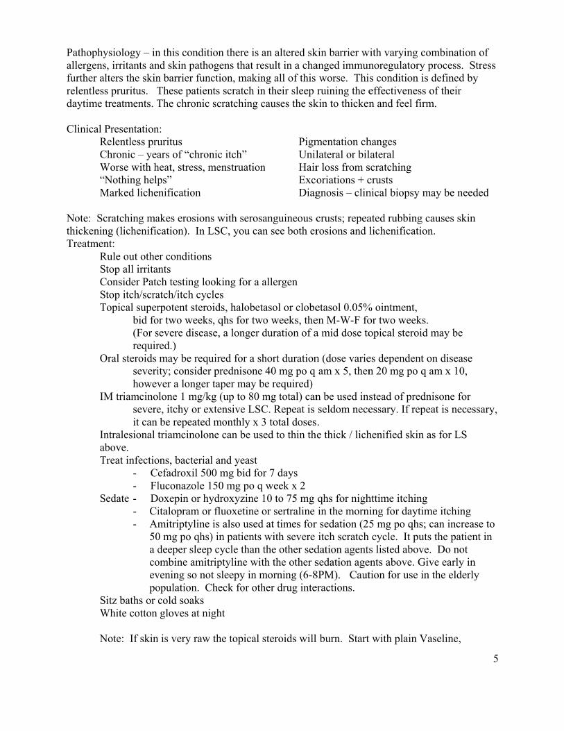
Pathophyallergensfurther alrelentlessdaytime t Clinical P R C W “N M Note: ScthickeninTreatmen
RSCST
O
IM
InabT
S
SW N
ysiology – ins, irritants anlters the skins pruritus. Ttreatments. T
PresentationRelentless pruChronic – yeaWorse with hNothing help
Marked liche
cratching mang (lichenificnt:
Rule out othetop all irritan
Consider Patctop itch/scra
Topical superbid fo(For srequir
Oral steroids severihowev
M triamcinosevereit can
ntralesional tbove.
Treat infectio- Ce- Fl
edate - D- Ci- Am
50a dcoevpo
itz baths or cWhite cotton
Note: If skin
n this conditind skin pathon barrier funcThese patienThe chronic
n: uritus ars of “chron
heat, stress, mps” nification
akes erosionscation). In L
er conditionsnts ch testing looatch/itch cycrpotent stero
or two weekssevere diseasred.) may be requ
ity; considerver a longer lone 1 mg/ke, itchy or exbe repeated triamcinolon
ons, bacterialefadroxil 50luconazole 1oxepin or hyitalopram ormitriptyline 0 mg po qhs)deeper sleep
ombine amitrvening so notopulation. Ccold soaks gloves at ni
is very raw
ion there is aogens that rection, makin
nts scratch inscratching c
nic itch”menstruation
s with serosaLSC, you can
s
oking for a acles oids, halobetas, qhs for twose, a longer d
uired for a shr prednisone taper may b
kg (up to 80 mxtensive LSCmonthly x 3
ne can be use
l and yeast 00 mg bid for150 mg po q ydroxyzine 1r fluoxetine ois also used
) in patients p cycle than triptyline witt sleepy in m
Check for oth
ght
the topical s
an altered skesult in a chang all of this n their sleep causes the sk
Pigm Unil
n Hair Exco Diag
anguineous cn see both er
allergen
asol or clobeo weeks, theduration of a
hort duration40 mg po q
be required)mg total) canC. Repeat is 3 total dosesed to thin th
r 7 days week x 2
10 to 75 mg or sertraline at times for with severe the other sedth the other smorning (6-8her drug inte
steroids will
kin barrier wanged immun
worse. Thiruining the e
kin to thicken
mentation chlateral or bilr loss from soriations + cgnosis – clin
crusts; repearosions and l
etasol 0.05%en M-W-F foa mid dose to
n (dose varieam x 5, then
n be used insseldom nece. e thick / lich
qhs for nighin the morni
r sedation (25itch scratch
dation agentssedation age8PM). Caut eractions.
l burn. Start
with varying cnoregulatorys condition ieffectivenesn and feel fir
hanges lateral scratching crusts nical biopsy
ated rubbinglichenificati
% ointment, or two weekopical steroi
es dependentn 20 mg po q
stead of predessary. If rep
henified skin
httime itchining for dayti5 mg po qhscycle. It pu
s listed abovents above. Gtion for use i
t with plain V
combinationy process. Sis defined bys of their rm.
may be need
g causes skinon.
s. d may be
t on disease q am x 10,
dnisone for peat is neces
n as for LS
g ime itchings; can increasuts the patienve. Do not Give early inin the elderly
Vaseline,
5
n of Stress y
ded
n
ssary,
se to nt in
n y
6
oral antibiotics, anti-yeast medication and nighttime sedation for 2-3 days, then start the topicals. LSC reoccurs due to sensitive skin in the area so it will need repeated management. LOOK FOR MORE THAN ONE CAUSE OR A COMBINATION OF CAUSES as it is not uncommon to have psoriasis, contact dermatitis and lichen simplex chronicus in the same patient.
LICHEN PLANUS (LP) Lichen planus is an autoimmune, mucocutaneous disorder of altered cell mediated immunity in older women affecting the skin and mucous membranes. Etiology: It is a disorder of altered cell mediated immunity with exogenous antigens targeting the epidermis. The diagnosis is often missed on the vulva and in the vagina. It tends to occur in menopausal women (age 40-60 years). It affects skin and mucous membrane – mouth, vulva, vagina, nails, scalp, esophagus, nose, conjunctiva of the eye, ears and bladder. Painful LP is usually erosive; patient can have LP plus chronic vulvar pain. Clinical Presentation: 1. Papulosquamous – typical papules and plaques with white lacy pattern on the
vulvar trigone and periclitoral area. It may be part of generalized LP. This can be itchy. It tends to respond to topical steroids.
2. Hypertrophic – least common with extensive white scarring and destruction (looks like LS)
– can be very itchy. Treatment tends to be resistant. 3. Erosive (vulvovaginal gingival syndrome) – destructive, scarred lichen planus on the
mucous membranes and vulva with a desquamative vaginitis, variable erosions plus atrophy, usually pain, burning and irritation rather than itch. The skin of the vulva often has a glazed erythema . Treatment tends to be resistant.
Note – LP involves the vulva and vagina, It may only be in the vagina.
Erosive LP (vulvovaginal gingival syndrome) Symptoms: Severe pain and burning Depression + anger
Dysuria Dyspareunia / apareunia Signs – painful, glossy red erosions (glazed erythema) and scarring are seen around the labia minora and vestibule. The borders may be white to smudgy or smoky gray. The scarring causes flattening of the vulva and loss of the labia minora.
- May see desquamative inflammatory vaginitis Vaginitis with vaginal erosions, atrophy, purulent malodorous discharge, vaginal synechiae and scarring. The vagina may be obliterated.
Note: up to 70% of women with vulvar LP have vaginal involvement.
7
This can be a chronic, destructive, debilitating and difficult condition. The vagina may be involved alone.
Diagnosis: Look at mouth and skin for evidence of LP Consider biopsy for H&E and immunofluorescence Biopsies may be nonspecific Differential diagnosis: Lichen sclerosus, drug eruption, cicatricial pemphigoid, graft vs. host disease Treatment:
Stop irritants Pain control Bland therapy for ulcers Sedation Superpotent steroid ointment (clobetasol) topically once to twice a day. Intralesional steroid – triamcinolone 3.3 up to 10 mg/ml q 3-4 wks x 3 (do not give high
dose in small area-erosions and ulcers may occur) Intravaginal steroid – hydrocortisone acetate foam 40-80 mg qhs
or 25 to 200 mg compounded suppository qhs (if using high dose steroids, use for short term use, then gradually decrease the dose). If severe – hydrocortisone acetate 10% compounded in a Replens like base –3 to 5 grams (300 mg to 500mg/dose) nightly for 14 days then 3 nights a week and continue to decrease dose as per response. (Some prefer to use every other night initially, and then gradually decrease the dose) Note: adrenal suppression and risk of candidiasis
IM Triamcinolone (Kenalog 40) 1 mg/kg every 4 weeks for 3 doses. (Dose up to a maximum of 80 mg total per dose) Repeat monthly for up to 3 months. Max 4 doses per year Prednisone 30-60 mg a day with taper Methotrexate 7.5-15 mg po or subcutaneously in abdomen or thigh, once a week with folate 1 mg daily Mycophenolate mofetil 250 mg/day building up to 3gm/day (pregnancy must be prevented) Acitretin 10 mg 3-7 days a week with fatty food for erosive disease. Counsel on no pregnancy as this is a teratogen. (see above for lichen sclerosus) Cyclosporine 3-4 mg / kg per day
Patient education and support needed Dilators Surgery for scarring followed by intravaginal treatment Other Treatments:
- Clobetasol propionate 0.05% ointment virginally using 1-2 grams nightly via a “Premarin type applicator” -Clobetasol propionate 0.05% ointment/Nystatin 100,000 units/gram/3% oxy-tetracycline in cream base - pimecrolimus (Elidel) 1% cream bid for mild LP -Topical tacrolimus (Protopic) 0.03 or 0.l% ointment (burns) as a steroid sparer -Hydroxychloroquine, etanercept (see below)

8
Course: uncertain - often very chronic-10% resolve, 50% asymptomatic and 15% do poorly

9
What are the various treatments for Lichen Planus? Papular lichen planus tends to respond to topical corticosteroids. Triamcinolone acetonide 0.1% ointment for mild disease and clobetasol propionate 0.05% ointment for severe disease. For erosive disease the following table contains many medications that have been tried for LP treatment. It is important to note that many of these medications are formulated for off label use. Agent Discussion Long term Anti-inflammatory antibiotics
This treatment works best for early erosive lichen planus Doxycycline or clindamycin used long-term. Consider adding weekly fluconazole to prevent yeast infection.
Steroids are often used for lichen planus
Vaginal LP Anusol HC 25 mg vaginal suppositories are used in the following manner: 1/2 of a Anusol HC suppository per vagina twice daily for 2 months, then daily for 2 months, then maintenance treatment at 1 to 3 times per week. However, many patients do not experience significant long-term response to intravaginal steroids. The vaginal vault tends to continue to scar. To keep the vault open and prevent adhesions it often will be necessary to use vaginal dilators. The dilator may be lubricated with a hydrocortisone cream. At times a stronger steroid may be required for vulvar LP (see text). Topical- Clobetasol propionate (Temovate®) 0.05% ointment Intralesional- triamcinolone acetonide 5-10 mg/ml As above, for stronger treatment: – hydrocortisone acetate foam 40-80 mg qhs or 25 to 200 mg suppository qhs (if using high dose steroids, use for short term use, then gradually decrease the dose). If severe – hydrocortisone acetate 10% compounded in a Replens like base –3 to 5 grams (300 mg to 500mg/dose) nightly for 14 days then 3 nights a week and continue to decrease dose as per response. (Some prefer to use every other night initially, then gradually decrease the dose) Oral- Oral prednisone may be required until healing has occurred. 30-40 mg qam with food for 3 weeks then slowly taper. As the skin heals, topical corticosteroids may be added as the prednisone is tapered. IM steroids (place into muscle in anterior thigh). Used for moderate disease. Dose 1 mg/kg (not to exceed 80 mg) every 4 weeks to every 8 weeks for up to 3 or 4 months. For Oral LP- Apply Clobetasol propionate (Temovate®) gel or ointment 0.05% to affected area up to qid Apply on a cotton ball in mouth for 5 min. Best to use in a dental tray for 15-30 min bid for gums. Some providers use dental molds to hold in medications in patients with gingival LP
10
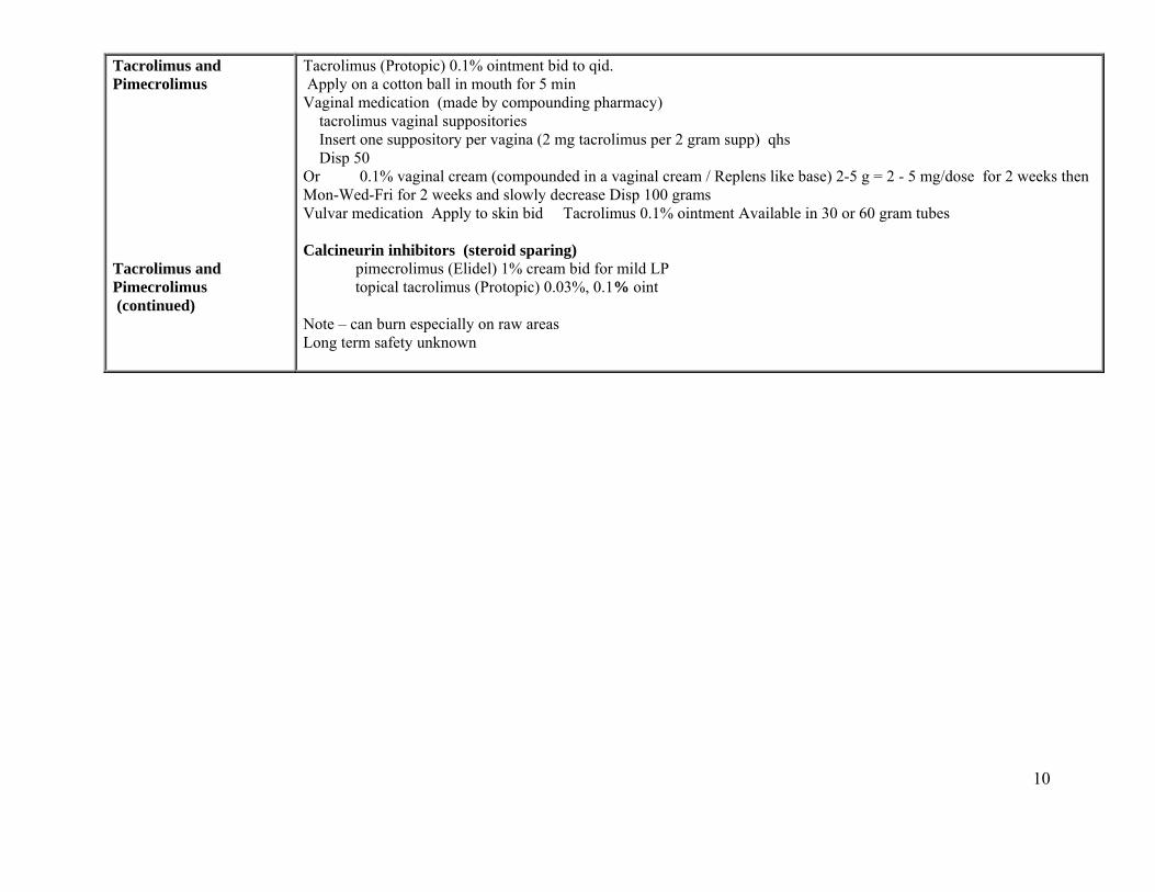
Tacrolimus and Pimecrolimus Tacrolimus and Pimecrolimus (continued)
Tacrolimus (Protopic) 0.1% ointment bid to qid. Apply on a cotton ball in mouth for 5 min Vaginal medication (made by compounding pharmacy) tacrolimus vaginal suppositories Insert one suppository per vagina (2 mg tacrolimus per 2 gram supp) qhs Disp 50 Or 0.1% vaginal cream (compounded in a vaginal cream / Replens like base) 2-5 g = 2 - 5 mg/dose for 2 weeks then Mon-Wed-Fri for 2 weeks and slowly decrease Disp 100 grams Vulvar medication Apply to skin bid Tacrolimus 0.1% ointment Available in 30 or 60 gram tubes Calcineurin inhibitors (steroid sparing) pimecrolimus (Elidel) 1% cream bid for mild LP topical tacrolimus (Protopic) 0.03%, 0.1% oint Note – can burn especially on raw areas Long term safety unknown

11
Less frequently used medications
Hydroxychloroquine (Plaquenil)
Occasionally used. Dose is 200 mg po bid.
Retinoids There is no documented successful use of retinoids for vulvovaginal lichen planus. There is only personal experience with Acitretin (Soriatane) .It can work well in low dose 30-70 mg/week. ( Isotretinoin has been used to treat oral lichen planus; however, discontinuation of the medication results in recurrence of the oral lesions.) Long-term use of retinoids may result in liver dysfunction.but not in the small doses recommended here. Liver function tests, cholesterol, triglycerides and complete blood cell counts should be monitored since laboratory changes are associated with the use of oral retinoids. Patients should be counseled concerning teratogenicity and need for optimal contraception. Acitretin is a strong teratogen that remains in the body for at least three months after the last dose . Topical retinoids (Tazarotene (tazarac) are often too irritating for this vulvar condition but have been used.
Cyclosporine Used topically and systemically. Topical cyclosporine provides a safe and often effective but very expensive alternative for mucous membrane disease. Pelisse et al. described the use of the oral or injectable form of the medication in 100 mg amounts directly to the affected skin four times a day initially. If several mucous membranes were affected for example, 100 mg was applied to the vulva, 100 mg inserted into the vagina, and 100 mg held in the mouth for as long as tolerated before spitting. As disease is controlled, the frequency of application can be tapered. Systemically it is dosed at 4-5 mg/kg/day for 3 months (used in severe disease). Occasionally, in patients with debilitating and painful disease not adequately treated by therapies discussed above, oral cyclosporine may be used. This medication should be used only by health care providers experienced in its use.
Cyclophosphamide Systemic antimetabolite Azathioprine Systemic antimetabolite Etanercept (Enbrel) This is used SQ (50 mg sq 2x/week until symptoms improve, then 25 mg sq 2x/week)) Mycophenolate mofetil (CellCept)
Oral use 250mg -3 g/d in divided dose
Methotrexate Oral or subcutaneous injection weekly. 7.5 to 15 mg oral or subcutaneously weekly using a 27 or 30 gauge needle. Need to give folate with this medication- 1 mg/d
12
Lichen Planus and Surgery
For scarred LP of the vagina - post surgery information
I. For dilation:
Dilation is vital to keep the vagina open in patients with vaginal lichen planus. Patients need specific instructions on size of dilator and how to use dilators. They may need a set of dilators and can to buy the dilator set from www.vaginismus.com. Start with the largest size that will fit, determined by surgery. Leave the dilator in once or twice a day for 15-20 minutes. For lubricating the dilator use either Vaseline or mineral oil. Hydrocortisone acetate cream or Estrace 0.01% vaginal cream can be used later.
II. To stop inflammation:
If not too severe 2-3 days preoperatively use prednisone 15-30 mg/d AM, with food, plus topical steroid. Keep on prednisone for 1 week post operatively then taper slowly at 5 mg/week. Use with the topical steroid (see below).
For more severe disease consider using a dose of intramuscular triamcinolone 1mg/kg up to a total of 80mg/dose to be given two days after surgery and repeat this monthly for up to three months. Follow and assess her to see if she is going to need other long-term systemic medication, cyclosporine, mycophenolate, methotrexate, etc. Once she is healed she may need a systemic anti-inflammatory. The medication will depend on the case. These medications can be used with intermittent doses of IM triamcinolone, also depending on the case.
A. For the vagina
Two days after surgery, when the stent is removed, the patient needs to start dilating with Vaseline on the dilator twice a day. Dilators must be used nightly. In 1 to 2 weeks if healing then consider 10% hydrocortisone acetate in a vaginal cream 300mg (3g) to 500 mg (5gms) nightly for a week then gradually decrease weekly to 1-3gram Mon-Wed-Fri depending on response. (The compounded prescription is 10% hydrocortisone acetate in vaginal cream base 100 g with 2 refills). As a steroid sparer consider tacrolimus 2 mg compounded suppository nightly, or 0.1% tacrolimus compounded vaginal cream 2 grams/dose. Note – tacrolimus can cause a burning sensation. Use fluconazole 150 mg weekly to prevent yeast as needed.
B. For the vulva - to start two days after surgery, if not very eroded, topical clobetasol 0.05% ointment in a thin film PM, If eroded use plain Vaseline for 2 weeks and then restart clobetasol. If tolerated consider using tacrolimus 0.1% ointment twice a day as a steroid sparer note - as above, it can cause a burning sensation.
III Follow up- patient needs to be seen often for support and to adjust treatment. Avoid sexual intercourse until well healed with adequate size.

13
Atrophic Vulvovaginitis Postmenopausal women not on estrogen replacement experience thinning of the vulvar and vaginal epithelium. They may also have thinning of the pubic hair and smoothness and thinning of the vulvar skin. The labia minora and majora lose substance and become more wrinkled; complete resorption of the labia minora occurs in some and may mimic the end stage of lichen sclerosus. Patients may be asymptomatic, but many are aware of a sensation of dryness that sometimes makes intercourse uncomfortable. Some patients complain of dysuria, urgency, and frequency as a result of atrophic urethritis. The diagnosis of atrophic vulvovaginitis is by clinical examination and a history of estrogen deficiency. Vulvovaginal atrophy from lack of estrogen can be seen with use of BCP, Depoprovera, nursing etc. Atrophic vaginitis is suspected when parabasal cells and inflammatory cells are seen on wet prep in a symptomatic patient. Atrophic vulvovaginitis complicates all vulvovaginal conditions. Without estrogen the barrier functions are weaker and the tissues more susceptible to irritation from day to day hygiene practices, sexual activity etc. This can be further compounded by an already disrupted barrier with lichen sclerosus, lichen planus, even VIN. Estrogen topically and, if appropriate, systemically can make a big difference.
CONTACT DERMATITIS Contact dermatitis is an inflammation of the skin resulting from an external agent that acts as an irritant or allergen. This reaction may be acute, subacute or chronic. Primary irritant contact dermatitis results from prolonged or repeated exposure to a caustic or physically irritating agent. (e.g. urine, feces, soap residue) Anyone exposed to such a product often enough will have a reaction. This is a non-immunologic reaction. The skin is directly damaged. Top three causes –
1. Over-washing (some patients become obsessed with cleanliness and wash the area with soap and water multiple times each day, causing irritation. Some may become fixated on symptoms and even use harsh cleansers. Patients may remain secretive and not report these habits.)
2. Use of creams with drying bases 3. Wetness (urine, feces, menstruation)
Allergic contact dermatitis results from a frank allergic reaction, to a low dose of a substance (e.g. poison ivy, neomycin or benzocaine). This is a type IV delayed hypersensitivity reaction. Top three causes – Neomycin, benzocaine and preservatives. Note: Irritant contact dermatitis is immediate; allergy takes 1-2 days. Clinical Presentation: The same for both types of reactions
Varying degree of itch, burning and irritation; can be acute or chronic. With an irritant there is a history of repeated exposure, e.g. repeated use of soaps, cleansers, chronic incontinence. Allergic contact dermatitis can be more acute with sudden onset of symptoms of itching and burning that can be more intense. On physical exam there can be an acute blistered erosive eruption but most of the time there are subacute or chronic changes with evidence of excoriation, honey colored crusting (with or without secondary infection) or just dryness, scaling and erythema. There may be altered pigmentation.
14
Diagnosis: Morphology of rash plus history of an irritant substance or an allergen. Biopsy may be needed to sort this out. To define allergic etiology, patch testing must be set up by a dermatologist or allergist.
TIPS ON VULVAR CONTACT DERMATITIS 1. Irritant contact dermatitis of the vulva is common. Factors that promote vulvar irritation with disruption of barrier function are: a. Lack of estrogen that causes the epidermal barrier to be weakened/thinned and less moist and pliable. The result if cracking/fissuring, etc. b. Overzealous hygiene with excessive washing with a washcloth or sponge using caustic soaps results in dry cracked and burned skin. Beware of the “dirty” vulva. Women are convinced that the area is dirty and needs to be scrubbed.
c. Excess maceration of the area from: Sweat, urine, wet pads of any type results in irritation Incontinence is a hidden epidemic Note – urine and feces burn enzymatically and/or chemically
d. Existing dermatoses, infection or tumors, e.g. lichen sclerosus, lichen planus, candidiasis are susceptible to irritants. 2. History of contactants may be difficult to elicit 3. Always stop all unnecessary vulvar contactants 4. Suspect allergic contact dermatitis with a sudden onset of intense itching and/or vesiculation and weeping 5. Always set up patch testing to rule out possible common allergens for patients with chronic or recurrent, poorly responsive vulvar dermatoses. Work with a dermatologist or allergist who can do the patch testing. The best screen is the North American Patch Test series (about 60 or more allergens) not the True Test Series as it may test for too few allergens – 25 to 35. 6. Reassess you vulvar patients for contact dermatitis as women commonly self treat themselves to “wash away” or “clean up” their itchy or burning vulva. Contact dermatitis can complicate all vulvar conditions. Treatment:
- Stop the irritant or allergen exposure - Topical corticosteroids – clobetasol 0.05% or halobetasol 0.05% ointment bid x 5-7 days,
then daily x 5-7 days (avoid long term use) - Bland emollients such as petrolatum or mineral oil and nighttime use sedation for sleeping - Antibiotics are needed for secondary infection – see lichen simplex chronicus above - If very severe, prednisone 1 mg/kg decreasing over 14 – 21 days or 1 dose of triamcinolone
acetonide IM l mg/kg (anterior thigh) (do not exceed 80 mg total IM) Caution in patients with diabetes- high dose steroids can interfere with their glucose control.
Common Vulvar Irritants: Soaps/cleansers Douches Medications -Trichloroacetic acid, 5FU Spermicides Sweat, urine, feces Panty liners
15
Common Vulvar Allergens: Benzocaine (Vagisil) Preservatives (parabens and propylene glycol) Neomycin (Neosporin) Condoms – latex Chlorhexidine (KY jelly) Lanolin Perfume Nail Polish
Some wipes and paper products contain the preservative methylchloroisothiazolinone/methylisothiazolinone and this can cause an allergic contact dermatitis.
Crohn’s Disease Crohn’s disease is a chronic inflammatory bowel disease. As many as 1 million Americans suffer from Crohn’s disease. Up to 120,000 people per year are diagnosed with moderate to severe disease. It most often appears in the second to fourth decade of life. In vulvar Crohn’s disease a diffuse lymphohistiocytic infiltrate is present on histologic evaluation. A loose noncaseating granuloma is seen. The absence of caseation excludes tuberculosis. Giant cells are numerous in Crohn’s disease. The most common symptoms of Crohn's disease are abdominal pain, cramping, and diarrhea, often following a meal. Rectal bleeding, weight loss, joint pains and fever may also occur. Anemia may be present. Some people find their symptoms are made worse by milk, alcohol, hot spices, or fiber. Sores in the anal area occur. Fistulas may form. Rare on vulva – 2% women have vulvar lesions Patterns of Crohn’s Disease on the vulva 1. Contiguous Direct fistulae from bowel to skin 2. Non-contiguous/metastatic Painful labial edema +/- ulcers “knife cut” ulcer Abscesses +/- Hidradenitis Suppurativa 3. Non-specific Aphthae – oral and vulvar Abscesses +/- Hidradenitis Suppurativa Extra-intestinal Manifestations of Crohn’s Disease Arthritis: Spondyloarthropathies (RA) Ocular: Conjunctivitis, uveitis, episcleritis Hepatobiliary: Primary sclerosing cholangitis Skin: Fistulas and abscesses Erythema nodosum Pyoderma gangrenosum Cheilitis, oral swelling, oral ulcers(aphthae) Buccal and pharyngeal ulceration Furunculosis, pustules, papules Genital swelling and ulcers (aphthae)
In females with colitis and odd vulvar symptoms consider Crohn’s – genital Involvement may occur before onset (or independent of) active bowel disease.

16
Diagnosis – by biopsy and recognizing clinical pattern e.g. knife-cut ulcers, aphthae. The treatment of vulvar Crohn's disease varies from the usual gastrointestinal treatment regimen. Generally for initial treatment, metronidazole is used. Ciprofloxacin may also be used as a single drug. Metronidazole and ciprofloxacin may be combined if needed for better response. For resistant disease as well as for the treatment of open, draining fistulas, the biologid drug Infliximab (Remicade®) may be used. It is an anti-tumor necrosis factor alpha (TNF) substance..Recently adalimumab (Humira) and certolizumab pegol (Cimzia), also biologics, are being used. Steroids and the 5-ASA drugs are generally effective only for bowel disease rather than for perineal disease, although an occasional patient with vulvar Crohn’s disease has responded well to steroids. Many patients with vulvar Crohn's disease do have bowel disease and require these drugs also. Immunosuppressants such as are 6-mercaptopurine and a related drug, azathioprine may also be required at times. For local treatment, for redness and swelling, ulcers etc. superpotent clobetasol or halobetasol 0.05% ointment can be used for short periods of time (2 weeks) intermittently. Tacrolimus (Protopic) 0.1% ointment can be used bid if no burning. For thick granulomatous areas and perianal thick “tags” triamcinolone 3.3-10 mg/ml can be injected 2-3 times spaced every 3-4 weeks. (see section on Edema below). HIDRADENITIS SUPPURATIVA Definition – Hidradenitis suppurativa is a chronic follicular occlusive disease, characterized by recurrent painful, deep-seated nodules and abscesses located primarily in the axillae, groins, perianal, perineal and inframammary regions. The Second International HS Research Symposium (San Francisco March 2009) adopted the following consensus definition. “HS is a chronic, inflammatory, recurrent, debilitating, skin follicular disease that usually presents after puberty with painful deep seated, inflamed lesions in the apocrine gland-bearing areas of the body, most commonly the axilla, inguinal and anogenital region”. HS is frequently misdiagnosed as “boils”. This results in delayed diagnosis, fragmented care, and progression to a chronic, disabling condition that has a profoundly negative impact on quality of life.
The prevalence of hidradenitis suppurativa (HS) is described as anywhere from 1 in one hundred to 1 in six hundred. Women are more commonly affected than men. Some studies have described a predilection in patients of afro-carib descent, but this has not been confirmed in all. 25% of patients present between the ages of 15 and 20 and 53% are aged 21 to 30. Female to male ratios range from 2:1 to 5:1. Prepubertal cases are rare, but occasional onset in neonates and infants has been described. . It is felt to arise secondary to some defect in the terminal follicular epithelium. The initial process is cornification of the follicular infundibulum followed by follicular occlusion. Folliculitis and destruction of the skin appendages and subcutaneous tissue occur. As the disease progresses, abscess and sinus tract formation occur. Apocrine glands become involved in the context of intense peri-follicular inflammation. Most recent papers concur that bacterial involvement is secondary and not causative to the disease process. The exact etiology of hidradenitis is unknown.
Diagnosis-Relies on the following diagnostic criteria:
1. Typical lesions: either deep-seated painful nodules (blind boils) in early primary lesions or abscesses, draining sinuses, bridged scars and “tombstone” open comedones in secondary lesions.
2. Typical topography: axillae, groin, genitals, perineal and perianal region, buttocks, infra- and inter-mammary folds.
17
3. Chronicity and recurrences. These three criteria must be met to establish the diagnosis. Multiple skin abscesses occur, with draining subcutaneous sinus tracts. Scarring and deformity are present in many individuals. Although biopsy is not absolutely required for diagnosis of HS, if you send tissue to pathology and tell them that the clinical picture is consistent with HS, they will likely look for the characteristic findings of follicular hyperkeratosis, active folliculitis or abscess, sinus tract formation, fibrosis, granuloma formation, apocrine and eccrine stasis and inflammation, fibrosis, fat necrosis, inflammation of the subcutis.
The basic problem is that people with HS have genetically ‘weak pores’ that rupture easily. New histologic findings show that the connective tissue wrap around the follicular tube is weak to non-existent at the point where the sebaceous glands attach to the follicle This defect leads to the following sequence of events: 1. The problem starts with innate and exogenous androgens acting on the follicle duct lining cells so that they build up and occlude the ducts. It is hypothesized that dietary factors that elevate insulin and insulin-like growth factor-1 sensitize the FPSU’s androgen receptors, creating the increase in end organ responsiveness that also leads to follicular occlusion. 2. The follicular duct content expands as keratinocytes accumulate and the wall of the follicle eventually ruptures due to the weakness in the follicle support. A number of genetic defects may play a role here. 3. Follicular rupture results in the release of numerous inflammatory stimuli and antigens, including keratin fragments, that trigger even more numerous elements of the innate and adaptive immune systems, leading to the development of an acute inflammatory response in the surrounding tissue. Extensive research has been done on the acute and chronic phase cellular and cytokine reactants in an effort to focus treatment appropriately for more effective therapy. 4. Attempted healing creates chronic inflammation and results in chronic tissue destruction through a foreign body-like reaction and subsequent resolution by scarring. 5. Mechanical factors can be important because any friction or shearing forces, from tight clothing to pinching the area can make it worse. Obesity with resulting sweating, maceration and friction can make things worse. Exogenous androgens such as progestins and drugs like lithium can also make things worse. Smoking is strongly associated with HS. It promotes follicular plugging in HS as it does in acne. High glycemic load diets, milk and milk products contribute to androgen sensitivity. 6. When the pores rupture, follicular stem cells can be released into the subcutis where they appear to trigger the formation of cysts and sinuses. Genetically weak-walled pores, distended under the influence of hormones and subject to friction and pressure, rupture and create painful inflammatory subcutaneous nodules.

18
Differential diagnosis –Multiple conditions are to be considered in the differential diagnosis of hidradenitis suppurativa. Infections Bacterial - Carbuncles, furuncles, abscesses, ischiorectal/perirectal abscess, Bartholin’s duct abscess Mycobacteria – TB STI– granuloma inguinale, lymphogranuloma venereum, syphilis Deep fungi – blastomyces, nocardia Tumors Cysts – epidermoid, Bartholin’s, pilonidal Miscellaneous Crohn’s, anal or vulvovaginal fistulae Clinical features – Early/primary lesions are a single, painful, deep-seated nodule 0.5-2cm, round, no “pointing” that may resolve, persist as a “silent” nodule that can recur, or abscess and drain and recur even if surgically drained. With time these can go on to chronic, recurrent lesions at same site, coalescing with fibrosis and sinus formation. Lesions persist for months with pain and drainage with foul odor. These can result in tertiary lesions with hypertrophic fibrous scarring with “bridged scars” forming rope-like bands with active, painful, inflammatory nodules and sinus tracts forming thick plaques over an area. Thick scarred areas can result in decreased mobility and lymphedema.
Lesion course – most form an abscess, rupture and drain purulent material then may resolve and /or recur, form a chronic sinus that can drain with a seropurulent and/or bloody discharge, ulcerate, burrow and rupture into nearby lesions.
TREATMENT PRINCIPLES Therapy and prognosis – Planning treatment follows severity grading. The first two stages respond to medical treatment whereas the third stage requires biologics and surgery. All patients will need thorough education and constant reassurance and support. Treatment - Define the frequency of the flares and the intensity of the pain when deciding upon
treatment. - A permanent cure is achieved only with wide, thorough, surgical excision
- Combine medical and surgical treatment Goals of treatment of hidradenitis:
1. To reduce the extent and progression of the disease to bring it to a milder stage 2. To heal existing lesions and prevent new ones from forming 3. To allow regression of scars and sinuses in cases of extensive hidradenitis suppurativa
Hurley’s criteria for Hidradenitis Suppurativa Staging Hurley’s criteria for Hidradenitis Suppurativa Staging – used to assess severity Treatment principles – choose treatment to fit disease severity staging
19
Stage I: Abscess formation, single or multiple without sinus tracts and cicatrisation/scarring. Stage II: Recurrent abscesses with sinus tracts and scarring. Single or multiple widely separated lesions Stage III: Diffuse or almost diffuse involvement or multiple interconnected tracts and abscess 70% stay in Stage I 28% progress to Stage II 4% progress to Stage III General Hidradenitis Suppurativa Treatment Education, diet and support Improve environment: Reduce friction in the area, heat, sweating and obesity
Loose clothing, boxer-type underwear Tampon use if appropriate / avoid pads
Use antiseptic washes Consider anti-androgen treatment Stop smoking
Antiseptic wash – triclosan cleanser Anti-androgen if appropriate Stop smoking
ZerodairydietwithlowglycemicloaddietAtallstages–especiallyifweightanissue–consider useofmetformintoimprovesensitivitytoinsulininpatientsonhighglycemicloaddiets.Loweringchronichyperglycemiareducesinsulinemiaandsodecreasestheimpactonandrogenreceptorswithapositiveoutcome.
Treatment - Hurley’s Stage I Abscess formation, single or multiple without sinus tracts and cicatrisation/scarring. This is the most limited form of disease and it is amenable to medical therapy. The majority of patients with Stage I have a few flares a year, however they can be well controlled. Medical Treatment for Stage 1 hidradenitis suppurativa Topical antibiotics Clindamycin 1% lotion bid Intralesional
Triamcinolone acetonide 10 mg/mL, 0.5 to 1 ml injected with a 30g needle into individual, painful, early papules / small nodules to suppress inflammation. Inject right into the center of the lesion
Systemic Antibiotics (for 7-10 days) - wide choice Tetracycline 250-500mg po qid or doxycycline 100 mg po bid or clindamycin 300 mg po bid, or
amoxicillin/ clavulanic acid 500mg-1gm po q 8h Caution in patients with diabetes- high dose steroids can interfere with their glucose control.
20
Adjunct preventive therapy Zinc gluconate 50 mg with copper 2mg po bid and vitamin C 500 mg tid Anti-androgens Yasmin – consider extended regimen (daily x 84 – 126 days) Yasmin plus spironolactone Finasteride 5 mg/d (Use of finasteride 5 mg per day in women and young girls as an antiandrogen for both therapy and long-term prevention) Surgical Treatment – not usually needed for Hurley’s Stage I General Care
Avoid irritants Loose clothing Stop smoking Weight loss
Maintenance
Continue above as needed Treatment - Hurley’s Stage II Recurrent abscesses with sinus tract formation and scarring, either single or multiple widely separated lesions The aim is to clear these patients or at least reduce them to stage I disease. If there are sinus tracts and scarring this will require combined medical and surgical therapy. For those with little scarring and much inflammation use antibiotics such as rifampin and /or clindamycin for 3 months and then decrease to maintenance on tetracyclines and/or high dose zinc and/or dapsone.
General care and intralesional treatment is the same as for stage I. Antibiotics for at least three months are usual, with a decreased dose for maintenance. Systemic antibiotics include tetracycline, as above or, for more extensive disease, clindamycin 300 mg twice a day often combined with rifampin 300 mg twice a day for three months. ( See below for prescribing details ) Dapsone 100 mg per day can be used. ( See below for prescribing details ) Long-term maintenance is with a tetracycline etc. (as below) is often recommended. The same adjunctive therapy with diet, no nicotine and zinc gluconate and anti-androgens - see above. A. Medical Treatment for Stage II
Topical antibiotics Clindamycin 1% lotion twice a day Systemic Antibiotics
Amoxicillin and clavulanic acid 3g loading then 1g po q8h for 5-7 days for acute painful lesions or Clindamycin 300 mg po bid with / without Rifampin 300 mg po bid or Dapsone 50 mg po and then 100 mg po with the appropriate blood work ( See below for prescribing details). Maintenance – Tetracycline 250-500 mg qid, doxycycline or minocycline 100 mg bid

21
Adjunct preventive therapy Zinc gluconate 50 mg with copper 2 mg po bid and Vitamin C 500 mg tid Anti-androgens Yasmin – consider extended regimen (daily x 84 – 126 days) Yasmin plus spironolactone Finasteride 5 mg/d Intralesional triamcinolone as in Stage I B. Surgical Treatment –If there are persistent chronic sinus tracts or cysts then obsessive surgical wide unroofing is necessary. Incision and drainage (I and D) should be avoided. Only do this for a tense abscess that is too painful to bear. Acute painful lesions sometimes develop into severely painful abscesses that need to be drained for pain relief only. This is not a curative procedure and needs concurrent antibiotics in full dose. Amoxicillin and clavulanic acid 3g in a single dose, then one gram po tid for 5-7 days is recommended. The lesion must be incised. Packing the wound for a few days may be needed to prevent premature superficial closure while the wound fills in from below C. and D. General Care and Maintenance- as for Stage I Treatment - Hurley’s Stage III Diffuse or almost diffuse involvement or multiple interconnected tracts and abscess This stage is a surgical disease and supportive concurrent medical treatment is both prophylactic and essential. This requires a staged medical – surgical team approach
A. Medical Treatment Pre-Op -These patients will need the anti-inflammatory effects of medical treatment to prepare them for surgical treatment. Corticosteroids 0.5 – 0.7 mg/kg/d methylprednisolone or prednisone (oral) Cyclosporine 4 mg/kg/d po Methotrexate 15 mg oral or subcutaneously weekly TNF- inhibitors Remicade 5 mg/kg I.V Q6 weeks – use with the help of a knowledgeable health care provider Clindamycin 300 mg po bid with Rifampin 300 mg po bid Note – Medical treatment at this stage is only palliative and temporary. After surgery to prevent new lesions they should avoid nicotine and follow the dietary recommendations. Antiandrogens may still be needed.
B. Surgical Treatment
Wide surgical unroofing and debriding of all cysts and sinuses and fistulous tissue by a knowledgeable surgeon. Healing can be by secondary intent or it may be accelerated with mesh grafting. Primary closure is avoided in active disease. At times skin flaps are required.
22
Pre-operative Clinic: Reminders for Hidradenitis Patients
1. Consider Nutrition consult - screening tool per nutrition: albumin and prealbumin with preop labs 2. Encourage tobacco cessation; discuss impact on wound healing, need for avoidance of nicotine replacement products post-operatively. 3. Give instructions for extensive bowel prep , use Golytely prep if h/o kidney or heart disease. The patient must be clear prior to OR. 4. Correct anemia prior to OR. 5. If not on OC’s, try to schedule surgery in luteal phase to avoid menses in post-operative time frame. 6. Counselling re extent of excision, possibility of recurrence, prolonged hospitalization (at bed rest) and healing time. 7. Counseling re clear liquid long term diet in hospital with TPN and rectal tube (Bard Dignicare). 8. Administer DLQI, Beck depression inventory, sexual health and function questionnaire, etc. if not recently
done. 9. Psychological needs to be addressed prior to OR 10. Discuss possible transfusion (need adequate HCT for adequate healing) 11. Arrange PIC line on POD 0 or 1 12. Arrange for a Clinitron specialty bed 13. Neurontin the day of OR (1200 mg) 14. Rule out Crohn disease serologic markers for Crohn's pANCA,
ASCA, OmpC and CBir1 Flagelin markers as well as consider upper GI evaluation as well as colonoscopy.
15. Consent for 3 procedures, plus numerous wound vac changes 1. Radical vulvectomy, excision of buttock and thighs and wound vac(s) placements 2. Wound vac removals and replacements, Split thickness skin graft after wound cleaning and wound vac(s) placements 3. Removal of wound vacs and staples 4. Additional wound vac placements and removals Will require 2 OR tables for extensive disease (rotate from prone to lithotomy) Consents for procedures Consent for radical vulvectomy for first procedure. Consent for skin grafts with possible skin flaps for second major procedure. Consent for removal of wound vacs for other procedures and skin care for other procedure. A bowel preparation prior to surgery is important if the anal area is involved and a wound VAC over that area is anticipated. It is a good idea anyways if a major area of the vulva is involved. The patients should be evaluated for malnutrition prior to surgery. OR 1 Intra-operative: Have Available for OR#1 (Radical Vulvectomy) Set Coag at 40/40 Ligasure Cautery Hand VAC machine, canister and dressings OF NOTE: Wound VAC must be on anterior vulvar aspect near mons. Make sure nothing is covering the holes on the wound VAC tube insertion point. It should be set for OR 1 continuous at 150. Consider 2 wound vacs if large area involved. One at superior aspect and one at mons level.
23
To prevent further leaks around the Foley and the rectal tube use a Hollister urostomy wafer cushion over the initial wound vac plastic covering, stock number 7806. They keep them with the urology supplies. Cut a slit to the center and use the inner, smaller part for the Foley, and the larger, outer part around the rectal tube. Bard Dignicare rectal tube Fill bulb with 45 cc fluid (saline or water). Deflate q 6 hours in 15 degrees Trendelenburg. It can be used for 29 days. Periodically milk the catheter to facilitate flow. Change the collection bag before it becomes too full (between 600 and 800 ml). If the catheter becomes blocked with solid particles it can be rinsed with water. VAC foams (Black (granu foam) for post-vulvectomy Supplies for aerobic and anaerobic culture of wound bed Stryker irrigator with X-Ray bag Yellow fin stirrups Can cover the first part of the area excised with tincoben on edge and iodine drape to cover the excised area. For large buttock resections start on prone, then cover the excised area with Styrofoam and wound vac, leaving a portion of the wound vac approaching the perineum unsealed. Cover this with towels , then roll to lithotomy position. This way, the buttock can be sealed easily. Make sure everything is dry, especially under the buttocks. Apply sticky plastic sheeting using ~1 inch strips around graft site in window pane fashion. This helps protect the skin and create a better seal. To prep the area for wound vac: 1) Make sure everything is dry, especially under the buttocks. Apply sticky plastic sheeting using ~1 inch strips around graft site in window pane fashion. This helps protect the skin and create a better seal. 2) Apply Adaptic dressings over graft. Ensure that there is overhang of the Adaptic over the edge of the incision sites so that if things bunch, the skin is still protected. 3) Cut foam to fit Vulvectomy site. Consider silver‐impregnated foam to improve antibacterial properties. Slits/holes are needed for the foley and rectal tube. 4) Apply Hollister wafer cushions around foley and rectal tube. For the rectal tube, cut a slit in the Hollister wafter to open it, then enlarge the hole a bit. Apply it around the tube and overlap to create a better seal. 5) Use window paning technique around Hollister wafers to get better seal. 6) Apply Mastisol to the skin ‐‐ this can even go over the window pane plastic. It's especially important over the buttocks. 7) Put plastic sheeting/Tegaderms/etc. over foam to get good seals everywhere.

24
8) When ready to attach wound vac, cut a quarter‐sized hole in plastic and apply the wound vac connector. 9) When starting suction, compress foam with hands to get as much air out as possible and get a better seal. 10) Connect wound vac as above.
Make sure the Foley is draining at the correct angle Make sure the rectal tube is at the correct angle. Irrigate with 60 cc water through Catheter irrigation port to make sure draining correctly Take back from OR on specialty bed. OR 2 (Skin Graft) ) (Consider Flap with this surgery if needed. If a flap is done, the edges of the flaps need to be excised to healthy tissue.) Stop Heparin 12 hours before OR Take to OR on specialty bed, then transfer to OR bed. Intra-operative: Have Available for OR#2 (Split Thickness Skin Graft)
Start in prone position if both sides are being done. Put on wound VAC in prone, then rotate to lithotomy. Complete wound VAC in lithotomy. Set Coag at 40/40 Yellow fin stirrups Stryker irrigator with X-Ray bag VAC machine, canister and dressings OF NOTE: Wound VAC must be on anterior vulvar aspect near mons. Make sure nothing is covering the holes on the wound VAC tube insertion point. Set at continuous at 150 if large area (less if small area). When placing wound vac, around the Foley and rectal tube, cut into smaller pieces to form a star around the wound tubes. Set wound vac at 150 continuous if large area involved Cover flaps with wound vac too. Make sure everything is dry, especially under the buttocks. Apply sticky plastic sheeting using ~1 inch strips around graft site in window pane fashion. This helps protect the skin and create a better seal. Bard Dignicare Rectal Tube Fill bulb with 45 cc fluid (saline or water). Deflate the bulb with patient in 15 degrees Trendelenburg qd for 5 mins. every 6 hours. Periodically milk the catheter to facilitate flow. Change the collection bag before it becomes too full ( between 600 and 800 ml). If the catheter becomes blocked with solid particles it can be rinsed with water. To prevent further leaks of the VAC, apply Ioban occlusive dressing to seal any holes in the Op-Site near and around the stool containment system. When doing flap, use Nylon. Remove stitches in 3 weeks postop. To prep the area for wound vac: 1) Make sure everything is dry, especially under the buttocks. Apply sticky plastic sheeting using ~1 inch strips around graft site in window pane fashion. This helps protect the skin and create a better seal.

25
2) Apply Adaptic dressings over graft. Ensure that there is overhang of the Adaptic over the edge of the incision sites so that if things bunch, the skin is still protected. 3) Cut foam to fit vulvar graft site. Consider silver‐impregnated foam to improve antibacterial properties. Slits/holes are needed for the foley and rectal tube. 4) Apply Hollister wafer cushions around foley and rectal tube. For the rectal tube, cut a slit in the Hollister wafter to open it, then enlarge the hole a bit. Apply it around the tube and overlap to create a better seal. 5) Use window paning technique around Hollister wafers to get better seal. 6) Apply Mastisol to the skin ‐‐ this can even go over the window pane plastic. It's especially important over the buttocks. 7) Put plastic sheeting/Tegaderms/etc. over foam to get good seals everywhere. 8) When ready to attach wound vac, cut a quarter‐sized hole in plastic and apply the wound vac connector. 9) When starting suction, compress foam with hands to get as much air out as possible and get a better seal. 10) For graft donor site, the entire site can be covered with Adaptic dressings. 11) Cut foam to cover donor site ‐‐ black foam is fine on leg. 12) Dry entire leg to ensure good plastic sheeting seal. 13) Put sheeting over donor site/foam. 14) Connect wound vac as above.
Adaptic (Non-adhering Dressing Curity Kendall) sheets to place over wound bed prior to placing foam over skin grafts. Can consider Xeroform gauze to cover thigh versus wound VAC on thigh For skin grafting procedure: Have available large curette used by plastic surgery for debridement. 12 to 17/1000 inch (14 or 15 ideal) 3 inch guard Meshed 1.5/1 Need extra carriers Have an assistant to gently lift up the skin graft as it piles up on the guard Change blade every 4 passes or so Consider if you will need to prep both thighs; wipe off thighs with water or saline prior to putting on mineral oil prior to doing skin graft When doing the skin graft use a 45 degree angle. Generally, do not use towel clips, just push down on the skin. Push down first to touch the skin, then start the motor. Regular staples Use 4’0’ monocryl on prepuce and labia if desired. For the flaps use 3’0’ vicryl buried stitches to reapproximate the skin through the dermis. Then close the skin with 3’0’ Nylon. The Nylon stitches should be removed in 3 weeks. To skin graft site After taking the graft, cover the thigh with epinephrine 1:1,000 (If small area can use 1% lidocaine with 1:200,000 epinephrine on raytec ) If using a Xeroform gauze to cover skin graft site; Staple at corners with Telfa and Stapler Place ABD over Xeroform, then Kerlex wrap and Ace bandage.(Remove Kerlex and ABD on POD 1). Another option for wrapping the leg which worked nicely was to use xeroform gauze covered with ABD, then Kerlex, then
26
cover with Bandnet 10” pack (precut Bandnet wrap). It is brought up over the heel and pulled up to the thigh. Leave Xeroform to dry and trim away dry areas the come off of the skin. Another option is to staple the xeroform onto the thigh, cover with ABDs, then wrap with Kerlex. Then remove POD 1. Use heat lamp to thigh after ABD removed. Consider 2 wound vacs if large area involved. One at superior aspect and one at vaginal level. Remove Wound VAC after 5 days in OR, and take out staples POD 5 To prevent further leaks around the Foley and the rectal tube use a Hollister urostomy wafer, stock number 7806. They keep them with the urology supplies. Cut a slit to the center and use the inner, smaller part for the Foley, and the larger, outer part around the rectal tube. For large buttock resections start on prone, do buttock flaps, then cover the flaps with dressing and place at the edge of the dressing a wound vac, leaving a portion of the wound vac approaching the perineum unsealed. Cover this with towels , then roll to lithotomy position. This way, the buttock can be sealed easily. POD #5 Remove Wound VACS after on the floor with SWAT (use liquid bandage remover) Irrigate wound VACS using a 60 cc syringe and (may need a catheter adapter (Christmas tree adapter) (blue one) Consider removing rectal tube and Foley versus leaving in for a day or two more. On skin grafts, place Xeroform gauze (double layer) with Bacitracin touching the graft and areas that may not have taken and cover with Kerlex, followed by ABD, and stretchy underwear. If too wet, leave to air. Change the kerlex and ABD qd to bid. Cut edges of Xeroform as it dries. Cotton flushes Burn net panties for compression OR 3 on POD # 12-14) Remove staples Post-operative Considerations
1. Check wound cultures, check if bacteria resistant to present antibiotic. If sterile culture, consider
discontinuing antibiotics. 2. Continue TPN 3. Sips and chips
Post-Op - They will need ongoing medical treatment for their hidradenitis after surgery. Rectal tube can be left in short term if needed.
27
Orders OR 1 Vulvectomy Post-op Orders
Immediate Post-op Admit to 8B Service: Attending: Diagnosis: S/P Complete Radical Vulvectomy Condition: Stable Allergies: Activity: Complete bedrest, do not elevate head of bed more than 20 degrees VS: q 1 hour X 2, q 2 hour X 2, then q 4 hours I/O’s q 4 hours Diet-sips and chips Hyperal Start sliding scale IV: D5NS with 20 meq/L KCl at 125 cc/hour, change to D5/0.45 NS with 20 meq/L KCL on POD#1, 80 cc/hr, decrease to KVO when tolerating po well SCD’s on and functioning at all times Incentive spirometry X 10 q 1 hour while awake Instruct patient in cough and deep breathing, q 1 hour while awake Physical therapy consult: supportive care while at bedrest, post-bedrest rehabilitation Occupational therapy consult: activities for bedrest Social work consult: home nursing needs, support VAC Therapy Order: VAC machines, canisters and dressings to be placed at patient’s Bedside Goal: Formation of granulation tissue in wound bed VAC to be applied to vulva Pressure setting: 150 mm Hg continuous if large area involved (if small area, 125 mm Hg) Never leave subatmospheric pressure off or more than 2 hours per 24 hour period Dressing will be changed POD 7 in the operating room Bard Dignicare bowel system to closed drainage. Every 6 hours, place patient in 15 degrees Trendelenburg and deflate the ballon (withdraw 45 cc from Balloon Inflation Port; wait 5 minutes, then place back 45 cc sterile water or saline). After this, take out of Trendelenburg. At other times, patient to be rotated from left lateral position to right lateral position every 2 hours. When buttock involved , do not have patient lying on back. Foley catheter to gravity drainage, do not remove Labs: CBCDP, Basic, iCal, Mg, Phos in am POD #1 (Consider labs in PACU depending on EBL/PRBC’s/pre-op Hct) Medications: PCA: Start/Managed per Anesthesia, encourage epidural per anesthesia
28
Toradol 30 mg IV X 24 hours, (use 15 mg if > 65 yrs or <50 kg,) change to PO Ibuprofen when tolerating PO well Neurontin Tylenol Ancef: 1 gram IV q 8 hours (May need revision when wound culture results
available.) Diflucan 150 mg PO q week Heparin 5000 units SQ q 12 hours; D/C heparin 12 hours prior to OR 1 week later, and 12 hours
prior to removal of wound vac 5 days after second surgery FeSO4 325 mg PO daily Tylenol 325-650 mg PO every 4-6 hours PRN mild pain/ headache (Not to Exceed 3000 mg/24 hours) Benadryl 12.5- 25 mg PO/IV q 6 hours PRN itching Ambien 5-10 mg PO qhs PRN sleep Phenergan 12.5-25 mg IV q 6 hours PRN nausea Zantac 150 mg PO twice daily
Lomotil- Start on Lomotil up to qid a day before going for skin graft OC’s: continue if patient on preoperatively, consider other menstrual suppression Tobacco service consult as indicated (No Nicotine containing products!) [Encourage tobacco cessation preop] (Review home medications and resume those indicated) Notify H.O. (pager 0005): temp > 100.4, SBP > 180 or < 80, DBP>95 or <50, HR >110 or < 60, UOP <120 cc/4 hours, dysfunction of VAC or rectal pouch, any sudden, rapid increase in bright, red blood in the tubing or canister of the VAC. Make sure they have a specialty bed. Orders OR 2 Post-op Skin Graft Admit to 8B Service: Attending: Diagnosis: S/P Vulvar skin graft Condition: Stable Allergies: Activity: Complete bedrest, do not elevate head of bed more than 20 degrees Patient to be rotated from left lateral position to right lateral position every 2 hours. VS: q 1 hour X 2, q 2 hour X 2, then q 4 hours I/O’s q 4 hours Diet: sips and chips Hyperal Start sliding scale IV: D5NS with 20 meq/L KCl at 125 cc/hour, change to D5/0.45 NS with 20 meq/L KCL on POD#1, 80 cc/hr, decrease to KVO when tolerating po well

29
SCD’s on and functioning at all times Incentive spirometry q 1 hour while awake Instruct patient in cough and deep breathing, q 1 hour while awake VAC Therapy Order: VAC machine, canister and dressings to be placed at patient’s bedside Goal: Formation of granulation tissue in wound bed VAC to be applied to vulva Pressure setting: 150 mm Hg continuous if large area involved (if small area 125 mm Hg) Never leave subatmospheric pressure off or more than 2 hours per 24 hour period Dressing will be changed POD 5 under conscious sedation or in operating room Bard Dignicare bowel system to closed drainage. Every 6 hours, place patient in 15 degrees Trendelenburg and deflate the ballon (withdraw 45 cc from Balloon Inflation Port; wait 5 minutes, then place back 45 cc sterile water or saline). After this, take out of Trendelenburg. At other times, patient to be rotated from left lateral position to right lateral position every 2 hours. When buttock involved , do not have patient lying on back. Abductor pillows Foley catheter to gravity drainage, do not remove Labs: CBCDP, Basic, iCal, Mg, Phos in am (Consider labs in PACU depending on EBL/PRBC’s/pre-op Hct) Medications: (Circle medications desired) PCA: Start/Managed per Anesthesia, encourage epidural per anesthesia Toradol 30 mg IV X 24 hours, (use 15 mg if > 65 yrs or <50 kg,) change to PO Ibuprofen when tolerating PO well Ancef: 1 grams IV q 8 hours X 48 hours Diflucan 150 mg PO q week Heparin 5000 units SQ q 12 hours
Lomotil –i po qid (NOT PRN), can decrease to tid, bid if needed. Neurontin 300 at bedtime
FeSO4 325 mg PO daily Tylenol 325-650 mg PO every 4-6 hours PRN mild pain/ headache. (Not to Exceed 3000 mg/24 hours) Benadryl 12.5- 25 mg PO/IV q 6 hours PRN itching Ambien 5-10 mg PO qHS PRN sleep Phenergan 12.5-25 mg IV q 6 hours PRN nausea Zantac 150 mg PO twice daily OC’s: continue if patient on preoperatively, consider other menstrual suppression Tobacco service consult as indicated (No Nicotine containing products!) Wound care for donor site (Remove Kerlex and ABD 24 hours after surgery; leave on Xeroform –cut edges as they dry): Heating lamp to donor site

30
Notify H.O. (pager 0005): temp > 100.4, SBP > 180 or < 80, DBP>95 or <50, HR >110 or < 60, UOP <120 cc/4 hours, dysfunction of VAC or rectal pouch, any sudden, rapid increase in bright, red blood in the tubing or canister of the VAC. Stop Heparin 12 hours before OR 3 Bring down to OR on specialty bed Orders Removal of Wound VAC Turn off wound vac 30 minutes before removal planned. Need to order a Christmas tree to put on tube of wound vac. Use 30 cc syringe and inject saline about 30 minutes before removal planned. Can also inject with 1% lidocaine vial and allow that to soak to decrease pain. There is a liquid adhesive remover that can be used to assist in removing the wound vac. Cover graft with Adaptic, then ABD then stretchy underwear. Cover thigh with Adaptic and Kerlex. The following day, remove the Adaptic from the graft and leave to air to dry. Can cover the graft with Kerlex if needed. Leave Adaptic on the thigh to dry and cut it as it drys. New orders: Consider leaving in rectal tube for a few more days, while TPN is being weaned. Start them on clear liquids to full liquid diet while rectal tube in during this time (the first 2 surgeries, keep NPO x chips and occasional sip) D/C lomotil when rectal tube out Advance diet When rectal tube out- Milk of magnesia 30 cc po q 6 hours, when stools start, prn Dressing changes-use saline to take off xeroform if needed. Do daily. Reapply xeroform with bacitracin daily, then cover with Kerlex, then an ABD. Patient to remain in bed for 4 days. If flap, will have gradual increase in sitting as follows:
The standard sitting protocol for these pts: 1) No weight bearing on buttocks for 3 weeks 2) Begin sitting protocol 15 mins TID for 2 days 3) Advance to 30 mins TID for 2 days 4) Advance to 45 mins TID for 2 days 5) Advance to 60 mins TID for 2 days 6) Continue with this advancement until she reached 120 mins TID and then she can sit without restrictions.
Rotate from left lateral position to right lateral position every 2 hours. After each sitting time period, the buttocks is checked to make sure that the flaps are tolerating the sitting (erythema, venous congestion, stress at suture line, early wound separation). Number of dressing changes per day: 2 D/C PICC line prior to home Send home on Stage 1-2 hidradenitis regimen (antibiotics, OCPs, or spironolactone dependent on age).
31
Arrange for visiting nurse. Cocoa butter to thighs once the xeroform comes off FOR HOME DRESSING CHANGES DESCRIBE the dressing change process including number of each type of dressing product: Using Toumy syringe and NS, irrigate all wounds. Apply __# of xeroform gauze (5 x 9) impregnated with bacitracin to all wounds. Apply a middle layer of 4 inch kerlix (total of __# of rolls) moistened with NS. Cover with ___# of Abd pads and hold in place with mesh panties. Products needed to provide dressing changes as ordered for 1 month: 180 4 inch kerlix #6715 180 abd pads 8 x 10 #6715 10 mesh panties #SBXL100 10 boxes of 50 xeroform gauze #433605 60 blue pads 1 tube bacitracin #001116 1 box tongue depressors #WOD3005 1 tuomy syringe #30962 Prognosis – The majority of patients are in stage 1 and can be controlled well. Stage 2 can be more difficult and Stage 3 is very difficult and requires a multi-disciplinary treatment approach. Average duration of disease is 20 years. Squamous cell carcinoma may occur in patients with HS. It tends to be seen in patients who have suffered from HS for ten years or more, will often be advanced in stage at diagnosis. Specific Drug Information for Medications Used in the Treatment of Hidradenitis Suppurativa CLINDAMYCIN In hidradenitis, clindamycin is used as an anti-inflammatory medication. – helps settle down the redness, swelling, etc. It is also a very effective medication for bacterial infections. Side effects Bowel inflammation can occur due to an overgrowth in the bowel of bacteria (C. difficile) that release a toxin. This can occur in a few patients. If there is any problem with diarrhea, stop the medication. Other side effects include upset stomach, vomiting, and skin rashes. Clindamycin can be taken with the rifampin or used separately. Dose – 150 - 300 mg po twice a day - to be taken with food. Use for 3-6 months. Interactions – can interact with birth control pills AMOXICILLIN / CLAVULANATE Used as an anti-inflammatory Dose – For acute nodules and incised abscessed lesions - amoxicillin and clavulanic acid 3g loading then 1g po q 8h for 5-7 days (taken with food). For indolent nodules, 500 mg po tid for 1-2 weeks. Side effects – allergy, GI upset, nausea, diarrhea, yeast, rashes Contraindications – hypersensitivity Indications – For acute nodular flares.
32
ZINC GLUCONATE Zinc gluconate is anti-inflammatory and helps in wound healing. Dose is 50 mg po bid or 30 mg po tid . This is suppressive rather than curative Side effects are occasional GI upset with nausea and / or diarrhea. Zinc in high doses can affect iron in the body with resulting anemia and drop in white count. Do not increase the dose of zinc. RIFAMPIN Rifampin 150 and 300 mg tablets – this is an antibacterial agent that is used for bacterial infections, both common ones and mycobacteria including tuberculosis. This medication is used in hidradenitis suppurativa as an anti-inflammatory and is usually combined with other medications. Dose - 150 – 300 mg po twice a day. Take on an empty stomach. It is occasionally given as 600 mg in one dose. It can be given with other medication such as clindamycin taken in two doses daily or may be given as a single dose with a large glass of water at 4 AM to prevent any interaction with the other medicines. Monitoring blood tests for Rifampin - baseline CBC, renal and liver function tests should be taken. Caution should be taken if there is pre-existing liver disease or liver function abnormalities. Repeat blood tests at 2-4 week intervals as needed. Drug interactions – many may occur Birth control pills – decreases effect of BCP Blood thinning drugs – increases INR / clotting time Heart drugs – digoxin, quinidine Beta blockers – verapamil Anti-convulsants –phenobarbital, phenytoin Anti-fungal drugs – ketoconazole Bronchodilators – theophylline Immunosuppressant drugs – cyclosporine Corticosteroids Sulfonylurea and other hypoglycemic medications Miscellaneous – acetaminophen, dapsone. Enalapril can result in an increase in blood pressure. Side effects Urine discoloration – orange red Permanent staining of soft contact lenses Allergic reactions Flu-like syndrome with fever, chills, headache, dizziness & rashes
Skin rashes – itching, hives, pimply reactions, and blisters, rarely erythema multiforme or toxic epidermal necrolysis
Dizziness, headache and fatigue can occur Rarely anemia and hepatitis DAPSONE
33
This is used as an anti-inflammatory. It reduces PMN/WBCs in tissue Dose – 50 - 100 mg po per day. Start at 50 mg/day for first 2-4 weeks Caution – the glucose-6 phosphate dehydrogenase should be measured. If this is low there is a higher risk of blood problems such as anemia. This can be more of a problem for some African Americans and Asians resulting in a more toxic reaction from the dapsone. Dapsone affects red blood cells so that they do not “live as long”. Usually red blood cells last for 120 days but when a patient is on dapsone this can decrease to 80 days causing the hemoglobin, to drop. This can be a problem in patients with heart, liver and kidney disease. A thorough history and physical with attention to the heart, liver and renal function is important. Patients must be checked to be sure there is no anemia. Contraindications to the use of dapsone include prior hypersensitivity and agranulocytosis. Paztient with severe allergy (hypersensitivity) to sulfonamides may be allergic to dapsone. If a mild allergy to sulfonamides, this is less likely. Relative contraindication would be significant cardiopulmonary disease, G-6PD deficiency, and severe sulfonamide allergy. Monitoring blood tests for patients for dapsone
1. G-6PD level must be assessed. 2. CBC with differential, liver function tests, BUN, creatinine and urinalysis. 3. Repeat blood work - CBC with differential, WBC and reticulocyte count every week for 4 weeks
and then every 2 weeks for 8 weeks and then about every 3-4 months. Check reticulocyte count to assess response to Dapsone hemolysis.
4. Liver function and renal function tests every 4 months for maintenance. Drug interactions 1. Dapsone levels are increased with trimethoprim, probenecid 2. Dapsone levels decreased with rifampin 3. Dapsone, if combined with hydroxychloroquine and sulfonamides, yields more red blood cell toxicity Cross Reactions Other sulfonamide type drugs - patients with severe allergic reactions to sulfonamide medications may be allergic to Dapsone. This is very rare. Adverse Effects
1. Hemolytic anemia, methemoglobinemia – symptoms headache, lethargy 2. Hepatotoxicity – mono-like syndrome 3. Peripheral neuropathy 4. Allergy – rashes etc. 5. GI upset
http://www.hs-foundation.org/ Behçet's Disease Is a very rare condition. It is uncommon in North America but not in the Middle East. Behçet’s disease was first described in 1937 by Hulusi Behçet, a Turkish dermatologist. It is defined by a triad, classically of oral ulcers, genital ulcers and uveitis. Oral ulceration is the most common cutaneous finding in Behçet's disease. The most common sites of involvement are the buccal mucosa, gums, tongue, lips, and pharynx. In order to
34
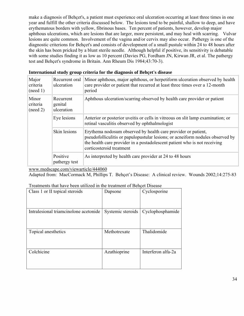
make a diagnosis of Behçet's, a patient must experience oral ulceration occurring at least three times in one year and fulfill the other criteria discussed below. The lesions tend to be painful, shallow to deep, and have erythematous borders with yellow, fibrinous bases. Ten percent of patients, however, develop major aphthous ulcerations, which are lesions that are larger, more persistent, and may heal with scarring. Vulvar lesions are quite common. Involvement of the vagina and/or cervix may also occur. Pathergy is one of the diagnostic criterions for Behçet's and consists of development of a small pustule within 24 to 48 hours after the skin has been pricked by a blunt sterile needle. Although helpful if positive, its sensitivity is debatable with some studies finding it as low as 10 percent (Davies PG, Fordham JN, Kirwan JR, et al. The pathergy test and Behçet's syndrome in Britain. Ann Rheum Dis 1984;43:70-3). International study group criteria for the diagnosis of Behçet's disease Major criteria (need 1)
Recurrent oral ulceration
Minor aphthous, major aphthous, or herpetiform ulceration observed by health care provider or patient that recurred at least three times over a 12-month period
Minor criteria (need 2)
Recurrent genital ulceration
Aphthous ulceration/scarring observed by health care provider or patient
Eye lesions Anterior or posterior uveitis or cells in vitreous on slit lamp examination; or retinal vasculitis observed by ophthalmologist
Skin lesions Erythema nodosum observed by health care provider or patient, pseudofolliculitis or papulopustular lesions; or acneiform nodules observed by the health care provider in a postadolescent patient who is not receiving corticosteroid treatment
Positive pathergy test
As interpreted by health care provider at 24 to 48 hours
www.medscape.com/viewarticle/444060 Adapted from: MacCormack M, Phillips T. Behçet’s Disease: A clinical review. Wounds 2002;14:275-83 Treatments that have been utilized in the treatment of Behçet Disease Class 1 or II topical steroids Dapsone
Cyclosporine
Intralesional triamcinolone acetonide Systemic steroids
Cyclophosphamide
Topical anesthetics Methotrexate
Thalidomide
Colchicine
Azathioprine
Interferon alfa-2a

35
DIFFERENTIAL DIAGNOSIS OF VULVAR EDEMA Swelling can be due to any of these conditions or combinations of inflammation, infiltration and lymphatic disruption or obliteration Inflammatory Edema I Allergic/Immune
1. Allergic Reaction a. Angioedema with or without urticaria b. Allergic Contact Dermatitis
2. Granulomatous Inflammation a. Crohn’s Disease b. Melkersson-Rosenthal Syndrome c. Sarcoidosis
II Infection – edema secondary to local infection 1. Cellulitis – streptococcal 2. Abscess – Bartholin’s duct 3. Candidiasis 4. Rare – Tuberculosis, actinomycosis, Granuloma Inguinale, Amebiasis, Blastomycosis,
Schistosomiasis
III Other 1. Direct Trauma 2. Hidradenitis suppurativa (HS) 3. Amyloidosis 4. Infiltrative neoplasm – inflammatory breast CA
Note – all infections, Crohn’s and HS can cause inflammatory edema the scarring and secondary obstructive lymphedema.
Obstructive Lymphedema I Congenital
1. Milroy’s disease (congenital lymphedema)
2. Lymphangioma
II Infection with secondary lymphatic damage 1. Recurrent cellulitis – streptococcal 2. Lymphogranuloma venereum 3. Filariasis
III Physical lymphatic obstruction with mass, tumor, or destructive process 1. Pregnancy 2. Pelvic or local trauma 3. Pelvic tumor
4. Post-radiation scarring 5. Congestive heart failure
36
IV Metabolic
1. Obesity
2. Renal failure
3. Hepatic failure
Note – Chronic obstructive lymphedema can result in lymphangiectasia/ acquired lymphangiomas. PROTOCOL FOR LYMPHANGIECTASIA AND CHRONIC LYMJPHEDEMA OF THE VULVA
A. First control infection. Usually it is Strep and occasionally Staph.
1. Gently cleanse with Cetaphil or another triclosan-containing antibacterial cleanser morning and night, pat dry. 2. Bleach baths can be very useful in reducing re-infection. Do 2-3 times a week. Add one half cup of household bleach (125 mL) to a 10 inch (25 cm) deep tub of comfortably warm bath water. Mix well. Soak for 5-7 minutes, ensuring penetration of the solution into all cracks and genital / buttock / skin folds, using a plastic cup and bare hands to spread over all involved areas. For sitz bath mix 1 ¼ tsp bleach in 1 gallon of water. 3. Antibiotic ointment (mupirocin ointment twice a day) and if skin is crusty, debride loose matter only. Do not rub. 4. Penicillin VK 500 mg qid for 2-4 weeks and then tid for 2-3 months, bid at least 6 months or more. Any flares, go back to four a day. If patient is doing very well, decrease to one or two a day indefinitely, for the next one or two years. Cephalexin 500 mg with the same dose may also be used.
(For intermittent flares, bump up the dose to 500 mg qid.) B. For the edema:
1. A brief course of prednisone or prednisolone starting at 20-30 mg in the morning for 2-3 weeks and then decreased gradually. Length of use of Prednisone depends on the response. Patients who flare acutely may require 30 mg per day for 1-2 weeks, then 15 mg per day for 1-2 weeks.
Chronic edema may require 20-30 mg per day for 2-3 weeks and a slow stretched-out course over 2-3 months, dropping 2.5 mg every 1-2 weeks.
2. If the edema is very indurated and woody use intralesional triamcinolone acetonide 10 mg/mL (Kenalog 10¨) instead of the oral steroid to soften or get rid of fibrosis.
a. Anesthetize the keratinized skin for one hour with topical EMLA or equivalent under occlusion.
b. If it is somewhat woody / indurated start with 10 mg and if quite woody use up to 40 mg total dose monthly (over large surface area). Inject with a 25-26g needle and use about a 1 cm grid. Inject into the subcutaneous tissue just enough to blanch the area. To soften this chronic lymphedema you can utilize it once a month.
3. Lymphatic massage:
This may be helpful for the vulva and for the lower legs. Some physiotherapists are trained to teach the patient how to do this at home, depending on the complexity of the problem.
37
4. For lymphangiomas that remain open and draining: A. For extensive involvement excisional surgery to debulk the area may be necessary. B. For more localized involvement or those for whom surgery is not and option -
a. Once you have the infection down and controlled then you can safely use the local anesthesia as above – 2.5% lidocaine 2.5% prilocaine in a cream base (EMLA) apply every ½ hour for 1 to 2 hours under occlusion, then local anesthesia 2% lidocaine with epinephrine.
b. To cleanse the area do not use alcohol. c. Electrodesiccate on a high setting and put the needle into each one of the small
lymphangiectatic “fish eggs” and cauterize them until they bubble, turn black and crust. d. Post-operatively: Soak in tepid water 1-2 times a day Mupirocin ointment 2 times a day to involved areas. Keflex 500 mg qid for 2 weeks then chronic penicillin VK Tylenol #2 for pain (acetaminophen and codeine) Loose ventilated clothing. Consider fluconazole suppression. Repeat the destruction when and if needed. ULCERS OF THE VULVA
Ulcers of the vulvar are diagnostically challenging. It is often very difficult to differentiate them from erosions. Erosions involve loss of the epidermis only, not the dermis, and they appear as deep red, often weeping, patches. Ulcers are deeper, extending into the dermis with a white or yellowish fibrinous base. Most diseases produce either erosions or ulcerations but often these overlap. Erosions can be transformed into ulcers by secondary infection, irritant contact dermatitis, rubbing and other trauma. The best example is severe herpes simplex virus (HSV) infection. The primary lesion of HSV is an intraepidermal vesicle that becomes a pustule that ruptures, creating an erosion. When severe, these erosions can ulcerate. An ulcer is characterized by loss of both epidermis and dermis. A diagnosis of a vulvar ulcer based on morphology alone is erroneous 40% of the time. Laboratory testing is usually required.
DIFFERENTIAL DIAGNOSIS In sorting out these conditions, try to identify the primary process. Is it a pustule within the epidermis as in candidiasis or herpes simplex, an intraepidermal vesicle in acute eczema (contact dermatitis), or a frank bulla (intraepidermal or sub-epidermal) as in the bullous diseases or drug eruptions. All these rupture, resulting in erosions and/or ulcerative disease. All of these can look much alike and it can be very difficult to differentiate them clinically, especially if there are secondary changes with crusting and bleeding, etc. A good history is important, as is the understanding that the history may be inaccurate. Many women have problems with discussing the genital area Note the following factors:
38
Age Immune status Epidemiology and demographics of their community Systemic disease Travel and sexual exposure History of abuse Pattern of recurrence Previous sexually
Previous/present treatment transmitted diseases Note the following factors specific for vulvar ulcers: Pain Systemic symptoms Induration Lymphadenopathy Friability Fever Number of lesions (single or multiple) Malaise Acute or chronic Headache Speed of onset Extragenital changes Tests for all ulcers: - HSV culture - CBC - Candida cultures - RPR (syphilis screen)
- HIV screen Serology as indicatedfor Epstein Barr virus (EBV) - antiviral capsid antigen – IgM for EBV and Serology for Mycoplasma Pneumoniae
- Biopsy for H&E +/- immunofluorescence Consider more extensive workup depending on the case, e.g. cultures, smears and serology. Biopsies are very important. Always biopsy the edge of the lesion – not the necrotic center. A wedge excision of the edge often gives the best information for the pathologist but may be impractical. Two smaller punch biopsies may be more appropriate Why biopsy? Because it is impossible to guess the cause of most ulcerative erosive conditions – biopsy gives the most information, especially for chronic ulcers. Although it is an uncomfortable procedure it can be made almost painless. One is adding an extra open area to an already tender area but your patient is already very stressed and wants to know the answer. Most common causes of primary vulvar ulcers (not erosions): INFECTIOUS Venereal Non Venereal Herpes simplex (HSV) EBV Immunosuppressed Mycoplasma pneumoniae Chancroid Granuloma inguinale Lymphogranuloma venereum Syphilis Human immunodeficiency virus NON-INFECTIOUS Tumors Aphthous ulcers Basal cell carcinoma Behçet’s disease Squamous cell carcinoma Crohn’s disease Factorial disease

39
Fissures
The infectious ulcers are classically due to the STIs. The most common cause of genital ulcers in the world is herpes simplex. HSV in any Immunosuppressed patient can present with ulcers. These can be chronic, severe, punched out, and widespread. These are typically seen in a HIV positive individual. The other conditions are syphilis, Chancroid, granuloma inguinale and rarely Lymphogranuloma venereum. These conditions are all quite uncommon in North America. Much more common are the non-infectious ulcers, particularly aphthae, which classically present as punched out, painful ulcers. They are mostly idiopathic but they can be associated with underlying conditions, see below. Aphthous ulcers are also seen in Behçet’s disease, Crohn’s disease and HIV. Crohn’s disease may present with the deep classic “knife-cut” type ulcers. Pyoderma gangrenosum can cause ulcers. Last in this group are the factitial ulcerations. Tumors, classically squamous cell carcinoma, also ulcerate. The limitation to this classification is the possibility of missing the less common conditions that could cause vulvar ulcers and erosions such as drugs, irritant contact dermatitis, secondary infected bullous diseases etc.
2. Etiologic classification vulvar ulcers and erosions:
INFECTIONS
a) Venereal b) Non-venereal Herpes simplex Candida Pseudomonas Chancroid Herpes zoster Histoplasmosis Granuloma Inguinale Varicella Cryptococcosis Lymphogranuloma venereum Hand Foot Mouth disease Tuberculosis Syphilis Staph & Strep, Actinomycosis Human Immunodeficiency Virus Typhoid ¶typhoid, Leishmaniasis Brucellosis Schistosomiasis
Diphtheria, Amebiasis Pseudomonas Epstein-Barr and Mycoplasma
pneumoniae Non-Bullous Dermatoses
Bullous Dermatoses
Premalignant and Malignant Tumors
Infections
Miscellaneous
Irritant contact dermatitis Drug Reaction* Fixed Drug Rxn LE Crohn’s Darier’s Behçet’s Pyo. gangrenosum Hidr. Suppurativa
a) Autoimmune BMM Pemphigoid P. vulgaris Bullous pemphigoid Linear IgA Disease EB Acquisita
Premalignant and Malignant Tumors VIN BCC SCC Extramammary Paget’s Disease
H. zoster Varicella Vaccinia Hand/Foot/Mouth Staph & Strep Typhoid Paratyphoid Brucellosis Diphtheria Pseudomonas Tuberculosis
Rheumatoid nodule Gangrene Acrodermatitis Lymphangiectasis Graft vs. Host Spider bite Hymenal Fissures Reiter’s Disease Wegener’s
40
Necrolytic Migratory Erythema
b) Non-autoimmune TEN / EM Contact Dermatitis Hailey-Hailey EB Inherited
Verrucous Carcinoma Melanoma Lymphoma Leukemia Hodgkins Langerhans cell histiocytosis
Histoplasmosis Actinomycosis Cryptococcosis Leishmaniasis Schistosomiasis Amebiasis
Granulomatosis Factitial Female Genital Mutilation
*12 meds for known to cause a drug reaction Antibiotics Sulfa PCN (not as much trouble as before (no polymers attached) cephalosporins Cardiovascular HCTZ Lasix Beta blockers Ace inihibitors Dilantin Miscellaneous Allopurinol Vaccines New biologicals NSAIDs
Of all this list, the most important causes of ulcers and erosions are, in North America are: Infections Venereal Non Venereal HSV Candida Syphilis EBV HIV M Pneumoniae Dermatoses Bullous Non-Bullous Contact dermatitis Aphthosis LS LP Drug Contact Crohn’s Tumors SCC
41
APHTHAE (aphthous ulcers) Canker sores on the vulva Very common in the mouth and not uncommon on the vulva Acute painful ulcer or ulcers of sudden onset Can be recurrent or chronic Minor or major in size, single or multiple Painful, non-sexually transmitted ulcers in young girls or women are referred to by may terms and there is no consenus on best term. See list below: Ulcus vulvae acutum Lipschütz ulcers Nonsexually acquired genital ulceration (NSAGU) Complex Aphthosis or aphthae Vulvar aphthous ulcers Acute vulvar ulcers Clinical: Average age is 14 (9-19) yrs, but patients can be older Sudden onset Usually multiple, painful, well demarcated punched-out ulcers Size: most <1cm; can be 1-3 cm Prodrome - flu-like with mild fever, headache, malaise There is not always a prodrome especially with recurrent cases in older patients Duration 1-3 weeks, can last months One episode, less common recurrent Often past history of oral aphthae – canker sores Not Behçet’s Associated with oral aphthae – complex aphthae The following associations have been made: Acute (more common) – these can recur Usually with a prodrome - fever, headache, malaise, GI upset These have been reported in the literature associated with:
EBV, Mycoplasma pneumoniae, viral upper respiratory infection (parvovirus, influenza, paramyxovirus) or gastroenteritis, Strep, CMV, Mumps, salmonella, toxoplasma gondii
Chronic or recurrent aphthae: No prodrome. Associations: Bowel disease - Crohn disease, Ulcerative colitis, Celiac disease Infections – HIV Behçet’s disease Medications – cytotoxic, NSAIDs Myeloproliferative disease, cyclic neutropenia, lymphopenia
42
Syndromes with Genital Aphthous Ulcers: rare
Sweet’s syndrome Mouth and Genital Ulcers Inflamed Cartilage - MAGIC Syndrome
Periodic Fever, Aphthae, Pharyngitis, Adenitis - PFAPA Syndrome Note Acute aphthae are probably immune complex related and can be precipitated by infection such as a viral illness. e.g. viral gastroenteritis or upper respiratory tract infection, influenza, CMV. Epstein Barr virus (EBV) could directly infect the skin or cause an immune complex reaction. Mycoplasma pneumoniae can do the same. Streptococcal infection has been found. Most common cause of acute onset aphthae in a 12-20 year old is probably an infection. For recurrent aphthae and complex aphthosis look for inflammatory bowel disease or, less likely, a lymphoproliferative problem. Diagnosis of exclusion Cultures negative, biopsies non-specific and blood work non-contributory Differential diagnosis: - HSV, Syphilis, HIV, Chancroid, LGV, Granuloma Inguinale - pyoderma gangrenosum - trauma - contact dermatitis Evaluation of Vulvar Aphthae: Thorough history and physical – eye, oral, genital Only testing for HSV may be necessary Biopsy rarely needed Lab tests that could be considered– CBC, diff Serology for HSV, HIV, EBV, syphilis, CMV, Mycoplasma pneumoniae Influenza – swab PCR HSV - swab for PCR – always rule out HSV For strep -throat swab and antistreptolysin O titer Tests as indicated for – paratyphoid and typhoid (stool, blood culture), TB enterocolitis, Yersinia GI investigations – for inflammatory bowel disease and celiac disease Note – in HIV + patients with genital ulcers - 60% of genital ulcers
are due to aphthae and 40% to HSV Treatment: depends on the severity. If mild comfort measures may be all that is needed Local therapy AGNO3 sticks Pain control – topical – 5% lidocaine ointment
43
- systemic – mild, moderate pain – NSAID severe - opioids Immunosuppression - Prednisone 40 – 60 mg each morning until pain resolves (3-7 days, then ½ dose 3-7 days) with food Methylprednisolone (Medrol) 4-8mg bid-tid 3-7 days then ½ dose 3-7days) with food Clobetasol or halobetasol 0.05% ointment AM & PM If not sure if HSV use antiviral meds until HSV test report available. Educate -Most often a one-time event, can recur For persistent or chronic aphthae: Oral corticosteroid for initial control - prednisone or methylprednisolone Intralesional triamcinolone (Kenalog 10) 5-10 mg/ml doxycycline 50 - 100 mg od colchicine 0.6 mg bid-tid if tolerated dapsone 50-150 mg per day dapsone + colchicine cyclosporine 100 mg up to tid decreasing to 100 mg 2-3 doses/week pentoxyfylline 400 mg tid thalidomide 100-150 mg per day TNF alpha inhibitors- infliximab, adalimumab Prognosis: Most often a one-time event Scarring can occur Occasionally recurrent Desquamative Inflammatory Vaginitis (DIV) Desquamative inflammatory vaginitis (DIV) is an erosive vulvovaginitis characterized by dyspareunia, and a profuse purulent vaginal discharge. There is significant vaginal cell exfoliation. Numerous parabasal cells are seen in vaginal smears, as well as large numbers of neutrophils (neutrophils/epithelium > 1:1 in at least 4 HPFs on wet smear). The pH is increased (> 4.5). Lactobacilli are decreased or absent, and there is often increased gram positive cocci and gram negative bacilli. When the speculum is inserted, fine red “dots” may be present in the vagina. Vaginal lichen planus can present with this appearance, as can atrophy. Can be seen commonly in Crohn’s disease. Rarely it can be seen with the chronic bullous diseases – cicatricial or classic Pemphigus. Treatment
The treatment varies among providers. Some prefer intravaginal clindamycin, while others prefer intravaginal steroids such as hydrocortisone in 25 mg doses. Some providers combine the clindamycin and hydrocortisone per vagina. Below is a treatment regimen that you might consider:
Clindamycin 2% cream; 1 applicator per vagina, qhs x 14 days as initial therapy

44
If that fails, try using clindamycin 2% per vagina (1 applicator) combined with a 25 mg hydrocortisone suppository per vagina every other night x 14 doses. When the patient does not respond to the above treatments consider:
Hydrocortisone 100 mg/gram in clindamycin 2% emollient cream base Insert 5 gram (applicator full) per vagina q.o.d. (at night) x 14 doses If recurrent, when controlled, decrease to 3 times a week and slowly decrease and stop
HERPES SIMPLEX VIRUS (HSV) (adapted from CDC STD Treatment Guidelines 2010) This is a common sexually transmitted disease worldwide and it is the most common cause of vulvar ulcers. A history of HSV is unreliable. Primary HSV is uncommon. The majority of patients present with non-primary recurrent disease. Etiology: 80% HSV 2 (Genital HSV 1 increasing with more “oral sex” up to 35%) HSV 1 recurs in 25% of cases HSV 2 recurs in 89% of cases Infection is usually from sexual contact. Most transmission occurs during periods of asymptomatic viral shedding. Most persons infected with HSV-2 have not been diagnosed with genital herpes. Many people have mild or unrecognized infections but they shed the virus intermittently in the genital tract. Thus, the majority of genital herpes infections are transmitted by persons unaware that they have the infection or who are asymptomatic when transmission occurs. Symptoms:
Primary HSV - Paresthesia for 2-3 days, followed by fever, malaise, headache and myalgia
There can be pain, moderate to severe (“deep boring pain” reflecting nerve involvement) Recurrent infection - there is more tingling, itching and burning before the onset of vesiculation Physical Examination Can be seen anywhere on the vulva, vagina, over cervix, anus, buttocks and thighs. Primary – red swollen vulva with extensive vesiculation, rapidly becoming pustular with open tender erosions lasting two weeks. Recurrent infection – lesions are less extensive and are clear in 5-7 days with
only mild swelling. Note – 91% of HSV 2 carriers are unaware of their infection yet 80% have symptoms. Women think they have: Vaginitis, GU infection, clothing irritation or hemorrhoids Symptoms can occur with no rash and no blistering in HSV sine eruption – herpes simplex without visible eruption Immunosuppressed HSV – chronic ulcers that gradually extend at the periphery. There may be varying degrees of necrosis. These are painful and indolent. Diagnosis: Cultures can be unreliable. Keep viral media refrigerated, on hand,
45
and up to date. Failure to detect HSV by culture or PCR does not indicate an absence of HSV infection, because viral shedding is intermittent. Scrape the base of an early lesion with 15 blade to get adequate material. The sensitivity of viral culture is low, especially for recurrent lesions, and declines rapidly as lesions begin to heal.
Virologic Tests
Cell culture and PCR are the preferred HSV tests PCR assays for HSV DNA are more sensitive and are increasingly used in many settings Viral culture isolates should be typed to determine which type of HSV is causing the infection. The use of cytologic detection of cellular changes of HSV infection is an insensitive and nonspecific method of diagnosis, both for genital lesions (i.e., Tzanck preparation) and for cervical Pap smears and therefore should not be relied upon.
Type-Specific Serologic Tests
Both laboratory-based assays and point-of-care tests that provide results for HSV-2 antibodies from capillary blood or serum during a clinic visit are available. The sensitivities of these glycoprotein G type-specific tests for the detection of HSV-2 antibody vary from 80%–98%, and false-negative results might be more frequent at early stages of infection. The specificities of these assays are ≥96%. False-positive results can occur, especially in patients with a low likelihood of HSV infection. Repeat or confirmatory testing might be indicated in some settings, especially if recent acquisition of genital herpes is suspected. IgM testing for HSV is not useful Since nearly all HSV-2 infections are sexually acquired, the presence of type-specific HSV-2 antibody implies anogenital infection. The presence of HSV-1 antibody alone is more difficult to interpret. Most persons with HSV-1 antibody have oral HSV infection acquired during childhood, which might be asymptomatic. However, acquisition of genital HSV-1 appears to be increasing, and genital HSV-1 also can be asymptomatic. Lack of symptoms in an HSV-1 seropositive person does not distinguish anogenital from orolabial or cutaneous infection, and regardless of site of infection, these persons remain at risk for acquiring HSV-2. Type-specific HSV serologic assays might be useful in the following scenarios: 1) recurrent genital symptoms or atypical symptoms with negative HSV cultures; 2) a clinical diagnosis of genital herpes without laboratory confirmation; or 3) a partner with genital herpes. HSV serologic testing should be considered for persons presenting for an STD evaluation (especially for those persons with multiple sex partners), persons with HIV infection, and MSM at increased risk for HIV acquisition. Differential diagnosis: Syphilis, chancroid, aphthous ulcers, Herpes zoster, HIV Note – patients with HIV can have vulvar ulcers. 60% are due to aphthous ulcers alone. The other 40% are due to HSV. Always look for multiple or atypical infections in these patients. Non-specific treatment for pain, discomfort etc. R/O other STD’s Treatments for the relief of discomfort The following non-specific treatments can alleviate the pain and discomfort of genital sores.
46
SALT BATHS (1 teaspoon of salt in 600 ml of water or a handful in a shallow bath) can be used to wash, soothe and dry the sores.
PAIN RELIEVERS LOOSE UNDERCLOTHES, preferably cotton (not nylon), can help minimize discomfort and allow
healing. For anyone experiencing extreme pain when urinating, the process can be less painful when done in a cool bath. Encourage plenty of fluids to dilute the urine.
NEW CDC STD TREATMENT GUIDELINES WERE RELEASED IN WINTER 2010
http://www.cdc.gov/std/treatment/2010/
First Clinical Episode of Genital Herpes
The initial genital herpes episode can last for more than 20 days. Symptoms include tingling, itching, burning or pain. Patients may experience a range of generalized symptoms, such as fever, aches and pains, swollen lymph nodes, as well as specific vulvar symptoms. For others, the initial infection can be mild with minimal symptoms. They may be totally unaware that they have had a herpes outbreak. The severity of symptoms for genital herpes varies in the population. The initial episode can be so mild as to pass unnoticed and a first recurrence may take place many years after the first infection.
Many patients with primary herpes present with mild clinical manifestations but later develop severe or prolonged symptoms. Newly acquired genital herpes can cause a prolonged clinical illness with severe genital ulcerations and neurologic involvement. Even persons with first-episode herpes who have mild clinical manifestations initially can develop severe or prolonged symptoms. Therefore, all patients with first episodes of genital herpes should receive antiviral therapy.
First Clinical Episode of Genital Herpes Recommended Regimens
Acyclovir 400 mg orally three times a day for 7-10 days, OR Acyclovir 200 mg orally five times a day for 7 -10 days, OR Famciclovir 250 mg orally three times a day for 7-10 days, OR Valacyclovir 1 g orally twice a day for 7-10 days.
NOTE: Treatment may be extended if healing is incomplete after 10 days of therapy.
Recurrent Episodes of HSV Disease Most patients with symptomatic, first-episode genital HSV-2 infection subsequently experience recurrent episodes of genital lesions; recurrences are much less frequent following initial genital HSV-1 infection. Antiviral therapy for recurrent genital herpes can be administered either continuously as suppressive therapy to reduce the frequency of recurrences or episodically, to ameliorate or shorten the duration of lesions.
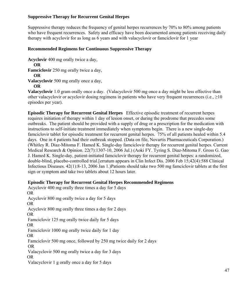
47
Suppressive Therapy for Recurrent Genital Herpes
Suppressive therapy reduces the frequency of genital herpes recurrences by 70% to 80% among patients who have frequent recurrences. Safety and efficacy have been documented among patients receiving daily therapy with acyclovir for as long as 6 years and with valacyclovir or famciclovir for 1 year
Recommended Regimens for Continuous Suppressive Therapy
Acyclovir 400 mg orally twice a day, OR Famciclovir 250 mg orally twice a day, OR Valacyclovir 500 mg orally once a day, OR Valacyclovir 1.0 gram orally once a day. (Valacyclovir 500 mg once a day might be less effective than other valacyclovir or acyclovir dosing regimens in patients who have very frequent recurrences (i.e., ≥10 episodes per year).
Episodic Therapy for Recurrent Genital Herpes Effective episodic treatment of recurrent herpes requires initiation of therapy within 1 day of lesion onset, or during the prodrome that precedes some outbreaks. The patient should be provided with a supply of drug or a prescription for the medication with instructions to self-initiate treatment immediately when symptoms begin. There is a new single-day famciclovir tablet for episodic treatment for recurrent genital herpes. 75% of all patients healed within 5.4 days. One in 4 patients had their outbreak stopped. (Data on file, Novartis Pharmaceuticals Corporation.) (Whitley R. Diaz-Mitoma F. Hamed K. Single-day famciclovir therapy for recurrent genital herpes. Current Medical Research & Opinion. 22(7):1307-10, 2006 Jul.) (Aoki FY. Tyring S. Diaz-Mitoma F. Gross G. Gao J. Hamed K. Single-day, patient-initiated famciclovir therapy for recurrent genital herpes: a randomized, double-blind, placebo-controlled trial.[erratum appears in Clin Infect Dis. 2006 Feb 15;42(4):588 Clinical Infectious Diseases. 42(1):8-13, 2006 Jan 1.)Patients should take two 500 mg famciclovir tablets at the first sign or symptom and take two tablets about 12 hours later.
Episodic Therapy for Recurrent Genital Herpes Recommended Regimens Acyclovir 400 mg orally three times a day for 5 days OR Acyclovir 800 mg orally twice a day for 5 days OR Acyclovir 800 mg orally three times a day for 2 days OR Famciclovir 125 mg orally twice daily for 5 days OR Famciclovir 1000 mg orally twice daily for 1 day OR Famciclovir 500 mg once, followed by 250 mg twice daily for 2 days OR Valacyclovir 500 mg orally twice a day for 3 days OR Valacyclovir 1 g orally once a day for 5 days

48
Few comparative studies of valacyclovir or famciclovir with acyclovir have been conducted. The results of these studies suggest that valacyclovir and famciclovir are comparable to acyclovir in clinical outcome. Ease of administration and cost also are important considerations for prolonged treatment.
Severe Disease Intravenous acyclovir therapy should be provided for patients who have severe disease or complications that necessitate hospitalization, such as disseminated infection, pneumonitis, hepatitis, or complications of the central nervous system (e.g., meningitis or encephalitis).
Treatment Immunosuppressed Patients With HSV Topical imiquimod 5% cream Usual treatment: acyclovir, famciclovir, valacyclovir foscarnet cidofovir
imiquimod 5% cream- 3 times a week for 2-8 weeks
Resources for Herpes
-American Social Health Association www.ashastd.org (patient information)
-International Herpes Alliance www.herpesalliance.com
-International Herpes Management Forum www.ihmf.org (geared to health care providers) Molluscum contagiosum Molluscum contagiosum is caused by a DNA poxvirus. The disease is more prevalent in children (lesions involve the face, trunk and extremities). Adults tend to have lesions most often near the genital areas. The incidence of molluscum has increased over the last 30 years. There are four main subtypes of molluscum contagiosum virus (MCV), MCV I, MCV II, MCV III and MCV IV. The disease is transmitted by direct skin contact. It presents clinically with a papular eruption of multiple umbilicated lesions. The central depression contains a white waxy curd-like core. The size of the papule ranges from 2-6 mm. The clinical appearance of molluscum contagiosum is the general diagnostic method, though it can be examined histologically (curetted or biopsied lesion). Large brick shaped inclusion bodies are seen. In-situ hybridization for MCV DNA has also been performed. Treatment of molluscum contagiosum Molluscum contagiosum is a self-limited disease, which will generally resolve in immunocompetent hosts. However, the time to resolution can be quite long. Treatment of molluscum contagiosum is advisable in healthy individuals to prevent autoinoculation or transmission.
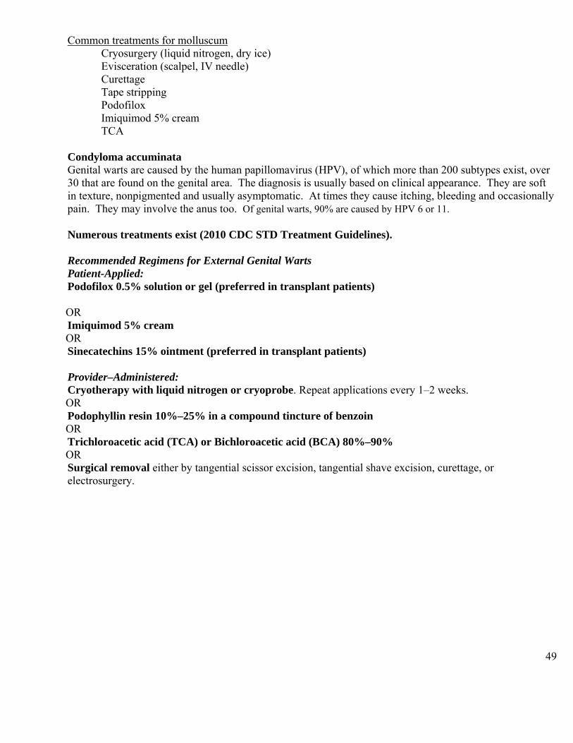
49
Common treatments for molluscum Cryosurgery (liquid nitrogen, dry ice) Evisceration (scalpel, IV needle) Curettage Tape stripping Podofilox Imiquimod 5% cream TCA
Condyloma accuminata Genital warts are caused by the human papillomavirus (HPV), of which more than 200 subtypes exist, over 30 that are found on the genital area. The diagnosis is usually based on clinical appearance. They are soft in texture, nonpigmented and usually asymptomatic. At times they cause itching, bleeding and occasionally pain. They may involve the anus too. Of genital warts, 90% are caused by HPV 6 or 11. Numerous treatments exist (2010 CDC STD Treatment Guidelines). Recommended Regimens for External Genital Warts Patient-Applied: Podofilox 0.5% solution or gel (preferred in transplant patients) OR Imiquimod 5% cream OR Sinecatechins 15% ointment (preferred in transplant patients) Provider–Administered: Cryotherapy with liquid nitrogen or cryoprobe. Repeat applications every 1–2 weeks. OR Podophyllin resin 10%–25% in a compound tincture of benzoin OR Trichloroacetic acid (TCA) or Bichloroacetic acid (BCA) 80%–90% OR Surgical removal either by tangential scissor excision, tangential shave excision, curettage, or electrosurgery.
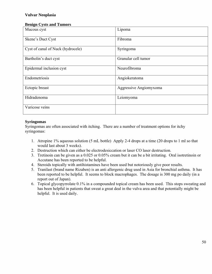
50
Vulvar Neoplasia Benign Cysts and Tumors Mucous cyst Lipoma
Skene’s Duct Cyst
Fibroma
Cyst of canal of Nuck (hydrocele)
Syringoma
Bartholin’s duct cyst
Granular cell tumor
Epidermal inclusion cyst
Neurofibroma
Endometriosis
Angiokeratoma
Ectopic breast
Aggressive Angiomyxoma
Hidradenoma
Leiomyoma
Varicose veins
Syringomas Syringomas are often associated with itching. There are a number of treatment options for itchy syringomas:
1. Atropine 1% aqueous solution (5 mL bottle) Apply 2-4 drops at a time (20 drops to 1 ml so that would last about 3 weeks).
2. Destruction which can either be electrodesiccation or laser CO laser destruction. 3. Tretinoin can be given as a 0.025 or 0.05% cream but it can be a bit irritating. Oral isotretinoin or
Accutane has been reported to be helpful. 4. Steroids topically with antihistamines have been used but notoriously give poor results. 5. Tranilast (brand name Rizaben) is an anti allergenic drug used in Asia for bronchial asthma. It has
been reported to be helpful. It seems to block macrophages. The dosage is 300 mg po daily (in a report out of Japan).
6. Topical glycopyrrolate 0.1% in a compounded topical cream has been used. This stops sweating and has been helpful in patients that sweat a great deal in the vulva area and that potentially might be helpful. It is used daily.
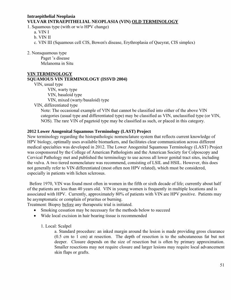
51
Intraepithelial Neoplasia VULVAR INTRAEPITHELIAL NEOPLASIA (VIN) OLD TERMINOLOGY 1. Squamous type (with or w/o HPV change)
a. VIN I b. VIN II
c. VIN III (Squamous cell CIS, Bowen's disease, Erythroplasia of Queyrat, CIS simplex) 2. Nonsquamous type
Paget ’s disease Melanoma in Situ
VIN TERMINOLOGY SQUAMOUS VIN TERMINOLOGY (ISSVD 2004)
VIN, usual type VIN, warty type VIN, basaloid type VIN, mixed (warty/basaloid) type
VIN, differentiated type Note: The occasional example of VIN that cannot be classified into either of the above VIN categories (usual type and differentiated type) may be classified as VIN, unclassified type (or VIN, NOS). The rare VIN of pagetoid type may be classified as such, or placed in this category.
2012 Lower Anogenital Squamous Terminology (LAST) Project New terminology regarding the histopathologic nomenclature system that reflects current knowledge of HPV biology, optimally uses available biomarkers, and facilitates clear communication across different medical specialties was developed in 2012. The Lower Anogenital Squamous Terminology (LAST) Project was cosponsored by the College of American Pathologists and the American Society for Colposcopy and Cervical Pathology met and published the terminology to use across all lower genital tract sites, including the vulva. A two tiered nomenclature was recommend, consisting of LSIL and HSIL. However, this does not generally refer to VIN differentiated (most often non HPV related), which must be considered, especially in patients with lichen sclerosus. Before 1970, VIN was found most often in women in the fifth or sixth decade of life; currently about half of the patients are less than 40 years old. VIN in young women is frequently in multiple locations and is associated with HPV. Currently, approximately 80% of patients with VIN are HPV positive. Patients may be asymptomatic or complain of pruritus or burning. Treatment: Biopsy before any therapeutic trial is initiated.
Smoking cessation may be necessary for the methods below to succeed Wide local excision in hair bearing tissue is recommended
1. Local: Scalpel
a. Standard procedure: an inked margin around the lesion is made providing gross clearance (0.5 cm to 1 cm) at resection. The depth of resection is to the subcutaneous fat but not deeper. Closure depends on the size of resection but is often by primary approximation. Smaller resections may not require closure and larger lesions may require local advancement skin flaps or grafts.
52
Special points: Vulvar skin thickness varies considerably by location. Particular care must be taken in the clitoral, urethral, anal and labia minora locations as the squamous epithelium is very thin. Resections in this area don’t require deep dissection and every effort should be made to minimize trauma.
2. CO2 laser- in non hair bearing areas Confidence that no invasive disease exists is important to patient selection.
3. LEEP: difficult to control depth of dissection 4. Medical
Imiquimod (Aldara®) has reported to be effective for VIN 3 (same dosage as for condyloma)(Off label use)
It is important to screen these patients with Anal Pap smears. Use a moistened Dacron swab or Cytobrush. Insert into the canal approximately 5-6 cm above the anal verge to the rectum. Rotate, applying pressure to the walls of the canal while removing the sampling device.
Anal Cytology Place the cytology sampling device (Dacron swab or Cytobrush moistened with water) into the anal canal until resistance is met (approximately 4 cm) Rotate/apply pressure to walls of canal while removing sampling device slowly (count to 10) Place in liquid media. Notify Pathology (Cytology) Department before you start these, so that they are prepared for them. HPV testing is not required on these specimens. Anal colposcopy After the cytology has been obtained, place an anoscope (use a clear plastic anoscope) with a small amount of lubricant into the anus. Then place an opened 4 x 4 soaked in 3 to 5% acetic acid over a cotton swab through the anoscope. Remove the anoscope, leaving in the 4 x 4 and cotton swab. Place a 4 x 4 with 3 to 5% acetic acid around the outer anus. Leave these on for about 3 minutes. Then, remove the 4 x 4’s and cotton swab and reinsert a lubricated anoscope. The anus is visualized sequentially, with a colposcope, keeping in mind the location of the dentate line.
Paget’s Disease of the Vulva Primary extramammary Paget’s Disease – an epidermotropic carcinoma arising within the epidermis or epidermal appendages (may arise in Toker cells) – no underlying carcinoma (most common) Secondary extramammary Paget’s disease – is a visceral carcinoma (anorectal, bladder or urethra) that is epidermotropic to the skin. Clinical Presentation:
Itching “rash” on perineum with eczematous, soft velvety papules slowly growing into crusty scaly plaques that do not respond to topical steroid Paget's disease of the skin is generally confined to the integument along the mid line. It occurs most commonly on the nipple and areola, where its presence signifies an underlying adenocarcinoma of the
53
breast. Extramammary lesions have been described in the genital, perianal, and axillary regions as well as the ear canal, all of which contain abundant apocrine glands. Vulvar Paget's disease appears as a red velvety area with white islands of hyperkeratosis and at times may be pinkish and eczematoid. It primarily occurs on the labia majora. Pruritus is present in over half of the patients. The mean age for Paget’s disease of the vulva is 65 years. Almost all of the patients are Caucasian. Signs
Red and white vulva - ulceration and hyperkeratosis Well demarcated Eczematoid
Symptoms Pruritus in over 50% Soreness Bleeding or discharge
When present on the vulva, it is most commonly an intraepithelial disease that tends to recur locally and has a minimal propensity to invade. Usually it is a slowly progressive, indolent, superficial process. It is rarely associated with an underlying skin appendage carcinoma such as a primary carcinoma of the rectum, urethra, or bladder Only about 25% of vulvar Extramammary Paget’s are associated with an underlying adenocarcinoma of an adnexal tissue or a Bartholin gland. Less commonly it is associated with a distant carcinoma of breast, GI, GU or the genital tract. Perianal Extramammary Paget’s is associated with underlying colorectal adenocarcinoma in 80% of cases. In view of the possible coexistence of sweat gland carcinoma of the vulva or another adjacent internal carcinoma, the overall prognosis for Paget's disease is less favorable than for VIN III. Clinical diagnosis based on gross appearance may be erroneous. Biopsy confirmation of the diagnosis is mandatory. Large, irregular Paget's cells containing clear, vacuolated pale cytoplasm are seen on histologic evaluation. Nuclei are vesicular. Mitotic figures are uncommon. Paget cells are most numerous in the tips and sides of the rete pegs and deep in the epithelium. They may be scattered throughout the outer keratinized layer. Paget cells, as well as the cells and secretions of normal eccrine and apocrine glands are rich in CEA. Markers The immunoprofile of vulvar Paget's disease includes cells that are typically positive for cytokeratin 7, keratin CAM5.2, EMA, CEA and GCDFP; mucin stains are also positive in a subset of the neoplastic cells (less cost). Work up to detect associated adenocarcinoma (location dependent)
H+P Pap Mammogram Hemoccult Cystoscopy Flex sigmoidoscopy vs BE vs colonoscopy
54
Treatment Paget’s disease of the vulva is generally treated with a wide local excision of the circumscribed lesions. It is important to remove the full thickness of the skin to the subcutaneous fat to be certain that all of the skin adnexal structures are excised. Even if resection margins are free of Paget's disease at the time of surgical excision, local recurrence remains a major risk. Laser therapy has been used on Paget’s disease (particularly recurrent Paget’s). On rare occasions, radiation therapy has been used to treat Paget’s disease. 5% imiquimod cream 1 to 5 times a week (frequency of application depends on tolerance) has been used for superficial involvement and when surgery would be poorly tolerated. Duration of treatment depends on response and this can be months. Atypical junctional melanocytic hyperplasia This is a preinvasive condition. If margins are not clear, a repeat resection should be performed. Melanoma in situ Clear margins should be obtained. Malignant Tumors Vulvar Cancer Most vulvar cancers are found in patients age 60 to 70 years. The risk for vulvar cancer continues to increase with age. The diagnosis is often delayed (mean = 1 year). It is usually unifocal. Most vulvar cancers are squamous cell carcinomas. Squamous carcinoma –87% Melanoma-6% Bartholin's Adenocarcinoma-4% Basal Cell carcinoma <2% Sarcoma <2%
55
2009 FIGO Staging System for Vulvar Cancer Carcinoma of the vulva. Stage I Tumor confined to the vulva
IA Lesions ≤2 cm in size, confined to the vulva or perineum and with stromal invasion ≤1.0 mm, no nodal metastasis
IB Lesions >2 cm in size or with stromal invasion >1.0 mm, confined to the Vulva or perineum, with negative nodes
Stage II Tumor of any size with extension to adjacent perineal structures (1/3 lower
urethra, 1/3 lower vagina, anus) with negative nodes Stage III Tumor of any size with or without extension to adjacent perineal structures
(lower urethra, lower vagina, anus) with positive inguino-femoral lymph nodes
IIIA (i) With 1 lymph node metastasis (≥5 mm), or (ii) 1–2 lymph node metastasis(es) (<5 mm)
IIIB (i) With 2 or more lymph node metastases (≥5 mm), or (ii) 3 or more lymph node metastases (<5 mm)
IIIC With positive nodes with extracapsular spread Stage IV Tumor invades other regional (2/3 upper urethra, 2/3 upper vagina) bladder mucosa, rectal mucosa), or distant structures
IVA Tumor invades any of the following: (i) upper urethral and/or upper vaginal mucosa, bladder mucosa, rectal mucosa; or fixed to pelvic bone or (ii) fixed or ulcerated inguino-femoral lymph nodes
IVB Any distant metastasis including pelvic lymph nodes a. The depth of invasion is defined as the measurement of the tumor from the epitheliostromal
junction of the adjacent most superficial dermal papilla to the deepest point of invasion.
Melanoma Clinical Features Lesions suspicious for melanoma are often characterized by the ABCDs. They are asymmetrical (A), have irregular or scalloped border (B), often black in color (C) or variegate with shades of red, white, or blue, and may have a diameter (D) greater than 6 millimeters. However, a melanoma with all or many of these characteristics is often more advanced. For melanoma, the earliest signs are change in size, shape, and color of a lesion. The earliest symptom is persistent pruritus in a lesion. Late features include bleeding, ulceration, pain, and tenderness. The most common presenting signs/symptoms for vulvar melanoma are bleeding, mass, discharge, pruritus, burning, pain, ulceration, foul odor, local discoloration, and dysuria. Vulvar melanoma is usually detected later than cutaneous melanoma simply due to location, resulting in more advanced lesions at presentation with a poorer prognosis. Melanoma may occur in any vulvar location
56
and has been reported as multifocal in up to 20% of cases, arising in the scattered background of malignant and premalignant melanocytic cells. Biopsy The first step for evaluating any lesion suspicious for melanoma is biopsy. Excisional biopsy is optimal, with narrow margins of 1-2 mm to provide the pathologist a complete specimen for accurate diagnosis and microstaging. Wide excision for diagnosis is not recommended in the event that sentinel lymph node biopsy (SLNB) is indicated. Sentinel node biopsy may not be as accurately performed following a wide local excision. The biopsy may be performed as an elliptical excision, punch, or saucerization (a modified shave technique extending to the adipose tissue. A superficial shave biopsy is never recommended due to the risk of transecting the lesion, which precludes accurate measurement of tumor thickness, the most important factor that determines treatment. If the lesion is too large or too close to vital structures to excise completely, an incisional biopsy may be performed. Several studies have documented that an incisional biopsy for melanoma does not increase the risk of tumor seeding, metastasis, or decrease survival. If the incisional biopsy reveals a melanocytic lesion with melanoma still in the differential diagnosis, complete excision of the lesion is still indicated due to possibility of biopsy sampling error. The biopsy should be interpreted by a pathologist with experience in the interpretation of pigmented lesions and melanoma. Results from large melanoma centers show that up to 20% of patients referred to a specialized melanoma center have significant alterations in diagnosis and/or microstaging parameters following review by a pigmented lesion pathology expert. The two most important factors in the cutaneous melanoma pathology report are tumor thickness measured in millimeters (Breslow depth) and ulceration status. Other potentially important factors include mitotic rate, microsatellitosis, angiolymphatic involvement, Clark level (anatomic measure of thickness), neurotropism, and extensive regression. Additional factors routinely documented in the pathology report include a gross and microscopic description of the specimen, histologic pattern subtype, tumor infiltrating lymphocytes, growth phase, presence of preexisting nevus, age, gender, anatomic site, and margin involvement. The most common histologic pattern subtype reported for vulvar melanoma is acral mucosal lentiginous; for cutaneous melanoma superficial spreading is the most common. The histologic subtype, in general, does not correlate with prognosis or survival after correction for tumor thickness, ulceration, and other prognostic parameters. Balch M., Buzaid AC, Soong SJ, Atkins MB, Cascinelli N, Coit DG, et. al. Final Version of the American Joint Committee on Cancer Staging System for Cutaneous Melanoma. J Clin Oncol 2001;19:3635-48.
Summary
An overview of the different types of vulvovaginal conditions has been given. Many vulvar conditions must be considered when a patient complains of discharge and itching. It is important to remember that
If TREATMENT IS NOT WORKING, RECONSIDER THE DIAGNOSIS.

57
Prescriptions for Vulvar Diseases
Pain Medications Xylocaine® (lidocaine)
5% Xylocaine® (lidocaine) ointment sig: apply to vulva prn Disp: 35 grams
Elavil® (amitriptyline) Start low and increase dose slowly. Initial amitriptyline prescription:
amitriptyline 10-25 mg Sig: 1 po qhs x 1 week; If sxs persist, 2 po qhs x 1 wk, if sxs persist, 3 po qhs x 1 wk; if sxs persist, 4 po qhs. Maintain nightly dose that relieves symptoms (Generally not to exceed 4 po qhs) Do not stop suddenly
Start at 10 mg in patients age 60 or older; increase by 10 mg weekly Future amitriptyline prescriptions
Amitriptyline ____mg Sig: i po qhs (comes in 10 mg, 25 mg, 50 mg, 75 mg, 100 mg and 150 mg tablets)
Neurontin®
Neurontin® (gabapentin) Sig: 300 mg po qd x 3 days; if sxs persist, 300 mg po bid x 3 days; if symptoms persist, 300 mg po tid. Stay on this dose for a month and increase gradually if needed. It comes in 100, 300, 400, 600 and 800 mg doses Do not exceed 2700 to 3600 mg total dose per day Do not give more than 1200 mg in a single dose
Gabapentin ointment 3% or 6% Sig: apply to affected area bid-tid Disp: 3 month supply
Lyrica® Lyrica® (pregabalin)
-50 mg po qd x 4 days, if sxs persist, 50 mg po bid x 4 days, if sxs persist, 50 mg po tid -Can gradually increase up to 100 mg po tid; doses up to 300 mg po bid have been used for pain control
Blocks
Bupivacaine ( 0.25% or 0.5%) and Kenalog® Draw up Kenalog® first (40 mg /cc) (can use up to 40 mg steroid in single dose per month) Combine with Bupivacaine (large area use 0.25%; small area use 0.5%) Inject into specific area or use as a pudendal block Can be repeated monthly
58
Do not use high doses on thin skin. Medications for localized pain or itching
Zonalon® (doxepin) 5 % cream Sig: apply to skin q d with gradual increase not to exceed qid Disp: 30 g
Topical Elavil® (amitriptyline) 2% with Baclofen 2% in water washable base (WWB)- squirt ½ cc from syringe onto finger and apply to affected area qd to tid Disp: 30 day supply Gabapentin 6% with Ketamine 5% WWB – 30mL apply ½-1 mL to Vulvar Vestibule twice daily for pain Amitriptyline 2% with Baclofen 2% WWB and Lidocaine 5% mg – 30mL Apply ½-1 mL to Vulvar Vestibule twice daily for pain Estradiol 0.1mg with Lidocaine 5% ointment – Disp 30g Apply thin layer over Vulva twice daily
for pain
Yeast medications Fluconazole 150 mg Sig: 1 po q 3 days x 3, then 1 po q week for up to 6 months (If using for longer than 6 months, check LFT’s) Do not use with active liver disease.
5 flucystosine 500 mg/5 grams compounded in a hydrophilic cream base -Insert 5 gram applicator (500 mg of active drug) full of mixture per vagina qhs x 14 days
Boric acid- fill 0-gel capsule halfway (600 mg) To treat active yeast infection - Insert per vagina nightly for 14 days For prevention of yeast - Insert per vagina twice weekly. Keep out of reach of children. Warn patients not to receive oral sex while on the boric acid. There is an herbal product called Yeast Arrest. It contains 600 mg boric acid, Oregon Grape Root and Calendula flowers. Gentian violet- 0.25% or 0.5% aqueous solution applied at home daily or it may be given in the office as a 1.0% solution (once weekly for up to three times). Warn patients that if they have oral sex, their partner’s teeth and lips could stain.
Medications for Lichen planus Anusol HC suppository 1/2 of a 25 mg suppository per vagina bid x 2 months Decrease to qd x 2 months Maintenance therapy of 1 - 3 x per week Hydrocortisone acetate 100 mg compounded suppository used QHS
59
Use for 2-4 weeks then use Mon Wed Fri for 2-4 weeks and change to milder 25 mg suppository as needed
Hydrocortisone acetate 10% compounded Vaginal cream used QHS – 4-5 gram q d (400 to 500 mg dose). For severe vaginal Lichen Planus
Use for 2-4 weeks then use Mon Wed Fri for 2-4 weeks and change to milder 25 mg suppository as needed
Tacrolimus For oral Lichen planus: Tacrolimus 0.1% in Orabase Sig: apply to mouth bid Disp 50 g
For vaginal lichen planus Tacrolimus vaginal suppositories Insert one supp per vagina (2 mg tacrolimus per 2 gm supp) qhs Disp 50
For vulvar Lichen planus Tacrolimus 0.1% Ointment Sig: apply to skin bid Available in 30 or 60 gram tubes
Folliculitis (swab for culture to r/o MRSA) Emgel® 2% topical gel (erythromycin) or 1% clindamycin lotion Sig: apply to skin bid Available in 27 or 50 gram bottles Other topical antibiotics include bacitracin, neomycin, mupirocin For fungal folliculitis Topical clotrimazole, miconazole
Oral terbinafine, itraconazole, griseofulvin
Furunculosis- very responsive to antibotics (swab for culture to r/o MRSA) Topical antibiotics (bacitracin, neomycin, mupirocin)
Oral antibiotics (dicloxacillin, cephalexin) Dial soap or Phisohex If wrinkled, I and D useful
For Recurrent Impetigo Staphlococcus +/- Steptococcus Take bacterial culture from site of infection, nose and gluteal cleft to find any hidden source of infection. Do bleach baths to reducing re-infection 2-3 times a week. Add one half cup of household bleach (125 mL) to a 10 inch (25 cm) deep tub of comfortably warm bath water. Mix well. Soak for 5-7 minutes, ensuring penetration of the solution into all cracks and genital / buttock / skin folds, using a plastic cup and bare hands to spread over all involved areas. For sitz bath mix 1 ¼ tsp bleach in 1 gallon of water. Treat with oral antibiotics as indicated by culture results. Use an antibiotic ointment (mupirocin ointment twice a day) bid for nose or gluteal cleft. If MRSA use retapamulin 1% ointment (Altabax) bid for 5 days. Desquamative inflammatory vaginitis Can utilize clindamycin 2% per vagina qhs x 14 days as initial therapy If that fails, try using clindamycin 2% per vagina combined with a 25 mg Anusol HC suppository per vagina every other night x 14 doses.

60
Another treatment for DIV that is used when the patient does not respond to the above treatments is: Hydrocortisone 100 mg/gram in clindamycin 2% emollient cream base Insert 5 gram (applicator full) per vagina q.o.d. (at night) x 14 doses
Steroid medications Clobetasol propionate ointment (Temovate®) 0.05% Sig: apply to vulva bid x 1 month, then qd x 2 months Disp: 30 g
Triamcinolone acetonide ointment 0.1% Sig: apply to vulva qd to bid Disp: 80 g Consider decreasing gradually to Triamcinolone acetonide ointment 0.025% qd to bid
TOPICAL CORTICOSTEROIDS Learn three to four ointments of different strengths, making appropriate selections as needed
- ointments are stronger than creams - ointments stay on longer than creams (creams are diluted and washed
away with body fluids) - ointments are less irritating and have fewer allergens than other bases
Patients may find one base more irritating than another. Be flexible. Do not use steroids for dysesthetic vulvodynia - steroids work by reducing inflammation, not pain
Note: Topical steroids are not a cure. Use the steroid potency that will do the job in the quickest period of time and then decrease to a lower potency. Either stop or maintain with the lowest potency or use intermittently as necessary. Tips: When considering topical corticosteroids, especially the superpotent types, consider:
There are more available than you need Use them in an educated way Limit the amount prescribed to 15g to 30 grams for high dose topical steroids Show the patient exactly how to use it – a tiny dab spread in a thin film just to the involved area is all that is necessary
Vulvar mucous membrane (vulvar trigone and inner labia minora) is remarkably steroid resistant. The outside of the labia minora and the labiocrural fold and the thighs will thin easily and develop striae. When the patient improves, decrease the frequency of topical steroid or manage with a low potency product.
Use under close supervision. At any suggestion of secondary yeast infection, add a topical or oral anti- fungal.
For example, for thick itchy dermatoses like lichen simplex chronicus – use name brand clobetasol or halobetasol 0.05% ointment bid for 1-2 weeks, once a week for 1-2 weeks and then M-W-F for 1-2 weeks and for long term maintenance either infrequent and intermittent usage each week of the same or switch to intermittent use of a mild ointment such as l% -2.5% hydrocortisone in petrolatum or a 1% hydrocortisone / 1% pramoxine cream mix. Effects of corticosteroids:
Vasoconstriction – decrease erythema and swelling Decreasing fibroblastic proliferation thins out thickened dermal lesions
61
Decreasing rapidly turning over keratinocytes thins out thickened epidermal lesions
Corticosteroid responsive vulvar dermatoses include: Thick and scaly (lichen sclerosus, lichen simplex chronicus, psoriasis, contact dermatitis)
Blistering erosive disease Bullous diseases Corticosteroid potency depends on:
Cortisone molecule Concentration of steroid in vehicle Partition co-efficient of steroid vehicle system Application frequency and length of time used Caution: steroids can be associated with irregular menses, increased BP, worsening of diabetes control, infection and glaucoma.
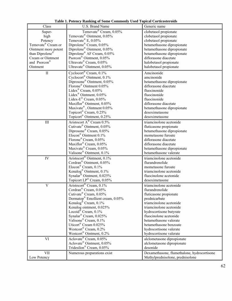
62
Table 1. Potency Ranking of Some Commonly Used Topical Corticosteroids
Class U.S. Brand Name Generic name
Super- high
Potency Temovate® Cream or Ointment more potent than Diprolene® Cream or Ointment and Psorcon® Ointment
Temovate® Cream, 0.05% Temovate® Ointment, 0.05% Temovate® E, 0.05% Diprolene® Cream, 0.05% Diprolene® Ointment, 0.05% Diprolene® AF Cream, 0.05% Psorcon® Ointment, 0.05% Ultravate® Cream, 0.05% Ultravate® Ointment, 0.05%
clobetasol propionate clobetasol propionate clobetasol propionate betamethasone dipropionate betamethasone dipropionate betamethasone dipropionate diflorasone diacetate halobetasol propionate halobetasol propionate
II Cyclocort® Cream, 0.1% Cyclocort® Ointment, 0.1% Diprosone® Ointment, 0.05% Florone® Ointment 0.05% Lidex® Cream, 0.05% Lidex® Ointment, 0.05% Lidex-E® Cream, 0.05% Maxiflor® Ointment, 0.05% Maxivate® , Ointment 0.05% Topicort® Cream, 0.25% Topicort® Ointment, 0.25%
Amcinonide amcinonide betamethasone dipropionate diflorasone diacetate fluocinonide fluocinonide fluocinonide diflorasone diacetate betamethasone dipropionate desoximetasone desoximetasone
III Aristocort A® Cream 0.5% Cutivate® Ointment, 0.05% Diprosone® Cream, 0.05% Elocon® Ointment 0.1% Florone® Cream, 0.05% Maxiflor® Cream, 0.05% Maxivate® Cream, 0.05% Valisone® Ointment, 0.1%
triamcinolone acetonide fluticasone propionate betamethasone dipropionate mometasone furoate diflorasone diacetate diflorasone diacetate betamethasone dipropionate betamethasone valerate
IV Aristocort® Ointment, 0.1% Cordran® Ointment, 0.05% Elocon® Cream, 0.1% Kenalog® Ointment, 0.1% Synalar® Ointment, 0.025% Topicort LP® Cream, 0.05%
triamcinolone acetonide flurandrenolide mometasone furoate triamcinolone acetonide fluocinolone acetonide desoximetasone
V Aristocort® Cream, 0.1% Cordran® Cream, 0.05% Cutivate® Cream, 0.05% Dermatop® Emollient cream, 0.05% Kenalog® Cream, 0.1% Kenalog ointment, 0.025% Locoid® Cream, 0.1% Synalar® Cream, 0.025% Valisone® Cream, 0.1% Uticort® Cream 0.025% Westcort® Cream, 0.2% Westcort® Ointment, 0.2%
triamcinolone acetonide flurandrenolide fluticasone propionate prednicarbate triamcinolone acetonide triamcinolone acetonide hydrocortisone butyrate fluocinolone acetonide betamethasone valerate betamethasone benzoate hydrocortisone valerate hydrocortisone valerate
VI Aclovate® Cream, 0.05% Aclovate® Ointment, 0.05% Tridesilon® Cream, 0.05%
alclometasone dipropionate alclometasone dipropionate desonide
VII Low Potency
Numerous preparations exist Dexamethasone, flumethalone, hydrocortisone Methylprednisolone, prednisolone
63
ALTERNATIVES TO CORTICOSTEROIDS Alternative topicals to corticosteroids are the Calcineurin inhibitors Calcineurin inhibitors: Pimecrolimus 1% cream (Elidel) Tacrolimus 0.03 and 0.l% ointment (Protopic) or compounded 0.l% vaginal cream or a 2g suppository. These are non-steroidal Does not cause atrophy May sting or burn initially when used topically Equivalent to mild to moderate topical steroids –Pimecrolimus to a mild topical steroid and tacrolimus equivalent to a moderate to strong topical steroid. These are topical immunosuppressants usually for maintenance of steroid responsive dermatoses Note: there is a black box warning on these medications. This is because of reports of skin cancers and lymphoma with systemic Calcineurin inhibitors used in organ transplant patients. This warning was also imposed because of one manufacturer’s failure to conduct safety studies. Note : Skin application results in minimal systemic exposure. Vaginal use can result in systemic absorption. Side effects of Calcineurin inhibitors: Burn, sting Infection – worsening of HSV, HPV, tinea, molluscum contagiosum Safety with regard to lichen sclerosus and squamous cell carcinoma? There are a number of studies showing good results with this medication in lichen sclerosus in adults and children. There are three reports of genital squamous cell carcinoma with patients who have used tacrolimus and one with squamous cell carcinoma on pimecrolimus. Treatment of choice for lichen sclerosus is still superpotent topical steroids For lichen planus that is difficult to treat with only partial control of topical steroids consider using tacrolimus and pimecrolimus. The response reported is between 55 and 94%. Summary of Calcineurin inhibitors: For lichen planus start with topical steroids and consider alternating with Calcineurin inhibitors. For lichen sclerosus with atrophy or reaction to topical steroids, consider usage, discuss the risks and follow carefully. No refills without follow-up vulvar exams. Consider for use in the following: vulvar dermatoses, psoriasis, Crohn’s, pemphigoid, etc. Systemic corticosteroids can be useful at times. A full discussion is beyond this lecture. IM triamcinolone acetonide (Kenalog 40) l mg per kg for an acute dermatosis (e.g. contact dermatitis or severe lichen simplex chronicus). This can be repeated in 3-4 weeks once or twice to get a severe condition under control. See appropriate monograph for all side effects of all corticosteroids and calcineurin inhibitors.
64
Caution in patients with diabetes- high dose steroids can interfere with their glucose control. TO DO FOR ALL VULVAR RASHES Educate
Support Stop: irritation, contact dermatitis, scratching Treat: infection – Candida, bacteria, atrophy, and inflammation Poor response: biopsy
CAUSES OF TREATMENT FAILURE
Non-compliance Poor education Fear of topical steroids Limited mobility
INCORRECT DIAGNOSIS
Associated problems
LS plus SCC or contact dermatitis Scarring
MOST COMMON ASYMPTOMATIC VULVAR DISEASES
Lichen sclerosus, Lichen planus, Malignancy – compounded by Ignorance Denial CAUSES OF POOR COMPLIANCE Fear of steroids Vulvar ignorance Miscommunication Physical impairment Secondary gain – no sex Phobic about touching “down there”
65
List of Lubricants This does not attempt to be a complete list, but rather describes commonly used lubricants. We do not officially recommend use of any one of these products, nor do we recommend any one product over any other products.
Slippery Stuff a silken gel that does not leave a sticky residue. It is hygienic, water-based and water-soluble, odorless, long lasting and latex compatible.
Astroglide: A long lasting, light lubrication that is odorless and flavorless. It is water soluble. Many like it
because it is a long lasting lubricant that does not become "stringy
Femigel Natural product from tea trees. For vaginal dryness.
K-Y Jelly: Generally considered an all-purpose lubricant that many people have found helpful with a
"medium" degree of thickness. Some report it comes out too fast and gets "gummy."
Lubrin: A suppository. Many post-menopausal women find this a helpful lubricant because, since it is
inserted into the vagina, it lasts longer. They indicate that it needs some time to melt inside the vagina because it is a suppository. For some women, they indicate that it is almost "too much" lubrication.
Moist Again Natural
Replens: A lubricant that is inserted by applicator into the vagina. It comes in a package of 12 single-use
applications. This vaginal gel is considered to have medium thickness and properties similar to Ortho Personal Lubricant. Women note that, like Lubrin, it does not dissolve too quickly. Must be used several times weekly.
Sylk: Made of Kiwi fruit vine and purified water. From New Zealand. Marketed through Whole Foods.
Mimics natural secretions.
Surgilube: Many consider this to be thicker than K-Y Jelly
Alboline - Most drug stores sell it in the cosmetic section. Is actually intended to remove make up and provide moisture to a the face.
Vitamin E oil: Available in health food stores, preferred by some women for natural, non-irritating qualities.
Vegetable oil (like olive oil) can also be used.
Egg whites have been used for lubrication.
Saliva has been used for lubrication
66
Pre-Seed is a vaginal lubricant that does not appear to cause significant damage to sperm
Agents for sexual enhancement Viafem – Aminophylline 30mg/mL 15mL Apply ½-1 mL to clitoris before intercourse Ergoloid Mesylates 0.5 mg/mL Nitroglyerine 1mg/mL L-Arginine 60mg/mL Pentoxyphylline 50mg/mL Trimix FM – Papaverine 30mg/mL 5mL Apply 0.5mL to clitoris one hour before intercourse Phentolamine 1mg/mL PGE1 20mcg/mL Testosterone 0.5mg/mL 30mL Apply 1/2mL to labia and 1/2mL to inner arm or Thigh q AM.
67
References General
Anemuller W, Recke A, Altgassen C, Kelling K. Developing an interdisciplinary consultation service for
vulvar disorders. Journal der Deutschen Dermatologischen Gesellschaft. 2012;10(5):350-7. Anonymous. ACOG Practice Bulletin No. 93: diagnosis and management of vulvar skin disorders. Obstetrics & Gynecology. 111(5):1243-53, 2008. Black MM, Ambrose-Rudolph C, Edwards L, Lynch P. Obstetric and Gynecologic Dermatology. 3nd Edition Mosby Inc. 2008. Bornstein J, Sideri M, Tatti S, Walker P, Prendiville W, Haefner HK. Nomenclature Committee of International Federation for Cervical Pathology and Colposcopy. 2011 terminology of the vulva of the International Federation for Cervical Pathology and Colposcopy. Journal of Lower Genital Tract Disease. 2012;16(3):290-5. Crum CP, Nucci MR, Lee KR. Diagnostic Gynecologic and Obstetric Pathology. Elsevier 2011. Dei M, Di Maggio F, Di Paolo G, Bruni V. Vulvovaginitis in childhood. Best Practice & Research in Clinical Obstetrics & Gynaecology. 2010;24(2):129-37. Edwards L. Genital Dermatology Atlas. Second Edition Lippincott Williams & Wilkins, 2011. Fisher BK, Margesson LJ. Genital Skin Disorders. Diagnosis and Treatment. Mosby, 1998. Foster DC. Vulvar disease. Obstetrics & Gynecology 2002;100:145-63. Fox H, Wells M. Recent advances in the pathology of the vulva. Histopathology 2003;42:209-16. Kaufman RH, Faro S, Brown D. Benign Diseases of the Vulva and Vagina, 5th ed, Mosby, 2005. Leibovitz A, Kaplun V, Saposhnicov N, Habot B. Vulvovaginal examinations in elderly nursing home women residents. Archives of Gerontology and Gerieatrics 2000;31:1-4. Margesson LJ. Vulvar Disease Pearls. Dermatol Clin 2006;24:145-55. Mayeaux EJ, and Cox JT. Modern Colposcopy Textbook and Atlas, 3nd edition. 2011 ASCCP www.asccp.org
Rodriguez MI, Leclair CM. Benign vulvar dermatoses. Obstetrical & Gynecological Survey.
2012;67(1):55-63.
Rome ES. Vulvovaginitis and other common vulvar disorders in children. Endocrine Development.
2012;22:72-83.
68
Spratt EA, Batra P, Fischer MK, Pomeranz MK. Vesicular lesions on the vulva. Archives of Dermatology.
2012;148(6):755-60. Summers PR, Hunn J. Unique dermatologic aspects of the postmenopausal vulva. Clinical Obstetrics and Gynecology. 2007;50:745-51. Nonneoplastic Epithelial Conditions/Lichen sclerosus Assmann T, Becker-Wegerich P, Grewe M et al. Tacrolimus ointment for the treatment of vulvar lichen sclerosus. Journal of the American Acadamy of Dermatology 2003;48:935-7. Baldo M, Bailey A, Bhogal B, Groves RW, Ogg G, Wojnarowska FT cells reactive with the NC16A domain of BP180 are present in vulval lichen sclerosus and lichen planus. Journal of the European Academy of Dermatology & Venereology. 2010;24(2):186-90. Baldo M, Bhogal B, Groves RW, Powell J, Wojnarowska F. Childhood vulval lichen sclerosus: autoimmunity to the basement membrane zone protein BP180 and its relationship to autoimmunity. Clin Exp Dermatol. 2010 Apr 26. Berger J, Telser A, Widschwendter M, Muller-Holzner E, Daxenbichler G, Marth C, Zeimet AG. Expression of retinoic acid receptors in non-neoplastic epithelial disorders of the vulva and normal vulvar skin. International Journal of Gynecologic Pathology 2000;19:95-102. Boyd AS. New and emerging therapies for lichenoid dermatoses. Dermatologic Clinics. 2000;18:21-9. Bracco GL, Carli P, Sonni L, Maestrini G, De Marco A, Taddei GL, et al. Clinical and histologic effects of topical treatments of vulval lichen sclerosus: A critical evaluation. Journal of Reproductive Medicine 1993;38:37-40. Bradford J, Fischer G. Long-term management of vulval lichen sclerosus in adult women. Aust N Z J Obstet Gynaecol. 2010;50(2):148-52. Burrows LJ, Creasey A, Goldstein AT. The treatment of vulvar lichen sclerosus and female sexual dysfunction. The Journal of Sexual Medicine. 2011;8(1): 219-22. Chi CC, Kirtschig G, Baldo M. Lewis F, Wang SH, Wojnarowska F. Systematic review and meta-analysis of randomized controlled trials on topical interventions for genital lichen sclerosus. Journal of the American Academy of Dermatology 2012;67(2):305-12. Chiesa-Vottero A, Dvoretsky PM, Hart WR. Histopathologic study of thin vulvar squamous cell carcinomas and associated cutaneous lesions: a correlative study of 48 tumors in 44 patients with analysis of adjacent vulvar intraepithelial neoplasia types and lichen sclerosus. American Journal of Surgical Pathology. 2006;30(3):310-8.
69
Cooper SM, Haefner HK, Abrahams-Gessel S, Margesson LJ. Vulvovaginal lichen planus treatment: a survey of current practices. Archives of Dermatology. 2008;144(11):1520-1. D'Antuono A, Bellavista S, Negosanti F, Zauli S, Baldi E, Patrizi A. Dermasilk briefs in vulvar lichen sclerosus: An adjuvant tool. Journal of Lower Genital Tract Disease. 2011;15(4): 287-91. Dalziel KL, Millard PR, Wojnarowska F. The treatment of vulval lichen sclerosus with a very potent topical steroid (clobetasol propionate 0.05%) cream. Br J Dermatol 1991;124:461-4. Dalziel KL, Wojnarowska F. Long-term control of vulval lichen sclerosus after treatment with a potent topical steroid cream. Journal of Reproductive Medicine 1993;39:25-7. Dalziel K, Shaw S. Lichen sclerosus. BMJ. 2010;340:c731. Edmonds E, Mavin S, Francis N, Ho-Yen D, Bunker C. Borrelia burgdorferi is not associated with genital lichen sclerosus in men. British Journal of Dermatology. 2009;160(2):459-60. Edwards L. Vulvovaginal Dermatology In: Dermatologic Clinics Consulting Editor Bruce H. Thiers, MD 2010;28(4) pp.639-833. www.derm.theclinics.com Engin B, Tufekci O, Yazici A, Ozdemir M. The effect of transcutaneous electrical nerve stimulation in the treatment of lichen simplex: a prospective study. Clinical & Experimental Dermatology. 2009;34(3):324-8. Farber EM, Nall L. Genital psoriasis. Cutis 1992;50:263-6. Fite C, Plantier F, Dupin N, Avril MF, Moyal-Barracco M. Vulvar verruciform xanthoma: Ten cases associated with lichen sclerosus, lichen planus, or other conditions. Archives of Dermatology. 2011;147(9): 1087-92. Goldstein AT, Metz A. Vulvar lichen planus. Clinical Obstetrics and Gynecology 2005;48;818-23. Goldstein AT, Creasey A, Pfau R, Phillips D, Burrows LJ. A double-blind, randomized controlled trial of clobetasol versus pimecrolimus in patients with vulvar lichen sclerosus. Journal of the American Academy of Dermatology. 2011;64(6): e99-104. Guerrero D, Guarch R, Ojer A, Casas JM, Mendez-Meca C, Esteller M, et al. Differential hypermethylation of genes in vulvar cancer and lichen sclerosus coexisting or not with vulvar cancer. International Journal of Cancer.Journal International Du Cancer. 2011;128(12):2853-64. Gutierrez-Pascual M, Vicente-Martin FJ,. Lopez-Estebaranz JL. Lichen sclerosus and squamous cell carcinoma. Actas Dermo-Sifiliograficas. 2012;103(1):21-8. Gunthert AR, Faber M. Knappe G. Hellriegel S. Emons G. Early onset vulvar Lichen Sclerosus in premenopausal women and oral contraceptives. European Journal of Obstetrics, Gynecology, & Reproductive Biology. 2008;137(1):56-60.
70
Gurumurthy M. Morah N, Gioffre G, Cruickshank ME. The surgical management of complications of vulval lichen sclerosus. European Journal of Obstetrics, Gynecology, & Reproductive Biology. 2012;162(1):79-82. Haverhoek E, Reid C, Gordon L, Marshman G, Wood J, Selva-Nayagam P. Prospective study of patch testing in patients with vulval pruritus. Australasian Journal of Dermatology. 2008;49(2):80-5. Higgins CA, Cruickshank ME. A population-based case-control study of aetiological factors associated with vulval lichen sclerosus. Journal of Obstetrics & Gynaecology. 2012;32(3):271-5. Jensen LS, Bygum A. Childhood lichen sclerosus is a rare but important diagnosis. Danish Medical Journal. 2012;59(5):A4424. Jones RW, Scurry J, Neill S, MacLean AB. Guidelines for the follow-up of women with vulvar lichen sclerosus in specialist clinics. American Journal of Obstetrics & Gynecology. 2009;198(5):496.e1-3. Kazandi M, Sahin C, Terek MC, Cirpan T, Oztekin K. Clinical evaluation of vulvar lichen sclerosus: case series. Eur J Gynaecol Oncol 2010;31(5):555-8.
Kroft J, Shier M. A novel approach to the surgical management of clitoral phimosis. Journal of Obstetrics & Gynaecology Canada: JOGC. 2012;34(5):465-71. LeFevre C, Hoffstetter S, Meyer S, Gavard J. Management of lichen sclerosus with triamcinolone ointment: Effectiveness in reduction of patient symptom scores. Journal of Lower Genital Tract Disease. 2011;15(3):205-9. Lynch PJ. 2006 International Society for the Study of Vulvovaginal Disease Classification of Vulvar Dermatoses: A Synopsis. J Low Gen Tract Dis 2007;11:1-2. Maronn ML, Esterly NB. Constipation as a feature of anogenital lichen sclerosus in children. Pediatrics. 2005;115(2):230-2. Neill SM, Tatnall FM, Cox NH. British Association of Dermatologists. Guidelines for the management of lichen sclerosus. British Journal of Dermatology. 2002;147:640-9. O'Connell TX, Nathan LS, Satmary WA, Goldstein AT. Non-neoplastic epithelial disorders of the vulva. American Family Physician. 2008;77(3):321-6. Patrizi A, Gurioli C, Medri M, Neri I. Childhood lichen sclerosus: a long-term follow-up. Pediatric Dermatology. 2010;27(1):101-3. Regauer S, Liegl B, Reich O. Early vulvar lichen sclerosus: a histopathological challenge. Histopathology. 2005;47(4):340-7. Regauer S. Residual anogenital lichen sclerosus after cancer surgery has a high risk for recurrence: A clinicopathological study of 75 women. Gynecologic Oncology. 2011;123(2):289-94.

71
Ridley CM. Genital lichen sclerosus (lichen sclerosus et atrophicus) in childhood and adolescence. Journal of the Royal Society of Medicine 1993;86:68. Rodriguez MI, Leclair CM. Benign vulvar dermatoses. Obstetrical & Gynecological Survey. 2012;67(1):55-63. Rosamilia LL, Schwartz JL, Lowe L, Gruber SB, Quint EH, Johnson TM, Reynolds RK, Haefner HK. Vulvar melanoma in a 10-year-old girl in association with lichen sclerosus. Journal of the American Academy of Dermatology. 2006;54(2 Suppl):S52-3. Ruan L, Xie Z, Wang H, Jiang J, Shi H, Xu J. High-intensity focused ultrasound treatment for non-neoplastic epithelial disorders of the vulva. International Journal of Gynaecology & Obstetrics. 2010;109(2):167-70. Sadalla JC, Lourenco SV, Sotto MN, Baracat EC, Carvalho JP. Claudin and p53 expression in vulvar lichen sclerosus and squamous-cell carcinoma. Journal of Clinical Pathology. 2011;64(10):853-7. Sadowska-Przytocka A, Danczak-Pazdrowska A, Szewczyk A, Czarnecka-Operacz M, Jenerowicz D, Osmola-Mankowska A, Olek-Hrab K. Treatment of genital lichen sclerosus in women--review. Ginekologia Polska. 2012;83(6):458-61.
Saunders H, Buchanan JA, Cooper S, Hollowood K, Sherman V, Wojnarowska F. The period prevalence of oral lichen planus in a cohort of patients with vulvar lichen sclerosus. Journal of the European Academy of Dermatology & Venereology. 2010;24(1):18-21. Saunders NA, Haefner HK. Vulvar lichen sclerosus in the elderly: pathophysiology and treatment update. Drugs & Aging. 2009;26(10):803-12. Sideri M et al. topical testosterone in the treatment of vulvar lichen sclerosus. International Journal of Gynaecology & Obstetrics 1994;46:53. Smith SD, Fischer G. Paediatric vulval lichen sclerosus. Australasian Journal of Dermatology. 2009;50(4):243-8. Smith YR, Haefner HK. Vulvar lichen sclerosus : pathophysiology and treatment. American Journal of Clinical Dermatology. 2004;5(2):105-25. Taube JM, Badger J, Kong CS, Dadras SS. Differentiated (simplex) vulvar intraepithelial neoplasia: A case report and review of the literature. The American Journal of Dermatopathology. 2011;33(3):e27-30. Tausch TJ, Peterson AC. Early aggressive treatment of lichen sclerosus may prevent disease progression. Journal of Urology. 2012;187(6):2101-5. Thorstensen KA, Birenbaum DL. Recognition and management of vulvar dermatologic conditions: lichen sclerosus, lichen planus, and lichen simplex chronicus. Journal of Midwifery & Women's Health. 2012;57(3):260-75.
72
Utas S, Ferahbas A, Yildiz S. Patients with vulval pruritus: patch test results. Contact Dermatitis. 2008;58(5):296-8. van der Avoort IA, Tiemes DE, van Rossum MM, van der Vleuten CJ, Massuger LF, de Hullu JA. Lichen sclerosus: treatment and follow-up at the departments of gynaecology and dermatology. Journal of Lower Genital Tract Disease. 2010;14(2):118-23. van de Nieuwenhof HP, Bulten J, Hollema H, Dommerholt RG, Massuger LF, van der Zee AG, et al. Differentiated vulvar intraepithelial neoplasia is often found in lesions, previously diagnosed as lichen sclerosus, which have progressed to vulvar squamous cell carcinoma. Modern Pathology : An Official Journal of the United States and Canadian Academy of Pathology, Inc 2011;24(2):297-305. Zamirska A, Reich A, Berny-Moreno J, Salomon J, Szepietowski JC. Vulvar pruritus and burning sensation in women with psoriasis. Acta Dermato-Venereologica.2008; 88(2):132-5. Lichen Planus
Bradford J, Fischer G. Management of Vulvovaginal Lichen Planus: A New Approach. Journal of Lower Genital Tract Disease. 2013;17(1):28-32.
Bradford J, Fischer G. Surgical Division of Labial Adhesions in Vulvar Lichen Sclerosus and Lichen Planus. Journal of Lower Genital Tract Disease. 2013;17(1):48-50. Chiu TL, Jones RW. Multifocal multicentric squamous cell carcinomas arising in vulvovaginal lichen planus. Journal of Lower Genital Tract Disease 2011;15(3):246-7. Cooper SM, Wojnarowska F. Influence of treatment of erosive lichen planus of the vulva on its prognosis. Archives of Dermatology 2006;142(3):289-94. Edwards L, Friedrich EG Jr. Desquamative vaginitis: Lichen planus in disguise. Obstetrics & Gynecology 1988;71:832. Edwards L. Vulvar lichen planus. Archives of Dermatology 1989;125:1677. Eisen D et al. Effect of topical cyclosporine rinse on oral lichen planus. A double-blind analysis. New England Journal of Medicine 1990;323:290. Eisen D. Hydroxychloroquine sulfate (Plaquenil) improves oral lichen planus: an open trial. Journal of the American Academy of Dermatology 1993;28:609. Eisen D. The vulvo-vaginal-gingival syndrome. The clinical characteristics of 22 patients. Archives of Dermatology 1994;130:1379. Fite C, Plantier F, Dupin N, Avril MF, Moyal-Barracco M. Vulvar verruciform xanthoma: Ten cases associated with lichen sclerosus, lichen planus, or other conditions. Archives of Dermatology 2011;147(9):1087-92.
73
Genadry R, Provost TT. Severe vulvar scarring in patients with erosive lichen planus: a report of 4 cases. Journal of Reproductive Medicine 2006;51(1):67-72. Helgesen AL, Gjersvik P, Jebsen P, Kirschner R, Tanbo T. Vaginal involvement in genital erosive lichen planus. Acta Obstetricia et Gynecologica Scandinavica. 2010;89(7):966-70. Lear JT, English JS. Erosive and generalized lichen planus responsive to azathioprine. Clinical and Experimental Dermatology 1996;21:56. Lonsdale-Eccles AA, Velangi S. Topical pimecrolimus in the treatment of genital lichen planus: a prospective case series. British Journal of Dermatology. 2005;153(2):390-4. Lotery HE, Galask RP. Erosive lichen planus of the vulva and vagina. Obstetrics & Gynecology 2003;101:1121-5. Panagiotopoulou N, Wong CS, Winter-Roach B. Vulvovaginal-gingival syndrome. J Obstet Gynaecol 2010 Apr;30(3):226-30.
Petruzzi M, De Benedittis M, Carriero C, Giardina C, Parisi G, Serpico R. Oro-vaginal-vulvar lichen planus: report of two new cases. Maturitas. 2005;50(2):140-50. Santegoets LA, Helmerhorst TJ, van der Meijden WI. A retrospective study of 95 women with a clinical diagnosis of genital lichen planus. J Low Genit Tract Dis 2010 Oct;14(4):323-8.
Simpson RC, Littlewood SM, Cooper SM, Cruickshank ME, Green CM, Derrick E,Yell J, Chiang N, Bell H, Owen C, Javed A, Wilson CL, McLelland J, Murphy R. Real-life experience of managing vulval erosive lichen planus: a case-based review and U.K. multicentre case note audit. British Journal of Dermatology. 2012;167(1):85-91.
Stany MP, Winter WE 3rd, Elkas JC, Rose GS. The use of acellular dermal graft for vulvovaginal reconstruction in a patient with lichen planus. Obstetrics & Gynecology. 2005;105(5 Pt 2):1268-71.
SuzukiV, Haefner HK, Piper CK, O’Gara C, Reed BD. Postoperative Sexual Concerns and Functioning in Patients Who Underwent Lysis of Vulvovaginal Adhesions. Journal of Lower Genital Tract Disease.
Webber NK. Setterfield JF. Lewis FM. Neill SM. Lacrimal canalicular duct scarring in patients with lichen planus. Archives of Dermatology. 2012;148(2):224-7.
Zaki I et al. The under-reporting of skin disease in association with squamous cell carcinoma of the vulva. Clinical and Experimental Dermatology 1996;21:334. Crohn’s Disease Andreani SM, Ratnasingham K, Dang HH, Gravante G, Giordano P. Crohn's disease of the vulva. International Journal Of Surgery. 2010;8(1):2-5.
74
Clemett D, Markham A. Prolonged-release mesalazine: a review of its therapeutic potential in ulcerative colitis and Crohn's disease. Drugs. 2000;59:929-56.
de la Poza G, Lopez-Sanroman A, Taxonera C, Marin-Jimenez I, Gisbert JP, Bermejo F, Opio V, Muriel A.
Genital fistulas in female Crohn's disease patients.: clinical characteristics and response to therapy.
Journal of Crohn's & colitis. 2012;6(3):276-80.
Hanauer SB. Updating the approach to Crohn's disease. Hospital Practice (Office Edition). 1999;34:77-8, 81-3, 87-93; discussion 94. Hoffmann JC, Zeitz M. Treatment of Crohn's disease. Hepato-Gastroenterology. 2000;47:90-100. Khaled A, Ezzine-Sebai N, Fazaa B, Zeglaoui F, Zermani R, & Kamoun MR. Chronic linear ulcerations of the inguino-crural and buttocks folds. Indian Journal of Dermatology 2011;56(1):101-3. Madnani NA, Desai D, Gandhi N, Khan KJ. Isolated crohn's disease of the vulva. Indian Journal of Dermatology, Venereology and Leprology 2011;77(3):342-4. McKinney A, Wallace JA, Alderdice JM. Crohn's disease of the labia minora. Ulster Medical Journal. 1995;64:92-4. Present DH, Rutgeerts P, Targan S, Hanauer SB, Mayer L, van Hogezand RA, Podolsky DK, Sands BE, Braakman T, DeWoody KL, Schaible TF, van Deventer SJ. Infliximab for the treatment of fistulas in patients with Crohn's disease. New England Journal of Medicine. 1999;340:1398-405. Preston PW, Hudson N, Lewis FM. Treatment of vulval Crohn's disease with infliximab. Clinical & Experimental Dermatology. 2006;31(3):378-80. Raza A. Anti-TNF therapies in rheumatoid arthritis, Crohn's disease, sepsis, and myelodysplastic syndromes. Microscopy Research & Technique. 2000;50:229-35. Regimbeau JM, Panis Y, Marteau P, Benoist S, Valleur P. Surgical treatment of anoperineal Crohn's disease: can abdominoperineal resection be predicted?. Journal of the American College of Surgeons. 1999;189:171-6. Rutgeerts P, Baert F. Immunosuppressive drugs in the treatment of Crohn's disease. European Journal of Surgery. 1998;164:911-5. Sandborn W, Sutherland L, Pearson D, May G, Modigliani R, Prantera C. Azathioprine or 6-mercaptopurine for inducing remission of Crohn's disease. Cochrane Database of Systematic Reviews [computer file]. (2):CD000545, 2000. 20257647. Steinhart AH, Ewe K, Griffiths AM, Modigliani R, Thomsen OO. Corticosteroids for maintaining remission of Crohn's disease. Cochrane Database of Systematic Reviews [computer file]. (2):CD000301, 2000. 20257615.

75
Urbanek M, Neill SM, McKee PH. Vulval Crohn's disease: difficulties in diagnosis. Clinical & Experimental Dermatology. 1996;21:211-4. Vettraino IM, Merritt DF. Crohn's disease of the vulva. American Journal of Dermatopathology. 1995;17:410-3. Hidradenitis suppurativa Hidradenitis Suppurativa. Ed. G. B. Jemec, J. Revuz, and J. J. Leyden. 1st ed. Berlin: Springer, 2006. Balik E., et al. "Surgical approach to extensive hidradenitis suppurativa in the perineal/perianal and gluteal regions." World J.Surg. 2009;33.3: 481-7. Chen E, Friedman HI.Management of regional hidradenitis suppurativa with vacuum-assisted closure and split thickness skin grafts. Annals of Plastic Surgery. 2011;67(4):397-440. Danby FW. Commentary: Unroofing for hidradenitis suppurativa, why and how. J Am Acad Derm 2010;63:481.et1-3. Danby FW, Margesson LJ. Hidradenitis suppurativa. Dermatol Clin 2010 Oct;28(4):779-93. Highton L, Chan WY, Khwaja N, Laitung JK. Treatment of hidradenitis suppurativa with intense pulsed light: A prospective study. Plastic and Reconstructive Surgery 2011;128(2):459-65. Jemec GB. "Hidradenitis suppurativa." J Cutan.Med.Surg. 2003;7.1: 47-56. Jemec GB "Medical treatment of hidradenitis suppurativa." Expert.Opin.Pharmacother. 2004;5.8 : 1767-70. Kurzen H, et al. "What causes hidradenitis suppurativa?" Exp.Dermatol. 2008;17.5: 455-6. Revuz J. "Hidradenitis Suppurativa." Dec. 2, 2006.Ed. A Tosti. 2004. http://www.orpha.net/data/patho/GB/uk-hidradenitis-suppurativa.pdf. Rhode JM, Burke WM, Cederna PS, Haefner HK. Outcomes of surgical management of stage III vulvar hidradenitis suppurativa. Journal of Reproductive Medicine.2008; 53(6):420-8. Sehgal VN, Verma P, Sawant S, Paul M. Contemporary surgical treatment of hidradenitis suppurativa (HS) with a focus on the use of the diode hair laser in a case. Journal of Cosmetic and Laser Therapy : Official Publication of the European Society for Laser Dermatology 2011;13(4):180-90. Sellheyer K, Krahl D. "What causes acne inversa (or hidradenitis suppurativa)?--the debate continues." J.Cutan.Pathol. 2008;35.8: 795-97. Shah N. "Hidradenitis suppurativa: a treatment challenge." Am Fam.Physician 2005;72.8: 1547-52. Simpson RC, Dyer MJ, Entwisle J, Harman KE. Positron emission tomography features of hidradenitis suppurativa. The British Journal of Radiology 2011;84(1004):e164-5.
76
Stewart EG, Marjesson LJ, Danby FW. Hidradenitis suppurativa. Uptodate.com 2008 Unal C, Yirmibesoglu OA, Ozdemir J, Hasdemir M. Superior and inferior gluteal artery perforator flaps in reconstruction of gluteal and perianal/perineal hidradenitis suppurativa lesions. Microsurgery 2011;31(7):539-44. Van der Zee HH, Prens EP, Boer J. Deroofing: A tissue-saving surgical technique for the treatment of mild to moderate hidradenitis suppurativa lesions. J Am Acad Dermatol 2010;63:475-80. Wiseman MC. "Hidradenitis suppurativa: a review." Dermatol Ther. 2004;17.1: 50-4. Wollina U, Tilp M, Meseg A, Schonlebe J, Heinig B, Nowak A. Management of severe anogenital acne inversa (hidradenitis suppurativa). Dermatologic Surgery. 2012;38(1):110-7. Yazdanyar S, Boer J, Ingvarsson G, Szepietowski JC, Jemec GB. Dapsone therapy for hidradenitis suppurativa: A series of 24 patients. Dermatology (Basel, Switzerland) 2011;222(4):342-6. Preliminary findings suggest hidradenitis suppurativa may be due to defective follicular support. Danby FW, Jemec GB, Marsch WCh, von Laffert M. Br J Dermatol. 2013 May;168(5):1034-9.
Pathogenesis and pharmacotherapy of Hidradenitis suppurativa. Nazary M, van der Zee HH, Prens EP J.Eur J Pharmacol. 2011 Dec 15;672(1-3):1-8.
Hidradenitis suppurativa: viewpoint on clinical phenotyping, pathogenesis and novel treatments. van der Zee HH, Laman JD, Boer J, Prens EP. Exp Dermatol. 2012 Oct;21(10):735-9.
Deroofing: A tissue-saving surgical technique for the treatment of mild to moderate hidradenitis suppurativa lesions. Van der Zee HH, Prens EP, Boer J. J Am Acad Dermatol 2010;63:475-80. Commentary: Unroofing for hidradenitis suppurativa, why and how. Danby FW. J Am Acad Derm 2010;63:481.e1-3.
Metformin in dermatology: an overview. Badr D, Kurban M, Abbas O. J Eur Acad Dermatol Venereol. 2013 Feb 26.1-8 Finasteride for the Treatment of Hidradenitis Suppurativa in Children and Adolescents. Randhawa HK, Hamilton J, Pope E. JAMA Dermatol. 2013 Mar 20:1-4. Acne inversa (Hidradenitis suppurativa): A review with a focus on pathogenesis and treatment. Wollina U, Koch A, Heinig B, Kittner T, Nowak A. Indian Dermatol Online J. 2013 Jan;4(1):2-11 Pharmacologic interventions for hidradenitis suppurativa: what does the evidence say? Alhusayen R, Shear NH. Am J Clin Dermatol. 2012 Oct 1;13:283-91.

77
Systemic therapy with immunosuppressive agents and retinoids in hidradenitis suppurativa: a systematic review. Blok JL, van Hattem S, Jonkman MF, Horváth B. Br J Dermatol. 2013 Feb;168(2):243-52. Contact Dermatitis Allergic contact dermatitis of the vulva. O'Gorman SM, Torgerson RR. Dermatitis. 2013 Mar-Apr;24(2):64-72 Contact sensitization in the anal and genital area. Bauer A, Oehme S, Geier J. Curr Probl Dermatol. 2011;40:133-41. Lymphedema Fadare O, Brannan, SM, Arin-Silasi D, Parkash V. Localized lymphedema of the vulva: A clinicopathologic study of 2 cases and a review of the literature. International Journal of Gynecological Pathology : Official Journal of the International Society of Gynecological Pathologists 2011;30(3):306-13. Harwood CA, Mortimer PS. Causes and clinical manifestations of lymphatic failure. Clin.Dermatol. 1995;13.5: 459-71. Lu S et al. "Localized lymphedema (elephantiasis): a case series and review of the literature." J.Cutan.Pathol. 2009;36.1: 1-20. Szuba A, Rockson SG. Lymphedema: classification, diagnosis and therapy. Vasc.Med. 1998;3.2: 145-56. Talwar A, Puri N, Sandhu HP. Vulval lymphoedema following pulmonary tuberculosis. Int.J.STD AIDS 2009'20.6: 437-9. Ulcers (including aphthous ulcers) Dixit S, Bradford J, Fischer G. Management of non-sexually acquired genital ulceration using oral and topical corticosteroids followed by doxycycline prophylaxis. J Am Acad Dermatol 2013;68:797-802. Esteve-Martinez A, Lopez-Davia J, Garcia-Rabasco A, Febrer-Bosch I, Miquel VA. Non-sexually transmitted acute ulcer of the vulva associated with influenza A virus infection. [Ulcera vulvar aguda de transmision no sexual asociada a infeccion por virus influenza A] Actas Dermo-Sifiliograficas 2011;102(1):63-4.
78
Farhi D, Wendling J, Molinari E, Raynal J, Carcelain G, Morand P, Avril MF, Francès C, Rozenberg F, Pelisse M, Dupin N. Non-sexually related acute genital ulcers in 13 pubertal girls: a clinical and microbiological study. Arch Dermatol. 2009 Jan;145(1):38-45. Huppert JS, Gerber MA, Deitch HR, Mortensen JE, Staat MA, Adams Hillard PJ. Vulvar ulcers in young females: a manifestation of aphthosis. J Pediatr Adolesc Gynecol. 2006 Jun;19(3):195-204 Lai K, Lambert E, Mercurio MG. Aphthous vulvar ulcers in adolescent girls: case report and review of the literature. J Cutan Med Surg. 2010 Jan-Feb;14(1):33-7 Lehman JS, Bruce AJ, Wetter DA, Ferguson SB, Rogers RS 3rd. Reactive nonsexually related acute genital ulcers: review of cases evaluated at Mayo Clinic. J Am Acad Dermatol. 2010 Jul;63(1):44-51. Lewis FM, Velangi SS. An overview of vulvar ulceration. Clinical Obstetrics and Gynecology 2005;48:824-37. Rakic VS, Colic MM, Lazovic GD. Unusual localisation of pressure ulcer--the vulva. International Wound Journal 2011;8(3):313-6. Rosman IS, Berk DR, Bayliss SJ, White AJ, Merritt DF. Acute genital ulcers in nonsexually active young girls: case series, review of the literature, and evaluation and management recommendations. Pediatric Dermatology. 2012;29(2):147-53. Spencer RJ, Young RH, Goodman A. The risk of squamous cell carcinoma in persistent vulvar ulcers. Menopause (New York, N.Y.) 2011;18(10):1067-71. Behçet’s Disease Behçet H. Ueber rezidiverende, aphtoese, durch ein Virus veru sachte Geschwuere am mund, am Auge, und an den Genitalien. Dermatologische Wochenschrift 1937;105:1152-7. Bills G, Kaufman RH, Bornstein J, Haefner H. Clinical question: ask the experts. Recurring painful vulvar ulcers. Journal of Lower Genital Tract Disease. 2005;9(1):55-8. Davatchi F. Diagnosis/Classification Criteria for Behcet's Disease. Pathology Research International. 2012:607921. Haidopoulos D, Rodolakis A, Stefanidis K, Blachos G, Sotiropoulou M, Diakomanolis E. Behcet's disease: part of the differential diagnosis of the ulcerative vulva. Clinical & Experimental Obstetrics & Gynecology. 2002;29(3):219-21. MacCormack M, Phillips T. Behçet’s Disease: A clinical review. Wounds 2002;14:275-83. www.medscape.com/viewarticle/444060
79
Maia S, Martins A, Santos C, Alves F. Genital ulcers: do not forget Behcet disease. BMJ Case Reports.
2012. Tursen U. Pathophysiology of the Behcet's Disease. Pathology Research International. 2012:493015. Desquamative Inflammatory Vaginitis Bradford J, Fischer G. Desquamative inflammatory vaginitis: differential diagnosis and alternate diagnostic criteria. Journal of Lower Genital Tract Disease. 2010;14(4):306-10. Byrd JA, Davis MD, Rogers RS 3rd. Recalcitrant symptomatic vulvar lichen planus: response to topical tacrolimus. Archives of Dermatology. 2004;140:715-20. Donders GG, Vereecken A, Bosmans E, Dekeersmaecker A, Salembier G, Spitz B. Definition of a type of abnormal vaginal flora that is distinct from bacterial vaginosis: aerobic vaginitis. BJOG: An International Journal of Obstetrics & Gynaecology. 2002;109(1):34-43. Edwards L. Desquamative vulvitis. Dermatologic clinics. Vulvar Diseases 1992;10:325-37. Franken H, Rotter W. Geburtsh u Frauenh 1954;14:154. Gardner HL. Desquamative inflammatory vaginitis: a newly defined entity. American Journal of Obstetrics & Gynecology. 1968;102(8):1102-5. Gardner HL. Desquamative inflammatory vaginitis. American Journal of Obstetrics & Gynecology. 1969;104:1225. Gray LA, Barnes ML Vaginitis in women, diagnosis and treatment. Am J Obstet Gynecol 1965;92:125-36. Hannon TR. Desquamative inflammatory vaginitis. American Journal of Obstetrics & Gynecology. 1969;10):1224-5. Jacobson M, Krumholz B, Franks A Jr. Desquamative inflammatory vaginitis. A case report. Journal of Reproductive Medicine. 1989;34:647-50. Murphy R. Desquamative inflammatory vaginitis. 2004;Dermatologic Therapy. 17:47-9. Newbern EC, Foxman B, Leaman D,. Sobel JD. Desquamative inflammatory vaginitis: an exploratory case-control study. Annals of Epidemiology. 2002;12:346-52. Nyirjesy P, Leigh RD, Mathew L, Lev-Sagie A, Culhane JF. Chronic vulvovaginitis in women older than 50 years: analysis of a prospective database. Journal of Lower Genital Tract Disease. 2012;16(1):24-9. Oates JK, Rowen D. Desquamative inflammatory vaginitis. A review. Genitourin Med 1990;66:275-9.

80
Peacocke M, Djurkinak E, Tsou HC, Thys-Jacobs S. Desquamative inflammatory vaginitis as a manifestation of vitamin D deficiency associated with Crohn disease: case reports and review of the literature. Cutis. 2010;86(1):39-46. Pereira N, Edlind TD, Schlievert PM, Nyirjesy P. Vaginal Toxic Shock Reaction Triggering Desquamative Inflammatory Vaginitis Journal of Lower Genital Tract Disease. 2013;17(1):88-91. Scheffey LC, Rakoff AE, Lang WR. An unusual case of exudative vaginitis (hydrorrhea vaginalis) treated with local hydrocortisone. Am J Obstet Gynecol 1956; 1956;72:208-11. Shippey SH, Malan TK. Desquamating vaginal mucosa from chlorhexidine gluconate. Obstetrics & Gynecology. 2004;103(5 Pt 2):1048-50. Sobel JD. Desquamative inflammatory vaginitis: a new subgroup of purulent vaginitis responsive to topical 2% clindamycin therapy. American Journal of Obstetrics & Gynecology. 1994;171:1215-20. Sobel JD, Reichman O, Misra D, Yoo W. Prognosis and treatment of desquamative inflammatory vaginitis. Obstetrics and Gynecology. 2011;117(4):850-5. Infectious Diseases
General Haefner HK. Current evaluation and management of vulvovaginitis. Clinical obstetrics and gynecology. J.B. Lippincott Company, 1999;42:184-95.
Merkley K. Vulvovaginitis and vaginal discharge in the pediatric patient. Journal of Emergency Nursing. 2005;31(4):400-2.
Say PJ, Jacyntho C. Difficult-to-manage vaginitis. Clinical Obstetrics and Gynecology 2005;44:753-68.
Sheeley A. Sorting out common causes of abnormal vaginal discharge. JAAPA. 2004;17(10):15-6, 18-20, 22.
Bacterial Vaginosis Austin MN, Beigi RH, Meyn LA, Hillier SL. Microbiologic response to treatment of bacterial vaginosis with topical clindamycin or metronidazole. Journal of Clinical Microbiology. 2005;43(9):4492-7.
Aroutcheva AA, Simoes JA, Behbakht K, Faro S. Gardnerella vaginalis isolated from patients with bacterial vaginosis and from patients with healthy vaginal ecosystems. Clinical Infectious Diseases 2001;33:1022-7.
Bahamondes MV, Portugal PM, Brolazo EM, Simoes JA, Bahamondes L. Use of a lactic acid plus lactoserum intimate liquid soap for external hygiene in the prevention of bacterial vaginosis recurrence after metronidazole oral treatment. Revista Da Associacao Medica Brasileira (1992). 2011;57(4):415-20.
Baylson FA, Nyirjesy P, Weitz MV. Treatment of recurrent bacterial vaginosis with tinidazole. Obstet Gynecol. 2004;104:931-2.
81
Bradshaw CS, Pirotta M, De Guingand D, Hocking JS, Morton AN, Garland SM, Fehler G, Morrow A, Walker S,. Vodstrcil LA, Fairley CK. Efficacy of oral metronidazole with vaginal clindamycin or vaginal probiotic for bacterial vaginosis: randomised placebo-controlled double-blind trial. PLoS ONE [Electronic Resource]. 2012;7(4):e34540.
Boskey ER. Alternative therapies for bacterial vaginosis: a literature review and acceptability survey. Alternative Therapies in Health & Medicine. 2005;11(5):38-43.
Faro S, Skokos CK. Clindesse Investigators Group. The efficacy and safety of a single dose of Clindesse vaginal cream versis a seven-dose regimen of Cleocin vaginal cream in patients with bacterial vaginosis. Infectious Diseases in Obstetrics & Gynecology. 2005;13(3):155-60.
Fredricks DN, Fiedler TL,. Marrazzo JM. Molecular identification of bacteria associated with bacterial vaginosis. New England Journal of Medicine. 2005;353(18):1899-911.
Hamrick M, Chambliss ML. Bacterial vaginosis and treatment of sexual partners. Archives of Family Medicine 2000;9:647-8.
Holzman C, Leventhal JM, Qiu H, Jones NM, Wang J. BV Study Group. Factors linked to bacterial vaginosis in nonpregnant women. American Journal of Public Health 2001;91:1664-70. Jamieson DJ, Duerr A, Klein RS, Paramsothy P, Brown W, Cu-Uvin S, Rompalo A, Sobel J. Longitudinal analysis of bacterial vaginosis: findings from the HIV epidemiology research study. Obstetrics & Gynecology 2001;98:656-63.
Kekki M, Kurki T, Pelkonen J, Kurkinen-Raty M, Cacciatore B, Paavonen J. Vaginal clindamycin in preventing preterm birth and peripartal infections in asymptomatic women with bacterial vaginosis: a randomized, controlled trial. Obstetrics & Gynecology 2001; 97:643-8.
Laxmi U, Agrawal S, Raghunandan C, Randhawa VS, Saili A. Association of bacterial vaginosis with adverse fetomaternal outcome in women with spontaneous preterm labor: a prospective cohort study. Journal of Maternal-Fetal & Neonatal Medicine. 2012;25(1):64-7.
Ness RB, Hillier SL, Richter HE et al. Douching in relation to bacterial vaginosis, lactobacilli, and facultative bacteria in the vagina. Obstetrics & Gynecology 2002;100:765-72.
Nyirjesy P, Petyon C, Weitz MV, Marhew L, Culhane JF. Causes of chronic vaginitis: analysis of a prospective database of affected women. Obstet Gynecol 2006;108:1185-91.
McGregor JA, French JI. Bacterial vaginosis in pregnancy. Obstetrical & Gynecological Survey 2000;55:1-19.
Okun N, Gronau KA, Hannah ME. Antibiotics for bacterial vaginosis or Trichomonas vaginalis in pregnancy: a systematic review. Obstetrics & Gynecology. 2005;105(4):857-68.

82
Reid G. Probiotics for urogenital health. Nutrition in Clinical Care. 2002; 5:3-8. Sewankambo N, Gray RH, Wawer MJ, et al. HIV-1 infection associated with abnormal vaginal flora morphology and bacterial vaginosis. Lancet 1997;350:546-50.
Sobel J, Peipert JF, McGregor JA, Livengood C, Martin M, Robbins J, Wajszczuk CP. Efficacy of clindamycin vaginal ovule (3-day treatment) vs. clindamycin vaginal cream (7-day treatment) in bacterial vaginosis. Infectious Diseases in Obstetrics & Gynecology. 2001;9:9-15.
Sobel JD. Bacterial vaginosis. Annual Review of Medicine 2000;51:349-56.
Subramanian C, Nyirjesy P, Sobel JD. Genital malodor in women: a modern reappraisal. [Review] Journal of Lower Genital Tract Disease. 2012;16(1):49-55.
Trichomonas Coelho DD. Metronidazole resistant trichomoniasis successfully treated with paromomycin. Genitourinary Medicine 1997;73:397-8. Klebanoff MA, Carey JC, Hauth JC,. Hillier SL, Nugent RP, Thom EA,. Ernest JM, Heine RP, Wapner RJ, Trout W, Moawad A, Leveno KJ, Miodovnik M, Sibai BM, Van Dorsten JP, Dombrowski MP, O'Sullivan MJ, Varner M, Langer O, McNellis D, Roberts JM. National Institute of Child Health and Human Development Network of Maternal-Fetal Medicine Units. Failure of metronidazole to prevent preterm delivery among pregnant women with asymptomatic Trichomonas vaginalis infection. New England Journal of Medicine 2001;345:487-93. Nyirjesy P, Weitz MV, Grelone SP, Fekete T. Paromomycin for metronidazole resistant trichomonosis. Lancet 1995;346:1110. Pearlman MD, Yashar C, Ernst S, Solomon W. An incremental dosing protocol for women with severe vaginal trichomoniasis and adverse reaction to metronidazole. American Journal of Obstetrics & Gynecology 1996;174:934-6. Prieto A, De Barrio M, Infante S, Torres A, Rubio M, Olalde S. Recurrent fixed drug eruption due to metronidazole elicited by patch test with tinidazole. Contact Dermatitis. 2005;53(3):169-70. Saurina G, DeMeo L, McCormack WM. Cure of metronidazole- and tinidazole-resistant trichomoniasis with use of high-dose oral and intravaginal tinidazole. Clinical Infectious Diseases 1998;26:1238-9. Schmid G, Narcisi E, Mosure D, Secor WE, Higgins J, Moreno H. Prevalence of metronidazole-resistant Trichomonas vaginalis in a gynecology clinic. Journal of Reproductive Medicine 2001;46:545-9. Sobel JD, Nagappan V, Nyirjesy P. Metronidazole-resistant vaginal trichomoniasis - An emerging problem. N Eng J Med 1999;341:292-3. Sobel JD, Nyirjesy P, Brown W. Tinidazole therapy for metronidazole-resistant vaginal trichomoniasis. Clinical Infectious Diseases 2001; 33:1341-6.
83
Soper D. Trichomoniasis: under control or undercontrolled?. American Journal of Obstetrics & Gynecology. 2004;190:281-90.
Candidiasis Achkar JM, Fries BC. Candida infections of the genitourinary tract. Clinical Microbiology Reviews. 2010;23(2):253-73.
Ainsworth J, Nail M, Fox A. Question: In females being treated with antibiotics, is probiotic use effective in reducing the incidence of vulvovaginal candidiasis?. Journal - Oklahoma State Medical Association. 2012;105(9):349-50.
Amouri I, Sellami H, Abbes S, Hadrich I, Mahfoudh N, Makni H, Ayadi A. Microsatellite analysis of Candida isolates from recurrent vulvovaginal candidiasis. Journal of Medical Microbiology. 2012;61(Pt 8):1091-6.
Bauters TG, Dhont MA, Temmerman MI, Nelis HJ. Prevalence of vulvovaginal candidiasis and susceptibility to fluconazole in women. American Journal of Obstetrics & Gynecology. 2002;187:569-74.
Challenor R, Pinsent S, Ekanayaka R. A case series of the management of symptomatic azole-resistant candida. International Journal of STD & AIDS. 2012;23(5):375-6.
D'Antuono A, Baldi E, Bellavista S, Banzola N, Zauli S, Patrizi A. Use of Dermasilk briefs in recurrent vulvovaginal candidosis: safety and effectiveness. Mycoses. 2012;55(3):e85-9.
Desai PC, Johnson BA. Oral fluconazole for vaginal candidiasis. American Family Physician 1996;54:1337-40, 1345-6.
Duerr A. Heilig CM. Meikle SF. Cu-Uvin S. Klein RS. Rompalo A. Sobel JD. HER Study Group. Incident and persistent vulvovaginal candidiasis among human immunodeficiency virus-infected women: Risk factors and severity. Obstetrics & Gynecology. 2003;101:548-56.
Dun E. Antifungal resistance in yeast vaginitis. Yale Journal of Biology & Medicine 2000;72:281-5.
Ehrstrom SM, Kornfeld D, Thuresson J, Rylander E. Signs of chronic stress in women with recurrent candida vulvovaginitis. American Journal of Obstetrics & Gynecology. 2005;193(4):1376-81.
Eriksson K, Carlsson B, Forsum U, Larsson PG. A double-blind treatment study of bacterial vaginosis with normal vaginal lactobacilli after an open treatment with vaginal clindamycin ovules. Acta Dermato-Venereologica. 2005;85(1):42-6.
Farmer MA, Taylor AM, Bailey AL, Tuttle AH, Macintyre LC, Milagrosa ZE, et al. Repeated vulvovaginal fungal infections cause persistent pain in a mouse model of vulvodynia. Science Translational Medicine. 2011;3(101):101ra91.
Faro S, Apuzzio J, Bohannon N, Elliott K, Martens MG, Mou SM, Phillips-Smith LE, Soper DE, Strayer A, Young RL. Treatment considerations in vulvovaginal candidiasis. The Female Patient 1997;22:39-56.
84
Ferris DG, Nyirjesy P, Sobel JD, Soper D, Pavletic A, Litaker MS. Over-the-counter antifungal drug misuse associated with patient-diagnosed vulvovaginal candidiasis. Obstetrics & Gynecology 2002;99:419-25.
Guaschino S, De Seta F, Sartore A, Ricci G, De Santo D, Piccoli M, Alberico S. Efficacy of maintenance therapy with topical boric acid in comparison with oral itraconazole in the treatment of recurrent vulvovaginal candidiasis. American Journal of Obstetrics & Gynecology 2001;184:598-602.
Hetticarachchi N, Ashbee HR, Wilson JD. Prevalence and management of non-albicans vaginal candidiasis. Sexually Transmitted Infections. 2010;86(2):99-100. Hoffstetter S, Barr S, LeFevre C, Gavard JA. Telephone triage: diagnosis of candidiasis based upon self-reported vulvovaginal symptoms. Journal of Lower Genital Tract Disease. 2012;16(3):251-5.
Kalkanci A, Guzel AB, Khalil II, Aydin M, Ilkit M, Kustimur S. Yeast vaginitis during pregnancy: susceptibility testing of 13 antifungal drugs and boric acid and the detection of four virulence factors. Medical Mycology. 2012;50(6):585-93.
MacNeill C, Weisz J, Carey JC. Clinical resistance of recurrent Candida albicans vulvovaginitis to fluconazole in the presence and absence of in vitro resistance. Journal of Reproductive Medicine. 2003; 48:63-8.
Mahmoudi Rad M, Zafarghandi ASh, Amel Zabihi M, Tavallaee M, Mirdamadi Y. Identification of Candida species associated with vulvovaginal candidiasis by multiplex PCR. Infectious Diseases in Obstetrics & Gynecology. 2012:872169.
Nyirjesy P. Chronic vulvovaginal candidiasis. American Family Physician. 2001;63:697-702.
Phillips AJ. Treatment of non-albicans Candida vaginitis with amphotericin B vaginal suppositories. American Journal of Obstetrics & Gynecology. 2005;192(6):2009-12; discussion 2012-3.
Pitsouni E, Iavazzo C, Falagas ME. Itraconazole vs fluconazole for the treatment of uncomplicated acute vaginal and vulvovaginal candidiasis in nonpregnant women: a metaanalysis of randomized controlled trials. American Journal of Obstetrics & Gynecology. 2008;198(2):153-60.
Richter SS, Galask RP, Messer SA, Hollis RJ, Diekema DJ, Pfaller MA. Antifungal susceptibilities of Candida species causing vulvovaginitis and epidemiology of recurrent cases. Journal of Clinical Microbiology. 2005;43(5):2155-62.
Ringdahl EN. Treatment of recurrent vulvovaginal candidiasis. American Family Physician 2000;61:3306-12, 3317.
Silverman NS, Morgan M, Nichols WS. Candida lusitaniae as an unusual cause of recurrent vaginitis and its successful treatment with intravaginal boric acid. Infectious Diseases in Obstetrics & Gynecology 2001;9:245-7.

85
Sobel JD, Kapernick PS, Zervos M, Reed BD, Hooton T, Soper D, Nyirjesy P, Heine MW, Willems J., Panzer H, Wittes H. Treatment of complicated Candida vaginitis: comparison of single and sequential doses of fluconazole. American Journal of Obstetrics & Gynecology 2001;185:363-9.
Sobel JD. Pathogenesis of recurrent vulvovaginal candidiasis. Current Infectious Disease Reports. 2002;4:514-9.
Sobel JD, Wiesenfeld HC, Martens M et al. Maintenance fluconazole therapy for recurrent vulvovaginal candidiasis. NEJM 2004;351:876-83.
Sobel JD. Management of recurrent vulvovaginal candidiasis: unresolved issues. Curr Infect Dis Rep 2006;8:481-6.
Thai L, Hart LL. Boric acid vaginal suppositories. Annals of Pharmacotherapy 1993;27:1355-7.
Van Kessel K, Assefi N, Marrazzo J, Eckert L. Common complementary and alternative therapies for yeast vaginitis and bacterial vaginosis: a systematic review. Obstetrical & Gynecological Survey. 2003;58:351-8.
Herpes Barbosa LD, Souto R, Association of oral acyclovir and imiquimod for the treatment of hypertrophic genital herpes simplex in HIV positive patients: report of two cases. An Bras Dermatol. 2011 Oct;86(5):1043-1045.
Corey L, Wald A, Patel R et al. Once-daily valacyclovir to reduce the risk of transmission of genital herpes. New England Journal of Medicine 2004;350:11-20,67-8. Freedman JM, Gacicia K. Management and treatment of genital herpes. A practical approach to patient care. Advance for Nurse Practitioners. 2002;10(4):71-4, 80. Hook EW 3rd. A new look at genital herpes: the critical role of the laboratory in diagnosis and management. Mlo: Medical Laboratory Observer. 2012;44(7):8, 10, 12; quiz 20. Kucera P, Gerber S, Marques-Vidal P, Meylan PR. Seroepidemiology of herpes simplex virus type 1 and 2 in pregnant women in Switzerland: an obstetric clinic based study. European Journal of Obstetrics, Gynecology, & Reproductive Biology. 2012;160(1):13-7. Lascaux AS, Caumes E. Successful treatment of acyclovir and foscarnet resistant Herpes simplex virus lesions with topical imiquimod in patients infected with human immunodeficiency virus type 1. J Med Virol. 2012 Feb;84(2):194-7. Lee AJ, Ashkar AA. Herpes simplex virus-2 in the genital mucosa: insights into the mucosal host response and vaccine development. Current Opinion in Infectious Diseases. 2012;25(1):92-9. Mohammed Fayaz A. Ao Z. Girilal M. Chen L. Xiao X. Kalaichelvan P. Yao X. Inactivation of microbial infectiousness by silver nanoparticles-coated condom: a new approach to inhibit HIV- and HSV-transmitted
86
infection. International J Nanomed 2012;7:5007-18. Moss JA. Malone AM. Smith TJ. Kennedy S. Kopin E. Nguyen C. Gilman J. Butkyavichene I. Vincent KL. Motamedi M. Friend DR. Clark MR. Baum MM. Simultaneous delivery of tenofovir and acyclovir via an intravaginal ring. Antimicrobial Agents & Chemotherapy. 2012;56(2):875-82. Perkins N, Nisbet M, Thomas M. Topical imiquimod treatment of aciclovir-resistant herpes simplex disease: case series and literature review. Sex Transm Infect. 2011 Jun;87(4):292-5. Richwald GA, Warren TJ. The diagnosis and management of genital herpes. The silent epidemic. www.medscape.com/viewprogram/1990 Roberts CM. Genital herpes. Evolving epidemiology and current management. JAAPA/Journal of the American Academy of Physician Assistants. 2003;16(2):36-40. Robinson JL. Vaudry WL. Forgie SE. Lee BE. Prevention, recognition and management of neonatal HSV infections. Expert Review of Antiinfective Therapy. 2012;10(6):675-85. Steben M. Genital herpes simplex virus. Clinical Obstetrics and Gynecology. 2005;48:838-44. Stoner KA. Reighard SD. Vicetti Miguel RD. Landsittel D. Cosentino LA. Kant JA. Cherpes TL. Recalcitrance of bacterial vaginosis among herpes-simplex-virus-type-2-seropositive women. Journal of Obstetrics & Gynaecology Research. 2012;38(1):77-83. Thompson C. Whitley R. Neonatal herpes simplex virus infections: where are we now?. Advances in Experimental Medicine & Biology. 2011;697:221-30. Yeung-Yue KA, Brentjens MH, Lee PC, Tyring SK. Herpes simplex viruses 1 and 2. Dermatologic Clinics. 2002;20(2):249-66.
Molluscum contagiosum Hanson D, Diven DG. Molluscum Contagiosum. Dermatology Online Journal 2003;9(2):2. Pruritus ani Edwards L. Pruritus ani and vulvae, pp 1054-6, In: Rakel R, Bope E (eds.) Conn’s Current Therapy, 2006.
Vulvodynia
Andrews JC. Vulvodynia interventions--systematic review and evidence grading. Obstetrical & Gynecological Survey. 2011;66(5):299-315.
Anemuller W, Recke A, Altgassen C, Kelling K. Developing an interdisciplinary consultation service for vulvar disorders. Journal der Deutschen Dermatologischen Gesellschaft. 2012;10(5):350-7.
87
Aung SKH. Sexual dysfunction: A modern medical acupuncture approach. Medical Acupuncture online Journal http://www.medicalacupuncture.org/aama_marf/journal/index.html. Argoff CE. A focused review on the use of botulinum toxins for neuropathic pain. Clinical Journal of Pain. 2002;18(6 Suppl):S177-81. Babula O, Linhares IM, Bongiovanni AM, Ledger WJ, Witkin SS. Association between primary vulvar vestibulitis syndrome, defective induction of tumor necrosis factor-alpha, and carriage of the mannose-binding lectin codon 54 gene polymorphism. American Journal of Obstetrics & Gynecology. 2008;198(1):101.e1-4. Baggish MS, Sze EH, Johnson R. Urinary oxalate excretion and its role in vulvar pain syndrome. American Journal of Obstetrics & Gynecology. 1997;177(3):507-11. Ben-David B, Friedman M. Gabapentin therapy for vulvodynia. Anesthesia & Analgesia 1999;89:1459-62. Bergeron S, Binik YM, Khalife S, Pagidas K, Glazer HI, Meana M, Amsel R. A randomized comparison of group cognitive--behavioral therapy, surface electromyographic biofeedback, and vestibulectomy in the treatment of dyspareunia resulting from vulvar vestibulitis. Pain 2001;1:297-306. Bergeron S, Brown C, Lord MJ, Oala M, Binik YM, Khalife S. Physical therapy for vulvar vestibulitis syndrome: a retrospective study. Journal of Sex & Marital Therapy. 2002;28(3):183-92. Bohm-Starke N, Rylander E. Surgery for localized, provoked vestibulodynia: a long-term follow-up study. Journal of Reproductive Medicine. 2008;53(2):83-9. Bornstein J, Cohen Y, Zarfati D, Sela S, Ophir E. Involvement of heparanase in the pathogenesis of localized vulvodynia. International Journal of Gynecological Pathology. 2008;27(1):136-41. Brown CS, Glazer HI, Vogt V, Menkes D, Bachmann G. Subjective and objective outcomes of botulinum toxin type A treatment in vestibulodynia: pilot data. J Reprod Med 2006;51:635-41. Cox KJ. Neville CE. Assessment and management options for women with vulvodynia. Journal of Midwifery & Women's Health. 2012;57(3):231-40. Danielsson I, Sjoberg I, Ostman C. Acupuncture for the treatment of vulvar vestibulitis: a pilot study. Acta Obstetricia et Gynecologica Scandinavica 2001;80:437-41. Danielsson I, Sjoberg I, Stenlund H, Wikman M. Prevalence and incidence of prolonged and severe dyspareunia in women: results from a population study. Scandinavian Journal of Public Health 2003;31:113-8. Donders G, Bellen G. Characteristics of the pain observed in the focal vulvodynia syndrome (VVS). Medical Hypotheses. 2012;78(1):11-4.

88
Farmer MA, Taylor AM, Bailey AL, Tuttle AH, Macintyre LC, Milagrosa ZE, et al. Repeated vulvovaginal fungal infections cause persistent pain in a mouse model of vulvodynia. Science Translational Medicine. 2011;3(101):101ra91. Gerber S, Bongiovanni AM,. Ledger WJ, Witkin SS. A deficiency in interferon-alpha production in women with vulvar vestibulitis. American Journal of Obstetrics & Gynecology. 2002;186(3):361-4. Glazer HI, Rodke G, Swencionis C, Hertz R, Young AW. Treatment of vulvar vestibulitis syndrome with electromyographic biofeedback of pelvic floor musculature. Journal of Reproductive Medicine 1995;40:283-90. Glazer HI, Marinoff SC, Sleight IJ. Web-enabled Glazer surface electromyographic protocol for the remote, real-time assessment and rehabilitation of pelvic floor dysfunction in vulvar vestibulitis syndrome. A case report. Journal of Reproductive Medicine. 2002;47(9):728-30. Goldstein AT, Marinoff SC, Haefner HK. Vulvodynia: Strategies for treatment. Clinical Obstetrics and Gynecology 2005;48:769-85. Goldstein AT, Klingman D, Christoper K, Johnson C, Marinoff SC. Surgical treatment of vulvar vestibulitis syndrome: outcome assessment derived from a postoperative questionnaire. J Sex Med 2006;3:923-31. Goldstein AT. Burrows L. Vulvodynia. Journal of Sexual Medicine. 2008;5(1):5-14; quiz 15. Haefner HK. Critique of new gynecologic surgical procedures: Surgery for vulvar vestibulitis. Clinical Obstetrics and Gynecology 2000;43:689-700. Haefner HK. Report of the International Society for the Study of Vulvovaginal Disease classification of vulvodynia. J Low Gen Tract Dis 2007;11:48-9. Harlow BL, Stewart EG. A population-based assessment of chronic unexplained vulvar pain: Have we underestimated the prevalence of vulvodynia? Journal of the American Medical Women’s Association 2003;58:82-8. Harlow BL, Abenhaim HA, Vitonis AF. Harnack L. Influence of dietary oxalates on the risk of adult-onset vulvodynia. Journal of Reproductive Medicine. 2008;53(3):171-8. Harlow BL, Vitonis AF, Stewart EG. Influence of oral contraceptive use on the risk of adult-onset vulvodynia. Journal of Reproductive Medicine. 2008;53(2):102-10. Hartmann EH, Nelson C. The perceived effectiveness of physical therapy treatment on women complaining of chronic vulvar pain and diagnosed with either vulvar vestibulitis syndrome or dysesthetic vulvodynia. Journal of the Section on Women’s Health 2001;25:13-8. Innamaa A, Nunns D. The management of vulval pain syndromes. Hospital Medicine (London). 2005;66(1):23-6.
89
Kandyba K, Binik YM. Hypotherapy as a treatment for vulvar vestibulitis syndrome: A case report. Journal of Sex & Marital Therapy. 2003;29:237-42. Lavy Y, Lev-Sagie A, Hamani Y et al. Modified vulvar vestibulectomy: Simple and effective surgery for the treatment of vulvar vestibulitis. Eur J Obstet Gynecol Reprod Biol 2005;120:91-5. Masheb RM, Lozano C, Richman S, Minkin MJ, Kerns RD. On the reliability and validity of physician ratings for vulvodynia and the discriminant validity of its subtypes. Pain Med. 2004;5:349-58. McKay E, Kaufman RH, Doctor U, Berkova Z, Glazer H, Redko V. Treating vulvar vestibulitis with electromyographic biofeedback of pelvic floor musculature. Journal of Reproductive Medicine 2001;46:337-42. McCormack WM, Spence MR. Evaluation of the surgical treatment of vulvar vestibulitis. European Journal of Obstetrics, Gynecology, & Reproductive Biology. 1999;86:135-8. Murina F, Bernorio R, Palmiotto R. The use of amielle vaginal trainers as adjuvant in the treatment of vestibulodynia: an observational multicentric study. Medscape journal of medicine. 2008;10(1):23. Nair AR, Klapper A, Kushnerik V, Margulis I, Del Priore G. Spinal cord stimulator for the treatment of a woman with vulvovaginal burning and deep pelvic pain. Obstetrics & Gynecology. 2008;111(2 Pt 2):545-7. Nunns D, Murphy R. Assessment and management of vulval pain. BMJ. 2012;344:e1723. Patsatsi A, Vavilis D, Theodoridis TD, Kellartzis D, Sotiriadis D, Tarlatzis BC. Vulvodynia: a case series of a poorly recognized entity. Clinical & Experimental Obstetrics & Gynecology. 2012;39(3):330-2. Peters KM, Carrico DJ, Diokno AC. Characterization of a clinical cohort of 87 women with interstitial cystitis/painful bladder syndrome. Urology. 2008;71(4):634-40. Polpeta NC, Giraldo PC, Juliato CR, Yoshida LP, do Amaral RL, Eleuterio J Jr. Electromyography and vaginal pressure of the pelvic floor muscles in women with recurrent vulvovaginal candidiasis and vulvodynia. Journal of Reproductive Medicine. 2012;57(3-4):141-7. Powell J, Wojnarowska F. Acupuncture for vulvodynia. Journal of the Royal Society of Medicine. 1999;92:579-81. Pukall CF, Binik YM, Khalifé S. A new instrument for pain assessment in vulvar vestibulitis syndrome. J Sex & Marital Ther 2004;30:69-78. Pukall CF, Strigo IA, Binik YM, et al. Neural correlates of painful genital touch in women with vulvar vestibulitis syndrome. Pain 2005;115:118-27. Rapkin AJ, McDonald JS, Morgan M. Multilevel local anesthetic nerve blockade for the treatment of vulvar vestibulitis syndrome. American Journal of Obstetrics & Gynecology. 2008;198(1):41.e1-5.
90
Reed BD, Haefner HK, Cantor L. Vulvar dysesthesia (Vulvodynia): A follow-up study. Journal of Reproductive Medicine 2003;48:409-16. Reed BD, Crawford S, Couper M, Cave C, Haefner HK. Pain at the vulvar vestibule: A web-based survey. Journal of Lower Genital Tract Disease 2004;8:48-57. Reed BD, Harlow SD, Sen A, Legocki LJ, Edwards RM, Arato N, Haefner HK. Prevalence and demographic characteristics of vulvodynia in a population-based sample. American Journal of Obstetrics & Gynecology. 2012;206(2):170.e1-9. Reed BD, Harlow SD, Sen A, Edwards RM, Chen D, Haefner HK. Relationship between vulvodynia and chronic comorbid pain conditions. Obstetrics & Gynecology. 2012;120(1):145-51. Sadownik LA, Seal BN, Brotto LA. Provoked vestibulodynia-women's experience of participating in a multidisciplinary vulvodynia program. Journal of Sexual Medicine. 2012;9(4):1086-93. Scheinfeld N. The role of gabapentin in treating diseases with cutaneous manifestations and pain. International Journal of Dermatology. 2003;42(6):491-5. Solomons CC, Melmed MH, Heitler SM. Calcium citrate for vulvar vestibulitis. A case report. Journal of Reproductive Medicine. 1991;36(12):879-82. Steege J. A conceptual model for the pathophysiology of vulvar vestibulitis syndrome. Obstetrical & Gynecological Survey. 2006;61(6):395-401; quiz 423. Steinberg AC, Oyama IA, Rejba AE, Kellogg-Spadt S,. Whitmore KE. Capsaicin for the treatment of vulvar vestibulitis. American Journal of Obstetrics & Gynecology. 2005;192(5):1549-53. Updike GM, Wiesenfeld HC. Insight into the treatment of vulvar pain: a survey of clinicians. American Journal of Obstetrics & Gynecology.2005;193(4):1404-9. Walsh KE, Berman JR, Berman LA, Vierregger K. Safety and efficacy of topical nitroglycerin for treatment of vulvar pain in women with vulvodynia: a pilot study. Journal of Gender Specific Medicine 2002;6:21-7. Weidinger S, Ring J, Kohn FM. IgE-mediated allergy against human seminal plasma. Chemical Immunology & Allergy. 2005;88:128-38. Xie Y, Shi L, Xiong X, Wu E, Veasley C, Dade C. Economic burden and quality of life of vulvodynia in the United States. Current Medical Research & Opinion. 2012;28(4):601-8.
Zolnoun DA, Hartmann KE, Steege JF. Overnight 5% lidocaine ointment for treatment of vulvar
vestibulitis. Obstet Gynecol 2003;102:84-7. Preinvasive and Invasive Diseases of the Vulva

91
Arbyn M, de Sanjose S, Saraiya M, Sideri M, Palefsky J, Lacey C, Gillison M, Bruni L, Ronco G, Wentzensen N, Brotherton J, Qiao YL, Denny L, Bornstein J, Abramowitz L, Giuliano A,Tommasino M, Monsonego J. EUROGIN 2011 roadmap on prevention and treatment of HPV-related disease. International Journal of Cancer. 2012;131(9):1969-82. Arunachalam M, Bassi A, Tripo L, Scarfi F, Galeone M, Franchi A, Difonzo E. Metastatic vulvar carcinoma. Journal of the American Academy of Dermatology. 2012;67(5):e205-6. Baandrup L, Varbo A, Munk C, Johansen C, Frisch M, Kjaer SK. In situ and invasive squamous cell carcinoma of the vulva in denmark 1978-2007-a nationwide population-based study. Gynecologic Oncology 2011;122(1):45-9. Balch CM, Soong SJ, Gershenwald JE, Thompson JF, Reintgen DS, Cascinelli N, et al. Prognostic factors analysis of 17,600 melanoma patients: validation of the American Joint Committee on Cancer melanoma staging system. Journal of Clinical Oncology 2001;19:3622-34. Balch CM, Buzaid AC, Soong SJ, Atkins MB, Cascinelli N, Coit DG, et al. Final version of the American Joint Committee on Cancer staging system for cutaneous melanoma. Journal of Clinical Oncology 2001;19:3635-48. Barnhill D, Smith M, Spears R, Ruiz B, Nolan T. Granular cell tumor of the vulva. Journal of the Louisiana State Medical Society. 2010;162(4):199-201. Barzon L, Militello V, Pagni S, Franchin E, Dal Bello F, Mengoli C, Palu G. Distribution of human papillomavirus types in the anogenital tract of females and males. Journal of Medical Virology. 2010;82(8):1424-30. Carter JS, Downs LS Jr. Vulvar and vaginal cancer. Obstetrics & Gynecology Clinics of North America. 2012;39(2):213-31. Cai Y. Sheng W. Xiang L. Wu X. Yang H. Primary extramammary Paget's disease of the vulva: the clinicopathological features and treatment outcomes in a series of 43 patients. Gynecologic Oncology. 2013;129(2):412-6. Committee on Gynecologic Practice of the American College of Obstetricians and Gynecologists and the American Society for Colposcopy and Cervical Pathology. Management of vulvar intraepithelial neoplasia. Journal of Lower Genital Tract Disease. 2012;16(1):1-3.
Courtney-Brooks M, Sukumvanich P, Beriwal S, Zorn KK, Richard SD, Krivak TC. Does the number of nodes removed impact survival in vulvar cancer patients with node-negative disease? Gynecologic Oncology. 2010;117(2):308-11. Crawford D, Nimmo M, Clement PB, Thomson T, Benedet JL, Miller D, Gilks CB. Prognostic factors in Paget’s disease of the vulva: A study of 21 cases. International Journal of Gynecologic Pathology 1999;18(4):351-9.
92
Darragh TM, Colgan TJ, Cox JT Et al. The lower anogenital squamous terminology standardization project for HPV-associated lesions: Background and consensus recommendations from the College of American Pathologists and the American Society for Colposcopy and Cervical Pathology. J Lower Genital Tract Dis 2012;16:205-42. Del Pino M. Rodriguez-Carunchio L. Ordi J. Pathways of vulvar intraepithelial neoplasia and squamous cell carcinoma. Histopathology. 2013;62(1):161-75. Dillner J, Arbyn M, Unger E, Dillner L. Monitoring of human papillomavirus vaccination. [Review] Clinical & Experimental Immunology. 2011;163(1):17-25. Fonseca-Moutinho JA. Recurrent vulvar cancer. Clinical Obstetrics and Gynecology 2005;48:879-83. Frega A. Sesti F. Sopracordevole F. Biamonti A. Scirpa P. Milazzo GN. Catalano A. Assorgi C. Lombardi D. Gentile M. Maniglio P. Ricciardi E. Cozza G. Marziani R. Moscarini M. Imiquimod 5% cream versus cold knife excision for treatment of VIN 2/3: a five-year follow-up. European Review for Medical & Pharmacological Sciences. 2013;17(7):936-40. Fuh KC, Berek JS. Current management of vulvar cancer. Hematology - Oncology Clinics of North America. 2012;26(1):45-62. FUTURE I/II Study Group. Dillner J, Kjaer SK, Wheeler CM, Sigurdsson K, Iversen OE, Hernandez-Avila M, Perez G, Brown DR, Koutsky LA, Tay EH, Garcia P, Ault KA, Garland SM, Leodolter S, Olsson SE, Tang GW, Ferris DG, Paavonen J, Lehtinen M, Steben M, Bosch FX, Joura EA, Majewski S, Munoz N, Myers ER, Villa LL, Taddeo FJ, Roberts C, Tadesse A, Bryan JT, Maansson R, Lu S, Vuocolo S, Hesley TM, Barr E, Haupt R. Four year efficacy of prophylactic human papillomavirus quadrivalent vaccine against low grade cervical, vulvar, and vaginal intraepithelial neoplasia and anogenital warts: randomised controlled trial. BMJ. 2010;341:c3493. Gil KM, von Gruenigen VE. Physical activity and gynecologic cancer survivorship. Recent Results in Cancer Research.Fortschritte Der Krebsforschung.Progres Dans Les Recherches Sur Le Cancer. 2011;186:305-15. Gray HJ. Advances in vulvar and vaginal cancer treatment. Gynecologic Oncology. 2010;118(1):3-5. Groenen SM, Timmers PJ, Burger CW. Recurrence rate in vulvar carcinoma in relation to pathological margin distance. International Journal of Gynecological Cancer. 2010;20(5):869-73. Gutierrez-Pascual M, Vicente-Martin FJ, Lopez-Estebaranz JL. Lichen sclerosus and squamous cell carcinoma.Actas Dermo-Sifiliograficas. 2012;103(1):21-8. Haefner HK, Tate JE, McLachlin CM, Crum CP. Vulvar intraepithelial neoplasia: age, morphological phenotype, papillomavirus DNA, and coexisting invasive carcinoma. Human Pathology 1995;26(2):147-54. Hakim AA, Terada KY. Sentinel node dissection in vulvar cancer. Current Treatment Options in Oncology 2006; 7(2):85-91.
93
Hatch KD, Davis JR. Complete resolution of Paget disease of the vulva with imiquimod cream. Journal of Lower Genital Tract Disease. 2008;12(2):90-4. Heller DM. Report of a new ISSVD classification of VIN. J Low Gen Tract Dis 2007;11:46-7. Heller DS, van Seters M, Marchitelli C, Moyal-Barracco M, Preti M, van Beurden M. Update on intraepithelial neoplasia of the vulva: proceedings of a Workshop at the 2009 World Congress of the International Society for the Study of Vulvovaginal Diseases, Edinburgh, Scotland, September 2009. Journal of Lower Genital Tract Disease. 2010;14(4):363-73. Hillemanns P, Wang X, Staehle S, Michels W, Dannecker C. Evaluation of different treatment modalities for vulvar intraepithelial neoplasia (VIN): CO(2) laser vaporization, photodynamic therapy, excision and vulvectomy. Gynecologic Oncology 2006;100(2):271-5. Horn LC, Wagner S. Frozen section analysis of vulvectomy specimens: results of a 5-year study period. International Journal of Gynecological Pathology. 2010;29(2):165-72. Hu DN, Yu GP, McCormick SA. Population-based incidence of vulvar and vaginal melanoma in various races and ethnic groups with comparisons to other site-specific melanomas Melanoma Research 2010;20:153–8. Jones RW, Joura EA. Analyzing prior clinical events at presentation in 102 women with vulvar carcinoma. Evidence of diagnostic delays. Journal of Reproductive Medicine 1999;44:766-8. Jones RW, MacLean AB. Re: "Is the assumed natural history of vulvar intraepithelial neoplasia III based on enough evidence? A systematic review of 3322 published patients". Gynecologic Oncology 2006;101(2):371-2. Joura EA, Garland SM, Paavonen J, Ferris DG, Perez G, Ault KA, Huh WK, Sings HL, James MK, Haupt RM. FUTURE I and II Study Group. Effect of the human papillomavirus (HPV) quadrivalent vaccine in a subgroup of women with cervical and vulvar disease: retrospective pooled analysis of trial data. BMJ. 2012;344:e1401.
Judson PL, Habermann EB, Baxter NN, Durham SB, Virnig BA. Trends in the incidence of invasive and in situ vulvar carcinoma. Obstetrics & Gynecology 2006;107(5):1018-22. Lam C, Funaro D. Extramammary Paget's disease: Summary of current knowledge. Dermatologic Clinics. 2010;28(4):807-26. Le T, Hicks W, Menard C, Hopkins L, Fung MF. Preliminary results of 5% imiquimod cream in the primary treatment of vulva intraepithelial neoplasia grade 2/3. American Journal of Obstetrics & Gynecology 2006; 194(2):377-80. Li X, Zhu L, Gu Y, Jin H, Wang C, Lang J. A multicenter study of the clinical characteristics of usual-type vulvar intraepithelial neoplasia in China. International Journal of Gynaecology & Obstetrics. 2012;117(1):18-22.

94
Likes WM, Stegbauer C, Hathaway D, Brown C, Tillmanns T. Use of the female sexual function index in women with vulvar intraepithelial neoplasia. Journal of Sex & Marital Therapy. 2006;32(3):255-66. Maniar KP. Ronnett BM. Vang R. Yemelyanova A. Coexisting high-grade vulvar intraepithelial neoplasia (VIN) and condyloma acuminatum: independent lesions due to different HPV types occurring in immunocompromised patients. American Journal of Surgical Pathology. 2013;37(1):53-60. Markowitz LE, Hariri S, Lin C, Dunne EF, Steinau M, McQuillan G, Unger ER. Reduction in human papillomavirus (HPV) prevalence among young women following HPV vaccine introduction in the United States, National Health and Nutrition Examination Surveys, 2003-2010. Journal of Infectious Diseases 2013:208:385-93. Meeuwis KA, van Rossum MM, van de Kerkhof PC, Hoitsma AJ, Massuger LF, de Hullu JA. Skin cancer and (pre)malignancies of the female genital tract in renal transplant recipients. Transplant International. 2010;23(2):191-9. Mahto M, Nathan M, O'Mahony C More than a decade on: review of the use of imiquimod in lower anogenital intraepithelial neoplasia. International Journal of STD & AIDS. 2010;21(1):8-16. Mulvany NJ, Allen DG. Differentiated intraepithelial neoplasia of the vulva. International Journal of Gynecological Pathology. 2008;27(1):125-35. Mehta V, Balachandran C, Rao L, Geeta V. Giant granular cell tumor of the vulva. Indian Journal of Dermatology, Venereology & Leprology. 2010;76(3):263-5. Narducci F, Samouelian V, Marchaudon V, Koenig P, Fournier C, Phalippou J, Leblanc E. Vacuum-assisted closure therapy in the management of patients undergoing vulvectomy. European Journal of Obstetrics, Gynecology, & Reproductive Biology. 2012;161(2):199-201. Nicoletto MO, Parenti A, Del Bianco P, Lombardi G, Pedrini L, Pizzi S, Carli P, Della Palma M, Pastorelli D, Corti L, Becagli L. Vulvar cancer: prognostic factors. Anticancer Research. 2010;30(6):2311-7. Oonk MH, de Hullu JA, van der Zee AG. Current controversies in the management of patients with early-stage vulvar cancer. Current Opinion in Oncology. 2010;22(5):481-6. Oonk MH, van de Nieuwenhof HP, van der Zee AG, de Hullu JA. Update on the sentinel lymph node procedure in vulvar cancer. Expert Review of Anticancer Therapy. 2010;10(1):61-9. Pantelic M, Durdevic S, Nikolic D, Maksimovic M. Surgical treatment of invasive vulvar cancer. Medicinski Pregled. 2012;65(3-4):97-101. Pisani C, Poggiali S, De Padova L, Andreassi A, Bilenchi R. Basal cell carcinoma of the vulva. Journal of the European Academy of Dermatology & Venereology 2006;20(4):446-8.
Ng SM, Nunns D, Lamb M. Bartholin's gland squamous cell carcinoma. Journal of Obstetrics & Gynaecology. 2012;32(3):318-9.
95
Poggiali S, Biagioli M, Bilenchi R, Rubegni P. Malignant melanoma of the vulva and melanosis. Minerva Ginecologica. 6 2010;2(2):169-70. Postow MA, Hamid O, Carvajal RD. Mucosal melanoma: pathogenesis, clinical behavior, and management. Current Oncology Reports. 2012;14(5):441-8. Preti M, Micheletti L, Massobrio M, Ansai S, Wilkinson EJ. Vulvar Paget disease: One century after first reported. Journal of Lower Genital Tract Disease 2003;7:122-34. Preti M, Van Seters M, Sideri M, Van Buerden M. Squamous vulvar intraepithelial neoplasia. Clinical Obstetrics and Gynecology. 2005;48;845-61. Preti M, Rouzier R, Mariani L, Wilkinson EJ. Superficially invasive carcinoma of the vulva: Diagnosis and treatment. Clinical Obstetrics and Gynecology 2005;48:862-8. Regauer S. Extramammary Paget's disease--a proliferation of adnexal origin? Histopathology 2006;48(6):723-9. Ribeiro F, Figueiredo A, Paula T, Borrego J. Vulvar intraepithelial neoplasia: evaluation of treatment modalities. Journal of Lower Genital Tract Disease. 2012;16(3):313-7. Rouzier R, Haddad B, Atallah D, Dubois P, Paniel BJ. Surgery for vulvar cancer. Clinical Obstetrics and Gynecology 2005;48:869-78. Santegoets LA, van Seters M, Heijmans-Antonissen C, Kleinjan A, van Beurden M, Ewing PC, Kuhne LC, Beckmann I, Burger CW, Helmerhorst TJ, Blok LJ. Reduced local immunity in HPV-related VIN: expression of chemokines and involvement of immunocompetent cells. International Journal of Cancer. 2008;123(3):616-22. Santoso JT, Long M, Crigger M, Wan JY, Haefner HK. Anal intraepithelial neoplasia in women with genital intraepithelial neoplasia. Obstetrics & Gynecology 2010;.116(3):578-82. Scurry J. International Society for the Study of Vulvovaginal Disease abandons grading of vulvar intraepithelial neoplasia. Australasian Journal of Dermatology. 2006;47(2):146-8. Selim MA, Hoang MP. A histologic review of vulvar inflammatory dermatoses and intraepithelial neoplasm. Dermatologic Clinics. 2010;28(4):649-67. Sendagorta E, Herranz P, Feito M, Ramirez P, Floristan U, Feltes R, Benito DM, Casado M. Successful treatment of three cases of primary extramammary Paget's disease of the vulva with Imiquimod--proposal of a therapeutic schedule. Journal of the European Academy of Dermatology & Venereology. 2010;24(4):490-2. Shaco-Levy R, Bean SM, Vollmer RT, Jewell E, Jones EL, Valdes CL, Bentley RC, Selim MA. Robboy SJ Paget disease of the vulva: a study of 56 cases. European Journal of Obstetrics, Gynecology, & Reproductive Biology. 2010;149(1):86-91.

96
Sideri M, Spinaci L, Spolti N, Schettino F. Evaluation of CO2 laser excision or vaporization for the treatment of vulvar intraepithelial neoplasia. Gynecologic Oncology1999;75:277-81. Stier EA. Goldstone SE. Einstein MH. Jay N. Berry JM. Wilkin T. Lee JY. Darragh TM. Da Costa M. Panther L. Aboulafia D. Palefsky JM. Safety and efficacy of topical cidofovir to treat high-grade perianal and vulvar intraepithelial neoplasia in HIV-positive men and women. AIDS. 2013 Feb 20;27(4):545-51. Sun NX, Li W. Aggressive angiomyxoma of the vulva: case report and literature review. [Review] Journal of International Medical Research. 2010;38(4):1547-52. Sun X, Zhang Y, Sun J, Feng S, Yan M, Cheng H. The comparative study of former and latest FIGO staging of vulva cancer. Minerva Chirurgica. 2012;67(2):187-95. Tcheung WJ, Selim MA, Herndon JE 2nd, Aberneth y AP, Nelson KC. Clinicopathologic study of 85 cases of melanoma of the female genitalia. Journal of the American Academy of Dermatology. 2012;7(4):598-605. Terlou A, Blok LJ, Helmerhorst TJ, van Beurden M. Premalignant epithelial disorders of the vulva: squamous vulvar intraepithelial neoplasia, vulvar Paget's disease and melanoma in situ. Acta Obstetricia et Gynecologica Scandinavica. 2010;89(6):741-8. Trifiro G, Travaini L., Sanvito F, Pacifici M, Mallia A, Ferrari ME, Vertua A, Maggioni A, Paganelli G, Sideri MG. Sentinel node detection by lymphoscintigraphy and sentinel lymph node biopsy in vulvar melanoma. European Journal of Nuclear Medicine & Molecular Imaging. 2010;37(4):736-41. van de Nieuwenhof HP, van der Avoort IA, Massuger LF, de Hullu JA. Letter to the Editor concerning "Topical imiquimod can reverse vulvar intraepithelial neoplasia: a randomized, double blinded study." Gynecologic Oncology 107 (2007) 219-22. Gynecologic Oncology. 2008;109(3):430-1; author reply 431. van de Nieuwenhof HP, van Kempen LC, Massuger LF, de Hullu JA. Differentiated-type vulval intraepithelial neoplasia has a high-risk association with vulval squamous cell carcinoma". International Journal of Gynecological Cancer. 2010;20(1):194. van den Einden LC. de Hullu JA. Massuger LF. Grefte JM. Bult P. Wiersma A. van Engen-van Grunsven ACh. Sturm B. Bosch SL. Hollema H. Bulten J. Interobserver variability and the effect of education in the histopathological diagnosis of differentiated vulvar intraepithelial neoplasia. Modern Pathology. 2013;26(6):874-80. van der Steen S, de Nieuwenhof HP, Massuger L, Bulten J, de Hullu JA. New FIGO staging system of vulvar cancer indeed provides a better reflection of prognosis. Gynecologic Oncology. 2010;119(3):520-5. van Seters M, Fons G, van Beurden M. Imiquimod in the treatment of multifocal vulvar intraepithelial neoplasia 2/3: Results of a pilot study. Journal of Reproductive Medicine 2002;47:701-5. van Seters M, ten Kate FJW, van Beurden M, Verheijen RHM, Meijer CJLM, Burger MPM, Helmerhorst, TJM. In the absence of (early) invasive carcinoma vulvar intraepithelial neoplasia associated with lichen sclerosus is mainly of undifferentiated type: new insights in histology and aetiology. J Clin Pathol, 2006; doi:10.1136/jcp.2005.031989 (JCP Online).
97
van Seters M, van Beurden M, ten Kate FJ, Beckmann I, Ewing PC, Eijkemans MJ, Kagie MJ, Meijer CJ, Aaronson NK, Kleinjan A, Heijmans-Antonissen C, Zijlstra FJ, Burger MP, Helmerhorst TJ. Treatment of vulvar intraepithelial neoplasia with topical imiquimod. New England Journal of Medicine. 2008;358(14):1465-73. Venkatesan A. Pigmented lesions of the vulva. Dermatologic Clinics. 2010;28(4):795-805. Verschraegen CF,Benjapibal M, Supakarapongkul W, Levy LB, Ross M, Atkinson EN, Bodurka-Bevers D,Kavanagh JJ, Kudelka AP, Legha SS. Vulvar melanoma at the M. D. Anderson Cancer Center: 25 years later. International Journal of Gynecological Cancer. 2001;11(5):359-64. Wallbillich JJ, Rhodes HE, Milbourne AM, Munsell MF, Frumovitz M, Brown J, Trimble CL, Schmeler KM. Vulvar intraepithelial neoplasia (VIN 2/3): comparing clinical outcomes and evaluating risk factors for recurrence. Gynecologic Oncology. 2012;127(2):312-5. Wechter ME, Reynolds K, Haefner HK et al. Vulvar melanoma: Review of diagnosis, staging, and therapy. Journal of Lower Genital Tract Disease. 2004;8:58-69. Westermann C. Fischer A. Clad A. Treatment of vulvar intraepithelial neoplasia with topical 5% imiquimod cream. International Journal of Gynaecology & Obstetrics. 2013;120(3):266-70. www.cdc.gov/cancer/knowledge/ Vaginal and Vulvar cancers Inside Knowledge Zannoni GF, Prisco MG, Vellone VG, De Stefano I, Scambia G, Gallo D. Changes in the expression of oestrogen receptors and E-cadherin as molecular markers of progression from normal epithelium to invasive cancer in elderly patients with vulvar squamous cell carcinoma. Histopathology. 2011;58(2):265-75.