defects in host immune function in tree frogs with chronic
TRANSCRIPT

Defects in Host Immune Function in Tree Frogs withChronic ChytridiomycosisSam Young1,2*, Paul Whitehorn2, Lee Berger1, Lee F. Skerratt1, Rick Speare1, Stephen Garland1,
Rebecca Webb1
1 James Cook University, One Health Research Group, School of Public Health, Tropical Medicine and Rehabilitation Sciences, James Cook University, Townsville,
Queensland, Australia, 2 Mogo Zoo, Mogo, New South Wales, Australia
Abstract
The amphibian chytrid fungus Batrachochytrium dendrobatidis (Bd) has caused mass mortality leading to populationdeclines and extinctions in many frog species worldwide. The lack of host resistance may be due to fungalimmunosuppressive effects that have been observed when Bd is incubated with cultured lymphocytes, but whether invivo host immunosuppression occurs is unknown. We used a broad range of hematologic and protein electrophoresisbiomarkers, along with various functional tests, to assess immune competence in common green (Litoria caerulea) andwhite-lipped (L. infrafrenata) tree frogs experimentally infected with Bd. Compared with uninfected frogs, Bd infection in L.caerulea caused a reduction in immunoglobulin and splenic lymphocyte responses to antigenic stimulation with sheep redblood cells, along with decreased white blood cell and serum protein concentrations, indicating possible impaired immuneresponse capability of Bd-infected frogs. This is the first in vivo study suggesting that infection with Bd causes multipledefects in systemic host immune function, and this may contribute to disease development in susceptible host species.Although L. infrafrenata failed to maintain Bd infection after exposure, white blood cell and serum globulin concentrationswere lower in recovered frogs compared with unexposed frogs, but antigen-specific serum and splenic antibody, andsplenic cellular, responses were similar in both recovered and unexposed frogs. This may indicate potential systemic costsassociated with infection clearance and/or redirection of host resources towards more effective mechanisms to overcomeinfection. No clear mechanism for resistance was identified in L. infrafrenata, suggesting that localized and/or innateimmune defense mechanisms may be important factors involved in disease resistance in this species.
Citation: Young S, Whitehorn P, Berger L, Skerratt LF, Speare R, et al. (2014) Defects in Host Immune Function in Tree Frogs with Chronic Chytridiomycosis. PLoSONE 9(9): e107284. doi:10.1371/journal.pone.0107284
Editor: Gourapura J. Renukaradhya, The Ohio State University, United States of America
Received May 19, 2014; Accepted August 12, 2014; Published September 11, 2014
Copyright: � 2014 Young et al. This is an open-access article distributed under the terms of the Creative Commons Attribution License, which permitsunrestricted use, distribution, and reproduction in any medium, provided the original author and source are credited.
Data Availability: The authors confirm that all data underlying the findings are fully available without restriction. All relevant data are contained within thepaper and its Supporting Information files.
Funding: Funding was provided by the Australian Government Department of the Environment, Water, Heritage and the Arts (grant number 60776, www.environment.gov.au) and the Australian Research Council (grant numbers FT100100375 and LP110200240, www.arc.gov.au). The funders had no role in studydesign, data collection and analysis, decision to publish, or preparation of the manuscript.
Competing Interests: With respect to competing interests and financial disclosure, two of the authors (Sam Young and Paul Whitehorn) are employees of MogoZoo. We have no other relevant declarations relating to employment, consultancy, patents, products in development or marketed products, etc. This does notalter adherence to PLOS ONE policies on sharing data and materials.
* Email: [email protected]
Introduction
The recent global spread of the emerging infectious disease
chytridiomycosis has caused declines and extinctions of many
amphibian species [1–3]. The causative fungal skin pathogen,
Batrachochytrium dendrobatidis (Bd), has had the most devastating
impact in remote and protected mountainous regions, where
abundant populations crashed within months of its arrival [1,2,4].
Environmental changes such as pollution and climate are generally
not considered primary factors in its emergence – Bd can clearly
cause high mortality rates in healthy, immune competent
populations [3]. Combined with its ability to spread rapidly
through host populations and persist even at low host densities, it
has had an unprecedented effect on amphibian biodiversity
[1,2,5–7]. If naıve susceptible amphibian populations survive
introduction of Bd, it becomes endemic with reduced mortality
rates and sometimes partial recovery, suggesting selection for host
resistance and/or waning pathogen virulence [8].
Morbidity and mortality rates in post-metamorphic amphibians
vary greatly among species and can reach up to 100% in
susceptible captive anuran species, including the common green
tree frog (Litoria caerulea) [1,9–12]. Fatal pathophysiological
changes include epidermal degeneration, inhibited epidermal
electrolyte transport, systemic electrolyte disturbances (hyponatre-
mia and hypokalemia) [11–13], severe hypovolemia secondary to
dehydration [13] and asystolic cardiac arrest [12].
Wide variation in susceptibility to chytridiomycosis exists
between species, populations and individuals. Within a species or
population, local environmental conditions and specific behav-
ioural characteristics can influence disease dynamics; Bd is
susceptible to heat and desiccation, and frogs inhabiting unfavour-
able habitats have improved survival [8,10,14,15]. Although
recent progress has been made in understanding aspects of
resistance to Bd, the mechanisms of immunity appear complex
and much remains unknown. The post-metamorphic amphibian
immune system is fundamentally similar to that of mammals,
PLOS ONE | www.plosone.org 1 September 2014 | Volume 9 | Issue 9 | e107284

demonstrating innate and adaptive responses including specific
cell-mediated and antibody responses, and immunoglobulin
isotype heterogeneity [16–19]. Innate host defense mechanisms,
such as antimicrobial skin peptides and symbiotic bacteria, may
influence susceptibility to Bd infection [20,21].
To date, little evidence of an effective localized or systemic
adaptive immune response in Bd-infected Rana, Silurana or
Litoria species has been found [9,21–24]. Activation of innate and
adaptive immunity has recently been suggested to be an important
component of natural Bd resistance in Xenopus laevis [25].
Knowledge of amphibian immune responses to fungal pathogens is
extremely limited and the contrasting findings of the few studies
available highlight the need to broaden the taxonomic focus of
future immunologic studies [23].
Furthermore, there is a critical knowledge gap about why Bd-
susceptible amphibians fail to mount an effective immune
response: is it due to pathogen immune evasion, host immuno-
suppression or a combination of the two? Negligible cellular
inflammation occurs in the skin of infected frogs, suggesting
sporangia may evade host immune recognition due to their
intracellular location within the superficial epidermis [26]. Recent
genetic, stress hormone and in vitro immune function studies
indirectly suggest Bd may actively suppress the host immune
response [27–31].
The overall aim of our study was to determine whether Bdinfection suppresses systemic innate and adaptive host immune
responses. We used diverse methods, previously established in
mammals and birds, to study immune structure and function in
Bd-infected and control frogs. Methodology involved measuring
1) mass and cellularity of immune organs, 2) total and differential
peripheral white blood cell (WBC) counts, 3) serum protein
fraction concentrations via gel electrophoresis, 4) in vivophytohemagglutinin (PHA) skin response, and 5) in vivo anti-
sheep red blood cell (SRBC) antibody response. Immunization
with SRBC to evaluate humoral immunity via serum and splenic
hemolytic antibody production has been previously reported in
three anuran species: Rana pipiens, Bufo arenarum and X. laevis[32–35]. The T-cell mitogen PHA has been used to evaluate
anuran splenocyte, thymocyte and lymphocyte proliferative
responses in vitro [31,36–38]. Only two reports describe the invivo PHA skin response test to assess cell-mediated immunity in
adult anurans [39,40], and apart from assessment of skin peptide
profiles [41], there are no reports describing innate or adaptive
immunity in Litoria species. Our results showed that all of the
methods, with the exception of the PHA skin test, were reliable for
assessing immune function in the species studied, and that chronic
Bd infection in L. caerulea caused multiple systemic immune
function defects. Litoria infrafrenata failed to maintain infection
with Bd after experimental exposure, but recovered frogs had
lower white blood cell and serum globulin responses compared
with unexposed frogs, suggesting potential costs associated with
infection clearance and/or redirection of host resources towards
more effective mechanisms to combat infection.
Materials and Methods
Ethics StatementThis study was carried out in strict accordance with the
recommendations in the Australian Code of Practice for the Care
and Use of Animals for Scientific Purposes of the National Health
and Medical Research Council. The research protocols were
approved by the James Cook University Animal Ethics Committee
(A1085) and the Queensland Parks and Wildlife Service (Scientific
Purposes Permit WISP03866106). All blood sampling and
initiation of in vivo tests were performed under tricaine
methanesulfonate general anaesthesia; frogs were euthanized at
the end of the study by cardiac exsanguination following tricaine
methanesulfonate general anaesthesia; all efforts were made to
minimise suffering throughout the study. All field locations and
activities, including collection of frogs from the wild, were
approved by the Queensland Parks and Wildlife Service (Scientific
Purposes Permit WISP03866106).
AnimalsFree-ranging clinically healthy adult individuals of the common
green tree frog (L. caerulea, n = 20) and the white-lipped tree frog
(L. infrafrenata, n = 20) were collected from widespread residen-
tial and semi-rural areas in and around Cairns and Townsville in
far northern Queensland, Australia. The two species were selected
based on their large body size, relative ease of capture, wide
distribution and stable conservation status. Each frog was placed,
using a new powder-free nitrile glove, into an individual plastic
holding container (706956150 mm3) for transport. Frogs were
housed in individual plastic containers (23062306350 mm3) in
temperature (20–22uC) and light (12L/12D) controlled quarantine
facilities at James Cook University (Cairns). Aged tap water was
changed daily and frogs were fed large domestic crickets (Achetadomestica) dusted with superfine calcium carbonate (Cattlekare,
Dandenong, VIC) and multivitamin powder (Reptivite, Zoo Med
Laboratories Inc., San Luis Obispo, CA), ad libitum each day.
Frogs from each species were randomly assigned equally
between two experimental trials. Before the trials commenced,
each frog was clinically examined by a veterinarian, weighed, and
a swab sample was collected from the ventral skin surfaces for
determination of Bd zoospore equivalents by real-time polymerase
chain reaction (PCR) analysis (James Cook University, Townsville)
[42]. All swab samples were analyzed in triplicate and compared
with James Cook University zoospore standards. All frogs were
negative for Bd prior to commencement of the experimental trials.
Experimental Design 1: Uninfected Tree FrogsThis was designed as a pilot study to validate functional immune
tests in healthy frogs from the two species. At the start of
Experiment 1 (day 0), L. caerulea (n = 10) and L. infrafrenata(n = 10) were anesthetized for sampling for general immunological
and hematological biomarkers and for initiation of functional tests
for immune competence. Anesthesia was induced by shallow
immersion in 0.20% (L. infrafrenata) or 0.25% (L. caerulea) ethyl
3-aminobenzoate methanesulfonic acid solution (tricaine meth-
anesulfonate, Sigma-Aldrich Inc., St Louis, MO) buffered with
10 mEql21 sodium bicarbonate solution (8.4%, Pro Care Animal
Health, Dandenong, VIC).
Blood samples (250–500 ml, ,1% body weight) were collected
for hematologic, plasma biochemical and serum protein electro-
phoretic analysis from dorsally recumbent frogs via cardiocentesis
with a 1 ml syringe and 25 g needle (Terumo Corporation, Binan,
Laguna). The PHA skin and the SRBC antibody response tests
were also initiated at this time.
On day 7, each frog was euthanized by cardiac exsanguination
following induction of anesthesia as previously described. Blood
samples were collected for hematologic, plasma biochemical and
serum protein electrophoretic analysis and for SRBC antibody
assay. Spleen, liver and kidneys were dissected, weighed and
recorded as % body weight. The spleen was immediately
processed for determination of total lymphocyte count, cell
viability and rosette formation by antibody-producing cells. These
measurements are described below.
Host Immune Function in Chytridiomycosis
PLOS ONE | www.plosone.org 2 September 2014 | Volume 9 | Issue 9 | e107284

Experimental Design 2: Bd-infected Tree FrogsAt the start of Experiment 2 (day 0), L. caerulea (n = 10) and L.
infrafrenata (n = 10) were anesthetized and blood samples were
collected for analysis as described in Experiment 1. Frogs were
then exposed to Bd via shallow immersion in a 25 ml bath of
dilute electrolyte solution (mmol l21: KH2PO4 1, CaCl2.H2O 0.2,
MgCl2 0.1) inoculated with 250,000 zoospores for 24 h, after
which they were returned to their holding containers with aged tap
water. During the exposure period, frogs were held in small
individual plastic containers (5061006150 mm3) with a lid to
ensure continuous contact of the ventral skin surfaces with the
inoculum.
Frogs were weighed and swabs collected for PCR at 10, 20, 30,
40, 50, 60, 75 and 82 d post-exposure. On day 30 post-exposure,
frogs were anesthetized and blood samples collected for hemato-
logic and plasma biochemical analysis. On day 75 post-exposure
(corresponding to day 0 in the uninfected frogs from Experiment
1), Bd-exposed frogs were again anesthetized for blood sampling
and for initiation of the PHA skin and SRBC antibody response
tests. One Bd-exposed L. infrafrenata failed to recover post-
anesthesia and was excluded from the trial. On day 82 post-
exposure (corresponding to day 7 in the uninfected frogs from
Experiment 1), each Bd-exposed frog (L. caerulea n = 10, L.infrafrenata n = 9) was euthanized by cardiac exsanguination
following anesthesia. Blood, spleen, liver and kidney samples were
collected as per Experiment 1. This time point for testing was
chosen to ensure that frogs infected early in the trial maintained
infection, but had not yet developed severe clinical signs of disease
which would confound our immune function assays.
Hematologic and Plasma Biochemical AnalysisBlood samples from each frog were processed according to
standard amphibian procedures [43,44]. Fresh blood smears were
air dried and fixed with 100% methanol; 200 ml was collected into
a 0.6 ml Microtainer pediatric lithium heparin tube (Becton and
Dickinson, Franklin Lakes, New Jersey); and 150–200 ml was
collected into a plain 1.0 ml microcentrifuge tube (Eppendorf AG,
Hamburg), centrifuged (5,5906g for 10 min) and the supernatant
decanted and refrigerated at 4uC until submission for serum
protein electrophoresis. Additional blood (500–1000 ml) from the
final sample on day 7 (healthy uninfected frogs, Experiment 1) and
day 82 (Bd-exposed frogs, Experiment 2) was collected into a plain
1.0 ml microcentrifuge tube, allowed to clot at room temperature
for 1 h, then centrifuged (5,5906g for 10 min) and the superna-
tant decanted and frozen at 270uC for later SRBC antibody
microtiter assay.
Total red blood cells (RBC), WBC and thrombocytes were
counted manually in a modified Neubauer hemocytometer at
4006 magnification with Natt-Herrick’s solution as the diluent
[43–45]. Differential WBC and polychromatophilic RBC were
counted at 10006magnification from Wright’s-stained (Clinipure
Wright’s Stain and Wright’s Buffer Concentrate, HD Scientific
Supplies Pty Ltd, Wetherill Park, NSW) blood smears. Well-mixed
whole blood (5 ml) was drawn into a pediatric microhematocrit
tube (Becton and Dickinson, Franklin Lakes, NJ) and centrifuged
(1126g for 2 min) for packed cell volume (PCV) measurement.
Hemoglobin (Hb) was assayed manually using the cyanomethe-
moglobin method modified for species with nucleated RBC
[46,47] and specifically for amphibians [44]. Mean corpuscular
volume (MCV), mean corpuscular Hb (MCH) and MCH
concentration (MCHC) were calculated from Hb, PCV and
RBC values using standard formulae [48].
Plasma biochemical analytes were measured from 100 ml of
whole blood using the automated bench-top VetScan VS2
Chemistry Analyzer and VetScan Avian/Reptilian Profile Plus
rotor (Abaxis Inc., Union City, CA) and included: aspartate
aminotransferase (AST), uric acid (UA), creatine kinase (CK),
glucose, calcium, phosphorus, potassium and sodium.
Serum Protein ElectrophoresisSerum samples (n = 97) were submitted to a commercial
reference laboratory (Gribbles Veterinary Pathology, Clayton,
VIC) for determination of total protein and protein fraction
(albumin, total globulins and a-1, a-2, b and c globulin)
concentrations. Electrophoresis was conducted according to the
manufacturer’s recommendations using the semi-automated aga-
rose gel electrophoresis system (Hydrasys, Sebia Inc., Norcross,
GA) and the split protein b1 and b2 gel reagent (Hydragel 30,
Sebia Inc., Norcross, GA). The resultant gel was fixed, stained and
scanned using the same equipment. Densitometer laser tracings
were used to measure protein fraction percentages [49], and
absolute values were determined on the basis of biuret total protein
measurement. The albumin-globulin (A–G) ratio was calculated
by dividing the albumin value by the sum of the globulin fraction
values.
PHA Skin Response TestThe PHA skin response test for T-cell mediated immunity was
initiated following standard avian and mammalian procedures
[50–52] adapted for anurans. A 0.1 ml dose of 0.5% PHA-P
(Sigma-Aldrich Inc., St Louis, MO) in phosphate-buffered saline
(PBS) (pH 7.4, Sigma-Aldrich Inc., St Louis, MO) was injected
intradermally in the interdigital webbing of the left hind foot
between the second and third phalanges with a 1 ml syringe and
27 g needle. The same volume of PBS was injected intradermally
as a control in the right hind foot interdigital webbing. The
thickness of each injection site was measured to the nearest
0.02 mm using manual vernier callipers (Mitutoyo Corporation,
Kanagawa) immediately before and then 6, 12, 24 and 48 h post-
injection. The PHA stimulation response was calculated as the
change in the thickness (mm) of the PHA-injected interdigital site
minus the change in thickness of the control site.
Serum SRBC Antibody AssayThe SRBC antibody response test was initiated following
standard avian and mammalian procedures [51,53] adapted for
anurans. Each frog was injected intracelomically with 0.5 ml of a
10% suspension of SRBC (Sigma-Aldrich Inc., St Louis, MO) in
PBS with a 1 ml syringe and 23 g needle.
Total (IgM and IgY) and 2-mercaptoethanol-resistant (IgY)
SRBC antibody activities were measured 7 d post-immunization
using a standard microtiter method [52,53]. Saline (PBS, 50 ml)
was added to each well in 96-well round-bottomed microtiter
plates (Eppendorf AG, Hamburg). Serum (50 ml) was added to the
first well of each row, and serial two-fold dilutions were performed
across rows. Fifty ml of 0.5% SRBC suspension in PBS was then
added to each well and the plates incubated at 37uC for 3 h and
then overnight at room temperature. Titers were recorded as
LOG10 of the reciprocal of the highest dilution showing
agglutination. To measure IgY titers, serum samples were
incubated for 60 min with 0.2 M 2-mercaptoethanol (Sigma-
Aldrich Inc., St Louis, MO) before dilution. All serum samples
were assayed in duplicate and the same batch of SRBC was used
for all immunizations and assays.
Host Immune Function in Chytridiomycosis
PLOS ONE | www.plosone.org 3 September 2014 | Volume 9 | Issue 9 | e107284

Ta
ble
1.
Fun
ctio
nal
test
resu
lts
for
imm
un
eco
mp
ete
nce
inu
nin
fect
ed
and
Ba
tra
cho
chyt
riu
md
end
rob
ati
dis
-exp
ose
dLi
tori
aca
eru
lea
and
L.in
fra
fren
ata
follo
win
gst
imu
lati
on
wit
hin
trad
erm
alp
hyt
oh
em
agg
luti
nin
(PH
A)
and
intr
ace
lom
icsh
ee
pre
db
loo
dce
lls.
Sp
eci
es
Lito
ria
cae
rule
aLi
tori
ain
fraf
ren
ata
Sta
tus
He
alt
hy
(n=
10
)In
fect
ed
a(n
=1
0)
He
alt
hy
(n=
10
)E
xp
ose
db
(n=
9)
Imm
un
eP
ara
me
ter
Me
an
SD
Me
an
SD
Pv
alu
eM
ea
nS
DM
ea
nS
DP
va
lue
Skin
PH
ASt
imu
lati
on
(mm
)0
.14
0.4
60
.04
0.0
80
.49
50
.25
0.3
92
0.1
90
.34
0.0
17
IgM
+Ig
YT
itre
(LO
G1
0)
4.7
3.1
3.7
1.3
0.3
70
4.9
2.3
4.6
2.7
0.8
00
IgY
Tit
re(L
OG
10)
3.4
c2
.54
.14
.20
.52
94
.0c
2.6
6.5
3.6
0.0
62
Sple
nic
Ce
llC
ou
nt
(x1
06)
36
.41
4.9
12
.78
.60
.00
04
0.6
14
.43
1.4
21
.00
.27
7
Sple
nic
Ce
llV
iab
ility
(%)
69
.01
1.5
35
.71
4.5
0.0
00
70
.41
1.4
62
.06
.80
.07
0
Ro
sett
e-f
orm
ing
Ce
lls(x
10
3)
24
23
13
69
89
86
29
0.0
05
85
54
97
75
53
56
0.6
23
Fin
alB
Wd
(g)
41
.51
9.1
59
.81
2.8
-3
7.2
12
.16
6.9
13
.4-
Kid
ne
y-B
WR
atio
0.6
00
.13
0.5
10
.16
0.1
64
0.6
40
.13
0.5
10
.06
0.0
11
Live
r-B
WR
atio
5.2
31
.61
2.8
90
.64
0.0
01
3.2
30
.65
3.3
91
.16
0.7
26
Sple
en
-BW
Rat
io0
.04
0.0
10
.04
0.0
10
.21
60
.13
0.0
70
.12
0.0
60
.55
5
a1
00
%o
f1
0e
xpo
sed
L.ca
eru
lea
we
rein
fect
ed
75
dp
ost
-exp
osu
reto
Bd
.b
0%
of
9e
xpo
sed
L.in
fra
fren
ata
we
rein
fect
ed
75
dp
ost
-exp
osu
reto
Bd
.c
n=
9.
do
i:10
.13
71
/jo
urn
al.p
on
e.0
10
72
84
.t0
01
Host Immune Function in Chytridiomycosis
PLOS ONE | www.plosone.org 4 September 2014 | Volume 9 | Issue 9 | e107284

Ta
ble
2.
Pre
-an
dp
ost
-im
mu
ne
stim
ula
tio
nh
em
ato
log
icva
lue
sfo
ru
nin
fect
ed
and
Ba
tra
cho
chyt
riu
md
end
rob
ati
dis
-in
fect
ed
Lito
ria
caer
ule
a.
Sta
tus
Un
infe
cte
d(n
=1
0)
Infe
cte
d(n
=1
0)
Imm
un
eS
tim
ula
tio
nP
re(d
ay
0)
Po
st(d
ay
7)
Pre
(da
y0
)P
ost
(da
y7
)
Pa
ram
ete
rM
ea
nS
DM
ea
nS
DP
va
lue
aM
ea
nS
DM
ea
nS
DP
va
lue
aP
va
lue
b
PC
V(%
)3
5.9
3.4
28
.94
.80
.00
43
6.1
4.0
34
.57
.50
.48
50
.07
4
Hb
(gd
l21)
8.7
1.2
6.2
1.4
0.0
00
9.0
2.4
8.6
1.8
0.6
70
0.0
42
RB
C(x
10
9l2
1)
69
41
35
58
81
20
0.1
37
55
39
05
80
13
20
.51
50
.09
8
MC
V(f
l)5
36
11
75
03
10
70
.40
26
63
91
60
61
07
0.2
37
0.6
97
MC
H(p
g)
12
92
51
07
24
0.0
21
16
54
41
51
26
0.4
93
0.6
75
MC
HC
(gl2
1)
24
53
82
14
30
0.0
10
24
74
62
52
33
0.8
27
0.1
37
Th
rom
bo
cyte
(61
09
l21)
34
.89
.03
0.5
7.3
0.2
99
27
.05
.92
3.6
4.0
0.0
42
0.8
23
WB
C(x
10
9l2
1)
24
.68
.93
9.7
15
.40
.02
66
.81
.98
.54
.00
.17
50
.04
4
Ne
utr
op
hil
(61
09
l21)
3.9
1.8
4.9
2.2
0.3
03
2.0
1.0
2.3
1.8
0.5
76
0.5
13
Lym
ph
ocy
te(6
10
9l2
1)
19
.07
.13
1.2
13
.90
.02
64
.11
.55
.01
.90
.17
90
.03
5
Ne
ut-
lym
ph
rati
o0
.22
0.1
00
.18
0.0
90
.22
40
.54
0.3
20
.46
0.2
40
.37
60
.69
4
Mo
no
cyte
(61
09
l21)
1.3
0.9
3.1
1.7
0.0
05
0.6
0.7
1.1
0.7
0.1
15
0.0
38
Eosi
no
ph
il(6
10
9l2
1)
0.5
00
.95
0.5
00
.68
0.8
14
0.0
60
.08
0.1
00
.12
0.0
55
0.6
76
Bas
op
hil
(61
09
l21)
0.0
00
.00
0.0
40
.12
0.3
43
0.0
00
.00
0.0
00
.00
-0
.34
3
Po
lych
rom
asia
(%)
6.9
4.0
13
.81
0.5
0.0
44
3.3
1.8
2.6
2.8
0.3
96
0.0
32
dB
W,
bo
dy
we
igh
t.a
Pai
red
-sam
ple
st-
test
sb
etw
ee
nd
ays
0an
d7
wit
hin
eac
hg
rou
p(u
nin
fect
ed
and
infe
cte
d).
bIn
de
pe
nd
en
t-sa
mp
les
t-te
sts
be
twe
en
the
two
gro
up
sfo
rth
ech
an
ge
ine
ach
vari
able
fro
md
ay0
tod
ay7
.d
oi:1
0.1
37
1/j
ou
rnal
.po
ne
.01
07
28
4.t
00
2
Host Immune Function in Chytridiomycosis
PLOS ONE | www.plosone.org 5 September 2014 | Volume 9 | Issue 9 | e107284
Ta
ble
3.
Pre
-an
dp
ost
-im
mu
ne
stim
ula
tio
np
lasm
ab
ioch
em
ical
and
seru
mp
rote
ine
lect
rop
ho
reti
cva
lue
sfo
ru
nin
fect
ed
and
Ba
tra
cho
chyt
riu
md
end
rob
ati
dis
-in
fect
ed
Lito
ria
caer
ule
a.
Sta
tus
Un
infe
cte
d(n
=1
0)
Infe
cte
d(n
=1
0)
Imm
un
eS
tim
ula
tio
nP
re(d
ay
0)
Po
st(d
ay
7)
Pre
(da
y0
)P
ost
(da
y7
)
Pa
ram
ete
rM
ea
nS
DM
ea
nS
DP
va
lue
aM
ea
nS
DM
ea
nS
DP
va
lue
aP
va
lue
b
AST
(Ul2
1)
69
37
73
34
0.8
00
68
14
97
42
0.0
68
0.2
68
CK
(Ul2
1)
49
53
44
63
64
96
0.5
66
28
21
85
55
53
37
0.0
74
0.6
32
Uri
cA
cid
( mm
ol
l21)
37
27
40
22
0.8
13
35
18
37
29
0.8
11
0.9
37
Glu
cose
(mm
ol
l21)
3.5
0.8
3.4
0.7
0.7
66
4.5
0.9
4.6
0.9
0.7
15
0.6
40
Cal
ciu
m(m
mo
ll2
1)
3.0
30
.46
3.0
00
.54
0.8
61
2.7
50
.40
2.7
20
.40
0.6
97
0.9
36
Ph
osp
ho
rus
(mm
ol
l21)
1.7
00
.46
1.4
90
.42
0.1
94
0.8
80
.23
0.8
30
.35
0.5
87
0.3
89
Ca-
Pra
tio
1.8
90
.58
2.1
60
.59
0.1
93
3.2
90
.82
3.7
81
.42
0.2
60
0.6
39
Po
tass
ium
(mm
ol
l21)
4.8
1.7
4.2
1.9
0.2
88
5.2
1.5
4.2
2.2
0.3
43
0.7
49
Sod
ium
(mm
ol
l21)
11
1.7
6.1
11
1.5
4.8
0.9
29
10
9.5
6.2
10
8.5
4.9
0.5
35
0.7
68
To
tal
Pro
tein
(gl2
1)
57
.46
.36
2.6
6.8
0.1
06
57
75
46
0.3
53
0.0
69
A-G
rati
o0
.57
0.1
20
.44
0.0
90
.00
80
.46
0.1
60
.54
0.1
70
.00
00
.00
0
Alb
um
in(g
l21)
20
.53
.41
8.7
2.2
0.1
30
18
51
84
0.6
04
0.1
06
To
tal
glo
bu
lins
(gl2
1)
36
.95
.24
3.9
6.6
0.0
24
39
.24
.93
5.8
6.6
0.2
07
0.0
10
a-1
glo
bu
lin(g
l21)
22
.74
.12
8.0
4.0
0.0
26
21
.53
.62
0.7
5.2
0.6
20
0.0
30
a-2
glo
bu
lin(g
l21)
6.9
1.2
8.2
3.1
0.3
02
9.2
1.5
7.9
2.0
0.0
92
0.0
72
bg
lob
ulin
(gl2
1)
5.2
1.6
4.9
1.5
0.6
96
5.8
1.7
5.0
1.0
0.0
77
0.5
41
cg
lob
ulin
(gl2
1)
2.2
0.8
2.8
1.1
0.0
23
2.7
1.0
2.2
0.7
0.0
72
0.0
04
aP
aire
d-s
amp
les
t-te
sts
be
twe
en
day
s0
and
7w
ith
ine
ach
gro
up
(un
infe
cte
dan
din
fect
ed
).b
Ind
ep
en
de
nt-
sam
ple
st-
test
sb
etw
ee
nth
etw
og
rou
ps
for
the
cha
ng
ein
eac
hva
riab
lefr
om
day
0to
day
7.
do
i:10
.13
71
/jo
urn
al.p
on
e.0
10
72
84
.t0
03
Host Immune Function in Chytridiomycosis
PLOS ONE | www.plosone.org 6 September 2014 | Volume 9 | Issue 9 | e107284

Host Immune Function in Chytridiomycosis
PLOS ONE | www.plosone.org 7 September 2014 | Volume 9 | Issue 9 | e107284

Splenic Lymphocyte Count and Cell ViabilityDetermination
Each spleen was divided equally by weight and processed
following standard avian and mammalian procedures [51–53].
One half was fixed in 10% neutral buffered formalin, the other
was homogenised into a single cell suspension in 1.0 ml Hanks
balanced salt solution (HBSS) (Sigma-Aldrich Inc., St Louis, MO)
with a scalpel blade. Total splenic lymphocytes in the cell
suspension were counted manually in a modified Neubauer
hemocytometer at 4006magnification.
Spleen cell viability was determined by incubating 0.2 ml spleen
cell suspension with 0.3 ml HBSS and 0.5 ml trypan blue solution
(Sigma-Aldrich Inc., St Louis, MO) for 10 min [54,55]. Stained
(non-viable) and unstained (viable) cells were counted manually in
a modified Neubauer hemocytometer at 4006magnification. Cell
viability % was calculated by dividing the mean number of
unstained cells by the mean total number of stained and unstained
cells.
Splenic Rosette-forming Cell AssayThis method was adapted from a combination of techniques
previously described for rosette-forming [56,57] and plaque-
forming [58,59] cell assays. Rosette-forming IgM-producing
splenic lymphocytes sensitized to SRBC were counted in two
monolayer chambers following incubation of 0.04 ml spleen single
cell suspension with 0.2 ml HBSS and 0.2 ml 10% SRBC
suspension at 37uC for 1 h. Rosette-forming cells (RFC) were
counted manually at 506magnification and the total number of
RFC calculated per spleen.
Culture and harvest of BdThe Bd isolate (Melbourne-L.lesueuri-00-LB-1-p19) was origi-
nally harvested from a clinically diseased captive juvenile L.lesueuri and cultured on tryptone/gelatin hydrolysate/lactose
(TGhL) agar with streptomycin sulfate and benzylpenicillin (Sigma
Aldrich Inc., St Louis, MO) [60]. Cultures were maintained in
half-strength TGhL broth at 4uC. Zoospores for frog inoculation
were harvested by flooding 4 d old agar plate cultures maintained
at 22uC with a dilute electrolyte solution (mmol l21: KH2PO4 1,
CaCl2.H2O 0.2, MgCl2 0.1) and counted in a hemocytometer
(Brand GMBH and CO KG, Wertheim) [9,61].
Statistical AnalysisIndependent-samples t-tests were used to compare functional
tests for immune competence in healthy frogs between the two
species, between healthy and Bd-exposed frogs within each of the
two species, and between infected L. caerulea with low Bd loads
(,1,000 zoospores) and high Bd loads (.1,000 zoospores).
Variables analyzed included skin PHA stimulation, serum IgY
and combined serum IgM/IgY titers, total splenic cell count,
splenic RFC count, splenic cell viability, ratios of kidney, liver and
spleen to body weight, and various hematologic, plasma
biochemical and protein electrophoretic parameters.
Paired-samples t-tests were used to compare various hemato-
logic, plasma biochemical and protein electrophoretic variables
pre- and post-immune stimulation within each experimental group
(healthy and Bd-exposed) for each species.
The software package PASW Statistics (Version 18, 2009, SPSS
Inc., Chicago, IL) was used for all analyses, and statistical
significance was set at #0.050 in all cases.
Results
Bd-infected Litoria caeruleaAt day 75 post-exposure when immune function tests were
initiated, 100% of exposed L. caerulea (10/10) tested positive for
Bd on PCR. Zoospore counts per sample ranged from 24 to .
10,000; six frogs had low Bd loads (,1,000 zoospores) and four
had high Bd loads (.1,000 zoospores). One frog with .10,000
zoospores showed mild clinical signs of disease including lethargy
and cutaneous erythema; all other frogs were clinically normal.
Mean splenic total lymphocyte and RFC counts, splenic cell
viability and liver-body weight ratio post-immune stimulation were
lower in Bd-infected L. caerulea (n = 10) compared with the
uninfected frogs (n = 10) (Table 1). Mean responses to all of the
functional tests for immune competence did not differ between
infected frogs with low and high Bd loads (P.0.050 in all cases).
Following immune stimulation of Bd-infected L. caerulea, mean
thrombocyte count decreased and A-G ratio increased; all other
hematologic, plasma biochemical and serum protein electropho-
retic parameters did not differ significantly pre- and post-
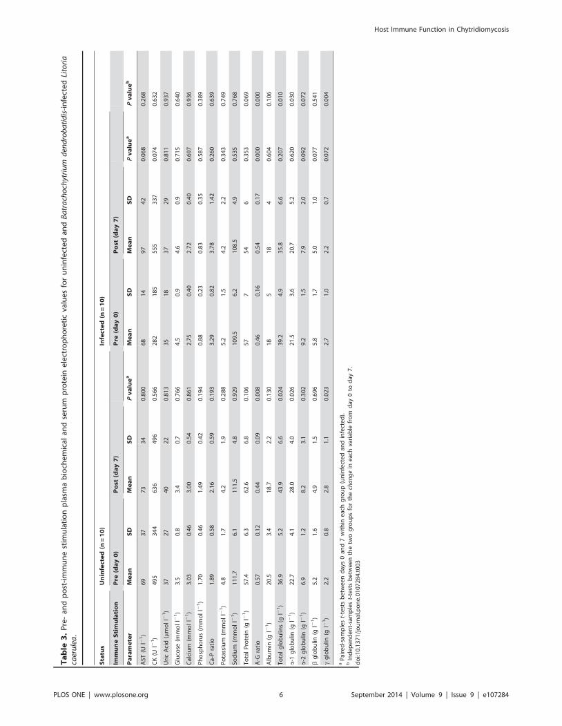
stimulation (Tables 2 and 3).
Compared with Bd-infected individuals, immune stimulation of
healthy uninfected L. caerulea caused a greater magnitude of
increase in total WBC (M+15.0 (SD 17.8) versus +1.7 (3.7)6109 l21)
(Fig. 1A), lymphocyte (M+12.2 (SD 14.5) versus +0.9 (1.8)6109 l21)
(Fig. 1B) and monocyte (M+1.8 (SD 1.5) versus +0.5 (0.9)6109 l21)
counts and polychromasia (M+6.9 (SD 9.3) versus 20.7 (2.5) %),
and a greater decrease in Hb concentration (M 22.6 (SD 1.2) versus
20.4 (2.9) g dl21) (Table 2).
Compared with Bd-infected individuals, immune stimulation of
healthy uninfected L. caerulea caused a significantly greater
increase in total globulins (M+7.0 (SD 8.2) versus 23.5 (8.1) g/L)
(Fig. 2A), a-1 globulin (M+5.3 (SD 6.3) versus 20.9 (5.4) g l21)
(Fig. 2B) and c globulin (M+0.6 (SD 0.7) versus 20.5 (0.8) g l21)
(Fig. 2C) concentrations, and a greater decrease in A-G ratio (M
20.13 (SD 0.12) versus +0.08 (0.03)) (Table 3).
Bd-infected Litoria infrafrenataAt day 75 post-exposure, 0% of the nine exposed L.
infrafrenata tested positive for Bd on PCR. Five frogs had low
Bd loads at either day 10 (n = 3) or 20 (n = 2) post-exposure, but all
of these were negative from day 30 onwards post-exposure, and
the other four frogs tested negative throughout the experiment.
Mean skin PHA response and kidney-body weight ratio were
lower in exposed uninfected L. infrafrenata compared with
healthy uninfected individuals post-immune stimulation; mean
responses to the other immune function tests did not differ
between the two groups (Table 1).
Following immune stimulation of Bd-exposed uninfected L.infrafrenata, mean Hb concentration and RBC and thrombocyte
counts decreased, while glucose concentration increased; all other
hematologic, plasma biochemical and serum protein electropho-
retic parameters did not differ significantly (Tables 4 and 5).
Compared with Bd-exposed uninfected individuals, immune
stimulation of healthy uninfected L. infrafrenata caused a greater
Figure 1. White blood cell counts of Litoria caerulea. Total white blood cell (WBC) (Fig. 1A) and lymphocyte (Fig. 1B) counts (x109 l21) pre- andpost-immune stimulation in healthy uninfected (n = 10) and Batrachochytrium dendrobatidis-infected (n = 10) Litoria caerulea. Bars are mean 6 SEM.*P,0.050 within each group.doi:10.1371/journal.pone.0107284.g001
Host Immune Function in Chytridiomycosis
PLOS ONE | www.plosone.org 8 September 2014 | Volume 9 | Issue 9 | e107284

increase in total WBC (M+14.7 (SD 12.4) versus 22.7 (15.5)6109 l21)
(Fig. 3A) and neutrophil (M+10.5 (SD 7.9) versus 22.8 (6.0)6109 l21)
(Fig. 3B) counts and polychromasia (M+9.8 (SD 8.0) versus +1.8 (3.0)
%), and a greater decrease in thrombocyte count (M 227.6 (SD 13.8)
versus 26.9 (8.3) 6109 l21) (Table 4).
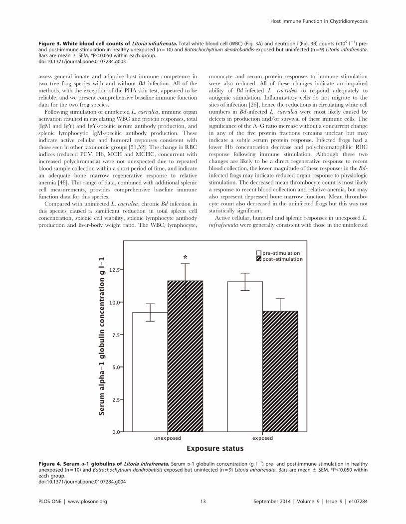
Compared with Bd-exposed uninfected individuals, immune
stimulation of healthy uninfected L. infrafrenata caused a
significantly greater increase in a-1 globulin concentration (M+2.5 (SD 3.1) versus 22.3 (3.1) g l21) (Fig. 4), and a greater decrease
in glucose concentration (M 20.7 (SD 0.9) versus +0.7 (0.8) mmol
l21) (Table 5). Changes in other variables post-immune stimula-
tion did not differ between the two groups.
Uninfected Tree FrogsFunctional immune competence test results for uninfected L.
caerulea and L. infrafrenata are presented in Table 1. Following
immune stimulation, mean total splenic RFC (P = 0.006) and liver
to body weight ratio (P = 0.003) were greater in L. caerulea, while
mean spleen to body weight ratio was greater in L. infrafrenata(P = 0.001). Mean responses to the other immune function tests
did not differ between the two species.
Pre- and post-immune stimulation hematologic values for
uninfected L. caerulea and L. infrafrenata are presented in
Tables 2 and 4 respectively. Following immune stimulation of L.caerulea, PCV, Hb, MCH and MCHC decreased, while total
WBC, lymphocyte and monocyte counts and polychromasia %
increased. In L. infrafrenata, total RBC and thrombocyte counts
decreased following immune stimulation, while total WBC,
neutrophil and monocyte counts, neutrophil-lymphocyte ratio,
and polychromasia % increased.
Pre- and post-immune stimulation plasma biochemical and
serum protein electrophoresis values for uninfected L. caeruleaand L. infrafrenata are presented in Tables 3 and 5 respectively.
Five protein fractions were defined in all samples submitted for
electrophoresis. Following immune stimulation of L. caerulea: total
globulins along with a-1 and c globulin fraction concentrations
increased, while the A–G ratio decreased; none of the plasma
biochemical values changed significantly. In L. infrafrenata:
plasma CK increased while glucose decreased post-immune
stimulation; total protein, albumin, total globulins and a-1 globulin
concentrations, and A–G ratio, all increased.
Discussion
Our results provide the first direct evidence suggesting that Bdinfection causes multiple in vivo systemic immune function defects
in a susceptible amphibian host, which likely enables disease
development. Antigenic stimulation of L. caerulea chronically
infected with Bd resulted in lower splenic lymphocyte, WBC,
serum protein and immunoglobulin responses compared with
uninfected frogs. Although L. infrafrenata failed to maintain
infection with Bd after experimental exposure, recovered frogs also
had reduced WBC and serum globulin concentrations compared
with unexposed frogs. Our work expands on recent studies
showing in vitro lymphocyte proliferation was impaired by Bd cells
and supernatants [31].
We successfully used diverse methods, many of which have not
previously been used outside of applied veterinary research, to
Figure 2. Serum globulin fractions of Litoria caerulea. Serum totalglobulins (Fig. 2A), a-1 globulin (Fig. 2B) and c globulin (Fig. 2C)concentrations (g l21) pre- and post-immune stimulation in healthyuninfected (n = 10) and Batrachochytrium dendrobatidis-infected (n = 10)Litoria caerulea. Bars are mean 6 SEM. *P,0.050 within each group.doi:10.1371/journal.pone.0107284.g002
Host Immune Function in Chytridiomycosis
PLOS ONE | www.plosone.org 9 September 2014 | Volume 9 | Issue 9 | e107284

Ta
ble
4.
Pre
-an
dp
ost
-im
mu
ne
stim
ula
tio
nh
em
ato
log
icva
lue
sfo
ru
nin
fect
ed
and
Ba
tra
cho
chyt
riu
md
end
rob
ati
dis
-exp
ose
db
ut
un
infe
cte
dLi
tori
ain
fra
fren
ata
.
Sta
tus
Un
infe
cte
d(n
=1
0)
Ex
po
sed
un
infe
cte
d(n
=9
)
Imm
un
eS
tim
ula
tio
nP
re(d
ay
0)
Po
st(d
ay
7)
Pre
(da
y0
)P
ost
(da
y7
)
Pa
ram
ete
rM
ea
nS
DM
ea
nS
DP
va
lue
aM
ea
nS
DM
ea
nS
DP
va
lue
aP
va
lue
b
PC
V(%
)2
6.9
7.0
25
.44
.10
.54
83
5.7
6.0
32
.34
.80
.07
90
.54
8
Hb
(gd
l21)
7.1
2.5
5.6
1.1
0.0
86
8.6
1.3
7.4
1.7
0.0
24
0.6
91
RB
C(x
10
9l2
1)
73
62
08
63
01
29
0.0
48
78
31
09
66
21
08
0.0
18
0.8
11
MC
V(f
l)3
83
15
54
21
12
40
.45
04
59
74
50
01
21
0.3
77
0.9
69
MC
H(p
g)
10
76
09
33
20
.52
51
11
19
11
63
80
.67
40
.45
7
MC
HC
(gl2
1)
29
11
26
22
02
90
.14
22
45
41
23
24
70
.30
60
.24
0
Th
rom
bo
cyte
(61
09
l21)
54
.81
2.9
27
.27
.20
.00
03
3.7
10
.12
6.8
7.0
0.0
36
0.0
01
WB
C(x
10
9l2
1)
32
.98
.74
7.6
10
.20
.00
53
3.7
27
.03
1.0
17
.40
.61
60
.01
5
Ne
utr
op
hil
(61
09
l21)
5.8
5.9
16
.45
.10
.00
21
2.1
14
.39
.31
0.0
0.2
04
0.0
01
Lym
ph
ocy
te(6
10
9l2
1)
24
.37
.72
5.5
6.2
0.7
26
17
.81
1.1
17
.31
1.1
0.8
60
0.7
00
Ne
ut-
lym
ph
rati
o0
.28
0.3
60
.67
0.2
40
.03
80
.66
0.4
70
.71
0.7
10
.72
60
.12
0
Mo
no
cyte
(61
09
l21)
2.1
1.4
5.6
3.5
0.0
23
3.3
3.9
4.0
3.1
0.5
22
0.1
31
Eosi
no
ph
il(6
10
9l2
1)
0.2
50
.44
0.0
40
.13
0.2
05
0.5
70
.76
0.3
40
.45
0.3
55
0.9
25
Bas
op
hil
(61
09
l21)
0.4
70
.79
0.0
00
.00
0.0
95
0.0
00
.00
0.0
00
.00
-0
.09
5
Po
lych
rom
asia
(%)
5.5
3.6
15
.37
.90
.00
49
.35
.11
1.1
6.7
0.1
08
0.0
13
aP
aire
d-s
amp
les
t-te
sts
be
twe
en
day
s0
and
7w
ith
ine
ach
gro
up
(un
infe
cte
dan
de
xpo
sed
).b
Ind
ep
en
de
nt-
sam
ple
st-
test
sb
etw
ee
nth
etw
og
rou
ps
for
the
cha
ng
ein
eac
hva
riab
lefr
om
day
0to
day
7.
do
i:10
.13
71
/jo
urn
al.p
on
e.0
10
72
84
.t0
04
Host Immune Function in Chytridiomycosis
PLOS ONE | www.plosone.org 10 September 2014 | Volume 9 | Issue 9 | e107284

Ta
ble
5.
Pre
-an
dp
ost
-im
mu
ne
stim
ula
tio
np
lasm
ab
ioch
em
ical
and
seru
mp
rote
ine
lect
rop
ho
reti
cva
lue
sfo
ru
nin
fect
ed
and
Ba
tra
cho
chyt
riu
md
end
rob
ati
dis
-exp
ose
db
ut
un
infe
cte
dLi
tori
ain
fra
fren
ata
.
Sta
tus
Un
infe
cte
d(n
=1
0)
Ex
po
sed
un
infe
cte
d(n
=9
)
Imm
un
eS
tim
ula
tio
nP
re(d
ay
0)
Po
st(d
ay
7)
Pre
(da
y0
)P
ost
(da
y7
)
Pa
ram
ete
rM
ea
nS
DM
ea
nS
DP
va
lue
aM
ea
nS
DM
ea
nS
DP
va
lue
aP
va
lue
b
AST
(Ul2
1)
24
84
06
17
19
30
.49
81
00
52
12
78
40
.30
90
.39
0
CK
(Ul2
1)
55
62
86
11
37
56
00
.01
39
18
86
41
27
69
29
0.2
18
0.5
39
Uri
cA
cid
( mm
ol
l21)
81
72
43
70
.25
13
.83
.45
.36
.60
.43
30
.30
2
Glu
cose
(mm
ol
l21)
3.6
0.6
2.9
0.8
0.0
36
3.4
0.4
4.2
1.0
0.0
20
0.0
02
Cal
ciu
m(m
mo
ll2
1)
2.2
92
.09
2.3
80
.99
0.8
27
2.9
00
.64
3.0
10
.80
0.6
28
0.9
56
Ph
osp
ho
rus
(mm
ol
l21)
1.5
40
.63
1.7
30
.76
0.4
04
1.5
90
.43
1.5
20
.32
0.4
12
0.2
79
Ca-
Pra
tio
1.3
80
.49
1.4
70
.40
0.6
08
1.8
80
.36
2.0
10
.45
0.3
88
0.8
41
Po
tass
ium
(mm
ol
l21)
3.7
0.8
3.8
0.9
0.5
91
4.8
2.0
4.2
1.5
0.1
83
0.1
45
Sod
ium
(mm
ol
l21)
10
7.7
2.7
11
0.9
4.1
0.0
65
11
0.0
3.6
10
8.7
3.1
0.3
36
0.1
38
To
tal
Pro
tein
(gl2
1)
29
63
47
0.0
07
40
83
85
0.5
00
0.0
65
A-G
rati
o0
.11
0.0
40
.13
0.0
40
.03
30
.21
0.0
60
.23
0.0
70
.17
50
.60
9
Alb
um
in(g
l21)
2.9
1.1
3.9
1.4
0.0
13
7.0
2.7
6.9
1.0
0.8
66
0.1
23
To
tal
glo
bu
lins
(gl2
1)
26
.16
.13
0.5
5.8
0.0
16
33
.25
.93
1.0
5.0
0.4
70
0.0
70
a-1
glo
bu
lin(g
l21)
9.2
2.1
11
.64
.20
.04
11
1.6
2.0
9.3
3.0
0.0
57
0.0
04
a-2
glo
bu
lin(g
l21)
5.3
1.7
6.8
2.9
0.4
18
6.1
2.6
5.7
2.3
0.6
54
0.0
79
bg
lob
ulin
(gl2
1)
10
.15
.51
1.0
4.0
0.1
92
13
.74
.21
4.6
2.7
0.6
65
0.9
68
cg
lob
ulin
(gl2
1)
1.4
0.8
1.2
0.3
0.2
71
1.3
0.4
1.4
0.3
0.8
83
0.3
17
aP
aire
d-s
amp
les
t-te
sts
be
twe
en
day
s0
and
7w
ith
ine
ach
gro
up
(un
infe
cte
dan
de
xpo
sed
).b
Ind
ep
en
de
nt-
sam
ple
st-
test
sb
etw
ee
nth
etw
og
rou
ps
for
the
cha
ng
ein
eac
hva
riab
lefr
om
day
0to
day
7.
do
i:10
.13
71
/jo
urn
al.p
on
e.0
10
72
84
.t0
05
Host Immune Function in Chytridiomycosis
PLOS ONE | www.plosone.org 11 September 2014 | Volume 9 | Issue 9 | e107284

Host Immune Function in Chytridiomycosis
PLOS ONE | www.plosone.org 12 September 2014 | Volume 9 | Issue 9 | e107284
assess general innate and adaptive host immune competence in
two tree frog species with and without Bd infection. All of the
methods, with the exception of the PHA skin test, appeared to be
reliable, and we present comprehensive baseline immune function
data for the two frog species.
Following stimulation of uninfected L. caerulea, immune organ
activation resulted in circulating WBC and protein responses, total
(IgM and IgY) and IgY-specific serum antibody production, and
splenic lymphocytic IgM-specific antibody production. These
indicate active cellular and humoral responses consistent with
those seen in other taxonomic groups [51,52]. The change in RBC
indices (reduced PCV, Hb, MCH and MCHC, concurrent with
increased polychromasia) were not unexpected due to repeated
blood sample collection within a short period of time, and indicate
an adequate bone marrow regenerative response to relative
anemia [48]. This range of data, combined with additional splenic
cell measurements, provides comprehensive baseline immune
function data for this species.
Compared with uninfected L. caerulea, chronic Bd infection in
this species caused a significant reduction in total spleen cell
concentration, splenic cell viability, splenic lymphocyte antibody
production and liver-body weight ratio. The WBC, lymphocyte,
monocyte and serum protein responses to immune stimulation
were also reduced. All of these changes indicate an impaired
ability of Bd-infected L. caerulea to respond adequately to
antigenic stimulation. Inflammatory cells do not migrate to the
sites of infection [26], hence the reductions in circulating white cell
numbers in Bd-infected L. caerulea were most likely caused by
defects in production and/or survival of these immune cells. The
significance of the A–G ratio increase without a concurrent change
in any of the five protein fractions remains unclear but may
indicate a subtle serum protein response. Infected frogs had a
lower Hb concentration decrease and polychromatophilic RBC
response following immune stimulation. Although these two
changes are likely to be a direct regenerative response to recent
blood collection, the lower magnitude of these responses in the Bd-
infected frogs may indicate reduced organ response to physiologic
stimulation. The decreased mean thrombocyte count is most likely
a response to recent blood collection and relative anemia, but may
also represent depressed bone marrow function. Mean thrombo-
cyte count also decreased in the uninfected frogs but this was not
statistically significant.
Active cellular, humoral and splenic responses in unexposed L.infrafrenata were generally consistent with those in the uninfected
Figure 3. White blood cell counts of Litoria infrafrenata. Total white blood cell (WBC) (Fig. 3A) and neutrophil (Fig. 3B) counts (x109 l21) pre-and post-immune stimulation in healthy unexposed (n = 10) and Batrachochytrium dendrobatidis-exposed but uninfected (n = 9) Litoria infrafrenata.Bars are mean 6 SEM. *P,0.050 within each group.doi:10.1371/journal.pone.0107284.g003
Figure 4. Serum a-1 globulins of Litoria infrafrenata. Serum a-1 globulin concentration (g l21) pre- and post-immune stimulation in healthyunexposed (n = 10) and Batrachochytrium dendrobatidis-exposed but uninfected (n = 9) Litoria infrafrenata. Bars are mean 6 SEM. *P,0.050 withineach group.doi:10.1371/journal.pone.0107284.g004
Host Immune Function in Chytridiomycosis
PLOS ONE | www.plosone.org 13 September 2014 | Volume 9 | Issue 9 | e107284

L. caerulea, and in other taxonomic groups [52,53]. Notable inter-
species differences in our results include the neutrophilic versus
lymphocytic WBC response and the hyperglycemia and elevated
CK post-immune stimulation in L. infrafrenata. Neutrophil
reference values in L. infrafrenata vary with season, and are
significantly higher compared with L. caerulea [44]. Furthermore,
the two species differ in temperament, with L. caerulea generally
calm and tolerant of handling, and L. infrafrenata often exhibiting
clinical signs of stress associated with handling and confinement.
This is likely to account for the neutrophilic WBC response and
the increased glucose and CK values in L. infrafrenata. The
reduced RBC count concurrent with increased polychromatophil-
ic response again was not unexpected due to repeated blood
sample collection, and indicate an adequate bone marrow
regenerative response.
All Bd-exposed L. infrafrenata failed to maintain infection
despite identical experimental conditions to those for L. caerulea,
and the five of nine frogs that tested positive early in the trial self-
cured. The sampling protocols were completed despite the loss of
infection, and results showed immunologic effects on some
systemic responses resulting from Bd exposure. Post-immune
stimulation WBC, neutrophil, polychromatophilic RBC, throm-
bocyte and a-1 globulin concentrations, and skin PHA response,
were reduced in the Bd-exposed L. infrafrenata compared with
uninfected frogs. However, immune stimulation caused similar
total and IgY-specific serum antibody, splenic lymphocytic IgM-
specific antibody, and splenic cellular, responses in both uninfected
and Bd-exposed L. infrafrenata. Variations in RBC indices and
plasma glucose were not unexpected as previously discussed.
In previous studies using the model species Silurana tropicalis,Bd infection appeared to cause down-regulation of some immune
genes including those associated with Toll-like receptors, comple-
ment pathways, and B- and T-lymphocytes [27,28]. Recently,
soluble factors in Bd culture supernatant inhibited in vitrolymphocyte proliferation assays, but did not reduce macrophage
activity [31]. Other studies found higher concentrations of urinary
corticosterone metabolites and plasma corticosterone, in Bd-
infected L. wilcoxii and L. caerulea respectively, indicating a
physiological stress response [30,62].
Stress hormones are known to alter normal WBC distribution,
and peripheral leukocyte profiles consistent with a classical
mammalian stress-related response include a relative neutrophilia,
lymphopenia and eosinopenia [63]. Peripheral neutrophilic and
eosinopenic responses have previously been reported in Bd-
infected larval anurans (Rana catesbeiana), although lymphocyte
abundance did not change [64]. Conflicting findings have been
reported in post-metamorphic anurans: juvenile L. chloris showed
relative peripheral neutropenic, eosinopenic and basophilic
responses to Bd infection [20], while L. caerulea showed a
lymphopenic response [62,65]. The amphibian leukocyte response
to stress varies according to species and many other intrinsic and
extrinsic factors, including season and sex [44,66,67], making
interpretation of leukocyte profiles alone difficult.
Previous laboratory and field studies show that L. caerulea is
highly susceptible to Bd [11,12,68], but little data is available for
L. infrafrenata. Our results indicate that L. infrafrenata is a
naturally resistant host. Pre-exposure data collected from L.infrafrenata did not differ greatly from L. caerulea, hence our
results do not suggest a clear mechanism for greater natural
resistance in L. infrafrenata, which may be related to localized
and/or innate immune defense mechanisms that we did not
measure. The skin peptide profiles of L. infrafrenata and L.caerulea differ greatly [41] and this could partly explain inter-
species differences in host resistance, although L. infrafrenata
produces no known major antibiotic peptides compared with at
least five that have been identified in L. caerulea [41].
The reduced WBC, neutrophil and globulin concentrations in
exposed L. infrafrenata may indicate direct host immune system
costs associated with infection clearance in frogs challenged post-
metamorphosis and/or redirection of host resources away from
systemic adaptive immune responses towards alternate immune
mechanisms involved in combating infection. Larval common
toads (Bufo bufo) experimentally exposed to low Bd doses usually
died at or soon after metamorphosis without detectable infections,
suggesting fitness costs attributable to exposure, control and
clearance in the absence of extensive pathogen proliferation [69].
However, exposed L. infrafrenata were still able to produce serum
antibody, and splenic cellular and antibody, responses of similar
magnitude to uninfected frogs following immune stimulation. This
suggests that adaptive immune responses may also play an
important role in Bd-resistant host species.
Despite our findings that Bd causes multiple defects in systemic
immune function in L. caerulea, an Australian survey found only
6.5% (13/199) of frogs with severe chytridiomycosis had
concurrent acute secondary infectious diseases [68]. The low
incidence of secondary infections may indicate that systemic
immune suppression is not generalized and distinct components
remain effective against opportunistic pathogen invasion, and/or
that systemic immune response capability is not reduced below a
critical point until late in the disease process. Total and IgY-
specific serum antibody responses did not differ between
uninfected and infected frogs of either species, suggesting that
serum immunoglobulin response as a component of systemic
adaptive immunity is not suppressed in chronic chytridiomycosis
and that it may not be an important component of disease
resistance. These findings further support the hypothesis that
localized and/or innate immune defense mechanisms may be key
factors.
Skin PHA stimulation was minimal and variable in contrast to
other taxa and this test was not found to be a sensitive indicator of
immune function in either of the two Litoria species in our study.
There was a slightly greater response in L. infrafrenata compared
with L. caerulea, but this was still minimal compared with other
species and quite variable. The PHA skin response in adult R.pipiens was also found to be highly variable and less sensitive for
detecting pesticide-related immune suppression compared with
hemocyanin-specific antibody and whole blood chemilumines-
cence tests [39], although in the cane toad (Rhinella marina) the
test was found to reliably quantify immune response in an assay
validation study [40]. Splenic cell viability assessed by trypan blue
exclusion resulted in lower mean viable cell counts in both of the
Litoria species in our study compared with avian and mammalian
species, but this test was still useful for comparing immune
function between Bd-infected and uninfected frogs. Future
adaptations of this method to improve viable cell harvest should
include the use of an amphibian-specific isotonic diluent;
mammalian isotonic solutions such as Hanks balanced salt solution
are hypertonic to amphibian cells [70] and may cause cell damage.
Although bisection of the spleen was necessary to perform multiple
splenic tests, improved cell viability may also be achieved by
processing the whole spleen for one single method.
A limitation associated with our experimental design is that the
two experimental trials were not conducted simultaneously. The
trial in uninfected frogs commenced as a pilot study to validate
methodology prior to application in the trial with Bd-infected
frogs, but insufficient specimens were available to include a
negative control group in the infection trial. This limitation was
largely compensated for by 1) standardizing timing of sample
Host Immune Function in Chytridiomycosis
PLOS ONE | www.plosone.org 14 September 2014 | Volume 9 | Issue 9 | e107284
collection relative to initiation of immune tests, 2) ensuring
experimental procedures were performed by the same person (SY)
using identical analytic equipment, reagents and methodology,
and 3) maintaining identical experimental laboratory and
husbandry conditions. Additionally, a large concurrent experiment
in our laboratory showed that immunity did not decrease in
uninfected L. caerulea held in captivity and blood sampled three
times over this period - with the exception of a mild but significant
decrease in WBC and lymphocyte counts on day 30, no
hematologic immune biomarkers varied significantly after 60 or
125 days [24]. Our baseline immune function data provide a
valuable tool for progressing future immunologic and Bdpathogenicity studies in amphibians. General mechanisms of
fungal immune suppression include inducing anti-inflammatory
cytokines, decreasing pro-inflammatory cytokines and comple-
ment evasion [71]. Further work on understanding the mecha-
nisms of Bd immune suppression is needed and may involve
exposing frogs directly to identified pathogen-secreted factors and
assessing immune function using the in vivo methods that we have
shown here to be sensitive indicators.
Our results may explain the lack of an adaptive immune
response to infection in some species [22,24,72], and suggest that if
vaccine development or other immune modulation is attempted in
future trials, it will be necessary to better understand the
mechanisms of immune suppression in order to overcome it.
While many of the immune tests we describe here will be valuable
for use in future immunologic studies, the total and differential
WBC and serum electrophoresis measurements, along with
antigen-specific serum antibody assays, have particularly wide
potential future applications as repeatable, ante-mortem methods
for assessment of amphibian immune function.
Acknowledgments
We thank Helen Martin and Deb Buckett at Gribbles Veterinary
Pathology for assistance with protein electrophoresis.
Author Contributions
Conceived and designed the experiments: SY LB LFS RS. Performed the
experiments: SY PW. Analyzed the data: SY. Contributed reagents/
materials/analysis tools: SY LB LFS SG RW. Contributed to the writing of
the manuscript: SY LB LFS RS SG RW. Performed PCR analyses: SG
RW.
References
1. Berger L, Speare R, Daszak P, Green DE, Cunningham AA, et al. (1998)
Chytridiomycosis causes amphibian mortality associated with population
declines in the rainforests of Australia and Central America. Proc Natl Acad
Sci USA 95: 9031–9036.
2. Lips KR, Brem F, Brenes R, Reeve JD, Alford RA, et al. (2006) Emerging
infectious disease and the loss of biodiversity in a Neotropical amphibian
community. Proc Natl Acad Sci USA 103: 3165–3170.
3. Skerratt LF, Berger L, Speare R, Cashins S, McDonald K, et al. (2007) Spread
of chytridiomycosis has caused the rapid global decline and extinction of frogs.
EcoHealth 4: 125–134.
4. Vredenburg VT, Knapp RA, Tunstall TS, Briggs CJ (2010) Dynamics of an
emerging disease drive large-scale amphibian population extinctions. Proc Natl
Acad Sci USA 107: 9689–9694.
5. Daszak P, Cunningham AA, Hyatt AD (2003) Infectious disease and amphibian
population declines. Divers Distrib 9: 141–150.
6. Woodhams DC, Alford RA (2005) Ecology of chytridiomycosis in rainforest
stream frog assemblages of tropical Queensland. Conserv Biol 19: 1449–1459.
7. Schloegel LM, Hero J-M, Berger L, Speare R, McDonald K, et al. (2006) The
decline of the sharp-snouted day frog: the first documented case of extinction by
infection in a free-ranging wildlife species? EcoHealth 3: 35–40.
8. McDonald KR, Mendez D, Muller R, Freeman AB, Speare R (2005) Decline in
the prevalence of chytridiomycosis in upland frog populations in North
Queensland, Australia. Pacific Cons Biol 11: 114–120.
9. Berger L, Marantelli G, Skerratt LF, Speare R (2005) Virulence of the
amphibian chytrid fungus, Batrachochytrium dendrobatidis, varies with the
strain. Dis Aquat Org 68: 47–50.
10. Woodhams DC, Alford RA, Marantelli G (2003) Emerging disease cured by
elevated body temperature. Dis Aquat Org 55: 65–77.
11. Voyles J, Berger L, Young S, Speare R, Webb R, et al. (2007) Electrolyte
depletion and osmotic imbalance in amphibians with chytridiomycosis. Dis
Aquat Org 77: 113–118.
12. Voyles J, Young S, Berger L, Campbell C, Voyles WF, et al. (2009) Pathogenesis
of chytridiomycosis, the cause of catastrophic amphibian declines. Science 326:
582–585.
13. Young S, Speare R, Berger L, Skerratt LF (2012) Chloramphenicol with fluid
and electrolyte therapy cures terminally ill green tree frogs (Litoria caerulea) with
chytridiomycosis. J Zoo Wildl Med 43: 330–337.
14. Rowley JJL, Alford RA (2007) Behaviour of Australian rainforest stream frogs
may affect the transmission of chytridiomycosis. Dis Aquat Org 77: 1–9.
15. Brem FMR, Lips KR (2008) Batrachochytrium dendrobatidis infection patterns
among Panamanian amphibian species, habitats and elevations during epizootic
and enzootic stages. Dis Aquat Org 81: 189–202.
16. Blomberg B, Bernard CCA, Du Pasquier L (1980) In vitro evidence for T-B
lymphocyte collaboration in the clawed toad Xenopus. Eur J Immunol 10: 869–
876.
17. Du Pasquier L, Schwager J, Flajnik MF (1989) The immune system of Xenopus.Annu Rev Immunol 7: 251–275.
18. Du Pasquier L, Robert J, Courtet M, Rainer M (2000) B-cell development in the
amphibian Xenopus. Immunol Rev 175: 201–213.
19. Whittington R, Speare R (1996) Sensitive detection of serum antibodies in the
cane toad Bufo marinus. Dis Aquat Org 26: 59–65.
20. Woodhams DC, Ardipradja K, Alford RA, Marantelli G, Reinert LK, et al.
(2007) Resistance to chytridiomycosis varies among amphibian species and iscorrelated with skin peptide defenses. Anim Conserv 10: 409–417.
21. Woodhams DC, Vredenburg VT, Simon M-A, Billheimer D, Shakhtour B, et al.(2007) Symbiotic bacteria contribute to innate immune defenses of the
threatened mountain yellow-legged frog, Rana mucosa. Biol Conserv 138:390–398.
22. Stice MJ, Briggs CJ (2010) Immunization is ineffective at preventing infectionand mortality due to the amphibian chytrid fungus Batrachochytriumdendrobatidis. J Wildl Dis 46: 70–77.
23. Rosenblum EB, Poorten TJ, Settles M, Murdoch GK (2012) Only skin deep:
shared genetic response to the deadly chytrid fungus in susceptible frog species.Mol Ecol 21: 3110–3120.
24. Young S (2012) Emerging amphibian diseases in Queensland and host immuneresponse to disease. PhD Thesis. Townsville: James Cook University. 258 p.
25. Ramsey JP, Reinert LK, Woodhams DC, Rollins-Smith LA (2010) Immune
defenses against Batrachochytrium dendrobatidis, a fungus linked to global
amphibian declines, in the South African clawed frog, Xenopus laevis. InfectImmun 78: 3981–3992.
26. Berger L, Speare R, Hyatt A (1999) Chytrid fungi and amphibian declines:overview, implications and future directions. In: Campbell A, editor. Declines
and disappearances of Australian frogs. Canberra: Environment Australia. pp.23–33.
27. Ribas L, Li MS, Doddington BJ, Robert J, Seidel JA, et al. (2009) Expressionprofiling the temperature-dependent amphibian response to infection by
Batrachochytrium dendrobatidis. PLoS ONE 4: e8408.
28. Rosenblum E, Poorten T, Settles M, Murdoch G, Robert J, et al. (2009)
Genome-wide transcriptional response of Silurana (Xenopus) tropicalis toinfection with the deadly chytrid fungus. PLoS ONE 4: e6494.
29. Rollins-Smith LA, Ramsey JP, Pask JD, Reinert LK, Woodhams DC (2011)
Amphibian immune defenses against chytridiomycosis: impacts of changing
environments. Integr Comp Biol 51: 552–562.
30. Kindermann C, Narayan EJ, Hero J-M (2012) Urinary corticosteronemetabolites and chytridiomycosis disease prevalence in a free-living population
of male Stony Creek frogs (Litoria wilcoxii). Comp Biochem Physiol A 162: 171–
176.
31. Fites JS, Ramsey JP, Holden WM, Collier SP, Sutherland DM, et al. (2013) Theinvasive chytrid fungus of amphibians paralyses lymphocyte responses. Science
342: 366–369.
32. Horton JD, Rimmer JJ, Horton TL (1976) The effect of thymectomy at different
stages of larval development on the immune response of the clawed toad to
sheep erythrocytes. J Exp Zool 196: 243–249.
33. Gearing AJ, Cribbin FA, Horton JD (1984) Restoration of the antibody responseto sheep erythrocytes in thymectomised Xenopus implanted with MHC-
compatible or MHC-incompatible thymus. J Embryol Exp Morphol 84: 287–
302.
34. Zettergren LD, Boldt BW, Petering DH, Goodrich MS, Weber DN, et al. (1991)Effects of prolonged low-level cadmium exposure on the tadpole immune
system. Toxicol Lett 55: 11–19.
35. Rosenberg CE, Salibian A, Fink NE (2002) An enzyme-linked immunosorbent
assay for measuring anti-sheep red blood cells antibodies in lead-exposed toads.J Pharmacol Toxicol Methods 47: 121–128.
Host Immune Function in Chytridiomycosis
PLOS ONE | www.plosone.org 15 September 2014 | Volume 9 | Issue 9 | e107284

36. Rollins-Smith LA, Parsons SC, Cohen N (1984) During frog ontogeny, PHA and
Con A responsiveness of splenocytes precedes that of thymocytes. Immunology52: 491–500.
37. Rollins-Smith LA, Blair PJ (1993) The effects of corticosteroid hormones and
thyroid hormones on lymphocyte viability and proliferation during developmentand metamorphosis of Xenopus laevis. Differentiation 54: 155–160.
38. Kinney KS, Cohen N (2005) Increased splenocyte mitogenesis followingsympathetic denervation in Xenopus laevis. Dev Comp Immunol 29: 287–293.
39. Gilbertson M-K, Haffner GD, Drouillard KG, Albert A, Dixon B (2003)
Immunosuppression in the Northern leopard frog (Rana pipiens) induced bypesticide exposure. Environ Toxicol Chem 22: 101–110.
40. Brown GP, Shilton CM, Shine R (2011) Measuring amphibian immunocom-petence: validation of the phytohemagglutinin skin-swelling assay in the cane
toad, Rhinella marina. Methods Ecol Evol 2: 341–348.41. Jackway RJ, Pukala TL, Donnellan SC, Sherman PJ, Tyler MJ, et al. (2011) Skin
peptide and cDNA profiling of Australian anurans: genus and species
identification and evolutionary trends. Peptides 32: 161–172.42. Boyle DG, Boyle DB, Olsen V, Morgan JAT, Hyatt AD (2004) Rapid
quantitative detection of chytridiomycosis (Batrachochytrium dendrobatidis) inamphibian samples using real-time Taqman PCR assay. Dis Aquat Org 60:
141–148.
43. Wright K (2001) Amphibian hematology. In: Wright KM, Whitaker BR, editors.Amphibian medicine and captive husbandry. Malabar: Krieger Publishing
Company. pp 129–146.44. Young S, Warner J, Speare R, Berger L, Skerratt LF, et al. (2012) Hematologic
and plasma biochemical reference values for health monitoring of wildAustralian tree frogs. Vet Clin Pathol 41: 478–492.
45. Natt MP, Herrick CA (1952) A new blood diluent for counting the erythrocytes
and leukocytes of the chicken. Poult Sci 31: 735.46. Drabkin DR (1945) Crystallographic and optical properties of human
hemoglobin. A proposal for the standardisation of hemoglobin. Am J Med Sci209: 268–270.
47. Melrose WD, Brown PB, Holdsworth MC, Bryant SL (1995) Haematology and
red cell enzymes of the Australian orange-bellied parrot, Neophema chrysogaster.Comp Hematol Int 5: 7–9.
48. Campbell TW, Ellis CK (2007) Avian and exotic animal hematology andcytology. Oxford: Blackwell Publishing. 287 p.
49. Zaias J, Cray C (2002) Protein electrophoresis: a tool for the reptilian andamphibian practitioner. J Herp Med Surg 12: 30–32.
50. Stadecker MJ, Lukic M, Dvorak A, Leskowitz S (1977) The cutaneous basophil
response to phytohemagglutinin in chickens. J Immunol 118: 1564–1568.51. Dean JH, Cornacoff JB, Rosenthal GJ, Luster MI (1989) Immune system:
evaluation of injury. In: Hayes AW, editor. Principles and methods of toxicology.New York: Raven Press. pp. 741–760.
52. Grasman KA (2002) Assessing immunological function in toxicological studies of
avian wildlife. Integ Comp Biol 42: 34–42.53. Fairbrother A, Fowles J (1990) Subchronic effects of sodium selenite and
selenomethionine on several immune functions in mallards. Arch EnvironContam Toxicol 19: 836–844.
54. Pappenheimer AM (1917) Experimental studies upon lymphocytes. I. The
reactions of lymphocytes under various experimental conditions. J Exp Med 25:633–650.
55. Strober W (1997) Trypan blue exclusion test of cell viability. Current Protocols
in Immunol 21: A.3B.1–A.3B.2.56. Zaalberg OB (1964) A simple method for detecting single antibody-forming cells.
Nature London 202: 1213.57. Behling UH, Campbell B, Chang C, Rampf C, Nowotny A (1976) Synthetic
glycolipid adjuvants. J Immunol 117: 847–851.
58. Jerne NK, Nordin AA (1963) Plaque formation in agar by single antibody-producing cells. Science 140: 405.
59. Cunningham AJ, Szenberg A (1968) Further improvements in the plaquetechnique for detecting single antibody forming cells. Immunol 14: 599–601.
60. Longcore JE, Pessier AP, Nichols DK (1999) Batrachochytrium dendrobatidis genet sp nov, a chytrid pathogenic to amphibians. Mycologia 91: 219–227.
61. Boyle DG, Hyatt AD, Daszak P, Berger L, Longcore JE, et al. (2003) Cryo-
archiving of Batrachochytrium dendrobatidis and other chytridiomycetes. DisAquat Org 56: 59–64.
62. Peterson JD, Steffen JE, Reinert LK, Cobine PA, Appel A, et al. (2013) Hoststress response is important for the pathogenesis of the deadly amphibian
disease, chytridiomycosis, in Litoria caerulea. PLoS ONE 8(4): e62146.
63. Dhabar F, Miller A, McEwen B, Spencer R (1996) Stress-induced changes inblood leukocyte distribution. Role of adrenal steroid hormones. J Immunol 157:
1638–1644.64. Davis AK, Keel MK, Ferreira A, Maerz JC (2010) Effects of chytridiomycosis on
circulating white blood cell distributions of bullfrog larvae (Rana catesbeiana).Comp Clin Pathol 19: 49–55.
65. Peterson JD, Steffen JE, Appel AG, Rollins-Smith LA, Mendonca MT (2011)
Chytridiomycosis is metabolically costly and induces sickness behavior andimmune effects that are linked to increased corticosterone secretion. Integr
Comp Biol 51(s1): e108.66. Garrido E, Gomariz RP, Leceta J, Zapata A (1987) Effects of dexamethasone on
the lymphoid organs of Rana perezi. Dev Comp Immunol 11: 375–384.
67. Pfeiffer CJ, Pyle H, Asashima M (1990) Blood cell morphology and counts in theJapanese newt (Cynops pyrrhogaster). J Zoo Wildl Med 21: 56–64.
68. Berger L, Speare R, Hines H, Marantelli G, Hyatt AD, et al. (2004) Effect ofseason and temperature on mortality in amphibians due to chytridiomycosis.
Aust Vet J 82: 31–36.69. Garner TWJ, Walker S, Bosch J, Leech S, Rowcliffe JM, et al. (2009) Life history
trade-offs influence mortality associated with the amphibian pathogen
Batrachochytrium dendrobatidis. Oikos 118: 783–791.70. Wright K (2001) Pharmacotherapeutics. In: Wright KM, Whitaker BR, editors.
Amphibian medicine and captive husbandry. Malabar: Krieger PublishingCompany. pp 309–330.
71. Chai LYA, Netea MG, Vonk AG, Kullberg B (2009) Fungal strategies for
overcoming host innate immune response. Med Mycol 47: 227–236.72. Cashins SD, Grogan LF, McFadden M, Hunter D, Harlow PS, et al. (2013)
Prior infection does not improve survival against the amphibian diseasechytridiomycosis. PLoS ONE 8(2): e56747. doi:10.1371/journal.pone.0056747.
Host Immune Function in Chytridiomycosis
PLOS ONE | www.plosone.org 16 September 2014 | Volume 9 | Issue 9 | e107284